Lower Limb Interjoint Postural Coordination One Year After First-Time Ankle Sprain
Upload
spanalumniCategory
view
1download
0
Heart 1996;75:469-476
Endothelial control of lower limb blood flow inchronic heart failure
David C Lindsay, Diana R Holdright, Debbie Clarke, Inder S Anand,Philip A Poole-Wilson, Peter Collins
Department ofCardiac Medicine,National Heart & LungInstitute, LondonD C LindsayD R HoldrightD ClarkeP A Poole-WilsonP CollinsVA Medical Centre,Minneapolis, MN55417, USAI S AnandCorrespondence to:Dr D C Lindsay, CardiacDepartment, GloucestershireRoyal Hospital, GreatWestern Road, GloucesterGLI 3NN.
Accepted for publication10 November 1995
AbstractBackground-Limitation of the bloodsupply to skeletal muscle in chronic heartfailure may contribute to the symptoms offatigue and diminished exercise capacity.The pathophysiology underlying thisabnormality is not known. The purpose ofthis study was to assess the effect ofendothelium dependent and independentvasodilator agents on blood flow in the legofpatients with heart failure.Methods and results-Blood flow in theleg was measured in patients with heartfailure (n = 20) and compared with that inpatients with ischaemic heart disease andnormal left ventricular function (n = 16)and patients with chest pain and normalcoronary arteries (n = 8). External iliacartery blood flow was measured usingintravascular Doppler ultrasound andquantitative angiography. Flow wasrecorded at rest and in response to bolusdoses of the endothelium independentvasodilator, papaverine. Endotheliumdependent responses were measured byinfusion of acetylcholine and substance P.Mean (SEM) baseline blood flow wasreduced at rest (2-9 (0-4) v 4 5 (0.3) mVs,P < 0 001) and vascular resistance wasraised (37.4 (3.6) v 27'1 (3.0) units, P <0.05) in patients with heart failure com-pared with that in controls. The peakblood flow response to papaverine (8 mg),acetylcholine (10-7-10-5 moIl), and sub-stance P (5 pmollmin) was reduced inheart failure, with greater impairment ofthe response to acetylcholine than sub-stance P. There was a correlation betweenbaseline blood flow in the heart failuregroup and diuretic dose (r = -0-62, P =0 003), New York Heart Association clas-sification (r = -0 65, P = 0.002), and leftventricular ejection fraction (r = 0-80, P =0.0004).Conclusions-There is reduced blood flowand raised vascular resistance at rest inthe legs of patients with heart failure. Thedegree of impaired blood flow in the legcorrelates with the severity of heart fail-ure. There is impairment of the responseto both endothelium dependent and inde-pendent vasodilators. Abnormal functionof the vascular myocyte in heart failuremay explain these results as would struc-tural abnormalities of the resistance ves-sels.
(Heart 1996;75:469-476)
Keywords: endothelium derived relaxing factor; acetyl-choline; vasodilatation; congestive heart failure
The commonest symptoms of patients withheart failure are fatigue and shortness ofbreath. The pathophysiological mechanismsunderlying these symptoms are complex, butrecent work has emphasised the role of neuraland hormonal signals from the periphery.While central haemodynamic variables areimportant in the genesis of symptoms in acuteheart failure, there is poor correlation betweenthe severity of symptoms in chronic heart fail-ure and the left ventricular ejection fraction orpulmonary capillary wedge pressure. 1-3Reduction of cardiac output is an importantfactor limiting exercise capacity when usinglarge groups of muscle, but is unlikely to be amajor limiting factor when using small musclegroups. Blood flow to skeletal muscle mayincrease by 20-fold with exercise.4 This abilityto increase limb blood flow with exercise isreduced in patients with chronic heart fail-ure. 56 Flow limitation in the leg and abnor-malities of skeletal muscle7-9 could be thesubstrate for signals giving rise to the charac-teristic symptoms of heart failure.'0The control of limb blood flow is influenced
by neural, hormonal, and locally released fac-tors. Endothelium derived relaxing factor(EDRF) contributes to basal peripheral vascu-lar tone in humans in vivo." Data from animalmodels12-'4 and humans'5-"7 suggest that therelease of EDRF, or the response to itsactions, is impaired in chronic heart failure.Most data from humans are derived fromstudies of forearm vasculature.5 18 Few studieshave reported the blood flow response ofthe leg5 15 despite activities associated with useof the legs being the commonest cause ofsymptoms.The aim of the present study was to com-
pare blood flow in the legs of patients withheart failure and those with normal leftventricular function in response to infusion ofthe endothelium dependent vasodilators,acetylcholine and substance P, and to theendothelium independent agent papaverine.The hypothesis being tested was that theendothelium dependent vasodilator responsesof the leg vasculature would be impaired inpatients with chronic heart failure.
Patients and methodsSTUDY POPULATIONPatients were recruited from those admittedfor routine cardiac catheterisation to the Royal
469
group.bmj.com on July 15, 2011 - Published by heart.bmj.comDownloaded from

Lindsay, Holdright, Clarke, Anand, Poole-Wilson, Collins
Brompton National Heart & Lung Hospital,London. All were informed of the procedures,and gave their written consent to the study inthe presence of an independent witness. Thestudies were approved by the EthicalCommittee of the National Heart & ChestHospitals.
Patients aged over 18 years undergoing rou-
tine cardiac catheterisation were consideredfor the study. Patients with unstable anginapectoris, severe coronary artery disease, signif-icant valvar stenosis, peripheral vascular dis-ease, diabetes mellitus, asthma, or systemichypertension (systolic blood pressure > 160mm Hg or diastolic blood pressure > 100 mmHg) were excluded. All vasoactive medication(excluding diuretics, digoxin and angiotensinconverting enzyme inhibitors) was stopped fora period of at least 16 h before the study.
Patients were characterised as havingchronic heart failure on the basis of their clini-cal history, physical findings, use of diuretictreatment, and left ventricular ejection frac-tion. Patients were divided into two groups:those with heart failure (group A) and thosewithout heart failure (group B). Two groups ofpatients were studied as controls in group B:patients with atheromatous coronary arterydisease and normal left ventricular functionand patients with chest pain and angiographi-cally normal coronary arteries. Patients werenot included in the latter group if they had evi-dence of ST segment change with exerciseelectrocardiography. Those with chest painand normal coronary arteries were believed tohave non-cardiac chest pain with entirely nor-
mal hearts, and thus represent a true controlgroup.The left ventricular ejection fraction was
measured from the left ventricular cinean-giogram. The severity of heart failure was
assessed by the New York Heart Associationclassification, dosage of diuretic, left ventricularend diastolic pressure. Exercise capacity wasmeasured in some patients as MVo, withtreadmill exercise testing (ml o,.kg-' .min- 1).The patients with heart failure were subdi-vided into four groups according to their dailydose of diuretics: no diuretic (group 1); lessthan or equal to 40 mg frusemide or equiva-lent (for example, bendrofluazide 5 mg daily)(group 2); greater than 40 mg frusemide to80 mg frusemide or its equivalent (for exam-
ple, bumetanide 1-2 mg daily) (group 3); andgreater than 80 mg frusemide or equivalent(group 4).
CATHETER LABORATORY PROTOCOLCardiac catheterisation was performed using an
eight French gauge sheath in situ in the femoralartery and catheterisation of the right heart wasundertaken according to clinical indication.Left ventricular and coronary angiograms were
performed using non-ionic x ray contrastmedium (Omnipaque; Nycomed AS, Oslo,Norway). Sublingual or systemic nitrates werenot administered during the study.
Blood flow velocity was measured using a
20 MHz pulsed wave Doppler catheter posi-tioned in the external iliac artery. The
catheters were either Schneider (three Frenchgauge Monorail; Schneider, Zurich,Switzerland) or Wessex (5 5 French gauge,Numed, New York, USA) Doppler catheters.Validation studies in the laboratory showedgood correlation between recorded and trueflows on an experimental test rig in whichthere was pulsatile circulation of blood in aclosed circuit (r = 0-98, P < 0 01).
FEMORAL ARTERIAL ANGIOGRAPHYCineangiography of the femoral and externaliliac arteries was performed by hand injectionof x ray contrast medium (10 ml) and animage was recorded of a radio-opaque calibra-tion marker. The field of view was coned tocentre on the external iliac artery and gonadalprotection was not used. The external iliacartery diameter was measured at the level ofthe tip of the catheter using a digital edgedetection system (microMipron software;Kontron Elektronik GMBH, Munich,Germany). The artery was assumed to be cir-cular in cross section when calculating vesselarea. Sequential angiograms were performedin 22 patients either during, or immediatelyafter administration of each vasoactive agent.Thereafter, the vessel diameter was deter-mined only at the beginning of the study, as itwas shown that there were trivial changes ofdiameter in response to pharmacological inter-ventions (see results).
ADMINISTRATION OF VASODILATOR AGENTSVasoactive drugs were delivered through theinfusion port of the Doppler catheter using asyringe pump and were continued for 3-5 min.The transit time of infusate through thecatheters was 30 s.
PapaverineAn ampoule of papaverine (40 mg) (MacarthyMedical, Romford, UK) was diluted to 10 mlwith sterile water and aliquots were adminis-tered by rapid bolus injection. Three doses ofpapaverine (4, 8, and 12 mg) were adminis-tered in a pilot study to 10 or less patientsfrom each group. No significant increment offlow to the increased doses was found, andtherefore a bolus of 8 mg was used for allpatients in the main study.
AcetylcholineThe desired final concentrations of acetyl-choline in the external iliac artery were 10-7,10-6, and 10-5 mol/l, and the blood flow wasassumed to be 200 ml/min to calculatethe required dilutions. Ampoules of acetyl-choline (20 mg) (Miochol, CooperVision,Southampton, UK) were diluted serially with5% dextrose immediately before the study.Solutions of 2 x 10-, (0-363 mg/ml), 2 x1 04, and 2 x 10⁻⁵ mol/l were prepared andinfused at 1 ml/min for final presumedconcentrations of 1 0-i, 10--6, and 10-7 mol/l,respectively.
Substance P and control solutionThere are no previous reports of infusion ofsubstance P into the leg. Three doses of sub-
470
group.bmj.com on July 15, 2011 - Published by heart.bmj.comDownloaded from

Endothelial control oflower limb bloodflow in chronic heartfailure
stance P (5, 10, and 25 pmol/min) wereadministered in a pilot study to 10 patientsfrom each group. There was a significantincrement of flow to the increased dose of sub-stance P (P < 0 01 in those without heart fail-ure and P < 0-02 in patients with heartfailure). A dose of 5 pmol/min was chosen forthe main study as this induced a similarresponse to acetylcholine 10-6 mol/l.
Aliquots of substance P (0-1 mg) were
made by dilution of original vials of thepeptide (1 mg), (Sigma Chemicals, Poole,UK) in 10% acetic acid and stored in sterilevials at -70°C. The final concentration of sub-stance P was confirmed by radioimmunoassay.Substance P adheres to plastic, although thisbinding may be reduced by prior exposure ofthe plastic to human serum. The final concen-
tration of the elute from the infusion tubingwas assayed, and there was found to be no sig-nificant difference between the concentrationof the eluent from a pretreated or ordinarysyringe and tubing. The infusion was thereforemade by serial dilution with normal saline(0 9%) immediately before the study.Substance P was infused at 1 ml/min in allcases. Control solutions were made bymatched dilution of sterile acetic acid (10%)in 0 9% saline.
RECORDING OF BLOOD FLOW AND VASCULARRESISTANCE IN THE LEGThe tip of the Doppler probe was manipulatedto a position recording the highest velocity andsimultaneous recordings were made of thephasic and electronically meaned blood flowvelocity. The baseline reading ofmean velocitywas made after stable readings had beenobserved for at least 60 s. The response to thepapaverine bolus and the infusions was
recorded as the maximum level of mean veloc-ity. External iliac artery blood flow was calcu-lated as the product of mean blood flowvelocity x external iliac artery area, assumingblood flow to be linear across the diameter ofthe vessel. The vascular resistance was calcu-lated as the product of mean blood pressuredivided by flow in the leg and reported in arbi-trary units ofmm Hg.s.ml'.
STATISTICAL ANALYSISData are presented as means (SEM) and were
analysed by Student's t test or analysis of vari-ance as appropriate. The level of significancewas P < 0 05.
Table 1 Clinical characteristics ofpatients
Patients withatheromatouscoronaty Patients withheart disease chest pain andand normal left normal coronary Patients withventnicularfunction arteries heart failure
(n = 16) (n = 8) (n = 20) Pvalue*
Age (years) 56-9 (3-0) 52-1 (3-9) 54 (2-6) NSEjection fraction (%) 68 (4) 78 (2-8) 35 (5-4) < 0-0001LVEDP (mm Hg) 11-7 (4-4) 13-5 (4 8) 17-2 (7-1) < 0-02Weight (kg) 76 (3 7) 83 (7-1) 79 (2 6) NSArterial pressure (mm Hg) 104 (3 5) 94 (4-5) 88 (4 0) < 0 05
Values are mean (SEM) (ANOVA). *Patients with versus those without heart failure (i.e. com-bining patients with atheromatous coronary heart disease and normal left ventricular functionand chest pain and normal coronary arteries). LVEDP, left ventricular end diastolic pressure.
ResultsPATIENT CHARACTERISTICSThree groups of patients were studied: 20patients with heart failure (16 men), 16patients with atheromatous coronary heart dis-ease and normal left ventricular function (12men), and eight patients with chest pain andnormal coronary arteries (five men). None ofthe latter patients had electrocardiographicchanges suggestive of myocardial ischaemiawith exercise.
Table 1 shows the baseline characteristics ofthe patient groups. The aetiology of heart fail-ure was idiopathic dilated cardiomyopathy ineight, ischaemic heart disease in nine, andmitral valve disease in three. The mean (SEM)MVo2 was 15 6 (1 2) ml o,.kg-'.min-I (n =9). Mean blood pressure was not significantlydifferent in the two groups of patients withoutheart failure, but was significantly lower inthose with heart failure than those without (88(4 0) v 99 (2-9) mm Hg, P < 0 05).
Digoxin was taken by 11 patients with heartfailure (six with atrial fibrillation) and oneeach of those with atheromatous coronaryheart disease and normal left ventricular func-tion and chest pain and normal coronary arter-ies, both of whom had atrial fibrillation.Angiotensin converting enzyme inhibitorswere taken by 13 patients with heart failureand by none of the other patients. The num-ber of patients with heart failure in eachdiuretic group was: three (group 1); three(group 2); six (group 3); and eight (group 4).Aspirin was taken by 11 patients with athero-matous coronary heart disease and normal leftventricular function and four with heart fail-ure, but was not used by any of those withchest pain and normal coronary arteries.
VESSEL DIAMETER MEASUREMENTSIn the initial studies the vessel diameter wasmeasured after each pharmacological inter-vention in eight patients with chest pain andnormal coronary arteries, seven with athero-matous coronary heart disease and normal leftventricular function, and seven with heart fail-ure. Analysis of variance showed that therewas no significant change of vessel diameterafter each intervention except in response tosubstance P (5 pmol/min) in patients withchest pain and normal coronary arteries. In thelatter patients vessel diameter increased by a
Table 2 Bloodflow in the legs of control patients
Bloodflow (ml/s)
Patients withatheromatouscoronary Patients withheart disease chest pain andand normal left normal coronaryventricular function arteries
Intervention (n = 16) (n = 8)
Baseline 4-3 (0-4) 5 1 (0-7)Papaverine 8 mg 12-2 (1-4) 13-1 (1-9)ACh
10-7 mol/l 5-4 (0 6) 4-9 (1-4)10 -6 mol/ 7-3 (0 8) 8-6 (2-0)10-5 mol/l 11 3 (1*8) 14-9 (4 0)
Substance P 5pmol/min 8-7 (12) 8-9 (1 6)
Values are mean (SEM). There were no significant differencesbetween control groups. ACh, acetylcholine.
471
group.bmj.com on July 15, 2011 - Published by heart.bmj.comDownloaded from

Lindsay, Holdright, Clarke, Anand, Poole-Wilson, Collins
Figure 1 Absolute bloodflow response toadministration of each drugfor patients with (n = 20)(a) and without heartfailure (n = 24) (U). Barsare group means (SEM).Base, baseline bloodflow;P8, papaverine 8 mg;ACh7, ACh6, and ACh5,acetyleholine 10-7, 10-6,and 10-5 molll; SP5,substance P 5 pmollmin.*P < 0-05 between the twogroups (t test).
25-25
-Z 20 Z 20
15 ~15-o 0*
a) 1
CO (~~~~~~~~~~0.0 * *<~ ~ ~ ~ <
0 _ ~0 D..... PR Ar7 A rkA AIkrh.crBase P8 A Ch7 A Ch6 A Ch5 SP5
factor of 1-07 (0-02) (P = 0 002). The maxi-mal increase of area (14%) was deemed incon-sequential compared with the increase of flowvelocity, which was many times greater, andwas thus not measured in each patient or usedto calculate flow.
DOPPLER FLOW MEASUREMENTSThe baseline blood flow and peak blood flowresponse to papaverine (8 mg), acetylcholine10-7-10- mol/l, and substance P (5pmol/min) for the control patients are shownin table 2 and are illustrated for those with andwithout heart failure in fig 1. There was nocorrelation between baseline blood flow andbody weight (r = 0-15). Table 3 shows thedata derived for the flow ratios(intervention/baseline flow ratio) and incre-
Table 3 Increment of bloodflow and ratio offlow in the legs of controls and patients withheartfailure
Patients without Patients withheart failure heartfailure
Intervention (n = 24) (n = 20) P value
Baseline blood flow (mi/s) 4-5 (0 3) 2-9 (0-4) < 0-001Increment of flow (mlVs)
Papaverine 8 mg 8-0 (0 8) 5-4 (0-8) 0-02ACh
10-7 mol/ 0-7 (0 2) 0-3 (0:2))10-6 moHl 3-3 (0-7) 1-9 (0 6) < 005t10-5 mol/l 8-2 (1-6) 5-4 (1-3)J
Substance P 5 pmol/min 4-2 (0 7) 3-1 (0 6) NSRatio of flow
Papaverine 8 mg 2-7 (0-1) 3-2 (0 3) NSACh
1-7 mol/l 1-2 (0-1) 1-2 (0-1))10-6 mol/l 1-8 (0 2) 1 6 (0 2) NS10-5 mol/l 2-6 (0-2) 2-9 (0-4)J
Substance P 5 pmol/min 1-9 (0-2) 2-1 (0-2) NS
Values are mean (SEM). *Patients with versus those without heart failure (i.e. combiningpatients with atheromatous coronary heart disease and normal left ventricular function and chestpain and normal coronary arteries). tP value determined by analysis of variance. ACh, acetyl-choline.
Table 4 Vascular resistance in the legs of controls and patients with heartfailure
Resistance (mm Hg. s. ml ')
Patients withatheromatouscoronaryheart disease Patients withand normal chest pain andleft ventricular normal coronary Patients without Patients withfunction arteries heart failure heart failure
Intervention (n = 16) (n = 8) (n = 24) * (n = 20) P valuetBaseline 29-7 (3-4) 22-9 (5-5) 27-1 (3-0) 37-4 (3-6) < 0-05Papaverine8mg 11-0(1-6) 8-9(2-1) 10-1 (1-3) 144(2-1) NSACh
10-7 mol/l 22-9 (2-9) 24-5 (7-6) 22-7 (3-1) 33-3 (3-3) < 0-0510-6 mol/l 18 9 (3 5) 15-6 (4-4) 17-0 (2-7) 29-5 (4-0) < 0-0210-5 mol/l 14-0 (3 0) 10-6 (3 6) 12-2 (2 4) 17-2 (2-7) NS
Substance P 5pmol/min 14 3 (2-3) 16-3 (5-9) 15-2 (3 2) 19-3 (2-5) NS
Values are mean (SEM) (ANOVA). *Combining patients with atheromatous coronary heart dis-ease and normal left ventricular function and chest pain and normal coronary arteries. tPatientswith versus those without heart failure.
tsase ra /Anb / A -no m uno oaro
Figure 2 Absolute bloodflow response to administrationof each drugfor patients with heart failure subdivided into"mild" (New York Heart Association (NYIHA) IIII,n = 11) (X) and "severe" (NYI-IA IIIIIV, n = 9) (-)groups. Bars are group means (SEM). Abbreviations asgiven in fig 1. *P < 0-05 (t test).
8
0
04-
a)
mC
6
4
2
o
.
U
U~~~~~~
_0*U
0 1 11 IIISeverity of heart failure (diuretic dose)
Figure 3 Correlation of baseline bloodflow in the leg withdiuretic dose in patients with heart failure. Severity ofheart failure was assessed by subdivision into patientgroups according to diuretic dose (see text). Group I, nodiuretic; group II, < 40 mgffrusemide; group III,< 80 mgffrusemide; group IV, > 80 mgfrusemide(r = -0-62, P < 0-005).
8
0
CO
6
4
o
.
U
I
U
11 III IV
Severity of heart failure (NYHA status)Figure 4 Correlation of baseline bloodflow in the leg withseverity of heartfailure. Severity ofheart failure wasassessed by subdivision into patient groups according to theNew York Heart Association (NYHA) classification(r = -0-65, P < 0-005).
mental changes (intervention flow minus base-line flow) after each infusion. Analysis of vari-ance showed the presence of heart failure to bea significant determinant of the increment offlow response to acetylcholine (P < 0-05).There was no change in blood pressure witheach intervention. Table 4 shows the derivedmeasures of vascular resistance in the leg inresponse to pharmacological interventions.The absolute flow to each intervention wasreduced in patients with more severe heart
472
2
group.bmj.com on July 15, 2011 - Published by heart.bmj.comDownloaded from

Endothelial control of lower limb bloodflow in chronic heart failure
Figure 5 Correlation ofbaseline bloodflow in theleg with left ventricularejection fraction (LVEF)in patients with heartfailure (r = 0-80, P <0-0005). Four patientswith symptoms and signs ofheart failure but withsignificant mitralregurgitation were excludedfrom the analysis becauseof their spuriouslyincreased LVEF (> 60%).
E
0
failubase(r =AsscP=tolicpaticdete:butandwas
and(r =
withgitateach> 6(patic
DistThisreducom
flowtionmol/the iof aclatioabsoagenT
thossymj
haerlar ewed,perilbe irTheliesandcharVascneur
nepi(e.g.rine'stanandvous
ure,
10 adrenergic blocking drugs into the femoralartery does not increase blood flow in the
8 _ leg.621 EDRF has been found to be importantin the maintenance of vascular tone in vivo.22 23
6 Our findings are not wholly in agreementwith those from other authors. We have shown
* impairment of the vasodilator response to both4 - * endothelium dependent (acetylcholine and
substance P) and independent agents2 (papaverine) in patients with heart failure, and
this impaired flow response correlates with the
o severity of heart failure. The study of bloodo 20 40 60 flow in the leg, rather than the arm, is of par-
Ejection fraction (%) ticular relevance as the reduction of exerciseinduced blood flow in the leg is a limiting factorto exercise capacity in patients with heart fail-ure.6 Only one previous study has examined
re (fig 2). There was a correlation between the response of the leg to endothelium depen-line blood flow and diuretic dose (fig 3) dent and independent vasodilator agents.'5-0-62, P = 0-003) and New York Heart This study by Katz et al'5 assessed blood flow in)ciation classification (fig 4) (r = -0-65, the leg semiquantitatively by transcutaneous0 002), but not left ventricular end dias- Doppler measurement of flow velocity in the
pressure (r = 0-335, P = 0-168) in superficial femoral artery. No formal compari-znts with heart failure. The MVo2 was son was made of basal flow velocity at rest inrmined in nine patients with heart failure, controls and patients with heart failure,no correlation was found between MVo, although it seemed marginally reduced inbaseline flow (r = 0 09, P = 0 74). There patients with heart failure. The increase ina correlation between baseline blood flow flow velocity to administration of intra-arterialleft ventricular ejection fraction (fig 5) acetylcholine was significantly reduced, and0-80, P = 0 0004) when the four patients there was blunting of the response to glycerylheart failure and significant mitral regur- trinitrate.
tion were excluded from the analysis (in Other studies of peripheral blood flow tocase left ventricular ejection fraction skeletal muscle in heart failure have measured
)%) and no correlation when these blood flow in the forearm using venous occlu-ents were included (r = 0-32, P = 0-18). sive plethysmography'6 or transcutaneous
Doppler ultrasonography.'7 In the formerstudy there was significant reduction of metha-
cussion choline induced vasodilatation, and in the latter; study shows that blood flow to the leg is of the response to acetylcholine. In contrast toiced at rest in patients with heart failure the present study, there was no impairment ofLpared with that in controls. The absolute the response to endothelium independentwas reduced in response to administra- actions of nitroprusside or glyceryl trinitrate,of papaverine, acetylcholine 10-71- 0-i respectively. It is not clear whether preserva-
/1, and substance P (5 pmol/min), as was tion of response to endothelium independentincrement of flow in response to each dose agents in these two studies reflects differencescetylcholine. There was a significant corre- in vascular tone between the arm and leg, asin between the severity of heart failure and there may be regional differences of vasomotorlute flow in response to the vasodilator control in patients with heart failure.24lts. Maximal vasodilatation cannot be achievedhe dominant symptoms of heart failure are safely in vivo by pharmacological means, ande of fatigue and breathlessness. These maximum blood flow achieved in these studiesptoms correlate poorly with simple is considerably less than may be attained duringnodynamic variables, such as left ventricu- peak exercise in a normal individual.4-jection fraction and pulmonary capillary Papaverine is probably an endothelium inde-ge pressure,'-3 and abnormalities in the pendent vasodilator in vivo in humans and haspheral vasculature in heart failure4 6 19 may been shown to be a powerful vasodilator of thenportant in the genesis of these symptoms. coronary circulation.25 The drug is adminis-control of peripheral vascular resistance tered in bolus form, but its use is limited bypredominantly in the resistance arterioles the occurrence of arrhythmias at high doses.is determined by functional or structural Many previous investigators have used onlyiges of the vessel wall, or both.20 acetylcholine or methacholine as endothelium)motor tone is determined by the effects of dependent vasodilators.'5-7 Acetylcholine hasrological stimuli (e.g. the release of norepi- vasodilator properties mediated through mus-irine and neuropeptides), endocrine stimuli carinic receptor activated release ofEDRF andangiotensin II or circulating norepineph- has muscarinic-vasoconstrictor properties
), and locally released paracrine sub- through its direct actions on vascular smoothces, such as endothelium derived relaxing muscle. The vasoconstrictor actions occur atcontracting factors. The sympathetic ner- high concentrations of 10-6 mol/l and above,
s system is activated in chronic heart fail- but altered sensitivity of the muscarinic recep-although acute administration of tor may exist in heart failure26 27 and has been
473
1
group.bmj.com on July 15, 2011 - Published by heart.bmj.comDownloaded from

Lindsay, Holdright, Clarke, Anand, Poole-Wilson, Collins
shown in a dog model of heart failure.28 Theuse of substance P in these experimentsallowed more specific determination of therole ofEDRF in the control of vasomotor tonein heart failure. Substance P has a direct effecton the release of EDRF and has no directeffects on vascular smooth muscle.29
It is not possible from the present study todetermine the mechanisms underlyingimpaired endothelium dependent responses inheart failure. The study of blood flow in theforearms by Drexler et al'7 is of interest as aspecific inhibitor of EDRF, NG-monomethyl-L-arginine, was infused locally into thebrachial artery. Infusion of this inhibitor pro-duced an exaggerated decrease in brachialarterial flow in patients with heart failure, sug-gesting that basal release ofEDRF is preservedor even enhanced in patients with heart fail-ure.
Atherosclerosis of any severity may causeimpairment of endothelial function30 32 and itwas for this reason patients with heart failureof ischaemic and non-ischaemic aetiology and acontrol group of similar individuals with orwithout demonstrable atheromatous coronaryartery disease were studied. Patients with chestpain, angiographically normal coronary arter-ies, and ST segment change with exercise elec-trocardiography (often termed "syndrome X")were excluded, as it has been suggested thatthese patients may have widespread abnormal-ities of vasomotor control.33 Hypertension,3435hypercholesterolaemia,3637 and diabetes melli-tus,38 have been implicated in the genesis ofvasomotor dysfunction.
There is no optimal method of representingchanges of blood flow in such studies. Resultsare sometimes presented as absolute bloodflows, but more often as the increment ofincreased blood flow above baseline value, thepercentage change, or as a ratio of peak flow tobaseline blood flow. Such representation canbe misleading in assessing the degree of abnor-mality of vasomotor control if the baselineblood flow is significantly different in the twogroups, as was the case in the present study.Some effects of angiotensin converting
enzyme inhibitors, including those on bloodflow in the leg, develop over several weeks,39and such actions would not be reversed unlessmedication was stopped for a lengthy period.The angiotensin converting enzyme inhibitor,captopril, has been reported to increaseendothelium dependent vasodilatation inpatients with essential hypertension.40 Digitalisglycosides may inhibit the release of EDRFand the response of vascular smooth muscle toits actions.4' Aspirin was taken by mostpatients with atheromatous coronary heart dis-ease and normal left ventricular function. Theeffects of cyclo-oxygenase inhibition on bloodflow responses have not been determined fully,although it has been reported that aspirin mayreduce the vasodilator properties of theangiotensin converting enzyme inhibitor,enalapril.42
Blood flow in the external iliac artery ineach individual was not known before theinvestigation, and thus an assumed blood flow
of 200 ml/min was used to calculate therequired dilution of each drug. The data fromthe literature suggest flows of 290-420 ml/minin normal controls64344 and 210-310 ml/minin patients with heart failure.64345 These mea-surements were made by various techniques,at differing levels in the external iliac orfemoral arteries, and in patients with heart fail-ure of varying severity. The flows observed inthe present study were within this range, sothat the actual drug concentration was close tothe expected concentration. The lesser base-line blood flow in the external iliac artery ofpatients with heart failure will have led tohigher concentrations of vasodilator agentsand will therefore have underestimated thedegree of impairment of vasodilator response.
Increased blood flow may itself cause reflexvasodilatation of the large arteries mediated bythe shear stress induced release of EDRF.46The compliance of the brachial artery isreduced in patients with heart failure.'8 Thechange of external iliac artery diameter in thisstudy was minimal, and angiograms weretherefore not performed after each interven-tion. The changes in blood flow shown in thisstudy must therefore be the result of changesin the microcirculation.
There was no significant difference in bodyweight between the controls and patients withheart failure. There was no correlationbetween body weight and baseline flow in theleg, although no specific measurement of legor skeletal muscle mass was made. Musclewasting may occur in patients with heart fail-ure, and a reduction of leg muscle mass mightaccount in part for the reduced blood flow andincreased resistance noted in our study.The cause of increased peripheral vascular
resistance in heart failure is not known. Theretention of sodium and water in oedematousheart failure may increase vascular tone anddiminish vasodilator reserve, possibly throughactivation of a sodium-calcium exchangemechanism47A49 in an endothelium indepen-dent manner. Plasma endothelin levels areincreased in chronic heart failure,50 as are levelsof tumour necrosis factor,5' and othercytokines, several of which influence vasculartone by endothelium dependent and indepen-dent effects.4' Furthermore, disturbance ofprostaglandin levels has been reported inchronic heart failure and may impair control ofvasomotor tone.5253 The reduced vasodilatorresponse to exercise in heart failure may bedue to a reduced response to local metabolites.
LIMITATIONS OF THE STUDYMedication, apart from angiotensin convertingenzyme inhibitors, digoxin or diuretic treat-ment, was stopped for at least 16 h before thestudy, although the longer term effects of suchtreatment are not known. The theoretical con-cerns about changes of external iliac arterydiameter in response to increased flow werenot supported by our data. The determinationof blood flow in patients with atrial fibrillationis problematic, although measurement ofmean blood flow minimises any beat to beatvariation. The optimum method of displaying
474
group.bmj.com on July 15, 2011 - Published by heart.bmj.comDownloaded from

Endothelial control oflower limb bloodflow in chronic heartfailure
and comparing flow after administration ofvasoactive drugs is not clear, particularly if thebaseline flow is different in the groups, as inthis study. No significant difference existedwhen comparing ratios or increments of flow,but it is likely that the absolute flow is the mostimportant physiological determinant.
ConclusionsThis study has shown derangement of vaso-motor control in the vasculature of the leg inpatients with chronic heart failure. Evidencehas been presented for impairment of theresponse to endothelium dependent (acetyl-choline and substance P) and independent(papaverine) agents. The underlying cause forthese abnormalities has not been elicited,although structural factors in the resistancevessels may be important. A correlation hasbeen shown between the severity of the heartfailure and impaired blood flow in the leg.These data confirm the importance of periph-eral abnormalities in the pathophysiology ofchronic heart failure and suggest possiblemechanisms for the genesis of symptoms inthis condition.
We are indebted to the patients who participated in this study,Sister G Maketo, the catheter laboratory staff, P Allibone, SPearson, and the staff on Paul Wood and York wards at theRoyal Brompton National Heart & Lung Hospital withoutwhose help and cooperation this study would not have beenpossible. We also thank Dr Ghatei for his assistance in assayingsubstance P. DCL is supported by a junior research fellowshipfrom the British Heart Foundation and DRH by a Bristol-Myers Squibb cardiovascular fellowship.
1 Sullivan MJ, Knight JD, Higginbotham MB, Cobb FR.Relation between central and peripheral hemodynamicsduring exercise in patients with chronic heart failure.Muscle blood flow is reduced with maintenance of arterialperfusion pressure. Circulation 1989;80:769-81.
2 Roleau J, Kortas C, Bichet D, de Champlain J.Neurohumoral and hemodynamic changes in congestiveheart failure: lack of correlation and evidence of compen-satory mechanisms. Am HeartJ 1988;116:746-57.
3 Fink LI, Wilson JR, Ferraro N. Exercise ventilation andpulmonary artery wedge pressure in chronic stable con-gestive heart failure. Am J Cardiol 1986;57:249-53.
4 Zelis R, Flaim SF. Alterations in vasomotor tone in conges-tive heart failure. Prog Cardiovasc Dis 1982;24:437-59.
5 Zelis R, Mason DT, Braunwald E. A comparison of theeffects of vasodilator stimuli on peripheral resistance ves-sels in normal subjects and in patients with congestiveheart failure. J Clin Invest 1968;47:960-70.
6 LeJemtel TH, Maskin CS, Lucido D, Chadwick BJ. Failureto augment maximal limb blood flow in response to one-leg versus two-leg exercise in patients with severe heartfailure. Circulation 1986;74:245-51.
7 Lipkin DP, Jones DA, Round JM, Poole-Wilson PA.Abnormalities of skeletal muscle in patients with chronicheart failure. IntJ Cardiol 1988;18:187-95.
8 Sullivan MJ, Green HJ, Cobb FR. Skeletal muscle bio-chemistry and histology in ambulatory patients with long-term heart failure. Circulation 1990;81:518-27.
9 Mancini DM, Coyle E, Coggan A, et al. Contribution ofintrinsic skeletal muscle changes to 31P NMR skeletalmuscle metabolic abnormalities in patients with chronicheart failure. Circulation 1989;80: 1338-46.
10 Piepoli M, Clark AL, Volterrani M, et al. Muscle ergoreflexrole in exertional dyspnea in chronic heart failure patients[abstract]. JAm Coil Cardiol 1993;21:480A.
11 Collier J, Vallance P. Endothelium-derived relaxing factoris an endogenous vasodilator in man. Br J Pharmacol1989;97:639-41.
12 Ontkean M, Gay R, Greenberg B. Diminished endothe-lium-derived relaxing factor activity in an experimentalmodel of chronic heart failure. Circ Res 1991;69:1088-96.
13 Lindsay DC, Jiang C, Brunotte F, et al. Impairment ofendothelium dependent responses in a rat model ofchronic heart failure: effects of an exercise training proto-col. Cardiovasc Res 1992;26:694-7.
14 Drexler H, Hablawetz E, Lu W, Riede U, Christes A.Effects of inhibition of nitric oxide formation on regionalblood flow in experimental myocardial infarction.Circulation 1992;86:255-62.
15 Katz SD, Biasucci L, Sabba C, et al. Impaired endothe-
lium-mediated vasodilation in the peripheral vasculatureof patients with congestive heart failure. JAm Coll Cardiol1992;19:918-25.
16 Kubo SH, Rector TS, Bank AJ, Williams RE, Heifetz SM.Endothelium-dependent vasodilation is attenuated inpatients with heart failure. Circulation 1991;84:1589-96.
17 Drexler H, Hayoz D, Miinzel T, et al. Endothelial functionin chronic congestive heart failure. Am Jf Cardiol 1992;69:1596-601.
18 Arnold JMO, Marchiori GE, Imrie JR, Burton GL,Pflugfelder PW, Kostuk WJ. Large artery function inpatients with chronic heart failure. Studies of brachialartery diameter and hemodynamics. Circulation 1991;84:2418-25.
19 Sinoway LI, Minotti JR, Davis D, et al. Delayed reversal ofimpaired vasodilation in congestive heart failure afterheart transplantation. Am Jf Cardiol 1988;61: 1076-9.
20 Bassenge E, Munzel T. Consideration of conduit and resis-tance vessels in regulation of blood flow. Am J Cardiol1988;62:40-4E.
21 Leier CV, Binkley PF, Cody RJ. Alpha-adrenergic compo-nent of the sympathetic nervous system in congestiveheart failure. Circulation 1990;82: 168-76.
22 Griffith TM, Edwards DH, Davies RL, Henderson AH.The role of EDRF in flow distribution: a microangio-graphic study of the rabbit isolated ear. Microvasc Res1989;37: 162-77.
23 Vallance P, Collier J, Moncada S. Effects of endothelium-derived nitric oxide on peripheral arteriolar tone in man.Lancet 1989;2:997-1000.
24 Leier CV. Regional blood flow responses to vasodilatorsand inotropes in congestive heart failure. Am J Cardiol1988;62:86-93E.
25 Zijlstra F, Serruys PW, Hugenholtz PG. Papaverine: theideal coronary vasodilator for investigating coronary flowreserve? A study of timing, magnitude, reproducibility,and safety of the coronary hyperaemic response to intra-coronary papaverine. Cathet Cardiovasc Diagn 1986;12:298-303.
26 Hodgson JM, Marshall JJ. Direct vasoconstriction andendothelium-dependent vasodilation. Mechanisms ofacetylcholine effects on coronary flow and arterial diame-ter in patients with nonstenotic coronary arteries.Circulation 1989;79: 1043-51.
27 Deighton NM, Zerkowski H, Brodde OE. Muscarinicreceptor number and functional responsiveness inpatients with mild-to-moderate or end-stage heart failure[abstract]. EurHeartJ 1990;11:71.
28 Vatner DE, Lee DL, Schwarz KR, et al. Impaired cardiacmuscarinic receptor function in dogs with heart failure. JClin Invest 1988;81:1836-42.
29 Crossman DC, Larkin SW, Fuller RW, Davies GJ, MaseriA. Substance P dilates epicardial coronary arteries andincreases coronary blood flow in humans. Circulation1989;80:475-84.
30 Ludmer PL, Selwyn AP, Shook TL, et al. Paradoxical vaso-constriction induced by acetylcholine in atheroscleroticcoronary arteries. N Engl_J Med 1986;315:1046-51.
31 Vita JA, Treasure CB, Nabel EG, et al. Coronary vasomotorresponse to acetylcholine relates to risk factors for coro-nary artery disease. Circulation 1990;81:491-7.
32 Werns SW, Walton JA, Hsia HH, Nabal EG, Sanz ML,Pitt B. Evidence of endothelial dysfunction in angio-graphically normal coronary arteries of patients withcoronary artery disease. Circulation 1989;79:287-91.
33 Sax FL, Cannon RO, Hanson C, Epstein SE. Impairedforearm vasodilator reserve in patients with microvascu-lar angina. NEnglJMed 1987;317:1366-70.
34 Linder L, Kiowski W, Biihler FR, Luscher TF. Indirectevidence for release of endothelium-derived relaxing factorin human forearm circulation in vivo. Blunted responsein essential hypertension. Circulation 1990;81:1762-7.
35 Luscher TF, Yang Z, Diederich D, Biihler FR.Endothelium-derived vasoactive substances: potentialrole in hypertension, atherosclerosis, and vascular occlu-sion. J Cardiovasc Pharmacol 1989;14(suppl 6):S63-9.
36 Wright CE, Angus JA. Effects of hypertension and hyperc-holesterolemia on vasodilatation in the rabbit.Hypertension 1986;8:361-71.
37 Creager MA, Cooke JP, Mendelsohn ME, et al. Impairedvasodilation of forearm resistance vessels in hypercholes-terolemic humans. Jf Clin Invest 1990;86:228-34.
38 Mayhan WG. Impairment of endothelium-dependent dila-tion of arterioles during diabetes mellitus. Am Jf Physiol1989;256:H621-5.
39 Drexler H, Banhardt U, Meinertz T, Wollschlager H,Lehmann M, Just H. Contrasting peripheral short-termand long-term effects of converting enzyme inhibition inpatients with congestive heart failure. A double-blind,placebo-controlled trial. Circulation 1989;79:491-502.
40 Hirooka Y, Imaizumi T, Takeshita A, Ando S, Haroda S.Impaired endothelial-dependent forearm vasodilation toacetylcholine in patients with essential hypertension andeffects of captopril [abstract]. Circulation 1990;82(supplIII)J:II-346.
41 Vanhoutte PM. Endothelial-derived relaxing and contractingfactors. Heart Failure 1989;5:5-12.
42 Hall D, Zeitler H, Rudolph W. Counteraction of thevasodilator effects of enalapril by aspirin in severe heartfailure. J7Am Coll Cardiol 1992;20:1549-55.
43 Wilson JR, Martin JL, Ferraro N, Weber KT. Effect ofhydralazine on perfusion and metabolism in the leg duringupright bicycle exercise in patients with heart failure.Circulation 1983;68:425-32.
475
group.bmj.com on July 15, 2011 - Published by heart.bmj.comDownloaded from
Lindsay, Holdright, Clarke, Anand, Poole- Wilson, Collins
44 Jorfeldt L, Wahren J. Leg blood flow during exercise inman. Clin Sci 1971;41:459-73.
45 Wilson JR, Ferraro N, Wiener DH. Effect of the sympa-thetic nervous system on limb circulation and metabo-lism during exercise in patients with heart failure.Circulation 1985;72:72-81.
46 Rubanyi GM, Romero JC, Vanhoutte PM. Flow-inducedrelease of endothelium-derived relaxing factor. Am JPhysiol 1986;250:Hl 145-9.
47 Zelis R, Mason DT. Diminished forearm arteriolar dilatorcapacity produced by mineralocorticoid-induced saltretention in man. Implications concerning congestiveheart failure and vascular stiffness. Circulation 1970;41:589-92.
48 Zelis R, Delea CS, Coleman HN, Mason DT. Arterialsodium content in experimental congestive heart failure.Circulation 1970;41:213-6.
49 Zelis R, Lee G, Mason DT. Influence of experimentaledema on metabolically determined blood flow. Circ Res1974;34:482-90.
50 McMurray JJ, Ray SG, Abdullah I, Dargie HJ, Morton JJ.Plasma endothelin in chronic heart failure. Circulation1992;85:1374-9.
51 Levine B, Kalman J, Mayer L, Fillit HM, Packer M.Elevated circulating levels of tumor necrosis factor insevere chronic heart failure. N Engl J Med 1990;323:236-41.
52 Kaiser L, Spickard RC, Olivier NB. Heart failure depressesendothelium-dependent responses in canine femoralartery. AmJ Physiol 1989;256:H962-7.
53 Dzau VJ, Swartz SL. Dissociation of the prostaglandin andrenin angiotensin systems during captopril therapy forchronic congestive heart failure secondary to coronaryartery disease. AmJ Cardiol 1987;60:1101-5.
ABSTRACTS IN CARDIOLOGY
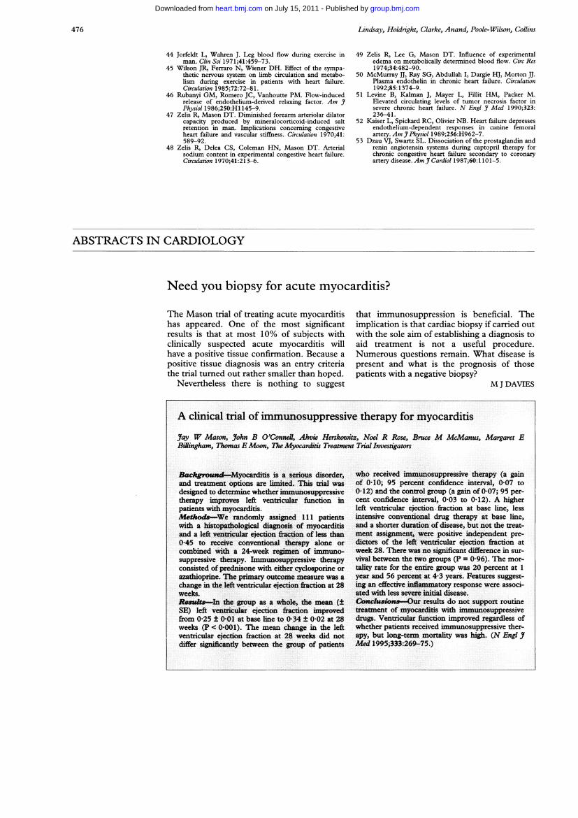
Need you biopsy for acute myocarditis?
The Mason trial of treating acute myocarditishas appeared. One of the most significantresults is that at most 10% of subjects withclinically suspected acute myocarditis willhave a positive tissue confirmation. Because apositive tissue diagnosis was an entry criteriathe trial turned out rather smaller than hoped.
Nevertheless there is nothing to suggest
that immunosuppression is beneficial. Theimplication is that cardiac biopsy if carried outwith the sole aim of establishing a diagnosis toaid treatment is not a useful procedure.Numerous questions remain. What disease ispresent and what is the prognosis of thosepatients with a negative biopsy?
M J DAVIES
476
group.bmj.com on July 15, 2011 - Published by heart.bmj.comDownloaded from

doi: 10.1136/hrt.75.5.469 1996 75: 469-476Heart
D. C. Lindsay, D. R. Holdright, D. Clarke, et al. chronic heart failure.Endothelial control of lower limb blood flow in
http://heart.bmj.com/content/75/5/469Updated information and services can be found at:
These include:
References http://heart.bmj.com/content/75/5/469#related-urls
Article cited in:
serviceEmail alerting
at the top right corner of the online article.Receive free email alerts when new articles cite this article. Sign up in the box
Notes
http://group.bmj.com/group/rights-licensing/permissionsTo request permissions go to:
http://journals.bmj.com/cgi/reprintformTo order reprints go to:
http://group.bmj.com/subscribe/To subscribe to BMJ go to:
group.bmj.com on July 15, 2011 - Published by heart.bmj.comDownloaded from
Copyright © 2022 FDOKUMEN