Elucidating the folding problem of helical peptides using empirical parameters
22
J. Mol. Biol. (1995) 245, 275–296 Elucidating the Folding Problem of Helical Peptides using Empirical Parameters. II². Helix Macrodipole Effects and Rational Modification of the Helical Content of Natural Peptides Victor Mun ˜ oz and Luis Serrano Explaining the helical behaviour of amino acid sequences in solution could EMBL, Meyerhofstrasse 1 Heidelberg D-69117 be one of the first steps in solving the protein folding problem in a rational Germany way. The information about the conformational behaviour of helical peptides in solution, as well as the a-helix stability in proteins, has been utilised to derive a database with the energy contributions for various interaction taking place in an a-helix: intrinsic helical propensities, side-chain to side-chain interactions, main-chain to main-chain hydrogen bonds, and capping effects. This database was implemented in a algorithm based on the helix-coil transition theory (AGADIR). Here, the effects on helix stability due to interactions between charged groups and the helix macrodipole are described, quantified and implemented in AGADIR. The algorithm correctly calculates the average helical behaviour in solution of 423 peptides analysed by circular dichroism and it describes the helicity at a residue level, as found when comparing the prediction for each amino acid residue with the data derived from nuclear magnetic resonance studies. Using AGADIR we have done a rational modification of peptides corresponding to protein secondary structure elements in order to increase their helical content. The circular dichroism analysis of the mutant peptides showed a very good agreement between the experimental and calculated helical content. Moreover, in certain specific cases in which strong tertiary contacts in folded proteins do not exist, the algorithm successfully predicts the length of mutagenised a-helices. It is interesting to note that the final values of the parameters used do not significantly differ in absolute terms from those extracted from mutagenesis studies in proteins. This indicates that the same physico-chemi- cal principles stand for both systems. Keywords: a-helix stability; protein folding; secondary structure prediction; biotechnology Introduction Protein folding is one of the most important problems in modern biochemistry remaining to be solved. It has been found that in the majority of cases proteins refold spontaneously in vitro to the native conformation, thus indicating that the three-dimen- sional information is contained in the linear sequence of amino acid residues. It also seems clear that the refolding polypeptide chain cannot explore all the available conformational space and, consequently, that one or more folding pathways should exist that will limit the conformational search (Anfinsen et al ., 1961). Experimental analysis of protein folding has revealed that secondary structure is formed very early in the folding process, while tertiary structure is acquired later (Matthews, 1993). Moreover, it has been found in many cases that isolated peptides have a strong tendency to populate significantly the same conformation that they have in the native state (Dyson & Wright, 1993). It is then possible to suggest that one of the earliest steps in the folding process will be the formation of secondary structure elements, concomitant or not with a hydrophobic collapse (Serrano et al ., 1992a ). It follows that if we want to understand protein folding in a rational way, we need to know what the factors are that determine the tendency of short amino acid sequences to populate different conformational states in solution. Abbreviations used: TFA, trifluoroacetic acid; EDT, ethane dithiol; TFE, trifluoroethanol. ² Papo I in this series is Mun ˜oz & Serano (1994). 0022–2836/95/030275–22 $08.00/0 7 1995 Academic Press Limited
-
Upload
independent -
Category
Documents
-
view
5 -
download
0
Transcript of Elucidating the folding problem of helical peptides using empirical parameters
J. Mol. Biol. (1995) 245, 275–296
Elucidating the Folding Problem of Helical Peptidesusing Empirical Parameters. II†. Helix MacrodipoleEffects and Rational Modification of the HelicalContent of Natural Peptides
Victor Mun oz and Luis Serrano
Explaining the helical behaviour of amino acid sequences in solution couldEMBL, Meyerhofstrasse 1Heidelberg D-69117 be one of the first steps in solving the protein folding problem in a rationalGermany way. The information about the conformational behaviour of helical peptides
in solution, as well as the a-helix stability in proteins, has been utilised toderive a database with the energy contributions for various interaction takingplace in an a-helix: intrinsic helical propensities, side-chain to side-chaininteractions, main-chain to main-chain hydrogen bonds, and capping effects.This database was implemented in a algorithm based on the helix-coiltransition theory (AGADIR). Here, the effects on helix stability due tointeractions between charged groups and the helix macrodipole aredescribed, quantified and implemented in AGADIR. The algorithm correctlycalculates the average helical behaviour in solution of 423 peptides analysedby circular dichroism and it describes the helicity at a residue level, as foundwhen comparing the prediction for each amino acid residue with the dataderived from nuclear magnetic resonance studies. Using AGADIR we havedone a rational modification of peptides corresponding to protein secondarystructure elements in order to increase their helical content. The circulardichroism analysis of the mutant peptides showed a very good agreementbetween the experimental and calculated helical content. Moreover, incertain specific cases in which strong tertiary contacts in folded proteins donot exist, the algorithm successfully predicts the length of mutageniseda-helices. It is interesting to note that the final values of the parameters useddo not significantly differ in absolute terms from those extracted frommutagenesis studies in proteins. This indicates that the same physico-chemi-cal principles stand for both systems.
Keywords: a-helix stability; protein folding;secondary structure prediction; biotechnology
Introduction
Protein folding is one of the most importantproblems in modern biochemistry remaining to besolved. It has been found that in the majority of casesproteins refold spontaneously in vitro to the nativeconformation, thus indicating that the three-dimen-sional information is contained in the linear sequenceof amino acid residues. It also seems clear that therefolding polypeptide chain cannot explore all theavailable conformational space and, consequently,that one or more folding pathways should exist thatwill limit the conformational search (Anfinsen et al.,
1961). Experimental analysis of protein folding hasrevealed that secondary structure is formed veryearly in the folding process, while tertiary structureis acquired later (Matthews, 1993). Moreover, it hasbeen found in many cases that isolated peptides havea strong tendency to populate significantly the sameconformation that they have in the native state(Dyson & Wright, 1993). It is then possible to suggestthat one of the earliest steps in the folding processwill be the formation of secondary structureelements, concomitant or not with a hydrophobiccollapse (Serrano et al., 1992a). It follows that if wewant to understand protein folding in a rational way,we need to know what the factors are that determinethe tendency of short amino acid sequences topopulate different conformational states in solution.
Abbreviations used: TFA, trifluoroacetic acid; EDT,ethane dithiol; TFE, trifluoroethanol.
† Papo I in this series is Munoz & Serano (1994).
0022–2836/95/030275–22 $08.00/0 7 1995 Academic Press Limited
The Folding Problem of Helical Peptides. II276
Currently, the attempts to describe the energeticsof systems formed by short polypeptide chains havebeen addressed to a-helices. The model most oftenused for that purpose, the helix-coil transition, isbased on the statistical mechanics theory and needsthe assumption of certain simplifications. In itssimplest version, as was postulated by Zimm &Bragg (1959), there were only two parameters, anelongation factor and a nucleation factor correspond-ing to equilibrium constants that are characteristic ofeach amino acid type responsible for the helicaltendency of a particular sequence. Both parametersare independent of the sequence environment of theamino acid residue. That means side-chain toside-chain interactions do not participate in thea-helix stability of a given peptide. A great deal ofwork has been devoted to the experimentaldetermination of nucleation and elongation factorsfor every type of amino acid (von Dreele et al., 1971),hoping that once parameterised, a complete pictureof the helix-coil transition would arise. Later on thetheory was modified in order to incorporate dataabout side-chain to side-chain interactions (Finkel-stein et al., 1991) as well as capping effects(Chakrabarty et al., 1993). These modifications,although they improve considerably the predictivepower of the helix-coil transition theory, still fellshort of explaining accurately the helical behaviourof complex heteropolypeptides in solution (Ganset al., 1991; Chen et al., 1992; Quian, 1993; Fukugitaet al., 1993; Park et al., 1993).
In a previous work we described an algorithm(AGADIR) that, based on the helix-coil theory andusing experimentally derived parameters, was ableto predict the average helical behaviour of 323peptides in aqueous solution at low temperatures (0to 4°C), ionic strengths (0 to 100 mM), and in the pHrange 6 to 7. This algorithm was able to describe thehelical behaviour of peptides in solution at a residuelevel (Munoz & Serrano, 1994, 1995). Here, weprovide a more detailed explanation of the methodand parameters used, as well as a more realisticdescription of the interactions between chargedresidues and the helix macrodipole, previouslyincluded in the capping effect. This results in a slightmodification of the values for some of the parameterspreviously described (Munoz & Serrano, 1994, 1995),and improves the predictive power of the algorithm.
Results
Variation of the helical content with the lengthof the peptide
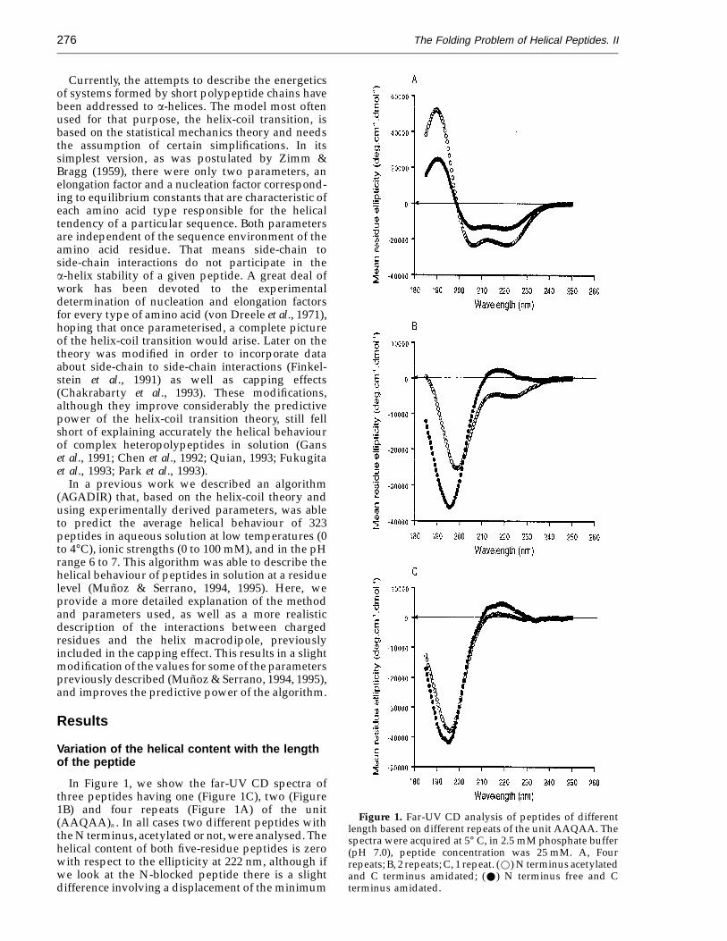
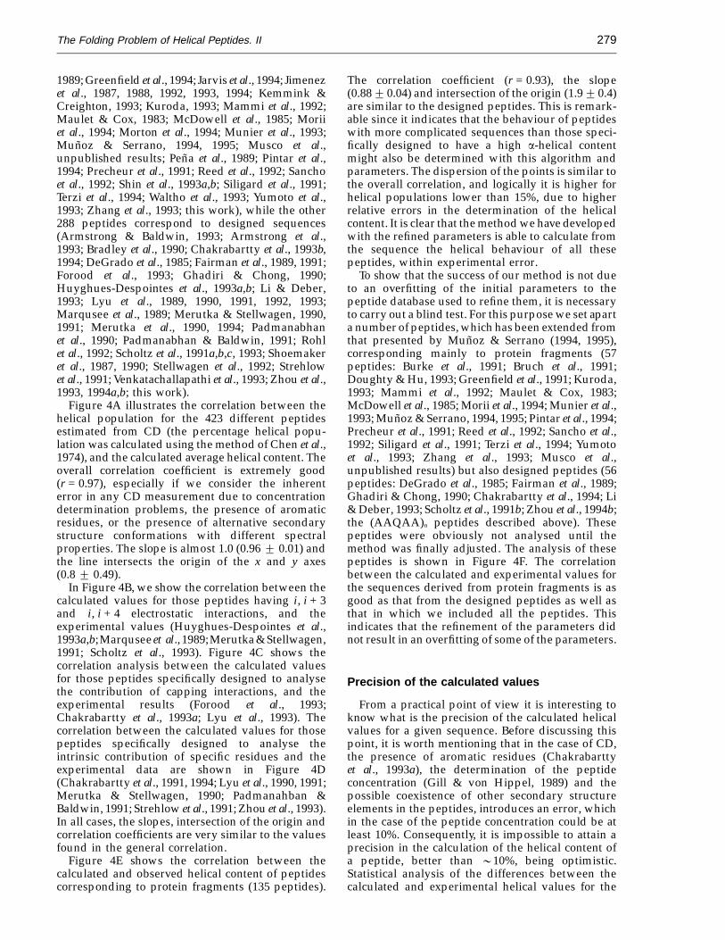
In Figure 1, we show the far-UV CD spectra ofthree peptides having one (Figure 1C), two (Figure1B) and four repeats (Figure 1A) of the unit(AAQAA)n . In all cases two different peptides withthe N terminus, acetylated or not, were analysed. Thehelical content of both five-residue peptides is zerowith respect to the ellipticity at 222 nm, although ifwe look at the N-blocked peptide there is a slightdifference involving a displacement of the minimum
Figure 1. Far-UV CD analysis of peptides of differentlength based on different repeats of the unit AAQAA. Thespectra were acquired at 5° C, in 2.5 mM phosphate buffer(pH 7.0), peptide concentration was 25 mM. A, Fourrepeats; B, 2 repeats; C, 1 repeat. (w) N terminus acetylatedand C terminus amidated; (W) N terminus free and Cterminus amidated.
The Folding Problem of Helical Peptides. II 277
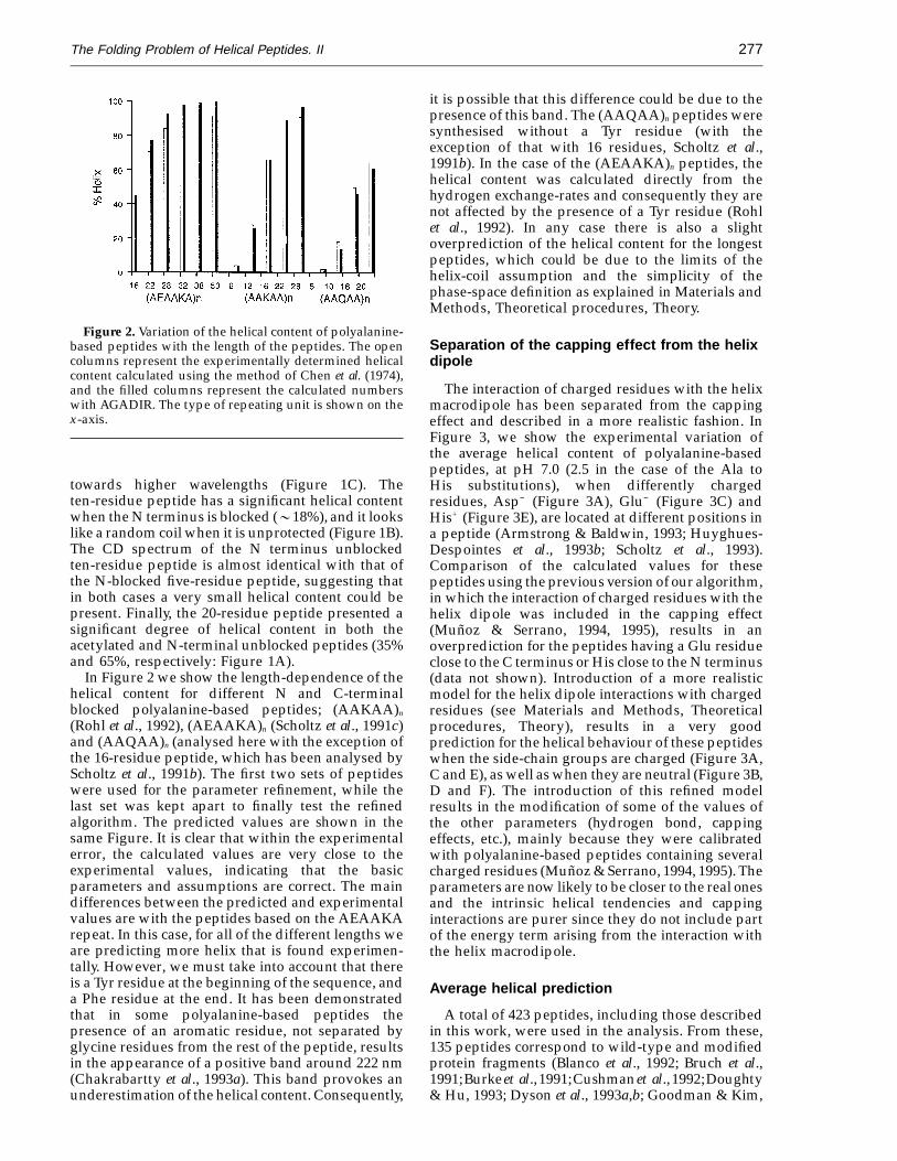
Figure 2. Variation of the helical content of polyalanine-based peptides with the length of the peptides. The opencolumns represent the experimentally determined helicalcontent calculated using the method of Chen et al. (1974),and the filled columns represent the calculated numberswith AGADIR. The type of repeating unit is shown on thex-axis.
it is possible that this difference could be due to thepresence of this band. The (AAQAA)n peptides weresynthesised without a Tyr residue (with theexception of that with 16 residues, Scholtz et al.,1991b). In the case of the (AEAAKA)n peptides, thehelical content was calculated directly from thehydrogen exchange-rates and consequently they arenot affected by the presence of a Tyr residue (Rohlet al., 1992). In any case there is also a slightoverprediction of the helical content for the longestpeptides, which could be due to the limits of thehelix-coil assumption and the simplicity of thephase-space definition as explained in Materials andMethods, Theoretical procedures, Theory.
Separation of the capping effect from the helixdipole
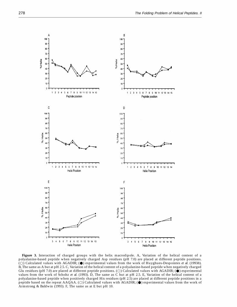
The interaction of charged residues with the helixmacrodipole has been separated from the cappingeffect and described in a more realistic fashion. InFigure 3, we show the experimental variation ofthe average helical content of polyalanine-basedpeptides, at pH 7.0 (2.5 in the case of the Ala toHis substitutions), when differently chargedresidues, Asp− (Figure 3A), Glu− (Figure 3C) andHis+ (Figure 3E), are located at different positions ina peptide (Armstrong & Baldwin, 1993; Huyghues-Despointes et al., 1993b; Scholtz et al., 1993).Comparison of the calculated values for thesepeptides using the previous version of our algorithm,in which the interaction of charged residues with thehelix dipole was included in the capping effect(Munoz & Serrano, 1994, 1995), results in anoverprediction for the peptides having a Glu residueclose to the C terminus or His close to the N terminus(data not shown). Introduction of a more realisticmodel for the helix dipole interactions with chargedresidues (see Materials and Methods, Theoreticalprocedures, Theory), results in a very goodprediction for the helical behaviour of these peptideswhen the side-chain groups are charged (Figure 3A,C and E), as well as when they are neutral (Figure 3B,D and F). The introduction of this refined modelresults in the modification of some of the values ofthe other parameters (hydrogen bond, cappingeffects, etc.), mainly because they were calibratedwith polyalanine-based peptides containing severalcharged residues (Munoz & Serrano, 1994, 1995). Theparameters are now likely to be closer to the real onesand the intrinsic helical tendencies and cappinginteractions are purer since they do not include partof the energy term arising from the interaction withthe helix macrodipole.
Average helical prediction
A total of 423 peptides, including those describedin this work, were used in the analysis. From these,135 peptides correspond to wild-type and modifiedprotein fragments (Blanco et al., 1992; Bruch et al.,1991;Burkeet al.,1991;Cushmanet al.,1992;Doughty& Hu, 1993; Dyson et al., 1993a,b; Goodman & Kim,
towards higher wavelengths (Figure 1C). Theten-residue peptide has a significant helical contentwhen the N terminus is blocked (018%), and it lookslike a random coil when it is unprotected (Figure 1B).The CD spectrum of the N terminus unblockedten-residue peptide is almost identical with that ofthe N-blocked five-residue peptide, suggesting thatin both cases a very small helical content could bepresent. Finally, the 20-residue peptide presented asignificant degree of helical content in both theacetylated and N-terminal unblocked peptides (35%and 65%, respectively: Figure 1A).
In Figure 2 we show the length-dependence of thehelical content for different N and C-terminalblocked polyalanine-based peptides; (AAKAA)n
(Rohl et al., 1992), (AEAAKA)n (Scholtz et al., 1991c)and (AAQAA)n (analysed here with the exception ofthe 16-residue peptide, which has been analysed byScholtz et al., 1991b). The first two sets of peptideswere used for the parameter refinement, while thelast set was kept apart to finally test the refinedalgorithm. The predicted values are shown in thesame Figure. It is clear that within the experimentalerror, the calculated values are very close to theexperimental values, indicating that the basicparameters and assumptions are correct. The maindifferences between the predicted and experimentalvalues are with the peptides based on the AEAAKArepeat. In this case, for all of the different lengths weare predicting more helix that is found experimen-tally. However, we must take into account that thereis a Tyr residue at the beginning of the sequence, anda Phe residue at the end. It has been demonstratedthat in some polyalanine-based peptides thepresence of an aromatic residue, not separated byglycine residues from the rest of the peptide, resultsin the appearance of a positive band around 222 nm(Chakrabartty et al., 1993a). This band provokes anunderestimation of the helical content. Consequently,
The Folding Problem of Helical Peptides. II278
Figure 3. Interaction of charged groups with the helix macrodipole. A, Variation of the helical content of apolyalanine-based peptide when negatively charged Asp residues (pH 7.0) are placed at different peptide positions.(w) Calculated values with AGADIR; (W) experimental values from the work of Huyghues-Despointes et al. (1993b).B, The same as A but at pH 2.5. C, Variation of the helical content of a polyalanine-based peptide when negatively chargedGlu residues (pH 7.0) are placed at different peptide positions. (w) Calculated values with AGADIR; (W) experimentalvalues from the work of Scholtz et al. (1993). D, The same as C but at pH 2.5. E, Variation of the helical content of apolyalanine-based peptide when positively charged His residues (pH 2.5) are placed at different peptide positions in apeptide based on the repeat AAQAA. (w) Calculated values with AGADIR; (W) experimental values from the work ofArmstrong & Baldwin (1993). F, The same as at E but pH 10.
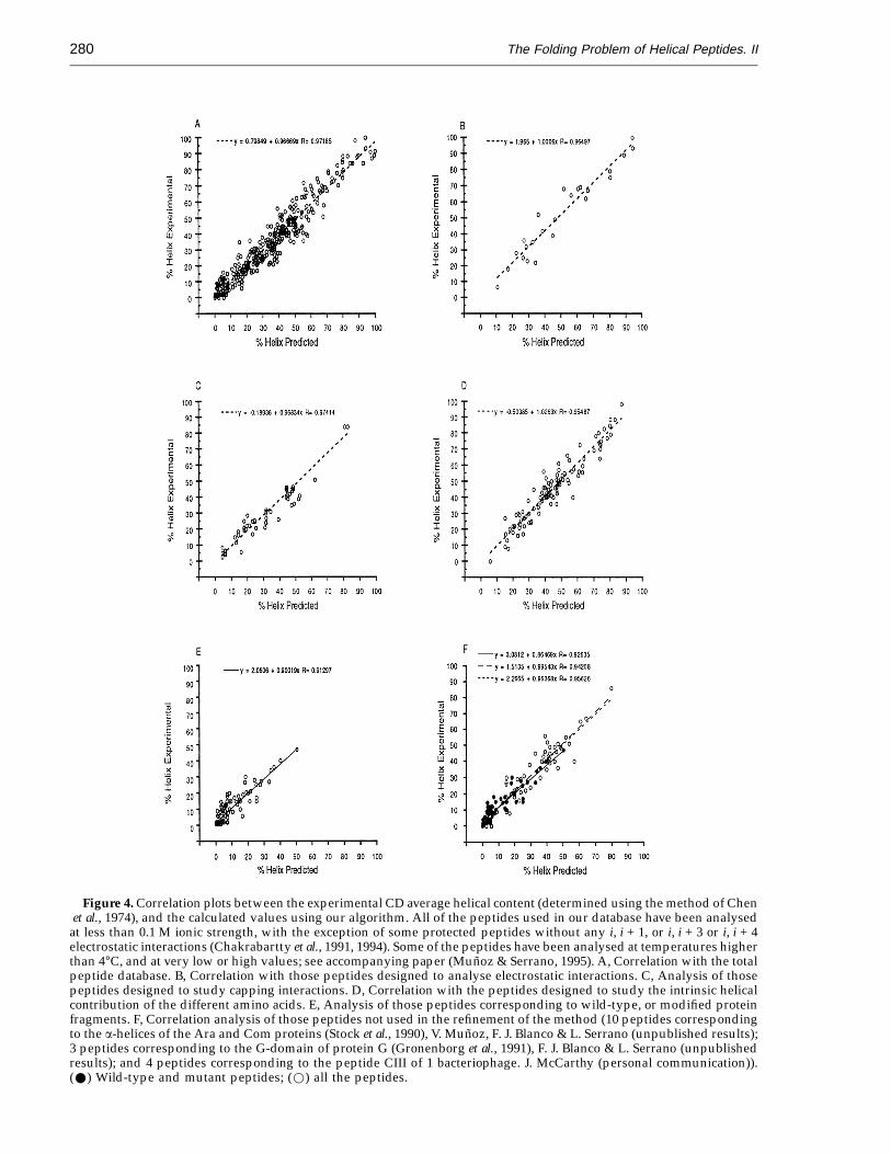
The Folding Problem of Helical Peptides. II 279
1989; Greenfield et al., 1994; Jarvis et al., 1994; Jimenezet al., 1987, 1988, 1992, 1993, 1994; Kemmink &Creighton, 1993; Kuroda, 1993; Mammi et al., 1992;Maulet & Cox, 1983; McDowell et al., 1985; Moriiet al., 1994; Morton et al., 1994; Munier et al., 1993;Munoz & Serrano, 1994, 1995; Musco et al.,unpublished results; Pena et al., 1989; Pintar et al.,1994; Precheur et al., 1991; Reed et al., 1992; Sanchoet al., 1992; Shin et al., 1993a,b; Siligard et al., 1991;Terzi et al., 1994; Waltho et al., 1993; Yumoto et al.,1993; Zhang et al., 1993; this work), while the other288 peptides correspond to designed sequences(Armstrong & Baldwin, 1993; Armstrong et al.,1993; Bradley et al., 1990; Chakrabartty et al., 1993b,1994; DeGrado et al., 1985; Fairman et al., 1989, 1991;Forood et al., 1993; Ghadiri & Chong, 1990;Huyghues-Despointes et al., 1993a,b; Li & Deber,1993; Lyu et al., 1989, 1990, 1991, 1992, 1993;Marqusee et al., 1989; Merutka & Stellwagen, 1990,1991; Merutka et al., 1990, 1994; Padmanabhanet al., 1990; Padmanabhan & Baldwin, 1991; Rohlet al., 1992; Scholtz et al., 1991a,b,c, 1993; Shoemakeret al., 1987, 1990; Stellwagen et al., 1992; Strehlowet al., 1991; Venkatachallapathi et al., 1993; Zhou et al.,1993, 1994a,b; this work).
Figure 4A illustrates the correlation between thehelical population for the 423 different peptidesestimated from CD (the percentage helical popu-lation was calculated using the method of Chen et al.,1974), and the calculated average helical content. Theoverall correlation coefficient is extremely good(r = 0.97), especially if we consider the inherenterror in any CD measurement due to concentrationdetermination problems, the presence of aromaticresidues, or the presence of alternative secondarystructure conformations with different spectralproperties. The slope is almost 1.0 (0.96 2 0.01) andthe line intersects the origin of the x and y axes(0.8 2 0.49).
In Figure 4B, we show the correlation between thecalculated values for those peptides having i, i + 3and i, i + 4 electrostatic interactions, and theexperimental values (Huyghues-Despointes et al.,1993a,b;Marqusee et al., 1989;Merutka&Stellwagen,1991; Scholtz et al., 1993). Figure 4C shows thecorrelation analysis between the calculated valuesfor those peptides specifically designed to analysethe contribution of capping interactions, and theexperimental results (Forood et al., 1993;Chakrabartty et al., 1993a; Lyu et al., 1993). Thecorrelation between the calculated values for thosepeptides specifically designed to analyse theintrinsic contribution of specific residues and theexperimental data are shown in Figure 4D(Chakrabartty et al., 1991, 1994; Lyu et al., 1990, 1991;Merutka & Stellwagen, 1990; Padmanahban &Baldwin, 1991; Strehlow et al., 1991; Zhou et al., 1993).In all cases, the slopes, intersection of the origin andcorrelation coefficients are very similar to the valuesfound in the general correlation.
Figure 4E shows the correlation between thecalculated and observed helical content of peptidescorresponding to protein fragments (135 peptides).
The correlation coefficient (r = 0.93), the slope(0.8820.04) and intersection of the origin (1.920.4)are similar to the designed peptides. This is remark-able since it indicates that the behaviour of peptideswith more complicated sequences than those speci-fically designed to have a high a-helical contentmight also be determined with this algorithm andparameters. The dispersion of the points is similar tothe overall correlation, and logically it is higher forhelical populations lower than 15%, due to higherrelative errors in the determination of the helicalcontent. It is clear that the method we have developedwith the refined parameters is able to calculate fromthe sequence the helical behaviour of all thesepeptides, within experimental error.
To show that the success of our method is not dueto an overfitting of the initial parameters to thepeptide database used to refine them, it is necessaryto carry out a blind test. For this purpose we set aparta number of peptides, which has been extended fromthat presented by Munoz & Serrano (1994, 1995),corresponding mainly to protein fragments (57peptides: Burke et al., 1991; Bruch et al., 1991;Doughty & Hu, 1993; Greenfield et al., 1991; Kuroda,1993; Mammi et al., 1992; Maulet & Cox, 1983;McDowell et al., 1985; Morii et al., 1994; Munier et al.,1993; Munoz & Serrano, 1994, 1995; Pintar et al., 1994;Precheur et al., 1991; Reed et al., 1992; Sancho et al.,1992; Siligard et al., 1991; Terzi et al., 1994; Yumotoet al., 1993; Zhang et al., 1993; Musco et al.,unpublished results) but also designed peptides (56peptides: DeGrado et al., 1985; Fairman et al., 1989;Ghadiri & Chong, 1990; Chakrabartty et al., 1994; Li& Deber, 1993; Scholtz et al., 1991b; Zhou et al., 1994b;the (AAQAA)n peptides described above). Thesepeptides were obviously not analysed until themethod was finally adjusted. The analysis of thesepeptides is shown in Figure 4F. The correlationbetween the calculated and experimental values forthe sequences derived from protein fragments is asgood as that from the designed peptides as well asthat in which we included all the peptides. Thisindicates that the refinement of the parameters didnot result in an overfitting of some of the parameters.
Precision of the calculated values
From a practical point of view it is interesting toknow what is the precision of the calculated helicalvalues for a given sequence. Before discussing thispoint, it is worth mentioning that in the case of CD,the presence of aromatic residues (Chakrabarttyet al., 1993a), the determination of the peptideconcentration (Gill & von Hippel, 1989) and thepossible coexistence of other secondary structureelements in the peptides, introduces an error, whichin the case of the peptide concentration could be atleast 10%. Consequently, it is impossible to attain aprecision in the calculation of the helical content ofa peptide, better than 010%, being optimistic.Statistical analysis of the differences between thecalculated and experimental helical values for the
The Folding Problem of Helical Peptides. II280
Figure 4. Correlation plots between the experimental CD average helical content (determined using the method of Chenet al., 1974), and the calculated values using our algorithm. All of the peptides used in our database have been analysed
at less than 0.1 M ionic strength, with the exception of some protected peptides without any i, i + 1, or i, i + 3 or i, i + 4electrostatic interactions (Chakrabartty et al., 1991, 1994). Some of the peptides have been analysed at temperatures higherthan 4°C, and at very low or high values; see accompanying paper (Munoz & Serrano, 1995). A, Correlation with the totalpeptide database. B, Correlation with those peptides designed to analyse electrostatic interactions. C, Analysis of thosepeptides designed to study capping interactions. D, Correlation with the peptides designed to study the intrinsic helicalcontribution of the different amino acids. E, Analysis of those peptides corresponding to wild-type, or modified proteinfragments. F, Correlation analysis of those peptides not used in the refinement of the method (10 peptides correspondingto the a-helices of the Ara and Com proteins (Stock et al., 1990), V. Munoz, F. J. Blanco & L. Serrano (unpublished results);3 peptides corresponding to the G-domain of protein G (Gronenborg et al., 1991), F. J. Blanco & L. Serrano (unpublishedresults); and 4 peptides corresponding to the peptide CIII of 1 bacteriophage. J. McCarthy (personal communication)).(W) Wild-type and mutant peptides; (w) all the peptides.
The Folding Problem of Helical Peptides. II 281
423 peptides of the database, gives an overallstandard deviation value of 5.86. The standarddeviation value is lower for those peptides havingless than 20% helical content (3 for 0 to 10% helicalcontent; 5 for those having between 10 and 20%). Thismeans that in the majority of the cases, we can cal-culate the helical content of a peptide with an error of26%, and in 99% of the cases with an error of 212%.
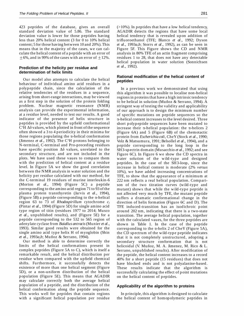
Prediction of the helicity per residue anddetermination of helix limits
Our model also attempts to calculate the helicalbehaviour of individual amino acid residues in apolypeptide chain, since the calculation of therelative tendencies of the residues in a sequence,arising from short-range interactions, may be viewedas a first step in the solution of the protein foldingproblem. Nuclear magnetic resonance (NMR)analysis can provide the experimental information,at a residue level, needed to test our results. A goodindicator of the presence of helix structure inpeptides is provided by the upfield conformationalCaH Dd values, which plotted in front of the sequenceoften showed a 3 to 4 periodicity in their minima forthose regions populating the a-helical conformation(Jimenez et al., 1992). The CaH protons belonging toN-terminal, C-terminal and Pro-preceding residueshave specific position Dd values, unrelated to thesecondary structure, and are not included in theplots. We have used these vaues to compare themwith the prediction of helical content at a residuelevel. In Figure 5A we show the good correlationbetween the NMR analysis in water solution and thehelicity per residue calculated with our method, forthe C-terminal 19 residues of murine interleukin-6(Morton et al., 1994) (Figure 5C) a peptidecorresponding to the amino acid region 71 to 93 of theplasma protein transthyretin (Jarvis et al., 1994),(Figure 5B) a peptide corresponding to the a-helicalregion 63 to 73 of Rhodospirillum cytochrome c2
(Pintar et al., 1994) (Figure 5D) for single amino acidtype region of titin (residues 1977 to 2014; Muscoet al., unpublished results), and (Figure 5E) for apeptide corresponding to the 532 to 565 region ofadenylate cyclase from Baiallus antracis (Munier et al.,1993). Similar good results were obtained for thesingle amino acid type helix H of myoglobin (Shinet al., 1993a,b; Munoz & Serrano, 1994).
Our method is able to determine correctly thelimits of the helical conformations present incomplex peptides (Figure 5A to C), which is itself aremarkable result, and the helical distribution perresidue when compared with the upfield chemicalshifts. Furthermore, it successfully detects theexistence of more than one helical segment (Figure5D), or a non-uniform distribution of the helicalpopulation (Figure 5E). This means that AGADIRmay calculate correctly both the average helicalpopulation of a peptide, and the distribution of thehelical conformation along the peptide sequence.This works well for peptides that contain regionswith a significant helical population per residue
(>10%). In peptides that have a low helical tendency,AGADIR detects the regions that have some localhelical tendency that is revealed upon addition oftrifluoroethanol (TFE; Blanco et al., 1992; Dysonet al., 1993a,b; Storrs et al., 1992), as can be seen inFigure 5F. This Figure shows the CD and NMRanalysis in 80% TFE of an actin fragment comprisingresidues 1 to 28, that does not have any detectablehelical population in water solution (Sonnichsenet al., 1992).
Rational modification of the helical content ofpeptides
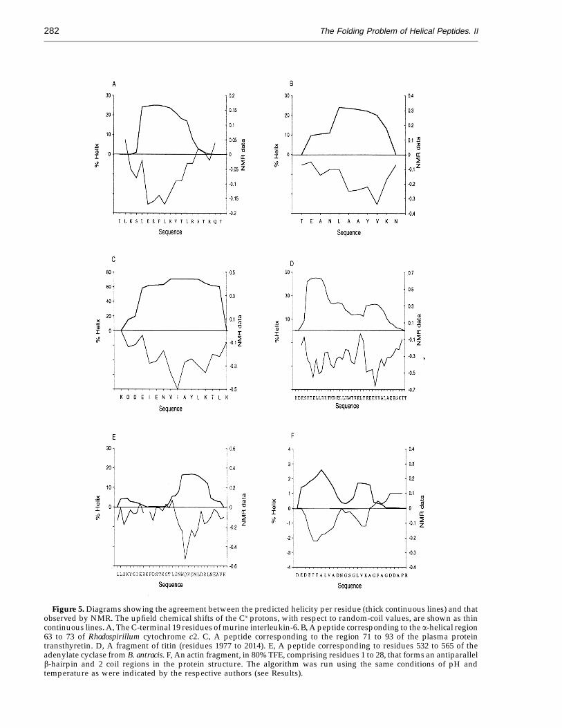
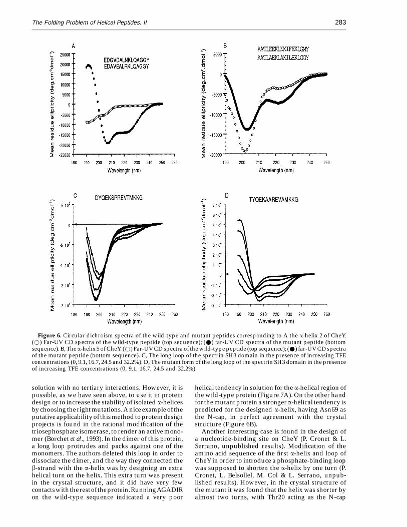
In a previous work we demonstrated that usingthis algorithm it was possible to localise non-helicalregions in proteins that have a high intrinsic tendencyto be helical in solution (Munoz & Serrano, 1994). Astringent way of testing the validity and applicabilityof our approach is to design the minimum numberof specific mutations on peptide sequences so thea-helical content increases to the level desired. Threeshort polypeptide sequences have been mutated toincrease their a-helical population: the a-helices 2(Figure 6A) and 5 (Figure 6B) of the chemotacticprotein from Escherichia coli, CheY (Stock et al., 1990;Volz & Matsumura, 1991; Belsollel et al., 1994), and apeptide corresponding to the long loop in theSH3-spectrin domain (Musacchio et al., 1992; and seeFigure 6C). In Figure 6 we show the CD spectra inwater solution of the wild-type and designedpeptides. In the case of the SH3-loop, since theincrease in helical content is moderate (2% versus10%), we have added increasing concentrations ofTFE, to show that the appearance of a minimum at222 nm reflects a real helical population. Compari-son of the two titration curves (wild-type andmutant) shows that while the wild-type peptide isnot affected very much by TFE, the mutant peptidesuffers a dramatic conformational change in thedirection of helix formation (Figure 6C and D). TheTFE induced-transition has an isodichroic pointaround 202 nm, indicating that there is a two-statetransition. The average helical population, togetherwith the calculated vaues, for the three peptides areshown in Table 1. In the case of the peptidecorresponding to the a-helix 2 of CheY (Figure 5A),the CD spectrum of the wild-type peptide indicatesthat it is not completely unstructured, adopting asecondary structure conformation that is nothelicoidal (V. Munoz, M. A. Jimenez, M. Rico & L.Serrano, unpublished results). After modification ofthe peptide, the helical content increases to a record40% for a short peptide (15 residues) that does nothave blocked ends and is not polyalanine-based.These results indicate that the algorithm issuccessfully calculating the effect of point mutationson the helical content of peptides.
Applicability of the algorithm to proteins
In principle, this algorithm is designed to calculatethe helical content of homopolymeric peptides in
The Folding Problem of Helical Peptides. II282
Figure 5. Diagrams showing the agreement between the predicted helicity per residue (thick continuous lines) and thatobserved by NMR. The upfield chemical shifts of the Ca protons, with respect to random-coil values, are shown as thincontinuous lines. A, The C-terminal 19 residues of murine interleukin-6. B, A peptide corresponding to the a-helical region63 to 73 of Rhodospirillum cytochrome c2. C, A peptide corresponding to the region 71 to 93 of the plasma proteintransthyretin. D, A fragment of titin (residues 1977 to 2014). E, A peptide corresponding to residues 532 to 565 of theadenylate cyclase from B. antracis. F, An actin fragment, in 80% TFE, comprising residues 1 to 28, that forms an antiparallelb-hairpin and 2 coil regions in the protein structure. The algorithm was run using the same conditions of pH andtemperature as were indicated by the respective authors (see Results).
The Folding Problem of Helical Peptides. II 283
Figure 6. Circular dichroism spectra of the wild-type and mutant peptides corresponding to A the a-helix 2 of CheY.(w) Far-UV CD spectra of the wild-type peptide (top sequence); (W) far-UV CD spectra of the mutant peptide (bottomsequence). B, The a-helix 5 of CheY. (w) Far-UV CD spectra of the wild-type peptide (top sequence); (W) far-UV CD spectraof the mutant peptide (bottom sequence). C, The long loop of the spectrin SH3 domain in the presence of increasing TFEconcentrations (0, 9.1, 16.7, 24.5 and 32.2%). D, The mutant form of the long loop of the spectrin SH3 domain in the presenceof increasing TFE concentrations (0, 9.1, 16.7, 24.5 and 32.2%).
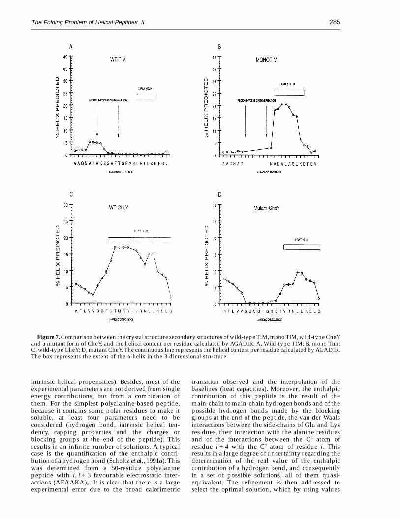
solution with no tertiary interactions. However, it ispossible, as we have seen above, to use it in proteindesign or to increase the stability of isolated a-helicesby choosing the right mutations. A nice example of theputative applicability of this method to protein designprojects is found in the rational modification of thetriosephosphate isomerase, to render an active mono-mer (Borchet et al., 1993). In the dimer of this protein,a long loop protrudes and packs against one of themonomers. The authors deleted this loop in order todissociate the dimer, and the way they connected theb-strand with the a-helix was by designing an extrahelical turn on the helix. This extra turn was presentin the crystal structure, and it did have very fewcontactswiththerestoftheprotein.RunningAGADIRon the wild-type sequence indicated a very poor
helical tendency in solution for the a-helical region ofthe wild-type protein (Figure 7A). On the other handfor the mutant protein a stronger a-helical tendency ispredicted for the designed a-helix, having Asn69 asthe N-cap, in perfect agreement with the crystalstructure (Figure 6B).
Another interesting case is found in the design ofa nucleotide-binding site on CheY (P. Cronet & L.Serrano, unpublished results). Modification of theamino acid sequence of the first a-helix and loop ofCheY in order to introduce a phosphate-binding loopwas supposed to shorten the a-helix by one turn (P.Cronet, L. Belsollel, M. Col & L. Serrano, unpub-lished results). However, in the crystal structure ofthe mutant it was found that the helix was shorter byalmost two turns, with Thr20 acting as the N-cap
The Folding Problem of Helical Peptides. II284
Table 1
Amino acid sequences of peptides corresponding towild-type regions of CheY and of the SH3 domain ofspectrin, as well as of the mutant sequences design toincrease the a-helical contentPeptide Sequence % Helixa % Predicted
CheY2 EDGVDALNKLQAGGY 2 4CheY2-Mo EDAVEALRKLQAGGY 39 40CheY5 AATLEEKLNKIFEKLGMY 13 7CheY5-Mo AATLAEKLAKILEKLGGY 20 20SH3Lo DYQEKSPREVAMKKG 2 2Sh3Lo-Mo TYQEKAAREVAMKKG 10 15
aThe experimental helical percentage was obtained by using themethod of Chen et al. (1974). All the peptides were analysed at pH7.0 in 5 mM phosphate buffer at 4°C.
parameter, s, of each participating residue. Thismakes it possible to assume it to be independent ofthe sequence and almost invariant upon changes intemperature (Scholtz et al., 1991c). Using thisdefinition we could calculate a sort of nucleationfactor obtained from the sum of four hydrogen bonds(3.12 kcal mol−1), s = 0.005, or 0.002 (if we consider avalue of 0.92 kcal mol−1 per hydrogen bond). In ourapproach the contributions of the main-chain tomain-chain hydrogen bonds and of the intrinsichelical propensities of the amino acid residues areclearly separated. We have assumed that the intrinsichelical propensities reflect the entropy loss uponfixing the different residues in a-helical dihedralangles. Then, the entropic cost of having the first fourresidues in helical angles, without formation of ahydrogen bond, is easily determined and isobviously strongly dependent on the particularsequence of amino acids and on the temperature.Our formulation, although equally involving twoparameters, could be more precise because, inprinciple, it is closer to the real energy contributions.
The partition function utilised here merits somecomment. From a strict point of view this partitionfunction might be considered a one-sequenceapproximation. The calculations performed involveonly the different individual possible helicalsegments and do not analyse all the possiblemolecular conformations with more than one helicalsegment. However, since we are calculating thepartition function for each amino acid residue withinthe polypeptide chain, and not for the whole peptide,more than one helical segment may be present at thesame time in a single molecule. In other words, theone-sequence approximation for the calculation ofhelicity of each residue renders an approximationshort of multiple sequence for the whole polypeptidechain. This is due to the total helicity of thepolypeptide chain arising from the average of thehelicities of the individual residues. One piece ofevidence for this statement is that our formulation isable to describe, in an appropriate way, the breakingof helices by bad helix-former residues when placedin the middle of the polypeptide chain and so it candetect the presence of more than one helical segmentin a polypeptide chain. The approach used in thiswork is therefore as simple as the one-sequenceapproximation but it provides a more realistic modelfor helix formation and stability, without involving somany calculations.
Refinement of the parameters
It is necessary to assess whether the set ofparameters being used accounts for the phenomenonto be described or is the consequence of overfittingto the database utilised. In our case, all of theparameters are derived from experimental data. Thisis somehow a warranty of their physical significance.However, there are important uncertainties thatmake the parameter refinement necessary.
Experimental error results in a range of valuesrather than in a discrete value for each parameter (i.e.
residue. Again that is what AGADIR calculates forthis sequence in solution (Figure 7C and D).
Discussion
Theoretical development
The work presented here and in a previous paper(Munoz & Serrano, 1994) is basically an attempt toput the large amount of experimental informationavailable to date into an appropriate theoreticalframework. The ultimate idea is to explain the helicalbehaviour of all linear, homopolymeric peptideshaving no tertiary interaction at a residue level. Thetheoretical framework utilised in this work is basedon the classical helix-coil transition as firstpostulated by Zimm & Bragg (1959) and by Lifson &Roig (1961). However, certain characteristics makeour approximation different from those modifi-cations, recently developed, of the original helix-coiltransition theory (Chen et al., 1992; Doig et al., 1994;Finkelstein et al., 1991; Gans et al., 1991; Quian, 1993).Our model, rather than describing the helix in termsof the two normal parameters, nucleation andelongation, attempts to differentiate the differentenergy contributions to the stability of the helicalconformation and thus a more extensive parameteri-sation is required. This makes it difficult to compareour energy contributions directly with the nucleationand elongation parameters extracted by severalgroups from their experimental data. Under ourtheoretical development it is possible to combine inone parameter the intrinsic tendency of a particularamino acid residue to populate helical dihedralangles with the enthalpy of hydrogen-bond for-mation, to obtain the classical elongation factor s. Thenucleation factor s is more difficult to compare. Inthe classical helix-coil transition theory, s does notarise from the entropic cost of fixing four amino acidresidues in helical angles without forming main-chain to main-chain hydrogen bonds, as hasfrequently been considered the case. Rather, itaccounts for the four main-chain to main-chainpossible hydrogen bonds that are not formed in aparticular helix, but are included in the elongation
The Folding Problem of Helical Peptides. II 285
Figure 7. Comparison between the crystal structure secondary structures of wild-type TIM, mono TIM, wild-type CheYand a mutant form of CheY, and the helical content per residue calculated by AGADIR. A, Wild-type TIM; B, mono Tim;C, wild-type CheY; D, mutant CheY. The continuous line represents the helical content per residue calculated by AGADIR.The box represents the extent of the a-helix in the 3-dimensional structure.
intrinsic helical propensities). Besides, most of theexperimental parameters are not derived from singleenergy contributions, but from a combination ofthem. For the simplest polyalanine-based peptide,because it contains some polar residues to make itsoluble, at least four parameters need to beconsidered (hydrogen bond, intrinsic helical ten-dency, capping properties and the charges orblocking groups at the end of the peptide). Thisresults in an infinite number of solutions. A typicalcase is the quantification of the enthalpic contri-bution of a hydrogen bond (Scholtz et al., 1991a). Thiswas determined from a 50-residue polyalaninepeptide with i, i + 3 favourable electrostatic inter-actions (AEAAKA)n . It is clear that there is a largeexperimental error due to the broad calorimetric
transition observed and the interpolation of thebaselines (heat capacities). Moreover, the enthalpiccontribution of this peptide is the result of themain-chain to main-chain hydrogen bonds and of thepossible hydrogen bonds made by the blockinggroups at the end of the peptide, the van der Waalsinteractions between the side-chains of Glu and Lysresidues, their interaction with the alanine residuesand of the interactions between the Cb atom ofresidue i + 4 with the Ca atom of residue i. Thisresults in a large degree of uncertainty regarding thedetermination of the real value of the enthalpiccontribution of a hydrogen bond, and consequentlyin a set of possible solutions, all of them quasi-equivalent. The refinement is then addressed toselect the optimal solution, which by using values
The Folding Problem of Helical Peptides. II286
within the experimental margins calculates correctlythe average helical behaviour of all the peptidesanalysed to date.
The refinement process needs to be carried outsequentially instead of doing a global fitting. In thisway it is possible to fix the parameters one by one orby small sets. The refined parameters presented hereshow some differences with those presented in theprevious work (Munoz & Serrano, 1994). Thesedifferences arise from the fact that the originalparameters were adjusted in the framework of amore simplistic model whereby interactions ofcharged residues with the helix macrodipole wereincluded in the capping effects. This simpleapproximation was found to work very well inpredicting the average helical behaviour of 323peptides. However, when the algorithm was appliedto some polyalanine-based peptides specificallydesigned to analyse the helical dipole (Armstrong &Baldwin, 1993; Huyghues-Despointes et al., 1993b;Scholtz et al., 1993), we found that the twointeractions (capping and helix dipole) needed to beseparated. Since most of the peptides used for theprevious refinement were polyalanine-based withcharged residues, the separation of the capping effectfrom the interaction with the helix dipole of chargedgroups done here results in slight changes in thevalues of the previous basic parameters. The moresignificant change is the reduction of the main-chain to main-chain hydrogen-bond contribution(0.78 kcal mol−1), below the experimental marginsdetermined by calorimetry for a polyalanine-basedpeptide (Scholtz et al., 1991a: 0.8 kcal mol−1 to1.2 kcal mol−1). This is an absolute requirement inorder to obtain the right cooperativity for theexperimentally observed length-dependent helix-coil transition of different polyalanine-based pep-tides (Rohl et al., 1992; Scholtz et al., 1991c; this work).Higher values, closer to the experimental data,always produced over-cooperative transitions, asis explained in Results. This discrepancy could bedue to our theoretical approximation, or to theexperimental error of the calorimetric studies. Inthe calorimetric analysis, and due to the broadtransition, it was not possible to determine thebase-lines of the unfolded and folded states. Then, tocalculate the base-lines and the thermodynamicparameters it was assumed that DCp was zero andthat the complete transition was a pure Gaussiancurve (Scholtz et al., 1991a). This could result in anoverestimation of the area of the curve (DHcal), andconsequently of the enthalpic contribution of ahydrogen bond. Moreover, in this peptide therecould be enthalpic contributions different from thehydrogen bond (see above). In support of this is thefact that DHcal for polyglutamic and polylysinepeptides is different (−1.1 versus −0.9 kcal mol − 1
per residue: Hermas, 1966), showing that factorsapart from hydrogen-bond formation contribute toDHcal.
Remarkably, the modification of the helix-coiltheory and the parameters described here predictvery well the helical content of more than 423
peptides analysed by CD. They also predict with ahigh degree of success how the helical population isdistributed along the polypeptide chain for thosepeptides analysed by NMR. The helix-coil transitiontheory using the appropriate parameters, it is able tocalculate correctly the average helical content of shortpolypeptide chains without tertiary interactions insolution, as well as the individual behaviour of thedifferent component residues.
Comparison with the parameters determinedby other authors
There are several scales of intrinsic helicaltendencies of the different amino acids, determinedfrom the thermodynamic analysis of proteins(Horovitz et al., 1992; Blaber et al., 1993) or peptides(von Dreele et al., 1971; Lyu et al., 1990; O’Neil &DeGrado, 1990; Horovitz et al., 1992; Chakrabarttyet al., 1994). Each scale has its own problems derivedfrom the system being used, as well as the differentexperimental conditions. Since every system iscompletely different from the others, but theresults are related (with the exception perhaps ofthat of von Dreele et al., 1971), it means that theaverage value of the different experimental scales(Horovitz et al., 1992; Blaber et al., 1993; Lyu et al.,1990; O’Neil & DeGrado, 1990; Horovitz et al., 1992;Chakrabartty et al., 1994), could diminish the contexteffects.
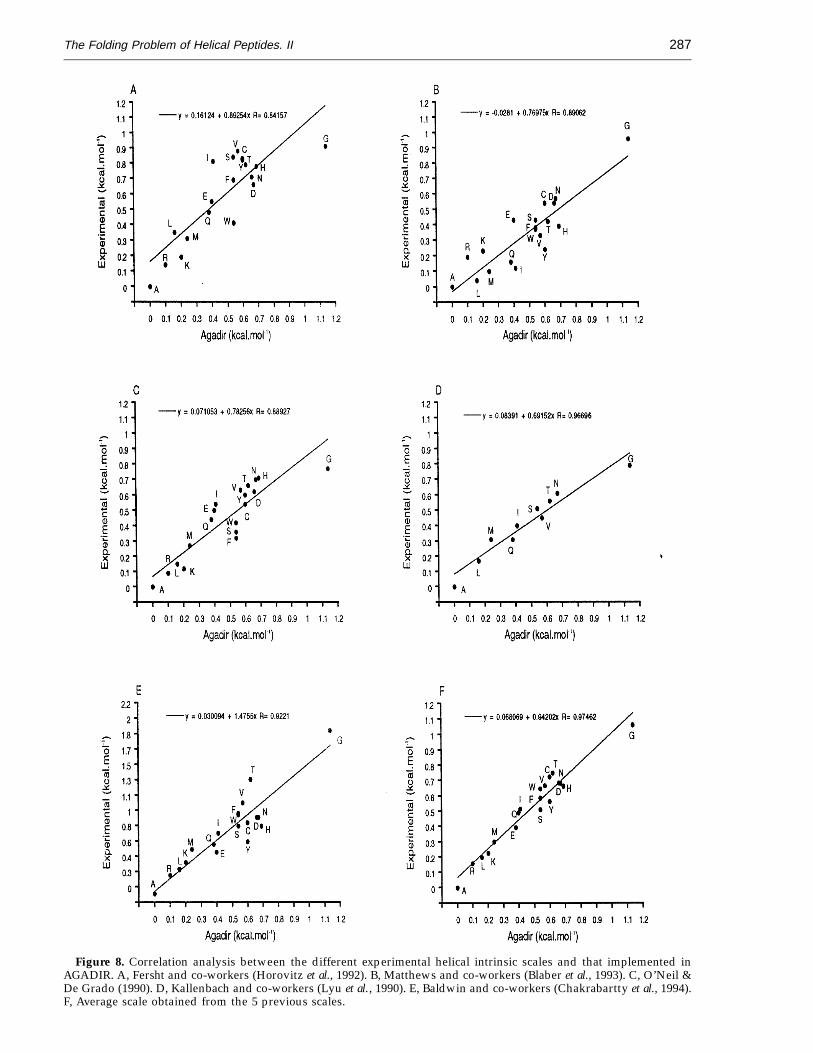
After refinement, the intrinsic helical propensitieswere slightly different from the initial values(slope = 0.84 and r = 0.89; data not shown). Com-parison of the refined values with the differentexperimental scales (Figure 8A to E), excluding Pro,indicates that the refined parameters correlateapproximately equally well with all of them (thecorrelation with the scale used by Lyu et al. (1990) isbetter but we must take into account that the numberof data is much smaller, 10 versus 19). The slopes aresimilar, varying from 0.7 to 0.9, with the exception ofthat of Baldwin and co-workers, which is higher, 1.48(Chakrabartty et al., 1994). The slopes of thecorrelations between the data of Baldwin andco-workers and the other scales (including AGADIR),are very similar, varying between 0.4 and 0.6 (datanot shown). This means that the differences in energybetween Ala and the different residues are muchhigher in this case than in all the other scales.Interestingly, our algorithm is able to calculatecorrectly the experimental data used by Baldwin andco-workers to derive their helix scale (Chakrabarttyet al., 1994: slope = 1 2 0.07; r = 0.89; intersection =−1.6 2 3; Figure 5F), although it was not used for therefinement of the parameters, without having suchlarge energy differences between the intrinsic helicalenergies of the amino acid residues.
Most importantly, the best correlation was foundwith the average data of the five experimental scales(slope = 0.94 and r = 0.97), indicating that we haveprobably reached values that are very close to thereal ones. Also, the fact that by using values similar
The Folding Problem of Helical Peptides. II 287
Figure 8. Correlation analysis between the different experimental helical intrinsic scales and that implemented inAGADIR. A, Fersht and co-workers (Horovitz et al., 1992). B, Matthews and co-workers (Blaber et al., 1993). C, O’Neil &De Grado (1990). D, Kallenbach and co-workers (Lyu et al., 1990). E, Baldwin and co-workers (Chakrabartty et al., 1994).F, Average scale obtained from the 5 previous scales.
The Folding Problem of Helical Peptides. II288
to those found in proteins we are able to reproducethe helical behaviour of 423 peptides in solution,indicates that the physico-chemical parameters inproteins and peptides are the same.
Conclusions
Here, we have described a more completeapproach to the folding of helical peptides insolution, in which we considered five differentparameters; hydrogen bond, intrinsic helical ten-dencies, capping interactions (including the effect ofblocking the ends of the peptides), charge-dipoleinteractions and side-chain to side-chain interactions.Using these five parameters we predicted the helicalbehaviour of 423 homopolymeric peptides insolution, as well as the effect of designed mutationson the helical content of different peptides. Moreimportant, as we have seen for several differentpeptides, AGADIR also calculates correctly thehelical population at a residue level. This webelieve is an important step in the direction of theelucidation of the folding problem of helicalpeptides, but we must be cautious since there aremany energy parameters involved and we aremaking several assumptions. The analysis of morepeptides by CD and NMR will allow a betterrefinement of the parameters, and will indicate if theassumptions made here are reasonable. In any case,it is clear that the parameters used in this workdescribe a-helix stability. When more peptides havebeen studied the model might be improved andtherefore further refinement could be needed. Thisshould be in only two directions: splitting ofpair-interactions groups into their components andinclusion in the theoretical framework of newlydiscovered factors.
Materials and Methods
Experimental procedures
Peptide synthesis
The solid-phase synthesis of the peptides was performedon an Abimed AMS422 multiple peptide synthesiser usingFmoc chemistry and PyBOP activation at a 0.025 mmolscale. After synthesis was completed, protecting groupswere removed and the peptide chains were cleaved fromthe resin with a mixture of 10 ml of TFA, 0.75 g of phenol,0.2 ml of EDT, 0.5 ml of thioanisole and 0.5 mlof water for three hours. The peptides were purifiedon a Vydac C-18 reverse phase column (20 mm ×250 mm, 0.01 mm particle) at a flow-rate of 10 ml/min.Solvent A was water containing 0.1% TFA and solvent Bwas 70% acetonitrile, 0.1% TFA in water. Peptidehomogeneity (>98%) was determined by HPLCusing an acetonitrile gradient of 0.7% per minute. Thepeptide composition was confirmed by amino acidanalysis and the molecular mass was checked bymatrix-assisted laser desorption time-of-flight massspectrometry.
Peptide concentration
The concentration of the different peptides wasdetermined by amino acid analysis, or UV absorbanceusing the method of Gill & von Hippel (1989). For peptidesthat do not contain Tyr or Trp residues, the concentrationwas determined by amino acid analysis. The error in bothcases is around 10%.
Circular dichroism analysis
Circular dichroism (CD) spectra were recorded on aJasco-710 instrument at a temperature of 5°C. The peptides(roughly 1.5 mg) were dissolved in 1 ml of 2.5 mM sodiumphosphate buffer (pH 7.0), unless otherwise indicated. Tocheck for concentration dependence of the CD spectra,different dilutions of the peptides (10 to 750 mM), usingcuvetteswithdifferentpathlengths(0.1 mmto0.5 cm),werescanned. CD spectra in the range 190 to 250 nm were ob-tained using the continuous scan option (100 nm/min scanspeed), with a one second response time and taking pointsevery 0.1 nm. For every sample we took 30 scans and the ex-periment was repeated three times on different days. The el-lipticity was calibrated using D-10-camphorsulphonic acid.
Determination of the helical percentage from the CDspectra
In order to estimate the helical population of thedifferent peptides we used the mean residue ellipticity at222 nm, taking into account the peptide length (Chen et al.,1974).
Theoretical procedures
Theory
The development of an appropriate statistical mechanicsframework to model the conformational behaviour ofpeptides in aqueous solution needs, as a first step, a precisedefinition of the physical system under study. In this casethe physical system is obviously a polypeptide chain inaqueous solution. The polypeptide chain as defined herecomprises n − 1 peptide bonds (for a peptide of n residues)of fixed length at fixed bond angles. The differentconformations of each residue are defined by the anglesbetween its alpha carbon atom and the atoms of theflanking peptide bonds. Following this definition, thephase space for each residue is restricted by means ofsimplicity to the allowed region of the Ramachandran plot.
As in the classical helix-coil transition formulations(Zimm & Brag, 1959), the phase space of each residue issimplified by considering only two states. The first is therandom-coil state, which consists of a heterogeneousensemble of conformations where each amino acid residueis able to explore all the allowed conformational space. Thesecond is the helical state, which is defined as the statewhere the amino acid residue being considered, and atleast three contiguous residues, are restricted to a-helicalangles plus the two capping residues (N-cap and C-cap).
The calculation of the helical propensity of each residueon a polypeptide chain may be addressed upon calculationof the molecular partition function. The molecularpartition function would be the summation of thestatistical weights of all the possible helical conformationsof the polypeptide chain, plus the statistical weight of themolecular random coil. A helical conformation would beany containing at least one helical segment (at least fourresidues in helical angles and the two caps) in the
The Folding Problem of Helical Peptides. II 289
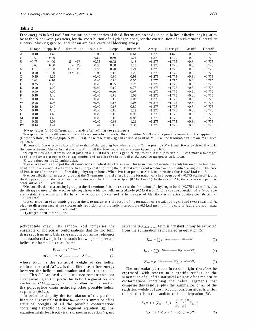
Table 2
Free energies in kcal mol −1 for the intrinsic tendencies of the different amino acids to be in helical dihedral angles, or tobe at the N or C-cap positions, for the contribution of a hydrogen bond, for the contribution of an N-terminal acetyl orsuccinyl blocking groups, and for an amide C-terminal blocking group
N-capa Capp. boxb (Pro N + 1)c Asp + 2d C-cape Intrinsicf Acetylg Succinylh Amidei Hbondj
A 0.40 0.40 0.00 0.00 0.61 −1.375 −1.875 −0.91 −0.775G −0.60 −0.60 −0.65 −0.40 1.71 −1.275 −1.775 −0.81 −0.775S −0.75 −1.00 S = −0'3 −0.75 −0.08 1.13 −1.275 −1.775 −0.81 −0.775T −0.65 −0.80 T = −0'3 −0.50 −0.08 1.18 −1.275 −1.775 −0.81 −0.775N −1.10 −1.00 N = −0'3 −1.10 −0.20 1.21 −1.275 −1.775 −0.81 −0.775D 0.00 −1.00 D = −0'3 0.00 0.08 1.20 −1.275 −1.775 −0.81 −0.775Q 0.50 0.25 −0.40 0.00 0.93 −1.275 −1.775 −0.81 −0.775E −0.08 −0.10 −0.40 0.08 0.95 −1.275 −1.775 −0.81 −0.775H 0.25 0.25 −0.40 −0.35 1.23 −1.275 −1.775 −0.81 −0.775K 0.00 0.00 −0.40 0.00 0.76 −1.275 −1.775 −0.81 −0.775R 0.00 0.00 −0.40 −0.10 0.67 −1.275 −1.775 −0.81 −0.775F 0.40 0.40 −0.40 0.08 1.08 −1.275 −1.775 −0.81 −0.775Y 0.40 0.40 −0.40 0.08 1.08 −1.275 −1.775 −0.81 −0.775W 0.08 0.08 −0.40 0.08 1.08 −1.275 −1.775 −0.81 −0.775L 0.40 0.40 −0.40 0.08 0.80 −1.275 −1.775 −0.81 −0.775V 0.40 0.40 −0.40 0.08 1.12 −1.275 −1.775 −0.81 −0.775I 0.40 0.40 −0.40 0.08 0.96 −1.275 −1.775 −0.81 −0.775M 0.40 0.40 −0.40 0.08 0.82 −1.275 −1.775 −0.81 −0.775C 0.08 0.08 −0.40 0.08 1.21 −1.275 −1.775 −0.81 −0.775P* 0.44 0.44 0.44 0.08 3.33 −1.275 −1.775 −0.81 −0.775
aN-cap values for 20 different amino acids after refining the parameters.bN-cap values of the different amino acid residues when there is Glu at position N + 3 and the possible formation of a capping box
(Harper & Rose, 1993; Dasgupta & Bell, 1993). In the case of having Gln or Asp at position N + 3, all the favourable values are multipliedby 0.625.
cFavourable free energy values added to that of the capping box when there is Glu at position N + 3, and Pro at position N + 1. Inthe case of having Gln or Asp at position N + 3, all the favourable values are multiplied by 0.625.
dN-cap values when there is Asp at position N + 2. If there is not a good N-cap residue, Asp at position N + 2 can make a hydrogenbond to the amide group of the N-cap residue and stabilise the helix (Bell et al., 1992; Dasgurpta & Bell, 1993).
eC-cap values for the 20 amino acids.fFree energy required to put the 20 amino acids in helical dihedral angles. This term does not incude the contribution of the hydrogen
bond, and in our model it reflects the free energy cost of putting the different amino acid residues in helical dihedral angles. In the caseof Pro, it includes the result of breaking a hydrogen bond. When Pro is at position N + 1, its intrinsic value is 0.66 kcal mol −1.
gNet contribution of an acetyl group at the N terminus. It is the result of the formation of a hydrogen bond (−0.775 kcal mol −1), plusthe disappearance of the electrostatic repulsion with the helix macrodipole (0.5 kcal mol −1). In the case of Ala, there is an extra positivecontribution of −0.1 kcal mol −1.
hNet contribution of a succinyl group at the N terminus. It is the result of the formation of a hydrogen bond (−0.775 kcal mol −1), plusthe disappearance of the electrostatic repulsion with the helix macrodipole (0.5 kcal mol −1), plus the introduction of a favourableelectrostatic interaction with the helix macrodipole (−0.5 kcal mol −1). In the case of Ala, there is an extra positive contribution of−0.1 kcal mol −1.
iNet contribution of an amide group at the C terminus. It is the result of the formation of a weak hydrogen bond (−0.31 kcal mol −1),plus the disappearance of the electrostatic repulsion with the helix macrodipole (0.5 kcal mol −1). In the case of Ala, there is an extrapositive contribution of −0.1 kcal mol −1.
jHydrogen bond contribution.
polypeptide chain. The random coil comprises theensemble of molecular conformations that do not fulfilthese requirements. Using the random coil as the referencestate (statistical weight 1), the statistical weight of a certainhelical conformation arises from:
KConfor = e − DGConfor/RT (1)
DGConfor = DGhelical-segment + DGrest, (2)
where KConfor is the statistical weight of the helicalconformation and DGConfor is the difference in free energybetween the helical conformation and the random coilstate. This DG can be divided into two components: onecorresponding to the particular helical segment we areanalysing (DGhelical-segment) and the other to the rest ofthe polypeptide chain including other possible helicalsegments (DGrest).
In order to simplify the formulation of the partitionfunction it is possible to define KHel as the summation of thestatistical weights of all the possible conformationscontaining a specific helical segment (equation (3)). Thisequation might be directly transformed in equation (4), and
since the DGhelical-segment term is constant it may be extractedfrom the summation as indicated in equation (5):
KHel = s e − (DGhelical-segment + DGrest)/RT (3)
KHel = s(e − DGhelical-segment/RTe − DGrest/RT) (4)
KHel = e − DGhelical-segment/RTs e − DGrest/RT. (5)
The molecular partition function might therefore beexpressed, with respect to a specific residue, as thesummation of all of the statistical weights of the molecularconformations containing the helical segments thatcomprise this residue, plus the summation of all of thestatistical weights of the molecular conformations in whichthis residue is in the random-coil state (equation (6)).
ZX = 1 + (ZN + ZC) + sn
j = 6
s(n − j + 1)
i = 1
KHelji
‘‘[x>i + j E x < i c KHelji = 0’’, (6)
The Folding Problem of Helical Peptides. II290
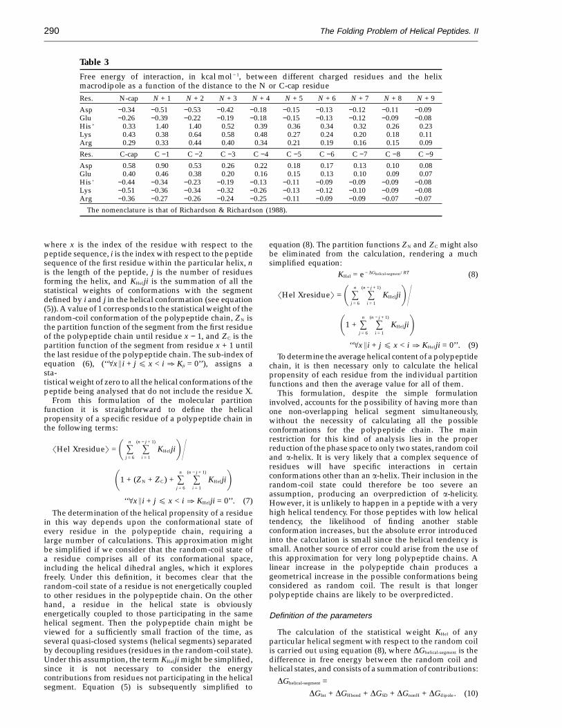
Table 3
Free energy of interaction, in kcal mol − 1, between different charged residues and the helixmacrodipole as a function of the distance to the N or C-cap residueRes. N-cap N + 1 N + 2 N + 3 N + 4 N + 5 N + 6 N + 7 N + 8 N + 9
Asp −0.34 −0.51 −0.53 −0.42 −0.18 −0.15 −0.13 −0.12 −0.11 −0.09Glu −0.26 −0.39 −0.22 −0.19 −0.18 −0.15 −0.13 −0.12 −0.09 −0.08His + 0.33 1.40 1.40 0.52 0.39 0.36 0.34 0.32 0.26 0.23Lys 0.43 0.38 0.64 0.58 0.48 0.27 0.24 0.20 0.18 0.11Arg 0.29 0.33 0.44 0.40 0.34 0.21 0.19 0.16 0.15 0.09
Res. C-cap C −1 C −2 C −3 C −4 C −5 C −6 C −7 C −8 C −9
Asp 0.58 0.90 0.53 0.26 0.22 0.18 0.17 0.13 0.10 0.08Glu 0.40 0.46 0.38 0.20 0.16 0.15 0.13 0.10 0.09 0.07His + −0.44 −0.34 −0.23 −0.19 −0.13 −0.11 −0.09 −0.09 −0.09 −0.08Lys −0.51 −0.36 −0.34 −0.32 −0.26 −0.13 −0.12 −0.10 −0.09 −0.08Arg −0.36 −0.27 −0.26 −0.24 −0.25 −0.11 −0.09 −0.09 −0.07 −0.07
The nomenclature is that of Richardson & Richardson (1988).
where x is the index of the residue with respect to thepeptide sequence, i is the index with respect to the peptidesequence of the first residue within the particular helix, nis the length of the peptide, j is the number of residuesforming the helix, and KHelji is the summation of all thestatistical weights of conformations with the segmentdefined by i and j in the helical conformation (see equation(5)). A value of 1 corresponds to the statistical weight of therandom-coil conformation of the polypeptide chain, ZN isthe partition function of the segment from the first residueof the polypeptide chain until residue x − 1, and ZC is thepartition function of the segment from residue x + 1 untilthe last residue of the polypeptide chain. The sub-index ofequation (6), (‘‘[x>i + j E x < i c Kji = 0’’), assigns asta-tistical weight of zero to all the helical conformations of thepeptide being analysed that do not include the residue X.
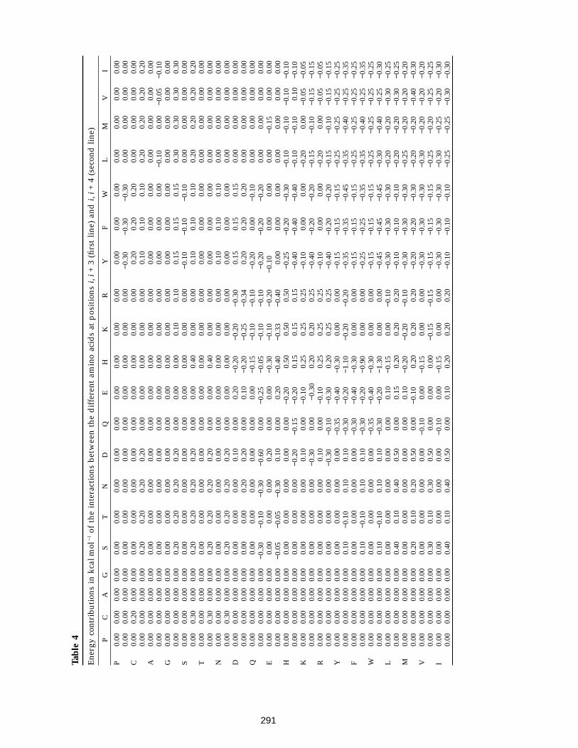
From this formulation of the molecular partitionfunction it is straightforward to define the helicalpropensity of a specific residue of a polypeptide chain inthe following terms:
�Hel Xresidue� = 0 sn
j = 6
s(n − j + 1)
i = 1
KHelji1>01 + (ZN + ZC) + s
n
j = 6
s(n − j + 1)
i = 1
KHelji1‘‘[x>i + j E x < i c KHelji = 0’’. (7)
The determination of the helical propensity of a residuein this way depends upon the conformational state ofevery residue in the polypeptide chain, requiring alarge number of calculations. This approximation mightbe simplified if we consider that the random-coil state ofa residue comprises all of its conformational space,including the helical dihedral angles, which it exploresfreely. Under this definition, it becomes clear that therandom-coil state of a residue is not energetically coupledto other residues in the polypeptide chain. On the otherhand, a residue in the helical state is obviouslyenergetically coupled to those participating in the samehelical segment. Then the polypeptide chain might beviewed for a sufficiently small fraction of the time, asseveral quasi-closed systems (helical segments) separatedby decoupling residues (residues in the random-coil state).Under this assumption, the term KHelji might be simplified,since it is not necessary to consider the energycontributions from residues not participating in the helicalsegment. Equation (5) is subsequently simplified to
equation (8). The partition functions ZN and ZC might alsobe eliminated from the calculation, rendering a muchsimplified equation:
KHel = e − DGhelical-segment/RT (8)
�Hel Xresidue� = 0 sn
j = 6
s(n − j + 1)
i = 1
KHelji1>01 + s
n
j = 6
s(n − j + 1)
i = 1
KHelji1‘‘[x>i + j E x < i c KHelji = 0’’. (9)
To determine the average helical content of a polypeptidechain, it is then necessary only to calculate the helicalpropensity of each residue from the individual partitionfunctions and then the average value for all of them.
This formulation, despite the simple formulationinvolved, accounts for the possibility of having more thanone non-overlapping helical segment simultaneously,without the necessity of calculating all the possibleconformations for the polypeptide chain. The mainrestriction for this kind of analysis lies in the properreduction of the phase space to only two states, random coiland a-helix. It is very likely that a complex sequence ofresidues will have specific interactions in certainconformations other than an a-helix. Their inclusion in therandom-coil state could therefore be too severe anassumption, producing an overprediction of a-helicity.However, it is unlikely to happen in a peptide with a veryhigh helical tendency. For those peptides with low helicaltendency, the likelihood of finding another stableconformation increases, but the absolute error introducedinto the calculation is small since the helical tendency issmall. Another source of error could arise from the use ofthis approximation for very long polypeptide chains. Alinear increase in the polypeptide chain produces ageometrical increase in the possible conformations beingconsidered as random coil. The result is that longerpolypeptide chains are likely to be overpredicted.
Definition of the parameters
The calculation of the statistical weight KHel of anyparticular helical segment with respect to the random coilis carried out using equation (8), where DGhelical-segment is thedifference in free energy between the random coil andhelical states, and consists of a summation of contributions:
DGhelical-segment =DGInt + DGHbond + DGSD + DGnonH + DGdipole. (10)
291
Tabl
e4
Ene
rgy
cont
ribu
tion
sin
kcal
mol
−1of
the
inte
ract
ions
betw
een
the
dif
fere
ntam
ino
acid
sat
pos
itio
nsi,
i+3
(firs
tlin
e)an
di,
i+4
(sec
ond
line)
PC
AG
ST
ND
QE
HK
RY
FW
LM
VI
P0.
000.
000.
000.
000.
000.
000.
000.
000.
000.
000.
000.
000.
000.
000.
000.
000.
000.
000.
000.
000.
000.
000.
000.
000.
000.
000.
000.
000.
000.
000.
000.
000.
00−0
.30
−0.3
0−0
.30
0.00
0.00
0.00
0.00
C0.
000.
200.
000.
000.
000.
000.
000.
000.
000.
000.
000.
000.
000.
200.
200.
200.
000.
000.
000.
000.
000.
000.
000.
000.
200.
200.
200.
200.
000.
000.
000.
000.
000.
100.
100.
100.
200.
200.
200.
20A
0.00
0.00
0.00
0.00
0.00
0.00
0.00
0.00
0.00
0.00
0.00
0.00
0.00
0.00
0.00
0.00
0.00
0.00
0.00
0.00
0.00
0.00
0.00
0.00
0.00
0.00
0.00
0.00
0.00
0.00
0.00
0.00
0.00
0.00
0.00
0.00
−0.1
00.
00−0
.05
−0.1
0G
0.00
0.00
0.00
0.00
0.00
0.00
0.00
0.00
0.00
0.00
0.00
0.00
0.00
0.00
0.00
0.00
0.00
0.00
0.00
0.00
0.00
0.00
0.00
0.00
0.20
0.20
0.20
0.20
0.00
0.00
0.00
0.10
0.10
0.15
0.15
0.15
0.30
0.30
0.30
0.30
S0.
000.
000.
000.
000.
000.
000.
000.
000.
000.
000.
000.
000.
00−0
.10
−0.1
0−0
.10
0.00
0.00
0.00
0.00
0.00
0.30
0.00
0.00
0.20
0.20
0.20
0.20
0.00
0.00
0.40
0.00
0.00
0.10
0.10
0.10
0.20
0.20
0.20
0.20
T0.
000.
000.
000.
000.
000.
000.
000.
000.
000.
000.
000.
000.
000.
000.
000.
000.
000.
000.
000.
000.
000.
300.
000.
000.
200.
200.
200.
200.
000.
000.
400.
000.
000.
000.
000.
000.
000.
000.
000.
00N
0.00
0.00
0.00
0.00
0.00
0.00
0.00
0.00
0.00
0.00
0.00
0.00
0.00
0.10
0.10
0.10
0.00
0.00
0.00
0.00
0.00
0.30
0.00
0.00
0.20
0.20
0.20
0.20
0.00
0.00
0.00
0.00
0.00
0.00
0.00
0.00
0.00
0.00
0.00
0.00
D0.
000.
000.
000.
000.
000.
000.
000.
100.
000.
20−0
.20
−0.2
0−0
.30
0.15
0.15
0.15
0.00
0.00
0.00
0.00
0.00
0.00
0.00
0.00
0.00
0.00
0.20
0.20
0.00
0.10
−0.2
0−0
.25
−0.3
40.
200.
200.
200.
000.
000.
000.
00Q
0.00
0.00
0.00
0.00
0.00
0.00
0.00
0.00
0.00
0.00
−0.1
5−0
.10
−0.1
0−0
.20
0.00
−0.1
00.
000.
000.
000.
000.
000.
000.
000.
00−0
.30
−0.1
0−0
.30
−0.6
00.
00−0
.25
−0.0
5−0
.10
−0.1
0−0
.20
−0.2
0−0
.20
0.00
0.00
0.00
0.00
E0.
000.
000.
000.
000.
000.
000.
000.
200.
000.
00−0
.30
−0.1
0−0
.20
−0.1
00.
000.
000.
00−0
.15
0.00
0.00
0.00
0.00
0.00
0.00
−0.0
5−0
.05
−0.3
00.
100.
000.
20−0
.40
−0.3
3−0
.40
0.00
0.00
0.00
0.00
0.00
0.00
0.00
H0.
000.
000.
000.
000.
000.
000.
000.
000.
00−0
.20
0.50
0.50
0.50
−0.2
5−0
.20
−0.3
0−0
.10
−0.1
0−0
.10
−0.1
00.
000.
000.
000.
000.
000.
000.
00−0
.20
−0.1
5−0
.20
0.15
0.15
0.15
−0.4
0−0
.40
−0.4
0−0
.10
−0.1
00.
10−0
.10
K0.
000.
000.
000.
000.
000.
000.
000.
100.
00−0
.10
0.25
0.25
0.25
−0.1
00.
000.
00−0
.20
0.00
−0.0
5−0
.05
0.00
0.00
0.00
0.00
0.00
0.00
0.00
−0.3
00.
00−0
.30
0.20
0.20
0.25
−0.4
0−0
.20
−0.2
0−0
.15
−0.1
0−0
.15
−0.1
5R
0.00
0.00
0.00
0.00
0.00
0.00
0.00
0.10
0.00
−0.1
00.
250.
250.
25−0
.10
0.00
0.00
−0.2
00.
00−0
.05
−0.0
50.
000.
000.
000.
000.
000.
000.
00−0
.30
−0.1
0−0
.30
0.20
0.25
0.25
−0.4
0−0
.20
−0.2
0−0
.15
−0.1
0−0
.15
−0.1
5Y
0.00
0.00
0.00
0.00
0.00
0.00
0.00
0.00
−0.3
5−0
.40
−0.3
00.
000.
00−0
.15
−0.1
5−0
.15
−0.2
5−0
.25
−0.2
5−0
.25
0.00
0.00
0.00
0.00
0.10
−0.1
00.
100.
10−0
.30
−0.2
0−1
.10
−0.2
0−0
.20
−0.3
5−0
.35
−0.4
5−0
.35
−0.4
0−0
.25
−0.3
5F
0.00
0.00
0.00
0.00
0.00
0.00
0.00
0.00
−0.3
0−0
.40
−0.3
00.
000.
00−0
.15
−0.1
5−0
.15
−0.2
5−0
.25
−0.2
5−0
.25
0.00
0.00
0.00
0.00
0.10
−0.1
00.
100.
10−0
.30
−0.2
0−0
.90
0.00
0.00
−0.2
5−0
.25
−0.3
5−0
.35
−0.4
0−0
.25
−0.3
5W
0.00
0.00
0.00
0.00
0.00
0.00
0.00
0.00
−0.3
5−0
.40
−0.3
00.
000.
00−0
.15
−0.1
5−0
.15
−0.2
5−0
.25
−0.2
5−0
.25
0.00
0.00
0.00
0.00
0.10
−0.1
00.
100.
10−0
.30
−0.2
0−1
.30
0.00
0.00
−0.4
5−0
.45
−0.4
5−0
.30
−0.4
0−0
.25
−0.3
0L
0.00
0.00
0.00
0.00
0.00
0.00
0.00
0.00
0.00
0.10
−0.1
50.
00−0
.10
−0.3
0−0
.30
−0.3
0−0
.20
−0.2
0−0
.30
−0.2
50.
000.
000.
000.
000.
400.
100.
400.
500.
000.
150.
200.
200.
20−0
.10
−0.1
0−0
.10
−0.2
0−0
.20
−0.3
0−0
.25
M0.
000.
000.
000.
000.
000.
000.
000.
000.
000.
10−0
.20
−0.2
0−0
.10
−0.3
0−0
.30
−0.3
0−0
.25
−0.2
0−0
.20
−0.2
00.
000.
000.
000.
000.
200.
100.
200.
500.
00−0
.10
0.20
0.20
0.20
−0.2
0−0
.20
−0.3
0−0
.20
−0.2
0−0
.40
−0.3
0V
0.00
0.00
0.00
0.00
0.00
0.00
0.00
0.00
−0.1
00.
00−0
.15
0.00
0.00
−0.3
0−0
.30
−0.3
0−0
.30
−0.2
0−0
.20
−0.2
00.
000.
000.
000.
000.
300.
100.
300.
500.
000.
000.
00−0
.15
−0.1
5−0
.15
−0.1
5−0
.15
−0.2
5−0
.20
−0.2
5−0
.25
I0.
000.
000.
000.
000.
000.
000.
000.
00−0
.10
0.00
−0.1
50.
000.
00−0
.30
−0.3
0−0
.30
−0.3
0−0
.25
−0.2
0−0
.30
0.00
0.00
0.00
0.00
0.40
0.10
0.40
0.50
0.00
0.10
0.20
0.20
0.20
−0.1
0−0
.10
−0.1
0−0
.25
−0.2
5−0
.30
−0.3
0
The Folding Problem of Helical Peptides. II292
(1) DGInt is the summation of the intrinsic tendency ofthe j residues to adopt the helical dihedral angles, and hasbeen defined (Munoz & Serrano, 1994, 1995). This termreflects the loss of conformational entropy and it isexpressed as the difference in free energy between thehelix and random-coil states (see Table 2). There is a verygood correlation (r = 0.92, data not shown), between theintrinsic values empirically determined here and thevalues determined by Monte Carlo simulations of thehydrophobic side-chains (Ala, Val, Leu, Ile, Phe, Tyr andTrp; Creamer & Rose, 1994). In the Monte Carlo simulationsthe hypothesis was that the difference in intrinsicpropensities of these residues was mainly due to the lossof conformational entropy, thus supporting our assump-tion that the intrinsic propensities of the different aminoacids reflect their loss of conformational entropy uponadopting a-helical angles.
(2) DGHbond is the sum of the net contribution of all themain-chain hydrogen bonds within the helical region, andreflects the difference in energy between a main-chainhydrogen bond made in the peptide and the hydrogenbonds made by the same groups with water molecules.This term is the main contributor to helix stability and theinitial value was obtained from calorimetric (Scholtz et al.,1991b) and theoretical studies (Ooi & Obatake, 1991).
(3) DGSD is the sum of the net contribution, with respectto the random-coil state, of all the side-chain to side-chaininteractions located at positions i, i + 3 and i, i + 4 withinthe helical region. It also includes a weakly attractive, aswell as repulsive, coulombic interaction in the helicalconformation between charged residues at positionsi, i + 1 (20.05 kcal mol−1; Munoz & Serrano, 1994, 1995; andsee Table 2).
(4) DGnonH is the sum of the net contribution to thestability of the helical region of all the residues that are notin the helical conformation. This term is different from zeroonly for the interactions of the helix with the first residuesbefore (N-cap), and after, the helical conformation (C-cap:Richardson & Richardson, 1988). Consequently, the largesthelix that can be formed in a peptide with n residuesconsists of n − 2 residues and the N and C-caps. In aprevious work the interaction of charged groups with thehelix dipole was included here (Munoz & Serrano, 1994,1995). This was clearly an oversimplification, since it isclear that charged groups located at different positionswithin the helix also interact with the helix dipole. In thiswork, we have separated the capping effect of chargedgroups from their interaction with the helix dipole (seeTable 2).
(5) Dipole interactions. This represents the interactionof charged groups with the helix macrodipole (Shoemakeret al., 1987). Experimental analysis on proteins (Serrano &Fersht, 1989; 1992b,c; Nicholson et al., 1991; Sancho et al.,1992) and peptides (Armstrong & Baldwin, 1993;Huyghues-Despointes et al., 1993b; Scholtz et al.,1993), as well as statistical analysis of the protein database(Richardson & Richardson, 1988; Dasgupta & Bell, 1993),have shown that negatively charged residues arefavourable at the N terminus of the helix, while positivelycharged residues are favourable at the C terminus. As hasbeen experimentally demonstrated, those effects are foundwhen the N or C-cap residue is charged and, as expectedfrom an electrostatic interaction, they take place at anyposition inside the helical conformation (Armstrong &Baldwin, 1993; Huyghues-Despointes et al., 1993b; Scholtzet al., 1993). To estimate the interaction of a charged groupin the helix macrodipole, we have placed in a polyalaninemodel helix different charged groups at all positions (usingtheir more favourable rotamers), and then measured and
averaged the distance between the charged groups and thefirst four amide groups or the last four carbonyl groups.Positions that are more than nine residues distant fromthe N or C-cap are assumed to have zero contribution.The average free energy value for the interaction of acharged histidine residue with the helix macrodipole(−0.6 kcal mol−1 with the C terminus; Sancho et al., 1992),and the average distance of the two NH side-chain groupsof the histidine residue to the four main-chain carbonylgroups of the last turn of the first a-helix of barnase (4.9 A;Mauguen et al., 1982; Baudet & Janin, 1991), have been usedas reference values to obtain the final energy values for theinteraction with the dipole of the different charged aminoacids at different positions:
DG = 20.6(4.92)/r2 kcal mol−1, (11)
where r is the average distance between the charged atomof the side-chain to the four main-chain carbonyl groupsof the last turn of the helix or the four amide groups of thefirst turn of the helix. We have considered only the residuesthat are totally or partly charged at pH 7.0 (Asp, Glu, His,Lys and Arg), since there are experimental data for them.The final energy contributions of the intercation of theseresidues with the helix dipole are presented in Table 4.
In non-blocked peptides there is a repulsion energybetween the main-chain ionisable end groups and the helixmacrodipole. We have then given a similar value to therepulsive interaction when the charged main-chain groupis in the N or C-cap residue, as for the experimental averagerepulsion of a charged histidine residue with the Nterminus of the helices in barnase (0.6 kcal mol − 1; Sanchoet al., 1992). Outside the N and C-cap positions themain-chain charged groups, as well as charged side-chaingroups, could interact with the helix macrodipole. Theresidues at these positions are in a random-coilconformation (very mobile) and we have considered theinteraction of their side-chain charged groups with thehelix dipole as negligible. On the other hand, themain-chain groups are more rigid, since they have a lesserdegree of freedom and they could have a more significantinteraction with the helix macrodipole. We have thenarbitrarily assigned a value of 0.3 kcal mol − 1 when themain-chain charged end-group belongs to the N − 1 orC + 1 residue, and a value of 0.15 kcal mol − 1 when itbelongs to the residue N − 2 or C + 2. At the rest of thepositions we have assumed it to be zero. These values havebeen tested against those peptides analysed by CD withblocked or unblocked ends, and we found them to besatisfactory (data not shown). These numbers also agreequite well with those published by Doig et al. (1994).
Parameter refinement
One of the critical points of this kind of analysis lies inthe acquisition of the energy contributions to parameterisethe algorithm. In our approach, we have used data fromstudies of helical peptides as well as of proteins, which isbetter than utilising arbitrary parameters derived fromtheoretical assumptions. However, the empirical par-ameters used are subject to experimental and statisticalerror (Munoz & Serrano, 1994, 1995). It is highlynoteworthy that with the initial parameters, without anyrefinement, a correlation of 0.7 is obtained (data notshown). This indicates that the theoretical approximationand the parameters are really describing the a-helixtendency of peptides in solution. Despite this, somerefinement is needed to check how far it is possible to gowith this kind of approximation and to improve the qualityof the parameters. We have carried out this refinement
The Folding Problem of Helical Peptides. II 293
sequentially as described (Munoz & Serrano, 1994, 1995),to avoid possible overfitting when trying to refine largesets of parameters, using the large body of experimentalinformation about helical peptides available. The refine-ment basically consisted of calibrating the energycontributions between themselves, fixing those par-ameters with an interval of experimental uncertainty toone discrete value and slightly modifying the side-chain toside-chain interactions to account for differences amongamino acids within the defined groups. It is important tonote that the changes made during the refinement havebeen generally supported by physicochemical reasoning.This is the main reason why we opted for a manualrefinement procedure rather than an automatic fitting. Themain difference between this refinement and the previousone is the separation of the capping effect and theinteraction of charged residues with the helix macrodipole.The introduction of this extra parameter, as well as of thetemperature and pH dependence (see the accompanyingpaper, Munoz & Serrano, 1995), results in a slightmodification of some of the previous values (Munoz &Serrano, 1994). The new values for the different refinedparameters are shown in Tables 2 to 4.
Acknowledgements
We are very grateful to Dr Francisco Blanco forcommunicating the results of the CD analysis of the proteinG fragments. We are very grateful to Dr Giovanna Muscoand Dr Annalisa Pastore for communicating the results ofthe CD and NMR analysis of the peptide corresponding tothe region 1977 to 2014 of titin.
ReferencesAnfinsen, C. B., Haber, E., Sela, M. & White, F. H. J.
(1961). The kinetics of formation of native ribonucleaseduring oxidation of the reduced polypeptide chain.Proc. Nat. Acad. Sci., U.S.A. 47, 1309–1314.
Armstrong, K. M. & Baldwin, R. L. (1993). Chargedhistidine affects a-helix stability at all positions in thehelix by interacting with the backbone charges. Proc.Nat. Acad. Sci., U.S.A. 90, 11337–11340.
Armstrong, K. M., Fairman, R. & Baldwin, R. L. (1993). The(i, i + 4) Phe-His interaction studied in an alanine-based a-helix. J. Mol. Biol. 230, 284–291.
Baudet, S. & Janin, J. (1991). Crystal structure of a barnased(GpC) complex at 1.9 A resolution. J. Mol. Biol. 214,123–132.
Bell, J. A., Becktel, W. J., Sauer, U., Baase, W. A. &Matthews, B. W. (1992). Dissection of helix capping inT4 lysozyme by structural and thermodynamicalanalysis of six aminoacid substitutions at Thr59.Biochemistry, 31, 3590–3596.
Bellsolel, L., Prieto, J., Serrano, L. & Coll, M. (1994).Magnesium binding to the bacterial chemotaxisprotein CheY results in large conformational changesinvolving its functional surface. J. Mol. Biol. 238,489–495.
Blaber, M., Zhang, X. & Matthews, B. (1993). Structuralbasis of amino acid a helix propensity. Science, 260,1637–1640.
Blanco, F. J., Jimenez, M. A., Rico, M., Santoro, J., Herranz,J. & Nieto, J. L. (1992). The homologous angiogenin andribonuclease N-terminal fragments fold into verysimilar helices when isolated. Biochem. Biophys. Res.Commun. 182, 1491–1498.
Borchet, T. V., Abagyan, R., Radha-Kishan, K. V., Zeelen,J. P. H. & Wierenga, R. K. (1993). The crystal structureof an engineered monomeric triosephosphate iso-merase, monoTIM: the correct modelling of an eightresidue loop. Curr. Opin. Struct. Biol. 1, 205–213.
Bradley, E. K., Thomason, J. F., Cohen, F. E., Kosen, P. A. K.& Kuntz, I. D. (1990). Studies of synthetic peptidesusing circular dichroism and nuclear magneticresonance. J. Mol. Biol. 215, 607–622.
Bruch, M. D., Dhingra, M. M. & Gierasch, L. M. (1991). Sidechain-backbone hydrogen bonding contributes to helixstability in peptides derived from an a-helical regionof carboxypeptidase A. Proteins: Struct. Funct. Genet.10, 130–139.
Burke, C., Mayo, K. H., Skubitz, A. P. N. & Furcht, L. T.(1991). 1H-NMR and CD secondary structure analysisof cell adhesion promoting peptide F-9 from laminin.J. Biol. Chem. 266, 19407–19412.
Chakrabartty, A., Schellman, J. A. & Baldwin, R. L. (1991).Large differences in the helix propensities of alanineand glycine. Nature (London), 351, 586–588.
Chakrabartty, A., Kortemme, T., Padmanabhan, S. &Baldwin, R. L. (1993a). Aromatic side-chain contri-bution to far-ultraviolet circular dichroism of helicalpeptides and its effect on measurement of helixpropensities. Biochemistry, 32, 5560–5565.
Chakrabartty, A., Doig, A. J. & Baldwin, R. L. (1993b). HelixN-cap propensities in peptides parallel those found inproteins. Proc. Nat. Acad. Sci., U.S.A. In the press.
Chakrabartty, A., Kortemme, T. & Baldwin, R. L. (1994).Helix propensities of the amino-acids measured inalanine-based peptides without helix-stabilizingsidechain interactions. Protein Sci. 3, 843–852.
Chen, C. C., Zhu, Y., King, J. A. & Evans, L. A. (1992). Amolecular thermodynamic approach to predict thesecondary structure of homopolypeptides in aqueoussystems. Biopolymers, 32, 1375–1392.
Chen, Y. H., Yang, J. T. & Chau, K. H. (1974). Determinationof the helix and b-form of proteins in aqueous solutionby circular dichroism. Biochemistry, 13, 3350–3359.
Creamer, T. P. & Rose, G. D. (1994). a-Helix-formingproperties in peptides and proteins. Proteins: Struct.Funct. Genet. 19, 85–97.
Cushman, J. A., Mishra, P. K., Bothner, A. A. & Khosla, M. S.(1992). Conformations in solution of angiotensin II,and its 1-7 and 1-6 fragments. Biopolymers, 32,1163–1171.
Dasgupta, S. & Bell, J. A. (1993). Design of helix ends. Int.J. Pept. Protein Res. 41, 499–511.
DeGrado, W. F., Prendergast, F. G., Wolfe, H. R., Jr & Cox,A. C. (1985). The design, synthesis, and characteris-ation of tight-binding inhibitors of calmodulin. J. Cell.Biochem. 29, 83–93.
Doig, A. J., Chakrabartty, A., Klinger, T. M. & Baldwin, R. L.(1994). Determination of the free energies ofN-capping in a-helices by modification of theLifson-Roig helix-coil theory to include N- andC-capping. Biochemistry, 33, 3396–3403.
Doughty, M. B. & Hu, L. (1993). The contribution of helicalpotential to the in vitro receptor binding activity of aneuropeptide Y N-terminal deletion fragment. Biopoly-mers, 33, 1201–1206.
Dyson, H. J. & Wright, P. (1993). Peptide conformation andprotein folding. Curr. Opin. Struct. Biol. 3, 60–65.
Dyson, H. J., Merutka, G., Waltho, J. P., Lerner, R. A. &Wright, P. E. (1993a). Folding of peptide fragmentscomprising the complete sequence of proteins. Modelsfor initiation of protein folding. I. Myohemerythrin.J. Mol. Biol. 226, 795–817.
The Folding Problem of Helical Peptides. II294
Dyson, H. J., Merutka, G., Waltho, J. P., Lerner, R. A. &Wright, P. E. (1993b). Folding of peptide fragmentscomprising the complete sequence of proteins. Modelsfor initiation of protein folding. II. Plastocyanin. J. Mol.Biol. 226, 818–835.
Fairman, R., Shoemaker, K. R., York, E. J., Stewart, J. M. &Baldwin, R. L. (1989). Further studies of the helix di-pole model: effects of a free a-NH +
3 or a-COO − groupon helix stability. Proteins: Struct. Funct. Genet. 5, 1–7.
Fairman, R., Armstrong, K. M., Shoemaker, K. R., York,E. J., Stewart, J. M. & Baldwin, R. L. (1991). Positioneffect on apparent helical propensities in theC-peptide helix. J. Mol. Biol. 221, 1395–1401.
Finkelstein, A. V., Badretinov, A. Y. & Ptitsyn, O. B. (1991).Physical reasons for secondary structure stability:a-helices in short peptides. Proteins: Struct. Funct.Genet. 10, 287–290.
Forood, B., Feliciano, E. J. & Nambiar, K. P. (1993). Stabili-zation of a-helical structures in short peptides via endcapping. Proc. Nat. Acad. Sci., U.S.A. 90, 838–842.
Fukugita, M., Lancaster, D. & Mitchard, M. G. (1993).Kinematics and thermodynamics of a folding hetero-polymer. Proc. Nat. Acad. Sci., U.S.A. 90, 6365–6368.
Gans, P. J., Lyu, P. C., Manning, M. C., Woody, R. W. &Kallenbach, N. R. (1991). The helix-coil transition inheterogeneous peptides with specific side-chaininteractions: theory and comparison with CD spectraldata. Biopolymers, 31, 1605–1614.
Ghadiri, M. R. & Choi, C. (1990). Secondary structurenucleation in peptides. Transition metal ion stabiliseda-helices. J. Amer. Chem. Soc. 112, 1630–1632.
Gill, S. C. & von Hippel, P. H. (1989). Calculation of proteinextinction coefficients from amino acid sequence data.Anal. Biochem. 182, 319–326.
Goodman, E. M. & Kim, P. S. (1989). Folding of a peptidecorresponding to a-helix in bovine pancreatic trypsininhibitor. Biochemistry, 28, 4333–4337.
Greenfield, N. J., Stafford, W. F. & Hitchcock-DeGregori,S. E. (1994). The effect of N-terminal tropomyosinpeptide and a-tropomyosin. Protein Sci. 3, 402–410.
Gronenborg, A. M., Filpula, D. R., Essig, N. Z., Achani, A.,Whitlow, M., Wingfield, P. T. & Clore, G. M. (1991). Anovel, highly stable fold of the immunoglobulinbinding domain of streptococcal protein G. Science,253, 657–661.
Harper, E. T. & Rose, G. D. (1993). Helix stop signals inproteins and peptides: the capping box. Biochemistry,32, 7605–7609.
Horovitz, A., Matthews, J. & Fersht, A. R. (1992). a-Helixstability in proteins. II. Factors that influence stabilityat an internal position. J. Mol. Biol. 227, 560–568.
Huyghues-Despointes, B. M. P., Scholtz, J. M. & Baldwin,R. L. (1993a). Helical peptides with three pairs ofAsp-Arg and Glu-Arg residues in different orien-tations and spacings. Protein Sci. 2, 80–85.
Huyghues-Despointes, B. M. P., Scholtz, J. M. & Baldwin,R. L. (1993b). Effect of a single aspartate on helixstability at different positions in a neutral alanine-basepeptide. Protein Sci. 2, 1604–1611.
Jarvis, J. A., Munro, S. L. A. & Craik, D. J. (1994). Structuralanalysis of peptide fragment 71-93 of transthyretin byNMR spectroscopy and electron microscopy: insightinto amyloid fibril formation. Biochemistry, 33, 33–41.
Jimenez, M. A., Nieto, J. L., Herranz, J., Rico, M. & Santoro,J. (1987). 1H NMR and CD evidence of the folding ofthe isolated ribonuclease 50-61 fragment. FEBS Letters,221, 320.
Jimenez, M. A., Rico, M., Herranz, J., Santoro, J. & Nieto,J. L. (1988). 1H-NMR assignment and folding of the
isolated ribonuclease 21-42 fragment. Eur. J. Biochem.175, 101–109.
Jimenez, M. A., Blanco, F. J., Rico, M., Santoro, J., Herranz,J. & Nieto, J. L. (1992). Periodic properties of protonconformational shifts in isolated protein helices. Eur.J. Biochem. 207, 39–71.
Jimenez, M. A., Bruix, M., Gonzalez, C., Blanco, F. J., Nieto,J. L., Herranz, J. & Rico, M. (1993). CD and 1H-NMRstudies on the conformational properties of peptidefragments from the C-terminal domain of thermo-lysin. Eur. J. Biochem. 211, 569–581.
Jimenez, M. A., Munoz, V., Rico, M. & Serrano, L. (1994).Helix stop and start signals in peptides and proteins:the capping box does not necessarily prevent helixelongation. J. Mol. Biol. 242, 487–496.
Kemmink, J. & Creighton, T. E. (1993). Conformations ofpeptides representing the entire sequence of bovinepancreatic trypsin inhibitor (BPTI) and their roles infolding. J. Mol. Biol. 234, 861–878.
Kuroda, Y. (1993). Residual helical structure in theC-terminal fragment of cytochrome c. Biochemistry, 32,1219–1224.
Li, S. & Deber, C. M. (1993). Peptide environment specifiesconformation. J. Biol. Chem. 268, 22975–22978.
Lifson, R. & Roig, A. (1961). On the theory of helix-coiltransitions in biopolymers. J. Chem. Phys. 34,1963–1974.
Lockhart, D. J. & Kim, P. S. (1993). Electrostatic screeningof charge and dipole interactions with the helixbackbone. Science, 260, 198–202.
Lyu, P. C., Marky, L. A. & Kallenbach, N. R. (1989). The roleof ion pairs in a-helix stability: two new designedhelical peptides. J. Amer. Chem. Soc. 111, 2733–2734.
Lyu, P. C., Liff, M. I., Marky, L. A. & Kallenbach, N. R.(1990). Side chain contributions to the stability ofalpha-helical structures in peptides. Science, 250,669–673.
Lyu, P. C., Wang, P. C., Liff, M. I. & Kallenbach, N. R. (1991).Local effect of glycine substitution in a model helicalpeptide. J. Amer. Chem. Soc. 113, 3568–3572.
Lyu, P. C., Zhou, H. X., Jelveh, N., Wemmer, D. E. &Kallenbach, N. R. (1992). Position-dependent stabiliz-ing effects in a-helices: N-terminal capping insynthetic model peptides. J. Amer. Chem. Soc. 114,6560–6562.
Lyu, P. C., Wemmer, D. E., Hongxing, X. Z., Pinker, R. J. &Kallenbach, N. R. (1993). Capping interactions inisolated a helices: position-dependent substitutioneffects and structure of a serine-capped peptide helix.Biochemistry, 32, 421–425.
Mammi, S., Foffani, M. T., Improta, S., Tessari, M.,Schierano, E. & Peggion, E. (1992). Conformation ofuteroglobin fragments. Biopolymers, 32, 341.
Marqusee, S., Robbins, V. H. & Baldwin, R. L. (1989).Unusually stable helix formation in short alanine-based peptides. Proc. Nat. Acad. Sci., U.S.A. 86,5286–5290.
Matthews, C. R. (1993). Pathways of protein folding. Annu.Rev. Biochem. 62, 653–683.
Mauguen, Y., Hartley, R. W., Dodson, E. J., Dodson, G. G.,Bricogne, G., Chothia, C. & Jack, A. (1982). Molecularstructure of a new family of ribonucleases. Nature(London), 297, 162–164.
Maulet, Y. & Cox, J. A. (1983). Structural changes inmelittin and calmodulin upon complex formation andtheir modulation by calcium. Biochemistry, 22,5680–5686.
McDowell, L., Sanayal, G. & Prendergast, F. G. (1985).Probable role of amphiphilicity in the binding of
The Folding Problem of Helical Peptides. II 295
mastoparan to camodulin. Biochemistry, 24, 2979–2984.
Merutka, G. & Stellwagen, E. (1990). Positional indepen-dence and additivity of amino acid replacements onhelix stability in monomeric peptides. Biochemistry,29, 894–898.
Merutka, G. & Stellwagen, E. (1991). Effect of amino acidion pairs on peptide helicity. Biochemistry, 30,1591–1594.
Merutka, G., Lipton, W., Shalongo, W., Park, S. H. &Stellwagen, E. (1990). Effect of central-residuereplacements on the helical stability of a monomericpeptide. Biochemistry, 29, 7511–7515.
Merutka, G., Morikis, D., Bruschweiler, R. & Wright, P. E.(1994). NMR evidence for multiple conformations in ahighly helical model peptide. Biochemistry, 32,13089–13097.
Morii, H., Honda, S., Ohashi, S. & Uedaira, H. (1994).a-Helical assembly of biologically active peptides anddesigned helix bundle protein. Biopolymers, 34,481–488.
Morton, C. J., Simpson, R. J. & Norton, R. S. (1994). Solutionstructure of synthetic peptides corresponding to theC-terminal helix of interleukin-6. Eur. J. Biochem. 219,97–107.
Munier, H., Blanco, F. J., Precheur, B., Dieisis, E., Nieto,J. L., Craescu, C. T. & Barzu, O. (1993). Characteris-ation of a synthetic calmodulin-binding peptidederived ? J. Biol. Chem. 268, 1695–1701.
Munoz, V. & Serrano, L. (1994). Elucidating the foldingproblem of helical peptides using empirical par-ameters. Nature: Struct. Biol. 1, 399–409.
Munoz, V. & Serrano, L. (1995). Elucidating the foldingproblem of helical peptides using empirical par-ameters. III: Temperature and pH dependence. J. Mol.Biol. 245, 297–308.
Musacchio, A., Noble, M. E. M., Pautit, R., Wierenga, R. &Saraste, M. (1992). Crystal structure of a Src-hom-ology 3 (SH3) domain. Nature (London), 359, 851.
Nicholson, H., Anderson, D. E., Dao-Pin, S. & Matthews,B. W. (1991). Analysis of the interaction betweencharged side chains and the a-helix dipole usingdesigned thermostable mutants of phage T4lysozyme. Biochemistry, 30, 9816.
O’Neil, K. T. & DeGrado, W. F. (1990). A thermodynamicscale for the helix-forming tendencies of the com-monly occurring amino acids. Science, 250, 646–650.
Ooi, T. & Obatake, M. (1991). Prediction of thethermodynamics of protein unfolding: the helix-coiltransition of poly(L-alanine). Proc. Nat. Acad. Sci.,U.S.A. 88, 2859–2863.
Padmanabhan, S. & Baldwin, R. L. (1991). Straight-chainnon-polar amino acids are good helix-formers inwater. J. Mol. Biol. 219, 135–137.
Padmanabhan, S., Marqusee, S., Ridgeway, T., Laue, T. M.& Baldwin, R. L. (1990). Relative helix-formingtendencies of nonpolar amino acids. Nature (London),344, 268–270.
Park, S. H., Shalongo, W. & Stellwagen, E. (1993). Residuehelix parameters obtained from dichroic analysis ofpeptides of defined sequence. Biochemistry, 32,7048–7053.
Pena, M. C., Rico, M., Jimenez, M. A., Herranz, J., Santoro,J. & Nieto, J. L. (1989). Conformational properties ofthe isolated 1-23 fragment of human hemoglobina-chain. Biochim. Biophys. Acta, 957, 380–389.
Pintar, A., Rooman, M. J., Hallenga, K. & Wodak, S. J.(1994). Conformational properties of four peptidescorresponding to a-helical regions of Rhodospirillum
cytochrome c2 and bovine calcium binding protein.Biochemistry, 33, 11158–11173.
Precheur, B., Siffert, O., Barzu, O. & Craescu, C. T. (1991).NMR and circular dichroic studies on the solutionconformation of a synthetic peptide derived from thecalmodulin-binding domain of Bordetella pertussisadenylate cyclase. Eur. J. Biochem. 196, 67–72.
Qian, H. (1993). Single-residue substitution in homo-polypeptides: perturbative helix-coil theory at a singlesite. Biopolymers, 33, 1605–1616.
Reed, J., Hull, W. E., Ponstingl, H. & Himes, R. H. (1992).Conformational properties of the b(400-436) andb(400-445) C-terminal peptides of porcine tubulin.Biochemistry, 31, 11888–11895.
Richardson, J. S. & Richardson, D. C. (1988). Amino acidpreferences for specific locations at the ends of ahelices. Science, 240, 1648–1652.
Rohl, C. A., Scholtz, J. M., York, E. J., Stewart, J. M. &Baldwin, R. L. (1992). Kinetics of proton exchange inhelical peptides of varying chain lengths: interpret-ation by the Lifson-Roig equation. Biochemistry, 31,1263–1269.
Sancho, J., Neira, J. L. & Fersht, A. R. (1992). An N-terminalfragment of barnase has residual structure similar tothat in a refolding intermediate. J. Mol. Biol. 224,749–758.
Sancho, S., Serrano, L. & Fersht, A. R. (1992). Histidineresidues at the N- and C-termini of a-helices:perturbed pKas and protein stability. Biochemistry, 31,2253.
Scholtz, J. M., Marqusee, S., Baldwin, R. L., York, E. J.,Stewart, J. M., Santoro, M. & Bolen, D. W. (1991a).Calorimetric determination of the enthalpy change forthe a-helix to coil transition of an alanine peptide inwater. Proc. Nat. Acad. Sci., U.S.A. 88, 2854–2858.
Scholtz, J. M., York, E. J., Stewart, J. M. & Baldwin, R. L.(1991b). A neutral water-soluble, a-helical peptide: theeffect of ionic strength on the helix-coil equilibrium.J. Amer. Chem. Soc. 113, 5102–5104.
Scholtz, M., Qian, H., York, E. J., Stewart, J. M. & Baldwin,R. L. (1991c). Parameters of helix-coil theory foralanine-based peptides of varying chain lengths inwater. Biopolymers, 31, 1463–1470.
Scholtz, J. M., Quian, H., Robbins, V. H. & Baldwin, R. L.(1993). The energetics of ion-pair and hydrogen-bond-ing interactions in a helical peptide. Biochemistry, 32,9668–9676.
Serrano, L. & Fersht, A. R. (1989). Capping and a-helixstability. Nature (London), 342, 296–299.
Serrano, L., Matouschek, A. R. & Fersht, A. R. (1992a). Thefolding of an enzyme VI: the folding pathway ofbarnase. Comparison with theoretical models. J. Mol.Biol. 224, 847–859.
Serrano, L., Neira, J. L., Sancho, J. & Fersht, A. R. (1992b).Effect of alanine versus glycine in a-helices on proteinstability. Nature (London), 356, 453–455.
Serrano, L., Sancho, J., Hirshberg, J. M. & Fersht, A. R.(1992c). a-Helix stability in proteins. I. Empiricalcorrelations concerning substitution of side chains atthe N- and C-caps and the replacement of alanine byglycine or serine at solvent-exposed surfaces. J. Mol.Biol. 227, 544–549.
Shin, H. C., Merutka, G., Waltho, J. P., Wright, P. E. &Dyson, H. J. (1993a). Peptide models of proteinfolding initiation sites. 2. The G-H turn region ofmyoglobin act as a helix stop signal. Biochemistry, 32,6348–6355.
Shin, H. C., Merutka, G., Waltho, J. P., Tennant, L. L., Dyson,H. J. & Wright, P. E. (1993b). Peptide models of protein
The Folding Problem of Helical Peptides. II296
folding initiation sites. 3. The G-H helical hairpin ofmyoglobin. Biochemistry, 32, 6356–6364.
Shoemaker, K. R., Kim, P. S., York, E. J., Stewart, J. M. &Baldwin, R. L. (1987). Tests of the helix dipole modelfor stabilisation of a-helices. Nature (London), 326,563–567.
Shoemaker, R. K., Fairman, R., Schultz, D. A., Robertson,A. D., York, E. J., Stewart, J. M. & Baldwin, R. L. (1990).Side chain interactions in the C-peptide helix Phe8 . . .His12. Biopolymers, 29, 1–11.
Siligard, G., Drake, A. F., Mascagni, P., Rowlands, D. J.,Brown, F. & Gibbons, W. A. (1991). A CD strategy forthe study of polypeptide folding/unfolding. Int. J.Pept. Protein Res. 38, 519–527.
Sonnichsen, F. D., Van Eyk, J. E., Hodges, R. S. &Sykes, B. D. (1992). Effect of trifluoroethanol onprotein secondary structure: an NMR and CD studyusing a synthetic actin peptide. Biochemistry, 31,8790–8798.
Stellwagen, E., Park, S., Shalongo, W. & Jain, A. (1992). Thecontribution of residue ion pairs to the helical stabilityof a model peptide. Biopolymers, 32, 1193–1200.
Stock, J. B., Stock, A. M. & Mottonen, J. M. (1990). Signaltransduction in bacteria. Nature (London), 344,395–400.
Storrs, R. W., Truckses, D. & Wemmer, D. E. (1992). Helixpropagation in trifluoroethanol solutions. Biopolymers,32, 1695–1702.
Strehlow, K. G. & Baldwin, R. L. (1989). Effect ofthe substitution Ala-Gly at each of five residuepositions in the C-peptide helix. Biochemistry, 28,2130–2133.
Strehlow, K. G., Robertson, A. D. & Baldwin, R. L. (1991).Proline for alanine substitutions in the C-peptide helixof ribonuclease A. Biochemistry, 30, 5810–5814.
Terzi, E., Holzemann, G. & Seelig, J. (1994). Reversiblecoil-b-sheet transition of the Alzheimer b-amyloidfragment (25-35). Biochemistry, 33, 1345–1350.
Venkatachallapathi, Y. V., Phillips, M. C., Epand, R. M.,Epand, R. F., Tytler, E. M., Segrest, J. P. &Anatharamaiah, G. M. (1993). Effect of end groupblockage on the properties of a class A amphipathic
helical peptide. Proteins: Struct. Funct. Genet. 15,349–359.
Volz, K. & Matsumura, P. (1991). Crystal structure ofEscherichia coli CheY refined at 1.7 A resolution. J. Biol.Chem. 266, 15511–15519.
von Dreele, P. H., Lotan, N., Ananthanarayan, V. S.,Andreatta, R. H., Poland, D. & Scheraga, H. A.(1971). Helix-coil stability parameters for thenaturally occurring amino acids in water. II.Characterization of host polymers and applicationof host-gues technique to random poly[(hydroxy-propyl)glutamine-co-(hydroxybuthyl)glutamine].Macromolecules, 4, 408–417.
Waltho, J. P., Feher, V. A., Merutka, G., Dyson, H. J. &Wright, P. E. (1993). Peptide models of protein foldinginitiation sites. 1. Secondary structure formation bypeptides corresponding to the G- and H-helices ofmyoglobin. Biochemistry, 32, 6337–6347.
Yumoto, N., Murase, S., Hattori, T., Yamamoto, H., Tatsu,Y. & Yoshikawa, S. (1993). Stabilization of a-helix inC-terminal fragments of neuropeptide Y. Biochem.Biophys. Res. Commun. 196, 1490–1495.
Zhang, M., Yuan, T. & Vogel, H. J. (1993). A peptide analogof the calmodulin-binding domain of myosin lightchain kinase adopts an a-helical structure in aqueoustrifluoroethanol. Protein Sci. 2, 1931–1937.
Zhou, N. E., Kay, C. M., Sykes, B. D. & Hodges, R. S. (1993).A single-stranded amphipatic a-helix in aqueoussolution: design, structural characterisation, and itsapplication for determining a-helical propensities ofamino acids. Biochemistry, 32, 6190–6197.
Zhou, H. X., Lyu, P., Wemmer, D. E. & Kallenbach, N. R.(1994a). a-Helix capping in synthetic model peptidesby reciprocal side chain-main chain interactions:evidence for an N-terminal ‘‘capping box’’. Proteins:Struct. Funct. Genet. 18, 1–7.
Zhou, H. X., Kay, C. M. & Hodges, R. S. (1994b). The roleof interhelical ionic interactions in controlling proteinfolding and stability. J. Mol. Biol. 237, 500–512.
Zimm, B. H. & Brag, J. K. (1959). Theory of the phasetransition between helix and random coil. J. Chem.Phys. 31, 526.
Edited by A. R. Fersht
(Received 28 June 1994; accepted 15 September 1994)