The immunological monitoring of alloreactive responses in liver transplant recipients: A review

ECT
I
lEotIcisc
Biology of Blood and Marrow Transplantation 13:530-542 (2007)� 2007 American Society for Blood and Marrow Transplantation1083-8791/07/1305-0001$32.00/0doi:10.1016/j.bbmt.2007.01.071
5
levation of Intracellular Cyclic AMP in AlloreactiveD4� T Cells Induces Alloantigen-Specific Tolerancehat Can Prevent GVHD Lethality In Vivo
Matthew J. O’Shaughnessy,1 Zong-Ming Chen,1 Irene Gramaglia,1 Patricia A. Taylor,1
Angela Panoskaltsis-Mortari,1 Christine Vogtenhuber,1 Ed Palmer,2 Thomas Grader-Beck,3
Vassiliki A. Boussiotis,3 and Bruce R. Blazar1
1University of Minnesota Cancer Center and Department of Pediatrics, Division of Bone Marrow Transplantation,Minneapolis, Minnesota; 2Laboratory of Transplantation Immunology and Nephrology, Department of Research,University Hospital-Basel, CH-4031 Basel, Switzerland; 3Department of Adult Oncology, Dana-Farber CancerInstitute, Harvard Medical School, Boston, Massachusetts
Correspondence and reprint requests: Bruce Blazar, MD, University of Minnesota Cancer Center and Departmentof Pediatrics, Division of Bone Marrow Transplantation, Minneapolis, MN 55455 (e-mail: [email protected]).
Received October 23, 2006; accepted January 11, 2007
ABSTRACTCyclic AMP (cAMP) is an important negative regulator of T cell activation, and an increased level of cAMP isassociated with T cell hyporesponsiveness in vitro. We sought to determine whether elevating intracellularcAMP levels ex vivo in alloreactive T cells during primary mixed lymphocyte reactions (MLR) is sufficient toinduce alloantigen-specific tolerance and prevent graft-versus-host disease (GVHD). Primary MLRs weretreated with exogenous 8Br-cAMP and IBMX, a compound that increases intracellular cAMP levels byinhibition of phosphodiesterases. T cell proliferation and IL-2 responsiveness in the treated primary MLRcultures were greatly reduced, and viable T cells recovered on day 8 also had impaired responses to restimu-lation with alloantigen compared to control-treated cells, but without an impairment to nonspecific mitogens.Labeling experiments showed that cAMP/IBMX inhibited alloreactive T cell proliferation by limiting thenumber of cell divisions, increasing susceptibility to apoptosis, and rendering nondeleted alloreactive T cellshyporesponsive to alloantigen restimulation. cAMP/IBMX-treated CD4� T cells had a markedly reducedcapacity for GVHD lethality in major histocompatibility complex class II disparate recipients, but maintainedthe capacity to mediate other CD4� T cell responses in vivo. Thus, our results provide the first preclinicalevidence of using cAMP-elevating pharmaceutical reagents to achieve long-term alloantigen-specific T celltolerance that is sufficient to prevent GVHD.© 2007 American Society for Blood and Marrow Transplantation
KEY WORDS
GVHD ● Tolerance ● T cells ● Mice ● Transgenic ● Immune responsettlvettipp
NTRODUCTION
Cyclic AMP (cAMP) is a second messenger uti-ized by cells to regulate a variety of cellular functions.levation of cAMP levels can stimulate proliferationf various cell types, including epithelial cells, hepa-ocytes, keratinocytes, and pancreatic islet � cells [1].n contrast, proliferation of fibroblasts, smooth mus-le, neuronal, and lymphoid cells are known to benhibited by cAMP [2,3]. In T lymphocytes, cAMPerves as an important negative regulator; elevated
AMP levels are associated with inhibition of activa- m30
ion and differentiation induced by signaling throughhe T cell receptor (TCR) [4,5]. In humans, cAMPevels in T cells from HIV-infected patients and indi-iduals with common variable immunodeficiency arelevated, which renders these T cells hyporesponsiveo mitogenic stimulation [6,7]. It has also been shownhat anergic T cell clones have increased levels ofntracellular cAMP associated with the elevation of27kip1 levels and the inhibition of antigen-specificroliferation and IL-2 production [8].
The inhibitory function of cAMP is mediated pri-
arily through cAMP-dependent protein kinase A
tcmt[appwtimccicva
tsawicfeua[
piac(ftapcpcia8
wwpWcfpa8
di
tCcmlpcm
M
M
RLCptgJwW3gsabMm
I
ptbis[dMc21IgtmPescmmro
cAMP Induces CD4� Alloantigen-Specific Tolerance 531
ype I (PKAI) [9]. This isoenzyme has been shown toolocalize with the TCR during activation [10], anday activate C-terminal Src kinase, leading to inhibi-
ion of Lck-mediated TCR � chain phosphorylation11]. cAMP-dependent PKAI can also mediate its neg-tive regulatory function via the formation of a com-lex between the cAMP response element bindingrotein and the cAMP response element modulator,hich serves as an inhibitor for IL-2 gene transcrip-
ion [12]. Elevated levels of cAMP have also beenmplicated in the inhibition of protein kinase C, which
ay lead to the dysfunction of Ras pathways that areritical for T cell activation and IL-2 production [13].AMP can also interfere with cell cycle progression byncreasing p27kip1 activity and decreasing cyclin D andyclin E expression, which eventually leads to deacti-ation of cyclin-dependent kinase (cdk)-2 and cdk4nd cell cycle arrest at the G1 phase [8,14].
Intracellular levels of cAMP are controlled by 2ypes of enzymes: adenyl cyclases, which use adeno-ine triphosphate as a substrate to synthesize cAMP,nd the cAMP-specific phosphodiesterases (PDEs),hich catalytically hydrolyze cAMP to biologically
nactive adenosine 5=-monophosphate [15,16]. Clini-ally approved reagents are being used to regulate theunctions of these enzymes in vivo, and thereforeasily alter intracellular cAMP levels. Current clinicalses of PDE inhibitors include the treatment ofsthma and chronic obstructive pulmonary disease17,18].
Given the inhibitory nature of cAMP in T cellroliferation and differentiation, we sought to exam-
ne the feasibility of using these pharmacologic re-gents to elevate intracellular cAMP in alloreactive Tells during primary mixed lymphocyte reactionsMLRs) to induce alloantigen-specific T cell toleranceor graft-versus-host disease (GVHD) prevention. Inhese studies, 8Br-cAMP, a cell-permeable cAMP an-log, and isobutyl-methylxanthine (IBMX), a com-ound that prevents the degradation of intracellularAMP by inhibiting PDEs [19], were used to treatrimary MLR cultures containing purified CD4� Tells as responders and irradiated major histocompat-bility complex (MHC) class II disparate splenocytess stimulators. Our data demonstrated that combined,Br-cAMP and IBMX-treated alloreactive T cellsere hyporesponsive to alloantigenic stimulator cells,hich was associated with an impairment in cell cyclerogression and increased susceptibility to apoptosis.
hen treated T cells obtained from primary MLRultures were provided CD3 and CD28 signals, theyailed to activate the Ras pathway as measured byhosphorylation of ERK1 and ERK2 MAP kinasesnd to produce cytokines including IL-2. In vivo,Br-cAMP/IBMX-treated cells had a substantially re-uced capacity to cause GVHD lethality when infused
nto MHC class II disparate recipients. However, a
reated cells retained the capacity to mediate otherD4� T cell responses similar to control cultured
ells. Taken together, our data are consistent with aodel where 8Br-cAMP/IBMX treatment induces al-
oantigen-specific T cell hyporesponsiveness during arimary MLR, which has potential for clinical appli-ation for prevention of GVHD and other T cell-ediated immune disorders.
ATERIALS AND METHODS
ice
B6.C.H2bm12/KhEg (termed bm12) mice and B6ag-1�/� mice were purchased from The Jacksonaboratory (Bar Harbor, ME). C57BL/6 (B6,D45.2�) and B6.Ly5.2/Cr (CD45.1�) mice wereurchased from the National Institutes of Health (Be-hesda, MD). B6 OT-II TCR transgenic mice wereenerated as described [20] and provided by Dr. Marcenkins (University of Minnesota, Minneapolis, MN)ith permission from Dr. William Heath (Thealter and Eliza Hall Institute, Victoria, Australia).
BBM74 TCR (B6 anti-bm12) transgenic mice wereenerated as described [21]. All mice were housed in apecific pathogen-free facility in microisolator cagesccording to NIH guidelines. Mouse studies haveeen reviewed and approved by the University ofinnesota Institutional Animal Care and Use Com-ittee.
n Vitro MLR Cultures
CD4� T cells were isolated from lymph nodereparations as described [22]. Purity of the prepara-ion was routinely �95% CD4� T cells as determinedy FACS. Responder CD4� T cells were mixed with
rradiated (30 Gy), T- and NK-cell depleted bm12plenic stimulators, which were prepared as described22]. Cells were cultured at 37°C and 10% CO2 for 8ays at 0.5 � 106/mL in 24-well plates (Costar, Acton,A) in DMEM (BioWhittaker, Walkersville, MD)
ontaining 10% FBS (Hyclone, Logan, UT), 50 mM-ME (Sigma, St. Louis, MO), 10 mM HEPES buffer,mM sodium pyruvate (Life Technologies, Grand
sland, NY), amino acid supplements (1.5 mM L-lutamine, L-arginine L-asparagine), (Sigma), and an-ibiotics (100 U/mL penicillin; 100 mg/mL strepto-ycin) (Sigma). IBMX (BIOMOL Research Labs,lymouth Meeting, PA) and 8Br-cAMP (8-bromoad-nosine 3=5=-cyclic monophosphate) (Sigma) were dis-olved in DMSO and added to cultures at final con-entrations of 25 �M and 50 �M, respectively. Toonitor primary responses, 96-well round-bottomicrotiter plates (Costar) were set up to contain 105
esponders and stimulators per well. To monitor sec-ndary responses, 5 � 104 washed, viable responders
nd 105 irradiated (30 Gy) bm12 stimulators were
p(a[spsdhflp
C
ttamw
C
�(tfafCc
F
omwe3bVtCtpcpca
M
ccc2C
uT�
C
wacFwX�l
I
p(Davaat[1cfCEIh(or
MI
efmCisdmmatdah
M. J. O’Shaughnessy et al.532
lated in the presence or absence of IL-2 (100 U/mL)Amgen, Thousand Oaks, CA). In some experiments,nti-CD3 plus anti-CD28 mAb-coated microspheres23] (Dr. Carl June, Philadelphia, PA) were used totimulate recovered T cells. Microtiter wells wereulsed with tritiated thymidine (1 �Ci/well) (Amer-ham Life Sciences, Buckinghamshire, United King-om) on the indicated days for 16 to 18 hours prior toarvesting and counted in the absence of scintillationuid on a �-plate reader (Packard Instrument Com-any, Meriden, CT) and analyzed in triplicate.
ytokine Analysis
Mixed lymphocyte reaction supernatants were ob-ained for cytokine analysis and analyzed using a mul-iplex assay kit (R & D Systems, Minneapolis, MN)nd a Luminex analyzer (Austin, TX) according to theanufacturer’s specifications. Sensitivity of the assayas 2-3 pg/mL.
FSE Staining
Freshly purified CD4� T cells were stained with 1M carboxyfluorescein diacetate succinimidyl ester
CFSE) (Molecular Probes, Eugene, OR) at a concen-ration of 2.5 � 106/mL in PBS at room temperatureor 2 minutes with shaking. Labeling was stopped byddition of FCS to a final concentration of 10%,ollowed by washing 2� with cold 2% FCS in PBS.alculations for responder frequency and proliferative
apacity were done as described [24].
low Cytometry
CD4� T cells were identified with either PerCP-r allophycocyanin-conjugated anti-CD4 (RM4-5)Ab. CD25 expression was determined by stainingith PE-conjugated anti-CD25 (PC61) mAb. In somexperiments, nonalloreactive OT-II and alloreactiveBBM74 transgenic CD4� T cells were identifiedased on positive staining with PE-conjugated anti-�5 TCR mAb and anti-V�8 TCR mAb, respec-
ively. All mAbs were from Pharmingen (San Diego,A), and staining was done according to manufac-
urer’s protocol. Determination of cell viability waserformed by labeling cells with FITC- or allophyco-yanin-conjugated Annexin V (Pharmingen) and/orropidium iodide according to manufacturer’s proto-ol. All flow cytometric experiments were done usingFACSCalibur (Becton Dickinson, San Jose, CA).
ACS Separation
3BBM74 TCR Tg T cells were separated fromongenic B6 CD4� T cells (CD45.1) from day 6ultures by staining with PE-conjugated mAb for theongenic marker CD45.1 (Pharmingen), followed byrounds of anti-PE microbeads (Miltenyi, Auburn,
A), and application to MACS cell separation col- hmns (Miltenyi Biotec). The purity of 3BBM74CR Tg cells in the negative selection fraction was92%.
ell Cycle Analysis
Freshly purified B6 CD4� T cells were stimulatedith plate-bound anti-CD3 (1 �g/mL) and soluble
nti-CD28 (5 �g/mL) at 1 million cells/mL in 24-wellulture plates. The cells were washed and stained withITC conjugated anti-CD4 mAb (Pharmingen),ashed again, and permeabilized with 0.1% Triton-100 for 5 minutes on ice. Propidium iodide (50g/mL) was added to the tubes and the samples ana-
yzed on a FACSCalibur for DNA content.
n Vitro Activation of T Cells and Immunoblotting
T cells isolated from MLR cultures were resus-ended at 107 cells/mL and incubated with anti-CD3εhybridoma 145-2C11, hamster IgG1, provided byr. Jeffrey Bluestone, San Franscisco, CA) (1 �g/mL)
nd anti-CD28 (hybridoma 37.51, hamster IgG1, pro-ided by Dr. James Allison, Berkeley, CA) (5 �g/mL)t 4°C for 20 min, washed, and incubated with goatntihamster IgG (10 �g/mL) at 37°C for indicatedimes, and cell lysates were prepared as described25]. Equal amounts of protein were analyzed by0% SDS-PAGE on microgels, transferred to nitro-ellulose membranes, and immunoblotted with mAbor pERK1/2 (Santa Cruz Biotechnology, Santa Cruz,A). Blots were stripped and reblotted with mAb forRK1/2 (Upstate Biotechnology, Lake Placid, NY).
mmunodetection was performed by incubation withorseradish peroxidase-conjugated anti-Ig (1:5000)Promega, Madison, WI) as indicated by the hostrigin of the primary antibody and detected by auto-adiography.
easurement of GVHD Lethality andn Vivo Responses
bm12 recipients were sublethally irradiated byxposing mice to 6 Gy total body irradiation (TBI)rom a 137Cesium source at a dose rate of 85 cGy/in. Four to 6 hours later, 105 freshly isolated B6D4� T cells or day 8 cultured cells were injected
nto the lateral tail vein. To measure contact hyper-ensitivity, 106 freshly isolated B6 CD4� T cells oray 8 cultured cells were injected into B6.Rag-1�/�
ice. After 30 days, mice were sensitized with 0.15L 3% 4-ethoxymethylene-2-phenyloxazolone (ox-
zolone) (Sigma) in ethanol:acetone (3:1) on shavedhoracic and abdominal skin and then rechallenged 6ays later on the right ear with 0.025 mL 1% ox-zolone in acetone:olive oil (4:1) to induce a contactypersensitivity (CH) response [26]. Seventy-two
ours after challenge, swelling in the right ear was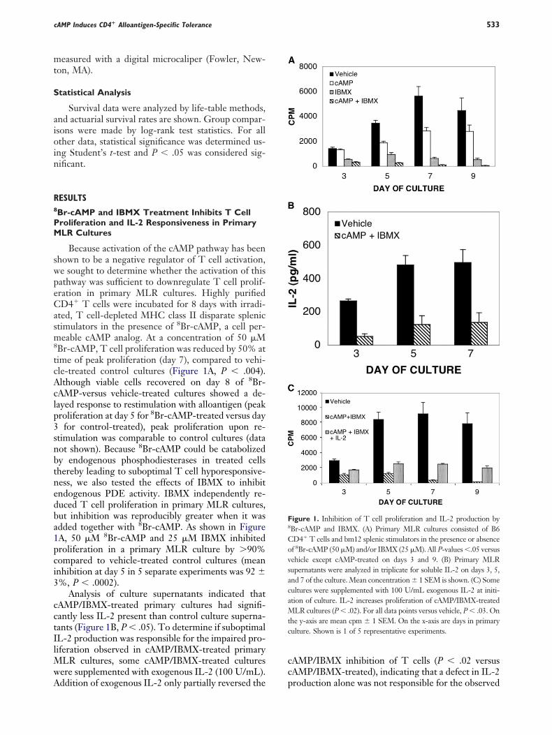
mt
S
aioin
R8
PM
swpeCasm8
tcAclp3snbtnedba1pci3
cctIlMwA
cc
F8
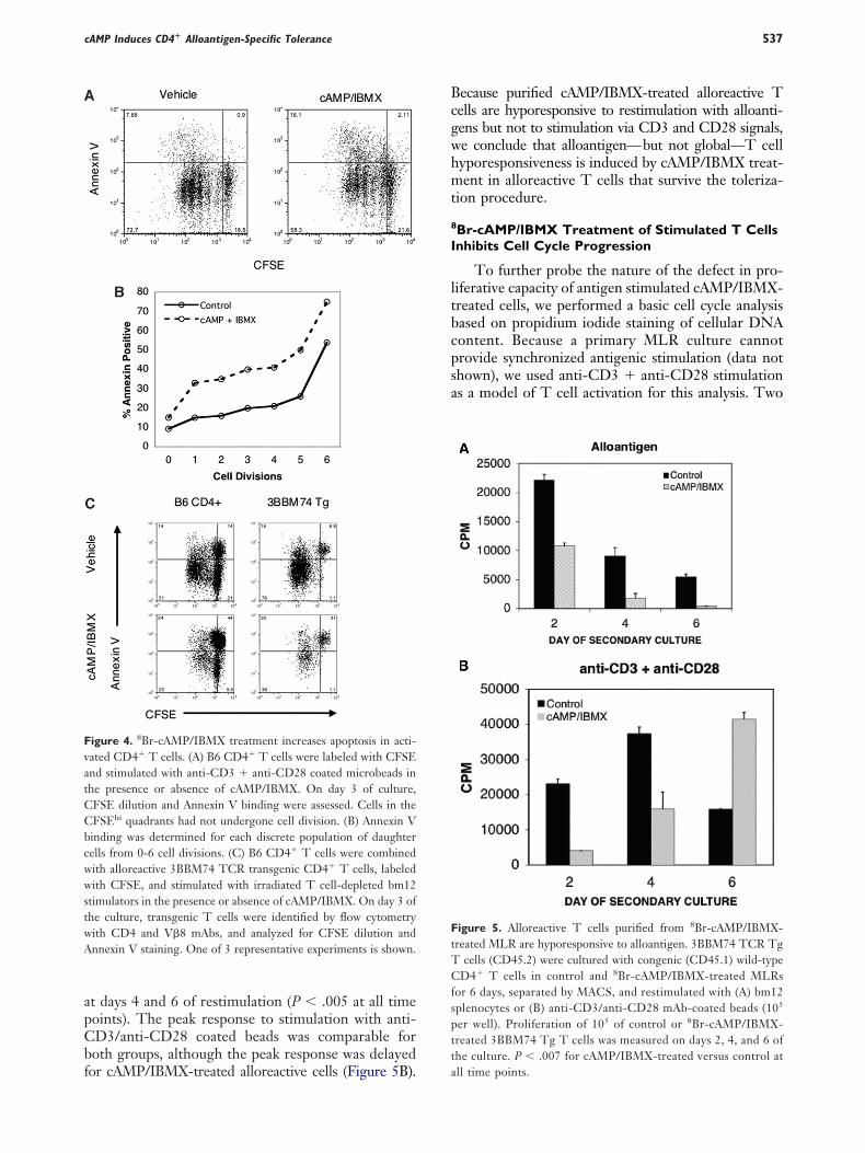
CovsacaMtc
cAMP Induces CD4� Alloantigen-Specific Tolerance 533
easured with a digital microcaliper (Fowler, New-on, MA).
tatistical Analysis
Survival data were analyzed by life-table methods,nd actuarial survival rates are shown. Group compar-sons were made by log-rank test statistics. For allther data, statistical significance was determined us-
ng Student’s t-test and P � .05 was considered sig-ificant.
ESULTS
Br-cAMP and IBMX Treatment Inhibits T Cellroliferation and IL-2 Responsiveness in PrimaryLR Cultures
Because activation of the cAMP pathway has beenhown to be a negative regulator of T cell activation,e sought to determine whether the activation of thisathway was sufficient to downregulate T cell prolif-ration in primary MLR cultures. Highly purifiedD4� T cells were incubated for 8 days with irradi-
ted, T cell-depleted MHC class II disparate splenictimulators in the presence of 8Br-cAMP, a cell per-eable cAMP analog. At a concentration of 50 �M
Br-cAMP, T cell proliferation was reduced by 50% atime of peak proliferation (day 7), compared to vehi-le-treated control cultures (Figure 1A, P � .004).lthough viable cells recovered on day 8 of 8Br-AMP-versus vehicle-treated cultures showed a de-ayed response to restimulation with alloantigen (peakroliferation at day 5 for 8Br-cAMP-treated versus day
for control-treated), peak proliferation upon re-timulation was comparable to control cultures (dataot shown). Because 8Br-cAMP could be catabolizedy endogenous phosphodiesterases in treated cellshereby leading to suboptimal T cell hyporesponsive-ess, we also tested the effects of IBMX to inhibitndogenous PDE activity. IBMX independently re-uced T cell proliferation in primary MLR cultures,ut inhibition was reproducibly greater when it wasdded together with 8Br-cAMP. As shown in FigureA, 50 �M 8Br-cAMP and 25 �M IBMX inhibitedroliferation in a primary MLR culture by �90%ompared to vehicle-treated control cultures (meannhibition at day 5 in 5 separate experiments was 92 %, P � .0002).
Analysis of culture supernatants indicated thatAMP/IBMX-treated primary cultures had signifi-antly less IL-2 present than control culture superna-ants (Figure 1B, P � .05). To determine if suboptimalL-2 production was responsible for the impaired pro-iferation observed in cAMP/IBMX-treated primary
LR cultures, some cAMP/IBMX-treated culturesere supplemented with exogenous IL-2 (100 U/mL).
ddition of exogenous IL-2 only partially reversed the pAMP/IBMX inhibition of T cells (P � .02 versusAMP/IBMX-treated), indicating that a defect in IL-2
igure 1. Inhibition of T cell proliferation and IL-2 production byBr-cAMP and IBMX. (A) Primary MLR cultures consisted of B6D4� T cells and bm12 splenic stimulators in the presence or absencef 8Br-cAMP (50 �M) and/or IBMX (25 �M). All P-values �.05 versusehicle except cAMP-treated on days 3 and 9. (B) Primary MLRupernatants were analyzed in triplicate for soluble IL-2 on days 3, 5,nd 7 of the culture. Mean concentration 1 SEM is shown. (C) Someultures were supplemented with 100 U/mL exogenous IL-2 at initi-tion of culture. IL-2 increases proliferation of cAMP/IBMX-treated
LR cultures (P � .02). For all data points versus vehicle, P � .03. Onhe y-axis are mean cpm 1 SEM. On the x-axis are days in primaryulture. Shown is 1 of 5 representative experiments.
roduction alone was not responsible for the observed
iclceccIes
ccm
S8
I
c
F8
(n(ocmyO
M. J. O’Shaughnessy et al.534
nhibited proliferation of the cAMP/IBMX-treatedells (Figure 1C). Alternatively, the elevated intracel-ular cAMP levels might have reduced the ability of Tells to respond to IL-2. Consistent with the latterxplanation, flow cytometric analysis of day 8 MLRells showed that whereas at least 50% of control-ultured cells expressed CD25, the subunit of theL-2 receptor, �20% of cAMP/IBMX-treated cellsxpressed CD25 in all experiments (see Figure 2D),uggesting that the failure to upregulate CD25 may
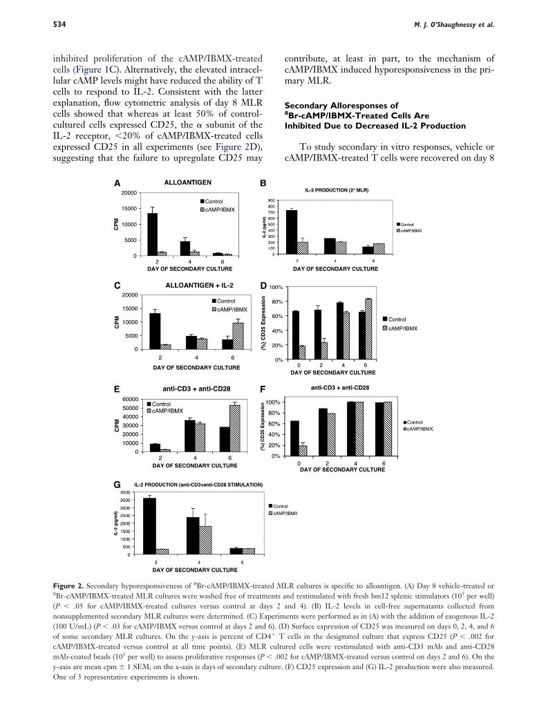
igure 2. Secondary hyporesponsiveness of 8Br-cAMP/IBMX-treaBr-cAMP/IBMX-treated MLR cultures were washed free of treatmP � .05 for cAMP/IBMX-treated cultures versus control at donsupplemented secondary MLR cultures were determined. (C) E100 U/mL) (P � .03 for cAMP/IBMX versus control at days 2 andf some secondary MLR cultures. On the y-axis is percent of CDAMP/IBMX-treated versus control at all time points). (E) MLRAb-coated beads (105 per well) to assess proliferative responses (P
-axis are mean cpm 1 SEM; on the x-axis is days of secondary cu
ne of 3 representative experiments is shown.ontribute, at least in part, to the mechanism ofAMP/IBMX induced hyporesponsiveness in the pri-ary MLR.
econdary Alloresponses ofBr-cAMP/IBMX-Treated Cells Arenhibited Due to Decreased IL-2 Production
To study secondary in vitro responses, vehicle orAMP/IBMX-treated T cells were recovered on day 8
R cultures is specific to alloantigen. (A) Day 8 vehicle-treated ornd restimulated with fresh bm12 splenic stimulators (105 per well)nd 4). (B) IL-2 levels in cell-free supernatants collected froments were performed as in (A) with the addition of exogenous IL-2) Surface expression of CD25 was measured on days 0, 2, 4, and 6cells in the designated culture that express CD25 (P � .002 for
red cells were restimulated with anti-CD3 mAb and anti-CD28for cAMP/IBMX-treated versus control on days 2 and 6). On the
(F) CD25 expression and (G) IL-2 production were also measured.
ted MLents a
ays 2 axperim
6). (D4� Tcultu
� .002lture.
onwr22tm2so9vAsc�adectb4uttabtstcraaca
iactvt2IpbttsChttc
rsTT
8
C
fdfsgtyrCabta1OhcubtvIcagtbrciItpbr
A8
C
tiadtii
cAMP Induces CD4� Alloantigen-Specific Tolerance 535
f a primary MLR, thoroughly washed, and equalumbers of viable T cells were plated and restimulatedith bm12 splenic stimulators. Prior to washing and
eplating, the viability of recovered cells was 54 0% (range � 34%-86%) for vehicle-treated cells and2 16% (range � 6%-48%) for cAMP/IBMX-reated cells (P � .02 versus vehicle-treated) as deter-ined by Annexin V/PI staining. As shown in Figure
A, vehicle-treated cells responded vigorously to re-timulation with alloantigen, whereas peak responsesf cAMP/IBMX-treated cells were reduced by 80%-0% (P � .05), despite the fact that equal numbers ofiable cells were plated in both secondary cultures.nalysis of culture supernatants for the presence of
oluble cytokines indicated that IL-2 was produced byAMP/IBMX-treated cells, but levels were reduced by70% (Figure 2B). To determine if inhibited second-
ry MLR responses resulted from decreased IL-2 pro-uction, we supplemented secondary cultures withxogenous IL-2 (100 U/mL). cAMP/IBMX-tolerizedells showed minimal response to IL-2 supplementa-ion at an early time point (day 2) of secondary culture,ut responded vigorously at later time points (days-6) when the culture contained exogenous IL-2 (Fig-re 2C). Increased proliferation in response to alloan-igen in the presence of exogenous IL-2 correspondedo the time when cAMP/IBMX-tolerized cells becamectivated and upregulated CD25 expression fromaseline levels of input cells in primary MLR cul-ure (range � 10%-15%) to �60% by day 4 ofecondary MLR culture (Figure 2D). Together,hese data indicate that cAMP/IBMX-treated Tells initially are hyporesponsive to alloantigenicestimulation, associated with low CD25 expressionnd IL-2 production at early time points of second-ry MLR culture, in contrast to control-treatedells, which had evidence of a primed response tolloantigen restimulation.
To determine whether the T cells were globallynhibited in their function, we chose anti-CD3 mAb �nti-CD28 mAb-coated microbeads to ensure a suffi-ient mitogenic stimulus to all T cells to determinehe response of the entire culture. These mAbs pro-ided a potent stimulus for proliferation of both con-rol cultured and cAMP/IBMX-cultured cells (FigureE). Compared to control-treated cells, cAMP/BMX-treated cells had evidence of a modestly lowerroliferation response on day 2 that was equivalenty day 4 of stimulation and exceeded that of con-rol-treated cells on day 6. In comparison to alloan-igen stimulation, cAMP/IBMX-treated cells that re-ponded to anti-CD3 � anti-CD28 mAbs upregulatedD25 expression earlier (Figure 2F) and producedigher levels of IL-2 (Figure 2G). Thus, cAMP/IBMXreatment renders alloreactive T cells hyporesponsiveo restimulation with alloantigen, but the remaining T
ells still have the capacity to mount a proliferative cesponse when stimulated through the TCR in a non-pecific manner with anti-CD3 and anti-CD28 mAbs.hese data indicate that cAMP/IBMX-treated CD4�
cells are not permanently and globally disabled.
Br-cAMP/IBMX Treatment Limits Proliferativeapacity of Alloreactive T Cells
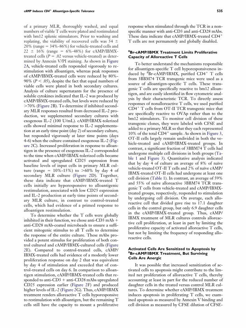
To better understand the mechanisms responsibleor alloantigen-specific T cell hyporesponsiveness in-uced by 8Br-cAMP/IBMX, purified CD4� T cellsrom 3BBM74 TCR transgenic mice were used as aource of alloantigen-specific T cells. These trans-enic T cells are specifically reactive to bm12 alloan-igen, and are easily identified in flow cytometric anal-sis by their characteristic TCR. To directly assessesponses of nonalloreactive T cells, we used purifiedD4� T cells from OT-II TCR transgenic mice that
re specifically reactive to OVAp rather than to them12 stimulators. To monitor cell division of theseransgenic clones, they were labeled with CFSE anddded to a primary MLR so that they each represented0% of the total CD4� sample. As shown in Figure 3,T-II cells largely remain undivided in both the ve-
icle-treated and cAMP/IBMX-treated groups. Inontrast, a significant fraction of 3BBM74 T cells hadndergone multiple cell divisions in both groups (Ta-le 1 and Figure 3). Quantitative analysis indicatedhat by day 4 of culture an average of 8% of naïveehicle-treated OT-II T cells and 2% of naïve cAMP/BMX-treated OT-II cells had undergone at least oneell division (Table 1). In contrast, an average of 59%nd 55% of naïve alloreactive 3BBM74 TCR trans-enic T cells from vehicle-treated and cAMP/IBMX-reated groups, respectively, responded to stimulationy undergoing cell division. On average, each allo-eactive cell that divided gave rise to 17.1 daughterells in the control group, but only 6.9 daughter cellsn the cAMP/IBMX-treated group. Thus, cAMP/BMX treatment of MLR cultures controls alloreac-ive cell proliferation, at least in part by limiting theroliferative capacity of activated alloreactive T cells,ut not by limiting the frequency of responding allo-eactive cells.
ctivated Cells Are Sensitized to Apoptosis byBr-cAMP/IBMX Treatment, But Survivingells Are Anergic
It was possible that increased sensitization of ac-ivated cells to apoptosis might contribute to the lim-ted net proliferation of alloreactive T cells, therebyccounting at least in part for the reduced number ofaughter cells in the treated versus control MLR cul-ures. To determine whether cAMP/IBMX treatmentnduces apoptosis in proliferating T cells, we exam-ned apoptosis as measured by Annexin V binding and
ell division as measured by CFSE dilution of CFSE-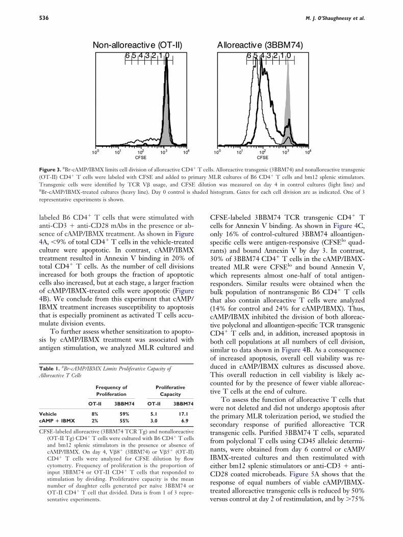
las4cttico4Itm
sa
Ccosr3twrbt(ctCbsodTct
wtstfnIeCrt
F(T8
r
TA
Vc
C
M. J. O’Shaughnessy et al.536
abeled B6 CD4� T cells that were stimulated withnti-CD3 � anti-CD28 mAbs in the presence or ab-ence of cAMP/IBMX treatment. As shown in FigureA, �9% of total CD4� T cells in the vehicle-treatedulture were apoptotic. In contrast, cAMP/IBMXreatment resulted in Annexin V binding in 20% ofotal CD4� T cells. As the number of cell divisionsncreased for both groups the fraction of apoptoticells also increased, but at each stage, a larger fractionf cAMP/IBMX-treated cells were apoptotic (FigureB). We conclude from this experiment that cAMP/BMX treatment increases susceptibility to apoptosishat is especially prominent as activated T cells accu-ulate division events.
To further assess whether sensitization to apopto-is by cAMP/IBMX treatment was associated withntigen stimulation, we analyzed MLR cultured and
igure 3. 8Br-cAMP/IBMX limits cell division of alloreactive CD4�
OT-II) CD4� T cells were labeled with CFSE and added to priransgenic cells were identified by TCR V� usage, and CFSE
Br-cAMP/IBMX-treated cultures (heavy line). Day 0 control is sepresentative experiments is shown.
able 1. 8Br-cAMP/IBMX Limits Proliferative Capacity oflloreactive T Cells
Frequency ofProliferation
ProliferativeCapacity
OT-II 3BBM74 OT-II 3BBM74
ehicle 8% 59% 5.1 17.1AMP � IBMX 2% 55% 3.0 6.9
FSE-labeled alloreactive (3BBM74 TCR Tg) and nonalloreactive(OT-II Tg) CD4� T cells were cultured with B6 CD4� T cellsand bm12 splenic stimulators in the presence or absence ofcAMP/IBMX. On day 4, V�8� (3BBM74) or V�5� (OT-II)CD4� T cells were analyzed for CFSE dilution by flowcytometry. Frequency of proliferation is the proportion ofinput 3BBM74 or OT-II CD4� T cells that responded tostimulation by dividing. Proliferative capacity is the meannumber of daughter cells generated per naïve 3BBM74 orOT-II CD4� T cell that divided. Data is from 1 of 3 repre-
vsentative experiments.
FSE-labeled 3BBM74 TCR transgenic CD4� Tells for Annexin V binding. As shown in Figure 4C,nly 16% of control-cultured 3BBM74 alloantigen-pecific cells were antigen-responsive (CFSElo quad-ants) and bound Annexin V by day 3. In contrast,0% of 3BBM74 CD4� T cells in the cAMP/IBMX-reated MLR were CFSElo and bound Annexin V,hich represents almost one-half of total antigen-
esponders. Similar results were obtained when theulk population of nontransgenic B6 CD4� T cellshat also contain alloreactive T cells were analyzed14% for control and 24% for cAMP/IBMX). Thus,AMP/IBMX inhibited the division of both alloreac-ive polyclonal and alloantigen-specific TCR transgenicD4� T cells and, in addition, increased apoptosis inoth cell populations at all numbers of cell division,imilar to data shown in Figure 4B. As a consequencef increased apoptosis, overall cell viability was re-uced in cAMP/IBMX cultures as discussed above.his overall reduction in cell viability is likely ac-
ounted for by the presence of fewer viable alloreac-ive T cells at the end of culture.
To assess the function of alloreactive T cells thatere not deleted and did not undergo apoptosis after
he primary MLR tolerization period, we studied theecondary response of purified alloreactive TCRransgenic cells. Purified 3BBM74 T cells, separatedrom polyclonal T cells using CD45 alleleic determi-ants, were obtained from day 6 control or cAMP/BMX-treated cultures and then restimulated withither bm12 splenic stimulators or anti-CD3 � anti-D28 coated microbeads. Figure 5A shows that the
esponse of equal numbers of viable cAMP/IBMX-reated alloreactive transgenic cells is reduced by 50%
s. Alloreactive transgenic (3BBM74) and nonalloreactive transgenicLR cultures of B6 CD4� T cells and bm12 splenic stimulators.n was measured on day 4 in control cultures (light line) andistogram. Gates for each cell division are as indicated. One of 3
T cellmary M
dilutiohaded h
ersus control at day 2 of restimulation, and by �75%
apCbf
Bcgwhmt
8
I
ltbcpsa
FtTCfsptt
FvatCCbcwwstwA
cAMP Induces CD4� Alloantigen-Specific Tolerance 537
t days 4 and 6 of restimulation (P � .005 at all timeoints). The peak response to stimulation with anti-D3/anti-CD28 coated beads was comparable foroth groups, although the peak response was delayed
igure 4. 8Br-cAMP/IBMX treatment increases apoptosis in acti-ated CD4� T cells. (A) B6 CD4� T cells were labeled with CFSEnd stimulated with anti-CD3 � anti-CD28 coated microbeads inhe presence or absence of cAMP/IBMX. On day 3 of culture,FSE dilution and Annexin V binding were assessed. Cells in theFSEhi quadrants had not undergone cell division. (B) Annexin Vinding was determined for each discrete population of daughterells from 0-6 cell divisions. (C) B6 CD4� T cells were combinedith alloreactive 3BBM74 TCR transgenic CD4� T cells, labeledith CFSE, and stimulated with irradiated T cell-depleted bm12
timulators in the presence or absence of cAMP/IBMX. On day 3 ofhe culture, transgenic T cells were identified by flow cytometryith CD4 and V�8 mAbs, and analyzed for CFSE dilution andnnexin V staining. One of 3 representative experiments is shown.
or cAMP/IBMX-treated alloreactive cells (Figure 5B). a
ecause purified cAMP/IBMX-treated alloreactive Tells are hyporesponsive to restimulation with alloanti-ens but not to stimulation via CD3 and CD28 signals,e conclude that alloantigen—but not global—T cellyporesponsiveness is induced by cAMP/IBMX treat-ent in alloreactive T cells that survive the toleriza-
ion procedure.
Br-cAMP/IBMX Treatment of Stimulated T Cellsnhibits Cell Cycle Progression
To further probe the nature of the defect in pro-iferative capacity of antigen stimulated cAMP/IBMX-reated cells, we performed a basic cell cycle analysisased on propidium iodide staining of cellular DNAontent. Because a primary MLR culture cannotrovide synchronized antigenic stimulation (data nothown), we used anti-CD3 � anti-CD28 stimulations a model of T cell activation for this analysis. Two
igure 5. Alloreactive T cells purified from 8Br-cAMP/IBMX-reated MLR are hyporesponsive to alloantigen. 3BBM74 TCR Tg
cells (CD45.2) were cultured with congenic (CD45.1) wild-typeD4� T cells in control and 8Br-cAMP/IBMX-treated MLRs
or 6 days, separated by MACS, and restimulated with (A) bm12plenocytes or (B) anti-CD3/anti-CD28 mAb-coated beads (105
er well). Proliferation of 105 of control or 8Br-cAMP/IBMX-reated 3BBM74 Tg T cells was measured on days 2, 4, and 6 ofhe culture. P � .007 for cAMP/IBMX-treated versus control at
ll time points.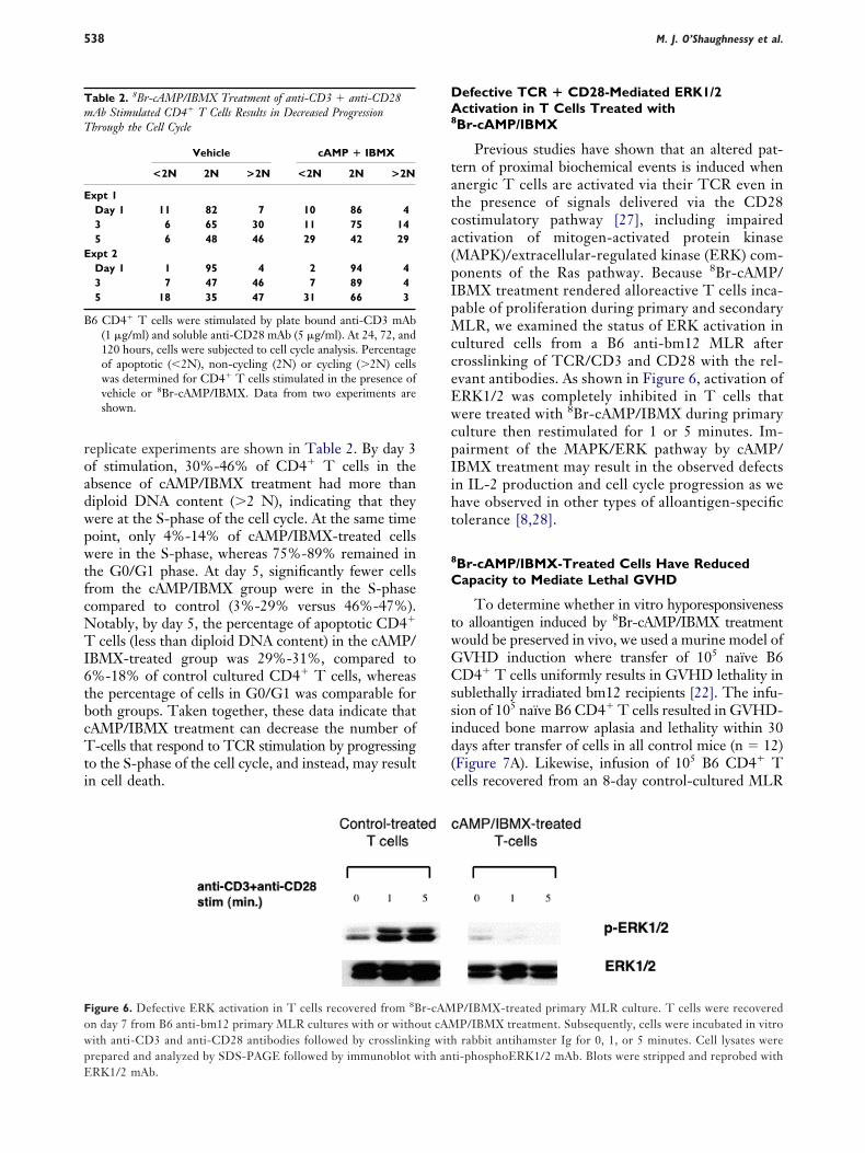
roadwpwtfcNTI6tbcTti
DA8
tatca(pIpMcceEwcpIiht
8
C
twGCssid(c
TmT
E
E
B
FowpE
M. J. O’Shaughnessy et al.538
eplicate experiments are shown in Table 2. By day 3f stimulation, 30%-46% of CD4� T cells in thebsence of cAMP/IBMX treatment had more thaniploid DNA content (�2 N), indicating that theyere at the S-phase of the cell cycle. At the same timeoint, only 4%-14% of cAMP/IBMX-treated cellsere in the S-phase, whereas 75%-89% remained in
he G0/G1 phase. At day 5, significantly fewer cellsrom the cAMP/IBMX group were in the S-phaseompared to control (3%-29% versus 46%-47%).otably, by day 5, the percentage of apoptotic CD4�
cells (less than diploid DNA content) in the cAMP/BMX-treated group was 29%-31%, compared to%-18% of control cultured CD4� T cells, whereashe percentage of cells in G0/G1 was comparable foroth groups. Taken together, these data indicate thatAMP/IBMX treatment can decrease the number of-cells that respond to TCR stimulation by progressing
o the S-phase of the cell cycle, and instead, may resultn cell death.
able 2. 8Br-cAMP/IBMX Treatment of anti-CD3 � anti-CD28Ab Stimulated CD4� T Cells Results in Decreased Progressionhrough the Cell Cycle
Vehicle cAMP � IBMX
<2N 2N >2N <2N 2N >2N
xpt 1Day 1 11 82 7 10 86 43 6 65 30 11 75 145 6 48 46 29 42 29
xpt 2Day 1 1 95 4 2 94 43 7 47 46 7 89 45 18 35 47 31 66 3
6 CD4� T cells were stimulated by plate bound anti-CD3 mAb(1 �g/ml) and soluble anti-CD28 mAb (5 �g/ml). At 24, 72, and120 hours, cells were subjected to cell cycle analysis. Percentageof apoptotic (�2N), non-cycling (2N) or cycling (�2N) cellswas determined for CD4� T cells stimulated in the presence ofvehicle or 8Br-cAMP/IBMX. Data from two experiments areshown.
igure 6. Defective ERK activation in T cells recovered from 8Bn day 7 from B6 anti-bm12 primary MLR cultures with or withoith anti-CD3 and anti-CD28 antibodies followed by crosslinkirepared and analyzed by SDS-PAGE followed by immunoblot w
RK1/2 mAb.efective TCR � CD28-Mediated ERK1/2ctivation in T Cells Treated with
Br-cAMP/IBMX
Previous studies have shown that an altered pat-ern of proximal biochemical events is induced whennergic T cells are activated via their TCR even inhe presence of signals delivered via the CD28ostimulatory pathway [27], including impairedctivation of mitogen-activated protein kinaseMAPK)/extracellular-regulated kinase (ERK) com-onents of the Ras pathway. Because 8Br-cAMP/BMX treatment rendered alloreactive T cells inca-able of proliferation during primary and secondaryLR, we examined the status of ERK activation in
ultured cells from a B6 anti-bm12 MLR afterrosslinking of TCR/CD3 and CD28 with the rel-vant antibodies. As shown in Figure 6, activation ofRK1/2 was completely inhibited in T cells thatere treated with 8Br-cAMP/IBMX during primary
ulture then restimulated for 1 or 5 minutes. Im-airment of the MAPK/ERK pathway by cAMP/BMX treatment may result in the observed defectsn IL-2 production and cell cycle progression as weave observed in other types of alloantigen-specificolerance [8,28].
Br-cAMP/IBMX-Treated Cells Have Reducedapacity to Mediate Lethal GVHD
To determine whether in vitro hyporesponsivenesso alloantigen induced by 8Br-cAMP/IBMX treatmentould be preserved in vivo, we used a murine model ofVHD induction where transfer of 105 naïve B6D4� T cells uniformly results in GVHD lethality in
ublethally irradiated bm12 recipients [22]. The infu-ion of 105 naïve B6 CD4� T cells resulted in GVHD-nduced bone marrow aplasia and lethality within 30ays after transfer of cells in all control mice (n � 12)Figure 7A). Likewise, infusion of 105 B6 CD4� Tells recovered from an 8-day control-cultured MLR
P/IBMX-treated primary MLR culture. T cells were recoveredP/IBMX treatment. Subsequently, cells were incubated in vitrorabbit antihamster Ig for 0, 1, or 5 minutes. Cell lysates were
ti-phosphoERK1/2 mAb. Blots were stripped and reprobed with
r-cAMut cAM
ng withith an
rIfs(trtca
CT
icspg
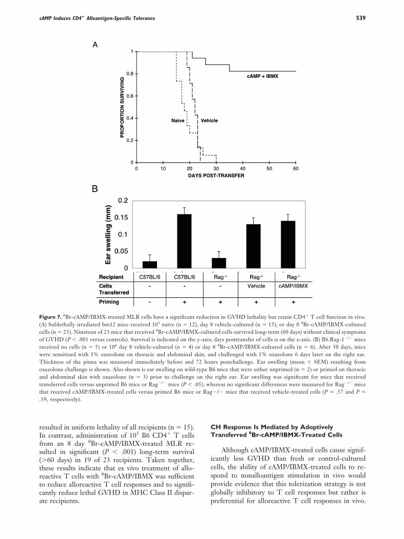
F(corwToatt.
cAMP Induces CD4� Alloantigen-Specific Tolerance 539
esulted in uniform lethality of all recipients (n � 15).n contrast, administration of 105 B6 CD4� T cellsrom an 8 day 8Br-cAMP/IBMX-treated MLR re-ulted in significant (P � .001) long-term survival�60 days) in 19 of 23 recipients. Taken together,hese results indicate that ex vivo treatment of allo-eactive T cells with 8Br-cAMP/IBMX was sufficiento reduce alloreactive T cell responses and to signifi-antly reduce lethal GVHD in MHC Class II dispar-
igure 7. 8Br-cAMP/IBMX-treated MLR cells have a significantA) Sublethally irradiated bm12 mice received 105 naïve (n � 12ells (n � 23). Nineteen of 23 mice that received 8Br-cAMP/IBMXf GVHD (P � .001 versus controls). Survival is indicated on theeceived no cells (n � 5) or 106 day 8 vehicle-cultured (n � 4)ere sensitized with 3% oxazolone on thoracic and abdominal shickness of the pinna was measured immediately before andxazolone challenge is shown. Also shown is ear swelling on wildnd abdominal skin with oxazolone (n � 3) prior to challengeransferred cells versus unprimed B6 mice or Rag�/� mice (P � .hat received cAMP/IBMX-treated cells versus primed B6 mice59, respectively).
te recipients. p
H Response Is Mediated by Adoptivelyransferred 8Br-cAMP/IBMX-Treated Cells
Although cAMP/IBMX-treated cells cause signif-cantly less GVHD than fresh or control-culturedells, the ability of cAMP/IBMX-treated cells to re-pond to nonalloantigen stimulation in vivo wouldrovide evidence that this tolerization strategy is notlobally inhibitory to T cell responses but rather is
ion in GVHD lethality but retain CD4� T cell function in vivo.vehicle-cultured (n � 15), or day 8 8Br-cAMP/IBMX-cultured
red cells survived long-term (60 days) without clinical symptoms; days posttransfer of cells is on the x-axis. (B) B6.Rag-1�/� mice8 8Br-cAMP/IBMX-cultured cells (n � 6). After 30 days, miced challenged with 1% oxazolone 6 days later on the right ear.rs postchallenge. Ear swelling (mean SEM) resulting from6 mice that were either unprimed (n � 2) or primed on thoracicright ear. Ear swelling was significant for mice that received
ereas no significant differences were measured for Rag�/� mice�/� mice that received vehicle-treated cells (P � .57 and P �
reduct), day 8
-cultuy-axis
or daykin, an72 hou-type Bon the05), whor Rag
referential for alloreactive T cell responses in vivo.

Tcnotrtttanetimrdaaeit1cm(dBciitrItte
D
ccstimrptucscgse
ocm
ilotcothcuwceIasmrptkbgtlftcsCnacICIoh
nacielt[itaTl
M. J. O’Shaughnessy et al.540
hus, we transferred 106 day 8 vehicle-cultured MLRells, 106 day 8 cAMP/IBMX-cultured MLR cells, oro cells to B6.Rag-1�/� recipient mice so that thenly responding T cell population was from the adop-ive transfer. After a 30-day period to allow T cellepopulation to occur, recipient mice were sensitizedo 3% oxazolone on thoracic and abdominal skin andhen rechallenged 8 days later with 1% oxazolone onhe right ear to induce a CH response. Because T cellctivation and homing to the site of inflammation areecessary steps for a CH response, measurement ofar swelling in the challenged ear provides evidencehat the adoptively transferred T cells could respondn vivo to a neo-antigen. The results of this experi-
ent, shown in Figure 7B, indicate that mice thateceived either day 8 control-cultured MLR cells oray 8 cAMP/IBMX-cultured cells exhibited similarmounts of ear swelling when rechallenged with ox-zolone (P � .57). The amount of ear swelling is alsoquivalent to sensitized wild-type B6 mice with anntact immune system (P � .59). Wild-type B6 micehat were not sensitized to oxazolone and B6.Rag-�/� mice that received no adoptively transferred Tells but were sensitized with oxazolone exhibitedinimal ear swelling when challenged with oxazolone
P � .05 versus transferred groups). Together, theseata indicate that the response that occurred in6.Rag-1�/� mice receiving adoptively transferredAMP/IBMX-treated cells resulted from a functionaln vivo response to oxazolone challenge that resultedn equivalent CD4� T cell responses as control-reated cells in vivo. Thus, despite having significantlyeduced capacity to mediate GVHD lethality, cAMP/BMX-treated cells have the ability to mount func-ional immune responses in vivo, which indicates thathe ex vivo cAMP/IBMX tolerization procedure pref-rentially inhibits alloreactive T cell responses in vivo.
ISCUSSION
Cyclic AMP is a known physiologic inhibitor of Tell activation. Although elevation of intracellularAMP has been shown to induce T cell hyporespon-iveness in vitro [8,29], it has not been formally provedhat this method could achieve immune tolerance thats sufficient for prevention and treatment of T cell-
ediated diseases such as autoimmune disorders, graftejection, and GVHD following bone marrow trans-lantation. In this paper, we showed that increasinghe intracellular cAMP level in alloreactive T cellssing 8Br-cAMP and IBMX during a primary MLRould induce alloantigen specific T cell hyporespon-iveness that was sufficient to significantly reduce theapacity of CD4� T cells to induce GVHD withoutlobally impairing CD4� T cell responses to othertimuli (oxazolone) in vivo. These data provide strong
vidence that suggests that pharmacologic regulation hf intracellular cAMP levels in pathogenic T cellsould be an appropriate therapeutic target for theodulation of unwanted immune responses.
Although an elevated level of intracellular cAMPs uniformly suppressive to a polyclonal T cell popu-ation, our data indicate that hyporesponsiveness onlyccurs in T cells that are antigen stimulated at theime of pharmacologically induced elevations in intra-ellular cAMP. We conclude this based on the resultsf in vitro experiments that showed cAMP/IBMX-reated cells recovered from a primary MLR wereyporesponsive to restimulation with alloantigen, butapable of intact responses to nonspecific TCR stim-lation. Responses to nonspecific TCR stimulationere intact at 4 but not 2 days after removal from
AMP/IBMX containing cultures. Thus, we favor thexplanation that the hyporesponsiveness of the cAMP/BMX-treated cells in secondary culture resulted fromntigen-specific tolerance or deletion without a sub-tantial induction of hyporesponsiveness in the re-aining nontolerized T cells. The relatively higher
esponse of control cultured cells to alloantigen re-riming and more modest increase in early responseso anti-CD3 � anti-CD28 mAbs may represent theinetic differences that would be expected to be seenetween cultures that contained a frequency of anti-en-primed T cells in contrast to the cAMP/IBMX-reated cells that were comprised primarily of nonal-oreactive, and hence primarily naïve, T cells. Theailure of IL-2 supplementation to restore the alloan-igen responses of cAMP/IBMX in secondary MLRultures indicates that IL-2 production is not respon-ible for this defective response. The correlation ofD25 antigen expression levels with the response ki-etics of alloantigen as well as anti-CD3 mAb �nti-CD28 mAb induced stimulation may reflect theonversion of antigen nonprimed, “naïve” cAMP/BMX-treated cells to antigen-primed, “stimulated”D4� T cells. Therefore, the kinetics of cAMP/
BMX-treated cells and CD25 upregulation in sec-ndary culture would lag behind control cells thatave already been stimulated in primary culture.
As mentioned previously, at least 2 major mecha-isms, anergy and deletion of alloreactive T cells, mayccount for the slow response of cAMP/IBMX-treatedultured cells to alloantigen restimulation in vitro andn vivo [30]. To date, it has not been clear whether anlevated cAMP level in T cells during antigen-stimu-ation could result in cell death. However, it is knownhat cAMP can mediate apoptosis in some tumor cells31] macrophages and pro-B cells [32]. Our findingsndicate that alloreactive T cells in cAMP/IBMX-reated MLRs had increased apoptosis, particularly inlloreactive cells that had undergone cell division.hus, induction of apoptosis in alloreactive T cells
ikely contributes to the observed in vitro and in vivo
yporesponsiveness.
issgtSalsefctdhddcmslceutastIri
taTphwbeoprboterIcsttpip
csnuaue
sdofaaFtpascswcCem
ttlsoipd
A
t(
R
cAMP Induces CD4� Alloantigen-Specific Tolerance 541
Although apoptosis limits the frequency of surviv-ng alloreactive T cells, a fraction of alloreactive cellsurvive the cAMP/IBMX treatment. Purification ofurviving Tg alloreactive cells indicated that an aner-ic phenotype was induced in these cells. It is knownhat failure of cell cycle progression from the G1 to-phase during TCR stimulation can lead to T cellnergy [33]. It was previously shown that an elevatedevel of cAMP can interfere with cell cycle progres-ion, possibly via a cAMP mediated increase of p27kip1
xpression [8,34]. p27kip1 is a cdk inhibitor whoseunction is to regulate G1 to S-phase transition of theell cycle [35]. In our experiments, 8Br-cAMP/IBMXreatment impaired cell cycle progression, resulting inecreased frequency of cells in the S-phase. Anotherallmark for anergic T cells is that they are viable buto not proliferate upon antigen stimulation or pro-uce IL-2 [36]. In our experiments, we showed that Tells recovered from a 8Br-cAMP/IBMX-treated pri-ary MLR culture did not proliferate and produced
ignificantly reduced IL-2 upon alloantigen restimu-ation. Interestingly, this secondary alloantigen-spe-ific hyporesponsiveness was reversible in the pres-nce of exogenous IL-2 at time periods of CD25pregulation, indicating that defective IL-2 produc-ion was present in 8Br-cAMP/IBMX-treated T cells,nd might be responsible in part for their unrespon-iveness to alloantigen restimulation. Alternatively,he slower kinetics of CD25 upregulation in cAMP/BMX-treated T cells was simply reflective of theesting state of the nonalloreactive T cells that dom-nated the cAMP/IBMX and not the control culture.
We have previously reported using the same cul-ure system and anti-CD154 mAb to induce tolerancend inhibit GVHD capacity of adoptively transferred
cells in vivo, exogenous IL-2 rescued both therimary and secondary MLR cultures from T cellyporesponsiveness, in contrast to cAMP/IBMX, inhich only the secondary alloantigen responses coulde rescued [37,38]. Despite the restorative effects ofxogenous IL-2 at 50 IU/mL on in vitro proliferationf alloreactive T cells exposed to anti-CD154 mAb inrimary MLR cultures, GVHD was not able to beeadily generated in vivo in sublethally irradiatedm12 mice given adoptively transferred CD4� T cellsbtained from anti-CD154 mAb-treated MLR cul-ures even when recipients were given high doses ofxogenous IL-2 for 2 weeks posttransfer [37]. Withespect to the IL-2 production defect in cAMP/BMX-treated cells, elevated intracellular cAMP levelould lead to IL-2 production failure in T cells viaeveral biochemical pathways, including inhibition ofhe PKC pathway, which eventually leads to dysfunc-ion of MAPK/ERK pathways and failure of IL-2roduction [13,34,39]. Whether any persistent signal-
ng pathway defects may be responsible for the IL-2
roduction defect in the 8Br-cAMP/IBMX-treated Tells even at a normalized intracellular cAMP level inecondary cultures remains to be determined. Alter-atively, cAMP/IBMX treatment may inhibit CD25pregulation per se, and when 8Br-cAMP and IBMXre no longer present (at the time of secondary stim-lation) CD25 upregulation can occur, which allowsxogenous IL-2 to restore proliferation.
The ability to selectively eliminate alloreactive re-ponses while preserving in vivo immune function ofonor T cells is an important discovery that could bef significant value in many clinical settings. Thiseature is particularly suitable for ex vivo tolerancepplications [30] because of the ability to regulatentigen access, and hence stimulation, in this setting.or example, one could use the method to specifically
arget GVHD causing alloreactive T cells but nototential viral-specific or tumor-specific T cells. Inddition to GVHD prophylaxis, the targeted antigenpecificity provided by an ex vivo tolerance approachould be advantageous for delayed lymphocyte infu-ion treatment of bone marrow transplant patientsith relapsed leukemia or severe viral infections. Be-
ause antileukemia and antiviral responses often requireD8� T cell function, future studies will be required to
xplore the effect of cAMP elevation on CD8� T cell-ediated alloresponses in vitro and in vivo.
In summary, we have shown that elevation of in-racellular cAMP levels results in tolerance sufficiento markedly inhibit GVHD while maintaining nonal-oreactive T cell functions in vivo. These studies makeignificant progress toward validation of the principalf using cAMP-elevating agents for ex vivo tolerancenduction and potential in vivo therapeutic use forrevention and therapy of T cell-mediated immuneisorders including GVHD.
CKNOWLEDGMENTThis study was supported by the National Insti-
utes of Health grants AI34495 (BRB), HL56067BRB), AOA43552 (VAB) and CA104596 (VAB).
EFERENCES
1. Dumont JE, Jauniaux JC, Roger PP. The cyclic AMP-mediatedstimulation of cell proliferation. Trends Biochem Sci. 1989;14:67-71.
2. Blomhoff HK, Blomhoff R, Stokke T, et al. cAMP-mediatedgrowth inhibition of a B-lymphoid precursor cell line Reh isassociated with an early transient delay in G2/M, followed by anaccumulation of cells in G1. J Cell Physiol. 1988;137:583-587.
3. Dugan LL, Kim JS, Zhang Y, et al. Differential effects of cAMPin neurons and astrocytes. Role of B-raf. J Biol Chem. 1999;274:25842-25848.
4. Bielekova B, Lincoln A, McFarland H, Martin R. Therapeuticpotential of phosphodiesterase-4 and -3 inhibitors in Th1-medi-ated autoimmune diseases. J Immunol. 2000;164:1117-1124.
5. Chen D, Rothenberg EV. Interleukin 2 transcription factors as mo-
1
1
1
1
1
1
1
1
1
1
2
2
2
2
2
2
2
2
2
2
3
3
3
3
3
3
3
3
3
3
M. J. O’Shaughnessy et al.542
lecular targets of cAMP inhibition: delayed inhibition kinetics andcombinatorial transcription roles. J Exp Med. 1994;179:931-942.
6. Hofmann B, Nishanian P, Nguyen T, Insixiengmay P, FaheyJL. Human immunodeficiency virus proteins induce the inhib-itory cAMP/protein kinase A pathway in normal lymphocytes.Proc Natl Acad Sci U S A. 1993;90:6676-6680.
7. Aandahl EM, Aukrust P, Skalhegg BS, et al. Protein kinase Atype I antagonist restores immune responses of T cells fromHIV-infected patients. FASEB J. 1998;12:855-862.
8. Boussiotis VA, Freeman GJ, Taylor PA, et al. p27kip1 functionsas an anergy factor inhibiting interleukin 2 transcription andclonal expansion of alloreactive human and mouse helper Tlymphocytes. Nat Med. 2000;6:290-297.
9. Kammer GM. The adenylate cyclase-cAMP-protein kinase Apathway and regulation of the immune response. Immunol To-day. 1988;9:222-229.
0. Skalhegg BS, Tasken K, Hansson V, Huitfeldt HS, Jahnsen T,Lea T. Location of cAMP-dependent protein kinase type I withthe TCR-CD3 complex. Science. 1994;263:84-87.
1. Vang T, Torgersen KM, Sundvold V, et al. Activation of theCOOH-terminal Src kinase (Csk) by cAMP-dependent proteinkinase inhibits signaling through the T cell receptor. J Exp Med.2001;193:497-507.
2. Powell JD, Lerner CG, Ewoldt GR, Schwartz RH. The -180site of the IL-2 promoter is the target of CREB/CREM bindingin T cell anergy. J Immunol. 1999;163:6631-6639.
3. Ramstad C, Sundvold V, Johansen HK, Lea T. cAMP-depen-dent protein kinase (PKA) inhibits T cell activation by phos-phorylating ser-43 of raf-1 in the MAPK/ERK pathway. CellSignal. 2000;12:557-563.
4. Naderi S, Gutzkow KB, Christoffersen J, Smeland EB, Blom-hoff HK. cAMP-mediated growth inhibition of lymphoid cellsin G1: rapid down-regulation of cyclin D3 at the level oftranslation. Eur J Immunol. 2000;30:1757-1768.
5. Kaminuma O, Mori A, Wada K, et al. A selective type 4phosphodiesterase inhibitor, T-440, modulates intracellular cy-clic AMP level and interleukin-2 production of Jurkat cells.Immunopharmacology. 1998;38:247-252.
6. Rascon A, Soderling SH, Schaefer JB, Beavo JA. Cloningand characterization of a cAMP-specific phosphodiesterase(TbPDE2B) from Trypanosoma brucei. Proc Natl Acad Sci U S A.2002;99:4714-4719.
7. Spina D. Phosphodiesterase-4 inhibitors in the treatment ofinflammatory lung disease. Drugs. 2003;63:2575-2594.
8. Rabe KF, Bateman ED, O’Donnell D, Witte S, BredenbrokerD, Bethke TD. Roflumilast—an oral anti-inflammatory treat-ment for chronic obstructive pulmonary disease: a randomisedcontrolled trial. Lancet. 2005;366:563-571.
9. Mann SC, Andrews PA, Howell SB. Modulation of cis-diam-minedichloroplatinum(II) accumulation and sensitivity by forskolinand 3-isobutyl-1-methylxanthine in sensitive and resistant humanovarian carcinoma cells. Int J Cancer. 1991;48:866-872.
0. Barnden MJ, Allison J, Heath WR, Carbone FR. DefectiveTCR expression in transgenic mice constructed using cDNA-based alpha- and beta-chain genes under the control of heter-ologous regulatory elements. Immunol Cell Biol. 1998;76:34-40.
1. Backstrom BT, Muller U, Hausmann B, Palmer E. Positiveselection through a motif in the alphabeta T cell receptor.Science. 1998;281:835-838.
2. Chen ZM, O’Shaughnessy MJ, Gramaglia I, et al. IL-10 andTGF-beta induce alloreactive CD4�CD25- T cells to acquire
regulatory cell function. Blood. 2003;101:5076-5083.3. Brice GT, Riley JL, Villinger F, et al. Development of ananimal model for autotransfusion therapy: in vitro character-ization and analysis of anti-CD3/CD28 expanded cells. J AcquirImmune Defic Syndr Hum Retrovirol. 1998;19:210-220.
4. Gudmundsdottir H, Wells AD, Turka LA. Dynamics and re-quirements of T cell clonal expansion in vivo at the single-celllevel: effector function is linked to proliferative capacity. J Im-munol. 1999;162:5212-5223.
5. Boussiotis VA, Barber DL, Nakarai T, et al. Prevention of Tcell anergy by signaling through the gamma c chain of the IL-2receptor. Science. 1994;266:1039-1042.
6. Taube M, Svensson L, Carlsten H. T lymphocytes are not thetarget for estradiol-mediated suppression of DTH in reconsti-tuted female severe combined immunodeficient (SCID) mice.Clin Exp Immunol. 1998;114:147-153.
7. Boussiotis VA, Barber DL, Lee BJ, Gribben JG, Freeman GJ,Nadler LM. Differential association of protein tyrosine kinaseswith the T cell receptor is linked to the induction of anergy andits prevention by B7 family-mediated costimulation. J Exp Med.1996;184:365-376.
8. Boussiotis VA, Chen ZM, Zeller JC, et al. Altered T-cellreceptor � CD28-mediated signaling and blocked cell cycleprogression in interleukin 10 and transforming growth factor-beta-treated alloreactive T cells that do not induce graft-versus-host disease. Blood. 2001;97:565-571.
9. Cone RE, Cochrane R, Lingenheld EG, Clark RB. Elevation ofintracellular cyclic AMP induces an anergic-like state in Th1clones. Cell Immunol. 1996;173:246-251.
0. Blazar BR, Korngold R, Vallera DA. Recent advances in graft-versus-host disease (GVHD) prevention. Immunol Rev. 1997;157:79-109.
1. Lanotte M, Riviere JB, Hermouet S, et al. Programmed celldeath (apoptosis) is induced rapidly and with positive cooper-ativity by activation of cyclic adenosine monophosphate-kinaseI in a myeloid leukemia cell line. J Cell Physiol. 1991;146:73-80.
2. Myklebust JH, Josefsen D, Blomhoff HK, et al. Activation ofthe cAMP signaling pathway increases apoptosis in humanB-precursor cells and is associated with downregulation ofMcl-1 expression. J Cell Physiol. 1999;180:71-80.
3. Powell JD, Lerner CG, Schwartz RH. Inhibition of cell cycleprogression by rapamycin induces T cell clonal anergy even inthe presence of costimulation. J Immunol. 1999;162:2775-2784.
4. Grader-Beck T, van Puijenbroek AA, Nadler LM, BoussiotisVA. cAMP inhibits both Ras and Rap1 activation in primaryhuman T lymphocytes, but only Ras inhibition correlates withblockade of cell cycle progression. Blood. 2003;101:998-1006.
5. Mohapatra S, Agrawal D, Pledger WJ. p27Kip1 regulates T cellproliferation. J Biol Chem. 2001;276:21976-21983.
6. Schwartz RH. Models of T cell anergy: is there a commonmolecular mechanism? J Exp Med. 1996;184:1-8.
7. Blazar BR, Taylor PA, Noelle RJ, Vallera DA. CD4(�) T cellstolerized ex vivo to host alloantigen by anti-CD40 ligand(CD40L:CD154) antibody lose their graft-versus-host diseaselethality capacity but retain nominal antigen responses. J ClinInvest. 1998;102:473-482.
8. Taylor PA, Panoskaltsis-Mortari A, Noelle RJ, Blazar BR.Analysis of the requirements for the induction of CD4� T cellalloantigen hyporesponsiveness by ex vivo anti-CD40 ligandantibody. J Immunol. 2000;164:612-622.
9. Wang L, Liu F, Adamo ML. Cyclic AMP inhibits extracellularsignal-regulated kinase and phosphatidylinositol 3-kinase/Akt
pathways by inhibiting Rap1. J Biol Chem. 2001;276:37242-37249.Copyright © 2022 FDOKUMEN