Elemental fingerprinting of tumorous and adjacent non-tumorous tissues from patients with colorectal...
13
Elemental fingerprinting of tumorous and adjacent non-tumorous tissues from patients with colorectal cancer using ICP-MS, ICP-OES and chemometric analysis Isela Lavilla Marta Costas Pilar San Miguel Jorge Millos Carlos Bendicho Received: 11 February 2009 / Accepted: 19 March 2009 Ó Springer Science+Business Media, LLC. 2009 Abstract Tumorous and adjacent non-tumorous paired biopsies from 38 patients with colorectal cancer were analyzed by inductively coupled plasma mass spectrometry and inductively coupled plasma optical emission spectrometry after low-volume microwave digestion. 18 elements were investigated: Ag, Al, Ca, Cd, Co, Cr, Cu, Fe, K, Mg, Mn, Mo, Ni, P, Pb, S, Se and Zn. Different chemometric tools were used for data evaluation: Wilcoxon signed rank test, Hieratical clustering analysis, principal compo- nent analysis (PCA) and linear discriminant analysis (LDA). With the exception of Al, tumours were observed to have significantly more elevated concen- trations of essential elements as compared to non- tumours. On the contrary, elements considered potentially carcinogenic such as Cr, Ni, Mo or Co do not display significant differences. When PCA was applied, different components were obtained for tumorous and non-tumorous tissues. When LDA was applied for the elements studied (including essential and non-essential elements) about 90% of cases were correctly classified. Keywords Colorectal cancer Trace and minor elements ICP-MS ICP-OES Chemometric analysis Introduction It has been well known for several decades that cancer is a disease in which element alterations are produced. Patients with different forms of cancer displayed significant differences in the level of some essential and non-essential elements when normal and malignant tissues are analyzed (Mulay et al. 1971; Gregoriadis et al. 1983; Rizk and Sky-Peck 1984; Drake and Sky-Peck 1989). Elemental concen- tration ranges in human tissues are so narrow for the correct functioning of the cells, that still today, the question of if these alterations are a consequence of the disease or can cause it is subjected to conjectures (Majewska et al. 2007). Many studies have targeted metal-induced carcin- ogenicity, emphasising their role in the generation of oxygen-free radicals (Halliwell and Gutteridge 1999; Leonard et al. 2004; Valko and Morris 2005). These radicals can cause changes in DNA bases, enhanced lipid peroxidation and homeostasis. As, Cd, Co, Cr, Cu, Fe, Ni and V are the most studied metal-induced oxidative stress (Valko et al. 2006). On the other hand, the effect of oxygen-free radicals is neutralized in the organisms by the antioxidant defense. The most efficient antioxidant I. Lavilla M. Costas J. Millos C. Bendicho (&) Departamento de Quı ´mica Analı ´tica y Alimentaria, A ´ rea de Quı ´mica Analı ´tica, Facultad de Quı ´mica, Universidad de Vigo, As Lagoas-Marcosende s/n, 36310 Vigo, Spain e-mail: [email protected] P. S. Miguel Centro Hospitalario Povisa, Servicio de Anatomı ´a Patolo ´gica, Vigo, Spain 123 Biometals DOI 10.1007/s10534-009-9231-6
-
Upload
independent -
Category
Documents
-
view
0 -
download
0
Transcript of Elemental fingerprinting of tumorous and adjacent non-tumorous tissues from patients with colorectal...
Elemental fingerprinting of tumorous and adjacentnon-tumorous tissues from patients with colorectal cancerusing ICP-MS, ICP-OES and chemometric analysis
Isela Lavilla Æ Marta Costas Æ Pilar San Miguel ÆJorge Millos Æ Carlos Bendicho
Received: 11 February 2009 / Accepted: 19 March 2009
� Springer Science+Business Media, LLC. 2009
Abstract Tumorous and adjacent non-tumorous
paired biopsies from 38 patients with colorectal
cancer were analyzed by inductively coupled plasma
mass spectrometry and inductively coupled plasma
optical emission spectrometry after low-volume
microwave digestion. 18 elements were investigated:
Ag, Al, Ca, Cd, Co, Cr, Cu, Fe, K, Mg, Mn, Mo, Ni,
P, Pb, S, Se and Zn. Different chemometric tools
were used for data evaluation: Wilcoxon signed rank
test, Hieratical clustering analysis, principal compo-
nent analysis (PCA) and linear discriminant analysis
(LDA). With the exception of Al, tumours were
observed to have significantly more elevated concen-
trations of essential elements as compared to non-
tumours. On the contrary, elements considered
potentially carcinogenic such as Cr, Ni, Mo or Co
do not display significant differences. When PCA was
applied, different components were obtained for
tumorous and non-tumorous tissues. When LDA
was applied for the elements studied (including
essential and non-essential elements) about 90% of
cases were correctly classified.
Keywords Colorectal cancer � Trace and
minor elements � ICP-MS � ICP-OES �Chemometric analysis
Introduction
It has been well known for several decades that
cancer is a disease in which element alterations are
produced. Patients with different forms of cancer
displayed significant differences in the level of some
essential and non-essential elements when normal
and malignant tissues are analyzed (Mulay et al.
1971; Gregoriadis et al. 1983; Rizk and Sky-Peck
1984; Drake and Sky-Peck 1989). Elemental concen-
tration ranges in human tissues are so narrow for the
correct functioning of the cells, that still today, the
question of if these alterations are a consequence of
the disease or can cause it is subjected to conjectures
(Majewska et al. 2007).
Many studies have targeted metal-induced carcin-
ogenicity, emphasising their role in the generation of
oxygen-free radicals (Halliwell and Gutteridge 1999;
Leonard et al. 2004; Valko and Morris 2005). These
radicals can cause changes in DNA bases, enhanced
lipid peroxidation and homeostasis. As, Cd, Co, Cr,
Cu, Fe, Ni and V are the most studied metal-induced
oxidative stress (Valko et al. 2006).
On the other hand, the effect of oxygen-free
radicals is neutralized in the organisms by the
antioxidant defense. The most efficient antioxidant
I. Lavilla � M. Costas � J. Millos � C. Bendicho (&)
Departamento de Quımica Analıtica y Alimentaria, Area
de Quımica Analıtica, Facultad de Quımica, Universidad
de Vigo, As Lagoas-Marcosende s/n, 36310 Vigo, Spain
e-mail: [email protected]
P. S. Miguel
Centro Hospitalario Povisa, Servicio de Anatomıa
Patologica, Vigo, Spain
123
Biometals
DOI 10.1007/s10534-009-9231-6

enzymes have an active metal centre; i.e. Cu, Mn or
Zn is part of superoxide dismutases (SODs), the first-
line antioxidant defense. Se takes part of glutathione
peroxidase, i.e. the major source of protection against
low levels of oxidative stress (Mates et al. 1999).
Tumours have a very high-energy demand and
high rate of glucolysis. In general, essential trace
elements such as Ca, Co, Cu, Fe, Mg, Mn, Mo, Ni, Se
or Zn form compounds with proteins, which have
important catalytic functions. In addition, the molec-
ular mechanisms involved in carcinogenesis for some
essential metals (as Ca, Fe, Mg, Ni, Zn) are likely to
include binding competition among metal ions at
chromatin and other regulatory molecules. This takes
place in both, target cells, which give rise to tumours,
and immune cells, which are responsible for control-
ling the tumour growth (Kasprzak 1997). Excess or
deficiency of the elements may be an important factor
in the development of malfunctions at cellular or
subcellular level.
More difficult it is to consider this subject with
regard to non-essential elements when their levels do
not appear to be pathological. Few studies can be
mentioned in this sense. Low non-cytotoxic concen-
trations of some elements such as As or Cd have been
related to the inhibition of nucleotide excision repair
(Hartwig 2000).
In recent years, the interest for understanding the
role of alterations in element homeostasis and in the
etiology of cancer has dramatically increased (Raju
et al. 2006). Comparative studies carried out recently
on normal and cancerous human tissues report
controversial results, especially for some elements
such as Cu, Fe, Ni, Se and Zn. In general, most of
authors found significantly elevated concentrations or
non-differences for several elements in malignant
tissues (Raju et al. 2006; Ionescu et al. 2006; Ebrahim
et al. 2007), however, decreases in concentration
have also been reported (Zoriy et al. 2006).
The aim of this work is to investigate the
elemental distribution in tumour and non-tumour
adjacent colorectal tissue using several chemometric
approaches. A case study with 38 patients was carried
out. Inductively coupled plasma mass spectrometry
(ICP-MS) and inductively coupled plasma optical
emission spectrometry (ICP-OES) were applied as
analytical techniques to determine 18 elements in 76
colorectal biopsies. Recently, the use of these mul-
tielemental analytical techniques has increased the
possibilities of obtaining accurate information in
biological tissues (Bocca et al. 2007; Alimonti et al.
2008). Ag, Al, Ca, Cd, Co, Cr, Cu, Fe, K, Mg, Mn,
Mo, Ni, Pb, Se and Zn were studied. S and P
were also included in this study owing to their
involvements in the demand of metabolic energy.
Chemometric tools were applied to obtain latent
information from data.
Materials and methods
Apparatus and reagents
Deionised water (18.3 MX) from a Milli-Q purifica-
tion system (Millipore, Molsheim, France) was used
throughout. Ultrapure grade nitric acid (Hyperpur-
Plus, Panreac, Barcelona, Spain) was used. Standard
working solutions were obtained from a multielement
standard stock solution ICP Multi Element Standard
Certipur� VI (Merck, Darmstadt, Germany) after
suitable dilution. Calibration curves were built from
three replicates measurements and, in all cases,
regression coefficients were higher than 0.999. Stan-
dard stock solutions (1,000 mg/L) of Ge, Rh and In
(Merck) were used as internal standards.
Concentrations of minor elements (Al, Ca, Cu, Fe,
K, Mg, P, S and Zn) were determined by ICP-OES
using a Perkin–Elmer Optima 4300 DV spectrometer
(Shelton, CT, USA), equipped with an AS-90 auto-
sampler, axial system, a high dynamic range detector
and a cross-flow type nebulizer for pneumatic
nebulization. The ICP-OES measurement conditions
for these elements were optimized to achieve the
maximum signal-to-background ratio.
Trace element concentrations were determined by
ICP-MS using a Thermo Elemental X7 Series ICP-
MS equipped with an ASX-520 autosampler (Omaha,
NE, USA). Two operation modes were used, standard
mode with guard electrode (plasma screen) and cell
collision technology (CCT). The instrument was
optimized in terms of sensitivity, resolution, mass
calibration and minimization of interferences (oxide
ratio and polyatomic interferences).
A Reisch (Haan, Germany) mixer mill MM 2000
was used for grinding the biopsy samples. Microwave
digestion of colorectal biopsies were carried out with
a Multiwave 3000 oven (Anton Paar, Graz, Austria),
equipped with eight PFA digestion vessels (100 ml of
Biometals
123

capacity). The dimensions of these vessels (20 cm
high, 2.5 cm of diameter) allow inserting three PFA
vials (6 cm high, 1 cm diameter, Savillex, Minne-
tonka, USA), one over the other. In this way, 24
sample digestions were performed in one digestion
run.
Different certified reference materials were used for
analytical validation purposes: BCR 185R (bovine
liver), NRC-CNRC TORT-2 (lobster hepatopancreas),
NRC-CNRC DORM-2 (dogfish muscle) and NRC-
CNRC DOLT-2 (dogfish liver).
All glassware was rinsed with acid and immersed
into a 10% v/v HNO3 solution prior to use. A class
100 laminar flow hood (Crumair, Barcelona, Spain)
into a clean room was used in this work.
Analyzed samples
Paired colorectal biopsies were supplied by the
anatomy pathological service of the Povisa Hospital
(Vigo, Galicia, NW of Spain) from 38 patients with
colorectal cancer before any chemotherapy or radio-
therapy treatment. They were not occupationally
exposed to heavy metals. Malignant and adjacent
normal tissues (76 samples) were provided.
Characteristics of colorectal biopsies are summa-
rized in Table 1. Classification of the tumours has
been carried out from the pathological analysis of
biopsies. Malignant tumour samples were adenocar-
cinomas, i.e. the most common type of colorectal
cancer (95% of cases). The patients presented differ-
ent stages or extents of disease: I (evidence of tumour
growth), II (local spread), III (extensive local and
regional spread) and IV (metastasis). The stage III
was the most spread among patients.
Biopsy samples were preserved in the hospital by
cryogenic freezing. Masses of biopsies were in the
range 0.1–1 g wet weight. In the laboratory, samples
were rinsed with deionised water and dried at 60�C to
constant weight. Dry samples were ground with a
mixer mill for 3 min at 60% amplitude yielding a
powdered sample with a particle size \100 lm.
Samples were stored in closed polyethylene vessels
and kept at 4�C in a refrigerator until analysis.
Microwave sample digestions were performed in
the PFA Teflon vials (6 ml) with 0.3 ml of HNO3
following the procedure described elsewhere (Millos
et al. 2008). Three subsamples of each biopsy (20–
30 mg mass) were digested. The same procedure was
carried out with blanks.
The accuracy of the proposed methodology was
tested by analyzing four certified reference materi-
als (BCR 185R, TORT 2, DORM 2 and DOLT 2).
The results obtained were in good agreement with
the certified values. For the non-certified elements,
the presence of matrix effects was investigated by
recovery experiments using the CRM BCR 185R. No
interferences were observed for these elements. The
precision of the method, expressed as relative stan-
dard deviation (RSD), was evaluated in terms of
repeatability. The RSDs from five independent
digestions were in the range 2–11% for all elements
and the precision from three replicates of the same
digestion was lower than 3%.
Chemometric analysis
Chemometric data analysis was performed with SPSS
for Windows version 14.0 (SPSS Inc., Chicago, IL).
Wilcoxon signed rank test, i.e. a non-parametric
statistical paired sample test, was used for assessing
the differences between the two populations (tumorous
and non-tumorous biopsies). Multivariate methods
such as Hieratical clustering analysis (HCA), principal
component analysis (PCA) and linear discriminant
analysis (LDA) were used as tools to investigate the
relationships between variables and samples.
Multivariate analysis of colon tissue data was
performed on both raw and log-transformed data. No
substantial differences were observed and the results
were used directly (concentration in lg/g).
Table 1 Characteristics of colorectal analyzed biopsies
Number of patients 38
Age 69 ± 11
Gender
Female 13 (34%)
Male 25 (66%)
Tumor type
Adenocarcinome 38
Tumor stage
I 2 (5%)
II 9 (24%)
III 24 (63%)
IV 3 (8%)
Biometals
123

Results and discussion
Elemental concentrations in paired colorectal
biopsies
Seventy-six colorectal biopsies (tumour and non
tumour) were analyzed in order to obtain the
elemental fingerprinting of these samples. Analytical
results, from three sample preparations and three
replicates of each one, are summarized in Table 2 as
mean, median, minimum and maximum levels for
each element. Elemental concentrations found in both
tumorous and non-tumorous biopsies were in the
following ranges: Ag, Co, Pb (\0.1 lg/g), Mo, Cd,
Ni, Mn, Se, Cr (0.1–1 lg/g); Cu, Al, Zn, Fe (10–
200 lg/g); Mg, Ca, K (300–1,500 lg/g).
A different variability of elemental concentrations
in tumorous and adjacent non-tumorous samples was
observed. The most abundant metals, i.e. Mg, K and
to a lesser extent Ca, displayed a similar variabil-
ity in tumorous and non-tumorous tissues (Mg 55
and 51%; K 88 and 84%; Ca 40 and 80% for
non-tumorous and tumorous biopsies, respectively).
A similar finding occurs for the essential elements
Zn, Cu and Se (CV: Zn 33 and 31%; Cu 41 and
43%; Se 48 and 46% for non-tumorous and
tumorous biopsies, respectively). For the remaining
metals, variability is higher in the group of tumor-
ous biopsies than in the non-tumorous ones and the
largest variability is found for the non-essential
elements. Thus, for Co, CVs are 50 and 140% and
for a non-essential element as Pb these values are
much higher, i.e. 140 and 248% in non-tumorous
and tumourous biopsies, respectively.
As can be observed in Fig. 1, tumorous tissues of
colon show higher element concentration than non-
tumorous tissues for the same patient. In this figure,
the average ratio of element concentration in tumor-
ous and healthy tissues is shown. Mn and Ca content
in tumours can exceed that of non-tumorous tissues
by a factor higher than 2; for Se, Mg, K and Al this
factor is between 2 and 1.5; for Cu, Cr, Fe, Mo, Ni,
Pb and Zn between 1.5 and 1; for Ag and Co is 1; and
only for Cd is \1.
Table 2 Elemental concentrations in tumorous and non-tumorous tissues
Element Non-tumorous tissue (lg/g), n = 38 Tumorous tissue (lg/g), n = 38 d Parameter
Mean Median Min–max Mean Median Min–max
Ag 0.02 0.010 0.002–0.16 0.02 0.01 0.002–0.09 0.819
Co 0.05 0.04 0.02–0.14 0.06 0.04 0.01–0.53 0.488
Pb 0.13 0.06 0.002–2.1 0.22 0.07 0.002–4.9 0.724
Mo 0.20 0.12 0.03–1.1 0.35 0.14 0.03–5.8 0.373
Cd 0.23 0.19 0.005–0.65 0.14 0.12 0.03–5.8 0.010*
Ni 0.54 0.35 0.02–5.6 1.27 0.40 0.02–27 0.509
Mn 1.1 0.6 0.23–7.7 2.3 1.5 0.66–20 0.001*
Se 0.9 0.7 0.10–1.9 1.6 1.3 0.75–3.8 0.000*
Cr 1.5 0.8 0.16–9.5 2.7 1.1 0.11–42 0.415
Cu 6.2 6.0 0.12–13.2 8.5 8.5 0.23–17.4 0.000*
Al 28 17 4.0–90 44 28 6.1–238 0.008*
Zn 79 79 29–131 89 90 31–150 0.100
Fe 125 106 18–313 194 154 20–500 0.003*
Mg 323 296 26–896 664 573 189–1,977 0.000*
Ca 459 416 81–955 1,462 978 372–7,217 0.000*
K 1,219 819 107–3,693 2,241 1,464 195–6,833 0.000*
P 2,130 1,954 248–5,918 5,134 5,107 1,447–10,994 0.000*
S 4,118 3,979 377–7,127 5,887 5,756 2,950–9,109 0.000*
Wilcoxon signed rank test (paired samples)
* Significant difference (P \ 0.05 and 0.01)
Biometals
123

Elemental concentrations reported in the literature
for colon tissues display a large variability (Table 3),
which can be attributed to different factors such as
diet, environment, age, etc.
Statistical comparison of data
Wilcoxon signed rank test was applied to establish
whether there were significant differences between
average elemental concentrations in non-tumorous
and tumorous tissues The values for the statistical
parameter (d) corresponding to the above test are
shown in Table 2. Tumorous tissues were observed to
have significantly more elevated contents of Mn, Se,
Cu, Al, Fe, Mg, Ca, K, P and S. In contrast, a
significantly lower level of Cd was found in tumour
tissues. No significant differences were found for Ag,
Co, Pb, Mo, Ni, Zn and Cr. Although non-significant
differences were found for Zn, significant differences
exist (P \ 0.05) for the Cu/Zn concentration ratio
when comparing the non-tumorous and tumorous
tissues (d = 0.037). It has been suggested that the
ratio Cu/Zn is a good indicator of the extent and
prognosis in carcinomas (Gupta et al. 2005).
We found that with the exception of Al, all the
elements that are significantly accumulated in tumor-
ous tissues correspond to the essential group. P and S
accumulation in tumorous tissues allows confirming a
major demand of metabolic energy. Phosphorylation
and de-phosphorylation of proteins are essential
processes for the growth of tumour cells. The total
S/P ratio in cell cultures has been showed a potential
tool to distinguish malignant cell lines. This ratio
between colorectal human tumorous and normal
colon tissue has been found to be in the range
1.57–1.93 (Bandura et al. 2004). In our work, an
average ratio of 1.69 has been found.
Elements such as Se, Cu, Zn and Mn are essential
trace elements involved in some enzymes that protect
the cells against the free radicals. Se is a cofactor of
glutathione peroxidase and Cu/Zn and Mn are
cofactors of SODs. Colorectal carcinogenesis has
been associated with serious oxidative stress. A
statistically significant increase in the level of Cu/
Zn-SOD, glutathione peroxidase (Se) and glutathione
reductase as well as lipid peroxidation products, has
been observed in tumorous tissues in comparison
with normal colon tissues (Skrzydlewska et al. 2005).
Selenium has been suggested that the intake of Se
reduces risk of colorectal cancer, but epidemiological
studies have not been able to show a consistent
protective action (Jacobs et al. 2004). Gastrointestinal
glutathione peroxidase (GI-GPx) and selenoprotein P
(SePP) are considered to provide protection against
reactive oxygen species, thereby reducing DNA
damage and preventing development of colon cancer.
Reduced SePP expression and increased GI-GPx
expression in colorectal cancers has been observed.
In general, the balance implies an increase in
selenoprotein expression (Al-Taie et al. 2004).
Enhanced levels and activity of enzyme Mn-SOD
has been well established in colorectal cancer (Jans-
sen et al. 1999). It is known that this enzyme
suppresses cell growth in different tumour cell lines.
It has been demonstrated that Mn-SOD induces p53-
dependent senescence in colorectal cancer cells
(Behrend et al. 2005). Immunohistochemical expres-
sion of Mn-SOD has been proposed as a marker of
malignant potential in colorectal carcinoma (Nozoe
et al. 2003).
Significant elements No significant elements
Ag Al Ca Cd Co Cu Cr Fe K Mg Mn Mo Ni Pb Se Zn
Element
Rat
io o
f el
emen
t co
ncen
trat
ion
(tum
or /
adja
cent
)
1
2
3
0
Fig. 1 Elemental
concentration ratios of
tumorous-to-adjacent non-
tumorous tissues
Biometals
123

Ta
ble
3S
om
ep
ub
lish
edd
ata
of
elem
enta
lco
nce
ntr
atio
ns
inco
lore
ctal
bio
psi
es
Ele
men
tG
reg
ori
adis
etal
.(1
98
3)
Dra
ke
and
Sk
y-P
eck
(19
89
)M
ajew
ska
etal
.(2
00
7)
Bo
cca
etal
.
(20
07
)
Ali
mo
nti
etal
.(2
00
8)
Ad
jace
nt
(n=
33
)
Tu
mo
ur
(n=
18
)
Ad
jace
nt
(n=
15
)
Tu
mo
ur
(n=
15
)
Ben
ign
colo
n
po
lyp
s(n
=4
2)
Tu
mo
ur
(n=
73
)
Hea
lth
y
(n=
10
)
Hea
lth
y
(n=
17
)
Ad
jace
nt
no
np
oly
po
tic
(n=
15
)
Po
lyp
oti
c
(n=
15
)
Ag
16
.1±
13
.1a
Co
35
.5±
28
.1a
29
.8±
15
.9a
39
.8±
25
.4a
19
±1
6a
Pb
10
7±
11
5a
13
6±
15
4a
1.3
6±
0.7
1b
1.0
3±
0.5
9b
13
7±
50
.0a
19
4±
14
5a
15
6±
10
9a
15
5±
14
5a
Mo
2.4
0±
0.5
8b
2.1
9±
0.2
3b
15
0±
55
.0a
Cd
19
9±
11
2a
22
2±
97
a2
98
±1
20
a9
7±
66
a
Ni
70
±6
1a
11
0±
76
a0
.94
±0
.54
b0
.88
±0
.32
b1
23
±4
0.0
a
Mn
70
±6
0a
78
±6
3a
2.9
2±
1.1
9b
1.6
8±
0.3
2b
3,3
43
±1
,25
8a
3,7
24
±1
,93
8a
4,9
50
±2
,50
0a
3,5
58
±1
,09
1a
Se
1.5
3±
0.3
3b
1.2
6±
0.2
8b
0.5
45
±0
.13
2b
0.8
16
±0
.57
7b
73
7±
35
9a
61
1±
35
1a
94
9±
37
7a
1,5
59
±5
27
a
Cr
1.3
1±
0.6
5b
1.2
1±
0.3
1b
44
5±
34
2a
36
5±
19
6a
50
9±
28
4a
26
1±
25
2a
Cu
1.2
7±
0.3
3b
1.7
6±
0.5
8b
14
.1±
4.4
b8
.9±
3.4
b3
.73
±1
.53
b3
.55
±2
.36
b2
6.0
±1
0.0
b2
7±
12
b2
9±
11
b2
0±
5b
Al
2,7
46
±1
,44
9a
3,7
26
±2
,09
9a
3,2
54
±1
,42
5a
2,8
17
±3
,20
1a
Zn
14
.4±
2.9
b1
4.3
±2
.8b
64
.1±
20
.5b
98
.2±
18
.7b
9.6
5±
4.5
0b
14
.8±
9.6
3b
89
.5±
19
.4b
94
±2
3b
10
0±
22
b9
3±
26
b
Fe
18
9±
50
.5b
12
9±
41
.2b
43
.1±
8.3
3b
45
.0±
33
.4b
12
1±
75
.0b
11
7±
66
b1
30
±4
8b
24
6±
15
0b
Mg
1,0
94
±2
39
b1
,07
9±
24
4b
1,1
05
±2
09
b1
,26
3±
31
8b
Ca
59
1±
25
3b
39
3±
19
5b
81
9±
30
4b
85
5±
24
6b
73
2±
27
1b
75
4±
25
2b
K1
,40
5±
49
5b
2,1
49
±6
86
b
aR
esu
lts
are
exp
ress
edas
ng
/gb
Res
ult
sar
eex
pre
ssed
aslg
/g
Biometals
123

Elevated Cu levels have been found in a wide
spectrum of tumours of cancer patients. This has been
mostly related to angiogenesis, i.e. development of
new blood vessels. Cancer cells synthesize their own
angiogenic stimulators or recruit endothelial cells to
synthesize them (Gupte and Mumper 2009). Vascular
endothelial growth factor (VEGF) is a key mediator
of angiogenesis. Copper ions have been identificated
as an important endogeneous angiogenesis stimula-
tors (Nasulewicz et al. 2004). Copper stimulates the
proliferation and migration of endothelial cells and is
required for the secretion of several angiogenic
factors by tumour cells (Lowndes and Harris 2005).
Cu deficiency has been proposed as an anti-cancer
strategy. Copper chelators reduce tumour growth and
microvascular density in animal models. (Goodman
et al. 2004). An unifying mechanism of action by
which copper chelation inhibits endothelial cell
proliferation and tumour secretion of angiogenic
factors remains to be elucidated, but possible targets
include copper-dependent enzymes, chaperones, and
transporters (Lowndes and Harris 2005).
Although, Zn is essential for cell growth as well as
in the control of gene transcription and differentiation
(Vallee and Auld 1990), accumulation of this element
in tumorous tissues was not observed in this work. Zn
homeostasis is coordinated through Zn transporter
proteins (ZnT), that include ZIP group increases the
intracellular level and cation diffusion facilitator
(CDF), and metallothioneines. Gurusamy et al.
(2008) explain the low levels of Zn found in liver
metastases in comparison with normal adjacent
tissues as a variation in the levels of Zn transporters
and a possible increase in the utilization of Zn by the
fast growing tumour tissues. An irregular distribution
of Zn in tumours might explain these results. Low Zn
levels in tumours and high Zn levels in marginal
areas of tumours have been observed (Ujiie et al.
1995). Thus, these results can be compatible with Zn
requirements of the tumorous cells for proliferation.
Zn causes growth arrest in colon cancer cells (Jaiswal
and Narayan 2004).
Total Fe in the body and high dietary Fe intake are
considered risk factors for colorectal cancer. It is due
to its redox characteristics and the ability to generate
reactive oxygen species (ROS) (Toyokuni 1996).
Significant associations between this kind of cancer
and iron intake, transferrin iron saturation and serum
iron has been found (Weinberg 1994; Richardson
et al. 2008). Colorectal cancer progression has been
recently associated with increased expression in Fe
import proteins and a block in Fe export. This results
in an increase of intracellular Fe, which may induce
proliferation and repress cell adhesion (Brookes et al.
2006). It has been well-established that tumorous
cells generally have higher levels of transferrin
receptor 1 (TfR1), a cell membrane-associated gly-
coprotein involved in iron homeostasis and cell
growth, than their normal adjacent cells (Larrick
and Cresswell 1979). More recently, transferrin
receptor 2 (TfR2) has been also found to be expressed
in a wide range of neoplastic cells and tumours
(Kawabata et al. 2000; Nakamaki et al. 2004). Excess
risk of colorectal cancer has been observed by Knekt
et al. (1994) in subjects with transferrin saturation
level exceeding 60% (in adult humans *25–30% of
Fe in the body is bound to ferritin (Eisenstein 2000).
Fe is crucial for cellular growth and then, for
continuous proliferation of the tumorous cells, it is
necessary an iron supplement. This made tumorous
cells express more transferrin receptors to produce
transferrin or transferrin-like proteins and to obtain
iron. Colon environment favors this process because
important quantities of unabsorbed iron are found in
the lumen of the colon (Weinberg 1994). In addition,
it is probable that Fe plays an important role in
angiogenesis (Richardson et al. 2008).
Ca is considered a chemopreventive agent for
colorectal cancer. This element can reduce the risk of
colorectal tumours by binding bile and fatty acids
(potentially carcinogenic compounds) in the bowel or
by the action of the calcium-sensing-receptors (CaS-
Rs) on colonic epithelium (Peters et al. 2004). Colon
tumours often start with the losses of functional APC
(adenomatous polyposis coli) followed by losses of a
top–down cryptal Ca gradient and functional cellular
CaSRs (Whitfield 2009). Low dietary calcium may be
related to inhibition of apoptosis and possibly to an
increase in cell proliferation (Owen 2001) and the
differentiation of colon cancer cell lines has been
associated with changes in Ca homeostasis (Aung
et al. 2007). In addition, Ca-ATPases are important in
many cell functions involved in intracellular signal
transduction, control of proliferation, programmed
cell death or synthesis of mature proteins (Chung
et al. 2006).
Magnesium plays an important role in cell prolif-
eration according to membrane magnesium mitosis
Biometals
123

model cell growth (Rubin 2007). It has been clearly
established that proliferating cells contain more
magnesium than quiescent cells and a decrease in
magnesium availability influences the cell prolifera-
tion rate. Scarce information has been published
about magnesium and tumour growth, but in contrast
with normal cells, these only ceased their growing
when extracellular magnesium decreased drastically.
The avidity of tumours towards magnesium explains
hypomagnesaemia in cancer and the chemotherapy for
its normalization (Wolf et al. 2009). Mg deficiency
modulates tumour expression of genes involved in the
control of cell cycle, stress response, proteolysis and
adhesion cells (Maier et al. 2007; Dai et al. 2007). Mg
has been also related to angiogenesis through the
endothelial cell migration (Lapidos et al. 2001) and
the VEGF (Maier et al. 2007). Recently, it has been
suggested that a high Mg intake may reduce the
occurrence of colorectal cancer (Larsson et al. 2005;
Folsom and Hong 2006; van den Brandt et al. 2007).
K is required by glycolytic enzymes, this has much
higher activity in carcinoma of rectum and colon than
in adjacent non tumour tissues. High activity of
pyruvate kinase in colon and rectum tumorous cells
has been observed, what would explain the accumu-
lation of this element in tumorous tissues (Reddy
et al. 2003).
Al is the most abundant metal in the earth0s crust,
however, is not an essential element for life. The
exposure to this element has been related to neuro-
degenerative diseases and more recently, to breast
cancer with aluminium based antiperspirants (Darbre
2005; Exley et al. 2007). It has been demonstrated
also that the administration of Al to rat is involved in
oxidative stress (Gonzalez et al. 2007). In spite of the
interest of this element, there are few studies in which
Al determination has been carried out in tumour
tissues. Al is omnipresent in everyday life in the
developed countries and this can result in an increase
in the body burden. Ng et al. (1997) found signifi-
cantly higher contents of this element in breast cancer
biopsies but no differences between healthy and
tumorous tissues were found in brain tissues (Andrasi
et al. 1995).
The IARC (International Agency for Research on
Cancer) considers Cd as a human carcinogen (IARC
1993). Cd is involved in genotoxic mechanisms as the
induction of single-strand DNA breaks, the inhibition
of DNA repair, the activation of protoncogenes and
the inhibition of apoptosis (Navarro Silvera and
Rohan 2007). Inconsistent results on Cd in tumorous
tissues have been published. Higher contents in breast
(Ionescu et al. 2006) and lung (Martin Mateo et al.
1990) tumorous tissues have been found in compar-
ison to non tumorous tissues. No differences have
been found in liver (Mai et al. 2006), lung (Kuo et al.
2006) or colon (Martin Mateo et al. 1990). A
decrease in Cd levels in kidney (Feustel et al. 1986)
and liver (Tashiro et al. 2003) tumours has also been
published. In adenomatous polyps, associated with a
greater risk of cancer, significantly lower levels have
been found (Alimonti et al. 2008). Metallothioneins
take part of Cd detoxication and they also protect the
tissues from the effects of free radicals; these
processes can be related to a decrease in cadmium
levels in tumorous tissues (Reddy et al. 2003).
For the other elements considered in this study (i.e.
Ag, Co, Cr, Mo, Ni and Pb), no differences were
found between tumorous and non-tumorous tissues.
Co, Cr, Mo and Ni have been related to induction of
oxidative stress produced in metal carcinogenesis
(Valko et al. 2006; Richardson-Boedler 2007).
Multivariate data analysis
For an insight into the similarity of samples, hierat-
ical clustering analysis was applied. Figure 2 shows a
dendogram for the 76 samples. Two possible cluster
solutions may be discussed. Cluster solution 2 is a
homogeneous grouping, where all samples corre-
sponded to tumorous tissues. Cluster 1 is an
inhomogeneous cluster where the adjacent tissues
and an important number of tumorous tissues are
grouped resulting in inconclusive results. When
cluster analysis is applied to 18 features (i.e.
elements), solutions can be explained as a function
of their concentration.
PCA was applied to two different subsets of data
(tumorous and non-tumorous biopsies). By applying
PCA with varimax rotation to the matrix formed by
18 variables (elements) and 38 non-tumorous sam-
ples, five principal components (PCs) were extracted
that accounted for 77.6% of the total variance. When
PCA was applied to tumorous samples (varimax
rotation, 18 variables and 38 samples), 5 PCs
described 79.0% of the total variance. Table 4 shows
the elements that load the different PCs and the
common variance for both tumorous tissues and non-
Biometals
123

tumorous ones. In non-tumorous tissues, the first PC
is loaded by essential elements related to the normal
metabolism and the antioxidant defense system. On
the contrary, these elements load different PCs in
tumorous tissues. Mo, Co, Ni and Cr load the first PC
in the tumorous tissues, whereas all these elements
load PC 2 in the non-tumorous tissues. In the
tumorous biopsies, PC 2 is loaded by essential
elements and displays a similar variance as compared
to PC 1. PCA confirms the need to consider groups of
elements in order to understand the variation of the
elemental concentrations in tissues.
Linear discriminant analysis (LDA) was used in the
classification and identification of the two groups of
samples (tumorous and non-tumorous biopsies) on the
basis of the values of the predictor variables (elemental
concentrations). The squared Mahalanobis distance
was used as a measurement of the differences between
the examined populations. When all elements were
used, one canonical discriminant function explained
the 100% of the variance (Wilks0 Lambda was 0.436,
P = 0.00). This discriminant function classified cor-
rectly the 90.8% of the cases (Table 5). In order to
reduce the number of elements necessary for classifi-
cation, the backward stepwise inclusion method was
taaaaaa t a aa aaa aaaaa a a a t a t t aaaaa t a t t t t t t a t t t t t t a t t t a t t a tt t t t aaa t a a a t a t t t t t t t t t
Lin
kage
dis
tanc
e
a non tumour adjacentt tumour
Fig. 2 Dendogram for the 76 samples analyzed
Table 4 Principal component analysis with varimax rotation for adjacent non-tumorous and tumorous tissues (18 features and 38
samples)
Component Non-tumorous tissues Tumorous tissues
Elements Common variance (%) Elements Common variance (%)
1 Cu, Se, Ca, Fe, K, Mg, P, S 32.2 Mo, Co, Ni, Cr 25.6
2 Mo, Co, Ni, Cr 19.2 Cu, Fe, K, P, S 23.6
3 Pb, Mn 12.7 Se, Ca, Mg 14.3
4 Ag, Al 7.0 Cd, Pb, Mn, Zn 9.8
5 Cd, Zn 6.5 Al 5.7
Accumulated variance 77.6 79.0
Table 5 Classification results for adjacent non-tumorous and
tumorous tissues discriminations
Group Number of cases Predicted group
Non-tumorous Tumorous
All elements
Adjacent 38 35 (92.1%) 3 (7.9%)
Tumour 38 4 (10.5%) 34 (89.5%)
Total 90.8% of the cases classified correctly
Without Ag and Pb
Adjacent 38 36 (94.7%) 2 (5.3%)
Tumour 38 4 (10.5%) 34 (89.5%)
Total 92.1% of the cases classified correctly
Biometals
123
used. Three elements were used in this model (P, Se
and S), however, only 85.5% of samples were correctly
classified. Then, others alternatives were used for
element reduction. If Ag and Pb are excluded (elements
with d parameter[0.7 in Wilcoxon signed rank test)
the 92.1% of the cases can be correctly classified
(Table 5). If we exclude those elements for which non-
significant differences were found between tumorous
and non-tumorous, such as Ag, Co, Pb, Mo, Ni and Cr,
this percentage decreased up to 84.2%. When the
different components obtained in the PCA were used as
elements for classification, the different LDAs did not
improve the classification accuracy. Results indicate
that between 92 and 95% of the non-tumorous biopsies
and 90% of the tumorous biopsies are correctly
classified only when an elevated number of elements
is used (16 or 18), including essential and non-essential
elements.
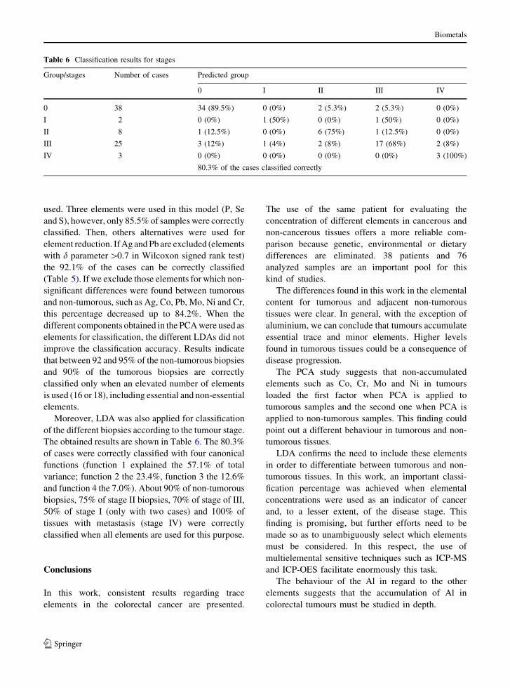
Moreover, LDA was also applied for classification
of the different biopsies according to the tumour stage.
The obtained results are shown in Table 6. The 80.3%
of cases were correctly classified with four canonical
functions (function 1 explained the 57.1% of total
variance; function 2 the 23.4%, function 3 the 12.6%
and function 4 the 7.0%). About 90% of non-tumorous
biopsies, 75% of stage II biopsies, 70% of stage of III,
50% of stage I (only with two cases) and 100% of
tissues with metastasis (stage IV) were correctly
classified when all elements are used for this purpose.
Conclusions
In this work, consistent results regarding trace
elements in the colorectal cancer are presented.
The use of the same patient for evaluating the
concentration of different elements in cancerous and
non-cancerous tissues offers a more reliable com-
parison because genetic, environmental or dietary
differences are eliminated. 38 patients and 76
analyzed samples are an important pool for this
kind of studies.
The differences found in this work in the elemental
content for tumorous and adjacent non-tumorous
tissues were clear. In general, with the exception of
aluminium, we can conclude that tumours accumulate
essential trace and minor elements. Higher levels
found in tumorous tissues could be a consequence of
disease progression.
The PCA study suggests that non-accumulated
elements such as Co, Cr, Mo and Ni in tumours
loaded the first factor when PCA is applied to
tumorous samples and the second one when PCA is
applied to non-tumorous samples. This finding could
point out a different behaviour in tumorous and non-
tumorous tissues.
LDA confirms the need to include these elements
in order to differentiate between tumorous and non-
tumorous tissues. In this work, an important classi-
fication percentage was achieved when elemental
concentrations were used as an indicator of cancer
and, to a lesser extent, of the disease stage. This
finding is promising, but further efforts need to be
made so as to unambiguously select which elements
must be considered. In this respect, the use of
multielemental sensitive techniques such as ICP-MS
and ICP-OES facilitate enormously this task.
The behaviour of the Al in regard to the other
elements suggests that the accumulation of Al in
colorectal tumours must be studied in depth.
Table 6 Classification results for stages
Group/stages Number of cases Predicted group
0 I II III IV
0 38 34 (89.5%) 0 (0%) 2 (5.3%) 2 (5.3%) 0 (0%)
I 2 0 (0%) 1 (50%) 0 (0%) 1 (50%) 0 (0%)
II 8 1 (12.5%) 0 (0%) 6 (75%) 1 (12.5%) 0 (0%)
III 25 3 (12%) 1 (4%) 2 (8%) 17 (68%) 2 (8%)
IV 3 0 (0%) 0 (0%) 0 (0%) 0 (0%) 3 (100%)
80.3% of the cases classified correctly
Biometals
123
Acknowledgments Financial support from the Spanish
Education and Science Ministry (project CTQ2006-04111/
BQU) is gratefully acknowledged.
References
Alimonti A, Bocca B, Lamazza A et al (2008) A study on
metals content in patients with colorectal polyps. J Tox-
icol Environ Health 71:342–347. doi:10.1080/15287
390701839133
Al-Taie OH, Uceyler N, Eubner U et al (2004) Expression
profiling and genetic alterations of the selenoproteins GI-
GPx and SePP in colorectal carcinogenesis. Nutr Cancer
48:6–14. doi:10.1207/s15327914nc4801_2
Andrasi E, Orosz L, Scheibeler H et al (1995) Concentrations
of elements in brain tumors. Mikrochim Acta 118:113–
121. doi:10.1007/BF01242234
Aung CS, Kruger WA, Poronnik P et al (2007) Plasma mem-
brane Ca2?-ATPase expression during colon cancer cell
line differentiation. Biochem Biophys Res Commun
355:932–936. doi:10.1016/j.bbrc.2007.02.050
Bandura DR, Ornatsky OI, Liao L (2004) Characterization of
phosphorus content of biological samples by ICP-DRC-
MS: potential tool for cancer research. J Anal At Spec-
trom 19:96–100. doi:10.1039/b308901k
Behrend L, Mohr A, Dick T et al (2005) Manganese superoxide
dismutase induces p53-dependent senescence in colorectal
cancer cells. Mol Cell Biol 25:7758–7769. doi:10.1128/
MCB.25.17.7758-7769.2005
Bocca B, Lamazza A, Pino A et al (2007) Determination of 30
elements in colorectal biopsies by sector field inductively
coupled plasma mass spectrometry. Rapid Commun Mass
Spectrom 21:1776–1782. doi:10.1002/rcm.3016
Brookes MJ, Hughes S, Turner FE et al (2006) Modulation of
iron transport proteins in human colorectal carcinogene-
sis. Gut 55:1449–1460. doi:10.1136/gut.2006.094060
Chung FY, Lin SR, Lu CY et al (2006) Sarco/endoplasmic
reticulum calcium-ATPase 2 expression as a tumor mar-
ker in colorectal cancer. Am Surg Pathol 30:969–974. doi:
10.1097/00000478-200608000-00006
Dai Q, Shrubsole MJ, Ness RM et al (2007) The relation of
magnesium and calcium intakes and a genetic polymor-
phism in the magnesium transporter to colorectal
neoplasia risk. Am J Clin Nutr 86:743–751
Darbre PD (2005) Aluminum, antiperspirants and breast can-
cer. J Inorg Biochem 99:1912–1919. doi:10.1016/
j.jinorgbio.2005.06.001
Drake EN II, Sky-Peck HH (1989) Discriminant analysis of
trace element distribution in normal and malignant human
tissues. Cancer Res 49:4210–4215
Ebrahim AM, Eltayeb MAH, Shaat MK et al (2007) Study of
selected trace elements in cancerous and non-cancerous
human breast tissues from Sudanese subjects using
instrumental neutron activation analysis. Sci Total Envi-
ron 383:52–58. doi:10.1016/j.scitotenv.2007.04.047
Eisenstein RS (2000) Iron regulatory proteins and the molec-
ular control of mammalian iron metabolism. Annu Rev
Nutr 20:627–662. doi:10.1146/annurev.nutr.20.1.627
Exley C, Charles LM, Barr L et al (2007) Aluminum in human
breast tissue. J Inorg Biochem 101:1344–1346. doi:
10.1016/j.jinorgbio.2007.06.005
Feustel A, Wennrich R, Dittrich M (1986) Studies of Cd, Zn
and Cu levels in human kidney tumours and normal kid-
ney. Urol Res 14:105–108. doi:10.1007/BF00257895
Folsom AR, Hong CP (2006) Magnesium intake and reduced
risk of colon cancer in a prospective study of women. Am
J Epidemiol 163:232–235. doi:10.1093/aje/kwj037
Gonzalez MA, Alvarez del Lujan M, Pisani GB et al (2007)
Involvement of oxidative stress in the impairment in bil-
iary secretory function induced by intraperitoneal
administration of aluminum to rats. Biol Trace Elem Res
116:329–348. doi:10.1007/BF02698017
Goodman VL, Brewer GJ, Merajver SD (2004) Copper defi-
ciency as an anti-cancer strategy. Endocr Relat Cancer
11:255–263. doi:10.1677/erc.0.0110255
Gregoriadis GC, Apostolidis NS, Romanos AN et al (1983) A
comparative study of trace elements in normal and can-
cerous colorectal tissues. Cancer 52:508–519. doi:10.1002/
1097-0142(19830801)52:3\508::AID-CNCR2820520322
[3.0.CO;2-8
Gupta SK, Singh SP, Shukla VK (2005) Copper, zinc and Cu/
Zn ratio in carcinoma of the gallbladder. J Surg Oncol
91:204–208. doi:10.1002/jso.20306
Gupte A, Mumper RJ (2009) Elevated copper and oxidative
stress in cancer cells as a target for cancer treatment. Cancer
Treat Rev 35:32–46. doi:10.1016/j.ctrv.2008.07.004
Gurusamy KS, Farquharson MJ, Craig C et al (2008) An
evaluation study of trace element content in colorectal
liver metastases and surrounding normal livers by X-ray
fluorescence. Biometals 21:373–378. doi:10.1007/s10534-
007-9126-3
Halliwell B, Gutteridge J (1999) Free radicals in biology and
medicine, 3rd edn. Oxford University Press, Oxford
Hartwig A (2000) Recent advances in metal carcinogenicity.
Pure Appl Chem 72:1007–1014. doi:10.1351/pac200072
061007
International Agency for Research on Cancer (1993) IARC
monographs on the evaluation of the carcinogenic risk to
man: beryllium, cadmium, mercury and exposures in the
glass manufacturing industry. IARC, Lyon
Ionescu JC, Novotny J, Stejskal V et al (2006) Increased levels
of transition metals in breast cancer tissue. Neuroendi-
cronol Lett 27:36–39
Jacobs ET, Jiang R, Alberts DS et al (2004) Selenium and
colorectal adenoma: results of a pooled analysis. J Natl
Cancer Inst 96:1669–1675
Jaiswal AS, Narayan S (2004) Zinc stabilizes adenomatous
polyposis coli (APC) protein levels and induces cell cycle
in colon cancer cells. J Cell Biochem 93:345–357. doi:
10.1002/jcb.20156
Janssen AML, Bosman CB, Kruidenier L et al (1999) Super-
oxide dismutases in the human colorectal cancer
sequence. J Cancer Res Clin Oncol 125:327–335. doi:
10.1007/s004320050282
Kasprzak KS (1997) Effects of calcium, magnesium, zinc and
iron on nickel carcinogenesis: inhibition versus enhance-
ment. In: Hadjiliadis ND (ed) Cytotoxic, mutagenic and
carcinogenic potential of heavy metals related to human
Biometals
123
environment, vol 26. Kluwer Academic Publishers,
Dordrecht, pp 93–106
Kawabata H, Germain RS, Vuong PT et al (2000) Transferrin
receptor 2-alpha supports cell growth both in iron-che-
lated cultured cells and in vivo. J Biol Chem 275:16618–
16625. doi:10.1074/jbc.M908846199
Knekt P, Reunamen A, Takkunen H et al (1994) Body iron
stores and risk of cancer. Int J Cancer 56:379–382. doi:
10.1002/ijc.2910560315
Kuo CY, Wong RH, Lin JY et al (2006) Accumulation of
chromium and nickel metals in lung tumors from lung
cancer patients in Taiwan. J Toxicol Environ Health
69:1337–1344. doi:10.1080/15287390500360398
Lapidos KA, Woodhouse EC, Kohn EC et al (2001) Mg??-
induced endothelial cell migration: substratum selectivity
and receptor-involvement. Angiogenesis 8:21–28. doi:
10.1023/A:1016619414817
Larrick JW, Cresswell P (1979) Modulation of cell surface iron
transferring receptors by cellular density and state acti-
vation. J Supramol Struct 11:579–586. doi:10.1002/jss.
400110415
Larsson SC, Bergkvist L, Wolk A (2005) Magnesium intake in
relation to risk of colorectal cancer in women. J Am Med
Assoc 293:86–89. doi:10.1001/jama.293.1.86
Leonard SS, Harris GK, Shi XL (2004) Metal-induced oxida-
tive stress and signal transduction. Free Radic Biol Med
37:1921–1942. doi:10.1016/j.freeradbiomed.2004.09.010
Lowndes SA, Harris AL (2005) The role of copper in tumour
angiogenesis. J Mammary Gland Biol Neoplasia 10:299–
310. doi:10.1007/s10911-006-9003-7
Mai FD, Chen BJ, Wu LC et al (2006) Imaging of single liver
tumor cells intoxicated by heavy metals using ToF-SIMS.
Appl Surf Sci 252:6809–6812. doi:10.1016/j.apsusc.2006.
02.227
Maier JAM, Nasulewicz-Goldeman A, Simonacci M et al
(2007) Insights into the mechanisms involved in magne-
sium-dependent inhibition of primary tumor growth. Nutr
Cancer 59:192–198
Majewska U, Banas D, Braziewicz J et al (2007) Trace element
concentration distributions in breast, lung and colon tis-
sues. Phys Med Biol 52:3895–3911. doi:10.1088/0031-
9155/52/13/016
Martin Mateo MC, Rabadan J, Boustamante J (1990) Com-
parative analysis of certain metals and tumor markers in
bronchopulmonary cancer and colorectal cancers. Metals
and tumor markers in the neoplastic process. Clin Physiol
Biochem 8:261–266
Mates JM, Perez-Gomez C, Nunez de Castro I (1999) Anti-
oxidants enzymes and human diseases. Clin Biochem
32:595–603. doi:10.1016/S0009-9120(99)00075-2
Millos J, Costas-Rodrıguez M, Lavilla I et al (2008) Multiel-
emental determination in breast cancerous and non-
cancerous biopsies by inductively coupled plasma-mass
spectrometry following small volume microwave-assisted
digestion. Anal Chim Acta 622:77–84. doi:10.1016/j.aca.
2008.05.066
Mulay IL, Roy R, Knox BE et al (1971) Trace metal analysis of
cancerous and noncancerous human tissues. J Natl Cancer
Inst 47:1–13
Nakamaki T, Kawabata H, Bungo S (2004) Elevated levels of
transferrin receptor 2 mRNA, not transferring receptor 1
mRNA, are associated with increased survival in acute
myeloid leukaemia. Br J Haematol 125:42–49. doi:
10.1111/j.1365-2141.2004.04866.x
Nasulewicz A, Mazur A, Opolski A (2004) Role of copper in
tumor angiogenesis—clinical implications. J Trace Elem
Med Biol 18:1–8. doi:10.1016/j.jtemb.2004.02.004
Navarro Silvera SA, Rohan TE (2007) Trace elements and
cancer risk: a review of the epidemiologic evidence.
Cancer Causes Control 18:7–27. doi:10.1007/s10552-006-
0057-z
Ng KH, Bradley DA, Looi LM (1997) Elevated trace element
concentrations in malignant breast tissues. Br J Radiol
70:375–382
Nozoe T, Honda M, Inutsuka S et al (2003) Significance of
immunohistochemical expression of manganese superox-
ide dismutase as a marker of malignant potential in
colorectal carcinoma. Oncol Rep 10:39–43
Owen RW (2001) Biomarkers in colorectal cancer, vol 154.
IARC Scientific Publications (Biomarkers in Cancer
Chemoprevention), pp 101–111
Peters U, Chatterjee N, McGlynn KA et al (2004) Calcium
intake and colorectal adenoma in a US colorectal cancer
early detection program. Am J Clin Nutr 80:1358–1365
Raju GJN, Sarita P, Kumar MR et al (2006) Trace elemental
correlation study in malignant and normal breast tissue by
PIXE technique. Nucl Instrum Methods Phys Res B
247:361–367
Reddy SB, Charles MJ, Raju GJN et al (2003) Trace elemental
analysis of carcinoma kidney and stomach by PIXE
method. Nucl Instrum Methods Phys Res B 207:345–355.
doi:10.1016/S0168-583X(03)00463-4
Richardson DR et al (2008) Cancer cell iron metabolism and the
development of potent iron chelators as anti-tumour agents.
Biochim Biophys Acta. doi:10.1016/j.bbagen.2008.04.003
Richardson-Boedler C (2007) Metal passivity as mechanism of
metal carcinogenesis: chromium, nickel, iron, copper,
cobalt, platinum, molybdenum. Toxicol Environ Chem
89:15–70. doi:10.1080/02772240601008513
Rizk SL, Sky-Peck HH (1984) Comparison between concen-
trations of trace elements in normal and neoplastic human
breast tissue. Cancer Res 44:5390–5394
Rubin H (2007) The logic of the membrane, magnesium,
mitosis (MMM) model for the regulation of animal cell
proliferation. Arch Biochem Biophys 458:16–23. doi:
10.1016/j.abb.2006.03.026
Skrzydlewska E, Sulkowski S, Koda M et al (2005) Lipid
peroxidation and antioxidant status in colorectal cancer.
World J Gastroenterol 11:403–406
Tashiro H, Kawamoto T, Okubo T et al (2003) Variation in the
distribution of trace elements in hepatoma. Biol Trace
Elem Res 95:49–63. doi:10.1385/BTER:95:1:49
Toyokuni S (1996) Iron induced carcinogenesis: the role or
redox regulation. Free Radic Biol Med 20:553–566. doi:
10.1016/0891-5849(95)02111-6
Ujiie S, Itoh Y, Kikuchi H, Wakui A (1995) Zinc distribution
in malignant tumors. Biomed Res Trace Elem 6:45–50
Valko M, Morris MTD (2005) Metals, toxicity and oxidative
stress. Curr Med Chem 12:1161–1208. doi:10.2174/092
9867053764635
Valko M, Rhodes CJ, Moncol J et al (2006) Free radicals,
metals and antioxidants in oxidative stress-induced
Biometals
123

cancer. Chem Biol Interact 160:1–40. doi:10.1016/j.cbi.
2005.12.009
Vallee BL, Auld DS (1990) Zinc coordination, function, and
structure of zinc enzymes and other proteins. Biochem-
istry 29:5647–5659. doi:10.1021/bi00476a001
van den Brandt PA, Smits KM, Goldbohm RA et al (2007)
Magnesium intake and colorectal cancer risk in the
Netherlands Cohort Study. Br J Cancer 96:510–513. doi:
10.1038/sj.bjc.6603577
Weinberg ED (1994) Association of iron with colorectal can-
cer. Biometals 7:211–216. doi:10.1007/BF00149550
Whitfield JF (2009) Calcium, calcium-sensing receptor and
colon cancer. Cancer Lett 275:9–16. doi:10.1016/j.canlet.
2008.07.001
Wolf FI et al (2009) Magnesium and tumors: ally or foe?
Cancer Treat Rev. doi:10.1016/j.ctrv.2009.01.003
Zoriy MV, Dehnhardt M, Reifenberger G et al (2006) Imaging
of Cu, Zn, Pb and U in human brain tumor resections by
laser ablation inductively coupled plasma mass spec-
trometry. Int J Mass Spectrom 257:27–33. doi:10.1016/
j.ijms.2006.06.005
Biometals
123