403170873 The Anorexia Nervosa Disorder that Reflected in ...
Upload
independentCategory
view
3download
0

SSeennssoorrss && TTrraannssdduucceerrss
Volume 99 December 2008 www.sensorsportal.com ISSN 1726-5479
Editor-in-Chief: professor Sergey Y. Yurish, phone: +34 696067716, fax: +34 93 4011989, e-mail: [email protected]
Editors for Western Europe Meijer, Gerard C.M., Delft University of Technology, The Netherlands Ferrari, Vittorio, UUnniivveerrssiittáá ddii BBrreesscciiaa,, IIttaaly Editors for North America Datskos, Panos G., OOaakk RRiiddggee NNaattiioonnaall LLaabboorraattoorryy,, UUSSAA Fabien, J. Josse, Marquette University, USA Katz, Evgeny, Clarkson University, USA
Editor South America Costa-Felix, Rodrigo, Inmetro, Brazil Editor for Eastern Europe Sachenko, Anatoly, Ternopil State Economic University, Ukraine Editor for Asia Ohyama, Shinji, Tokyo Institute of Technology, Japan
Editorial Advisory Board
Abdul Rahim, Ruzairi, Universiti Teknologi, Malaysia Ahmad, Mohd Noor, Nothern University of Engineering, Malaysia Annamalai, Karthigeyan, National Institute of Advanced Industrial Science
and Technology, Japan Arcega, Francisco, University of Zaragoza, Spain Arguel, Philippe, CNRS, France Ahn, Jae-Pyoung, Korea Institute of Science and Technology, Korea Arndt, Michael, Robert Bosch GmbH, Germany Ascoli, Giorgio, George Mason University, USA Atalay, Selcuk, Inonu University, Turkey Atghiaee, Ahmad, University of Tehran, Iran Augutis, Vygantas, Kaunas University of Technology, Lithuania Avachit, Patil Lalchand, North Maharashtra University, India Ayesh, Aladdin, De Montfort University, UK Bahreyni, Behraad, University of Manitoba, Canada Baoxian, Ye, Zhengzhou University, China Barford, Lee, Agilent Laboratories, USA Barlingay, Ravindra, RF Arrays Systems, India Basu, Sukumar, Jadavpur University, India Beck, Stephen, University of Sheffield, UK Ben Bouzid, Sihem, Institut National de Recherche Scientifique, Tunisia Benachaiba, Chellali, Universitaire de Bechar, Algeria Binnie, T. David, Napier University, UK Bischoff, Gerlinde, Inst. Analytical Chemistry, Germany Bodas, Dhananjay, IMTEK, Germany Borges Carval, Nuno, Universidade de Aveiro, Portugal Bousbia-Salah, Mounir, University of Annaba, Algeria Bouvet, Marcel, CNRS – UPMC, France Brudzewski, Kazimierz, Warsaw University of Technology, Poland Cai, Chenxin, Nanjing Normal University, China Cai, Qingyun, Hunan University, China Campanella, Luigi, University La Sapienza, Italy Carvalho, Vitor, Minho University, Portugal Cecelja, Franjo, Brunel University, London, UK Cerda Belmonte, Judith, Imperial College London, UK Chakrabarty, Chandan Kumar, Universiti Tenaga Nasional, Malaysia Chakravorty, Dipankar, Association for the Cultivation of Science, India Changhai, Ru, Harbin Engineering University, China Chaudhari, Gajanan, Shri Shivaji Science College, India Chen, Jiming, Zhejiang University, China Chen, Rongshun, National Tsing Hua University, Taiwan Cheng, Kuo-Sheng, National Cheng Kung University, Taiwan Chiriac, Horia, National Institute of Research and Development, Romania Chowdhuri, Arijit, University of Delhi, India Chung, Wen-Yaw, Chung Yuan Christian University, Taiwan Corres, Jesus, Universidad Publica de Navarra, Spain Cortes, Camilo A., Universidad Nacional de Colombia, Colombia Courtois, Christian, Universite de Valenciennes, France Cusano, Andrea, University of Sannio, Italy D'Amico, Arnaldo, Università di Tor Vergata, Italy De Stefano, Luca, Institute for Microelectronics and Microsystem, Italy Deshmukh, Kiran, Shri Shivaji Mahavidyalaya, Barshi, India Dickert, Franz L., Vienna University, Austria Dieguez, Angel, University of Barcelona, Spain Dimitropoulos, Panos, University of Thessaly, Greece Ding Jian, Ning, Jiangsu University, China Djordjevich, Alexandar, City University of Hong Kong, Hong Kong Ko, Sang Choon, Electronics and Telecommunications Research Institute, Korea South
Donato, Nicola, University of Messina, Italy Donato, Patricio, Universidad de Mar del Plata, Argentina Dong, Feng, Tianjin University, China Drljaca, Predrag, Instersema Sensoric SA, Switzerland Dubey, Venketesh, Bournemouth University, UK Enderle, Stefan, University of Ulm and KTB Mechatronics GmbH,
Germany Erdem, Gursan K. Arzum, Ege University, Turkey Erkmen, Aydan M., Middle East Technical University, Turkey Estelle, Patrice, Insa Rennes, France Estrada, Horacio, University of North Carolina, USA Faiz, Adil, INSA Lyon, France Fericean, Sorin, Balluff GmbH, Germany Fernandes, Joana M., University of Porto, Portugal Francioso, Luca, CNR-IMM Institute for Microelectronics and
Microsystems, Italy Francis, Laurent, University Catholique de Louvain, Belgium Fu, Weiling, South-Western Hospital, Chongqing, China Gaura, Elena, Coventry University, UK Geng, Yanfeng, China University of Petroleum, China Gole, James, Georgia Institute of Technology, USA Gong, Hao, National University of Singapore, Singapore Gonzalez de la Rosa, Juan Jose, University of Cadiz, Spain Granel, Annette, Goteborg University, Sweden Graff, Mason, The University of Texas at Arlington, USA Guan, Shan, Eastman Kodak, USA Guillet, Bruno, University of Caen, France Guo, Zhen, New Jersey Institute of Technology, USA Gupta, Narendra Kumar, Napier University, UK Hadjiloucas, Sillas, The University of Reading, UK Hashsham, Syed, Michigan State University, USA Hernandez, Alvaro, University of Alcala, Spain Hernandez, Wilmar, Universidad Politecnica de Madrid, Spain Homentcovschi, Dorel, SUNY Binghamton, USA Horstman, Tom, U.S. Automation Group, LLC, USA Hsiai, Tzung (John), University of Southern California, USA Huang, Jeng-Sheng, Chung Yuan Christian University, Taiwan Huang, Star, National Tsing Hua University, Taiwan Huang, Wei, PSG Design Center, USA Hui, David, University of New Orleans, USA Jaffrezic-Renault, Nicole, Ecole Centrale de Lyon, France Jaime Calvo-Galleg, Jaime, Universidad de Salamanca, Spain James, Daniel, Griffith University, Australia Janting, Jakob, DELTA Danish Electronics, Denmark Jiang, Liudi, University of Southampton, UK Jiang, Wei, University of Virginia, USA Jiao, Zheng, Shanghai University, China John, Joachim, IMEC, Belgium Kalach, Andrew, Voronezh Institute of Ministry of Interior, Russia Kang, Moonho, Sunmoon University, Korea South Kaniusas, Eugenijus, Vienna University of Technology, Austria Katake, Anup, Texas A&M University, USA Kausel, Wilfried, University of Music, Vienna, Austria Kavasoglu, Nese, Mugla University, Turkey Ke, Cathy, Tyndall National Institute, Ireland Khan, Asif, Aligarh Muslim University, Aligarh, India Kim, Min Young, Koh Young Technology, Inc., Korea South Sandacci, Serghei, Sensor Technology Ltd., UK Sapozhnikova, Ksenia, D.I.Mendeleyev Institute for Metrology, Russia

Kockar, Hakan, Balikesir University, Turkey Kotulska, Malgorzata, Wroclaw University of Technology, Poland Kratz, Henrik, Uppsala University, Sweden Kumar, Arun, University of South Florida, USA Kumar, Subodh, National Physical Laboratory, India Kung, Chih-Hsien, Chang-Jung Christian University, Taiwan Lacnjevac, Caslav, University of Belgrade, Serbia Lay-Ekuakille, Aime, University of Lecce, Italy Lee, Jang Myung, Pusan National University, Korea South Lee, Jun Su, Amkor Technology, Inc. South Korea Lei, Hua, National Starch and Chemical Company, USA Li, Genxi, Nanjing University, China Li, Hui, Shanghai Jiaotong University, China Li, Xian-Fang, Central South University, China Liang, Yuanchang, University of Washington, USA Liawruangrath, Saisunee, Chiang Mai University, Thailand Liew, Kim Meow, City University of Hong Kong, Hong Kong Lin, Hermann, National Kaohsiung University, Taiwan Lin, Paul, Cleveland State University, USA Linderholm, Pontus, EPFL - Microsystems Laboratory, Switzerland Liu, Aihua, University of Oklahoma, USA Liu Changgeng, Louisiana State University, USA Liu, Cheng-Hsien, National Tsing Hua University, Taiwan Liu, Songqin, Southeast University, China Lodeiro, Carlos, Universidade NOVA de Lisboa, Portugal Lorenzo, Maria Encarnacio, Universidad Autonoma de Madrid, Spain Lukaszewicz, Jerzy Pawel, Nicholas Copernicus University, Poland Ma, Zhanfang, Northeast Normal University, China Majstorovic, Vidosav, University of Belgrade, Serbia Marquez, Alfredo, Centro de Investigacion en Materiales Avanzados, Mexico Matay, Ladislav, Slovak Academy of Sciences, Slovakia Mathur, Prafull, National Physical Laboratory, India Maurya, D.K., Institute of Materials Research and Engineering, Singapore Mekid, Samir, University of Manchester, UK Melnyk, Ivan, Photon Control Inc., Canada Mendes, Paulo, University of Minho, Portugal Mennell, Julie, Northumbria University, UK Mi, Bin, Boston Scientific Corporation, USA Minas, Graca, University of Minho, Portugal Moghavvemi, Mahmoud, University of Malaya, Malaysia Mohammadi, Mohammad-Reza, University of Cambridge, UK Molina Flores, Esteban, Benemérita Universidad Autónoma de Puebla,
Mexico Moradi, Majid, University of Kerman, Iran Morello, Rosario, DIMET, University "Mediterranea" of Reggio Calabria,
Italy Mounir, Ben Ali, University of Sousse, Tunisia Mukhopadhyay, Subhas, Massey University, New Zealand Neelamegam, Periasamy, Sastra Deemed University, India Neshkova, Milka, Bulgarian Academy of Sciences, Bulgaria Oberhammer, Joachim, Royal Institute of Technology, Sweden Ould Lahoucin, University of Guelma, Algeria Pamidighanta, Sayanu, Bharat Electronics Limited (BEL), India Pan, Jisheng, Institute of Materials Research & Engineering, Singapore Park, Joon-Shik, Korea Electronics Technology Institute, Korea South Penza, Michele, ENEA C.R., Italy Pereira, Jose Miguel, Instituto Politecnico de Setebal, Portugal Petsev, Dimiter, University of New Mexico, USA Pogacnik, Lea, University of Ljubljana, Slovenia Post, Michael, National Research Council, Canada Prance, Robert, University of Sussex, UK Prasad, Ambika, Gulbarga University, India Prateepasen, Asa, Kingmoungut's University of Technology, Thailand Pullini, Daniele, Centro Ricerche FIAT, Italy Pumera, Martin, National Institute for Materials Science, Japan Radhakrishnan, S. National Chemical Laboratory, Pune, India Rajanna, K., Indian Institute of Science, India Ramadan, Qasem, Institute of Microelectronics, Singapore Rao, Basuthkar, Tata Inst. of Fundamental Research, India Raoof, Kosai, Joseph Fourier University of Grenoble, France Reig, Candid, University of Valencia, Spain Restivo, Maria Teresa, University of Porto, Portugal Robert, Michel, University Henri Poincare, France Rezazadeh, Ghader, Urmia University, Iran Royo, Santiago, Universitat Politecnica de Catalunya, Spain Rodriguez, Angel, Universidad Politecnica de Cataluna, Spain Rothberg, Steve, Loughborough University, UK Sadana, Ajit, University of Mississippi, USA Sadeghian Marnani, Hamed, TU Delft, The Netherlands
Saxena, Vibha, Bhbha Atomic Research Centre, Mumbai, India Schneider, John K., Ultra-Scan Corporation, USA Seif, Selemani, Alabama A & M University, USA Seifter, Achim, Los Alamos National Laboratory, USA Sengupta, Deepak, Advance Bio-Photonics, India Shankar, B. Baliga, General Monitors Transnational, USA Shearwood, Christopher, Nanyang Technological University, Singapore Shin, Kyuho, Samsung Advanced Institute of Technology, Korea Shmaliy, Yuriy, Kharkiv National University of Radio Electronics,
Ukraine Silva Girao, Pedro, Technical University of Lisbon, Portugal Singh, V. R., National Physical Laboratory, India Slomovitz, Daniel, UTE, Uruguay Smith, Martin, Open University, UK Soleymanpour, Ahmad, Damghan Basic Science University, Iran Somani, Prakash R., Centre for Materials for Electronics Technol., India Srinivas, Talabattula, Indian Institute of Science, Bangalore, India Srivastava, Arvind K., Northwestern University, USA Stefan-van Staden, Raluca-Ioana, University of Pretoria, South Africa Sumriddetchka, Sarun, National Electronics and Computer Technology
Center, Thailand Sun, Chengliang, Polytechnic University, Hong-Kong Sun, Dongming, Jilin University, China Sun, Junhua, Beijing University of Aeronautics and Astronautics, China Sun, Zhiqiang, Central South University, China Suri, C. Raman, Institute of Microbial Technology, India Sysoev, Victor, Saratov State Technical University, Russia Szewczyk, Roman, Industrial Research Institute for Automation and
Measurement, Poland Tan, Ooi Kiang, Nanyang Technological University, Singapore, Tang, Dianping, Southwest University, China Tang, Jaw-Luen, National Chung Cheng University, Taiwan Teker, Kasif, Frostburg State University, USA Thumbavanam Pad, Kartik, Carnegie Mellon University, USA Tian, Gui Yun, University of Newcastle, UK Tsiantos, Vassilios, Technological Educational Institute of Kaval, Greece Tsigara, Anna, National Hellenic Research Foundation, Greece Twomey, Karen, University College Cork, Ireland Valente, Antonio, University, Vila Real, - U.T.A.D., Portugal Vaseashta, Ashok, Marshall University, USA Vazques, Carmen, Carlos III University in Madrid, Spain Vieira, Manuela, Instituto Superior de Engenharia de Lisboa, Portugal Vigna, Benedetto, STMicroelectronics, Italy Vrba, Radimir, Brno University of Technology, Czech Republic Wandelt, Barbara, Technical University of Lodz, Poland Wang, Jiangping, Xi'an Shiyou University, China Wang, Kedong, Beihang University, China Wang, Liang, Advanced Micro Devices, USA Wang, Mi, University of Leeds, UK Wang, Shinn-Fwu, Ching Yun University, Taiwan Wang, Wei-Chih, University of Washington, USA Wang, Wensheng, University of Pennsylvania, USA Watson, Steven, Center for NanoSpace Technologies Inc., USA Weiping, Yan, Dalian University of Technology, China Wells, Stephen, Southern Company Services, USA Wolkenberg, Andrzej, Institute of Electron Technology, Poland Woods, R. Clive, Louisiana State University, USA Wu, DerHo, National Pingtung University of Science and Technology,
Taiwan Wu, Zhaoyang, Hunan University, China Xiu Tao, Ge, Chuzhou University, China Xu, Lisheng, The Chinese University of Hong Kong, Hong Kong Xu, Tao, University of California, Irvine, USA Yang, Dongfang, National Research Council, Canada Yang, Wuqiang, The University of Manchester, UK Ymeti, Aurel, University of Twente, Netherland Yong Zhao, Northeastern University, China Yu, Haihu, Wuhan University of Technology, China Yuan, Yong, Massey University, New Zealand Yufera Garcia, Alberto, Seville University, Spain Zagnoni, Michele, University of Southampton, UK Zeni, Luigi, Second University of Naples, Italy Zhong, Haoxiang, Henan Normal University, China Zhang, Minglong, Shanghai University, China Zhang, Qintao, University of California at Berkeley, USA Zhang, Weiping, Shanghai Jiao Tong University, China Zhang, Wenming, Shanghai Jiao Tong University, China Zhou, Zhi-Gang, Tsinghua University, China Zorzano, Luis, Universidad de La Rioja, Spain Zourob, Mohammed, University of Cambridge, UK
Sensors & Transducers Journal (ISSN 1726-5479) is a peer review international journal published monthly online by International Frequency Sensor Association (IFSA).
Available in electronic and CD-ROM. Copyright © 2007 by International Frequency Sensor Association. All rights reserved.

SSeennssoorrss && TTrraannssdduucceerrss JJoouurrnnaall
CCoonntteennttss
Volume 99 Issue 12 December 2008
www.sensorsportal.com ISSN 1726-5479
Research Articles
Pushing Low‐Cost Sensors to Market A NanoMarkets White Paper, NanoMarkets, ...................................................................................... 1 Design and Development of an Embedded System for Measurement of Temperature and Remote Communication Using Fiber Optic Cable Raghavendra Rao Kanchi, Sreelekha Kande and Ramanjappa Thogata.......................................... 7 Reflective Type Small-Angle Sensor Based on Multiple Total Internal Reflections in Heterodyne Interferometry Shinn-Fwu Wang, Po-Chin Chiu, Tsung-Hsun Yang ......................................................................... 18 An Inter-digital Capacitive Electrode Modified as a Pressure Sensor T. J. Ginson and J. Philip.................................................................................................................... 25 A New Rotational Velocity Meter for Dynamic Testing Hou Xiaoyan ....................................................................................................................................... 33 Fault Diagnosis and Condition Monitoring of an All Geared Lathe Machine Using Piezoelectric Sensor Amiya Bhaumik, Nabin Sardar, Nirmal Kumar Mandal ...................................................................... 43 Modal Analysis and Experimental Determination of Optimum Tool Shank Overhang of a Lathe Machine Nabin Sardar, Amiya Bhaumik, Nirmal Kumar Mandal ...................................................................... 53 Test Device for Under Water Tidal Flow Power Generation Measurement Ibrahim Al-Bahadly and Paul Pinfold.................................................................................................. 66 Development of Portative System for Quality Detection in Indian Honey Using Gas Sensor Rohini P. Mudhalwadkar, Dr. Ashok Ghatol ....................................................................................... 82 Electrostrictive Effect in Cancer Cell Reflected in Capacitance Relaxation Phenomena Tapas Kumar Basak, T. Ramanujam, Suman Halder, Poonam Goyal, Prachi Mohan Kulshreshtha, Shweta Pandey, Himanshu Tripathi ................................................................................................... 90 Wireless Pressure Sensor Using Non-contact Differential Variable Reluctance Transducer K. Prabakar, Usharani Ravi and J. Jayapandian................................................................................ 102
Authors are encouraged to submit article in MS Word (doc) and Acrobat (pdf) formats by e-mail: [email protected] Please visit journal’s webpage with preparation instructions: http://www.sensorsportal.com/HTML/DIGEST/Submition.htm
International Frequency Sensor Association (IFSA).

Sensors & Transducers Journal, Vol. 99, Issue 12, December 2008, pp. 90-101
90
SSSeeennnsssooorrrsss &&& TTTrrraaannnsssddduuuccceeerrrsss
ISSN 1726-5479© 2008 by IFSA
http://www.sensorsportal.com
Electrostrictive Effect in Cancer Cell Reflected in Capacitance Relaxation Phenomena
Tapas Kumar Basak, T. Ramanujam, Suman Halder, Poonam Goyal, Prachi
Mohan Kulshreshtha, Shweta Pandey, Himanshu Tripathi Krishna Engineering College, 95 Loni Road, Mohan Nagar, Ghaziabad, India
Received: 5 November 2008 /Accepted: 22 December 2008 /Published: 30 December 2008 Abstract: The present paper has focus on the composite dielectric property of the cancer cell on concomitant with the capacitance relaxation phenomena. In this respect it has been found from MAT lab simulation the electrostrictive process in cancer cell is a complex one for which the electrostatic surfaces surrounding the cell changes with the incremental changes in the capacitance present in the capacitance relaxation curve. From these incremental changes in capacitance it is also possible to find out the electrostrictive energy of the cancer cell. It is interesting to note that the electrostrictive energy corresponding to the cell incremental changes in the capacitance is more in the first order system than that present in the second order system representing the equivalent configuration of the composite dielectric associated with the cell membrane. This is due the fact that during the process DNA synthesis and cell division the change in capacitance of the membrane for the first order system is relatively slow. Copyright © 2008 IFSA. Keywords: Electrosrictive energy, Cancer cell capacitance relaxation, Composite dielectric 1 Introduction This paper reflects the electrostictive energy variation in carcinogenic cell membrane during metastasis, concomitant with capacitance relaxation phenomena [united States patent No. 5691178,1997]]’ The electrostrictive phenomena is due to composite dielectric property of the carcinogenic cell membrane for which there is excess negative charge on the outer surface of the membrane. It is well known that the cell membranes are composed of a bilayer of highly mobile lipid molecules that electrically behaves as an insulator (dielectric). The insulating properties of the cell membrane also restrict the movement of charged ions and electrons across the membrane except through specialized membrane spanning protein ion channels [9-16] and membrane spanning protein semiconductors [26] respectively. Thus the lipid structure of a cell membrane makes it relatively

Sensors & Transducers Journal, Vol. 99, Issue 12, December 2008, pp. 90-101
91
semiconductors [26] respectively. Thus the lipid structure of a cell membrane makes it relatively impermeable to the passage of charged molecules. As the cell membrane is a leaky dielectric, this means that any condition, illness or change in dietary intake that affects the composition of the cell membranes and their associated minerals can affect and alter the cellular capacitance. [1, 2, 4]. The major charge carriers of biological organisms are negatively charged electrons, positively charged hydrogen protons, positively charged sodium, potassium, calcium, magnesium ions and negatively charged anions particularly phosphate ions. As the cell membrane is selectively permeable to sodium and potassium ions, a different concentration of these ions and other charged mineral ions will build up on either side of the membrane. The different concentrations of these will cause the outer surface to have a relatively higher positive charge than the inner membrane surface and creates an electrical potential difference across the membrane. [18]. so this potential difference is due to the imbalance in electrical charges between the inside of the cell and the outside of the cell. Almost all normal cells have a membrane potential of about -60 to –100 mV. Cancer cells also have different lipid and sterol content than normal cells [9-16, 27]. Cancer cells have altered membrane composition and membrane permeability, which results in the movement of potassium, magnesium and calcium out of the cell and the accumulation of sodium and water into the cell [28]. With The result of these mineral movements, membrane composition changes, leaving to energy abnormalities , and membrane charge distribution abnormalities falls there occurs a decline in the normal membrane potential and rise of membrane capacitance. [19-22]. The outermost electrically negative zone of cell membrane is composed of negatively charged sialic acid molecules that cap the tips of glycoprotein and glycolipids that extend outward from the cell membrane like tree branches. The outermost negative zone is separated from the positive cell membrane surface by a distance of about 20 micrometers. This outermost calyx zone of steady negativity that makes each cell act as a negatively charged body; every cell creates a negatively charged field around itself that influences any other charged body close to it [37]. Therefore the cancer cells exhibit both lower electrical membrane potentials and lower electrical impedance than normal cells [17, 21 ,29].The excessive amount of negative charge on the exterior surface of the cell responsible for carcinogenic change and genetic change result from development of cellular electrical abnormalities.[23] Also the depolarization (fall in membrane potential) of the cancer cell membrane due to the accumulation of excess negative surface charges may precede and create the reduction in intracellular potassium and the rise in the intracellular sodium ions[23].The cancer cells have significantly more sialic acid molecules in their cell coat and as a result cancer cells have a greater surface negativity. It has been found that the cells become more electronegative in the course of cancerization, that membrane degeneration occurs in the initial phase of carcinogenesis first in the external cell membrane and then in the inner mitochondrial membrane and these degenerative changes in the surface membrane causes these membranes to become more permeable to water-soluble substances so that potassium, magnesium, and calcium migrate from the cells and sodium and water accumulate in the cell interior. The degenerative changes in the inner membrane of the mitochondria cause loss of storage of critical mitochondrial enzymes, for which mitochondria cancer cells degenerate and are reduced in numbers [28]. Two of the most outstanding electrical features of cancer cells are that they constantly maintain their membrane potential at a low value and their intracellular concentration of sodium is of high magnitude [19-21]. This sustained elevation of intracellular sodium may act as a mitotic trigger causing cells to go into cell division (mitosis) [21]. In the resting phase normal cells maintain a high membrane potential of around -60 mV to -100 mV, but when cells begin cell division and DNA synthesis, the membrane potential falls to around –15 mV [24].During this process of DNA synthesis the composite dielectric property of cell the cell membrane has a different values of permittivity and conductivity for which the there is large variation of elecctrostrictive energy. [1, 24].
Sensors & Transducers Journal, Vol. 99, Issue 12, December 2008, pp. 90-101
92
The electrical conductivity and permittivity of cancerous cells has been found to be greater than those of normal cells of normal tissues [25]. The cancerous cells demonstrate greater permittivity, and moreover cover a larger surface area for which there is large increase in capacitance concomitant with the Capacitance Relaxation phenomenon. [15]. The normal and carcinogenic cells may be thought of as a composite dielectric material with the relative permittivity
ε=εn+ εc This composite dielectric property accounts for elecrtostrictive phenomenon [1].for which the surface with the negative charges increases during the growth of cancer cell concomitant with it increases the capacitance. 2. Modeling and Simulation 2.1. Modeling and Simulation of the First Order Composite Dielectric (Case 1) The block diagram of the model is shown below. The transfer function of the composite dielectric will be represented by second order system shown in the block diagram:
From the US Patent [15] of Prof. T. K. Basak we have taken different values of capacitance as an input from capacitance from capacitance relaxation curve as shown below (Fig. 1).
Fig. 1. Capacitance relaxation curve.

Sensors & Transducers Journal, Vol. 99, Issue 12, December 2008, pp. 90-101
93
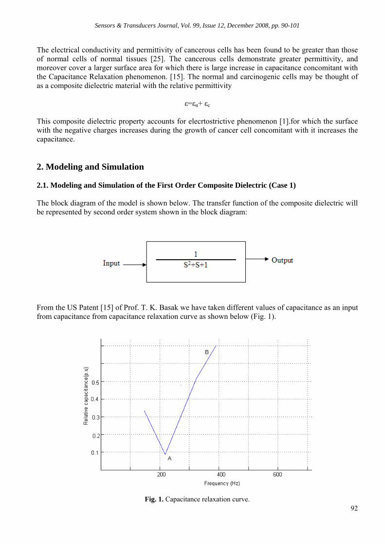
From the capacitance relaxation curve shown in the figure1, we have taken different values of capacitance as input of the model. Moreover, the input of the model is restricted to different values of capacitance in the rising part of the capacitance relaxation curve i.e. from the point A to B. For example we have taken the input as increase of capacitance from 0.1 p.u to 0.5 p.u in steps of 0.1 p.u in the forward path of the rising part of the curve i.e. from A to B. For the reverse path i.e. from B to A same process is repeated. The composite dielectric is regarded as 2nd order system having transfer function (1/(S2+S+1)). The output is focused to trace out the locus for which it is possible to obtain electrostatic surfaces containing negative charges with different values of capacitances in the capacitance relaxation curve. The technique for tracing out the locus in real-imaginary axis, the Nyquist criterion has been used with the aid of MATLAB 6.5. In this way we have obtained different electrostatic surface for the forward and reverse path of the rising phase of the capacitance relaxation curve. The per unit values are taken for the normalization of a particular phenomena [8]. Let the capacitance change from 0.4 to 0.5 p.u is termed as ∆C1. The electrostatic surfaces obtained due to forward and reverse path of ∆C1 are given in Figs. 2-4.
Fig. 2. Electrostatic surface containing negative charge in the forward path of ∆C1.
Fig. 3. Electrostatic surface containing negative charge in the reverse path of ∆C1.
Sensors & Transducers Journal, Vol. 99, Issue 12, December 2008, pp. 90-101
94
Fig. 4. Shaded area representing the variation in electrostatic energy indicating difference in electrostatic surfaces due to ∆C1.
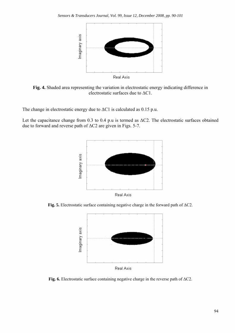
The change in electrostatic energy due to ∆C1 is calculated as 0.15 p.u. Let the capacitance change from 0.3 to 0.4 p.u is termed as ∆C2. The electrostatic surfaces obtained due to forward and reverse path of ∆C2 are given in Figs. 5-7.
Fig. 5. Electrostatic surface containing negative charge in the forward path of ∆C2.
Fig. 6. Electrostatic surface containing negative charge in the reverse path of ∆C2.
Sensors & Transducers Journal, Vol. 99, Issue 12, December 2008, pp. 90-101
95
Fig. 7. Shaded area representing the variation in electrostatic energy indicating difference in electrostatic surfaces due to ∆C2.
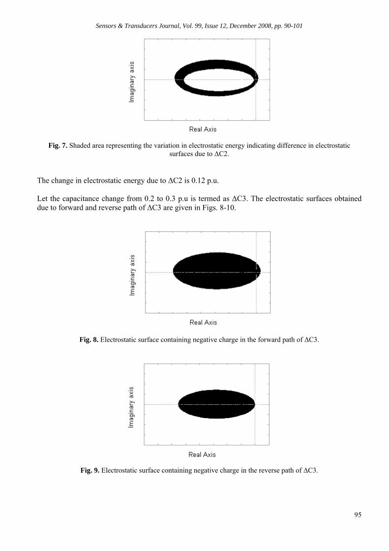
The change in electrostatic energy due to ∆C2 is 0.12 p.u. Let the capacitance change from 0.2 to 0.3 p.u is termed as ∆C3. The electrostatic surfaces obtained due to forward and reverse path of ∆C3 are given in Figs. 8-10.
Fig. 8. Electrostatic surface containing negative charge in the forward path of ∆C3.
Fig. 9. Electrostatic surface containing negative charge in the reverse path of ∆C3.

Sensors & Transducers Journal, Vol. 99, Issue 12, December 2008, pp. 90-101
96
Fig. 10. Shaded area representing the variation in electrostatic energy indicating difference in electrostatic surfaces due to ∆C3.
The change in electrostatic energy due to ∆C3 is 0.06 p.u. Let the capacitance change from 0.1 to 0.2 p.u is termed as ∆C4. The electrostatic surfaces obtained due to forward and reverse path of ∆C4 are given in Figs. 11-13.
Fig. 11. Electrostatic surface containing negative charge in the forward path of ∆C4.
Fig. 12. Electrostatic surface containing negative charge in the reverse path of ∆C4.
Sensors & Transducers Journal, Vol. 99, Issue 12, December 2008, pp. 90-101
97
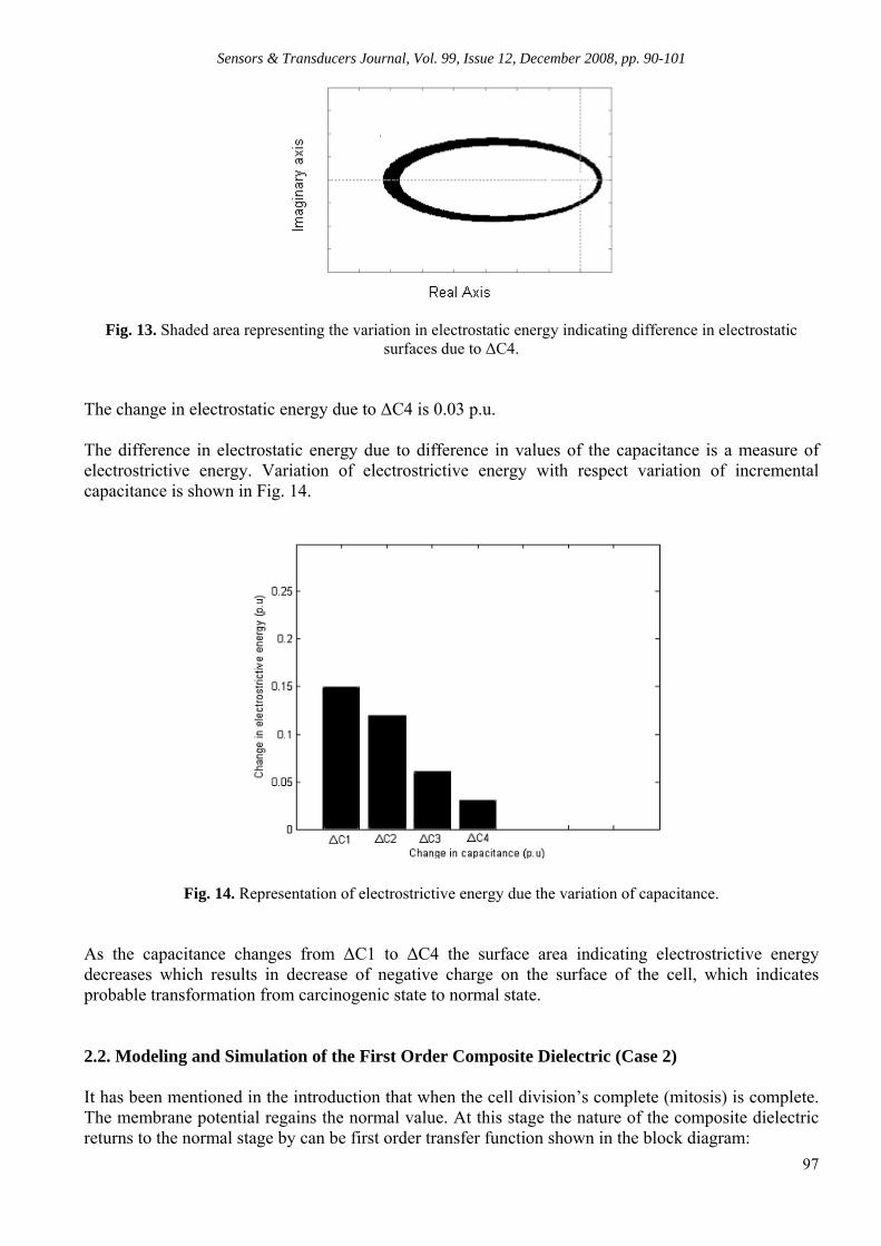
Fig. 13. Shaded area representing the variation in electrostatic energy indicating difference in electrostatic surfaces due to ∆C4.
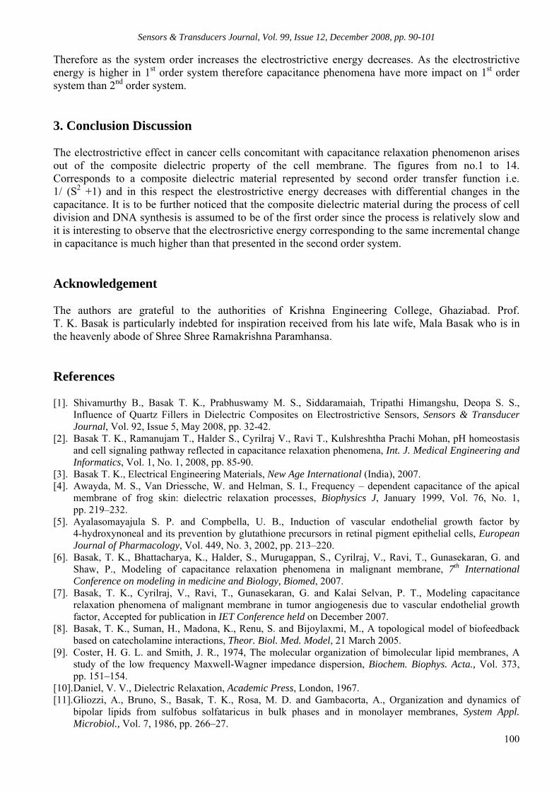
The change in electrostatic energy due to ∆C4 is 0.03 p.u. The difference in electrostatic energy due to difference in values of the capacitance is a measure of electrostrictive energy. Variation of electrostrictive energy with respect variation of incremental capacitance is shown in Fig. 14.
Fig. 14. Representation of electrostrictive energy due the variation of capacitance.
As the capacitance changes from ∆C1 to ∆C4 the surface area indicating electrostrictive energy decreases which results in decrease of negative charge on the surface of the cell, which indicates probable transformation from carcinogenic state to normal state. 2.2. Modeling and Simulation of the First Order Composite Dielectric (Case 2) It has been mentioned in the introduction that when the cell division’s complete (mitosis) is complete. The membrane potential regains the normal value. At this stage the nature of the composite dielectric returns to the normal stage by can be first order transfer function shown in the block diagram:

Sensors & Transducers Journal, Vol. 99, Issue 12, December 2008, pp. 90-101
98
From the US Patent [15] of Prof. T.K.Basak we have taken different values of capacitance as an input from capacitance from capacitance relaxation curve as shown in Fig. 15.
Fig. 15. Capacitance relaxation curve. The input of the model is restricted to different values of capacitance in the rising part of the capacitance relaxation curve i.e. from the point A to B. Here we have taken the input as increase of capacitance from 0.4 p.u to 0.5 p.u in the forward path of the rising part of the curve for the reverse path i.e. from 0.5 p.u to 0.4 p.u same process is repeated. At the initial phase of metastasis the composite dielectric is regarded as 1st order system having transfer function (1/(S+1)). The output is focused to trace out the locus for which it is possible to obtain electrostatic surfaces containing negative charges with different values of capacitances in the capacitance relaxation curve. The technique for tracing out the locus in real-imaginary axis, the Nyquist criterion has been used with the aid of MATLAB 6.5. In this way we have obtained different electrostatic surface for the forward and reverse path of the rising phase of the capacitance relaxation curve. The per unit values are taken for the normalization of a particular phenomena [8] It is stated earlier that capacitance change from 0.4 to 0.5 p.u is termed as ∆C1. The electrostatic surfaces containing negative charges obtained due to forward and reverse path of ∆C1 for the 1st order system are given in Figs. 16-18.

Sensors & Transducers Journal, Vol. 99, Issue 12, December 2008, pp. 90-101
99
Fig. 16. Electrostatic surface containing negative charge in the forward path of ∆C1.
Fig. 17. Electrostatic surface containing negative charge in the reverse path of ∆C1.
Fig. 18. Shaded area representing the variation in electrostatic energy indicating difference in electrostatic surfaces due to ∆C1.
The change in electrostatic energy for the 1st order system due to ∆C1 is calculated as 0.4 p.u. Therefore it has been found from the results of simulation, that the electrosrisrictive energy due to ∆C1 for the 1st order system is 0.4 p.u where as in 2nd order system the electrostrictive energy due to ∆C1 is 0.15 p.u. as the system order increases the electrostrictive energy decreases concomitant with capacitance relaxation phenomenon.
Sensors & Transducers Journal, Vol. 99, Issue 12, December 2008, pp. 90-101
100
Therefore as the system order increases the electrostrictive energy decreases. As the electrostrictive energy is higher in 1st order system therefore capacitance phenomena have more impact on 1st order system than 2nd order system. 3. Conclusion Discussion The electrostrictive effect in cancer cells concomitant with capacitance relaxation phenomenon arises out of the composite dielectric property of the cell membrane. The figures from no.1 to 14. Corresponds to a composite dielectric material represented by second order transfer function i.e. 1/ (S2 +1) and in this respect the elestrostrictive energy decreases with differential changes in the capacitance. It is to be further noticed that the composite dielectric material during the process of cell division and DNA synthesis is assumed to be of the first order since the process is relatively slow and it is interesting to observe that the electrosrictive energy corresponding to the same incremental change in capacitance is much higher than that presented in the second order system. Acknowledgement The authors are grateful to the authorities of Krishna Engineering College, Ghaziabad. Prof. T. K. Basak is particularly indebted for inspiration received from his late wife, Mala Basak who is in the heavenly abode of Shree Shree Ramakrishna Paramhansa. References [1]. Shivamurthy B., Basak T. K., Prabhuswamy M. S., Siddaramaiah, Tripathi Himangshu, Deopa S. S.,
Influence of Quartz Fillers in Dielectric Composites on Electrostrictive Sensors, Sensors & Transducer Journal, Vol. 92, Issue 5, May 2008, pp. 32-42.
[2]. Basak T. K., Ramanujam T., Halder S., Cyrilraj V., Ravi T., Kulshreshtha Prachi Mohan, pH homeostasis and cell signaling pathway reflected in capacitance relaxation phenomena, Int. J. Medical Engineering and Informatics, Vol. 1, No. 1, 2008, pp. 85-90.
[3]. Basak T. K., Electrical Engineering Materials, New Age International (India), 2007. [4]. Awayda, M. S., Van Driessche, W. and Helman, S. I., Frequency – dependent capacitance of the apical
membrane of frog skin: dielectric relaxation processes, Biophysics J, January 1999, Vol. 76, No. 1, pp. 219–232.
[5]. Ayalasomayajula S. P. and Compbella, U. B., Induction of vascular endothelial growth factor by 4-hydroxynoneal and its prevention by glutathione precursors in retinal pigment epithelial cells, European Journal of Pharmacology, Vol. 449, No. 3, 2002, pp. 213–220.
[6]. Basak, T. K., Bhattacharya, K., Halder, S., Murugappan, S., Cyrilraj, V., Ravi, T., Gunasekaran, G. and Shaw, P., Modeling of capacitance relaxation phenomena in malignant membrane, 7th International Conference on modeling in medicine and Biology, Biomed, 2007.
[7]. Basak, T. K., Cyrilraj, V., Ravi, T., Gunasekaran, G. and Kalai Selvan, P. T., Modeling capacitance relaxation phenomena of malignant membrane in tumor angiogenesis due to vascular endothelial growth factor, Accepted for publication in IET Conference held on December 2007.
[8]. Basak, T. K., Suman, H., Madona, K., Renu, S. and Bijoylaxmi, M., A topological model of biofeedback based on catecholamine interactions, Theor. Biol. Med. Model, 21 March 2005.
[9]. Coster, H. G. L. and Smith, J. R., 1974, The molecular organization of bimolecular lipid membranes, A study of the low frequency Maxwell-Wagner impedance dispersion, Biochem. Biophys. Acta., Vol. 373, pp. 151–154.
[10]. Daniel, V. V., Dielectric Relaxation, Academic Press, London, 1967. [11]. Gliozzi, A., Bruno, S., Basak, T. K., Rosa, M. D. and Gambacorta, A., Organization and dynamics of
bipolar lipids from sulfobus solfataricus in bulk phases and in monolayer membranes, System Appl. Microbiol., Vol. 7, 1986, pp. 266–27.

Sensors & Transducers Journal, Vol. 99, Issue 12, December 2008, pp. 90-101
101
[12]. Kell, D. B. and Harris C. M., On the dielectrically observable consequences of the diffusional motions of lipids and membranes, I. Theory and overview. Eur. Biophysics J., Vol. 12, 1985, pp. 181–197.
[13]. Pething, R., Dielectric and electronic properties of bio logical materials, John Willey & Sons, New York, 1979.
[14]. Schwan, H. P., Electrical properties of tissues and cell suspensions, in Advances in Biological and Medical Physics, Academic Press, New York, 1957, pp. 147–209.
[15]. Shaw, P., Basak, T. K. and Ghosh, N. C., Capacitance relaxation phenomena in cartilaginous membrane, Everyman’s Science, Vol. XL, No. 6, February–March 2006. Shaw, P., Basak, T. K. and Ghosh, N. C., Novel method for detecting malignancies in membrane, Science & Culture, May–June 2006, Nos. 5–6.
[16]. Aidley D. J., Stanfield P. R., Ion Channels: Molecules in Action, Cambridge, UK, Cambridge University Press, 1996.
[17]. Blad B., Baldetorp B., Impedance spectra of tumour tissue in comparison with normal tissue: A possible clinical application for electrical impedance tomography, Physiological Measurement, 17, Suppl 4A, 1996, pp. A105-115.
[18]. Charman R. A., Electrical Properties of Cells and Tissues, In Clayton’s Electrotherapy 10th edition (eds. S. Kitchen and S. Bazin), London, UK, WB Saunders Company Ltd., 1996.
[19]. Cone C. D., Variation of the transmembrane potential level as a basic mechanism of mitosis control. Oncology, 1970, 24, pp. 438-470.
[20]. Cone C. D., The role of surface electrical transmembrane potential in normal and malignant mitogenesis, Ann. NY Acad Sci., 1975, 238, pp. 420-435.
[21]. Cone C. D., Transmembrane Potentials and Characteristics of Immune and Tumor Cells. Boca Raton, Florida, CRC Press, 1985.
[22]. Cope F. W., A medical application of the Ling Association-Induction Hypothesis: The high potassium, low sodium diet of the Gerson cancer therapy, Physiol Chem Phys, 1978, 10, 5, pp. 465-468.
[23]. Cure J. C., Cancer an electrical phenomenon, Resonant, 1991, 1, 1. [24]. Cure J. C., On the electrical characteristics of cancer, Paper presented at the Second International Congress
of Electrochemical Treatment of Cancer, Jupiter, Florida, October 1995. [25]. Foster K. R., Schepps J. L., Dielectric properties of tumor and normal tissues at radio through microwave
frequencies, J. Microwave Power, 1981, 16, pp. 107-119. [26]. Oschman J. L., Energy Medicine: The Scientific Basis, Edinburgh, England, Churchill Livingstone, 2000. [27]. Revici E., Research in Pathophysiology as Basis for Guided Chemotherapy, with Special Application to
Cancer, Princeton, NJ, D. Van Nostrand Company, 1961. [28]. Seeger P. G., Wolz S., Successful Biological Control of Cancer: By Combat Against the Causes.
Gesamtherstellung, Neuwieder Verlagsgesellschaft GmbH, 1990. [29]. Stern R. G., Carcinogenesis and the plasma membrane, Med Hypotheses, 1999, May, 52, 5, pp. 367-372.
___________________
2008 Copyright ©, International Frequency Sensor Association (IFSA). All rights reserved. (http://www.sensorsportal.com)


SSeennssoorrss && TTrraannssdduucceerrss JJoouurrnnaall
GGuuiiddee ffoorr CCoonnttrriibbuuttoorrss
Aims and Scope Sensors & Transducers Journal (ISSN 1726-5479) provides an advanced forum for the science and technology of physical, chemical sensors and biosensors. It publishes state-of-the-art reviews, regular research and application specific papers, short notes, letters to Editor and sensors related books reviews as well as academic, practical and commercial information of interest to its readership. Because it is an open access, peer review international journal, papers rapidly published in Sensors & Transducers Journal will receive a very high publicity. The journal is published monthly as twelve issues per annual by International Frequency Association (IFSA). In additional, some special sponsored and conference issues published annually. Topics Covered Contributions are invited on all aspects of research, development and application of the science and technology of sensors, transducers and sensor instrumentations. Topics include, but are not restricted to: • Physical, chemical and biosensors; • Digital, frequency, period, duty-cycle, time interval, PWM, pulse number output sensors and transducers; • Theory, principles, effects, design, standardization and modeling; • Smart sensors and systems; • Sensor instrumentation; • Virtual instruments; • Sensors interfaces, buses and networks; • Signal processing; • Frequency (period, duty-cycle)-to-digital converters, ADC; • Technologies and materials; • Nanosensors; • Microsystems; • Applications. Submission of papers Articles should be written in English. Authors are invited to submit by e-mail [email protected] 6-14 pages article (including abstract, illustrations (color or grayscale), photos and references) in both: MS Word (doc) and Acrobat (pdf) formats. Detailed preparation instructions, paper example and template of manuscript are available from the journal’s webpage: http://www.sensorsportal.com/HTML/DIGEST/Submition.htm Authors must follow the instructions strictly when submitting their manuscripts. Advertising Information Advertising orders and enquires may be sent to [email protected] Please download also our media kit: http://www.sensorsportal.com/DOWNLOADS/Media_Kit_2008.pdf

Copyright © 2022 FDOKUMEN