Antimicrobial metal–organic frameworks incorporated into electrospun fibers
Upload
independentCategory
view
5download
0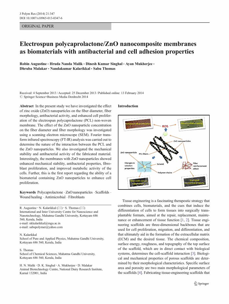
ORIGINAL PAPER
Electrospun polycaprolactone/ZnO nanocomposite membranesas biomaterials with antibacterial and cell adhesion properties
Robin Augustine & Hruda Nanda Malik & Dinesh Kumar Singhal & Ayan Mukherjee &
Dhruba Malakar & Nandakumar Kalarikkal & Sabu Thomas
Received: 4 September 2013 /Accepted: 25 December 2013 /Published online: 13 February 2014# Springer Science+Business Media Dordrecht 2014
Abstract In the present study we have investigated the effectof zinc oxide (ZnO) nanoparticles on the fiber diameter, fibermorphology, antibacterial activity, and enhanced cell prolifer-ation of the electrospun polycaprolactone (PCL) non-wovenmembrane. The effect of the ZnO nanoparticle concentrationon the fiber diameter and fiber morphology was investigatedusing a scanning electron microscope (SEM). Fourier trans-form infrared spectroscopy (FT-IR) analysis was carried out todetermine the nature of the interaction between the PCL andthe ZnO nanoparticles. We also investigated the mechanicalstability and antibacterial activity of the fabricated material.Interestingly, the membranes with ZnO nanoparticles showedenhanced mechanical stability, antibacterial properties, fibro-blast proliferation, and improved metabolic activity of thecells. Further, this is the first report regarding the ability of abiomaterial containing ZnO nanoparticles to enhance cellproliferation.
Keywords Polycaprolactone .ZnOnanoparticles . Scaffolds .
Wound healing . Antimicrobial . Fibroblasts
Introduction
Tissue engineering is a fascinating therapeutic strategy thatcombines cells, biomaterials, and the cues that induce thedifferentiation of cells to form tissues into surgically trans-plantable formats, aimed at the repair, replacement, mainte-nance or enhancement of tissue function [1, 2]. Tissue engi-neering scaffolds are three-dimensional backbones that areused for cell proliferation, migration, and differentiation, andthat ultimately aid in the formation of the extracellular matrix(ECM) and the desired tissue. The chemical composition,surface energy, roughness, and topography of the top surfaceof the scaffold, which are in direct contact with biologicalsystems, determines the cell-scaffold interaction [3]. Biologi-cal and mechanical properties of porous scaffolds are deter-mined by their morphological characteristics. Specific surfacearea and porosity are two main morphological parameters ofthe scaffolds [4]. Fabricating tissue-engineering scaffolds that
R. Augustine :N. Kalarikkal (*) : S. Thomas (*)International and Inter University Centre for Nanoscience andNanotechnology, Mahatma Gandhi University, Kottayam 686560, Kerala, Indiae-mail: [email protected]: [email protected]
N. KalarikkalSchool of Pure and Applied Physics, Mahatma Gandhi University,Kottayam 686 560, Kerala, India
S. ThomasSchool of Chemical Sciences, Mahatma Gandhi University,Kottayam 686 560, Kerala, India
H. N. Malik :D. K. Singhal :A. Mukherjee :D. MalakarAnimal Biotechnology Centre, National Dairy Research Institute,Karnal 132001, India
J Polym Res (2014) 21:347DOI 10.1007/s10965-013-0347-6

exactly mimic the structure of the extracellular matrix at thenanoscale is one of the major challenges in the field of tissueengineering and the development of nanofibers can potentiallymeet this challenge [5]. Sufficient porosity and pore size areessential to facilitate cell seeding and diffusion throughout thescaffold of both cells and nutrients [6]. According to Oh et al.,the optimum pore size and spatial distribution for proper cellgrowth and tissue regeneration vary with the type of tissue tobe constructed [7]. The scaffold should have adequate me-chanical properties compatible with those of the tissue at theimplantation site or mechanical properties that are sufficient toprotect cells from damaging compressive or tensile forceswithout affecting appropriate molecular signals [8].
The process of electrospinning enables the production ofcontinuous fibers with dimensions on the scale of nanometersor submicrons from a wide range of natural and syntheticpolymers [9]. A high surface-area-to-volume ratio and thehigh porosity on the sub-micrometer length scale make thesematerials suitable for many biomedical applications [10–14].Their applicability in regenerative medicine and tissue engi-neering has significantly increased the amount of research inthis arena. For the past few decades, a considerable effort hasbeen made by many researchers to improve the properties ofelectrospun, polycaprolactone, non-woven mats [11, 15, 16].
Since polycaprolactone (PCL) is biocompatible and biode-gradable, it has immense potential as biomaterial for variousbiomedical applications including tissue engineeringscaffolds, wound dressings, hemostats, etc [17–19].Electrospinning of PCL has been reported in many studiesover the past few years. Efforts have been made to improveboth the mechanical and morphological features ofelectrospun-PCL non-woven membranes.
The incorporation of various filler materials like nanopar-ticles and blending with other polymers have resulted insignificant improvement in the overall performance of bioma-terials. The major goal of such efforts have been aimed atreducing the fiber diameter, achieving fibers with uniformfiber diameters, and maintaining good mechanical properties.In one study, electrospun PCL fibers incorporatedwith CaCO3
or HA nanoparticles were fabricated and shown to improvecell attachment [11].
Zinc oxide (ZnO) is an inorganic compound used widely ineveryday applications. ZnO is currently listed as a ‘generallyrecognized as safe’ (GRAS) material by the Food and DrugAdministration and is used as a food additive. Nanoparticlesof ZnO and their application in coating systems have attracteda great deal of attention in recent years because of theirmultifunction properties, especially in terms of antibacterialactivity. Li et al. studied the antibacterial and physical prop-erties of poly (vinyl chloride) (PVC)-based film coated withZnO nanoparticles and found that the particles have a goodpotential to be used as an active coating system for foodpackaging [20] . Compress ion-molded PCL-ZnO
nanocomposites are already reported in the literature and aresuperior to the bare polymer in terms of increased barrierproperties and mechanical properties [21]. Microbial invasionand proliferation on biomaterials is a major problem that limitsthe success of such materials. For these reasons, while design-ing a material for medical use, it is extremely important toensure its resistance tomicrobial attack [22]. The effectivenessof polyurethane membranes with zinc oxide nanoparticles toresist both bacteria and fungi has already been reported [23].
The role of reactive oxygen species (ROS) in cell prolifer-ation and wound healing through the activation of growthfactors has been discussed previously [24–28]. The ability ofZnO nanoparticles to generate ROS is also well-established[29–33]. A recent report suggested that ZnO nanoflowersinduced the proliferation and migration of endothelial cellsand led to the formation of new blood vessels [34].
The element zinc participates in DNA and RNA synthesis,which are directly related to cell replication, which entails thedifferentiation of cells, including fibroblasts, and cell tran-scription. Zinc is also essential to enzyme systems that influ-ence cell division and proliferation. [35, 36]. Fujiwara et al.reported that zinc enhances vascular smooth muscle cell pro-liferation induced by basic and acidic fibroblast growth fac-tors, as well as thrombospondin [37]. Endopeptidases likematrixmetaloproteinases (MMPs), which require zinc for cat-alytic activity and are capable of digesting ECM and basementmembrane components, are important for fibroblast prolifera-tion [38]. The increased expression of MMPs leads to theproteolytic breakdown of the basement membrane and ECMleading to the release of FGFs, which enhances cell prolifer-ation [39]. Sudheesh Kumar et al. reported the use of ZnOnanoparticles containing β-chitin hydrogel bandages withantimicrobial and wound healing properties. The fabricatedmaterials possessed good biocompatibility to human dermalfibroblast cells [40]. Thus, the zinc oxide in its nano-dimensions may have enhanced activity in fibroblast prolifer-ation due to its large surface area and subsequent increasedcatalytic efficacy.
In the following, we propose the design of novel tissue-engineering scaffolding materials with tunable morphology,high mechanical stability, antibacterial properties, and en-hanced tissue proliferation abilities made from electrospunmats of PCL fibers filled with ZnO nanoparticles. This is thefirst report regarding the ability of biomaterials containing ZnOnanoparticles to enhance the mammalian cell proliferation.
Materials and Methods
Materials
The polycaprolactone (Mw 70,000) used in this study waspurchased from Sigma Aldrich, St. Luis, USA. The ZnO
347, Page 2 of 17 J Polym Res (2014) 21:347

nanoparticles were of an average particle size of 60 nm andpurchased from Sigma Aldrich, St. Luis, USA. Acetone wasobtained fromMerck, Mumbai, India. All the reagents used inthis study were of analytical grade quality and therefore usedwithout further purification.
Methods
Electrospinning of Polycaprolactone with ZnO Nanoparticles
Electrospun membranes of PCL, into which we incorporatedZnO nanoparticles of particle size range∼60 nm, were pre-pared by electrospinning 15 wt.% PCL in acetone. Theelectrospinning apparatus was assembled by Holmarc, India,and consisted of a syringe pump, a high-voltage power supply,and a 10-ml syringe with an attached 21G-diameter needle.The needle-to-collector distancewasmaintained at 15 cmwithan applied voltage of 18 kV. The feeding rate of the solutionwas precisely controlled by a syringe pumping system, whichwas adjusted to a flow rate of 1 ml/h. As a collector, a thinaluminum sheet of 7 cm2 was attached to the fixed collector,which was grounded properly.
Polycaprolactone solutions with different concentration ofZnO nanoparticles were prepared in acetone. Briefly, a lowerconcentration range (0.1 to 1 wt.%) and a comparativelyhigher concentration range (1–6 wt.%) of ZnO nanoparticleswere accurately weighed and ultrasonicated for 15 min toproperly disperse in acetone. Then, a known quantity ofPCL was added to the above solution so that the final solutioncontained 15 wt.% PCL, and the mixture was stirred with amagnetic stirrer for 12 h to ensure the dissolution of the pelletsand proper mixing. About 10ml of the prepared solutions withdifferent wt% of ZnO nanoparticles were taken in plasticsyringes and electrospun individually on aluminum sheets.
Characterization
The fabricated PCL/ZnO nanocomposite membranes werecharacterized using various techniques like scanning electronmicroscopy (SEM), energy-dispersive X-ray spectroscopy(EDX), Fourier transform infrared spectroscopy (FTIR), andX-ray diffraction (XRD) analysis. Mechanical properties werealso determined using a universal testing machine accordingto ASTM standards. The antimicrobial property of the fabri-cated material was evaluated by the disc diffusion method.
Scanning Electron Microscopy
The morphological features of the fabricated membranes wereobserved by SEM. The neat PCL membrane as well as thePCL/ZnO nanocomposite membranes were carefully sec-tioned with an approximate size of 3-mm length and 0.5-mmwidth using a sharp scissor, and then mounted onto an SEM
grid. Prior to the examination, each sample was coated withplatinum using a JEOL JFC 1600 Autofine coater. A JEOLJSM 6390 scanning electron microscope at 10 kV was used toanalyze the samples. The average fiber diameter of eachsample was quantified from the SEM image using ImageJsoftware. Measurements were made at 100 random positionsand the average of these measurements gave the diameter ofthe nanofibers.
Energy-dispersive X-ray Spectroscopy
The presence of ZnO nanoparticles in the polycaprolactonemembrane was investigated by EDX analysis using an OxfordSwift ED attached to a JEOL JSM 6390 SEM, based on theenergy and intensity distribution of X-ray signals generated bythe electron beam striking the surface of the specimen.
Fourier Transform Infrared Spectroscopy
Fourier transform infrared spectroscopy (FT-IR) spectra wereobtained with a Perkin Elmer, spectrum 400 FTIR spectrom-eter. The powders of ZnO nanoparticles, and PCL/ZnO nano-composite membranes with different wt% of ZnO nanoparti-cles were subjected to IR analysis. The spectra were collectedover a range of 400–4,000 cm-1.
X-Ray Diffraction Analysis
X-Ray diffraction (XRD) was used to understand the crystal-linity and the structure of the fabricated materials. XRD wasrecorded in the 2θ range of 5°–80° using a model D8-Advance of Bruker (Germany), of CuKα radiation, of whichthe energy was 8.04 keVand the wavelength was 1.54 Å. Theapplied voltage was 40 kVand the current was 25 mA.
Mechanical Measurements
Tensile tests of the electrospinning nanofibrous membranewere carried out with a TinusOlsen H50KTUniversal TestingMachine according to the ASTM D 882 standard by applyinga 500-N load cell at a crosshead speed of 1 mm/min. All thesamples were cut into rectangles with dimensions of 6×1 cm2
and vertically mounted in between two mechanical grippingunits of the tester, leaving a 3-cm gauge length for mechanicalloading. The sample thicknesses were measured with an elec-tronic micrometer having a precision of 1 μm. The averagevalues of tensile property were obtained from the results offive tests and expressed as the mean±standard deviation (SD).
Antimicrobial Activity: Disc Diffusion Technique
In vitro antibacterial activity of the PCL/ZnO nanocompositemembranes was evaluated by the disc diffusion method
J Polym Res (2014) 21:347 Page 3 of 17, 347

according to the National Committee for Clinical LaboratoryStandards (NCCLS, 2001). Freshly prepared Mueller-Hintonagar was poured into glass petri dishes to get a uniform depthof approximately 4 mm and allowed to cool. Escherichia coliand Staphylococcus aureus were used as standard organismsfor Gram-negative and Gram-positive bacteria respectively.Three to five well-isolated colonies of the samemorphologicaltype of both bacteria were selected from an agar plate cultureand inoculated into Mueller Hinton Broth media and incubat-ed at 35 °C until they achieved the turbidity of the 0.5McFarland standards. To standardize the inoculum density, aBaSO4 turbidity standard, equivalent to a 0.5 McFarlandstandard was used. A sterile cotton swab was dipped into thestandardized bacterial suspension. The Mueller-Hinton agarplate was inoculated by streaking the swab over the entiresterile agar surface. The discs of electrospun PCL membraneswith various concentrations of ZnO nanoparticles were cutinto approximately 6-mm-diameter pieces and placed onto thesurface of the inoculated MHA plates. Discs of PCL mem-branes without nanoparticles were maintained as controls. Theplates were incubated at 35 °C overnight to get a confluentlawn of bacterial growth.
The sensitivity of the microorganisms to membranes wasdetermined by measuring the diameter of inhibitory zones onthe agar surface around the discs. All the tests were carried outin triplicate. The diameters of the inhibition zones were mea-sured in millimeters.
Fibroblast Attachment and Proliferation
A small piece of tissue was taken from the ear of a goat usingan ear puncturing machine. The hairs were removed and thesample was washed three to four times with DPBS(Dulbecco’s phosphate-buffered saline). Then, the tissue piecewas dissected into very small pieces with a sterile scalpel andwashed with DPBS. Pieces of tissue were placed into 6-welldishes containing fibroblast culture medium (DMEM supple-mented with 10 % fetal bovine serum). According to theoutgrowth of cells, a portion of the tissue pieces was attachedand the remaining portion was removed after 2-3 days. Thesecells formed a confluent monolayer and were used for furthersubculturing. The medium from the 6-well culture dish wasdiscarded and 0.5 ml of 0.25 % trypsin-EDTA was added tothe dish and kept in a 5 % CO2 incubator for 2 min todisaggregate the cells. The cells were separated by centrifu-gation at 1,000 rpm for 5 min. The supernatant was discarded,1 ml of the fresh fibroblast medium was added, and thesuspension was plated at the rate of 500 μl into a 25-cm2
tissue culture flask containing 2 ml of fresh culture mediumand incubated in a CO2 incubator with 5 % CO2 supply under38.5 °C. The medium was replaced after each 48 h interval.Fresh passaged fibroblast cells were layered down in the six-well culture dish containing polycaprolactone membranes
with different concentrations of ZnO nanoparticles and incu-bated in an incubator with 5%CO2 supply under 38.5 °C for 4days. After every 12 h, the culture plates were observed underan inverted microscope (No. X51, Olympus, Japan) and im-ages were taken using a CCD (charge-coupled device) cameraattached to the microscope.
Cell Proliferation Assay
The effects of the PCL membranes containing different con-centrations of ZnO nanoparticles on the in vitro cell prolifer-ation were assayed by XTT (2,3-bis-(2-methoxy-4-nitro-5-sulfophenyl)-2H-tetrazolium-5-carboxanilide) assay accord-ing to the manufacturer’s instruction (XTT cell proliferationassay kit, catalog No. 10010200, Cayman Chemical Compa-ny, MI, USA) with some minor modifications. XTT assay isused to assess cell viability as a function of redox potential.Actively respiring cells convert the water-soluble XTT to awater-soluble, orange colored formazan product. Briefly, me-dium from the six-well culture dish containing membraneswas discarded and 0.5 ml of 0.25 % trypsin-EDTAwas addedto the dish and kept in a 5 % CO2 incubator for 2 min todisaggregate the cells. The cells were pelleted by centrifuga-tion at 1,000 rpm for 5 min and the supernatant was discarded.The fresh fibroblast medium was added and the suspensionwas plated in a 96-well plate at a density of 0.5×10⁵ cells/wellin 100 μl of culture medium for 24 h at 37 °C in a CO2
incubator. Then, 10 μl of the reconstituted XTT mixture wasadded to each well and mixed gently for 1 min in an orbitalshaker. The cells were incubated at 37 °C in a CO2 incubatorfor 2 h. The absorbance of each sample was measured at awavelength of 450 nm in a NanoQuant microplate reader.
The results from the three individual experiments wereaveraged, expressed as the mean±standard deviation (SD)and statistically analyzed using a t-test. A p value less than0.05 was considered statistically significant.
Results and Discussion
Effect of ZnO Concentration on Fiber Morphology
Analyzing the effect of ZnO nanoparticles on the fiber mor-phology and structure revealed the spider web structure of themembranes (Figs. 1, 2, 3, 4, 5, 6, 7, 8, 9, 10 and 11). It may beobserved from the micrographs that the resulting PCL andPCL-ZnO nanocomposite fiber membranes are almost uni-form in fiber diameter and are highly porous.
The effect of ZnO nanoparticles of various concentrations, alower range of 0.1–0.9 wt.%, and a higher range of 1–6 wt.%significantly changed the fiber morphology provided that otherexperimental parameters remained constant. In Figs. 1–11,these morphological changes are clearly observable. Figure 1
347, Page 4 of 17 J Polym Res (2014) 21:347

shows the SEM image of a neat PCL membrane where thesurface of individual fibers is very smooth. As the ZnO nano-particle content in the fibers increases, the surface of fibersbecomes rougher due to the agglomerates of ZnO nanoparti-cles. From 2 wt.% ZnO concentration and onward, the effect isvery evident (Figs. 6, 7 and 8), which was in agreement withthe results obtained by Patcharaporn et al [11].
Some special morphological features like highly intercon-nected fibers (Fig. 9) and individual fibers with secondarypores (Fig. 11) were also observed. These features can be
explained in terms of the changes in the viscosity of thespinning solution. The interconnected fiber formation occurswhen the fibers dry, only after reaching them on the collector.Many other studies also reported observing the secondarypore formation on the individual fibers [41, 42]. This mightbe due to the breath figure formation, which is directly relatedto ambient factors like temperature, viscosity of the solution,nature of the solvent, and humidity. Fast evaporation ofhighly-volatile solvents like acetone from the surface of ejac-ulated polymer solution makes it cold and leads to the
Fig. 1 Scanning electronmicrograph of electrospun neat polycaprolactonemembrane (a), fiber diameter distribution (b), and the pore space distribution (c)
Fig. 2 Scanning electron micrograph of electrospun polycaprolactone membrane with 0.2 wt.% ZnO nanoparticles (a), fiber diameter distribution (b),and the pore space distribution (c)
J Polym Res (2014) 21:347 Page 5 of 17, 347
condensation of water vapor on the surface. During the evap-oration of condensed water, droplets from the surface of fibersleave an imprint on the surface, reported as breath fig-ures, which have been extensively studied by variousother researchers [42–47].
The challenges of particle dispersion and agglomerationwere encountered during the electrospinning in our study,when the ZnO nanoparticle loading was increased above
6 wt.%. At this concentration, formation of the continuouspolymer solution jet and subsequent fiber formation becamedifficult due to the high viscosity of the solution.
Effects of ZnO Nanoparticles on Fiber Diameter
The fiber diameter of the electrospun submicron fibers wasobserved by SEM and calculated using ImageJ software.
Fig. 3 Scanning electron micrograph of electrospun polycaprolactone membrane with 0.5 wt.% ZnO nanoparticles (a), fiber diameter distribution (b),and the pore space distribution (c)
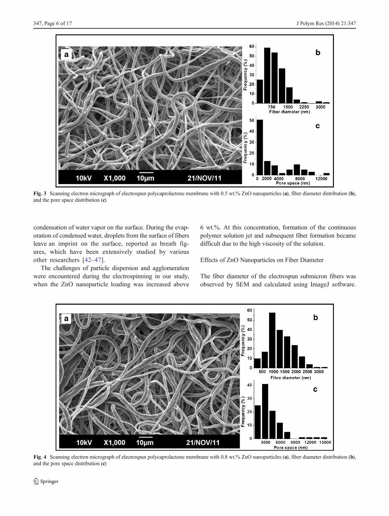
Fig. 4 Scanning electron micrograph of electrospun polycaprolactone membrane with 0.8 wt.% ZnO nanoparticles (a), fiber diameter distribution (b),and the pore space distribution (c)
347, Page 6 of 17 J Polym Res (2014) 21:347
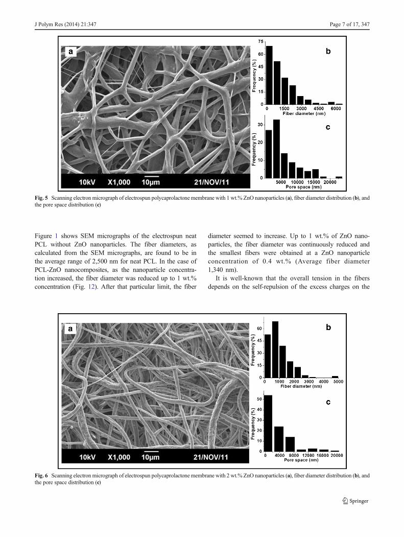
Figure 1 shows SEM micrographs of the electrospun neatPCL without ZnO nanoparticles. The fiber diameters, ascalculated from the SEM micrographs, are found to be inthe average range of 2,500 nm for neat PCL. In the case ofPCL-ZnO nanocomposites, as the nanoparticle concentra-tion increased, the fiber diameter was reduced up to 1 wt.%concentration (Fig. 12). After that particular limit, the fiber
diameter seemed to increase. Up to 1 wt.% of ZnO nano-particles, the fiber diameter was continuously reduced andthe smallest fibers were obtained at a ZnO nanoparticleconcentration of 0.4 wt.% (Average fiber diameter1,340 nm).
It is well-known that the overall tension in the fibersdepends on the self-repulsion of the excess charges on the
Fig. 5 Scanning electron micrograph of electrospun polycaprolactone membrane with 1 wt.% ZnO nanoparticles (a), fiber diameter distribution (b), andthe pore space distribution (c)
Fig. 6 Scanning electron micrograph of electrospun polycaprolactone membrane with 2 wt.% ZnO nanoparticles (a), fiber diameter distribution (b), andthe pore space distribution (c)
J Polym Res (2014) 21:347 Page 7 of 17, 347

jet. The addition of ZnO nanoparticles resulted in the accu-mulation of a higher charge density on the surface of theejected jet during the process of electrospinning, and theoverall electric charges carried by the electrospinning jetsignificantly increased [48]. As the charges carried by the jetincreased, higher elongation forces that could overcome theself-repulsion were brought down to the jet under the electricalfield. Thus, as the charge density increased, the diameter of thefinal fibers became substantially smaller and the diameter
distribution of fibers became narrower [49]. At higher con-centrations of the filler, the viscosity of the solution tended toincrease, which led to the apparent increase in fiber diameter[50–52].
Energy-dispersive X-ray Spectroscopy (EDX)
The EDX spectrum of PCL and PCL/ZnO nano-compositemembranes confirmed the presence of the ZnO nanoparticles
Fig. 7 Scanning electron micrograph of electrospun polycaprolactone membrane with 4 wt.% ZnO nanoparticles (a), fiber diameter distribution (b), andthe pore space distribution (c)
Fig. 8 Scanning electron micrograph of electrospun polycaprolactone membrane with 6 wt.% ZnO nanoparticles (a), fiber diameter distribution (b), andthe pore space distribution (c)
347, Page 8 of 17 J Polym Res (2014) 21:347

trapped within the PCL matrix. It also confirmed that the ZnOnanoparticles reached the collector along with the polymersolution during the electrospinning process. Spectra of neatPCL membrane and ZnO nanoparticle-incorporated PCLmembranes at varying filler content are shown in Fig. 13. Inthe case of neat PCL, there are some sharp, low-energy peaksthat correspond to the elements carbon and oxygen (Fig. 13a),whereas, in the case of PCL/ZnO composite membranes, threeadditional peaks could be observed at the energy levels 1 keV,8.5 keV, and 9 keV, which are the characteristics of zinc.While using 0.2 wt.% ZnO nanoparticle concentration, thepeak intensities corresponding to zinc seemed to be very low,and one at the 9.5 Kev level could not be detected. This can beattributed to the fact that elements in low abundance willgenerate X-ray peaks that may not be resolvable from thebackground radiation. Moreover, there was a remarkable in-crease in the intensity of the three characteristic peaks of zinc,as well as one peak of oxygen as we increased the ZnOnanoparticle content from 0.2 to 6 wt.% (Fig. 13b–f).
FT-IR Analysis of the Fabricated Membrane
FT-IR analysis of polycaprolactone/ZnO nanocompositemembranes revealed the incorporation of ZnO into the PCL
matrix. Figure 14 shows the FT-IR spectra of ZnO,polycaprolactone, and PCL/ZnO nanocomposites. FT-IRspectrum of neat PCL shows an intense peak at 1,721 cm-1 ,which is due to the presence of the ester carbonyl group thatcorresponds to the –CO (stretching) in the PCL polymer. Thepeaks at 2,869 and 2,940 cm-1 are related to the C–H bond ofsaturated carbons. The ZnO/polycaprolactone composite andpolycaprolactone alone presented the same spectra in the wavenumber range of 700–3,100 cm-1 , while the peak located at500–700 cm-1 is attributed to the stretching of the Zn–O bond.
The FTIR spectrum of ZnO has shown nearly 100 %transmittance. The neat PCL has around 94 % transmittance.Interestingly, the composite has a transmittance range in be-tween that of pure ZnO nanoparticles and neat PCL. As theZnO content in the composite increased, transmittance ofalmost all the peaks other than those in the range 500–700 cm-1 tended to increase. Furthermore, the decrease inthe intensity of the carbonyl stretching vibrations at 1,718-1,721 cm-1 indicates the interaction between the carbonyl
Fig. 9 Interconnected fiber web formed at 1 wt. v% of ZnO nanoparticleconcentration
Fig. 10 Formation of aggregates of ZnO nanoparticles on PCL fibers at6 wt.% ZnO nanoparticle content
Fig. 11 SEM image of secondary pore formation on fibers at 2 wt.% ofZnO nanoparticle concentration
0 1 2 3 4 5 61200
1400
1600
1800
2000
2200
2400
2600
2800
Dia
met
er o
f fib
ers
(nm
)
ZnO nanoparticle concentration (Wt%)
Fig. 12 Effect of ZnO concentration on the fiber diameter
J Polym Res (2014) 21:347 Page 9 of 17, 347

groups of PCL with ZnO nanoparticles. This physical inter-action may weaken the covalent C=O double bond. Thismight be the reason for the decrease of intensity of the peak
at 1,718-1,721 cm-1. Additionally, there was a small shift inthe peak position to a lower energy level as the concentrationof ZnO nanoparticles increased (Fig. 14(b)), revealing the
Fig. 13 EDX (Energy-dispersive X-ray spectrum) of neat PCLmembrane (a), PCLmembranes with 0.2 wt.% (b), 1 wt.% (c), 2 wt.% (d), 4 wt.% (e) and6 wt.% (f) ZnO nanoparticle concentrations
1760 1740 1720 1700
20
30
40
50
60
70
80
90
100
% T
Wavenumber (cm-1)
ZnO 6% 2% 0.5% PCL
1721
1720
1719
1718
4000 3500 3000 2500 2000 1500 1000 500
40
60
80
100
% T
Wavenumber (cm-1)
ZnO 6% 2% 0.5% PCL
2940
2869
17211158
a b
Fig. 14 FT-IR spectra of neat PCL, ZnO nanoparticles and the composites of both at varying concentrations of ZnO nanoparticles (a). Decrease in theintensity of carbonyl vibrations as the ZnO concentration increases (b)
347, Page 10 of 17 J Polym Res (2014) 21:347

interaction of ZnO nanofillers with the PCL polymer chains.The interaction might be of physical interactions like VanderWaals forces that probably weaken the strength of the esterbond present in PCL, as shown in Scheme 1.
X-ray Diffraction Analysis
The effect of ZnO nanoparticles on the crystalline behavior ofelectrospun polycaprolactone nanofibers was studied by X-raydiffraction (XRD) analysis. The XRD patterns of the neat PCLmembrane and ZnO/PCL nanocomposites are shown in Fig. 15.
The results show that the neat PCL membrane containsthree distinct reflections at the Bragg angles of about 21.4,
22.0 and 23.7°, corresponding to the (110), (111), and (200)planes of the orthorhombic crystal structure, respectively [21].The behavior of the dominant crystalline phase in the PCL/ZnO nanocomposites can be predicted from the XRD patternsin order to indicate the change in the dominant crystallinephase with increasing ZnO content. The crystallinity of poly-mer matrix usually tends to decrease with the addition ofnanofillers, and the amorphous phase increases accordingly[53]. In the case of neat PCL, a clear and sharp diffractionpeak is evident at 2θ=21.2°, 22.0°, and 23.7°, illustrating thesemi-crystalline nature of neat PCL. The addition of filler upto 1 wt.% resulted in no significant variation in the diffractionpeaks. However, compared with the neat PCL nanofibermembrane, the diffraction peaks broaden as a result of thereduction in crystallinity and the increase in amorphous natureat higher filler content, which might be due to the complexa-tion between polymer and filler [12]. At a ZnO content of0.2 wt.% in the PCL matrix, the peak area of all the threecrystalline peaks slightly decreased, but increasing the ZnOcontent to more than 1 wt.% resulted in a drastic change incrystalline behavior. This clearly indicates that the crystallinenature of PCL decreased with increasing ZnO concentra-tion to a great extent. This was due to the fact that themobility of the PCL chain was restricted by ZnO aggre-gates, such that it was difficult for PCL to crystallize,resulting in a decrease in the crystallinity of PCL in thecomposite. Lim et al. reported that electrospun PCLnanofibers with smaller diameters have a higher degreeof molecular orientation, crystallinity, stiffness, andstrength. According to their work, nanofiber diameterand the resulting crystalline morphology are influencedby whether complete crystallization of polymer chainstook place before or after the electrospinning jet reachedthe collector. The former result leads to the formation ofsmaller fibers with fibrillar structure and aligned lamel-lae, but the latter leads to the formation of a misalignedlamellar structure [54]. A similar trend can be observedhere as well. At a lower concentration of ZnO, a non-woven membrane with smallest diameter is obtained
Scheme 1 Diagrammaticrepresentation of theinteraction of ZnOnanoparticles withPCL chains
30 60
0
200
400
600
800
0
10
20
300
20
40
-200
20406080
100-10
0
10
20
30
4030 60
Inte
nsity
()a
.u)
2 Theta (Degrees)
ZnO nanoparticles
PCL with 4% ZnO
PCL with 2% ZnO
PCL alone
PCL with 0.5% ZnO
Fig. 15 XRD analysis of PCL and PCL/ZnO nanocomposites
J Polym Res (2014) 21:347 Page 11 of 17, 347
(Fig. 12). Up to 1 % ZnO concentration, smaller fiberswere obtained compared to those seen in the neat PCLand these fibers showed a comparatively higher crystal-linity than at the higher ZnO content where larger fiberswere obtained. A diagrammatic representation of thedispersion of ZnO nanoparticles at lower and higherconcentrations are depicted in Scheme 2.
Mechanical Properties
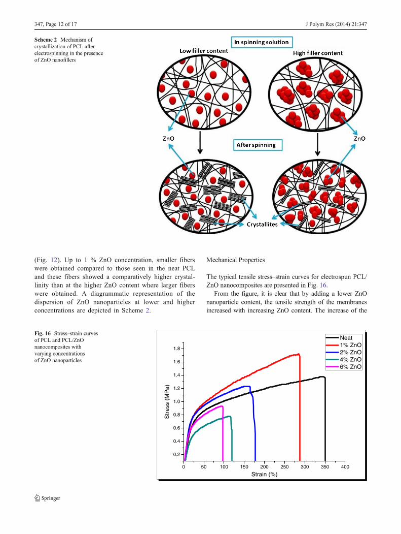
The typical tensile stress–strain curves for electrospun PCL/ZnO nanocomposites are presented in Fig. 16.
From the figure, it is clear that by adding a lower ZnOnanoparticle content, the tensile strength of the membranesincreased with increasing ZnO content. The increase of the
Scheme 2 Mechanism ofcrystallization of PCL afterelectrospinning in the presenceof ZnO nanofillers
0 50 100 150 200 250 300 350 400
0.2
0.4
0.6
0.8
1.0
1.2
1.4
1.6
1.8
Str
ess
(MP
a)
Strain (%)
Neat1% ZnO2% ZnO4% ZnO6% ZnO
Fig. 16 Stress–strain curvesof PCL and PCL/ZnOnanocomposites withvarying concentrationsof ZnO nanoparticles
347, Page 12 of 17 J Polym Res (2014) 21:347

tensile modulus of the composite proceeds linearly with theZnO content up to 1 wt.%, while further increasing the ZnOnanoparticle content (2, 4, and 6 wt.%) led to a decrease intensile strength of the PCL/ZnO nanocomposite membranes.The smaller weight percentage of the ZnO nanoparticles re-sulted in a strong reinforcing effect, increasing the tensilemodulus and inhibiting polymer drawing. The PCL mem-brane with 1 wt.% ZnO nanoparticles showed superior tensilestrength compared with the neat PCL membrane. At higherZnO content, the tensile behavior reverses and all the mechan-ical properties seemed to be retarded. This is due to the factthat nanoparticles have high surface energy and are easy toaggregate, which leads to the poor dispersion of them inpolymer matrix. As the content of ZnO increased, this tenden-cy was also increased and the aggregates of ZnO formed in thepolymer matrix acted as stress concentration centers thatprevented the stress transfer from the polymer matrix to thefillers. Thus, at lower percentages of ZnO content, there willbe a uniform dispersion of ZnO in PCL polymer matrix,provided there is more uniform stress distribution, minimizedformation of stress-concentration centers, increased interfacialarea for stress transfer from the polymer matrix to the fillers,and good mechanical properties [54, 55]. Also, the PCL–ZnOnanofibers with less than 1 wt.% filler content showed thefinest fiber sizes, and since many nanofibers formmembranes,they can thus provided more contact and stronger cohesionamong the fibers. Furthermore, as mentioned earlier, increas-ing the filler content above 1 wt.% led to a drastic decrease inthe crystallinity of the polymer. Crystalline polymers arestronger than amorphous ones. Electrospun polycaprolactonenanofibers with smaller diameters have a higher degree ofcrystallinity, molecular orientation, strength, and stiffness,but lower ductility [54]. A similar trend has been observedin our investigation for ZnO concentration of up to 1 wt.%, inwhich range the non-woven membrane with the smallestdiameter was obtained (Fig. 12). From the tensile testing(Table 1) it is clear that the modulus was higher for thissample. Maximum elongation (%), modulus, and break stressalso showed similar trends. Nanofibers had a randomly
oriented distribution in the membrane. When the membranewas uniaxially stretched, only fibers along the elongationdirection were stretched; fibers of the other direction generallyslid and turned to the elongation direction, and then alsostretched during this process (Fig. 17).
Thus, the observed irregularities in the stress–strain curveand the modulus might be due to this random orientation offibers [54]. These results indicated that nanocomposite fiberswith lower ZnO content, less agglomeration, and finest fibersizes would produce fibers with better mechanical properties.
Antibacterial Properties
The antibacterial activity of the ZnO nanoparticle-filledpolycaprolactone fiber mats was assessed by observing theiractivity (based on the disc diffusion method) against bothGram-negative (Escherichia coli) and Gram-positive (Staph-ylococcus aureus) bacteria. The activity of the neat PCLmembranes against these bacteria was used as a control. Theresults of the antimicrobial activity analysis are shown inFig. 18 and Table 2.
From Fig. 18, it is clear that the fabricated membrane hadgood antimicrobial activity against both E. coli and S. aureus.According to the results obtained, neat PCL membranes andfiber mats with a ZnO nanoparticle content less than 5 wt.%,showed no activity against the tested bacteria. The PCL mem-brane containing 5 % ZnO nanoparticles showed statisticallysignificant antibacterial activity with an inhibitory zone diam-eter of 8.76±1.2 (P=0.0278) and 9.98±0.6 (P=0.0082)against E. coli and S.aureus, respectively. The PCLmembranecontaining 6 % ZnO nanoparticles showed an inhibitory zonediameter of 9.81±0.8 (P=0.0095) and 10.22±1.3 (P=0.0067)against E. coli and S.aureus, respectively. The antimicrobialactivity was evident only with the 5 and 6 wt.% of ZnOnanoparticles. This was due to the fact that at lower concen-trations, the nanoparticles were trapped inside the polymermatrix and thus, those in direct contact with bacterial cellswere very few in number. The antibacterial activity of the ZnO
Table 1 Mechanical properties of electrospun PCL/ZnO nanoparticlecomposite membrane
Sample FillerconcentrationWt.%
Break stress(MPa)
Maximumelongation (%)
Modulus(MPa)
1 0 1.40±0.21 342±11 3.70±0.23
2 0.5 1.53±0.12 280±6 4.25±0.15
3 1 1.60±0.23 283±8 5.52±0.21
4 2 1.17±0.12 170±5 4.28±0.12
5 4 0.93±0.04 120±9 3.73±0.2
6 6 0.98±0.11 112±8 3.78±0.21
Fig. 17 Sliding and orientation of individual fibers in the elongationdirection during the uniaxial tensile test. Arrowmarkdenotes the directionof applied stress
J Polym Res (2014) 21:347 Page 13 of 17, 347

nanoparticles occurs only if the nanoparticles are in directcontact with bacterial cell wall. Further, in the disc diffusionmethod the antibacterial agent should be able to diffuse intothe agar medium to show the antibacterial activity. At higherconcentrations of ZnO nanoparticles, the interaction betweenthe polymer matrix and the filler will be apparently low due tohigher filler-filler interactions. Thus, there will be more free-dom for the entrapped nanoparticles to diffuse into the agarmedium and maintain a minimum concentration of ZnO toeffectively inhibit bacterial growth. The antibacterial activityof the ZnO/PCLmembranes was higher against S. aureus thanagainst E. coli. Reddy et al. have reported similar results forZnO nanoparticles [56]. The reason for such an observationcould be explained in terms of the difference in the cell wallstructure of these bacteria. The outer cell membrane of Gram-negative bacteria contains lipopolysaccharide in its outer leaf-let and phospholipids in the inner leaflet. But Gram-positivebacteria lack such a lipopolysaccharide layer. Selahattin et al.proposed that the higher susceptibility of Gram-positive bac-teria against ZnO nanoparticles could be related to differencesin cell wall structure, metabolism, cell physiology, or thedegree of contact points [57]. Many researchers have reportedthat the antibacterial activity of zinc oxide could be due todamage to the membrane of bacterial cells by hydrogen per-oxide or to the affinity between zinc oxide nanoparticles andbacterial surfaces [58–61].
Based on the results obtained from the disc diffusion tech-nique, it is clear that the fabricated PCL/ZnO nanocompositemembrane can successfully avoid bacterial growth at theimplantation site.
Effects of Different Nanoparticle Concentrationson the Proliferation of Adult Goat Fibroblast Cells
The effects of ZnO nanoparticle concentration on cell prolif-eration are shown in Figs. 19, 20, and 21. From the visibleobservation of the membranes, it was clear that the color of themembrane changed from white to pale pink due to the attach-ment and proliferation of fibroblasts through the membrane(Fig. 19).
Both the membranes containing ZnO nanoparticles 0.5 %and 1 % appeared to be more pinkish due to the enhancedgrowth of cells (19(A) and 19(B)). In comparison with lower
Fig. 18 Plates showing theantibacterial activity of thefabricated PCL membranes withdifferent concentrations of ZnOnanoparticles against E. coli (plate(a)) and S. aureus (plate (b)). Inboth plates are 2 wt.% (a), 3 wt.%(b), 4 wt.% (c), 5 wt.% (d), and6 wt.% (e) ZnO nanoparticles, andPCL membrane alone (f)
Table 2 Inhibition zone diameter from disc diffusion method
Sample Inhibition zone diameter (mm)
E. coli S. aureus
Neat PCL membrane 6.00±0 6.00±0
PCL/2 % ZnO 6.00±0 6.00±0
PCL/3 % ZnO 6.00±0 6.00±0
PCL/4 % ZnO 6.00±0 6.00±0
PCL/5 % ZnO 8.76±1.2 9.98±0.6
PCL/6 % ZnO 9.81±0.8 10.22±1.3
Fig. 19 Photograph showing the growth of adult goat fibroblast cells onthe electrospun membranes after 24 h of culture. PCL membranes with0.5 % ZnO nanoparticles (a), 1 % ZnO nanoparticles (b), 2 % ZnOnanoparticles (c) and the PCL membrane without nanoparticles (d)
347, Page 14 of 17 J Polym Res (2014) 21:347
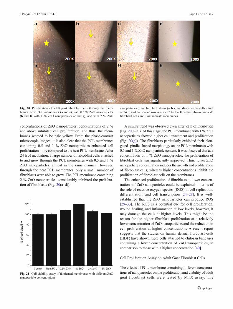
concentrations of ZnO nanoparticles, concentrations of 2 %and above inhibited cell proliferation, and thus, the mem-branes seemed to be pale yellow. From the phase-contrastmicroscopic images, it is also clear that the PCL membranescontaining 0.5 and 1 % ZnO nanoparticles enhanced cellproliferation more compared to the neat PCLmembrane. After24 h of incubation, a large number of fibroblast cells attachedto and grew through the PCL membranes with 0.5 and 1 %ZnO nanoparticles, almost in the same manner. However,through the neat PCL membranes, only a small number offibroblasts were able to grow. The PCL membrane containing2 % ZnO nanoparticles considerably inhibited the prolifera-tion of fibroblasts (Fig. 20(a–d)).
A similar trend was observed even after 72 h of incubation(Fig. 20(e–h)). At this stage, the PCLmembrane with 1 % ZnOnanoparticles showed higher cell attachment and proliferation(Fig. 20(g)). The fibroblasts particularly exhibited their elon-gated spindle-shapedmorphology on the PCLmembranes with0.5 and 1% ZnO nanoparticle content. It was observed that at aconcentration of 1 % ZnO nanoparticles, the proliferation offibroblast cells was significantly improved. Thus, lower ZnOnanoparticle concentration induces the growth and proliferationof fibroblast cells, whereas higher concentrations inhibit theproliferation of fibroblast cells on the membranes.
The enhanced proliferation of fibroblasts at lower concen-trations of ZnO nanoparticles could be explained in terms ofthe role of reactive oxygen species (ROS) in cell replication,differentiation, and cell transcription [24–28]. It is well-established that the ZnO nanoparticles can produce ROS[29–33]. The ROS is a potential cue for cell proliferation,wound healing, and inflammation at low levels, however, itmay damage the cells at higher levels. This might be thereason for the higher fibroblast proliferation at a relativelylower concentration of ZnO nanoparticles and the reduction incell proliferation at higher concentrations. A recent reportsuggests that the studies on human dermal fibroblast cells(HDF) have shown more cells attached to chitosan bandagescontaining a lower concentration of ZnO nanoparticles, incomparison to those with a higher concentration [40].
Cell Proliferation Assay on Adult Goat Fibroblast Cells
The effects of PCL membrane containing different concentra-tions of nanoparticles on the proliferation and viability of adultgoat fibroblast cells were tested by MTX assay. The
Fig. 20 Proliferation of adult goat fibroblast cells through the mem-branes. Neat PCL membranes (a and e), with 0.5 % ZnO nanoparticles(b and f), with 1 % ZnO nanoparticles (c and g), and with 2 % ZnO
nanoparticles (dand h). The first row (a, b, c, and d) is after the cell cultureof 24 h, and the second row is after 72 h of cell culture. Arrows indicatefibroblast cells and stars indicate membranes
Control Neat PCL 0.5% ZnO 1% ZnO 2% znO 6% ZnO0
20
40
60
80
100
120
% o
f cel
l via
bilit
y
Fig. 21 Cell viability assay of fabricated membranes with different ZnOnanoparticle concentrations
J Polym Res (2014) 21:347 Page 15 of 17, 347
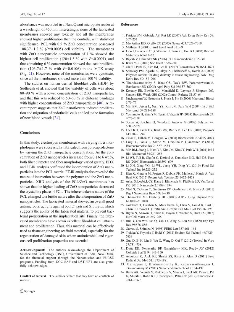
absorbance was recorded in a NanoQuant microplate reader ata wavelength of 450 nm. Interestingly, none of the fabricatedmembranes showed any toxicity and all the membranesshowed higher proliferation than the controls, with statisticalsignificance. PCL with 0.5 % ZnO concentration possessed108.37±1.2 % (P=0.0005) cell viability . The membraneswith ZnO nanoparticle concentration of 1 % showed thehighest cell proliferation (120±1.5 % with P<0.0001), andthat containing 6 % concentration showed the least prolifera-tion (103.7±1.7 % with P=0.0106) in the MTX assay(Fig. 21). However, none of the membranes were cytotoxic,since all the membranes showed more than 100 % viability.
The studies on human dermal fibroblast cells (HDF) bySudheesh et al. showed that the viability of cells was about80–90 % with a lower concentration of ZnO nanoparticles,and that this was reduced to 50–60 % in chitosan bandageswith higher concentrations of ZnO nanoparticles [40]. A re-cent report suggests that ZnO nanoflowers induced prolifera-tion and migration of endothelial cells and led to the formationof new blood vessels [34].
Conclusions
In this study, electrospun membranes with varying fiber mor-phologies were successfully fabricated from polycaprolactoneby varying the ZnO nanoparticle concentration. As the con-centration of ZnO nanoparticles increased from 0.1 to 6 wt.%,both fiber diameter and fiber morphology varied greatly. EDXand FT-IR analysis confirmed the incorporation of ZnO nano-particles into the PCLmatrix. FT-IR analysis also revealed thenature of interaction between the polymer and the ZnO nano-particles. XRD analysis of the PCL/ZnO membranes hasshown that the higher loading of ZnO nanoparticles decreasedthe crystalline phase of PCL. The inherent elastic nature of thePCL changed to a brittle nature after the incorporation of ZnOnanoparticles. The fabricated material showed an overall goodantimicrobial activity against both E. coliand S. aureus,whichsuggests the ability of the fabricated material to prevent bac-terial proliferation at the implantation site. Finally, the fabri-cated membranes have shown excellent fibroblast cell attach-ment and proliferation. Thus, this material can be effectivelyused as tissue-engineering scaffold material, especially for theregeneration of damaged skin where antimicrobial and rigor-ous cell proliferation properties are essential.
Acknowledgments The authors acknowledge the Department ofScience and Technology (DST), Government of India, New Delhi,for the financial support through the Nanomission and PURSEprograms. Funding from UGC SAP and DST-FIST are also grate-fully acknowledged.
Conflict of Interest The authors declare that they have no conflicts ofinterest.
References
1. Patrícia BM, Gabriela AS, Rui LR (2007) Adv Drug Deliv Rev 59:207–233
2. MacArthur BD, Oreffo RO (2005) Nature 433:7021–70193. Mathieu H (2001) J Surf Interf Anal 32:3–94. Li WJ, Laurencin CT, Caterson EJ, Tuan RS, Ko FKJ (2002) Biomed
Mater Res 60:613–6215. Rajesh V, Dhirendra SK (2006) Int J Nanomedicine 1:15–306. Ikada YJR (2006) Soc Interf 3:589–6017. Oh SH, Park IK, Kim JM, Lee JH (2007) Biomaterials 28:1664–16718. Sokolsky PM, Agashi K, Olaye A, Shakesheff K, Domb AJ (2007)
Polymer carriers for drug delivery in tissue engineering. Adv DrugDeliv Rev 59:187–206
9. Thandavamoorthy S, Bhat GS, Tock RW, Parameswaran S,Ramkumar SSJ (2005) Appl Poly Sci 96:557–569
10. Kenawy ER, Bowlin GL, Mansfield K, Layman J, Simpson DG,Sanders EH, Wnek GEJ (2002) Control Release 81:57–64
11. Patcharaporn W, Neeracha S, Prasit P, Pitt S (2006) Macromol Biosci6:70–77
12. Min BM, Jeong L, Nam YS, Kim JM, Park WH (2004) Int J BiolMacromol 34:281–288
13. Yoshimoto H, Shin YM, Terai H, Vacanti JP (2003) Biomaterials 24:2077–2082
14. Seema A, Joachim H, Wendorff, Andreas G (2008) Polymer 49:5603–5621
15. Leea KH, Kimb HY, Khilb MS, Rab YM, Lee DR (2003) Polymer44:1287–1294
16. Cevat E, Dilhan M, Hongjun W (2008) Biomaterials 29:4065–407317. Luigi C, Paola L, Mario M, Orsolina P, Gianfranco P (2008)
Biomacromolecules 9:1527–153418. Min BM, Jeong L, NamYS, Kim JM, Kim JY, ParkWH (2004) Intl J
Biol Macromol 34:281–28819. Li WJ, Tuli R, Okafor C, Derfoul A, Danielson KG, Hall DJ, Tuan
RS (2004) Biomaterials 26:599–60920. Li XH, Xing YG, Li WL, Jiang YH, Ding YL (2010) Food Sci
Technol Int 16:225–23221. Elen K, Murariu M, Peeters R, Dubois PH, Mullens J, Hardy A, Van
Bael MK (2012) Polym Adv Technol 23:1422–142822. Aslan S, Loebick CZ, Kang S, ElimelechM, Pfefferle LD, Van Tassel
PR (2010) Nanoscale 2:1789–179423. Vlad S, Ciobanu C, Gradinaru RV, Gradinaru LM, Nistor A (2011)
Dig J Nanomater Bios 6:921–93024. Thannickal VJ, Fanburg BL (2000) AJP - Lung Physiol 279:
6L1005–6L102825. Goldkorn T, Balaban N, Matsukuma K, Chea V, Gould R, Last J,
Chan C, Chavez C (1998) Am J Respir Cell Mol Biol 19:786–79826. Bryan N, Ahswin H, Smart N, Bayon Y, Wohlert S, Hunt JA (2012)
Eur Cell Mater 24:249–26527. Huo Y, Qiu WY, Pan Q, Yao YF, Xing K, Lou MF (2009) Exp Eye
Res 89:876–88628. Gamou S, Shimizu N (1995) FEBS Lett 357:161–16429. Toduka Y, Toyooka T, Ibuki Y (2012) Environ Sci Technol 46:7629–
763630. Guo D, Bi H, Liu B, Wu Q, Wang D, Cui Y (2012) Toxicol In Vitro
27:731–73831. Dutta RK, Nenavathu BP, Gangishetty MK, Reddy AV (2012)
Colloids Surf B 94:143–15032. Ashutosh K, Alok KP, Shashi SS, Rishi S, Alok D (2011) Free
Radical Bio Med 51:1872–188133. Mariappan P, Krishnamoorthy K, Kadarkaraithangam J,
Govindasamy M (2011) Nanomed-Nanotechnol 7:184–19234. Barui AK, Veeriah V, Mukherjee S, Manna J, Patel AK, Patra S, Pal
K, Murali S, Rohit KR, Chatterjee S, Patra CR (2012) Nanoscale 4:7861–7869
347, Page 16 of 17 J Polym Res (2014) 21:347

35. Branda˜o-Neto J, Stefan V, Mendoc BB, BloiseW, Castro AV (1995)Nutr Res 15:335–358
36. Salgueiro MJ, Zubillaga MB, Lysionek AE, Caro RA, Weill R,Boccio JR (2012) Nutrition 18:510–519
37. Fujiwara Y, Kaji T (1997) Res Commun Mol Pathol Pharmacol 97:95–106
38. Newell KJ, Witty JP, Rodgers WH, Matrisian LM (1994) MolCarcinogen 10:199–120
39. Mack CF, Knox JD, Powell WC, Nagle RB, Bowden GT (1993) Int JRadiat Oncol 27:217
40. Sudheesh Kumar PT, Vinoth KL, Mincy R, Raja B, Tamura H, NairSV, Jayakumar R (2013) Pharm Res 523-537
41. Silke M, Jean SS, Bruce CD, John FR (2002) Macromolecules 35:8456–8466
42. Fashandi H, Karimi M (2012) Polymer 53:5832–584943. Park MS, Kim JK (2004) Langmuir 20(13):5347–535244. Srinivasarao M, Collings D, Philips A, Patel S (2001) Science
292(5514):79–8345. Francois B, Pitois O, Francois J (1995) Adv Mater 7(12):1041–104446. Limaye AV, Narhe RD, Dhote AM, Ogale SB (1996) Phys Rev Lett
76(20):3762–376547. Peng J, Han Y, Yang Y, Li B (2004) Polymer 45(2):447–452
48. GuipingM,Dongzhi Y, JunN (2009) PolymAdvTechnol 20:147–15049. Zong XH, Kim K, Fang DF, Ran SF, Hsiao BJ, Chu BJ (2002)
Polymer 43:4403–441250. Baumgarten PKJ (1971) Colloid Interf Sci 36:71–7951. Fong H, Chun I, Reneker DH (1999) Polymer 40:4585–459252. Doshi J, Reneker DHJ (1995) Electrostatics 35(2–3):151–16053. Li ZH, Su GY, Wang XY, Gao D (2005) S Solid State Ion 176:1903–
190854. Lim CT, Tan EPS, Ng SY (2008) Appl Phys Lett 92:141908–14191055. Moniruzzaman M, Chattopadhyay J, Billups WE, Winey KI (2007)
Nano Lett 7:1178–118556. Reddy KM, Kevin F, Jason B, Denise GW, Cory H, Alex PJ (2007)
Appl Phys Lett 90(21):1–357. Selahattin A, Kadri G, Ramazan C (1998) Tr J Med Sc 28:
595–59758. Sawai J, Kawada E, Kanou F, Igarashi H, Hashimoto A, Kokugan T,
Shimizu MJ (1996) Chem Eng Jpn 29:627–63359. Sawai J, Igarashi H, Hashimoto A, Kokugan T, Shimizu MJ (1996)
Chem Eng Jpn 29:251–25660. Yamamoto O (2001) Int J Inorg Mater 3:643–64661. Stoimenov PK, Klinger RL, Marchin GL, Klabunde KJ (2002)
Langmuir 18:6679–6686
J Polym Res (2014) 21:347 Page 17 of 17, 347
Copyright © 2022 FDOKUMEN