A gel-free 3D microfluidic cell culture system Biomaterials
8
A gel-free 3D microfluidic cell culture system Siew-Min Ong a, b , Chi Zhang a, b, c , Yi-Chin Toh a , So Hyun Kim d , Hsien Loong Foo a, c , Choon Hong Tan c, e , Danny van Noort a, * , Sungsu Park d , Hanry Yu a, b, c, f, g, h, i a Institute of Bioengineering and Nanotechnology, A*STAR, The Nanos, #04-01, 31 Biopolis Way, Singapore 138669, Singapore b NUS Graduate School for Integrative Sciences and Engineering, Centre for Life Sciences (CeLS), #05-01, 28 Medical Drive, Singapore 117456, Singapore c Graduate Program in Bioengineering, Centre for Life Sciences (CeLS), #05-01, 28 Medical Drive, Singapore 117456, Singapore d Ewha Womans University, Division of Nano Sciences,11-1 Daehyun-dong, Seodaemun-gu, Seoul 120-750, Republic of Korea e Department of Chemistry, Faculty of Science, National University of Singapore, 3 Science Drive 3, Singapore 117543, Singapore f Department of Physiology, Yong Loo Lin School of Medicine, #03-03, 2 Medical Drive, Singapore 117597, Singapore g Singapore-MIT Alliance, E4-04-10, 4 Engineering Drive 3, Singapore 117576, Singapore h NUS Tissue-Engineering Programme, DSO Labs, National University of Singapore, Singapore 117597, Singapore i Department of Haematology-Oncology, National University Hospital, Singapore 119074, Singapore article info Article history: Received 24 March 2008 Accepted 9 April 2008 Available online 2 May 2008 Keywords: 3D in vitro cell culture Cell aggregates Microfluidics Perfusion culture Transient inter-cellular polymeric linker Hydrogel abstract 3D microfluidic cell culture systems offer a biologically relevant model to conduct micro-scale mam- malian cell-based research and applications. Various natural and synthetic hydrogels have been suc- cessfully incorporated into microfluidic systems to support mammalian cells in 3D. However, embedment of cells in hydrogels introduces operational complexity, potentially hinders mass transfer, and is not suitable for establishing cell-dense, ECM-poor constructs. We present here a gel-free method for seeding and culturing mammalian cells three-dimensionally in a microfluidic channel. A combination of transient inter-cellular polymeric linker and micro-fabricated pillar arrays was used for the in situ formation and immobilization of 3D multi-cellular aggregates in a microfluidic channel. 3D cellular constructs formed this way are relieved of hydrogel embedment for cellular support. Two mammalian cell lines (A549 and C3A) and a primary mammalian cell (bone marrow mesenchymal stem cells) were cultured in the gel-free 3D microfluidic cell culture system. The cells displayed 3D cellular morphology, cellular functions and differentiation capability, affirming the versatility of the system as a 3D cell per- fusion culture platform for anchorage-dependent mammalian cells. Ó 2008 Elsevier Ltd. All rights reserved. 1. Introduction Microfluidic cell culture systems offer many advantages for ap- plications in cell-based research. The precision of microfabrication allows the presentation of a controllable and reproducible micro- environment for mammalian cell culture formerly unattainable by standard tissue culture techniques [1]. The ease of multiplexing for high throughput analyses [2] and possibilities of integrating microfluidic components, such as concentration gradient genera- tors [3,4] and computer-controlled on-chip valves, pumps and analytical systems [1,5], further add to the appeal of microfluidic cell culture systems. Transparent microfluidic platforms are also compatible with optical-based in situ assays that utilize fluores- cence reporters and high resolution imaging modalities, such as confocal microscopy, to probe cellular events or monitor cellular responses [6]. Hence, microfluidic cell culture systems have found applications in many areas of mammalian cell-based research, such as cell migration studies [7], gene expression analysis [8], cellular function and differentiation studies [9], drug metabolism and tox- icity testing [10], and disease diagnosis [11]. To improve the biological relevance of microfluidic cell culture systems, there is emphasis to move from two-dimensional (2D) cell culture (where cells are cultured as a monolayer on a flat substrate) to three-dimensional (3D) cell culture (where cells are supported in all directions by either neighboring cells or extracellular matrix (ECM)) in microfluidic systems [12,13]. Cells cultured in 3D display gene expression profiles and biological activities that resemble the in vivo situation more closely than the cells cultured in 2D mono- layer [14]. A common strategy to effect 3D cell culture is to embed cells three-dimensionally in hydrogels [13,15]. Here, we refer to hydrogel as a network of hydrophilic polymer chains that are wa- ter-insoluble [16]. Cells are first suspended in liquid hydrogels, and then embedded three-dimensionally when gelation sets in. Various natural and synthetic hydrogels have been incorporated into microfluidic cell culture systems to support cells in 3D [13,17,18]. However, there are several limitations associated with the use of * Corresponding author. Tel.: þ65 6824 7120; fax: þ65 6478 9080. E-mail address: [email protected] (D. van Noort). Contents lists available at ScienceDirect Biomaterials journal homepage: www.elsevier.com/locate/biomaterials 0142-9612/$ – see front matter Ó 2008 Elsevier Ltd. All rights reserved. doi:10.1016/j.biomaterials.2008.04.022 Biomaterials 29 (2008) 3237–3244
-
Upload
khangminh22 -
Category
Documents
-
view
3 -
download
0
Transcript of A gel-free 3D microfluidic cell culture system Biomaterials

lable at ScienceDirect
Biomaterials 29 (2008) 3237–3244
Contents lists avai
Biomaterials
journal homepage: www.elsevier .com/locate/biomateria ls
A gel-free 3D microfluidic cell culture system
Siew-Min Ong a,b, Chi Zhang a,b,c, Yi-Chin Toh a, So Hyun Kim d, Hsien Loong Foo a,c,Choon Hong Tan c,e, Danny van Noort a,*, Sungsu Park d, Hanry Yu a,b,c, f,g,h, i
a Institute of Bioengineering and Nanotechnology, A*STAR, The Nanos, #04-01, 31 Biopolis Way, Singapore 138669, Singaporeb NUS Graduate School for Integrative Sciences and Engineering, Centre for Life Sciences (CeLS), #05-01, 28 Medical Drive, Singapore 117456, Singaporec Graduate Program in Bioengineering, Centre for Life Sciences (CeLS), #05-01, 28 Medical Drive, Singapore 117456, Singapored Ewha Womans University, Division of Nano Sciences, 11-1 Daehyun-dong, Seodaemun-gu, Seoul 120-750, Republic of Koreae Department of Chemistry, Faculty of Science, National University of Singapore, 3 Science Drive 3, Singapore 117543, Singaporef Department of Physiology, Yong Loo Lin School of Medicine, #03-03, 2 Medical Drive, Singapore 117597, Singaporeg Singapore-MIT Alliance, E4-04-10, 4 Engineering Drive 3, Singapore 117576, Singaporeh NUS Tissue-Engineering Programme, DSO Labs, National University of Singapore, Singapore 117597, Singaporei Department of Haematology-Oncology, National University Hospital, Singapore 119074, Singapore
a r t i c l e i n f o
Article history:Received 24 March 2008Accepted 9 April 2008Available online 2 May 2008
Keywords:3D in vitro cell cultureCell aggregatesMicrofluidicsPerfusion cultureTransient inter-cellular polymeric linkerHydrogel
* Corresponding author. Tel.: þ65 6824 7120; fax:E-mail address: [email protected] (D. v
0142-9612/$ – see front matter � 2008 Elsevier Ltd.doi:10.1016/j.biomaterials.2008.04.022
a b s t r a c t
3D microfluidic cell culture systems offer a biologically relevant model to conduct micro-scale mam-malian cell-based research and applications. Various natural and synthetic hydrogels have been suc-cessfully incorporated into microfluidic systems to support mammalian cells in 3D. However,embedment of cells in hydrogels introduces operational complexity, potentially hinders mass transfer,and is not suitable for establishing cell-dense, ECM-poor constructs. We present here a gel-free methodfor seeding and culturing mammalian cells three-dimensionally in a microfluidic channel. A combinationof transient inter-cellular polymeric linker and micro-fabricated pillar arrays was used for the in situformation and immobilization of 3D multi-cellular aggregates in a microfluidic channel. 3D cellularconstructs formed this way are relieved of hydrogel embedment for cellular support. Two mammaliancell lines (A549 and C3A) and a primary mammalian cell (bone marrow mesenchymal stem cells) werecultured in the gel-free 3D microfluidic cell culture system. The cells displayed 3D cellular morphology,cellular functions and differentiation capability, affirming the versatility of the system as a 3D cell per-fusion culture platform for anchorage-dependent mammalian cells.
� 2008 Elsevier Ltd. All rights reserved.
1. Introduction
Microfluidic cell culture systems offer many advantages for ap-plications in cell-based research. The precision of microfabricationallows the presentation of a controllable and reproducible micro-environment for mammalian cell culture formerly unattainable bystandard tissue culture techniques [1]. The ease of multiplexing forhigh throughput analyses [2] and possibilities of integratingmicrofluidic components, such as concentration gradient genera-tors [3,4] and computer-controlled on-chip valves, pumps andanalytical systems [1,5], further add to the appeal of microfluidiccell culture systems. Transparent microfluidic platforms are alsocompatible with optical-based in situ assays that utilize fluores-cence reporters and high resolution imaging modalities, such asconfocal microscopy, to probe cellular events or monitor cellularresponses [6]. Hence, microfluidic cell culture systems have found
þ65 6478 9080.an Noort).
All rights reserved.
applications in many areas of mammalian cell-based research, suchas cell migration studies [7], gene expression analysis [8], cellularfunction and differentiation studies [9], drug metabolism and tox-icity testing [10], and disease diagnosis [11].
To improve the biological relevance of microfluidic cell culturesystems, there is emphasis to move from two-dimensional (2D) cellculture (where cells are cultured as a monolayer on a flat substrate)to three-dimensional (3D) cell culture (where cells are supported inall directions by either neighboring cells or extracellular matrix(ECM)) in microfluidic systems [12,13]. Cells cultured in 3D displaygene expression profiles and biological activities that resemble thein vivo situation more closely than the cells cultured in 2D mono-layer [14]. A common strategy to effect 3D cell culture is to embedcells three-dimensionally in hydrogels [13,15]. Here, we refer tohydrogel as a network of hydrophilic polymer chains that are wa-ter-insoluble [16]. Cells are first suspended in liquid hydrogels, andthen embedded three-dimensionally when gelation sets in. Variousnatural and synthetic hydrogels have been incorporated intomicrofluidic cell culture systems to support cells in 3D [13,17,18].However, there are several limitations associated with the use of
S.-M. Ong et al. / Biomaterials 29 (2008) 3237–32443238
hydrogels to effect 3D cell culture in microfluidic systems. Animal-derived hydrogels (e.g., collagen) can be variable in compositionand properties [14,19], while some synthetic hydrogels (e.g.,poly(ethylene glycol) (PEG)) require cytotoxic ultra-violet photo-polymerization [15,20]. Mass transport of oxygen and nutrientsthrough dense hydrogels, which are required to mechanicallywithstand fluid perfusion, may also be inefficient [21]. Further-more, hydrogels need to be spatially localized within the micro-fluidic channel so as not to impede fluid flow. This requirement mayimpose additional operational steps, such as sealing of the micro-fluidic channel after hydrogel incorporation [22] or using hydro-dynamic focusing for in situ gelation within the microfluidicchannel [17,23], which can exacerbate the design and operationalcomplexity of the microfluidic system. More importantly, the use ofhydrogels to embed cells may not be ideal for forming 3D in vitromodels that are cell-dense and ECM-poor. For instance, solid tumormodels are predominantly established by cell aggregation intospheroids rather than hydrogel embedment [24,25]. Liver modelsbased on hepatocyte spheroids also exhibited better polarity anddifferentiated functions as compared to models that are based onhydrogel embedment [26,27]. Thus, there is a need to develop a gel-free microfluidic system to establish these cell-dense and ECM-poor 3D in vitro models in microfluidics.
We present here a novel method for seeding and culturingmammalian cells three-dimensionally in microfluidic systemswithout the use of hydrogels. A combination of transient inter-cellular polymeric linker (hereafter referred to as ‘‘inter-cellularlinker’’) [28] and micro-fabricated pillar arrays [13] was used for thein situ formation and immobilization of 3D multi-cellular aggre-gates in a microfluidic channel. Operation of this gel-free 3Dmicrofluidic cell culture system (hereafter referred to as ‘‘gel-free3D-mFCCS’’) does not encounter the limitations nor requires addi-tional operational steps that the above-described hydrogel-basedsystems do. In addition, 3D cellular constructs formed this way arerelieved of hydrogel embedment for cellular support, giving a cell-dense construct. We demonstrated the biological versatility of thissystem by culturing two mammalian cell lines (A549 and C3A) anda primary mammalian cell (bone marrow mesenchymal stem cells(BMSCs)) in the gel-free 3D-mFCCS. All the cells were viable and
c
c
a
cell reservoir
cell reservoir
perfusi
perfusi
b
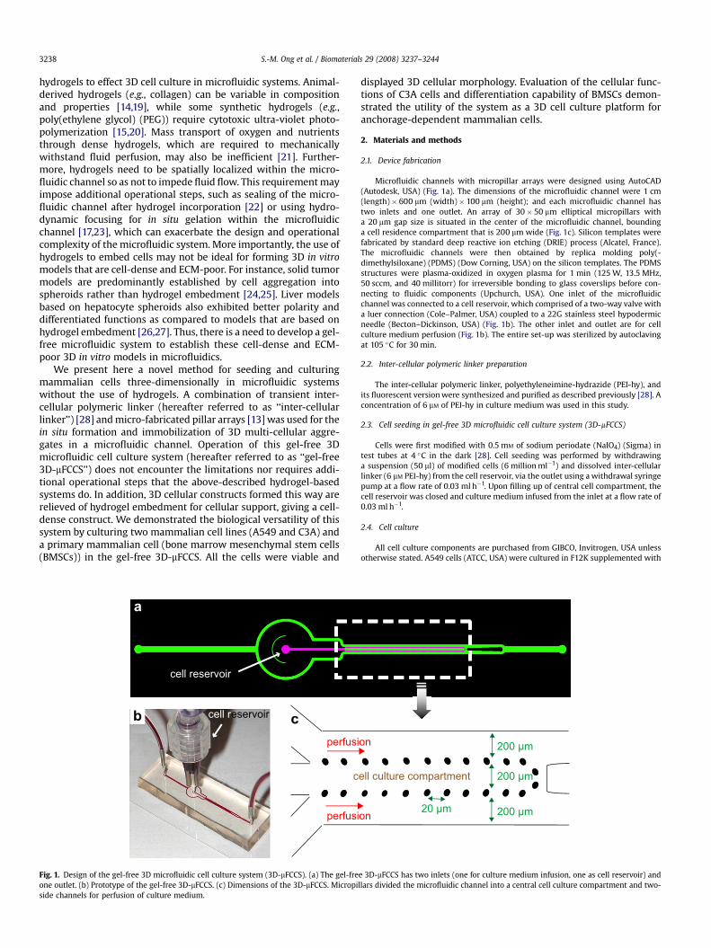
Fig. 1. Design of the gel-free 3D microfluidic cell culture system (3D-mFCCS). (a) The gel-freone outlet. (b) Prototype of the gel-free 3D-mFCCS. (c) Dimensions of the 3D-mFCCS. Micropiside channels for perfusion of culture medium.
displayed 3D cellular morphology. Evaluation of the cellular func-tions of C3A cells and differentiation capability of BMSCs demon-strated the utility of the system as a 3D cell culture platform foranchorage-dependent mammalian cells.
2. Materials and methods
2.1. Device fabrication
Microfluidic channels with micropillar arrays were designed using AutoCAD(Autodesk, USA) (Fig. 1a). The dimensions of the microfluidic channel were 1 cm(length)� 600 mm (width)� 100 mm (height); and each microfluidic channel hastwo inlets and one outlet. An array of 30� 50 mm elliptical micropillars witha 20 mm gap size is situated in the center of the microfluidic channel, boundinga cell residence compartment that is 200 mm wide (Fig. 1c). Silicon templates werefabricated by standard deep reactive ion etching (DRIE) process (Alcatel, France).The microfluidic channels were then obtained by replica molding poly(-dimethylsiloxane) (PDMS) (Dow Corning, USA) on the silicon templates. The PDMSstructures were plasma-oxidized in oxygen plasma for 1 min (125 W, 13.5 MHz,50 sccm, and 40 millitorr) for irreversible bonding to glass coverslips before con-necting to fluidic components (Upchurch, USA). One inlet of the microfluidicchannel was connected to a cell reservoir, which comprised of a two-way valve witha luer connection (Cole–Palmer, USA) coupled to a 22G stainless steel hypodermicneedle (Becton–Dickinson, USA) (Fig. 1b). The other inlet and outlet are for cellculture medium perfusion (Fig. 1b). The entire set-up was sterilized by autoclavingat 105 �C for 30 min.
2.2. Inter-cellular polymeric linker preparation
The inter-cellular polymeric linker, polyethyleneimine-hydrazide (PEI-hy), andits fluorescent version were synthesized and purified as described previously [28]. Aconcentration of 6 mM of PEI-hy in culture medium was used in this study.
2.3. Cell seeding in gel-free 3D microfluidic cell culture system (3D-mFCCS)
Cells were first modified with 0.5 mM of sodium periodate (NaIO4) (Sigma) intest tubes at 4 �C in the dark [28]. Cell seeding was performed by withdrawinga suspension (50 ml) of modified cells (6 million ml�1) and dissolved inter-cellularlinker (6 mM PEI-hy) from the cell reservoir, via the outlet using a withdrawal syringepump at a flow rate of 0.03 ml h�1. Upon filling up of central cell compartment, thecell reservoir was closed and culture medium infused from the inlet at a flow rate of0.03 ml h�1.
2.4. Cell culture
All cell culture components are purchased from GIBCO, Invitrogen, USA unlessotherwise stated. A549 cells (ATCC, USA) were cultured in F12K supplemented with
200 µm
200 µm
200 µm20 µm
ell culture compartment
on
on
e 3D-mFCCS has two inlets (one for culture medium infusion, one as cell reservoir) andllars divided the microfluidic channel into a central cell culture compartment and two-
S.-M. Ong et al. / Biomaterials 29 (2008) 3237–3244 3239
10% fetal calf serum, 1.5 g L�1 sodium bicarbonate, 2 mM L-glutamine, 100 units ml�1
penicillin and 100 g ml�1 streptomycin. C3A cells (ATCC, USA) were cultured inMinimum Essential Medium supplemented with 10% fetal calf serum, 1.5 g L�1 so-dium bicarbonate, 1 mM sodium pyruvate, 100 units ml�1 penicillin and 100 g ml�1
streptomycin. Bone marrow stem cells were harvested from the bone marrow ofmale Wistar rats and cultured in low glucose Dulbecco’s Modified Eagle’s Mediumsupplemented with 10% fetal calf serum and 1.5 g L�1 sodium bicarbonate. Osteo-genic medium was prepared by supplementing basal medium with 100 nM dexa-methasone, 50 mM ascorbic acid 2-phosphate and 10 mM b-glycerophosphate(Merck, Singapore).
2.5. Perfusion culture in gel-free 3D-mFCCS
The cells are cultured in a one-pass perfusion manner with a syringe pump(Cole–Palmer) at 0.03 ml h�1. The microfluidic system was placed onto a heatingplate (MEDAX GmbH & Co. KG, Germany) maintained at 37 �C throughout the cul-ture period in a sterile hood. Sixty millimolar of Hepes buffer (GIBCO, Invitrogen,USA) was added to the culture medium to maintain its pH at 7.4–7.6.
2.6. Cell viability staining
Cell viability of A549, C3A, and BMSCs after 3 days of perfusion culture in the gel-free 3D-mFCCS was assessed by perfusing 5 mM of Calcein AM (Molecular Probes,USA) and 25 mg ml�1 of propidium iodide at 0.5 ml h�1 for 30 min and viewingimmediately by confocal microscopy (Fluoview 300, Olympus, Japan).
2.7. F-actin staining
F-actin distribution in all cell types was assessed after 3 days of perfusion culturein the gel-free 3D-mFCCS. In situ F-actin staining was performed after fixation with3.7% paraformaldehyde (PFA) (30 min) by infusing the microfluidic channel with0.5% Triton-X 100 (USB Corp, USA) (30 min), 0.2% bovine serum albumin (BSA)(30 min), 0.2 mg ml�1 of TRITC-phalloidin (Invitrogen, Singapore) (20 min) and 1�PBS (15 min) at 0.5 ml h�1. 2D monolayer cultures were fixed with 3.7% PFA (15 min)and stained by incubating with 0.5% Triton-X 100 (10 min), 0.2% BSA (15 min), and0.2 mg ml�1 of TRITC-phalloidin (20 min).
2.8. Scanning electron microscopy (SEM)
SEM samples were prepared by bonding the PDMS microfluidic channels ontoa polyethylene (PE) film (Diversified Biotech, USA) instead of a glass coverslip. The
a
Wit
cell cell
cel
ceNaIO4
Inter-cellularlinker
cell Modified cell surface with aldehydes
Inter-cellular linker
cell
b
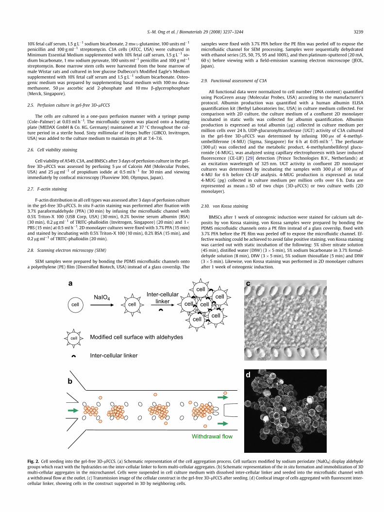
Fig. 2. Cell seeding into the gel-free 3D-mFCCS. (a) Schematic representation of the cell agggroups which react with the hydrazides on the inter-cellular linker to form multi-cellular aggmulti-cellular aggregates in the microchannel. Cells were suspended in cell culture mediua withdrawal flow at the outlet. (c) Transmission image of the cellular construct in the gel-frecellular linker, showing cells in the construct supported in 3D by neighboring cells.
samples were fixed with 3.7% PFA before the PE film was peeled off to expose themicrofluidic channel for SEM processing. Samples were sequentially dehydratedwith ethanol series (25, 50, 75, 95 and 100%), and then platinum-sputtered (20 mA,60 s) before viewing with a field-emission scanning electron microscope (JEOL,Japan).
2.9. Functional assessment of C3A
All functional data were normalized to cell number (DNA content) quantifiedusing PicoGreen assay (Molecular Probes, USA) according to the manufacturer’sprotocol. Albumin production was quantified with a human albumin ELISAquantification kit (Bethyl Laboratories Inc, USA) in culture medium collected. Forcomparison with 2D culture, the culture medium of a confluent 2D monolayerincubated in static wells was collected for albumin quantification. Albuminproduction is expressed as total albumin (mg) collected in culture medium permillion cells over 24 h. UDP-glucuronyltransferase (UGT) activity of C3A culturedin the gel-free 3D-mFCCS was determined by infusing 100 mM of 4-methyl-umbelliferone (4-MU) (Sigma, Singapore) for 6 h at 0.05 ml h�1. The perfusate(300 ml) was collected and the metabolic product, 4-methylumbelliferyl glucu-ronide (4-MUG), was analyzed using capillary electrophoresis with laser inducedfluorescence (CE-LIF) [29] detection (Prince Technologies B.V., Netherlands) atan excitation wavelength of 325 nm. UGT activity in confluent 2D monolayercultures was determined by incubating the samples with 300 ml of 100 mM of4-MU for 6 h before CE-LIF analysis. 4-MUG production is expressed as total4-MUG (pg) collected in culture medium per million cells over 6 h. Data arerepresented as mean� SD of two chips (3D-mFCCS) or two culture wells (2Dmonolayer).
2.10. von Kossa staining
BMSCs after 1 week of osteogenic induction were stained for calcium salt de-posits by von Kossa staining. von Kossa samples were prepared by bonding thePDMS microfluidic channels onto a PE film instead of a glass coverslip, fixed with3.7% PFA before the PE film was peeled off to expose the microfluidic channel. Ef-fective washing could be achieved to avoid false positive staining. von Kossa stainingwas carried out with static incubation of the following: 5% silver nitrate solution(45 min), distilled water (DIW) (3� 5 min), 5% sodium bicarbonate in 3.7% formal-dehyde solution (8 min), DIW (3� 5 min), 5% sodium thiosulfate (5 min) and DIW(3� 5 min). Likewise, von Kossa staining was performed in 2D monolayer culturesafter 1 week of osteogenic induction.
hdrawal flow
d
cell
l cell
cellll
c
regation process. Cell surfaces modified by sodium periodate (NaIO4) display aldehyderegates. (b) Schematic representation of the in situ formation and immobilization of 3Dm with dissolved inter-cellular linker and seeded into the microfluidic channel withe 3D-mFCCS after seeding. (d) Confocal image of cells aggregated with fluorescent inter-
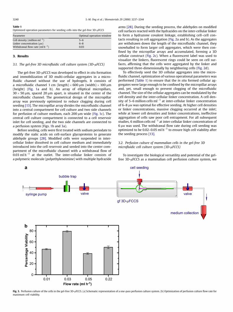
Table 1Optimized operation parameters for seeding cells into the gel-free 3D-mFCCS
Parameter Optimal operation window
Cell density (million ml�1) 5–6Linker concentration (mM) 6–8Withdrawal flow rate (ml h�1) 0.03
S.-M. Ong et al. / Biomaterials 29 (2008) 3237–32443240
3. Results
3.1. The gel-free 3D microfluidic cell culture system (3D-mFCCS)
The gel-free 3D-mFCCS was developed to effect in situ formationand immobilization of 3D multi-cellular aggregates in a micro-fluidic channel without the use of hydrogels. It consists ofa microfluidic channel 1 cm (length)� 600 mm (width)� 100 mm(height) (Fig. 1a and b). An array of elliptical micropillars,30� 50 mm, spaced 20 mm apart, is situated in the center of themicrofluidic channel. The geometrical design of the micropillararray was previously optimized to reduce clogging during cellseeding [13]. The micropillar array divides the microfluidic channelinto a central compartment for cell culture and two side channelsfor perfusion of culture medium, each 200 mm wide (Fig. 1c). Thecentral cell culture compartment is connected to a cell reservoirinlet for cell seeding, and the two side channels are connected toa perfusion system (Figs. 1b and 3a).
Before seeding, cells were first treated with sodium periodate tomodify the sialic acids on cell-surface glycoproteins to generatealdehyde groups [28]. Modified cells were suspended in inter-cellular linker dissolved in cell culture medium and immediatelyintroduced into the cell reservoir and seeded into the center com-partment of the microfluidic channel with a withdrawal flow of0.03 ml h�1 at the outlet. The inter-cellular linker consists ofa polymeric molecule (polyethyleneimine) with multiple hydrazide
Fig. 3. Perfusion culture of the cells in the gel-free 3D-mFCCS. (a) Schematic representation omaximum cell viability.
arms [28]. During the seeding process, the aldehydes on modifiedcell surfaces reacted with the hydrazides on the inter-cellular linkerto form a hydrazone covalent linkage, establishing cell–cell con-tacts resulting in cell aggregation (Fig. 2a and b). As the aggregatesare withdrawn down the length of the microfluidic channel, theysnowballed to form larger cell aggregates, which were then con-fined by the micropillar arrays and accumulated, forming a 3Dcellular construct (Fig. 2c). When a fluorescent label was used tovisualize the linkers, fluorescent rings could be seen on cell sur-faces, affirming that the cells were aggregated by the linker andsupported three-dimensionally by neighboring cells (Fig. 2d).
To effectively seed the 3D cellular aggregates into the micro-fluidic channel, optimization of various operational parameters wasperformed (Table 1) to ensure that the in situ formed cellular ag-gregates were large enough to be confined by the micropillar arraysand, yet, small enough to prevent clogging of the microfluidicchannel. The size of the cellular aggregates can be modulated by thecell density and the inter-cellular linker concentration. A cell den-sity of 5–6 million cells ml�1 at inter-cellular linker concentrationof 6–8 mM was optimal for effective seeding. At higher cell densitiesor linker concentrations, massive clogging occurred at the inlet;while at lower cell densities and linker concentrations, ineffectiveaggregation of cells saw poor cell entrapment. For all subsequentstudies, 6 million cells ml�1 at inter-cellular linker concentration of6 mM was used. The withdrawal flow rate during cell seeding wasoptimized to be 0.02–0.05 ml h�1 to ensure high cell viability afterthe seeding process [13].
3.2. Perfusion culture of mammalian cells in the gel-free 3Dmicrofluidic cell culture system (3D-mFCCS)
To investigate the biological versatility and potential of the gel-free 3D-mFCCS as a mammalian cell perfusion culture system, we
f a one-pass perfusion culture system. (b) Optimization of perfusion culture flow rate for
S.-M. Ong et al. / Biomaterials 29 (2008) 3237–3244 3241
cultured a few model anchorage-dependent mammalian cells, suchas the human lung epithelial cell line A549, human liver cell lineC3A, and rat primary progenitor cells (BMSCs), using a one-passperfusion system (Fig. 3a). A one-pass perfusion system allows themaintenance of a constant cell culture microenvironment overtime, without accumulation of metabolites or depletion of oxygenand nutrients that is experienced by re-circulating perfusion cul-tures [30]. The culture medium was oxygenated by passing throughoxygen-permeable tubing before entering the bubble trap. Theperfusion flow rate to culture the 3D cellular construct was0.03 ml h�1, optimized for maximal cell viability (Fig. 3b). Culturemedium at the end of the perfusion circuit can be collected forassessment of cellular functions.
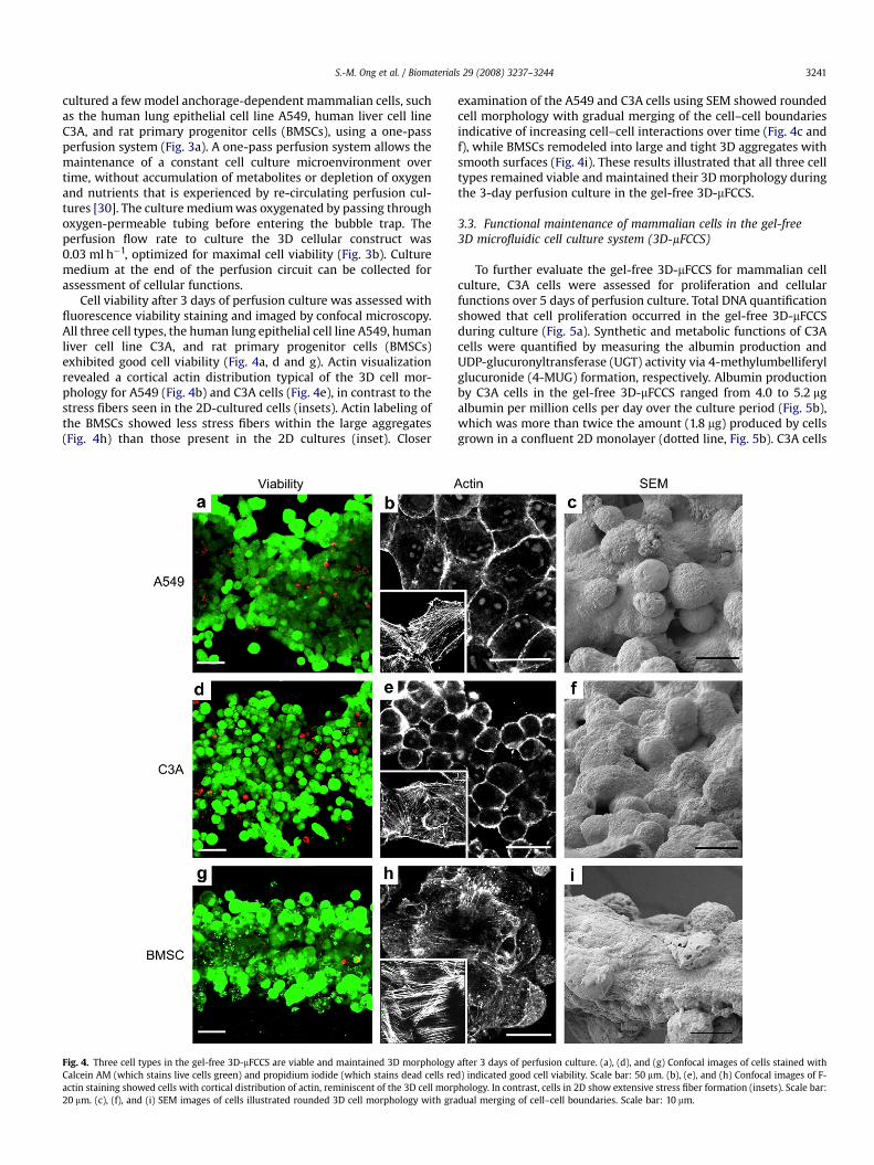
Cell viability after 3 days of perfusion culture was assessed withfluorescence viability staining and imaged by confocal microscopy.All three cell types, the human lung epithelial cell line A549, humanliver cell line C3A, and rat primary progenitor cells (BMSCs)exhibited good cell viability (Fig. 4a, d and g). Actin visualizationrevealed a cortical actin distribution typical of the 3D cell mor-phology for A549 (Fig. 4b) and C3A cells (Fig. 4e), in contrast to thestress fibers seen in the 2D-cultured cells (insets). Actin labeling ofthe BMSCs showed less stress fibers within the large aggregates(Fig. 4h) than those present in the 2D cultures (inset). Closer
Fig. 4. Three cell types in the gel-free 3D-mFCCS are viable and maintained 3D morphologyCalcein AM (which stains live cells green) and propidium iodide (which stains dead cells reactin staining showed cells with cortical distribution of actin, reminiscent of the 3D cell morp20 mm. (c), (f), and (i) SEM images of cells illustrated rounded 3D cell morphology with gra
examination of the A549 and C3A cells using SEM showed roundedcell morphology with gradual merging of the cell–cell boundariesindicative of increasing cell–cell interactions over time (Fig. 4c andf), while BMSCs remodeled into large and tight 3D aggregates withsmooth surfaces (Fig. 4i). These results illustrated that all three celltypes remained viable and maintained their 3D morphology duringthe 3-day perfusion culture in the gel-free 3D-mFCCS.
3.3. Functional maintenance of mammalian cells in the gel-free3D microfluidic cell culture system (3D-mFCCS)
To further evaluate the gel-free 3D-mFCCS for mammalian cellculture, C3A cells were assessed for proliferation and cellularfunctions over 5 days of perfusion culture. Total DNA quantificationshowed that cell proliferation occurred in the gel-free 3D-mFCCSduring culture (Fig. 5a). Synthetic and metabolic functions of C3Acells were quantified by measuring the albumin production andUDP-glucuronyltransferase (UGT) activity via 4-methylumbelliferylglucuronide (4-MUG) formation, respectively. Albumin productionby C3A cells in the gel-free 3D-mFCCS ranged from 4.0 to 5.2 mgalbumin per million cells per day over the culture period (Fig. 5b),which was more than twice the amount (1.8 mg) produced by cellsgrown in a confluent 2D monolayer (dotted line, Fig. 5b). C3A cells
after 3 days of perfusion culture. (a), (d), and (g) Confocal images of cells stained withd) indicated good cell viability. Scale bar: 50 mm. (b), (e), and (h) Confocal images of F-hology. In contrast, cells in 2D show extensive stress fiber formation (insets). Scale bar:dual merging of cell–cell boundaries. Scale bar: 10 mm.

Alb
um
in
P
ro
du
ctio
n
(µµg
/ m
illio
n cells)
To
tal C
ell N
um
ber
(T
ho
usan
d)
b
a
4-M
UG
P
ro
du
ctio
n
(p
mo
l / m
illio
n cells)
c
70
80
60
50
40
30
20
10
0
Day 1 Day 3 Day 5
Day 1 Day 3 Day 5
Day 1 Day 3 Day 5
6
5
4
3
2
1
0
0.8
0.2
0.4
0.6
0
Fig. 5. C3A cells cultured in the gel-free 3D-mFCCS proliferated and exhibited goodcellular functions. (a) Total cell number measured showed cell proliferation inthe gel-free 3D-mFCCS. (b) and (c) Functional assessment of C3A cells cultured inthe gel-free 3D-mFCCS by albumin production (b) and UDP-glucuronyltransferase(UGT) activity via 4-methylumbelliferyl glucuronide (4-MUG) production (c)showed good cell function compared to cells in a confluent 2D monolayer (dottedlines).
S.-M. Ong et al. / Biomaterials 29 (2008) 3237–32443242
in the gel-free 3D-mFCCS also demonstrated higher UGT activity,producing 0.47–0.63 pmol 4-MUG per million cells (Fig. 5c), ascompared to 0.21 pmol 4-MUG produced by a confluent 2Dmonolayer (dotted line, Fig. 5c). The proliferation and maintenanceof good cell functionality of C3A cells in the gel-free 3D-mFCCSthroughout the 5-day culture demonstrated the reliability of thegel-free 3D-mFCCS for culturing and maintaining cellular functionsin 3D.
To evaluate the gel-free 3D-mFCCS for the culture of sensitiveprimary cells, we investigated the differentiation competence ofthe BMSCs by differentiating them down the osteogenic lineage.BMSCs in the gel-free 3D-mFCCS were perfused with osteogenicinduction medium for 1 week. von Kossa staining for calcium de-posits was positive for BMSCs aggregates in the gel-free 3D-mFCCS(Fig. 6a), similar to von Kossa staining observed in standard con-fluent 2D cultures after a 1 week of osteogenic induction (Fig. 6b).This suggests that the gel-free 3D-mFCCS may be useful for theculture and study of sensitive primary cells such as the bonemarrow-derived adult stem cells.
4. Discussion
We present here a novel method for 3D perfusion cell culture ofmammalian cells in microfluidic channels without the use ofhydrogels. This gel-free 3D microfluidic cell culture system (gel-free3D-mFCCS) utilizes inter-cellular polymeric linker to in situ form 3Dmulti-cellular aggregates in microfluidic channels, which are thenconfined by a micropillar array. The inter-cellular linker acts asa ‘‘cell glue’’ to stabilize the multi-cellular aggregates, such that thecells are supported three-dimensionally by neighboring cells.Compared to microfluidic systems that utilize hydrogels for 3D cellembedment [12,17,22], the use of inter-cellular linker to effect 3Dcell culture in the gel-free 3D-mFCCS facilitates the establishment ofa more natural extracellular matrix (ECM) environment. This isbecause the polymeric inter-cellular linker is transient (i.e. gradu-ally disappears from the cell surfaces with a half-life of w2 days),thus allowing cells to secrete and remodel their own ECM envi-ronment for maintaining their 3D structural integrity and mor-phology [28]. The presence of an ECM environment endogenous toa specific cell type in the gel-free 3D-mFCCS may promote more invivo-like cellular phenotypes when compared to microfluidic sys-tems incorporating exogenous ECM hydrogels since cellular phe-notypes are highly dependent on the chemical and mechanicalproperties of the ECM [14].
From an operational point of view, seeding and forming a 3D cellconstruct in the gel-free 3D-mFCCS is simple. Cells are chemicallymodified, suspended with the inter-cellular linker, and introducedinto the microfluidic channel; the inter-cellular linker induces theformation of 3D multi-cellular aggregates, which are then confinedby the micropillar array within the channel. After the central cellcompartment of the channel is filled with cells, the cell reservoir isclosed; and culture medium is immediately perfused, completingthe cell seeding process. Where hydrogels are used to provide 3Dcellular support in other microfluidic systems, additional steps arerequired to induce in situ matrix gelation within the microfluidicchannel either by photo-polymerization [31], self-assembly ofoligopeptides [17], hydrodynamic focusing [17], or polyelectrolytecomplex coacervation [13,23]. These steps are more time-consuming and/or require the use of additional fluidic components,such as valves, pumps and connectors to control the delivery ofdifferent reagents (Supplementary Fig. S1). In some hydrogel-basedsystems, suspension of cells in hydrogels is loaded into openmicrofluidic channels while sealing of the channel is carried outonly after the hydrogel has solidified [20,22]. Such operationalcomplexity increases the likelihood of contamination and failureand poses additional challenges for multiplexing design and oper-ation which can be alleviated by using the inter-cellular linker inplace of the hydrogels for 3D cellular support.
We demonstrated the utility of the gel-free 3D-mFCCS by suc-cessfully culturing and maintaining the 3D phenotypes of modelanchorage-dependent mammalian cells, which included carcinomacell lines (A549 and C3A) and primary bone marrow mesenchymalstem cells (BMSCs). These cell types represent biologically relevant3D in vitro models that can be established with the gel-free
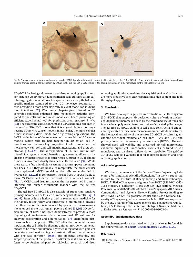
Fig. 6. Primary bone marrow mesenchymal stem cells (BMSCs) can be differentiated into osteoblasts in the gel-free 3D-mFCCS after 1 week of osteogenic induction. (a) von Kossastaining showed calcium salt deposition by BMSCs in the gel-free 3D-mFCCS, similar to the staining obtained in a 2D monolayer control (b). Scale bar: 50 mm.
S.-M. Ong et al. / Biomaterials 29 (2008) 3237–3244 3243
3D-mFCCS for biological research and drug screening applications.For instance, A549 human lung epithelial cells cultured as 3D cel-lular aggregates were shown to express increased epithelial cell-specific markers compared to their 2D monolayer counterparts,thus providing a more physiologically relevant model for studyinglung infections [32]. C3A human hepatocytes cultured as 3Dspheroids exhibited enhanced drug metabolism activities com-pared to the cells cultured in 2D monolayer, hence providing anefficient experimental tool for predicting drug responses in vivo[33]. The successful culture of A549 and C3A carcinoma cell lines inthe gel-free 3D-mFCCS shows that it is a good platform for engi-neering 3D in vitro cancer models, in particular, the multi-cellulartumor spheroid (MCTS) model for drug testing applications. TheMCTS model is one of the most studied and established 3D cancermodels, where cells are held together in 3D by cell–cell in-teractions, and features key properties of solid tumors such asmorphology, cell–cell and cell–matrix interactions, and drug pen-etration [14,24,25]. The incorporation of 3D cell culture intomicrofluidic systems would benefit drug screening efforts, as in-creasing evidence shows that cancer cells cultured in 3D resembletumors in vivo more closely than cells cultured in 2D [34]. Whilethere exists a few microfluidic systems that can support carcinomacell lines in 3D, they are unable to recapitulate the multi-cellulartumor spheroid (MCTS) model as the cells are embedded inhydrogels [13,17,22]. In comparison, the gel-free 3D-mFCCS is able toform MCTS-like cell-dense constructs with cell–cell contacts(Fig. 4). MCTS-based drug testing can thus be performed in a min-iaturized and higher throughput manner with the gel-free3D-mFCCS.
The gel-free 3D-mFCCS is also capable of supporting sensitiveprimary mammalian cells, such as mesenchymal stem cells (Figs. 4and 6). Stem cells are highly prized in regenerative medicine fortheir ability to self-renew and differentiate into multiple lineages.Its differentiation fate is influenced by specialized microenviron-ments or cell niche that remain poorly defined [35,36]. 3D perfu-sion culture of BMSCs has been demonstrated to provide a morephysiological environment than conventional 2D cultures forstudying proliferation and differentiation [37]. Microfluidic plat-forms such as the gel-free 3D-mFCCS offer the potential for ma-nipulating the cell niche by allowing different microenvironmentalfactors to be tested simultaneously when integrated with gradientgenerators, and maintaining a constant cell microenvironmentwith one-pass perfusion [30,38]. The biological versatility andsimple operation of the gel-free 3D-mFCCS make it a suitable plat-form to be further adapted for biological research and drug
screening applications, enabling the acquisition of in vitro data thatare more predictive of in vivo responses in a high content and highthroughput approach.
5. Conclusion
We have developed a gel-free microfluidic cell culture system(3D-mFCCS) that supports 3D perfusion culture of various anchor-age-dependent mammalian cells by the combined use of transientinter-cellular polymeric linker and micro-fabricated pillar arrays.The gel-free 3D-mFCCS exhibits a cell-dense construct and endog-enously created extracellular microenvironment. We demonstratedthe biological versatility of the gel-free 3D-mFCCS by culturing an-chorage-dependent mammalian cell lines (A549 and C3A) andprimary bone marrow mesenchymal stem cells (BMSCs). The cellsshowed good cell viability and preserved 3D cell morphology,exhibited higher cell functionality over cells cultured in 2Dmonolayer, and displayed differentiation capability. The gel-free3D-mFCCS will be a valuable tool for biological research and drugscreening applications.
Acknowledgements
We thank the members of the Cell and Tissue Engineering Lab-oratory for stimulating scientific discussions. This work is supportedin part by the Institute of Bioengineering and Nanotechnology,BMRC, A*STAR of Singapore and grants from BMRC (R185-001-045-305), Ministry of Education (R-185-000-135-112), National MedicalResearch Council (R-185-000-099-213) and Singapore-MIT AllianceComputational and Systems Biology Flagship Project funding toHYU. SMO is an A*STAR graduate scholar and CZ is a National Uni-versity of Singapore graduate research scholar. SHK was supportedby the SRC program of the Korea Science and Engineering Founda-tion (KOSEF) through the Center for Intelligent Nano-Bio Materialsat Ewha Womans University (R11-2005-008-02003-0).
Appendix. Supplementary data
Supplementary data associated with this article can be found, inthe online version, at doi:10.1016/j.biomaterials.2008.04.022.
References
[1] El-Ali J, Sorger PK, Jensen KF. Cells on chips. Nature 27 Jul 2006;442(7101):403–11.
S.-M. Ong et al. / Biomaterials 29 (2008) 3237–32443244
[2] Wang Z, Kim MC, Marquez M, Thorsen T. High-density microfluidic arrays forcell cytotoxicity analysis. Lab Chip Jun 2007;7(6):740–5.
[3] Dittrich PS, Manz A. Lab-on-a-chip: microfluidics in drug discovery. Nat RevDrug Discov Mar 2006;5(3):210–8.
[4] Ye N, Qin J, Shi W, Liu X, Lin B. Cell-based high content screening using anintegrated microfluidic device. Lab Chip Dec 2007;7(12):1696–704.
[5] Mitchell P. Microfluidics – downsizing large-scale biology. Nat Biotechnol Aug2001;19(8):717–21.
[6] Taylor DL, Woo ES, Giuliano KA. Real-time molecular and cellular analysis:the new frontier of drug discovery. Curr Opin Biotechnol Feb 2001;12(1):75–81.
[7] Nie FQ, Yamada M, Kobayashi J, Yamato M, Kikuchi A, Okano T. On-chip cellmigration assay using microfluidic channels. Biomaterials Sep 2007;28(27):4017–22.
[8] King KR, Wang S, Irimia D, Jayaraman A, Toner M, Yarmush ML. A high-throughput microfluidic real-time gene expression living cell array. Lab ChipJan 2007;7(1):77–85.
[9] Tourovskaia A, Figueroa-Masot X, Folch A. Differentiation-on-a-chip: a micro-fluidic platform for long-term cell culture studies. Lab Chip Jan 2005;5(1):14–9.
[10] Kane BJ, Zinner MJ, Yarmush ML, Toner M. Liver-specific functional studies ina microfluidic array of primary mammalian hepatocytes. Anal Chem 1 Jul2006;78(13):4291–8.
[11] Cheng X, Irimia D, Dixon M, Sekine K, Demirci U, Zamir L, et al. A microfluidicdevice for practical label-free CD4(þ) T cell counting of HIV-infected subjects.Lab Chip Feb 2007;7(2):170–8.
[12] Ling Y, Rubin J, Deng Y, Huang C, Demirci U, Karp JM, et al. A cell-laden mi-crofluidic hydrogel. Lab Chip Jun 2007;7(6):756–62.
[13] Toh YC, Zhang C, Zhang J, Khong YM, Chang S, Samper VD, et al. A novel 3Dmammalian cell perfusion-culture system in microfluidic channels. Lab ChipMar 2007;7(3):302–9.
[14] Abbott A. Cell culture: biology’s new dimension. Nature 21 Aug 2003;424(6951):870–2.
[15] Albrecht DR, Underhill GH, Wassermann TB, Sah RL, Bhatia SN. Probing therole of multicellular organization in three-dimensional microenvironments.Nat Methods May 2006;3(5):369–75.
[16] Nisbet DR, Crompton KE, Horne MK, Finkelstein DI, Forsythe JS. Neural tissueengineering of the CNS using hydrogels – a review. J Biomed Mater Res B ApplBiomater 27 Dec 2007.
[17] Kim MS, Yeon JH, Park JK. A microfluidic platform for 3-dimensional cellculture and cell-based assays. Biomed Microdevices Feb 2007;9(1):25–34.
[18] Tan W, Desai TA. Layer-by-layer microfluidics for biomimetic three-dimensional structures. Biomaterials Mar–Apr 2004;25(7–8):1355–64.
[19] Griffith LG, Swartz MA. Capturing complex 3D tissue physiology in vitro. NatRev Mol Cell Biol Mar 2006;7(3):211–24.
[20] Ng CP, Pun SH. A perfusable 3D cell-matrix tissue culture chamber for in situevaluation of nanoparticle vehicle penetration and transport. BiotechnolBioeng 29 Oct 2007.
[21] Golden AP, Tien J. Fabrication of microfluidic hydrogels using molded gelatinas a sacrificial element. Lab Chip Jun 2007;7(6):720–5.
[22] Figallo E, Cannizzaro C, Gerecht S, Burdick JA, Langer R, Elvassore N, et al.Micro-bioreactor array for controlling cellular microenvironments. Lab ChipJun 2007;7(6):710–9.
[23] Toh YC, Ng S, Khong YM, Samper V, Yu H. A configurable three-dimensionalmicroenvironment in a microfluidic channel for primary hepatocyte culture.Assay Drug Dev Technol Apr 2005;3(2):169–76.
[24] Kunz-Schughart LA. Multicellular tumor spheroids: intermediates betweenmonolayer culture and in vivo tumor. Cell Biol Int 1999;23(3):157–61.
[25] Sutherland RM. Cell and environment interactions in tumor microregions: themulticell spheroid model. Science 8 Apr 1988;240(4849):177–84.
[26] Koide N, Sakaguchi K, Koide Y, Asano K, Kawaguchi M, Matsushima H, et al.Formation of multicellular spheroids composed of adult rat hepatocytes indishes with positively charged surfaces and under other nonadherent envi-ronments. Exp Cell Res Feb 1990;186(2):227–35.
[27] Walker TM, Rhodes PC, Westmoreland C. The differential cytotoxicity ofmethotrexate in rat hepatocyte monolayer and spheroid cultures. Toxicol InVitro Oct 2000;14(5):475–85.
[28] Ong SM, He L, Thuy Linh NT, Tee YH, Arooz T, Tang G, et al. Transient inter-cellular polymeric linker. Biomaterials Sep 2007;28(25):3656–67.
[29] Toh YC, Zhang J, Khong YM, Du YN, Sun WX, Yu H. Integrating sensitivequantification of hepatic metabolic functions by capillary electrophoresis withlaser-induced fluorescence detection. Analyst 2008;132(3):326–30.
[30] Kim L, Vahey MD, Lee HY, Voldman J. Microfluidic arrays for logarithmicallyperfused embryonic stem cell culture. Lab Chip Mar 2006;6(3):394–406.
[31] Koh WG, Pishko MV. Fabrication of cell-containing hydrogel microstructuresinside microfluidic devices that can be used as cell-based biosensors. AnalBioanal Chem Aug 2006;385(8):1389–97.
[32] Carterson AJ, Honer zu Bentrup K, Ott CM, Clarke MS, Pierson DL,Vanderburg CR, et al. A549 lung epithelial cells grown as three-dimensionalaggregates: alternative tissue culture model for Pseudomonas aeruginosapathogenesis. Infect Immun Feb 2005;73(2):1129–40.
[33] Elkayam T, Amitay-Shaprut S, Dvir-Ginzberg M, Harel T, Cohen S. Enhancingthe drug metabolism activities of C3A – a human hepatocyte cell line – bytissue engineering within alginate scaffolds. Tissue Eng May 2006;12(5):1357–68.
[34] Kunz-Schughart LA, Freyer JP, Hofstaedter F, Ebner R. The use of 3-D culturesfor high-throughput screening: the multicellular spheroid model. J BiomolScreen Jun 2004;9(4):273–85.
[35] Calvi LM, Adams GB, Weibrecht KW, Weber JM, Olson DP, Knight MC, et al.Osteoblastic cells regulate the haematopoietic stem cell niche. Nature 23 Oct2003;425(6960):841–6.
[36] Spradling A, Drummond-Barbosa D, Kai T. Stem cells find their niche. Nature 1Nov 2001;414(6859):98–104.
[37] Braccini A, Wendt D, Jaquiery C, Jakob M, Heberer M, Kenins L, et al. Three-dimensional perfusion culture of human bone marrow cells and generation ofosteoinductive grafts. Stem Cells Sep 2005;23(8):1066–72.
[38] Khademhosseini A, Langer R, Borenstein J, Vacanti JP. Microscale technologiesfor tissue engineering and biology. Proc Natl Acad Sci USA 21 Feb 2006;103(8):2480–7.