Strain and deformation angle for a steel pipe elbow using ...
Upload
independentCategory
view
1download
0
Elbow Position Affects Distal Radioulnar Joint Kinematics
Eric Fu, BS*, Guoan Li, PhD**, Sebastiaan Souer, MD*, Santiago Lozano-Calderon, MD, JamesH. Herndon, MD*, Jesse B. Jupiter, MD*, and Neal C. Chen, MD†* Hand and Upper Extremity Service, Massachusetts General Hospital** Bioengineering Laboratory, Massachusetts General Hospital
AbstractPrevious in vivo and in vitro studies of forearm supination/pronation suggest that distal radioulnarjoint kinematics may be affected by elbow flexion. The primary hypotheses tested by this study werethat in vivo: 1) ulnar variance changes with elbow flexion and forearm rotation and 2) the arc offorearm rotation changes in relationship to elbow flexion.
Materials and Methods—Changes in radioulnar kinematics during forearm supination/pronationand elbow flexion (0–90°) were studied in five uninjured subjects using computed tomography, dual-orthogonal fluoroscopy, and three-dimensional modeling. Analysis of variance and post-hoc testingwas performed.
Results—Proximal translation of the radius was greatest with the elbow flexed to 90° with the armin mid-pronation. With the arm in mid-pronation, the translation of the radius was significantlygreater at 0° versus 45° of elbow flexion (0.82 ± 0.59 mm v. 0.65 ± 0.80 mm, F: 4.49, Post Hoc:0.055; p = 0.05), and significantly smaller at 45° versus 90° of elbow flexion (0.65 ± 0.80 mm v.0.97 ± 0.35 mm, F: 4.49, Post Hoc: 0.048; p = 0.05). Proximal translation of the radius in mid-pronation was significantly greater than when the forearm was in a supinated position when the elbowwas at 0° or 90° flexion (F: 14.90, post-hoc: < 0.01; p < 0.01, F: 19.11, post-hoc: < 0.01, p < 0.01).The arc of forearm rotation was significantly decreased at 0° compared to 90° of elbow flexion (129.3± 22.2° v 152.8 ± 14.4°, F: 3.29, post-hoc: 0.79; p = 0.09). The center of rotation shifted volarly andulnarly with increasing elbow extension.
Discussion—Elbow position affects the kinematics of the distal radioulnar joint. The kinematicsof the distal radioulnar joint are primarily affected by forearm rotation and secondarily affected byelbow flexion. These findings have clinical relevance to our understanding of ulnar impaction, andhow elbow position affects the proximal-distal translation of the radius. These findings haveimplications upon the treatment of ulna impaction, radiographic evaluation of the distal ulna, andfuture biomechanical studies.
Keywordselbow; distal radioulnar; kinematics; biomechanics; forearm
†To whom all correspondence should be addressed: Neal C. Chen MD, Medsport, University of Michigan, 24 Frank Lloyd Wright Drive,P.O. Box 0391, Ann Arbor, MI 48106, [email protected], (734) 930-7390 (office), (734) 930-7402 (fax).Study performed at: Bioengineering Laboratory, Department of Orthopaedics, Massachusetts General Hospital/Harvard Medical School,Boston, MAPublisher's Disclaimer: This is a PDF file of an unedited manuscript that has been accepted for publication. As a service to our customerswe are providing this early version of the manuscript. The manuscript will undergo copyediting, typesetting, and review of the resultingproof before it is published in its final citable form. Please note that during the production process errors may be discovered which couldaffect the content, and all legal disclaimers that apply to the journal pertain.
NIH Public AccessAuthor ManuscriptJ Hand Surg Am. Author manuscript; available in PMC 2010 September 1.
Published in final edited form as:J Hand Surg Am. 2009 September ; 34(7): 1261–1268. doi:10.1016/j.jhsa.2009.04.025.
NIH
-PA Author Manuscript
NIH
-PA Author Manuscript
NIH
-PA Author Manuscript

IntroductionRadiographic and cadaveric studies have been used to investigate in vivo and in vitro ulnarvariance changes with forearm supination and pronation, in healthy and pathologic subjects(1–4). Ulnar variance is standardized in these studies where the shoulder is placed in 90° ofabduction and the elbow flexed to 90° the forearm in neutral rotation. However, it is unclearwhether the position of elbow flexion intrinsically affects ulna variance.
Tay et al. used finite helical axis analysis to describe the in vivo kinematics of the forearm andfound proximal-distal translation of the radius relative to the ulna with rotation of the forearm(5), suggesting that elbow position may affect distal forearm kinematics. However, becauseimaging techniques such as computed tomography and magnetic resonance imaging havegeometric limitations, and plain radiography is not as precise as these other techniques; theeffect of elbow flexion on the distal radioulnar joint has not been well studied.
Recently, a combined dual fluoroscopic and MR/CT imaging technique has been developedthat allows measurement of joint motion without constraints of the target joint (6,7). Thistechnique has been successfully applied to the lower extremity (8–10). In this study, we utilizedthis imaging technique to explore the relationship of distal radioulnar joint kinematics withelbow and forearm positions. The primary hypotheses tested are: 1) proximal-distal translationof the radius relative to the ulna changes in relationship to elbow flexion and forearm rotationand 2) the arc of forearm rotation changes in relationship to elbow flexion.
Materials and MethodsThis study was approved by the Institutional Review Board and informed consent was obtainedfor each subject. Five right dominant upper extremities without a history of previous injuryfrom right-hand dominant male volunteers (age 24 to31) were imaged while in neutral forearmrotation and elbow extension by computed tomography (GE Light Speed Pro 16-Slice scanner).Parallel axial CT images separated at 0.625mm with a resolution of 512 × 512 pixels wereobtained. Each CT image was processed using a Canny filter programmed in a commerciallyavailable mathematics software package (Matlab, Mathworks, Canton, MA). The Canny filtercalculates gradients in pixel intensity to detect edges between objects. The calculated edgeswere used to help trace the outlines of the radius and ulna within the axial plane image usingsolid-modeling software (Rhinoceros®, Robert McNeel & Associates, Seattle, WA). Thecontours were then placed in the appropriate plane in a 3D space, and the surface was meshedusing the solid-modeling software.
After CT image-based computer 3D models were constructed, each subject was imaged usingtwo orthogonally placed fluoroscopes (BV Pulsera, Philips, Bothell, WA) at elbow flexionangles of 0°, 45°, and 90° as the subject rotated his/her forearm from maximum supination,50% supination, neutral, 50% pronation, and maximum pronation (Fig 1a). The wrist wasplaced in zero degrees of extension and zero degrees of radioulnar deviation and themetacarpalphalangeal, proximal interphalangeal, and distal interphalangeal joints were held inextension. These positions were confirmed with goniometer. Each subject practiced the testingpositions five times before actual testing. The degree of prononation/supination was estimatedfrom the pre-conditioning trials with the understanding that the true amount of rotation wouldbe defined with the modeling techniques. During imaging, the plane of the C-arm was co-planarto the floor and the subject was seated with the elbow stabilized on a radiolucent table (Figure1). The subject held each position which was confirmed with a goniometer and then the imagewas acquired. The entire testing procedure took about 10 minutes.
The orthogonal images were then imported into a solid modeling software (Rhinoceros®,Robert McNeel & Associates, Seattle, WA Rhinoceros, Seattle, Wash) and used to determine
Fu et al. Page 2
J Hand Surg Am. Author manuscript; available in PMC 2010 September 1.
NIH
-PA Author Manuscript
NIH
-PA Author Manuscript
NIH
-PA Author Manuscript

the in vivo forearm positions at each of the targeted flexion angles. The orthogonal imageswere placed in the software to reproduce the positions of the two intensifiers of the fluoroscopeduring image acquisition. The forearm model was imported into the virtual space and wasviewed simultaneously from two orthogonal directions corresponding to the positions of thex-ray source of the fluoroscope during image acquisition. The 3D forearm model was thenmanipulated in six degrees of freedom inside the 3D C-arm until its projections viewed fromtwo orthogonal directions matched the outlines of the fluoroscopic images obtained, thusreproducing the in vivo forearm position using the 3D anatomical models. The method hasbeen previously validated on a knee model to an accuracy of 0.1 mm position and 0.1 degreesrotation (6,7).
To describe the six degrees of freedom (6DOF) kinematics of the distal radioulnar joint(DRUJ), anatomically based ulnar and radial Cartesian coordinate systems similar to thatreported previously in the literature were constructed for each subject (Figure 2). The positionof the forearm in neutral position during CT scanning was used as a reference. For the ulnarcoordinate system, the longitudinal axis (z-axis) was defined as the line passing through thelongitudinal axis of the ulna. The y-axis was defined as the line passing from the center of theulnar head to through the center of the ulnar styloid base. The x-axis was an axis perpendicularto those two axes pointing dorsally. The radial longitudinal axis (z-axis) was the long-axis ofthe radius. The y-axis was defined as the line passing through the anatomic center of the distalradius to the tip of the radial styloid. The x-axis was an axis perpendicular to those 2 axespointing dorsally. In this manner, a coordinate system for each wrist could be established in aconsistent way. The axes used to describe the radius are similar to those used clinically todescribe the forearm.
The 6DOF kinematics of the wrist was described by the relative position and orientation of thedistal radius with respect to the ulnar head using a script written by our laboratory forRhinoceros solid modeling software. The three-dimensional positions were determined by theposition of the origin of the distal radius coordinate system in the ulnar head coordinate system.The orientation was represented by the relative orientation of the distal radius coordinatesystem with respect to the ulnar styloid coordinate system using 3 Euler angles (in x-y-zsequence) (Figure 3). In this study, 6DOF was expressed using the radial displacements alongthe x,y, and z-axes of the ulna for anterior/posterior, ulnar/radial, and proximal/distaldisplacements and rotation about the z-axis for pronation/supination. Proximal translation ofthe radius relative to the ulna was defined as negative, whereas distal translation was positive.
In order to characterize the overall effect of flexion and rotation at a specified elbow flexionangle, we examined the difference observed in proximal-distal translation between fullsupination and full pronation (terminal difference) versus the difference observed between thepeak proximal and peak distal translation (peak difference).
To compare the actual path of rotation of the radius to the expected path of a perfect circle, wedefined a circular path about the center of the ulnar coordinate system using the center of theradial coordinate systems and the “best fit circle” function within Rhinoceros (Figure 4). Linesextending from the center of the ulnar coordinate system were extended to the centers of theradial coordinate systems such that they intersected with the path of the best fit circle. Thedifference in distance between the actual and expected path of rotation were then measuredalong this line.
Because of the small sample size of the cohort, significance was pre-defined using an alpha of0.10. Analysis of Variance (ANOVA) was used to compare the motion of the forearm amongthe different elbow flexion and rotation angles with Statistica 6.0 (StatSoft, Tulsa OK). Post-hoc testing using a Neuman Keul’s test was performed to evaluate differences between groups.
Fu et al. Page 3
J Hand Surg Am. Author manuscript; available in PMC 2010 September 1.
NIH
-PA Author Manuscript
NIH
-PA Author Manuscript
NIH
-PA Author Manuscript

ResultsProximal/Distal Translation
Traditional ulna variance is measured at 90 degrees of elbow flexion and neutral rotation. Inour subjects, ulna variance based on the model would be −0.24 ± 0.57 mm.
The proximal and distal translation of the radius relative to the ulna varied with elbow flexion.With the arm in mid-pronation, the translation of the radius was significantly greater at 0°versus 45° of elbow flexion (0.82 ± 0.59 mm v. 0.65 ± 0.80 mm, F: 4.49, Post Hoc: 0.055; p= 0.05), and significantly smaller at 45° versus 90° of elbow flexion (0.65 ± 0.80 mm v. 0.97± 0.61 mm, F: 4.49, Post Hoc: 0.048; p = 0.05) (Figure 5).
When the elbow was at 0° or 90° flexion, the proximal and distal translation of the radiusrelative to the ulna was not maximal at extremes of pronation and supination. Rather, peakdisplacement was observed at mid-supination and mid-pronation. With the elbow in fullextension, translation of the radius was significantly more in mid-supination than fullsupination (−1.10 ± 0.35 mm v. −0.24 ± 0.34 mm, F: 14.90, Post Hoc: 0.005; p < 0.01). Withthe elbow in full extension, translation of the radius was also significantly greater than whenin mid-pronation than full pronation (0.82 ± 0.59 mm v. 0.3 ± 0.24 mm, F: 14.90, Post Hoc:0.06; p < 0.01). Translation of the radius in mid-pronation was significantly greater when theforearm was in either mid-supination or terminal supination with the elbow at 0° or 90° flexion(F: 14.90, post-hoc: < 0.01; p < 0.01, F: 19.11, post-hoc: < 0.01, p < 0.01). Similarly, translationof the radius in mid-supination was significantly less than any other position of forearm rotationwith the elbow at 0° or 90° flexion (F: 14.90, post-hoc: < 0.01; p < 0.01, F: 19.11, post-hoc: <0.01, p <0.01).
In general, at each flexion angle, the peak difference was greater than the terminal difference.At 0° of elbow flexion, the terminal difference was significantly smaller than peak differencesat all elbow flexion angles (F: 5.49, post-hoc: <0.05 for all flexion angles; p = 0.002). At 90°flexion, the peak difference was significantly greater than the terminal differences at all elbowflexion angles (F: 5.49, post-hoc: <0.05 for all flexion angles; p = 0.002).
Arc of Pronation/SupinationFrom maximal supination to maximal pronation, the DRUJ demonstrated an average of 141.8°± 18.4° of total rotation for all angles of elbow flexion. Total arc of rotation increased withprogressive elbow flexion. As elbow flexion increases, end-range pronation increases (F: 3.67,post-hoc 0° v 45°: 0.10, post-hoc 0° v 90°: 0.049; p = 0.07). The arc of rotation was significantlysmaller at 0° compared to 90° of elbow flexion (129.3 ± 22.2° v. 152.8 ± 14.4°, F: 3.29, post-hoc: 0.79; p=0.09), (Table 1).
The radius rotated about the ulna in a circular path with a center of rotation located in the ulnarhead with only minor deviations in the radius of rotation. At ninety-degrees of elbow flexion,the center of rotation was nearly coincident with the anatomic center of the ulnar head. Thecenter of rotation of the radius shifted volarly and ulnarly with increasing elbow extension(Table 2, Figure 6). The location of the center of rotation was significantly different when theelbow was flexed to 90° as compared to the elbow in full extension (F: 3.31, post-hoc: 0.09; p= 0.08). The diameter of the circular path traversed by the radius relative to the ulna alsochanged with elbow flexion. The diameter was significantly different between 0° v. 45° (57.9± 8.3 mm v. 54.3 ± 4.4 mm, F: 3.13, post-hoc 0° v. 45°: 0.07; p = 0.10) (Table 2).
Fu et al. Page 4
J Hand Surg Am. Author manuscript; available in PMC 2010 September 1.
NIH
-PA Author Manuscript
NIH
-PA Author Manuscript
NIH
-PA Author Manuscript

DiscussionThis study examined the kinematics of the forearm using a combined dual fluoroscopy imagingsystem and CT imaging technique. To our knowledge, this is the first application of this typeof technology to understanding the kinematics of the forearm. Strengths of this non-invasiveimaging system are that this technique allows examination of a distal joint while a proximaljoint is unconstrained and examination of forearm motion in the entire functional range ofmotion (6–10).
The first hypothesis, that ulnar variance changes with elbow flexion, was supported by ourfindings. Our data suggests that when the elbow is at 45° flexion, the radius translatesproximally less than when the elbow is at full extension or 90° flexion. The maximum amountof distal or proximal translation of the radius relative to the ulna did not occur at terminalpronation or supination, but at mid-arc. One possible reason why maximal translation occursduring mid arc is that as the forearm approaches terminal rotation, the corresponding distalradioulnar ligament becomes taut and restricts longitudinal translation. The proximal structuressuch as the biceps and brachialis may also limit translation as the forearm approaches terminalsupination with elbow extension.
Previous studies have suggested that elbow flexion and forearm rotation influence ulnavariance. For example, Schuurman et al. examined ulna variance with the forearm in neutralrotation with the elbow at 90° of flexion compared to the forearm in full supination with theelbow at full extension and found differences of −0.18 ± 1.56 mm v. −0.86 ± 1.47 mm (1). Yehet al. studied ulnar variance at maximum pronation, neutral rotation, and maximum supinationusing anteroposterior radiographs and found a total proximal-distal translation of 0.6 mm withconfidence intervals of 0.4 mm to 0.8 mm, but also did not control for elbow flexion other thanin neutral rotation (2). Using static CT scans, Tay et al. found proximal-distal translation of1.67mm when moving from full pronation to full supination (5). We found a similar magnitudeof mean maximum change between maximum pronation and maximum supination.
When comparing examining previous data, it is not clear to what degree elbow flexion orforearm rotation contributes to translation of the radius relative to the ulna. In general, we foundthat forearm rotation has a greater relative contribution to proximal-distal translation thanelbow flexion. Rotation leads to changes of up to 2 mm, while elbow flexion leads to changesof up to 0.5 mm. Ultimately, our data suggests that ulna impaction on the carpus is most likelyto occur when the forearm is in mid-pronation and the elbow is at 90 degrees of flexion or fullextension.
The second hypothesis, that arc of rotation changes with elbow flexion was supportedstatistically. The total arc of rotation increases with increasing elbow flexion. We observedthat as elbow flexion increased, the center of rotation of the radius approached the anatomiccenter of the ulna head; in addition, the diameter of that arc of rotation decreased significantlywith elbow flexion. This suggests that the precession of the radius about the ulna changes withelbow flexion. Using a custom jig, Shaaban et al. found that total arc of rotation increasedsignificantly from 0° to 45°, remained constant from 45° to 90°, and then decreased at fullflexion (11). This was, in general, consistent with our results. The decrease of range of motionin extension may be a result of the biceps and other soft tissue restraints around the proximalradioulnar joint becoming tight at this position.
These findings are relevant in radiographic evaluations of the distal radioulnar joint and ulnavariance and understanding injuries of the forearm. Studies have examined various methodsof quantifying distal radioulnar joint dorso-volar translation and ulna variance (12,13). Inevaluating ulna variance, there is emphasis on elbow flexion and neutral forearm rotation, andthe effect of proximal and distal joint positioning upon radiographic evaluation has been
Fu et al. Page 5
J Hand Surg Am. Author manuscript; available in PMC 2010 September 1.
NIH
-PA Author Manuscript
NIH
-PA Author Manuscript
NIH
-PA Author Manuscript
recognized by previous authors (1,14,15); however, this has not been systematically studiedpreviously. Our results suggest that maximal distal radioulnar translation occurs when theelbow is flexed or fully extended and in mid-pronation.
Similar to Baeyens et al., we did not observe any volar/dorsal translation at the distal radioulnarjoint at either maximum supination or pronation (16). Tay et al. demonstrated volar/dorsaltranslation of the radius relative to the ulna in vivo during resisted supination and pronationwhile gripping an external handle (5). Schiund et al. have also demonstrated a mean change ofulnar variance of 0.5mm during maximal grasp of a dynamometer (15). We did not alter fingeror wrist flexion-extension throughout the experiment, suggesting that during elbow flexion,muscle forces across the distal radioulnar joint are such that the ulna remains centered in thesigmoid notch in vivo if the wrist and hand are not closed or experiencing an external load.
LimitationsLimitations of this study were the small number of subjects available for study. Ideally therewould be a larger number of subjects and female subjects included however, practicalconsiderations limited the number of participants and female subject enrollement. The smallcohort also did not have any outliers with an ulna variance greater or less than two millimeters.The inclusion of these subjects may give insight into how deviations affect our measurementsof translation or forearm kinematics. A second limitation was that it was not possible to performfluoroscopy that encompassed the entire elbow and wrist joint at the same time because of thereceiver size on the fluoroscopes. This would allow simultaneous analysis of the proximal anddistal radioulnar joints. In addition, the position of the elbow could be more accuratelydescribed rather than using a goniometer for positioning. Finally, an ideal study would alsoinclude more positions of elbow flexion and points along the rotational arc and under dynamicforearm motions.
In conclusion, application of three dimensional solid modeling techniques in conjunction withdual orthogonal fluoroscopy demonstrates that the translation of the radius relative to the ulna,the arc of forearm rotation, and the size of the arc of forearm rotation changes with elbowflexion. These findings have implications in the treatment of ulna impaction, radiographicevaluation, and understanding of forearm kinematics. Future studies examining integrating softtissue structures and dynamic forearm loading may be helpful in understanding the contributionof the distal radioulnar ligaments, triangular fibrocartilage, and interosseous membrane to theseobservations could help further elucidate our understanding of the in vivo kinematics of thedistal radioulnar joint.
AcknowledgmentsThis research was funded by a Basic Science Grant from the American Foundation for Surgery of the Hand. Wegratefully acknowledge the financial support of the Department of Orthopaedic Surgery at Massachusetts GeneralHospital. We thank Jeffrey Bingham, Shaobai Wang, and Michal Kozanek for their technical assistance.
References1. Schuurman AH, Maas M, Dijkstra PF, Kauer JM. Assessment of ulnar variance: a radiological
investigation in a Dutch population. Skeletal Radiol 2001;30:633–8. [PubMed: 11810155]2. Yeh GL, Beredjiklian PK, Katz MA, Steinberg DR, Bozentka DJ. Effects of forearm rotation on the
clinical evaluation of ulnar variance. J Hand Surg 2001;26A:1042–1046.3. Epner RA, Bowers WH, Guilford WB. Ulnar variance—the effect of wrist positioning and roentgen
filming technique. J Hand Surg 1982;7:298 –305.4. Palmer AK, Werner FW. Biomechanics of the distal radioulnar joint. Clin Orthop 1984;187:26 –35.
[PubMed: 6744728]
Fu et al. Page 6
J Hand Surg Am. Author manuscript; available in PMC 2010 September 1.
NIH
-PA Author Manuscript
NIH
-PA Author Manuscript
NIH
-PA Author Manuscript

5. Tay SC, Berger RA, Tomita K, Tan ET, Amrami KK, An KN. In vivo three-dimensional displacementof the distal radioulnar joint during resisted forearm rotation. J Hand Surg 2007;32A:450–8.
6. Li G, Wuerz TH, DeFrate LE. Feasibility of using orthogonal fluoroscopic images to measure in vivojoint kinematics. J Biomech Eng 2004;126:314–8. [PubMed: 15179865]
7. Li G, Van de Velde SK, Bingham JT. Validation of a non-invasive fluoroscopic imaging techniquefor the measurement of dynamic knee joint motion. J Biomech 2008;41:1616–22. [PubMed:18394629]
8. Li G, DeFrate LE, Sun H, Gill TJ. In vivo elongation of the an terior cruciate ligament and posteriorcruciate ligament during knee flexion. Am J Sports Med 2004;32:1415–20. [PubMed: 15310565]
9. Li G, Papannagari R, Li M, Bingham J, Nha KW, Allred D, Gill T. Effect of posterior cruciate ligamentdeficiency on in vivo translation and rotation of the knee during weightbearing flexion. Am J SportsMed 2008;36:474–9. [PubMed: 18057390]
10. Jordan SS, DeFrate LE, Nha KW, Papannagari R, Gill TJ, Li G. The in vivo kinematics of theanteromedial and posterolateral bundles of the anterior cruciate ligament during weightbearing kneeflexion. Am J Sports Med 2007;35:547–54. [PubMed: 17261571]
11. Shaaban H, Pereira C, Williams R, Lees VC. The effect of elbow position on the range of supinationand pronation of the forearm. J Hand Surg Eur Vol 2008;33:3–8. [PubMed: 18332013]
12. Park MJ, Kim JP. Reliability and normal values of various computed tomography methods forquantifying distal radioulnar joint translation. J Bone Joint Surg 2008;90:145–53. [PubMed:18171969]
13. Pan CC, Lin YM, Lee TS, Chou CH. Displacement of the distal radioulnar joint of clinically symptom-free patients. Clin Orthop Relat Res 2003;415:148–56. [PubMed: 14612641]
14. Tomaino MM. The importance of the pronated grip x-ray view in evaluating ulnar variance. J HandSurg 2000;25A:352–7.
15. Schuind FA, Linscheid RL, An KN, Chao EYS. Changes in wrist and forearm configuration withgrasp and isometric contraction of the elbow flexors. J Hand Surg 1992;17A:698–703.
16. Baeyens JP, Van Glabbeek F, Goossens M, Gielen J, Van Roy P, Clarys JP. In vivo 3Darthrokinematics of the proximal and distal radioulnar joints during active pronation and supination.Clin Biomech (Bristol, Avon) 2006;21 (Suppl 1):S9–12.
Fu et al. Page 7
J Hand Surg Am. Author manuscript; available in PMC 2010 September 1.
NIH
-PA Author Manuscript
NIH
-PA Author Manuscript
NIH
-PA Author Manuscript

Figure 1.Dual fluoroscopic imaging system. Subject with elbow at 45° flexion and forearm in neutralrotation. Images were captured at elbow flexion angles of 0°, 45°, and 90° with the forearm atmaximum supination, mid-supination, neutral, mid-pronation, and maximum pronation.
Fu et al. Page 8
J Hand Surg Am. Author manuscript; available in PMC 2010 September 1.
NIH
-PA Author Manuscript
NIH
-PA Author Manuscript
NIH
-PA Author Manuscript

Figure 2.The coordinate systems used to describe radioulnar kinematics. The ulnar y-axis, a line thecenter of the ulnar head to the center of the ulna styloid, was used to delineate supination frompronation.
Fu et al. Page 9
J Hand Surg Am. Author manuscript; available in PMC 2010 September 1.
NIH
-PA Author Manuscript
NIH
-PA Author Manuscript
NIH
-PA Author Manuscript

Figure 3.Change in ulna variance was measured from the neutral position for each angle of elbowflexion. Ulna variance was measured as the distance along the ulnar z-axis between the centerof the radial coordinate system and the center of the ulnar coordinate system. Proximaltranslation of the radial coordinate system relative to the ulna coordinate was defined asnegative, whereas distal translation was positive.
Fu et al. Page 10
J Hand Surg Am. Author manuscript; available in PMC 2010 September 1.
NIH
-PA Author Manuscript
NIH
-PA Author Manuscript
NIH
-PA Author Manuscript

Figure 4.Center of rotation was determined by finding the best fit circle of the center points of the radialcoordinate system. Deviations from this circular path were random and minor.
Fu et al. Page 11
J Hand Surg Am. Author manuscript; available in PMC 2010 September 1.
NIH
-PA Author Manuscript
NIH
-PA Author Manuscript
NIH
-PA Author Manuscript

Figure 5.Proximal-distal translation of the radius relative to the ulna. Proximal translation of the radiusrelative to the ulna was defined as negative, whereas distal translation was positive.
Fu et al. Page 12
J Hand Surg Am. Author manuscript; available in PMC 2010 September 1.
NIH
-PA Author Manuscript
NIH
-PA Author Manuscript
NIH
-PA Author Manuscript
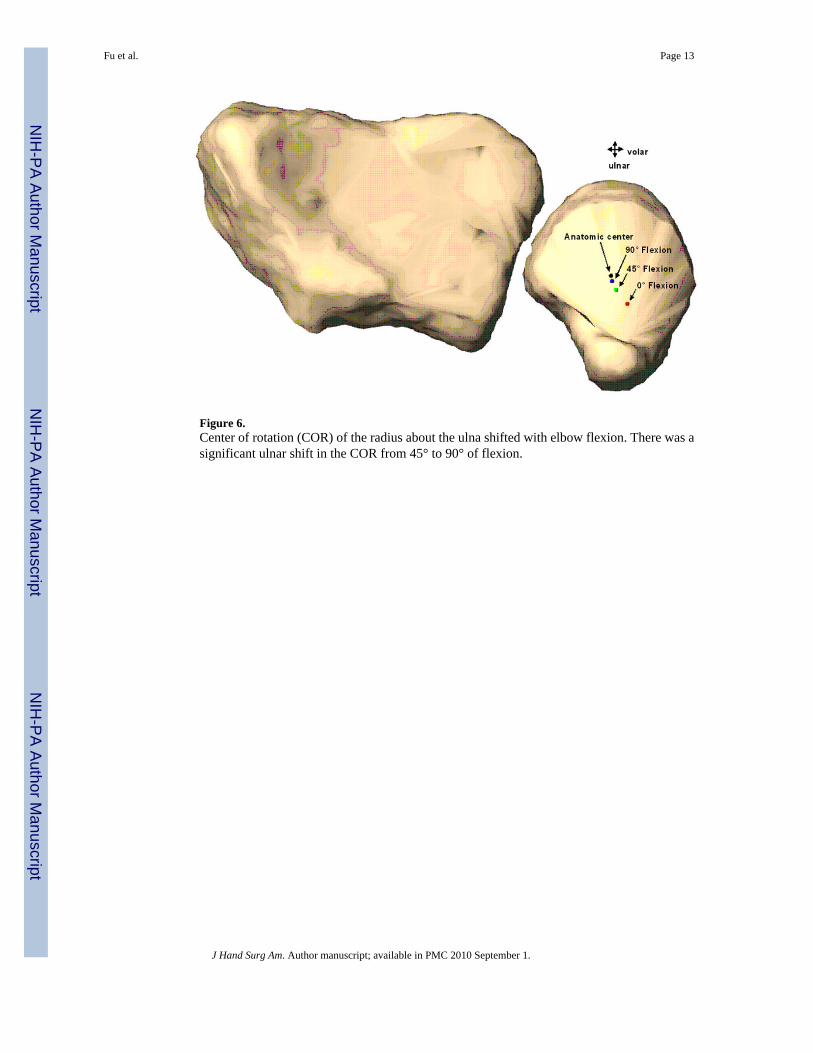
Figure 6.Center of rotation (COR) of the radius about the ulna shifted with elbow flexion. There was asignificant ulnar shift in the COR from 45° to 90° of flexion.
Fu et al. Page 13
J Hand Surg Am. Author manuscript; available in PMC 2010 September 1.
NIH
-PA Author Manuscript
NIH
-PA Author Manuscript
NIH
-PA Author Manuscript

NIH
-PA Author Manuscript
NIH
-PA Author Manuscript
NIH
-PA Author Manuscript
Fu et al. Page 14
Table 1Total arc of rotation, maximum pronation, and maximum supination compared across elbow flexion angle. There wasa significant difference between the range of rotation at 0° elbow flexion and 90°.
Elbow Flexion 0° 45° 90°
Total Arc of Rotation 129.3 ± 22.2° 143.4 ± 11.7° 152.8 ± 14.4°
Maximum Pronation 41.1 ± 15.5° 57.7° ± 8.3° 57.3 ± 5.9°
Maximum Supination 88.2 ± 12.3° 85.7 ± 8.0° 95.5 ± 18.2°
J Hand Surg Am. Author manuscript; available in PMC 2010 September 1.

NIH
-PA Author Manuscript
NIH
-PA Author Manuscript
NIH
-PA Author Manuscript
Fu et al. Page 15Ta
ble
2U
lnar
and
vola
r cha
nges
of t
he C
OR
of t
he ra
dius
abou
t the
uln
a. T
here
was
a si
gnifi
cant
diff
eren
ce in
the u
lnar
dis
plac
emen
t of t
he C
OR
betw
een
45° a
nd 9
0° o
f elb
ow fl
exio
n.
Elb
ow F
lexi
onU
lnar
Dis
plac
emen
tV
olar
Dis
plac
emen
tA
rc o
f Rot
atio
n D
iam
eter
0°3.
1 ±
2.3m
m1.
7 ±
1.2m
m57
.9 ±
8.3
mm
45°
1.5
± 1.
4mm
0.5
± 0.
8mm
54.3
± 4
.4 m
m
90°
0.5
± 1.
5mm
0.1
± 0.
6mm
55.0
± 5
.5 m
m
J Hand Surg Am. Author manuscript; available in PMC 2010 September 1.
Copyright © 2022 FDOKUMEN