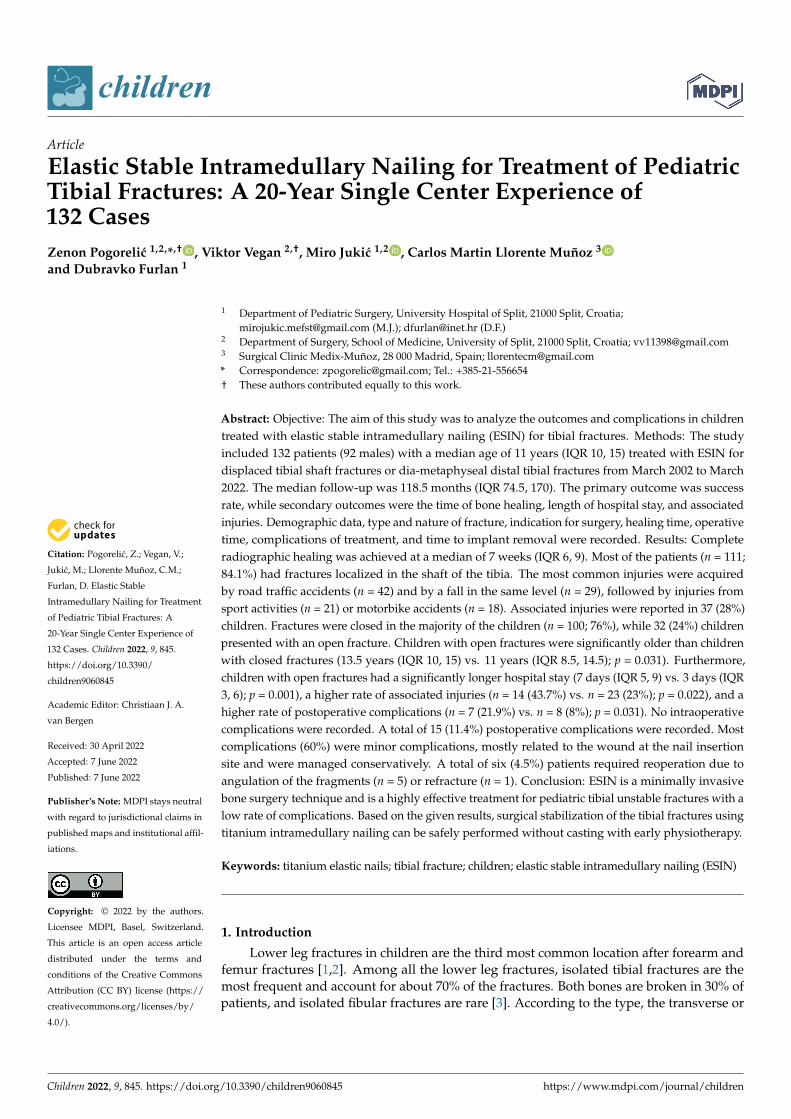
Elastic Stable Intramedullary Nailing for Treatment of Pediatric ...
12
Citation: Pogoreli´ c, Z.; Vegan, V.; Juki´ c, M.; Llorente Muñoz, C.M.; Furlan, D. Elastic Stable Intramedullary Nailing for Treatment of Pediatric Tibial Fractures: A 20-Year Single Center Experience of 132 Cases. Children 2022, 9, 845. https://doi.org/10.3390/ children9060845 Academic Editor: Christiaan J. A. van Bergen Received: 30 April 2022 Accepted: 7 June 2022 Published: 7 June 2022 Publisher’s Note: MDPI stays neutral with regard to jurisdictional claims in published maps and institutional affil- iations. Copyright: © 2022 by the authors. Licensee MDPI, Basel, Switzerland. This article is an open access article distributed under the terms and conditions of the Creative Commons Attribution (CC BY) license (https:// creativecommons.org/licenses/by/ 4.0/). children Article Elastic Stable Intramedullary Nailing for Treatment of Pediatric Tibial Fractures: A 20-Year Single Center Experience of 132 Cases Zenon Pogoreli´ c 1,2, * ,† , Viktor Vegan 2,† , Miro Juki´ c 1,2 , Carlos Martin Llorente Muñoz 3 and Dubravko Furlan 1 1 Department of Pediatric Surgery, University Hospital of Split, 21000 Split, Croatia; [email protected] (M.J.); [email protected] (D.F.) 2 Department of Surgery, School of Medicine, University of Split, 21000 Split, Croatia; [email protected] 3 Surgical Clinic Medix-Muñoz, 28 000 Madrid, Spain; [email protected] * Correspondence: [email protected]; Tel.: +385-21-556654 † These authors contributed equally to this work. Abstract: Objective: The aim of this study was to analyze the outcomes and complications in children treated with elastic stable intramedullary nailing (ESIN) for tibial fractures. Methods: The study included 132 patients (92 males) with a median age of 11 years (IQR 10, 15) treated with ESIN for displaced tibial shaft fractures or dia-metaphyseal distal tibial fractures from March 2002 to March 2022. The median follow-up was 118.5 months (IQR 74.5, 170). The primary outcome was success rate, while secondary outcomes were the time of bone healing, length of hospital stay, and associated injuries. Demographic data, type and nature of fracture, indication for surgery, healing time, operative time, complications of treatment, and time to implant removal were recorded. Results: Complete radiographic healing was achieved at a median of 7 weeks (IQR 6, 9). Most of the patients (n = 111; 84.1%) had fractures localized in the shaft of the tibia. The most common injuries were acquired by road traffic accidents (n = 42) and by a fall in the same level (n = 29), followed by injuries from sport activities (n = 21) or motorbike accidents (n = 18). Associated injuries were reported in 37 (28%) children. Fractures were closed in the majority of the children (n = 100; 76%), while 32 (24%) children presented with an open fracture. Children with open fractures were significantly older than children with closed fractures (13.5 years (IQR 10, 15) vs. 11 years (IQR 8.5, 14.5); p = 0.031). Furthermore, children with open fractures had a significantly longer hospital stay (7 days (IQR 5, 9) vs. 3 days (IQR 3, 6); p = 0.001), a higher rate of associated injuries (n = 14 (43.7%) vs. n = 23 (23%); p = 0.022), and a higher rate of postoperative complications (n = 7 (21.9%) vs. n = 8 (8%); p = 0.031). No intraoperative complications were recorded. A total of 15 (11.4%) postoperative complications were recorded. Most complications (60%) were minor complications, mostly related to the wound at the nail insertion site and were managed conservatively. A total of six (4.5%) patients required reoperation due to angulation of the fragments (n = 5) or refracture (n = 1). Conclusion: ESIN is a minimally invasive bone surgery technique and is a highly effective treatment for pediatric tibial unstable fractures with a low rate of complications. Based on the given results, surgical stabilization of the tibial fractures using titanium intramedullary nailing can be safely performed without casting with early physiotherapy. Keywords: titanium elastic nails; tibial fracture; children; elastic stable intramedullary nailing (ESIN) 1. Introduction Lower leg fractures in children are the third most common location after forearm and femur fractures [1,2]. Among all the lower leg fractures, isolated tibial fractures are the most frequent and account for about 70% of the fractures. Both bones are broken in 30% of patients, and isolated fibular fractures are rare [3]. According to the type, the transverse or Children 2022, 9, 845. https://doi.org/10.3390/children9060845 https://www.mdpi.com/journal/children
-
Upload
khangminh22 -
Category
Documents
-
view
1 -
download
0
Transcript of Elastic Stable Intramedullary Nailing for Treatment of Pediatric ...
Citation: Pogorelic, Z.; Vegan, V.;
Jukic, M.; Llorente Muñoz, C.M.;
Furlan, D. Elastic Stable
Intramedullary Nailing for Treatment
of Pediatric Tibial Fractures: A
20-Year Single Center Experience of
132 Cases. Children 2022, 9, 845.
https://doi.org/10.3390/
children9060845
Academic Editor: Christiaan J. A.
van Bergen
Received: 30 April 2022
Accepted: 7 June 2022
Published: 7 June 2022
Publisher’s Note: MDPI stays neutral
with regard to jurisdictional claims in
published maps and institutional affil-
iations.
Copyright: © 2022 by the authors.
Licensee MDPI, Basel, Switzerland.
This article is an open access article
distributed under the terms and
conditions of the Creative Commons
Attribution (CC BY) license (https://
creativecommons.org/licenses/by/
4.0/).
children
Article
Elastic Stable Intramedullary Nailing for Treatment of PediatricTibial Fractures: A 20-Year Single Center Experience of132 CasesZenon Pogorelic 1,2,*,† , Viktor Vegan 2,†, Miro Jukic 1,2 , Carlos Martin Llorente Muñoz 3
and Dubravko Furlan 1
1 Department of Pediatric Surgery, University Hospital of Split, 21000 Split, Croatia;[email protected] (M.J.); [email protected] (D.F.)
2 Department of Surgery, School of Medicine, University of Split, 21000 Split, Croatia; [email protected] Surgical Clinic Medix-Muñoz, 28 000 Madrid, Spain; [email protected]* Correspondence: [email protected]; Tel.: +385-21-556654† These authors contributed equally to this work.
Abstract: Objective: The aim of this study was to analyze the outcomes and complications in childrentreated with elastic stable intramedullary nailing (ESIN) for tibial fractures. Methods: The studyincluded 132 patients (92 males) with a median age of 11 years (IQR 10, 15) treated with ESIN fordisplaced tibial shaft fractures or dia-metaphyseal distal tibial fractures from March 2002 to March2022. The median follow-up was 118.5 months (IQR 74.5, 170). The primary outcome was successrate, while secondary outcomes were the time of bone healing, length of hospital stay, and associatedinjuries. Demographic data, type and nature of fracture, indication for surgery, healing time, operativetime, complications of treatment, and time to implant removal were recorded. Results: Completeradiographic healing was achieved at a median of 7 weeks (IQR 6, 9). Most of the patients (n = 111;84.1%) had fractures localized in the shaft of the tibia. The most common injuries were acquiredby road traffic accidents (n = 42) and by a fall in the same level (n = 29), followed by injuries fromsport activities (n = 21) or motorbike accidents (n = 18). Associated injuries were reported in 37 (28%)children. Fractures were closed in the majority of the children (n = 100; 76%), while 32 (24%) childrenpresented with an open fracture. Children with open fractures were significantly older than childrenwith closed fractures (13.5 years (IQR 10, 15) vs. 11 years (IQR 8.5, 14.5); p = 0.031). Furthermore,children with open fractures had a significantly longer hospital stay (7 days (IQR 5, 9) vs. 3 days (IQR3, 6); p = 0.001), a higher rate of associated injuries (n = 14 (43.7%) vs. n = 23 (23%); p = 0.022), and ahigher rate of postoperative complications (n = 7 (21.9%) vs. n = 8 (8%); p = 0.031). No intraoperativecomplications were recorded. A total of 15 (11.4%) postoperative complications were recorded. Mostcomplications (60%) were minor complications, mostly related to the wound at the nail insertionsite and were managed conservatively. A total of six (4.5%) patients required reoperation due toangulation of the fragments (n = 5) or refracture (n = 1). Conclusion: ESIN is a minimally invasivebone surgery technique and is a highly effective treatment for pediatric tibial unstable fractures with alow rate of complications. Based on the given results, surgical stabilization of the tibial fractures usingtitanium intramedullary nailing can be safely performed without casting with early physiotherapy.
Keywords: titanium elastic nails; tibial fracture; children; elastic stable intramedullary nailing (ESIN)
1. Introduction
Lower leg fractures in children are the third most common location after forearm andfemur fractures [1,2]. Among all the lower leg fractures, isolated tibial fractures are themost frequent and account for about 70% of the fractures. Both bones are broken in 30% ofpatients, and isolated fibular fractures are rare [3]. According to the type, the transverse or
Children 2022, 9, 845. https://doi.org/10.3390/children9060845 https://www.mdpi.com/journal/children
Children 2022, 9, 845 2 of 12
short oblique fractures of the tibia are most commonly seen [4,5]. The tibia is most suscepti-ble to trauma in the area of transition from the middle to the distal third due to anatomicalchanges in the cross-section of the bone from triangular to round in shape [6]. The choice ofthe treatment method depends on the bone age, body weight and general condition of thepatient, and the type, angle, and the location of the fracture. In the earliest and preschoolage, fractures are usually treated conservatively because short-term immobilization issufficient to stabilize fractures with periosteal callus and the high potential for remodelingcorrects almost all deformities. In older children, bone healing is slower, and the requiredstability of the fragments over time is difficult to achieve with immobilization. In total, only10% of all fractures in children require surgical treatment [7]. Several recent studies pointedout that pediatric tibial fractures have been increasingly treated by a surgical approach [8,9].According to good medical practice and guidelines, surgery should be undertaken only incases of severe dislocation that could not be reduced, fracture instability, open fractures,compartment syndrome, neurovascular dysfunction, failed non-operative management,or in cases of polytrauma [10]. Surgical techniques for the treatment of tibial fractures inchildren include elastic stable intramedullary osteosynthesis with titanium nails (ESIN),Kirschner wires, osteosynthetic plate placement, and external fixation [11,12]. The useof elastic stable intramedullary nails has almost become a routine treatment method forpediatric diaphyseal fractures of long bones in the last few years. There is much evidencethat proves this method has the benefits of early immediate stability to the involved bonesegment, which permits early mobilization and return to normal activities of the patientswithout immobilization, with very low complication rates [13–16]. The benefits of ESIN,compared to other surgical techniques, include shorter surgical time, minimal soft-tissuedissection, improved cosmesis, less pain, early mobilization, and relatively easy implantremoval [13–16]. Although many authors recommend immobilization in the postoperativeperiod, our previous study clearly showed that immobilization is not necessary and thatthere is no increased number of complications if immobilization did not occur [13,15,17–19].The aim of this retrospective analysis was to evaluate the outcomes of treatment andcomplication rates of tibial fractures treated with ESIN in children and adolescents in arepresentative cohort of 132 patients in order to underline the safeness and efficiency ofthis technique. Moreover, our goal was to present that immobilization is not required afterESIN osteosynthesis.
2. Materials and Methods2.1. Patients
A retrospective search of 132 children (92 males) who underwent ESIN for tibialfractures, from March 2002 to March 2022 in a Clinic of Pediatric Surgery at UniversityHospital of Split, Croatia, was performed. The median follow-up was 118.5 (IQR 74.5,170) months, while the median age was 12 (IQR 10, 15) years. Inclusion criteria were thediagnosis of displaced tibial shaft or dia-metaphyseal distal tibial fractures in patients ofboth genders, between 3 and 17 years of age treated with ESIN, followed up for a minimumof three months. Exclusion criteria were patients outside the predetermined age range,patients with conservative treatment of fractures or treated operatively with other methods(expert tibial nail, external fixation, plating), patients with proximal tibial fractures, patientswith epiphyseal injuries, patients with closed physis or body weight > 70 kg, patients withfollow-up shorter than three months, and patients with incomplete data. For the purposeof this study, the patients were subdivided in two subgroups regarding the localizationof the fracture. The patients from the first subgroup had a tibial shaft fracture, while thepatients from the second group had a distal tibial fracture. The outcomes of treatment werecompared between the groups.
The Institutional Review Board (IRB) of our hospital approved the study (IRB reference,500-03-/22-01/16, date of approval 3 March 2022).
Children 2022, 9, 845 3 of 12
2.2. Outcomes of the Study and Hypothesis
The primary outcome of the study was the success rate of the ESIN method in thetreatment of lower leg fractures, which is presented as a number of postoperative compli-cations. Postoperative complications included wound complications, bleeding, fragmentangulation, and refracture. Secondary outcomes were the time of bone healing, length ofhospital stay, and associated injuries.
Hypothesis (H1). The ESIN is an effective method of treating lower leg fractures in childrenwith excellent healing results and a low complication rate.
2.3. Study Design
This study was designed as a retrospective cross-sectional cohort study. Accordingto the structure, this study was categorized as qualitative research, while according to theintervention and data processing, it was of a descriptive type. The source of data wereelectronic case records of the patients. All patients underwent emergency surgery, usingthe ESIN method, due to lower leg fractures. The age, gender, type of fracture, indicationfor surgery, fracture mechanism, lateralization, associated injuries, healing time, operativetime, complications of treatment, and time to nail removal were analyzed for each patient.
2.4. Radiographic Assessments and Indications for Surgery
All children underwent full-length anteroposterior (AP) and lateral (LL) radiographsof the lower leg. Displacement was assessed on mentioned radiographs.
The indications for surgery were open fractures, polytrauma, loss of reduction af-ter conservative treatment, compartment syndrome, or initially severely displaced andunstable fractures (displacement for more than two-thirds of the diameter and/or angula-tion > 30◦ after manipulation). In regard to type of fracture, the most commonly severelydisplaced mild oblique, transverse, and spiral fractures were selected for surgery. Age limitwas not strictly selected but the patients with closed physis and body weight > 70 kg weretreated using an expert tibial nail.
2.5. Surgical Procedure
The surgery was performed under general anesthesia in the supine position. Titaniumintramedullary nails (TEN; Synthes® GmbH, Oberdorf, Switzerland) were used in allpatients. The diameter and length of the nails were selected according to the bone lengthand child’s age (two nails must fill at least two-thirds of the medulla at the narrowest partof the bone). After preparation of the surgical field, the fracture site and proximal tibialepiphysis were marked under fluoroscopy. Two mini longitudinal incisions were madeon the medial and lateral side at the level of the tibial metaphysis, proximal to the desiredbony entrance. The starting point for nail insertion was located 1.5–2 cm distal to the boneepiphysis below the tibial tubercles. The cortical bone was shown by blunt dissection ofsoft tissues. Before the insertion, the nails were manually bent into a slight “C” shapethat allowed fixation of the nail at three points. A drill was used to pierce the bone cortexwhich made it easier for the nail to enter the intramedullary canal. Both nails were thenintroduced into the intramedullary canal through the input incisions anteromedially andanterolaterally to the level of the fracture. Under fluoroscopic supervision, the fracturewas reduced in both planes. After fluoroscopic confirmation, the first nail continued toadvance toward the distal metaphysis of the tibia. If AP and LL X-rays confirmed that thedistal position of the first intramedullary nail was correct, the second nail was inserted.Both titanium nails were pushed through the intramedullary canal towards the distal untiltheir tips reached just above the distal tibial epiphysis, with special attention paid to notcrossing the distal tibial physis, and at the end of the procedure, shortened at subcutaneouslevel. The surgical incisions were sutured using non-absorbable nylon sutures. Aftersurgery, there was no casting in any of the age groups and physical therapy was started
Children 2022, 9, 845 4 of 12
on the second or third postoperative day, depending on the patient’s condition and/orassociated injuries.
2.6. Pain Management, Physical Therapy, and Follow-Up
All the patients were kept in hospital after the surgery. Most children with tibialfractures have the strongest pain within the first 48 h after the injury and use analgesiamostly for three days after injury. Our protocol includes fentanyl in a dose of 1.5 µg/kgimmediately after surgery. After that period, Ibuprofen in a dose of 10 mg/kg and parac-etamol in a dose of 15 mg/kg, individually or in combination, are the analgesics mostcommonly used, with no clear superiority.
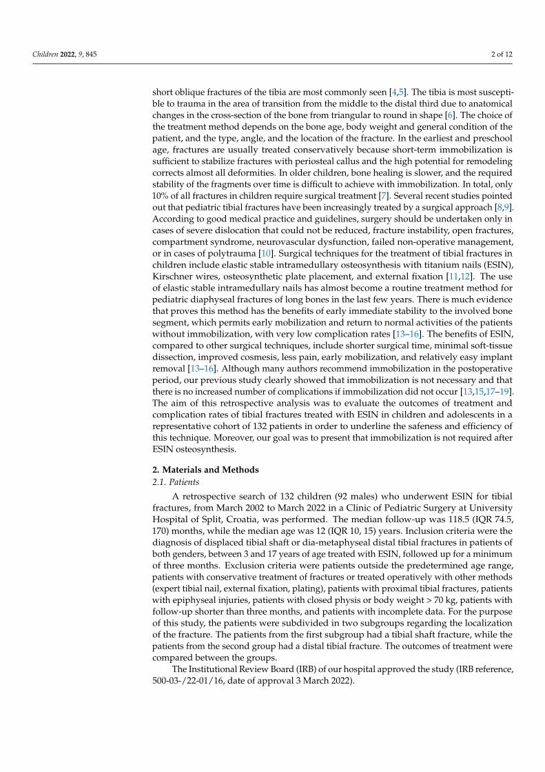
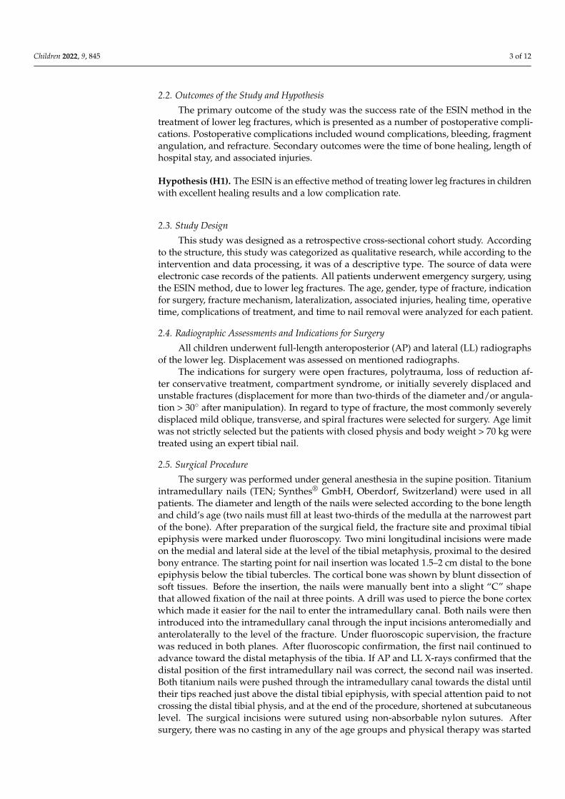
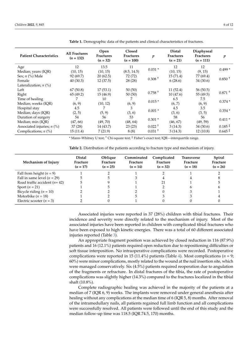
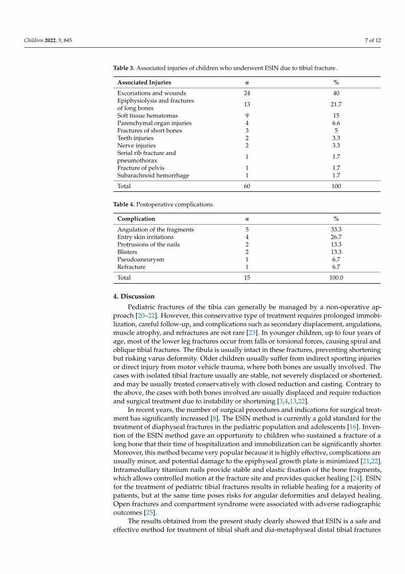
On the first postoperative day, strict rest is required, mostly due to pain control. Fromthe second or third postoperative day (individually, depending on age and other factors,such as general condition of the patient or associated injuries), physical therapy and gettingout of bed with crutches begins. After the patient learns to walk stably using crutches, ifthe other parameters are satisfactory, the patient is discharged to home care, continuedwith ambulatory physical therapy after discharge. For the first three or four weeks, thepatient does not step on the operated leg, and then, after radiological verification of thefracture, begins gradual weight bearing. For the first few days, the bearing is approximatelyone-seventh of the body weight, after which the bearing gradually increases. On average,after 8 to 10 weeks of osteosynthesis, after radiologically verifying a good callus, a crutch-free gait begins. Each patient underwent an intraoperative X-ray after repositioning thebone fragments and placing of titanium elastic nails. Control X-rays were taken sevendays after the procedure, and after one, three, and six months, or until healing of the bonewas completed (Figure 1). Radiological evaluation was carried out using standard APand LL radiographs at each visit to evaluate the consolidation of the fracture and identifycomplications such as secondary displacement, shortening, nail migration, delayed union,nonunion or malunion, and re-fracture (Figure 2). Nonunion was defined as the lackof appropriate healing within six months from index surgery. Malunion was defined asangular deformity of greater than 5–10◦ (depending on patient’s age) in the coronal orsagittal plane. Limb length inequality >1 cm was considered as limb shortening. All nailswere removed under general anesthesia when the radiological healing was evident at themedian of six months.
Figure 1. Displaced tibial shaft fracture in 11-year-old female patient: (A) preoperative AP radiograph;(B) preoperative LL radiograph; (C) AP radiograph one month after surgery; (D) LL radiograph onemonth after surgery; (E) AP radiograph three months after surgery; (F) LL radiograph three monthsafter surgery.
Children 2022, 9, 845 5 of 12
Figure 2. Displaced distal tibial fracture in 9-year-old male patient: (A) preoperative AP radiograph;(B) preoperative LL radiograph; (C) AP radiograph one month after surgery; (D) LL radiograph onemonth after surgery; (E) AP radiograph three months after surgery; (F) LL radiograph three monthsafter surgery.
2.7. Statistical Analysis
The data were analyzed using Microsoft Excel for Windows Version 16.0 (MicrosoftCorporation, Redmond, WA, USA), and Statistical Package for Social Sciences, version19.0 (IBM SPSS Corp, Armonk, NY, USA) software programs. Distributions of quantitativedata were described by medians and interquartile range (IQR), while categorical variableswere expressed in absolute numbers and percentages. Differences in median values ofquantitative variables between the examined groups were tested by the Mann–WhitneyU-test. A comparison of different categories of variations was performed by the Chi-squaretest. In cases where the frequency rate of individual variants was low, Fisher’s exact testwas used. All values of p < 0.05 were considered statistically significant.
3. Results
In the selected study period, which included 132 children operated on using ESINfor tibial fractures, there were 92 (69.7%) boys and 40 (30.3%) girls. The median age was12 (IQR 10, 15) years. There were 100 (75.8%) closed and 32 (24.2%) open fractures. Medianduration of hospital stay was 4.5 (IQR 2, 5) days. Most of the patients (n = 111 (84.1%)) had afracture localized in the shaft of the tibia, while the other 21 (15.9%) had a fracture localizedin the distal part of the bone (Table 1). A total of 32 complicated fractures were recorded.Using Gustilo - Anderson classification a total of 16 fractures (50%) were categorized asgrade 1, 13 (40.6%) as a grade 2 and 3 (9.4%) as a grade 3. Most common mechanism ofinjury was traffic accident (31.8%) and the most common type of fracture was complicatedfracture (24.2%) (Table 2).
Statistical comparison of data between patients who had open and closed fracturesshowed that children with open fractures were significantly older than children with closedfractures (13.5 years (IQR 10, 15) vs. 11 years (IQR 8.5, 14.5); p = 0.031). Furthermore,children with open fractures had a significantly longer hospital stay (7 days (IQR 5, 9) vs.3 days (IQR 3, 6); p = 0.001), a higher rate of associated injuries (n = 14 (43.7%) vs. n = 23(23%); p = 0.022), and a higher rate of postoperative complications (n = 7 (21.9%) vs. n = 8(8%); p = 0.031). No statistically significant difference was found between the examinedgroups in relation to the sex of the patient (p = 0.308), duration of surgery (p = 0.301), andlateralization of the fracture (p = 0.758).
Children 2022, 9, 845 6 of 12
Table 1. Demographic data of the patients and clinical characteristics of fractures.
Patient Characteristics All Fractures(n = 132)
OpenFractures(n = 32)
ClosedFractures(n = 100)
pDistal
Fractures(n = 21)
DiaphysealFractures(n = 111)
p
Age 12 13.5 110.031 *
12 120.499 *Median; years (IQR) (10, 15) (10, 15) (8.5, 14.5) (10, 15) (9, 15)
Sex; n (%) Male 92 (69.7) 20 (62.5) 72 (72)0.308 † 15 (71.4) 77 (69.4)
0.850 †Female 40 (30.3) 12 (37.5) 28 (28) 6 (28.6) 34 (30.6)Lateralization; n (%)Left 67 (50.8) 17 (53.1) 50 (50)
0.758 † 11 (52.4) 56 (50.5)0.871 †
Right 65 (49.2) 15 (46.9) 50 (50) 10 (47.6) 55 (49.5)Time of healing 7 10 7
0.015 *6.5 7.5
0.374 *Median; weeks (IQR) (6, 9) (10, 12) (6, 9) (6, 7) (6, 9)Hospital stay 4.5 7 3
0.001 *4.5 3.5
0.354 *Median; days (IQR) (2, 5) (5, 9) (3, 6) (3, 6) (3, 5)Duration of surgeryMedian; min (IQR)
54(47, 66)
56(49, 70)
53(48, 64) 0.301 * 58
(46, 67)56
(49, 59) 0.411 *
Associated injuries; n (%) 37 (28) 14 (43.7) 23 (23) 0.022 † 3 (14.3) 34 (30.6) 0.185 ‡
Complications; n (%) 15 (11.4) 7 (21.9) 8 (8) 0.031 † 3 (14.3) 12 (10.8) 0.645 ‡
* Mann–Whitney U test; † Chi-square test; ‡ Fisher’s exact test; IQR—interquartile range.
Table 2. Distribution of the patients according to fracture type and mechanism of injury.
Mechanism of InjuryDistal
Fracture(n = 17)
ObliqueFracture(n = 25)
ComminutedFracture(n = 14)
ComplicatedFracture(n = 32)
TransverseFracture(n = 18)
SpiralFracture(n = 26)
Fall from height (n = 9) 1 2 1 2 1 2Fall in same level (n = 29) 5 5 3 4 4 8Road traffic accident (n= 42) 5 9 1 21 1 5Sport (n = 21) 1 5 1 2 6 6Bicycle riding (n = 10) 2 2 2 0 3 1Motorbike (n = 18) 1 2 5 3 3 4Electric scooter (n = 3) 2 0 1 0 0 0
Associated injuries were reported in 37 (28%) children with tibial fractures. Theirincidence and severity were directly related to the mechanism of injury. Most of theassociated injuries have been reported in children with complicated tibial fractures whohave been exposed to high kinetic energies. There was a total of 60 different associatedinjuries reported (Table 3).
An appropriate fragment position was achieved by closed reduction in 116 (87.9%)patients and 16 (12.1%) patients required open reduction due to repositioning difficulties orsoft tissue interposition. No intraoperative complications were recorded. Postoperativecomplications were reported in 15 (11.4%) patients (Table 4). Most complications (n = 9;60%) were minor complications, mostly related to the wound at the nail insertion site, whichwere managed conservatively. Six (4.5%) patients required reoperation due to angulationof the fragments or refracture. In distal fractures of the tibia, the rate of postoperativecomplications was slightly higher (14.3%) compared to the fractures localized in the tibialshaft (10.8%).
Complete radiographic healing was achieved in the majority of the patients at amedian of 7 (IQR 6, 9) weeks. The implants were removed under general anesthesia afterhealing without any complications at the median time of 6 (IQR 5, 8) months. After removalof the intramedullary nails, all patients regained full limb function and all complicationswere successfully resolved. All patients were followed until the end of this study and themedian follow-up time was 118.5 (IQR 74.5, 170) months.
Children 2022, 9, 845 7 of 12
Table 3. Associated injuries of children who underwent ESIN due to tibial fracture.
Associated Injuries n %
Excoriations and wounds 24 40Epiphysiolysis and fracturesof long bones 13 21.7
Soft tissue hematomas 9 15Parenchymal organ injuries 4 6.6Fractures of short bones 3 5Teeth injuries 2 3.3Nerve injuries 2 3.3Serial rib fracture andpneumothorax 1 1.7
Fracture of pelvis 1 1.7Subarachnoid hemorrhage 1 1.7
Total 60 100
Table 4. Postoperative complications.
Complication n %
Angulation of the fragments 5 33.3Entry skin irritations 4 26.7Protrusions of the nails 2 13.3Blisters 2 13.3Pseudoaneurysm 1 6.7Refracture 1 6.7
Total 15 100.0
4. Discussion
Pediatric fractures of the tibia can generally be managed by a non-operative ap-proach [20–22]. However, this conservative type of treatment requires prolonged immobi-lization, careful follow-up, and complications such as secondary displacement, angulations,muscle atrophy, and refractures are not rare [23]. In younger children, up to four years ofage, most of the lower leg fractures occur from falls or torsional forces, causing spiral andoblique tibial fractures. The fibula is usually intact in these fractures, preventing shorteningbut risking varus deformity. Older children usually suffer from indirect sporting injuriesor direct injury from motor vehicle trauma, where both bones are usually involved. Thecases with isolated tibial fracture usually are stable, not severely displaced or shortened,and may be usually treated conservatively with closed reduction and casting. Contrary tothe above, the cases with both bones involved are usually displaced and require reductionand surgical treatment due to instability or shortening [3,4,13,22].
In recent years, the number of surgical procedures and indications for surgical treat-ment has significantly increased [8]. The ESIN method is currently a gold standard for thetreatment of diaphyseal fractures in the pediatric population and adolescents [16]. Inven-tion of the ESIN method gave an opportunity to children who sustained a fracture of along bone that their time of hospitalization and immobilization can be significantly shorter.Moreover, this method became very popular because it is highly effective, complications areusually minor, and potential damage to the epiphyseal growth plate is minimized [21,22].Intramedullary titanium nails provide stable and elastic fixation of the bone fragments,which allows controlled motion at the fracture site and provides quicker healing [24]. ESINfor the treatment of pediatric tibial fractures results in reliable healing for a majority ofpatients, but at the same time poses risks for angular deformities and delayed healing.Open fractures and compartment syndrome were associated with adverse radiographicoutcomes [25].
The results obtained from the present study clearly showed that ESIN is a safe andeffective method for treatment of tibial shaft and dia-metaphyseal distal tibial fractures
Children 2022, 9, 845 8 of 12
with a low number of complications and relatively short length of hospital stay. Most ofthe complications were graded as minor (entry skin irritation, inflammation of the wound),while severe complications such as angulation of the fragments or refracture were rarelyreported. Most of the authors recommend casting for a few weeks after surgery, probablydue to fear of displacement of fragments or angulation [21,26–28]. In this study, we clearlyshowed that titanium intramedullary nailing may be safely performed without casting andone of the most important benefits of this method is early physiotherapy. Several previousreports on upper and lower extremities support this constancy [13,15,17–19].
Swindells and Rajan performed a systematic review of seven different retrospectivestudies regarding tibial ESIN with outcomes of 210 patients [29]. The authors of thosestudies described several indications for use of ESIN in pediatric patients, but most ofthem stated that the main indication was unstable fractures. The longest mean healingtime in those studies was 20.7 weeks, reported by Srivastava et al. [21], and the shortestwas 7 weeks, reported by Kubiak et al. [24]. Reported complication rates were similar,ranging from 12% to 35%. The most commonly reported complications were delayed union,malunion, nonunion, leg length discrepancy, and infections. All seven studies concludedthat ESIN is an effective and safe method for treatment of unstable fractures of the tibialshaft in children and adolescents. They also concluded that most of the pediatric tibialfractures can be treated by a non-operative approach, but in cases where the surgery cannotbe avoided, ESIN provides an acceptable and valuable option.
Griffet et al. reported that all 86 children included in their study were able to haveunrestricted physical activity six months after the treatment with the ESIN method fortibial fractures [30]. They showed that the fixation of pediatric diaphyseal tibial fracturesusing ESIN is an effective method of treatment in pediatric patients and adolescents.
Uludag et al. published a study which included 20 children. The mean time ofradiographic healing was 11 weeks and there were six (30%) instances of irritation andinfections at the nail entry site [31]. Furthermore, they reported that three of six patientswith an open fracture had infections of the wound. All their patients gained full range ofmotion of the ankle and knee joints. They concluded that intramedullary fixation with ESINprovides favorable outcomes in the treatment of unstable pediatric tibial shaft fracturesthat cannot be reduced with conservative treatment modalities.
Onta et al. reported that their 18 patients had a mean hospitalization time of 5.7 daysand that all children achieved radiographic healing at the mean time of 13.3 weeks [32]. Allof their patients had excellent final results and all of them had full range of movement atthe knee joint. Four children had minor complications (nail protrusion and skin irritation),which were managed non-operatively. They concluded that ESIN had benefits of low bloodloss compared to plating, and they also reported easier nursing care, early ambulation, andno complications of prolonged immobilization.
Shen et al. published a study with 21 children that went under the surgery procedurewith the ESIN method due to severely displaced dia-metaphyseal distal tibial fractures.They reported a mean hospitalization time of 3.9 days, and the mean time of the nail removalwas 7.1 months [33]. A total of 19 patients achieved radiographic healing at the mean timeof 9.6 weeks and two patients had delayed healing 10 months after the surgery. Theirstudy showed good functional and radiological results in the pediatric population whohad severely displaced DTDMJ (distal tibial diaphyseal metaphyseal junction) fracturesthat could not be casted.
Kc et al. reported that in their study, which included 45 children treated with the ESINmethod due to fracture of tibia, that they had a mean healing time of 11.17 weeks [34]. Theyrecorded 20 postoperative complications which included 2 malunions, 4 delayed unions,3 limb shortening, 2 limb lengthening, 6 nail prominences and skin irritations, 2 superficialinfections on the nail entry site, and 1 refracture. However, none of their patients requiredsecondary surgical intervention due to those complications. They concluded that ESIN isa simple, easy, reliable, and effective method for management of pediatric tibial fractures
Children 2022, 9, 845 9 of 12
with shorter operative time, lesser blood loss, shorter length of hospital stay, and adequatetime for bone healing.
Pennock et al. in their study compared 44 patients who underwent ESIN with 26 pa-tients who received open reduction with internal fixation (ORIF) for tribal shaft frac-tures [28]. Patients that underwent ORIF had a longer mean surgical time and their meancasting time was seven weeks; minor complications were recorded in 10 (38%) patients andmajor ones were recorded in 3 (12%) patients. Patients that underwent ESIN had a shortersurgical time, and their mean casting time was 10.5 weeks; minor complications wererecorded in 12 (27%) cases, while major complications were recorded in 8 (18%) cases. Theyconcluded that patients treated with ORIF tend to heal and mobilize a few weeks faster,they have slightly more anatomic reductions at final healing, and they are less likely torequire implant removal. Moreover, they pointed out that both ESIN and ORIF treatmentscontribute to a faster return to activities and that potential advantages of ORIF must bebalanced with the potential increased risk of wound complications.
There is no exact consensus among the authors which age or height should be setas a limit for ESIN. Many authors used ESIN for treatment of pediatric and adolescenttibial fractures without any limits [13,22,33,35]. In previous studies, the rate of delayedhealing was shown to vary by around 10% [13,22,33]. Gordon et al. in their study reportedthat patients with delayed union were older and they concluded the lack of stability maybe the key factor behind the delayed healing [22]. Although there is a wide age (height)range in the majority of the studies, age of the patients ranges between 10 and 12 years ofage [13,22,23,26,33,35,36]. A recent study showed that patients with tibial fractures whoweigh 50 kg or less and with proximal tibial growth plates wide open can be treated withelastic stable intramedullary nailing, while more mature adolescents benefit from rigidintramedullary nailing as rigid nailing allows more precise fracture alignment withoutincreased risk of growth disturbance [36]. Similar findings were observed in another recentstudy [37]. Hanf-Osetek et al. in their study compared children weighing less than 50 kg ormore than 50 kg and found that the use of ESIN in displaced tibial shaft fractures in growingchildren weighing 50 kg or more is acceptable and safe [38]. A recent study performed byThabet et al. compared adolescents treated for tibial shaft fractures using ESIN, interlockingnails, plates, and screws or external fixators and showed that open fractures had highercomplication rates but no statistically significant differences in complication rates betweenthe fixation methods was observed [39]. In general, the good results using the ESIN methodmay be obtained when the surgeon has a good knowledge of the method, respects andunderstands indications for surgery, and the main principles of the correction of the fractureand its stability [40].
In our study, of the sample of 132 children, the mean time of radiographic healingwas seven weeks. We recorded complications in fifteen (11.36%) patients, which includedfive angulations of the fragments, four entry site irritations, two protrusions of the nails,two skin blisters, one pseudoaneurysm, and one refracture. Six (4.5%) of our patientsneeded a reoperation due to angulations of the fragment and refracture. The median ofsurgical time was 56 min. These results are similar to previous reports. Although mostof the previously mentioned studies reported usage of a postoperative cast or a slab, ourdepartment policy is that after the surgical procedure with ESIN, no type of immobilizationis needed. According to our positive results of this study, which included 132 patients, weproved that cast immobilization is not needed after the surgery procedure with the ESINmethod due to tibia fracture.
The results of this study must be interpreted within the context of several limita-tions. First, this was a single-center study, and the data were collected retrospectively.Furthermore, sample size was relatively small (although significantly higher than in mostof the published reports). Next, there was a lack of a comparison group because a verylow number of the patients were treated with other surgical techniques (such as plating,external fixation, or expert tibial nail). The majority of pediatric tibial fractures can besuccessfully treated conservatively by non-operative management and only the patients
Children 2022, 9, 845 10 of 12
who fail conservative treatment (or are open/polytrauma/initially unable to reduce) areselected for surgery (most commonly ESIN). Accordingly, we were not able to design anadequate control group. Moreover, as this was a retrospective study, we could not findthe data regarding functional outcome for the majority of the patients, especially for thoseoperated on in earlier years. Prospective, multicenter studies with a larger sample sizeneed to be conducted in the future before any definite conclusions in this regard shouldbe drawn.
5. Conclusions
ESIN fulfills all criteria of minimally invasive bone surgery and is a highly effectivetreatment for pediatric tibial unstable fractures with a low rate of complications. Further-more, the results of this study clearly showed that titanium intramedullary nailing may besafely performed without casting, and one of the most important benefits of this method isearly physiotherapy.
Author Contributions: Conceptualization, Z.P. and D.F.; Formal analysis, V.V. and Z.P.; Writing—original draft preparation, M.J., V.V. and C.M.L.M.; Writing—review and editing, Z.P., C.M.L.M. andD.F. All authors have read and agreed to the published version of the manuscript.
Funding: This research received no external funding.
Institutional Review Board Statement: The study was conducted according to the guidelines of theDeclaration of Helsinki and approved by the Institutional Review Board of the University Hospital ofSplit (IRB reference, 500-03-/22-01/16, date of approval 3 March 2022).
Informed Consent Statement: Not applicable.
Data Availability Statement: The data presented in this study are available upon request of therespective author.
Conflicts of Interest: The authors declare no conflict of interest.
References1. Metaizeau, J.D.; Denis, D. Update on leg fractures in paediatric patients. Orthop. Traumatol. Surg. Res. 2019, 105, 143–151.
[CrossRef]2. Palmu, S.A.; Auro, S.; Lohman, M.; Paukku, R.T.; Peltonen, J.I.; Nietosvaara, Y. Tibial fractures in children. A retrospective 27-year
follow-up study. Acta Orthop. 2014, 85, 513–517. [CrossRef]3. Martus, J.E. Operative fixation versus cast immobilization: Tibial shaft fractures in adolescents. J. Pediatr. Orthop. 2021, 41, 33–38.
[CrossRef]4. Joeris, A.; Lutz, N.; Wicki, B.; Slongo, T.; Audigé, L. An epidemiological evaluation of pediatric long bone fractures—A
retrospective cohort study of 2716 patients from two Swiss tertiary pediatric hospitals. BMC Pediatr. 2014, 14, 314. [CrossRef]5. Weber, B.; Kalbitz, M.; Baur, M.; Braun, C.K.; Zwingmann, J.; Pressmar, J. Lower leg fractures in children and adolescents-
comparison of conservative vs. ECMES Treatment. Front. Pediatr. 2021, 9, 597870. [CrossRef]6. Fox, J.; Enriquez, B.; Bompadre, V.; Carlin, K.; Dales, M. Observation Versus Cast Treatment of Toddler’s Fractures. J. Pediatr.
Orthop. 2022, 42, e480–e485. [CrossRef]7. Patel, N.K.; Horstman, J.; Kuester, V.; Sambandam, S.; Mounasamy, V. Pediatric tibial shaft fractures. Indian J. Orthop. 2018, 52,
522–528. [CrossRef]8. Stenroos, A.; Laaksonen, T.; Nietosvaara, N.; Jalkanen, J.; Nietosvaara, Y. One in three of pediatric tibia shaft fractures is currently
treated operatively: A 6-year epidemiological study in two university hospitals in Finland treatment of pediatric tibia shaftfractures. Scand. J. Surg. 2018, 107, 269–274. [CrossRef]
9. Kleiner, J.E.; Raducha, J.E.; Cruz, A.I., Jr. Increasing rates of surgical treatment for paediatric tibial shaft fractures: A nationaldatabase study from between 2000 and 2012. J. Child. Orthop. 2019, 13, 213–219. [CrossRef]
10. Cruz, A.I., Jr.; Raducha, J.E.; Swarup, I.; Schachne, J.M.; Fabricant, P.D. Evidence-based update on the surgical treatment ofpediatric tibial shaft fractures. Curr. Opin. Pediatr. 2019, 31, 92–102. [CrossRef]
11. Mashru, R.P.; Herman, M.J.; Pizzutillo, P.D. Tibial shaft fractures in children and adolescents. J. Am. Acad. Orthop. Surg. 2005, 13,345–352. [CrossRef] [PubMed]
12. Hogue, G.D.; Wilkins, K.E.; Kim, I.S. Management of pediatric tibial shaft fractures. J. Am. Acad. Orthop. Surg. 2019, 27, 769–778.[CrossRef]
Children 2022, 9, 845 11 of 12
13. Furlan, D.; Pogorelic, Z.; Biocic, M.; Juric, I.; Budimir, D.; Todoric, J.; Šušnjar, T.; Todoric, D.; Meštrovic, J. Elastic stableintramedullary nailing for pediatric long bone fractures: Experience with 175 fractures. Scand. J. Surg. 2011, 100, 208–215.[CrossRef]
14. Egger, A.; Murphy, J.; Johnson, M.; Hosseinzadeh, P.; Louer, C. Elastic stable intramedullary nailing of pediatric tibial fractures.JBJS Essent. Surg. Tech. 2020, 10, e19.00063. [CrossRef]
15. Pogorelic, Z.; Gulin, M.; Jukic, M.; Biliškov, A.N.; Furlan, D. Elastic stable intramedullary nailing for treatment of pediatricforearm fractures: A 15-year single centre retrospective study of 173 cases. Acta Orthop. Traumatol. Turc. 2020, 54, 378–384.[CrossRef]
16. Cosma, D.; Vasilescu, D.E. Elastic stable intramedullary nailing for fractures in children—Specific applications. Clujul Med. 2014,87, 147–151. [CrossRef]
17. Pogorelic, Z.; Vodopic, T.; Jukic, M.; Furlan, D. Elastic stable intramedullary nailing for treatment of pediatric femoral fractures; A15-year single centre experience. Bull. Emerg. Trauma. 2019, 7, 169–175. [CrossRef]
18. Pogorelic, Z.; Kadic, S.; Milunovic, K.P.; Pintaric, I.; Jukic, M.; Furlan, D. Flexible intramedullary nailing for treatment of proximalhumeral and humeral shaft fractures in children: A retrospective series of 118 cases. Orthop. Traumatol. Surg. Res. 2017, 103,765–770. [CrossRef]
19. Pogorelic, Z.; Capitain, A.; Jukic, M.; Žufic, V.; Furlan, D. Flexible intramedullary nailing for radial neck fractures in children.Acta Orthop. Traumatol. Turc. 2020, 54, 618–622. [CrossRef]
20. Goodwin, R.C.; Gaynor, T.; Mahar, A.; Oka, R.; Lalonde, F.D. Intramedullary flexible nail fixation of unstable pediatric tibialdiaphyseal fractures. J. Pediatr. Orthop. 2005, 25, 570–576. [CrossRef]
21. Srivastava, A.K.; Mehlman, C.T.; Wall, E.J.; Do, T.T. Elastic stable intramedullary nailing of tibial shaft fractures in children. J.Pediatr. Orthop. 2008, 28, 152–158. [CrossRef]
22. Gordon, J.E.; Gregush, R.V.; Schoenecker, P.L.; Dobbs, M.B.; Luhmann, S.J. Complications after titanium elastic nailing of pediatrictibial fractures. J. Pediatr. Orthop. 2007, 27, 442–446. [CrossRef] [PubMed]
23. O’Brien, T.; Weisman, D.S.; Ronchetti, P.; Piller, C.P.; Maloney, M. Flexible titanium nailing for the treatment of the unstablepediatric tibial fracture. J. Pediatr. Orthop. 2004, 24, 601–609. [CrossRef]
24. Kubiak, E.N.; Egol, K.A.; Scher, D.; Wasserman, B.; Feldman, D.; Koval, K.J. Operative treatment of tibial fractures in children: Areelastic stable intramedullary nails an improvement over external fixation? J. Bone Jt. Surg. Am. 2005, 87, 1761–1768. [CrossRef]
25. Pennock, A.T.; Huang, S.G.; Pedowitz, J.M.; Pandya, N.K.; McLaughlin, D.C.; Bastrom, T.P.; Ellis, H.B. Risk factors for adverseradiographic outcomes after elastic stable intramedullary nailing of unstable diaphyseal tibia fractures in children. J. Pediatr.Orthop. 2020, 40, 481–486. [CrossRef]
26. Sankar, W.N.; Jones, K.J.; David Horn, B.; Wells, L. Titanium elastic nails for pediatric tibial shaft fractures. J. Child. Orthop. 2007,1, 281–286. [CrossRef] [PubMed]
27. Vallamshetla, V.R.; De Silva, U.; Bache, C.E.; Gibbons, P.J. Flexible intramedullary nails for unstable fractures of the tibia inchildren. An eight-year experience. J. Bone Jt. Surg. Br. 2006, 88, 536–540. [CrossRef] [PubMed]
28. Pennock, A.T.; Bastrom, T.P.; Upasani, V.V. Elastic Intramedullary Nailing Versus Open Reduction Internal Fixation of PediatricTibial Shaft Fractures. J. Pediatr. Orthop. 2017, 37, e403–e408. [CrossRef]
29. Swindells, M.G.; Rajan, R.A. Elastic intramedullary nailing in unstable fractures of the paediatric tibial diaphysis: A systematicreview. J. Child. Orthop. 2010, 4, 45–51. [CrossRef]
30. Griffet, J.; Leroux, J.; Boudjouraf, N.; Abou-Daher, A.; El Hayek, T. Elastic stable intramedullary nailing of tibial shaft fractures inchildren. J. Child. Orthop. 2011, 5, 297–304. [CrossRef]
31. Uludag, A.; Tosun, H.B. Treatment of unstable pediatric tibial shaft fractures with titanium elastic nails. Medicina 2019, 55, 266.[CrossRef] [PubMed]
32. Onta, P.R.; Thapa, P.; Sapkota, K.; Ranjeet, N.; Kishore, A.; Gupta, M. Outcome of diaphyseal fracture of tibia treated with flexibleintramedullary nailing in pediatrics age group; A prospective study. Am. J. Public Health 2015, 3, 65–68.
33. Shen, K.; Cai, H.; Wang, Z.; Xu, Y. Elastic stable intramedullary nailing for severely displaced distal tibial fractures in children.Medicine 2016, 95, e4980. [CrossRef]
34. Kc, K.M.; Acharya, P.; Sigdel, A. Titanium Elastic Nailing System (TENS) for tibia fractures in children: Functional outcomes andcomplications. JNMA J. Nepal. Med. Assoc. 2016, 55, 55–60. [CrossRef] [PubMed]
35. Aslani, H.; Tabrizi, A.; Sadighi, A.; Mirblok, A.R. Treatment of open pediatric tibial fractures by external fixation versus flexibleintramedullary nailing: A comparative study. Arch. Trauma Res. 2013, 2, 108–112. [CrossRef]
36. Widbom-Kolhanen, S.; Helenius, I. Intramedullary nailing of paediatric tibial fractures: Comparison between flexible and rigidnails. Scand. J. Surg. 2021, 110, 265–270. [CrossRef]
37. Williams, K.A.; Thier, Z.T.; Mathews, C.G.; Locke, M.D. Physeal-Sparing Rigid Intramedullary Nailing in Adolescent Tibial ShaftFractures: A Pilot Study. Cureus 2021, 13, e13893. [CrossRef]
38. Hanf-Osetek, D.; Bilski, P.; Łabadz, D.; Snela, S. Tibial shaft fractures in children: Flexible intramedullary nailing in growingchildren especially weighing 50 kg (110 lbs) or more. J. Pediatr. Orthop. B 2022. [CrossRef]
Children 2022, 9, 845 12 of 12
39. Thabet, A.M.; Craft, M.; Pisquiy, J.; Jeon, S.; Abdelgawad, A.; Azzam, W. Tibial shaft fractures in the adolescents: Treatmentoutcomes and the risk factors for complications. Injury 2022, 53, 706–712. [CrossRef] [PubMed]
40. Lascombes, P.; Haumont, T.; Journeau, P. Use and abuse of flexible intramedullary nailing in children and adolescents. J. Pediatr.Orthop. 2006, 26, 827–834. [CrossRef]