Footprints of water and energy inputs in food production – Global perspectives
Upload
khangminh22Category
view
1download
0
Article
Bacterial Footprints in Elastic Pillared Microstructures
Susarrey-Arce, Arturo, Hernández-Sánchez, José Federico, Marcello,Marco, Diaz-Fernandez, Yuri, Oknianska, Alina, Sorzabal-Bellido, Ioritz, Tiggelaar, Roald, Lohse, Detlef, Gardeniers, Han, Snoeijer, Jacco, Marin, Alvaro and Raval, Rasmita
Available at http://clok.uclan.ac.uk/33618/
Susarrey-Arce, Arturo, Hernández-Sánchez, José Federico, Marcello, Marco, Diaz-Fernandez, Yuri, Oknianska, Alina, Sorzabal-Bellido, Ioritz, Tiggelaar, Roald, Lohse, Detlef, Gardeniers, Han et al (2018) Bacterial Footprints in Elastic Pillared Microstructures. ACS Applied Bio Materials, 1 (5). pp. 1294-1300. ISSN 2576-6422
It is advisable to refer to the publisher’s version if you intend to cite from the work.http://dx.doi.org/10.1021/acsabm.8b00176
For more information about UCLan’s research in this area go to http://www.uclan.ac.uk/researchgroups/ and search for <name of research Group>.
For information about Research generally at UCLan please go to http://www.uclan.ac.uk/research/
All outputs in CLoK are protected by Intellectual Property Rights law, includingCopyright law. Copyright, IPR and Moral Rights for the works on this site are retainedby the individual authors and/or other copyright owners. Terms and conditions for useof this material are defined in the policies page.
CLoKCentral Lancashire online Knowledgewww.clok.uclan.ac.uk
1 Bacterial Footprints in Elastic Pillared Microstructures2 Arturo Susarrey-Arce,*,†,○ Jose Federico Hernandez-Sanchez,*,‡,○ Marco Marcello,¶
3 Yuri Diaz-Fernandez,† Alina Oknianska,§ Ioritz Sorzabal-Bellido,† Roald Tiggelaar,∥ Detlef Lohse,⊥
4 Han Gardeniers,# Jacco Snoeijer,*,⊥ Alvaro Marin,⊥ and Rasmita Raval*,†
5†Open Innovation Hub for Antimicrobial Surfaces at the Surface Science Research Centre and Department of Chemistry, University
6 of Liverpool, Oxford Street, Liverpool L69 3BX, United Kingdom
7‡Division of Physical Sciences and Engineering and Clean Combustion Research Center, King Abdullah University of Science and
8 Technology, Thuwal 23955-6900, Saudi Arabia
9¶Institute of Integrative Biology, University of Liverpool, Biosciences Building, Liverpool L69 7ZB, United Kingdom
10§School of Health Sciences, Liverpool Hope University, Hope Park, Liverpool L16 9JD, United Kingdom
11∥NanoLab Cleanroom, MESA+ Institute for Nanotechnology, University of Twente, P.O. Box 217, Enschede 7500AE, The
12 Netherlands
13⊥Physics of Fluids Group, MESA+ Institute for Nanotechnology, J.M. Burgers Centre for Fluid Dynamics, University of Twente,
14 P.O. Box 217, Enschede 7500AE, The Netherlands
15#Mesoscale Chemical Systems, MESA+ Institute for Nanotechnology, University of Twente, P.O. Box 217, Enschede 7500AE, The
16 Netherlands
17 *S Supporting Information
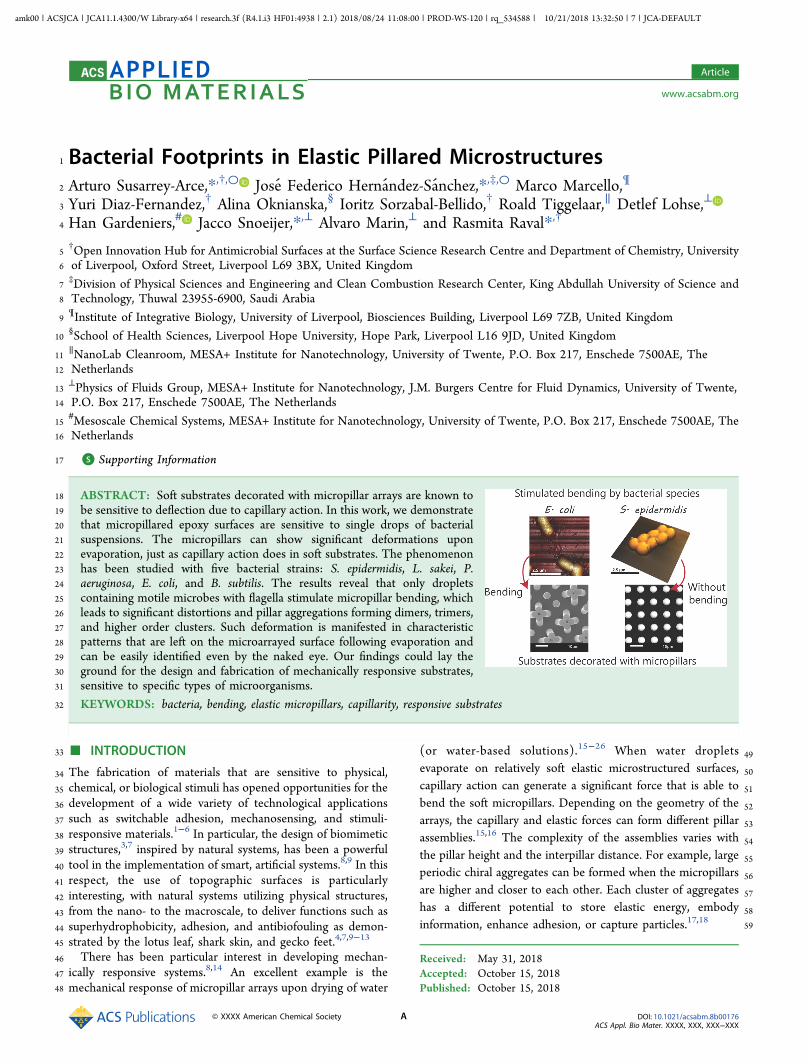
18 ABSTRACT: Soft substrates decorated with micropillar arrays are known to19 be sensitive to deflection due to capillary action. In this work, we demonstrate20 that micropillared epoxy surfaces are sensitive to single drops of bacterial21 suspensions. The micropillars can show significant deformations upon22 evaporation, just as capillary action does in soft substrates. The phenomenon23 has been studied with five bacterial strains: S. epidermidis, L. sakei, P.24 aeruginosa, E. coli, and B. subtilis. The results reveal that only droplets25 containing motile microbes with flagella stimulate micropillar bending, which26 leads to significant distortions and pillar aggregations forming dimers, trimers,27 and higher order clusters. Such deformation is manifested in characteristic28 patterns that are left on the microarrayed surface following evaporation and29 can be easily identified even by the naked eye. Our findings could lay the30 ground for the design and fabrication of mechanically responsive substrates,31 sensitive to specific types of microorganisms.
32 KEYWORDS: bacteria, bending, elastic micropillars, capillarity, responsive substrates
33 ■ INTRODUCTION
34 The fabrication of materials that are sensitive to physical,35 chemical, or biological stimuli has opened opportunities for the36 development of a wide variety of technological applications37 such as switchable adhesion, mechanosensing, and stimuli-38 responsive materials.1−6 In particular, the design of biomimetic39 structures,3,7 inspired by natural systems, has been a powerful40 tool in the implementation of smart, artificial systems.8,9 In this41 respect, the use of topographic surfaces is particularly42 interesting, with natural systems utilizing physical structures,43 from the nano- to the macroscale, to deliver functions such as44 superhydrophobicity, adhesion, and antibiofouling as demon-45 strated by the lotus leaf, shark skin, and gecko feet.4,7,9−13
46 There has been particular interest in developing mechan-47 ically responsive systems.8,14 An excellent example is the48 mechanical response of micropillar arrays upon drying of water
49(or water-based solutions).15−26 When water droplets
50evaporate on relatively soft elastic microstructured surfaces,
51capillary action can generate a significant force that is able to
52bend the soft micropillars. Depending on the geometry of the
53arrays, the capillary and elastic forces can form different pillar
54assemblies.15,16 The complexity of the assemblies varies with
55the pillar height and the interpillar distance. For example, large
56periodic chiral aggregates can be formed when the micropillars
57are higher and closer to each other. Each cluster of aggregates
58has a different potential to store elastic energy, embody59information, enhance adhesion, or capture particles.17,18
Received: May 31, 2018Accepted: October 15, 2018Published: October 15, 2018
Article
www.acsabm.org
© XXXX American Chemical Society A DOI: 10.1021/acsabm.8b00176ACS Appl. Bio Mater. XXXX, XXX, XXX−XXX
amk00 | ACSJCA | JCA11.1.4300/W Library-x64 | research.3f (R4.1.i3 HF01:4938 | 2.1) 2018/08/24 11:08:00 | PROD-WS-120 | rq_534588 | 10/21/2018 13:32:50 | 7 | JCA-DEFAULT

60 The demonstration of mechanically responsive topographic61 surfaces to bacterial stimuli during evaporation of small62 droplets is of great interest and has not been demonstrated63 before. Furthermore, the deflections seen in our systems are64 significant, leading to pillar aggregations into dimers, trimers,65 and higher order clusters. Recently, the formation of biofilm66 strings and networks between topographic pillars has been67 demonstrated in liquid media;27 however, the mechanical68 response of the pillars to bacterial presence upon evaporation69 is not observed. Chew and coauthors have shown small70 deflections of macropillared surfaces in response to the71 differential pressure exerted by biofilm growth within a growth72 chamber over a 24 h period,28 while Biais29 and Ng30 et al.73 have investigated the interaction of bacterial pili with pillared74 structures.75 Here, we demonstrate how epoxy-made soft surfaces76 containing micropillar arrays interact with suspensions of77 different bacterial species. Our results suggest that the presence78 of motile bacteria with flagella drastically increases the79 mechanical response of the pillars, actively bending soft80 topographical substrates in the area contained within the81 contact line. In contrast, solutions containing nonmotile82 bacteria do not generate such responses. We attribute this to83 the ability of motile bacteria to interact with each other and84 with their topographical environment. Importantly, the85 response of the microarray is sensitive to the type and86 concentration of bacteria in the solution. These promising87 results could lay the foundation for the development of devices88 that are selectively responsive to specific microorganisms,89 paving the way to construct smart, fast, and cost-effective90 diagnostic tools.
91 ■ RESULTS AND DISCUSSION92 One of the key parameters in the mechanical response of soft93 micropillar arrays is the aspect ratio of a single pillar. We94 investigated the effect of the pillar aspect ratio by fabricating95 regular patterns of cylindrical pillars with a constant diameter96 (5 μm) and interspacing (5 μm) and with variable height97 (from 5 to 45 μm). The patterns were created on epoxy resin98 using a method described before31−35 based on casting99 uncured epoxy on a negative polydimethilsiloxane (PDMS)100 mold, followed by curing and mechanically removing of the101 mold. The micropatterns were transferred efficiently, with a102 high degree of fidelity, as shown by scanning electron
f1 103 microscopy (SEM) imaging (Figure 1 and Figure S1).104 These microstructured substrates can be susceptible to105 elastocapillary forces in the presence of pure liquids. Therefore,106 we evaluated the effect of pure water over a surface decorated107 with micropillars with lengths varying from 5 to 45 μm (Figure108 1) during the evaporation of water droplets (Figure 1). In109 these experiments, the liquid filled up the space between the110 pillars, resulting in an almost square-shaped droplet contour.111 Once the droplet spreads on the substrate, the liquid contact112 line is blocked by the pillared structure and remains113 immobilized (pinned) for the rest of the drying process.31
114 Figure 1b shows that after complete evaporation, there is115 almost no trace of the droplet, except at the droplet contour,116 where lines of pillars were bent by capillary action at the117 contact line shown in Video S1.18−23,31
118 In the systems studied, the pillar lattice was kept constant119 (i.e., l = d = 5 μm), but different pillar heights (h) ranging from120 h = 5 to 45 μm were fabricated. Thus, a range of121 micropatterned surfaces were generated with different aspect
122ratios (i.e., h/d = 3 to h/d = 9). For large aspect ratio123structures, we observed significant perturbation of the124micropillars in the area within the contact line boundary.125Imaging at low magnifications, or even examination by the126naked eye, revealed that the inner part of the pattern was127opaque, suggesting that the whole array of pillars inside the128dried droplet perimeter was modified (Figure 1c). Higher129magnification SEM imaging showed that this optical contrast
Figure 1. (a) Representative SEM image of pillared structure (H15),showing the topographic descriptors for the array. The pillars have acylindrical shape and a height (h) of 15 μm and a diameter (d) of 5μm forming a square lattice with an interpillar distance (l) = 5 μm.(b) Pure water droplet evaporating on the H15 substrate withmicropillars leaving a distinct square-shaped contact line with noperturbation of pillars within this contour. (c) Pure water dropletevaporating on the H22 substrate with micropillars leaving a distinctshaped contact line pattern with significant modification of themicropillars within the contact line boundary. Time needed isrepresented in a dimensionless form as the ratio between the elapsedtime (t) and the final evaporation time (tf). (d−i) Pillared structureswith constant (d = 5 μm) and different pillar heights (h) of (d) 15 μm(H15), (e) 22 μm (H22), (f) 28 μm (H28), (g) 33 μm (H33), (h) 38μm (H38), and (i) 45 μm (H45). SEM images are presented for thedifferent heights after evaporation of pure water droplets, probing thesensitivity of the structures to pure elastocapillary bending.
ACS Applied Bio Materials Article
DOI: 10.1021/acsabm.8b00176ACS Appl. Bio Mater. XXXX, XXX, XXX−XXX
B

130 effect was caused by local bending of the micropillars (Figure131 1d−i), with the pillars bent toward each other forming clusters132 and adopting complex geometries, e.g., dimer (white box),133 tetramer (blue box), hexamer (red box), octamer (yellow box),134 and nonamer (orange box). Similar effects have been reported135 before for larger pillar aspect ratios18,24,25 and were attributed136 to the elastocapillary coalescence of the flexible structures.15,18
137 In our experiments, as the aspect ratio decreased, the clusters138 contained lower numbers of aggregated pillars until a critical139 aspect ratio h/d = 3, for which no clusters were observed in the140 inner part of the droplet (Figure 1d).141 The deformation of the pillars, upon water evaporation, is142 induced by the surface tension (γ) of the water/air meniscus143 connecting the pillars, and the corresponding force scales as Fc144 ∼ γr, where r = d/2 is the pillar radius.21,36 The natural145 elasticity of the pillars resists deformation with an elastic force146 FE ∼ Elr4/h3, where E is the Young modulus and l the147 interpillar distance.18 This expression is analogous to the usual148 beam theory for slender objects, showing that the resistance to149 bending decreases strongly when the pillars height increases. If150 we define the pillar bending sensitivity as the ratio of capillary151 and elastic forces, Fc/FE = γ/El(h/r)3, we can conclude that it152 is directly proportional to the cubic power of the pillar aspect153 ratio h/r; i.e., slender pillars are more prone to be bent by154 surface tension, while wide pillars tend to be more stable.155 Under our experimental conditions, no pillar coalescence is156 observed in the area within the contact line boundary from157 pure water when the aspect ratio is below h/d = 3,31 suggesting158 that this is the critical aspect ratio threshold for which capillary159 action equals restoration mechanical stress on the micropillars.160 It is important to note that in this analysis, we are not161 considering the effect of the contact line. This effect is162 expected to have an enhanced deforming effect, but an163 accurate evaluation of this factor is beyond existing164 phenomenological modeling capabilities and will be the subject165 of future studies. Consequently, all of the results described166 below applies exclusively to the inner part of the dried pattern167 left by the droplet, ignoring possible contact line effects.168 Bacterial-Triggered Coalescence of Pillars. From the169 elastocapillary assay discussed in the previous section, we170 identified the critical region within the topographic parameter171 space where the micropillared structure is able to resist172 capillary deformation in the presence of pure water droplets.173 Such a surface opens up the possibility to sense the presence of174 a second entity introduced into water (i.e., bacterial cells),175 which could induce a response in its own right. This critical176 structure corresponds to an aspect ratio h/d ≈ 3 and pillar177 height h = 15 μm (H15, Figure 1d), as discussed in the178 previous section.179 We, therefore, investigated the drying process of droplets180 containing different bacteria species over the H15 pillared181 structures. Similar to the case of pure water droplets, a pinned182 square drop shape is found. However, the patterns observed183 within the contact line formed after complete evaporation of184 the droplets were surprisingly different for some bacteria as185 clearly observed in Video S2.186 Five different bacterial species, with a wide range of187 morphological and biological characteristics were investigated:188 S. epidermidis, L. sakei, P. aeruginosa, E. coli, and B. subtilis. The189 patterns formed after evaporation of droplets containing
f2 190 different bacteria on H15 pillar substrates (Figure 2) can be191 classified in two main groups: one group displaying significant192 bending of the pillars within the pattern (P. aeruginosa, E. coli,
193and B. subtilis) and another group that does not induce any194responsive bending of the pillars in the center of the dried195patterns (S. epidermidis and L. sakei). These distinct behaviors196could be observed even by the naked eye in the form of a local197change in contrast at the surface (Figure 2, 5×). At higher198magnifications, the difference is clearly revealed to be199associated with the coalescence of adjacent pillars (Figure 2,20040× and SEM (100×)).201We attempted to correlate these results to the general202 t1characteristics of the bacterial species used in this work (Table203 t11). Atomic force microscopy (AFM) imaging confirmed the204expected size and cell morphology for these bacteria: Gram-205negative (−) P. aeruginosa and E. coli as well as Gram-positive206(+) B. subtilis and L. sakei present a rod-like shape, while207Gram-positive (+) S. epidermidis has a spheroidal shape208(Figure S2). In addition, L. sakei and S. epidermidis are not209motile (no flagella present), while the other three strains have210flagella. From these considerations, we can conclude that the211different pattern types showed in Figure 2 (bending vs212nonbending) cannot be explained considering bacteria cell
Figure 2. Typical patterns left over H15 substrates after theevaporation of different bacterial species: (a1−a3) S. epidermidis,(b1−b3) L. sakei, (c1−c3) P. aeruginosa, (d1−d3), E. coli, (e1−e3) B.subtilis. Here, the concentration of the different bacterial species is 107
CFU/mL. The different columns correspond to different degrees ofmagnifications: 5× (left column), 40× (central column) by using aconfocal microscope, and >100× with SEM (right column).
ACS Applied Bio Materials Article
DOI: 10.1021/acsabm.8b00176ACS Appl. Bio Mater. XXXX, XXX, XXX−XXX
C

213 morphology only. Similarly, the stiffness of the cell envelop214 does not appear to play a critical role, with rigid Gram-positive215 bacteria and softer Gram-negative bacteria distributed among216 both pattern groups.217 Interestingly, the different response of the microstructures218 upon evaporation of the bacterial solutions correlates with the219 presence or absence of flagella. Bacteria with flagella clearly220 induce a bending response in the H15 pillars, while221 nonflagellated bacteria are unable to bend the pillars when222 used at the same bacterial concentration.223 For the bacteria that induce a mechanical response, a224 concentration dependence is observed, with deformation of225 pillar clusters at the center of the dried droplet observed for226 bacteria concentrations between 107 CFU/mL and 109 CFU/227 mL, while none is observed for lower bacteria concentrations228 (105 CFU/mL). At low concentrations, only the perimeter229 near the corners of the dried square pattern presented
f3 230 coalescence of the pillars (Figure 3a−c). This can be attributed231 to the coffee-stain-like effect, able to drag bacterial cells toward232 the droplet contact line, increasing the local concentration of233 bacteria during evaporation.31 Interestingly, bacterial cells234 without flagella confirm the absence of responsivity at different235 cell concentrations (Figure 3d−f).
236No clear correlation was observed between bacterial species237and the cluster symmetries obtained (e.g., dimer, trimer,238tetramer, etc.). However, the data suggests that the assemblies239emerge due to perturbation of the balance between capillary240forces and elastic restoration forces in the presence of bacteria241with flagella. In the next section, we discuss a possible242mechanism for this distinctive behavior.243Possible Origin of Bacteria-Induced Coalescence. In244the previous sections, we determined the critical pillar aspect245ratio, below which surface tension forces were not able to246induce pillar coalescence in pure water. Interestingly, the247responsivity is dramatically enhanced when the droplets248contain flagellated bacteria. While the bending process at the249perimeter of the contact line appears similar in both cases,250coalescence within the central area is triggered at smaller251aspect ratios by the presence of bacteria with flagella. This252enhanced pillar bending effect results in characteristic patterns253on the substrate, distinct for motile and nonmotile bacteria.254The possible origin of the enhanced pillar bending may be255related to the ability of the bacteria with flagella to adhere to256more than one pillar (Figure S3), thus connecting adjacent257pillars and inducing a mechanical deformation. In the presence258of bacteria with flagella, we observed, at SEM, after drying,259structures bridging bent pillars, while nonflagellated bacteria260appeared attached to single pillars. The morphology of the261single bacterial cells cannot be distinguished, probably due to262distortions on the cell envelop after evaporation, in the absence263of fixation.264These effects can also be understood by comparing the265length scales of bacterial structures and pillar interspacing266distances. The average size of the capsule for a single bacterial267cell is below 2 μm (Table 1), while flagella can reach tens of268μm beyond the outer cell membrane.37 Considering that in our269microstructured surfaces the interpillar distance was 5 μm,270bacteria without flagella will predominantly fall between the
Table 1. General Characteristics of the Different BacterialStrains Used in the Studya
strain gram shape L × Wa (μm2) flagella
(a) P. aeruginosa − rod 1.4(±0.2) × 0.8(±0.2) yes(b) E. coli − rod 1.7(±0.2) × 0.9(±0.2) yes(c) B. subtilis + rod 1.8(±0.4) × 0.80(±0.2) yes(d) L. sakei + rod 1.5(±0.4) × 0.8(±0.2) no(e) S. epidermidis + spherical 1.3(±0.3) × 1.3(±0.3) noaAFM images of cells are presented in Figure S2.
Figure 3. Effect of bacteria concentration on the bending pattern for E. coli and S. epidermidis on the H15 pillared substrate. Representative opticalmicroscopy images for (a) 105 CFU/mL, (b) 107 CFU/mL, and (c) 109 CFU/mL. Scale bar in panels a−f is 100 μm.
ACS Applied Bio Materials Article
DOI: 10.1021/acsabm.8b00176ACS Appl. Bio Mater. XXXX, XXX, XXX−XXX
D

271 pillars or strongly adhere38 to single pillars. On the other hand,272 bacteria with flagella,32 in which appendage sizes exceed the273 interpillar distance, can potentially interact with more than one274 pillar, leading to the observed pillar deformation.275 In support of this, we found evidence of bacterial matter276 residing between the bent pillars, after complete evaporation of
f4 277 droplets containing flagellated bacteria (Figure 4). Non-278 flagellated bacteria, on the other hand, are found attached to279 individual pillars only, forming nonconnecting structures (see280 Figures S4−S7).
281 Although a more detailed investigation of bacterial behavior282 during the actual drying process is necessary to confirm the283 hypothesis proposed, our results support the potential use of284 pillared soft substrates to discriminate between motile and285 nonflagellated bacteria using a cost-effective and immediate286 assay based on droplet-drying, which can be performed and287 quickly analyzed by the naked eye. In addition, discrimination288 of bacterial concentration is also possible, with only samples289 containing concentrations above a critical threshold producing290 a response. We envision that by tuning the properties of the291 substrates, a more subtle differentiation between different292 microorganisms and different bacterial concentrations could be293 achieved in the future with this presented novel, easy to294 fabricate, and cost-effective technology.
295 ■ CONCLUSIONS296 We show that soft micropillared surfaces can be tailor-made297 sensitive to the presence of isolated bacterial cells in a single298 drop. The evaporation of water droplets and bacterial299 suspensions over fabricated micropillar arrays leads to very300 distinct micropillar deformations and patterns. Once the301 threshold for elastocapillary pillar coalescence is found, we302 observe that only bacteria with flagella can promote pillar303 coalescence. Such responsive micropillared surfaces could304 provide a platform for the development of fast and cost-305 effective self-responsive surfaces for bacterial detection and306 differentiation.
307■ EXPERIMENTS AND METHODS308The epoxy micropillars were fabricated by casting EPO-TEK OG142-30913 from Epoxy Technology into a negative replica PDMS mold, as310described.31,32 After the resin was casted, a 1.1 mm thick glass slide311was placed over the mold and placed below an ultraviolet light for 20312min until the epoxy pillar was cured. The epoxy micropillars were313mechanically removed from the mold. The SEM images of the epoxy314pillars are shown in Figure S1. After the sample preparation, we315measured the Young modulus (E) of the bulk material and the316micropillar via an axial compression test. The E value for the bulk317material was 1 ± 0.3 GPa, and the E value for the H15 substrate was3180.5 ± 0.2 GPa.319Bacterial cultures were performed following recommended growing320conditions for each species. P. aeruginosa ATCC-8626, E. coli ATCC-32110798, and S. epidermidis ATTC-12228 were grown overnight at 37322°C in liquid broth medium (Oxoid Ltd., Thermo Fisher). B. subtilis323subsp. subtilis ATCC-6051 and L. sakei DSMZ-20017 were grown324overnight at 30°C in MRS broth medium from Oxoid Ltd., Thermo325Fisher. All of the cells cultures were then centrifuged and redispersed326in sterile deionized water two times, finally adjusting the bacterial327concentration to 107 colony-forming units per milliliter (CFU/mL),328unless differently specified. Note that colony counting was performed329after cell redispersion in deionized water to ensure cell viability.330The evaporation of all droplets was carried out placing a droplet of3315−10 μL ± 4 μL on the epoxy substrates. For droplets containing332bacteria, experiments were performed in triplicates drying 5 droplets333over substrates independently. The images were collected with a334CMOS camera PCO Sensicam at 1 frames per second (fps). The335droplet completely evaporated in approximately 2100 ± 300 s.336Evaporation experiments were assessed at room temperature (21 ± 3337°C) in an atmosphere with a relative humidity of 35 ± 5%.338The contact angle measurements of water and bacterial suspension339droplets on epoxy surfaces were carried out by placing a water droplet340with bacterial suspension of 107 CFU/mL on the epoxy substrates.341The contact angle (CA) for H15 was 100° ± 7°, whereas the CA was34292° ± 5° for H22, H28, and H33. For longer pillars like H38 and343H45, the CA was 88° ± 3°. CA hysteresis was carried out in a similar344manner as CA measurements but by tilting the substrate 45°.345Experiments were performed for the H15 substrate with and without346bacterial containing droplets only, the CA hysteresis was 50° ± 8°. No347significant differences in CA and CA hysteresis were observed348between water droplets and the deposited bacterial containing349droplets. CA values are shown in Table S1.350Transmission light microscopy images of the dried patterns were351collected with a Zeiss 510 confocal microscope equipped with ×10,352×20, and ×40 air objectives. AFM measurements from the Supporting353Information were obtained using a Bruker Multimode 8 and a354Keysights 5500 instrument. Prior to AFM morphological analysis, a355droplet of bacteria suspension (107 CFU/mL) was deposited onto an356oxygen plasma-treated epoxy flat substrate and dried at room357temperature. Estimated length (L) × width (Wa) in Table 1 are358reported within a standard deviation of 10−25% obtained by359measuring 15−20 cells per bacterial strains. These tests were carried360out independently in triplicates. Top-view scanning electron361microscopy (SEM) imaging was performed at 20 kV. Side-view362SEM was recorded after fracturing the epoxy/glass with a diamond363cutter at accelerating voltages of 3 kV. Prior to SEM inspection in a364JSM-6610 JEOL system, all samples were coated with 20 nm of365chromium to increase the electrical conductivity. SEM images are366presented without fixation, which involves several solvent exchange367steps39 preserving the bacterial footprints after droplet evaporation.
368■ ASSOCIATED CONTENT369*S Supporting Information370The Supporting Information is available free of charge on the371ACS Publications website at DOI: 10.1021/acsabm.8b00176.
372SEM images of some of the pillared arrays fabricated;373AFM images of bacterial cells dried over flat epoxy
Figure 4. Representative SEM images of H15 pillared structures afterdrying of bacterial suspensions, showing motile bacteria (B. subtilisand E. coli) bridging the bent pillars. The concentration of thedifferent bacterial species is 107 CFU/mL.
ACS Applied Bio Materials Article
DOI: 10.1021/acsabm.8b00176ACS Appl. Bio Mater. XXXX, XXX, XXX−XXX
E

374 surfaces; close-ups of E.coli dried over the H15 pillared375 substrate; additional SEM images of bacteria on H15376 pillared structures; contact angle values for water and377 bacterial suspensions on different pillared structures378 (PDF)379 Video S1: droplet contour impalement (AVI)380 Video S2: pillar bending by B. subtilis at the latest stages381 of evaporation (AVI)
382 ■ AUTHOR INFORMATION383 Corresponding Authors384 *E-mail: [email protected] *E-mail: [email protected] *E-mail: [email protected] *E-mail: [email protected] ORCID389 Arturo Susarrey-Arce: 0000-0003-2572-223X390 Detlef Lohse: 0000-0003-4138-2255391 Han Gardeniers: 0000-0003-0581-2668392 Author Contributions
○393 A.S.-A. and J.F.H.-S. contributed equally to this work394 Notes395 The authors declare no competing financial interest.
396 ■ ACKNOWLEDGMENTS397 We would like to thank Dr. Joanna Wnetrzak and the398 Liverpool Centre for Cell Imaging (CCI) for help with399 experimental design and technical support. We also acknowl-400 edge the support of the Nanoinvestigation Centre at University401 of Liverpool (NICAL) for access to the SEM facility. Stefan402 Schlautmann (Mesoscale Chemical Systems, MESA+ Institute403 of Nanotechnology, University of Twente) is also acknowl-404 edged for sample fabrication. This work was partly funded by405 BBSRC (BB/R012415/1).
406 ■ REFERENCES(1)407 Tawfick, S.; De Volder, M.; Copic, D.; Park, S. J.; Oliver, C. R.;
408 Polsen, E. S.; Roberts, M. J.; Hart, A. J. Engineering of Micro-and409 Nanostructured Surfaces with Anisotropic Geometries and Properties.410 Adv. Mater. 2012, 24, 1628−1674.
(2)411 Le, V.; Lee, J.; Chaterji, S.; Spencer, A.; Liu, Y.-L.; Kim, P.; Yeh,412 H.-C.; Kim, D.-H.; Baker, A. B. Syndecan-1 in Mechanosensing of413 Nanotopological Cues in Engineered Materials. Biomaterials 2018,414 155, 13−24.
(3)415 Chakrapani, N.; Wei, B.; Carrillo, A.; Ajayan, P. M.; Kane, R. S.416 Capillarity-Driven Assembly of Two-Dimensional Cellular Carbon417 Nanotube Foams. Proc. Natl. Acad. Sci. U. S. A. 2004, 101, 4009−418 4012.
(4)419 Boesel, L. F.; Greiner, C.; Arzt, E.; Del Campo, A. Gecko-420 Inspired Surfaces: a Path to Strong and Reversible Dry Adhesives.421 Adv. Mater. 2010, 22, 2125−2137.
(5)422 Prieto-Lopez, L.; Williams, J. Using Microfluidics to Control Soft423 Adhesion. J. Adhes. Sci. Technol. 2016, 30, 1555−1573.
(6)424 Prieto-Lopez, L. O.; Williams, J. A. Switchable Adhesion Surfaces425 with Enhanced Performance Against Rough Counterfaces. Biomimetics426 2016, 1, 2.
(7)427 Kwak, M. K.; Jeong, H.-E.; Kim, T.-i.; Yoon, H.; Suh, K. Y. Bio-428 Inspired Slanted Polymer Nanohairs for Anisotropic Wetting and429 Directional Dry Adhesion. Soft Matter 2010, 6, 1849−1857.
(8)430 Trichet, L.; Le Digabel, J.; Hawkins, R. J.; Vedula, S. R. K.;431 Gupta, M.; Ribrault, C.; Hersen, P.; Voituriez, R.; Ladoux, B.432 Evidence of a Large-Scale Mechanosensing Mechanism for Cellular433 Adaptation to Substrate Stiffness. Proc. Natl. Acad. Sci. U. S. A. 2012,434 109, 6933−6938.
(9) 435Liu, K.; Jiang, L. Bio-Inspired Design of Multiscale Structures for436Function Integration. Nano Today 2011, 6, 155−175.
(10) 437Asayesh, F.; Zarabadi, M. P.; Greener, J. A New Look at438Bubbles During Biofilm Inoculation Reveals Pronounced Effects on439Growth and Patterning. Biomicrofluidics 2017, 11, 064109.
(11) 440Dean, B.; Bhushan, B. Shark-Skin surfaces for Fluid-Drag441Reduction in Turbulent Flow: a Review. Philos. Trans. R. Soc., A 2010,442368, 4775−4806.
(12) 443Guo, Z.; Liu, W. Biomimic from the Superhydrophobic Plant444Leave in Nature: Binary Structure and Unitary Structure. Plant Sci.4452007, 172, 1103−1112.
(13) 446Feng, L.; Zhang, Y.; Cao, Y.; Ye, X.; Jiang, L. The Effect of447Surface Microstructures and Surface Compositions on the Wett-448abilities of Flower Petals. Soft Matter 2011, 7, 2977−2980.
(14) 449Fratzl, P.; Barth, F. G. Biomaterial Systems for Mechanosensing450and Actuation. Nature 2009, 462, 442−448.
(15) 451Bico, J.; Roman, B.; Moulin, L.; Boudaoud, A. Adhesion:452Elastocapillary Coalescence in Wet Hair. Nature 2004, 432, 690−690.
(16) 453Yeh, Y.-H.; Cho, K.-H.; Chen, L.-J. Effect of Softness of454Polydimethylsiloxane on the Hydrophobicity of Pillar-Like Patterned455Surfaces. Soft Matter 2012, 8, 1079−1086.
(17) 456Nill, P.; Goehring, N.; Loeffler, R.; Peschel, A.; Kern, D. P.457Studying Bacterial Adhesion Forces: Staphylococcus Aureus on458Elastic Poly (Dimethyl) Siloxane Substrates. Microelectron. Eng.4592011, 88, 1825−1827.
(18) 460Pokroy, B.; Kang, S. H.; Mahadevan, L.; Aizenberg, J. Self-461Organization of a Mesoscale Bristle Into Ordered, Hierarchical462Helical Assemblies. Science 2009, 323, 237−240.
(19) 463Chandra, D.; Yang, S. Stability of High-Aspect-Ratio Micro-464pillar Arrays Against Adhesive and Capillary Forces. Acc. Chem. Res.4652010, 43, 1080−1091.
(20) 466Chandra, D.; Yang, S. Capillary-Force-Induced Clustering of467Micropillar Arrays: is it Caused by Isolated Capillary Bridges or by the468Lateral Capillary Meniscus Interaction Force? Langmuir 2009, 25,46910430−10434.
(21) 470Roman, B.; Bico, J. Elasto-Capillarity: Deforming an Elastic471Structure with a Liquid Droplet. J. Phys.: Condens. Matter 2010, 22,472493101.
(22) 473Marchand, A.; Weijs, J. H.; Snoeijer, J. H.; Andreotti, B. Why is474Surface Tension a Force Parallel to the Interface? Am. J. Phys. 2011,47579, 999−1008.
(23) 476Weijs, J. H.; Andreotti, B.; Snoeijer, J. H. Elasto-Capillarity at477the Nanoscale: on the Coupling between Elasticity and Surface478Energy in Soft Solids. Soft Matter 2013, 9, 8494−8503.
(24) 479Yang, M. T.; Fu, J.; Wang, Y.-K.; Desai, R. A.; Chen, C. S.480Assaying Stem Cell Mechanobiology on Microfabricated Elastomeric481Substrates with Geometrically Modulated Rigidity. Nat. Protoc. 2011,4826, 187−213.
(25) 483Wei, Z.; Schneider, T.; Kim, J.; Kim, H.-Y.; Aizenberg, J.;484Mahadevan, L. Elastocapillary Coalescence of Plates and Pillars. Proc.485R. Soc. London, Ser. A 2015, 471, 20140593.
(26) 486Ledesma-Aguilar, R.; Laghezza, G.; Yeomans, J. M.; Vella, D.487Using Evaporation to Control Capillary Instabilities in Micro-488Systems. Soft Matter 2017, 13, 8947−8956.
(27) 489Jahed, Z.; Shahsavan, H.; Verma, M. S.; Rogowski, J. L.; Seo, B.490B.; Zhao, B.; Tsui, T. Y.; Gu, F. X.; Mofrad, M. R. K. Bacterial491Networks on Hydrophobic Micropillars. ACS Nano 2017, 11, 675−492683.
(28) 493Chew, S. C.; Kundukad, B.; Teh, W. K.; Doyle, P.; Yang, L.;494Rice, S. A.; Kjelleberg, S. Mechanical Signatures of Microbial Biofilms495in Micropillar-Embedded Growth Chambers. Soft Matter 2016, 12,4965224−5232.
(29) 497Biais, N.; Ladoux, B.; Higashi, D.; So, M.; Sheetz, M.498Cooperative Retraction of Bundled Type IV Pili Enables Nanonewton499Force Generation. PLoS Biol. 2008, 6, 1−7.
(30) 500Ng, D.; Harn, T.; Altindal, T.; Kolappan, S.; Marles, J. M.; Lala,501R.; Spielman, I.; Gao, Y.; Hauke, C. A.; Kovacikova, G.; Verjee, Z.;502Taylor, R. K.; Biais, N.; Craig, L. The Vibrio cholerae Minor Pilin
ACS Applied Bio Materials Article
DOI: 10.1021/acsabm.8b00176ACS Appl. Bio Mater. XXXX, XXX, XXX−XXX
F

503 TcpB Initiates Assembly and Retraction of the Toxin-Coregulated504 Pilus. PLoS Pathog. 2016, 12, 1−31.
(31)505 Susarrey-Arce, A.; Marin, A.; Massey, A.; Oknianska, A.; Díaz-506 Fernandez, Y.; Hernandez-Sanchez, J. F.; Griffiths, E.; Gardeniers, J.507 G. E.; Snoeijer, J. H.; Lohse, D.; Raval, R. Pattern Formation by508 Staphylococcus Epidermidis via Droplet Evaporation on Microoillars509 Arrays at a Surface. Langmuir 2016, 32, 7159−7169.
(32)510 Hochbaum, A. I.; Aizenberg, J. Bacteria Pattern Spontaneously511 on Periodic Nanostructure Arrays. Nano Lett. 2010, 10, 3717−3721.
(33)512 Li, X.; Cheung, G. S.; Watson, G. S.; Watson, J. A.; Lin, S.;513 Schwarzkopf, L.; Green, D. W. The Nanotipped Hairs of Gecko Skin514 and Biotemplated Replicas Impair and/or Kill Pathogenic Bacteria515 with High Efficiency. Nanoscale 2016, 8, 18860−18869.
(34)516 Kim, P.; Epstein, A. K.; Khan, M.; Zarzar, L. D.; Lipomi, D. J.;517 Whitesides, G. M.; Aizenberg, J. Structural Transformation by518 Electrodeposition on Patterned Substrates (STEPS): A New Versatile519 Nanofabrication Method. Nano Lett. 2012, 12, 527−533.
(35)520 Pokroy, B.; Epstein, A. K.; Persson-Gulda, M. C. M.; Aizenberg,521 J. Fabrication of Bioinspired Actuated Nanostructures with Arbitrary522 Geometry and Stiffness. Adv. Mater. 2009, 21, 463−469.
(36)523 Hadjittofis, A.; Lister, J. R.; Singh, K.; Vella, D. Evaporation524 Effects in Elastocapillary Aggregation. J. Fluid Mech. 2016, 792, 168−525 185.
(37)526 Haiko, J.; Westerlund-Wikstrom, B. The Role of the Bacterial527 Flagellum in Adhesion and Virulence. Biology 2013, 2, 1242−1267.
(38)528 Hizal, F.; Choi, C.-H.; Busscher, H. J.; van der Mei, H. C.529 Staphylococcal Adhesion, Detachment and Transmission on Nano-530 pillared Si Surfaces. ACS Appl. Mater. Interfaces 2016, 8, 30430−531 30439.
(39)532 Susarrey-Arce, A.; Sorzabal-Bellido, I.; Oknianska, A.; McBride,533 F.; Beckett, A. J.; Gardeniers, J. G. E.; Raval, R.; Tiggelaar, R. M.; Diaz534 Fernandez, Y. A. Bacterial Viability on Chemically Modified Silicon535 Nanowire Arrays. J. Mater. Chem. B 2016, 4, 3104−3112.
ACS Applied Bio Materials Article
DOI: 10.1021/acsabm.8b00176ACS Appl. Bio Mater. XXXX, XXX, XXX−XXX
G
Copyright © 2022 FDOKUMEN