Effects of visual deprivation on gait dynamic stability
7
The Scientific World Journal Volume 2012, Article ID 974560, 7 pages doi:10.1100/2012/974560 The cientificWorldJOURNAL Research Article Effects of Visual Deprivation on Gait Dynamic Stability Marco Iosa, Augusto Fusco, Giovanni Morone, and Stefano Paolucci Clinical Laboratory of Experimental Neurorehabilitation, Fondazione Santa Lucia IRCCS, 00179 Rome, Italy Correspondence should be addressed to Marco Iosa, [email protected] Received 26 October 2011; Accepted 28 December 2011 Academic Editor: Gilles Allali Copyright © 2012 Marco Iosa et al. This is an open access article distributed under the Creative Commons Attribution License, which permits unrestricted use, distribution, and reproduction in any medium, provided the original work is properly cited. Vision can improve bipedal upright stability during standing and affect spatiotemporal parameters during walking. However, little is known about the effects of visual deprivation on gait dynamic stability. We have tested 28 subjects during walking under two different visual conditions, full vision (FV) and no vision (NV), measuring their upper body accelerations. Lower accelerations were found in NV for the reduced walking speed. However, the normalized accelerations were higher in the NV than in the FV condition, both in anteroposterior (1.05 ± 0.21 versus 0.88 ± 0.16, P = 0.001) and laterolateral (0.99 ± 0.26 versus 0.78 ± 0.19, P< 0.001) directions. Vision also affected the gait anteroposterior harmony (P = 0.026) and, interacting with the environment, also the latero-lateral one (P = 0.017). Directly (as main factor of the ANOVA) or indirectly (by means of significant interactions with other factors), vision affected all the measured parameters. In conclusion, participants showed an environment-dependent reduction of upper body stability and harmony when deprived by visual feedback. 1. Introduction Maintaining balance during walking is one of the fundamen- tal motor skills needed in bipedal locomotion. This dynamic stability can be defined as the capacity to move the body segments in a coordinated fashion so that the body can be displaced with a proper speed, keeping it more constant as possible for the conservation of momentum [1, 2] and minimizing upper body oscillations and hence the risk of fall [3, 4]. In fact, in an unstable gait, walking speed fluctuates, causing higher accelerations and hence inertial forces and perturbations that need to be controlled. Conversely, stable walking suitably exploits information about the orientation of the swaying body in respect to the environment mainly provided by vestibular system, lower extremities mechanore- ceptors, and vision [5, 6]. The visual system can provide information not only about target distance and presence of obstacles, but also in maintaining balance during walking [7] and in adjusting trajectories when an obstacle appears or if the target is shifted [8]. Already in 1946, Edwards observed a small but signif- icant increase in subjects’ sway when standing under low illumination [9]. Many other successive studies investigated the impact of vision on standing balance. Some other ones investigated the effects on spatio-temporal gait parameters and joint kinematics of alterations in the optic flow field [10] or of visual deprivation [11–13]. However, little is known about the effects of visual deprivation on gait dynamic stability. Hallemans and colleagues have recently performed an interesting cohort of studies about the effects of visual deprivation on the biomechanics of gait patterns [7, 11–13]. One of these studies was focused on the gait dynamic stability [7]. In this study, the authors did not found any difference in terms of trunk and pelvis angular range of motion between full- and no-vision conditions in healthy subjects. Conversely they found interesting results comparing healthy adults with those with visual impairments. The absence of upper body differences between the two visual conditions for healthy subjects could be imputable to many different reasons: the range of motions were not normalized for the different walking speeds, the gait analysis was carried out in an uncluttered environment (the experimental setting, as the authors properly highlighted), upper body accelerations were not investigated, and possible gender differences were not taken into account (especially because the main statistical analysis was not pairwise within subjects and between conditions).
-
Upload
independent -
Category
Documents
-
view
0 -
download
0
Transcript of Effects of visual deprivation on gait dynamic stability

The Scientific World JournalVolume 2012, Article ID 974560, 7 pagesdoi:10.1100/2012/974560
The cientificWorldJOURNAL
Research Article
Effects of Visual Deprivation on Gait Dynamic Stability
Marco Iosa, Augusto Fusco, Giovanni Morone, and Stefano Paolucci
Clinical Laboratory of Experimental Neurorehabilitation, Fondazione Santa Lucia IRCCS, 00179 Rome, Italy
Correspondence should be addressed to Marco Iosa, [email protected]
Received 26 October 2011; Accepted 28 December 2011
Academic Editor: Gilles Allali
Copyright © 2012 Marco Iosa et al. This is an open access article distributed under the Creative Commons Attribution License,which permits unrestricted use, distribution, and reproduction in any medium, provided the original work is properly cited.
Vision can improve bipedal upright stability during standing and affect spatiotemporal parameters during walking. However, littleis known about the effects of visual deprivation on gait dynamic stability. We have tested 28 subjects during walking under twodifferent visual conditions, full vision (FV) and no vision (NV), measuring their upper body accelerations. Lower accelerationswere found in NV for the reduced walking speed. However, the normalized accelerations were higher in the NV than in the FVcondition, both in anteroposterior (1.05 ± 0.21 versus 0.88 ± 0.16, P = 0.001) and laterolateral (0.99 ± 0.26 versus 0.78 ± 0.19,P < 0.001) directions. Vision also affected the gait anteroposterior harmony (P = 0.026) and, interacting with the environment,also the latero-lateral one (P = 0.017). Directly (as main factor of the ANOVA) or indirectly (by means of significant interactionswith other factors), vision affected all the measured parameters. In conclusion, participants showed an environment-dependentreduction of upper body stability and harmony when deprived by visual feedback.
1. Introduction
Maintaining balance during walking is one of the fundamen-tal motor skills needed in bipedal locomotion. This dynamicstability can be defined as the capacity to move the bodysegments in a coordinated fashion so that the body can bedisplaced with a proper speed, keeping it more constantas possible for the conservation of momentum [1, 2] andminimizing upper body oscillations and hence the risk of fall[3, 4]. In fact, in an unstable gait, walking speed fluctuates,causing higher accelerations and hence inertial forces andperturbations that need to be controlled. Conversely, stablewalking suitably exploits information about the orientationof the swaying body in respect to the environment mainlyprovided by vestibular system, lower extremities mechanore-ceptors, and vision [5, 6]. The visual system can provideinformation not only about target distance and presence ofobstacles, but also in maintaining balance during walking [7]and in adjusting trajectories when an obstacle appears or ifthe target is shifted [8].
Already in 1946, Edwards observed a small but signif-icant increase in subjects’ sway when standing under lowillumination [9]. Many other successive studies investigatedthe impact of vision on standing balance. Some other ones
investigated the effects on spatio-temporal gait parametersand joint kinematics of alterations in the optic flow field [10]or of visual deprivation [11–13]. However, little is knownabout the effects of visual deprivation on gait dynamicstability.
Hallemans and colleagues have recently performed aninteresting cohort of studies about the effects of visualdeprivation on the biomechanics of gait patterns [7, 11–13].One of these studies was focused on the gait dynamic stability[7]. In this study, the authors did not found any differencein terms of trunk and pelvis angular range of motionbetween full- and no-vision conditions in healthy subjects.Conversely they found interesting results comparing healthyadults with those with visual impairments. The absence ofupper body differences between the two visual conditionsfor healthy subjects could be imputable to many differentreasons: the range of motions were not normalized for thedifferent walking speeds, the gait analysis was carried outin an uncluttered environment (the experimental setting, asthe authors properly highlighted), upper body accelerationswere not investigated, and possible gender differences werenot taken into account (especially because the main statisticalanalysis was not pairwise within subjects and betweenconditions).

2 The Scientific World Journal
In another study, in fact, trunk accelerations and inter-step trunk-acceleration variability increased when light wassuddenly reduced [14]. In this last study, the assessmentof the ability to maintain balance during walking wasperformed using accelerometers, in accordance with themost recent literature [1, 4, 15]. Furthermore, the accelero-metric parameters were compared between the two visualconditions for similar walking speeds. However, this studyinvestigated the modulation of gait during the suddentransition from normal to marginal lighting, and not blindwalking.
Furthermore, there are other two important factors totake into account for assessing the effects of visual depri-vation on gait dynamic stability: gender and environment.Gender differences have already been observed in terms ofupper body accelerations during walking [16]. Furthermore,locomotor control was found different between indoor andoutdoor environments during blindfolded [17] and normal[18] walking, between gaits on different surfaces [1], betweenoverground versus treadmill walking [19], and between gaitsunder different conditions of optic flow [10].
The aim of the present study was to investigate subjects’upper body accelerations during walking without visualfeedback. The relationships between changes in gait spatio-temporal parameters and gait stability were also investigated.Because of the above reasons, male and female participantswere tested indoors and outdoors, and both gender andenvironment have been included in this study as factorsunder investigation.
2. Materials and Methods
2.1. Participants and Protocol. Twenty-eight healthy volun-teers were enrolled in this study (mean age: 28.14 ± 5.04years). They were asked to stand on a line marked on thefloor and then to walk straight for 10 m at a self-selectedspeed until arriving at another line taped on the ground. Tohighlight the target, an experimenter stood on the arrivingline and moved away immediately before subjects started towalk (as previously explained to the subjects). Firstly, theywere tested in walking to the target blindfolded (no vision,NV), then under visual control (full vision, FV).
To take into account also the possible gender differencesin upper body acceleration during walking [16] each sub-group was formed by 7 females and 7 males. To take intoaccount the possible influence of environment [17], fifteenparticipants were tested in an indoor hall (IN) and an otherfifteen participants in an outdoor paved area (OUT) in themiddle of a big lawn. Both environments have a length ofabout 18 m and a width of about 4 m, and they were quietand well illuminated.
Both environment and gender had been used as between-subject factors.
Our subjects were naıve to blind without any previouspractice of blindfolded walking in the two environmentsbefore testing to avoid drift after-effect due to previouspractice [20]. For this reason, two different groups of subjectswere tested in the two environments instead of a test-retest
study design because memory can play a fundamental roleinto blindfolded walking [20].
This study was conducted in accordance with the Dec-laration of Helsinki about experiments on human subjects,and it was approved by the local ethical committee. Signedinformed consent was obtained from each participant.Finally, this study was part of the project Assessment ofAltered Motor Schemas financed by the Italian Ministry ofHealth (Grant RC11G.15).
2.2. Measurement Settings. During the test, subjects wore anelastic belt with a wearable inertial sensor device (FreeSense,Sensorize s.r.l., Rome; sampling frequency = 100 Hz) locatedon an area of their back corresponding to the L2-L3 spinousprocesses, close to their body center of mass, as schematicallyshown in Figure 1. This device is lightweight (93 g) andcontains a triaxial accelerometer to measure accelerationsalong the three body axes (antero-posterior, AP; latero-lateral, LL; and craniocaudal, CC) and three gyroscopes tomeasure angular velocities around the above axes. Duringthe test, all the subjects wore their commonly used shoes(avoiding special shoes such as high-heeled ones, flip-flops,ballerinas, or boots).
2.3. Parameter Computation. In the NV condition, partici-pants were asked to stop walking when they thought theyhad achieved the target and to maintain that position. Anexperimenter measured their walked distance (WD) with agraduated tape. Time and the number of performed steps(Time and Ns) were determined from the recorded peaks ofAP acceleration (see Figure 1); the mean step frequency wascomputed as SF = Ns/Time and the mean walking speed asWS = WD/Time [17].
Upper body accelerations were analyzed after the sub-traction of their mean values and after low-pass filteringat 20 Hz and were summarized in six parameters for eachbody axis [4, 15, 16]. The parameters related to gait stabilitywere averaged among the three values of three consecutivesteps performed in the central part of the walking pathwayand included the following: the acceleration root meansquare (aRMS), a measure of acceleration dispersion, whichcoincides with the standard deviation because of signal meansubtraction [1]; the acceleration harmonic ratio (aHR), theratio between the sum of even/odd (for AP and V) orodd/even (for LL) harmonic amplitudes calculated via thediscrete Fourier transform, an indicator of the smoothnessand rhythmicity of acceleration patterns [1]. To take intoaccount the expected slower gait in NV, we have alsocomputed the accelerations normalized in respect to walkingspeed [21]: nRMS = aRMS·SL/WS2, and the ratio betweenAP and LL components in respect to CC componentsanalyzing aRMSAP/aRMSCC and aRMSLL/aRMSCC [22].
To take into account unsteady movements (beginningof walking and its stop), aRMS was computed also on theentire walking trial (atRMS). Furthermore, the mean angularvelocity around cranio-caudal axis (�) and its absolute value(|�|) were computed to assess the mean rotation in thehorizontal plane during the entire trial.

The Scientific World Journal 3
0 2 4 6 8 10
ω
AccAP
AccLL
AccCC
Time(s)
0.1 deg/sfor ω
5 m/s2
for Acc
10
0.1 deg/sfor ω
5 m/s2
for AccAPCC
LL
Figure 1: Schematic representation of gait stability assessment. A schematic representation of the walking test performed by a subject andthe collected raw signals. Each subject performed the test wearing the red belt including the black device located on the back. Raw signalsof cranio-caudal angular velocity (�, black) and of acceleration signals along antero-posterior (AccAP, red), latero-lateral (AccLL, green), andcranio-caudal (AccCC, blue) axes are shown.
2.4. Statistical Analysis. The mean ± standard deviationwas computed for all the investigated parameters. Repeatedmeasure analysis of variance was performed on the abovecomputed parameters along each direction to take intoaccount the roles of the following factors: vision (V: FVversus NV, with-in subject factor), environment (E: IN versusOUT, between subject factor), gender (G: male versus female,between subject factor), and all their possible interactions.
Linear regression was used to assess the relationshipbetween parameters, such as between aRMS and WS. Therelevant Pearson coefficient (R) was used to assess thestrength of these correlations. SPSS 17.0 was used for allstatistical analyses, and the significance level was set at 0.05.
3. Results
All the estimated gait spatio-temporal parameters resultedhighly changed when vision was excluded. When blind-folded, participants walked for a shorter distance in a longertime. So, their walking speed resulted reduced of about 26%due to a reduction of step length of about 16% and of stepfrequency of about 11% (see Table 1).
The reduction of walking speed in the NV conditionimplied the reduction of upper body accelerations (withsimilar results for aRMS and atRMS, see Table 2). However,the values of accelerations normalized for velocity, resultedsignificantly higher in the NV condition, especially alonglatero-lateral (27%) and antero-posterior (19%) axes. Inter-estingly, this change in latero-lateral direction was mainlyimputable to male subjects (FV: 0.78± 0.23, NV: 1.14± 0.25;+48%, P < 0.001, paired t-test), whereas for females thischange was lower and not statistically significant (FV: 0.78±0.14, NV: 0.84± 0.18, +7%, P = 0. 277).
The mean angular velocity, as well as its absolute value,did not result significantly affected by visual deprivation. It
means that the final position of subjects was translated butnot rotated in respect to their starting position. However,we have observed in some subjects the presence of a lateraltranslation during blindfolded walking, but we did notmeasure these lateral errors. Also the normalized cranio-caudal acceleration was not affected by vision, confirmingthat cranio-caudal accelerations is usually less informativethan antero-posterior and latero-lateral ones [1, 22].
Vision affected the harmony of gait only in antero-posterior direction. In fact, aHRAP resulted significantlylower during blindfolded walking (NV) than during walkingunder full-vision condition (FV). On the other hand, aHRLLresulted affected by the interaction between visual andenvironmental factor: aHRLL resulted higher indoors thanoutdoors when subjects walked in the FV condition (in:4.40 ± 3.18 versus out: 2.54 ± 0.89), whereas the oppositewhen subjects walked in the NV condition (in: 2.97 ± 1.27versus out: 3.43± 1.19).
This interaction between vision and environment greatlyaffected also other gait parameters, both spatio-temporalones and those related to dynamic stability. Figure 2 showsthe aRMSAP values in respect to the relevant WS in thetwo visual conditions and in the two environments. Therelationship between aRMSAP and WS was similar betweenall subjects in the FV condition. In both the environments,the WS was reduced in the NV condition. However, out-doors, this WS reduction did not affect the linear relationshipwith the upper body accelerations and the regression linewas a sort of continuation of the observed relationshipin the FV condition. Conversely, indoors, the relationshipbetween aRMSAP and WS was still linear, but shifted in therange of higher accelerations. Finally, aRMSAP/aRMSCC andaRMSLL/aRMSCC resulted more affected by visual depriva-tion indoors than outdoors (Figure 3).

4 The Scientific World Journal
Table 1: Mean ± standard deviation of gait spatio-temporal parameters in full (FV) versus no vision (NV) condition, and the P values ofthe effects of vision (V), environment (E), gender (G), and all their possible interactions on step length (SL), step frequency (SF), walkingspeed (WS), walked distance (WD), and walked time (Time).
Parameters FV NV V E G V ∗ E V ∗ G E ∗ G V ∗ E ∗ G
SL 0.64± 0.05 0.54± 0.09 <0.001 0.022 0.091 0.009 0.454 0.977 0.784
SF 0.87± 0.06 0.77± 0.10 <0.001 0.142 0.928 0.011 0.234 0.142 0.082
WS 1.12± 0.11 0.83± 0.18 <0.001 0.002 0.391 0.013 0.883 0.389 0.519
WD 10.00± 0.00 8.86± 1.57 <0.001 0.005 0.551 0.005 0.551 0.555 0.555
Time 8.98± 0.83 10.95± 2.11 <0.001 0.224 0.209 0.715 0.607 0.096 0.121
Table 2: Effects on gait stability parameters. Mean ± standard deviation of gait stability parameters in full- versus no-vision condition,and the P-values of the effects of vision (V), environment (E), gender (G), and all their possible interactions on upper body accelerationsevaluated on three central strides (aRMS), over the entire trial (atRMS), acceleration root mean square normalized by walking speed (nRMS)and by cranio-caudal acceleration (aRMSLL/aRMSCC and aRMSAP/aRMSCC), harmonic ratio (aHR), mean angular velocity (�), and itsmodule (|�|).
Gait stability Parameters FV NV V E G V ∗ E V ∗ G E ∗ G V ∗ E ∗ G
aRMS
CC 2.76 ± 0.59 1.87 ± 0.68 <0.001 0.119 0.590 <0.001 0.283 0.982 0.683
LL 1.51 ± 0.36 1.23 ± 0.36 <0.001 0.251 0.714 0.029 0.003 0.799 0.731
AP 1.72 ± 0.28 1.32 ± 0.36 <0.001 0.029 0.467 0.009 0.127 0.839 0.796
atRMS
CC 2.38 ± 0.46 1.64 ± 0.56 <0.001 0.047 0.558 0.001 0.404 0.953 0.868
LL 1.41 ± 0.26 1.17 ± 0.32 <0.001 0.092 0.257 0.018 <0.001 0.947 0.577
AP 1.55 ± 0.24 1.22 ± 0.32 <0.001 0.010 0.394 0.004 0.137 0.709 0.818
nRMS
CC 1.41 ± 0.28 1.45 ± 0.31 0.451 0.934 0.275 0.019 0.089 0.471 0.664
LL 0.78 ± 0.19 0.99 ± 0.26 <0.001 0.306 0.026 0.558 0.002 0.693 0.574
AP 0.88 ± 0.16 1.05 ± 0.21 0.001 0.664 0.166 0.689 0.157 0.346 0.513
aRMSLL/aRMSCC 0.56 ± 0.12 0.69 ± 0.16 <0.001 0.147 0.242 0.011 0.006 0.882 0.793
aRMSAP/aRMSCC 0.64 ± 0.12 0.74 ± 0.14 <0.001 0.656 0.861 0.004 0.729 0.908 0.670
aHR
CC 8.74 ± 3.34 8.10 ± 4.07 0.425 0.741 0.170 0.442 0.161 0.563 0.957
LL 3.47 ± 2.48 3.20 ± 1.23 0.554 0.230 0.751 0.017 0.400 0.725 0.876
AP 7.54 ± 2.95 5.95 ± 2.19 0.026 0.366 0.547 0.667 0.636 0.887 0.826
� 1.71 ± 4.34 0.29 ± 2.70 0.085 0.143 0.877 0.691 0.135 0.384 0.568
|�| 2.52 ± 3.91 1.59 ± 2.17 0.188 0.114 0.758 0.653 0.559 0.958 0.989
4. Discussion
Our results showed that visual deprivations affected gaitdynamic stability. Previous studies have already showed thatvisual deprivation in healthy subjects affected their spatio-temporal gait parameters [11], lower limb kinematics [12],interlimbs coordination [13], and trunk stability duringvisual adaptation to dark [14]. Our study highlighted that,during target-directed walking, visual deprivation affectedgait dynamic stability in a different manner between twodifferent environments.
In accordance with previous studies, we have observed aslower preferred walking in the NV condition [7, 11]. Thiscan be a conservative strategy allowing for more time forhaptic foot exploration and reducing uncertainty and fear (offalling or hitting a wall) [7, 17].
In fact, our results showed that the reduced walkingspeed can limit the upper body accelerations, facilitating thedynamic balance control. The strategy of reducing walkingspeed for reducing upper body instabilities has been alreadyobserved in patients with stroke when they were overstrainedby a prolonged walking [4] and in elderly with visualimpairments [23].
In the full-vision condition, we have found values ofupper body acceleration aRMS and aHR similar to thosealready found for healthy subjects in previous studies[1, 21, 24]. Also the observed gender differences were similarto those previously observed in latero-lateral direction,with higher accelerations for males than for females [16].Interestingly, also the interaction between vision and genderaffected upper body stability, confirming that males are lessable to control LL accelerations.

The Scientific World Journal 5
2.52.32.11.91.71.51.3
10.90.70.5
0.4 0.6 0.8 1 1.2 1.4
WS (m/s)
FV—inNV—in
FV—outNV—out
aRM
S AP
(m/s
2)
Figure 2: Effects of vision and environment on the relation betweengait stability and speed. Values of antero-posterior acceleration rootmean square are shown for all subjects in respect to their walkingspeed (together with the relevant regression lines) in the two visualconditions (FV versus NV) and the two environments (in versusout).
FV NV FV NV
In Out
Nor
mal
ized
RM
S
aRMSAP /aRMSCC
aRMSLL /aRMSCC
1
0.9
0.8
0.7
0.6
0.5
0.4
0.3
0.2
0.1
0
Figure 3: Effects of visual deprivation on gait stability in two dif-ferent environments. Mean ± standard deviation of the normalizedvalues of acceleration root mean square along antero-posterior (redbars) and latero-lateral (green bars) directions in full-versus. no-vision conditions (FV versus NV) and in the two environments (inversus out).
These results were similar between parameters estimatedin the central part of walking pathway (aRMS) and thoseevaluated on the entire pathway of the trial including thebeginning and the end of movements (atRMS). On the otherhand, starting to walk in the dark, such as when people wakeup in the night for example to go to the bathroom, can bedifferent to adaptation to dark from normal lighting, suchas when people entered in a dark room coming from an
illuminated one [14]. As expected, the normalized accelera-tions along both AP and LL axes resulted higher in the NVcondition, indicating that the capacity to control dynamicstability was challenging in blind walking [7]. It confirmsthat vestibular and proprioceptive information cannot fullycompensate for the loss of visual information to producea normal gait pattern [12]. However, O’Connor and Kuo[25] found that the visual feedback information is primarilyused to control balance in the latero-lateral direction whilein the antero-posterior one gait stability can be passivelyobtained through the dynamics of walking, in accordancewith the biomechanical model proposed by Bauby and Ko[26]. In fact, they observed higher step width RMS than steplength RMS in presence of an optic flow perturbation [25].Conversely, we found that also AP-normalized accelerationswere higher in the NV versus the FV condition. Many reasonsare possible to explain this difference. First of all, despite theconnection between step width and trunk kinematics [27],the upper body accelerations along AP and LL axes werefound more related to the body dynamic stability than tothe step width [28]. Moreover, the maintenance of balancecan probably be more challenging also along AP directionwhen vision is completely excluded than when it is onlyperturbed. However, as suggested by O’Connor and Kuo[25], also our results depicted a scenario confirming that theprocesses involved in controlling movements along the APand LL axes can be quite different. In fact, we found thataHRLL was affected only by the interaction between visionand environment, strengthening that the movements in theLL direction could be less controlled, allowing for adaptationto different environmental contexts [25]. On the contrary,vision was the only factor affecting the aHRAP, that is, theharmony of gait along the main direction of straight walking.
Our results confirmed that vestibular, acoustic, andproprioceptive information cannot fully compensate forthe visual deprivation to produce a normal gait pattern[12]. The vision-environment interaction affecting both gaitspatio-temporal parameters and gait stability can be dueto different but possibly coexistent reasons. The first oneis that external acoustic feedbacks can differently affect thegait stability in the two different environments. In fact, theperformances of subjects during blindfolded walking werefound more similar in two different environments whenalso acoustic information were removed [17]. The secondone is related to the potential role of the environment as aselective tuner between different strategies more based onsensory feedbacks or on internal representation of externalworld [17, 29]. Finally, also motor imagery can play afundamental role during blindfolded walking. It is wellknown that the production of the basic locomotor rhythm islargely dependent upon activity of central pattern generatorswithin the spinal cord [30]. However, real-life gait alsodepends upon supraspinal structures that are involved inadapting walking movements to environmental and moti-vational demands [31]. It is conceivable that walking inthe dark requires an involvement of the cortical structuresalready shown to be involved in locomotion imagination,foot positioning, and dynamic postural control [32]. Forexample, cortical structures outside primary motor regions
6 The Scientific World Journal
such as supplementary motor area and cerebellum werefound related to subjects’ estimation of the timing to covera previously seen path, emphasizing their role in imagininggait movements [32]. Target-directed walking without visualsupport, more than just going for walk in the dark, can implythe need of imagining the relative movement of the target inrespect of the subject [33].
Motor imagery commonly involves a blend of kinestheticand visual forms of movement imagination [34]. Interest-ingly, it has been recently shown that kinesthetic motorimagery, more than visual motor imagery, can modulatebody sway during balance control [35]. The environmentalconstraints can affect this kinesthetic motor imagery morethan the visual one, with a higher involvement of corticalstructures outside primary motor regions when highergait stability is required [32]. Furthermore, the role ofkinesthetic information seemed to be fundamental duringgait development even in presence of visual feedback. In fact,toddlers showed the need of a foot kinesthetic explorationof a visible obstacle to walk over it [36]. On the other hand,some pathologies, such as Parkinson’s disease, can alter theintegration of kinesthetic information with other sensorialinformation altering the motor control [37].
Further studies are needed to investigate the potentialrole of motor imagery during walking without visual control,and probably about the capacity of walkers to imagine anoptic flow for judging the remaining distance and/or time-to-contact with their target.
The accelerometric assessment of the capacity to controlgait dynamic stability has been shown to be informativeduring aging [38] and in many different pathologies (suchas stroke [4, 22], cerebral palsy [39], and dystrophy [40]).This capacity was found to be superior in people with avisual impairment [7]. Conversely, visual deprivation canhighlight less severe dynamic instabilities, as observed forobese children [41], and as well-known by neurologistsand physiotherapists administering the Functional GaitAssessment test [42].
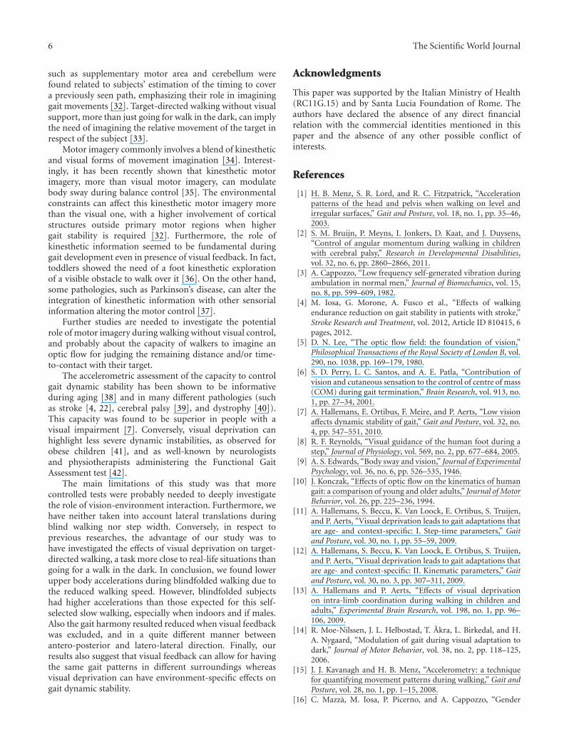
The main limitations of this study was that morecontrolled tests were probably needed to deeply investigatethe role of vision-environment interaction. Furthermore, wehave neither taken into account lateral translations duringblind walking nor step width. Conversely, in respect toprevious researches, the advantage of our study was tohave investigated the effects of visual deprivation on target-directed walking, a task more close to real-life situations thangoing for a walk in the dark. In conclusion, we found lowerupper body accelerations during blindfolded walking due tothe reduced walking speed. However, blindfolded subjectshad higher accelerations than those expected for this self-selected slow walking, especially when indoors and if males.Also the gait harmony resulted reduced when visual feedbackwas excluded, and in a quite different manner betweenantero-posterior and latero-lateral direction. Finally, ourresults also suggest that visual feedback can allow for havingthe same gait patterns in different surroundings whereasvisual deprivation can have environment-specific effects ongait dynamic stability.
Acknowledgments
This paper was supported by the Italian Ministry of Health(RC11G.15) and by Santa Lucia Foundation of Rome. Theauthors have declared the absence of any direct financialrelation with the commercial identities mentioned in thispaper and the absence of any other possible conflict ofinterests.
References
[1] H. B. Menz, S. R. Lord, and R. C. Fitzpatrick, “Accelerationpatterns of the head and pelvis when walking on level andirregular surfaces,” Gait and Posture, vol. 18, no. 1, pp. 35–46,2003.
[2] S. M. Bruijn, P. Meyns, I. Jonkers, D. Kaat, and J. Duysens,“Control of angular momentum during walking in childrenwith cerebral palsy,” Research in Developmental Disabilities,vol. 32, no. 6, pp. 2860–2866, 2011.
[3] A. Cappozzo, “Low frequency self-generated vibration duringambulation in normal men,” Journal of Biomechanics, vol. 15,no. 8, pp. 599–609, 1982.
[4] M. Iosa, G. Morone, A. Fusco et al., “Effects of walkingendurance reduction on gait stability in patients with stroke,”Stroke Research and Treatment, vol. 2012, Article ID 810415, 6pages, 2012.
[5] D. N. Lee, “The optic flow field: the foundation of vision,”Philosophical Transactions of the Royal Society of London B, vol.290, no. 1038, pp. 169–179, 1980.
[6] S. D. Perry, L. C. Santos, and A. E. Patla, “Contribution ofvision and cutaneous sensation to the control of centre of mass(COM) during gait termination,” Brain Research, vol. 913, no.1, pp. 27–34, 2001.
[7] A. Hallemans, E. Ortibus, F. Meire, and P. Aerts, “Low visionaffects dynamic stability of gait,” Gait and Posture, vol. 32, no.4, pp. 547–551, 2010.
[8] R. F. Reynolds, “Visual guidance of the human foot during astep,” Journal of Physiology, vol. 569, no. 2, pp. 677–684, 2005.
[9] A. S. Edwards, “Body sway and vision,” Journal of ExperimentalPsychology, vol. 36, no. 6, pp. 526–535, 1946.
[10] J. Konczak, “Effects of optic flow on the kinematics of humangait: a comparison of young and older adults,” Journal of MotorBehavior, vol. 26, pp. 225–236, 1994.
[11] A. Hallemans, S. Beccu, K. Van Loock, E. Ortibus, S. Truijen,and P. Aerts, “Visual deprivation leads to gait adaptations thatare age- and context-specific: I. Step-time parameters,” Gaitand Posture, vol. 30, no. 1, pp. 55–59, 2009.
[12] A. Hallemans, S. Beccu, K. Van Loock, E. Ortibus, S. Truijen,and P. Aerts, “Visual deprivation leads to gait adaptations thatare age- and context-specific: II. Kinematic parameters,” Gaitand Posture, vol. 30, no. 3, pp. 307–311, 2009.
[13] A. Hallemans and P. Aerts, “Effects of visual deprivationon intra-limb coordination during walking in children andadults,” Experimental Brain Research, vol. 198, no. 1, pp. 96–106, 2009.
[14] R. Moe-Nilssen, J. L. Helbostad, T. Akra, L. Birkedal, and H.A. Nygaard, “Modulation of gait during visual adaptation todark,” Journal of Motor Behavior, vol. 38, no. 2, pp. 118–125,2006.
[15] J. J. Kavanagh and H. B. Menz, “Accelerometry: a techniquefor quantifying movement patterns during walking,” Gait andPosture, vol. 28, no. 1, pp. 1–15, 2008.
[16] C. Mazza, M. Iosa, P. Picerno, and A. Cappozzo, “Gender

The Scientific World Journal 7
differences in the control of the upper body accelerationsduring level walking,” Gait and Posture, vol. 29, no. 2, pp. 300–303, 2009.
[17] M. Iosa, A. Fusco, G. Morone, and S. Paolucci, “Walking there:environmental influence on walking-distance estimation,”Behavioural Brain Research, vol. 226, no. 1, pp. 124–132, 2012.
[18] O. Bock and R. Beurskens, “Changes of locomotion in old agedepend on task setting,” Gait and Posture, vol. 32, no. 4, pp.645–649, 2010.
[19] S. A. Kautz, M. G. Bowden, D. J. Clark, and R. R. Neptune,“Comparison of motor control deficits during treadmilland overground walking poststroke,” Neurorehabilitation andNeural Repair, vol. 25, no. 8, pp. 756–764, 2011.
[20] J. W. Philbeck, A. J. Woods, J. Arthur, and J. Todd, “Progressivelocomotor recalibration during blind walking,” Perception andPsychophysics, vol. 70, no. 8, pp. 1459–1470, 2008.
[21] C. Mizuike, S. Ohgi, and S. Morita, “Analysis of stroke patientwalking dynamics using a tri-axial accelerometer,” Gait andPosture, vol. 30, no. 1, pp. 60–64, 2009.
[22] M. Iosa, A. Fusco, G. Morone et al., “Assessment of upper bodydynamic stability during walking in patients with subacutestroke,” Journal of Rehabilitation Research and Development. Inpress.
[23] J. M. Wood, P. F. Lacherez, A. A. Black, M. H. Cole, M. Y. Boon,and G. K. Kerr, “Postural stability and gait among older adultswith age-related maculopathy,” Investigative Ophthalmologyand Visual Science, vol. 50, no. 1, pp. 482–487, 2009.
[24] C. Mazza, M. Iosa, F. Pecoraro, and A. Cappozzo, “Controlof the upper body accelerations in young and elderly womenduring level walking,” Journal of NeuroEngineering and Reha-bilitation, vol. 5, article no. 30, 2008.
[25] S. M. O’Connor and A. D. Kuo, “Direction-dependent controlof balance during walking and standing,” Journal of Neuro-physiology, vol. 102, no. 3, pp. 1411–1419, 2009.
[26] C. E. Bauby and A. D. Kuo, “Active control of lateral balance inhuman walking,” Journal of Biomechanics, vol. 33, no. 11, pp.1433–1440, 2000.
[27] C. P. Hurt, N. Rosenblatt, J. R. Crenshaw, and M. D. Grabiner,“Variation in trunk kinematics influences variation in stepwidth during treadmill walking by older and younger adults,”Gait and Posture, vol. 31, no. 4, pp. 461–464, 2010.
[28] R. Moe-Nilssen and J. L. Helbostad, “Interstride trunk acceler-ation variability but not step width variability can differentiatebetween fit and frail older adults,” Gait and Posture, vol. 21, no.2, pp. 164–170, 2005.
[29] R. N. Shepard, “Ecological constraints on internal representa-tion: resonant kinematics of perceiving, imagining, thinking,and dreaming,” Psychological Review, vol. 91, no. 4, pp. 417–447, 1984.
[30] S. Grillner and P. Wallen, “Central pattern generators forlocomotion, with special reference to vertebrates,” AnnualReview of Neuroscience, vol. 8, pp. 233–261, 1985.
[31] D. M. Armstrong, “The supraspinal control of mammalianlocomotion,” Journal of Physiology, vol. 405, pp. 1–37, 1988.
[32] M. Bakker, F. P. De Lange, R. C. Helmich, R. Scheeringa, B.R. Bloem, and I. Toni, “Cerebral correlates of motor imageryof normal and precision gait,” NeuroImage, vol. 41, no. 3, pp.998–1010, 2008.
[33] J. J. Gibson, “The visual perception of objective motion andsubjective movement,” Psychological Review, vol. 61, no. 5, pp.304–314, 1954.
[34] P. Dechent, K. D. Merboldt, and J. Frahm, “Is the humanprimary motor cortex involved in motor imagery?” CognitiveBrain Research, vol. 19, no. 2, pp. 138–144, 2004.
[35] E. C. Rodrigues, T. Lemos, B. Gouvea, E. Volchan, L. A.Imbiriba, and C. D. Vargas, “Kinesthetic motor imagerymodulates body sway,” Neuroscience, vol. 169, no. 2, pp. 743–750, 2010.
[36] N. Dominici, Y. P. Ivanenko, G. Cappellini, M. L. Zampagni,and F. Lacquaniti, “Kinematic strategies in newly walkingtoddlers stepping over different support surfaces,” Journal ofNeurophysiology, vol. 103, no. 3, pp. 1673–1684, 2010.
[37] M. Sabate, C. Llanos, and M. Rodrıguez, “Integration ofauditory and kinesthetic information in motion: alterations inParkinson’s disease,” Neuropsychology, vol. 22, no. 4, pp. 462–468, 2008.
[38] H. B. Menz, S. R. Lord, and R. C. Fitzpatrick, “Age-relateddifferences in walking stability,” Age and Ageing, vol. 32, no.2, pp. 137–142, 2003.
[39] M. Iosa, T. Marro, S. Paolucci, and D. Morelli, “Stability andharmony of gait in children with cerebral palsy,” Research inDevelopmental Disabilities, vol. 33, no. 1, pp. 129–135, 2012.
[40] M. Iosa, C. Mazza, F. Pecoraro, I. Aprile, E. Ricci, and A.Cappozzo, “Control of the upper body movements duringlevel walking in patients with facioscapulohumeral dystrophy,”Gait and Posture, vol. 31, no. 1, pp. 68–72, 2010.
[41] E. D’Hondt, V. Segers, B. Deforche et al., “The role of visionin obese and normal-weight children’s gait control,” Gait andPosture, vol. 33, no. 2, pp. 179–184, 2011.
[42] D. M. Wrisley, G. F. Marchetti, D. K. Kuharsky, and S. L.Whitney, “Reliability, internal consistency, and validity ofdata obtained with the functional gait assessment,” PhysicalTherapy, vol. 84, no. 10, pp. 906–918, 2004.