Expression of Nitric Oxide Synthase Isoforms in Bone and Bone Cell Cultures
Upload
independentCategory
view
1download
0
1
PLATELET CONCENTRATE AND BONE PLUG AS ENHANCERS OF
HAMSTRING TENDON AUTOGRAFT MATURATION IN ANTERIOR CRUCIATE
LIGAMENT RECONSTRUCTION
Authors:
1. Mario Orrego MD - Correspondence Author*†
Departamento de Traumatología Hospital Militar de Santiago.
Holanda 050, Vitacura, Santiago, Chile.
Phone: +56-2-3653059
Mail: [email protected].
2. Catalina Larrain MD*
Departamento de Traumatología Hospital Militar de Santiago.
Holanda 050, Vitacura, Santiago, Chile.
3. Rodrigo Mardones MD*‡
Departamento de Traumatología Hospital Militar de Santiago.
Holanda 050, Vitacura, Santiago, Chile.
Clínica Las Condes, Lo Fontecilla 0441, Las Condes, Santiago, Chile.
4. Julio Rosales MD*
Departamento de Radiología Hospital Militar de Santiago.
Holanda 050, Vitacura, Santiago, Chile.

2
5. Luis Valenzuela MD*†
Departamento de Traumatología Hospital Militar de Santiago.
Holanda 050, Vitacura, Santiago, Chile.
6. José Matas MD*
Departamento de Traumatología Hospital Militar de Santiago.
Holanda 050, Vitacura, Santiago, Chile.
* Hospital Militar de Santiago
† Clínica Dávila
‡ Clínica Las Condes

3
PLATELET CONCENTRATE AND BONE PLUG AS ENHANCERS OF
HAMSTRING TENDON AUTOGRAFT MATURATION IN ANTERIOR CRUCIATE
LIGAMENT RECONSTRUCTION
ABSTRACT
- Background: The Semitendinosus and Gracilis tendon graft (ST-G) requires a longer maturation
period than its bone-tendon-bone (BTB) homologue. The use of enhancement factors as a Platelet
Concentrate and a Bone Plug would accelerate the maturation process.
- Methods: 108 patients requiring ACL reconstruction were prospectively randomized into 4 groups:
Control (27 patients), Platelet Concentrate (26 patients), Bone Plug (28 patients) and a combination
of Platelet Concentrate and Bone Plug (27 patients). Magnetic Resonance Imaging (MRI) studies
were carried out 3 and 6 months after surgery. Maturation of the graft was evaluated at the femoral
tunnel using MRI maturation criteria: intensity signal, osteoligamentous interface and widening of
the femoral tunnel.
- Results: At the 3rd month, no differences were found among the groups regarding MRI maturation
criteria. At the 6th month, in the Control group 76% of the patients had a low intensity signal, while
in the Platelet Concentrate group the low intensity signal was present in 100% of the patients (p =
0.036). No significant differences were observed regarding the interface among the various groups
in the study, despite there being a positive trend (absence of interface) with the use of the Platelet
Concentrate (87% of the patients with Platelet Concentrate vs. 65% of the patients in the Control
group). Tunnel widening was seen in 40% of the patients in the control group, 33% of the patients of
the Platelet Concentrate group, 20% of the patients of the group with Platelet Concentrate plus
Bone plug and in 12% of the patients of the Bone Patelet group (p = 0.047).

4
In all 108 cases there was maturation, comparing results obtained at the 3rd and 6th month,
regarding all parameters.
- Conclusions: The ST-G graft matured in all the groups between third and sixth month.
The maturation process evaluated with MRI increased within the use of the Platelet Concentrate as
measured by signal intensity.
The use of a Bone Plug effectively prevented tunnel widening.
The BP and PC combination does not have a synergic effect as compared to PC or BP individually.
- Level of Evidence: Level I, Randomized controlled trial.
Key Words: Reconstruction of the Anterior Cruciate Ligament, Semitendinosus, Gracilis, Platelet
Concentrate, Growth Factors, Bone Plug, Structural Bone Graft.

5
INTRODUCTION
Rupture of the anterior cruciate ligament (ACL) is the most frequent severe
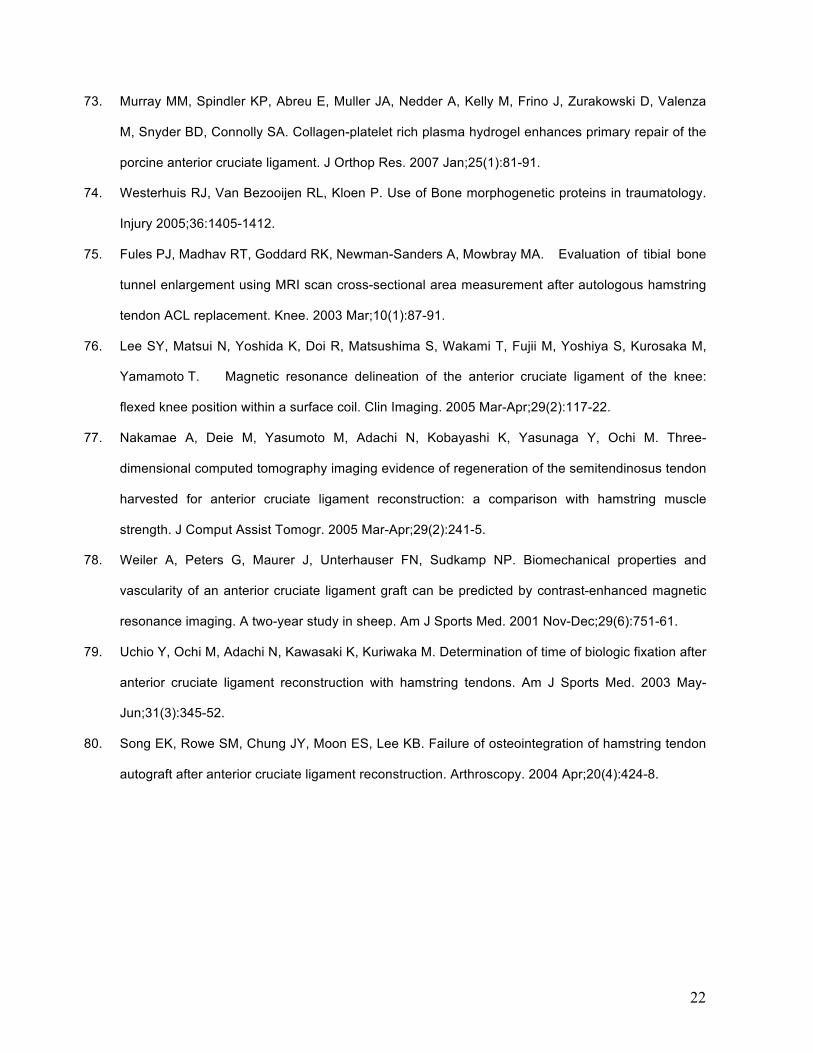
traumatic injury of the knee. Numerous articles state that the general incidence is 1
for every 3000, with 100,000 to 200,000 new reconstructions per year in USA 1,2,3,4 .
The explosive growth of sports activities, as well as the greater range of age
groups who practice these activities, explains the rise on incidence and creates
concern about the future management of this pathology 5,6,7 .
In our country, reconstruction of the ACL has experienced a vertiginous
growth in the last decade in terms of frequency, variety of techniques and types of
grafts used to replace the ACL 8.
At present, most of the reconstructions are made with autografts or
allografts9. The “Gold Standard” for the autograft group is the use of the central third
of the knee extensor mechanism, a technique known as bone-tendon-bone (BTB)
10,11. Nevertheless, it is often associated with residual pain in the anterior pole of the
donor knee, as well as to a number of complications stemming from the harvesting
in the donor location 12,13,14,15. Therefore, other alternatives are being tried, such as
the use of semitendinous-gracilis, whose clinical results are equivalent 4,8,16.
However, this technique does have some theoretical disadvantages, mainly those
related to slower maturation and integration within the bone tunnels
17,18,19,20,21,22,23,24; difficulties in fixation and greater residual lassitude of the graft
12,14,25,26,27,28,29 . The use of bone plugs at the ends of the graft have resulted in better
integration to the tunnels in vitro 30,31 and in vivo studies21,31,32,33,34,35,36,37, 38,39,40,41.

6
Ligament autograft maturation process goes through the various stages
common to all healing process (inflammation; proliferation or reparation; and
remodeling) 42,43,44. Studies have shown that growth factors are present in each of
these stages 45,46,47,48,49 . Growth factors are protein chains released by the many
cells involved in this process. These factors activate membrane receptors in the cell
walls (integrines). Their main roles are to stimulate cell differentiation from stem cells
and to act as mitogenic, quimiotactic and stimulating elements in the production of
collagen matrixes. There are five main mega-families of growth factors involved in
the muscle-skeletal repair process, platelet-derived growth factor (PDGF),
transforming growth factor beta (TGF- ), insulinic growth factor (IGF), basic
fibroblast growth factor (bFGF), and vascular endothelial growth factor (VEGF) 44.
Numerous animal model studies have shown that the healing process can be
enhanced by adding one or more of these growth factors 3,44,49,50,51,52,53,54,55,56,57,58,59.
In particular, their use on ligamentous tissue has been shown to have a positive
effect on the hystological aspect and on the biomechanical endurance in various
models 54,60,61,62,63
One of the best sources of growth factors are blood platelets, which release
large quantities of activated microgranules, which are rich in growth factors, during
fibrin clot formation 64. Platelet-concentrate-rich samples have been used in humans
and animals to accelerate the healing process 27,46,50,52,64,65,66,67,68,69,70. In clinical
practice, several authors related to maxillofacial surgery have shown the usefulness
of platelet-rich plasma obtained from autologous samples taken from patients who
undergo implant procedures 71,72. Its use is currently being explored for muscle-
skeletal pathologies 3,45,49,50,51,54,59,62,63,67,73,74,75 .

7
The objective of this study is to evaluate the potential enhancing effect of the
platelet concentrate and/or the bone plug when the ACL is reconstructed using an
ST-G graft.
MATERIALS AND METHODS
Study Design
A double-blind, prospectively randomized study was performed.
One hundred and sixteen (116) patients who had been operated on by the
same team of Hospital Militar de Santiago surgeons were prospectively included in
the study between January 2005 and December 2006. Of the 116 patients included
in the study, 86% (99/116) were males and 14% (17/116) were females. The
average age of the patients was 30 years, ranging from 15 to 57 years old. In 60% of
the cases (70/116) the injured knee was the right one, while 40% of the patients had
been injured in their left knees. In 63% (73/116) of the cases, the injury had been
caused while engaging in contact sports. The inclusion criteria were: a mature
skeleton; clinical instability and MRI showing total rupture of the ACL, without any
other associated capsulo-ligamentosus injuries; and voluntary acceptance to
participate in the study, according to the protocol authorized by the hospital’s ethics
committee.
The patients were randomly divided into 4 groups of 29 people each. Eight (8)
of the patients included in the study were lost during the follow-up, so the final
sample included 108 people, who were divided as follows:

8
- Group I, Control: ACL Reconstruction with quadruple ST-G fixed proximally
with a biodegradable transfixing screw (Arthrex, Naples, Florida) and a
distally biodegradable interferencial screw (Arthrex, Naples, Florida) (Figure
N°1). 27 patients
- Group II, ACL Reconstruction plus Platelet Concentrate GPS II ® (PC)
(Biomet…) 26 patients
- Group III, ACL Reconstruction plus Bone Plug (BP) Biomet Trefin kit ®
(Biomet….) 28 patients
- Group IV, ACL Reconstruction plus Platelet Concentrate and Bone Plug (PC
+ BP). 27 patients
These four groups were comparable.
Surgical Technique
Collection of Platelet Concentrate (PC):
A blood sample of 67 cc of blood was taken from the patient during anesthetic
induction in order to collect the PC and the thrombin.
1. PC: 3 cc of an anti-clotting agent is added to 57 cc of blood using the
Biomet GPS II® kit, to which an ultra-filtering system is added in order to obtain a
higher platelet concentration. The sample is spun during 15 minutes and, after this
period, approximately 10 ml of PC are collected.

9
2. Thrombin: 10 ml of the patient’s blood (without the anti-clotting agent) are
poured into the test tube. After the sample clots are added, it is centrifuged at 200
RPM during 5 minutes and, after this period, the skim is collected. Ten parts of this
product are mixed with one part of Calcium Chloride (10:1).
Once the process is completed, the application system is set up (Figure N°2).
Adding the PC: Once the quadruple ST-G graft is prepared, 5 cc of the PC
are added between the strands of the graft, helping it to adhere and integrate itself
by repeatedly puncturing it throughout its whole length with a N° 21 needle.
Next, the graft with the concentrate is inserted along the bone tunnels (Figure
N°3). Once it is fixed, 1 cc of PC is injected into the femoral tunnel.
Collection of the Bone Plug:
Using the trefin kit (Biomet,…..) (Figure N°4-A) when the tibia tunnel is made,
a spongy plug of approximately 7 mm in diameter and 30 mm long is obtained
(Figure N°4-B). This plug is compacted into its final dimensions of 2.5 mm x 25 mm
(Figure N°4-C). Once the ST-G graft is fixed, the plug is placed interferentially at the
level of the femoral tunnel (Figure N°5).
Imaging Assessment:
Patients were assessed on the basis of sagital MRI (General Electric® 2 T)
sections in T2. This was done by a radiologist, blinded to the groups, specialized in
muscle-skeletal images, 3 months and 6 months after surgery 75,76.

10
The following variables were analyzed:
- Graft Signal Intensity: In sagital sections in T2 we analized the graft’s signal
intensity at the femoral tunnel. There were high intensity signals, intensity similar to
the synovial fluid (Figure N°6-A), intermediate intensity signals, comparable to the
muscle’s (Figure N°6-B) and low intensity signals, equivalent to the posterior
cruciate ligament (Figure N°6-C). According to Weiler et al 79 a mature graft should
have a low intensity signal 77,78,79.
- Osteoligamentous Interface: In MRI axial and sagital sections in T2 we look for
the presence of interface, appearing as a high-intensity halo, between the graft and
the bone femoral tunnel (Figure N°7). The absence of an interface reflects major
integration of the graft, meaning a more mature graft 79,80.
- Tunnel Widening: The width of the femoral tunnel was measured in sagital MRI
sections in T2 at the third and sixth month after surgery. A difference greater than 2
mm between the two measures was considered to be positive. The lack of widening
of the femoral tunnel means a more integrated and mature graft (Figure N°8) 20.
Data Analysis:
The data obtained from the study were analized using chi-square’s test and
Fisher’s Exact test. Our power analysis was XX patients each group.

11
RESULTS:
IMAGING RESULTS AT THREE MONTHS:
- Signal Intensity: in the control group 12 of the 27 patients (44%) had a low intensity
signal; in the Bone Plug group 11 of the 28 patients (43%), in the Platelet
Concentrate group 12 of the 26 patients (46%) and in the Bone Plug plus Platelet
Concentrate group 11 of the 27 patients (40%) had a low intensity signal. The values
and statistic analysis are shown in Table N°1 and Graphic N°1. No significant
differences were found among the groups.
- Osteoligamentous Interface: in the control group 12 of the 27 patients (44%)
showed no interface; in the Bone Plug group 12 of the 28 patients (43%), in the
Platelet Concentrate group 11 of the 26 patients (42%) and in the Bone Plug plus
Patelet Concentrate 11 of the 27 patients (42%) showed no interface. The values
and statistic analysis are shown in Table N°2 and Graphic N°2. No significant
differences were found among the groups.
IMAGINOLOGIC RESULTS AT SIX MONTHS:
- Signal Intensity: in the control group 21 of the 27 patients (77%) had a low intensity
signal; in the Bone Plug group 25 of the 28 patients (89%), in the Platelet
Concentrate group 26 of the 26 patients (100%) and in the Bone Plug plus Platelet
Concentrate group 25 of the 27 patients (93%) had a low intensity signal. The values

12
and statistic analysis are shown in Table N°1 and Graphic N°1. The difference
between the control and the Platelet Concentrate group were statistically significant
(p = 0.036). No significant differences were found among the other groups.
- Osteoligamentous Interface: in the control group 18 of the 27 patients (67%)
showed no interface; in the Bone Plug group 19 of the 28 patients (68%), in the
Platelet Concentrate group 23 of the 26 patients (88%) and in the Bone Plug plus
Patelet Concentrate 19 of the 27 patients (70%) showed no interface. The values
and statistic analysis are shown in Table N°2 and Graphic N°2. No significant
differences were found among the groups.
- Tunnel Widening: in the control group 16 of the 27 patients (59%) showed no
tunnel widening; in the Bone Plug group 25 of the 28 patients (89%), in the Platelet
Concentrate group 18 of the 26 patients (69%) and in the Bone Plug plus Platelet
Concentrate group 22 of the 27 patients (81%) showed no tunnel widening. The
values and statistic analysis are shown in Table N°3 and Graphic N°3. The
difference between the control and the Bone Plug group were statistically significant
(p = 0.047). No significant differences were found among the other groups.
The data obtained at the third month and at the sixth month (intensity signal
and osteoligamentous interface) were compared finding statistically significant
differences in all variables, which reflects a progression of the maturation of the
graft.

13
DISCUSSION
Biological solutions to these types of injuries are being used with increasing
frequency in clinical practice3,45,49,50,51,54,59,62,63,67,73,74. However, most studies and
experimental models are done employing animals, and make frequent references to
growth factors3,44,49,50,51,52,53,54,55,56,57,58,59. Clearly, there still remain many unknowns
in this line of works. Also, there is a surprising shortage of published works regarding
the use of platelet concentrate27,46,50,52,64,65,66,67,68,69,70. This study was designed to
analyze experimental evidence on the use of autologous elements (bone plug and
platelet concentrate), which do not put the patient at risk or compromise the results
of conventional surgery, and which can be theoretically useful in aiding the natural
healing process. As far as the platelet concentrate and its large content of growth
factors is concerned, its use seems reasonable, given that the proportions or
interactions of various growth factors are not being altered. It has been shown, that
not only the proportion of growth factors, but the timing and duration of their
interaction can have a positive or negative impact on the final outcome. The use of a
bone plug is based on the mechanical knowledge regarding graft behavior inside the
tunnels. This plug acts as a natural interferential fixation, which does not add any
exogenous inflammatory phenomena, as sometimes happens with bioabsorbable
materials. In addition, it is fixed close to the articular surface and it should
theoretically reduce the “bunji” and windshield wiper effects, which are frequent in
this type of surgery and are related to the tunnel widening
phenomenon17,21,30,31,32,33,34,35,36,37, 38,39,40,41.

14
The results that were obtained in the study are encouraging, though not
sufficiently conclusive. A positive trend was noticed when these elements were
used. Paradoxically, however, the combination of both elements does not appear to
be as beneficial, which could suggest that they interfere with each other. The
potential difficulties faced by the radiologist when interpreting images, in relation to
the bone plug and graft maturation in the femoral tunnel, must also be considered.
Finally, the factors that have been recognized as having the greatest
influence on this healing process are the environment and the presence of
transportation media for stem cells, either with or without growth factors. This
interaction was not studied by our group given the difficulty of homogenizing and
controlling all these variables.
CONCLUSION
The use of a platelet concentrate or of a bone plug, each on its own, favorably
influences ST-G graft maturation in the ACL reconstruction.
ACKNOWLEDGMENTS
Juan Durruty MD
Hernán Sudy MD
Matías Salineros MD
Alexander Tomic MD

15
BIBLIOGRAFÍA:
1. Insall & Scott. “Surgery of the knee” Edición en español. Marbán , 2004
2. Brown CH Jr, Carson EW. Revision anterior cruciate ligament surgery. Clin Sports Med. 1999
Jan;18(1):109-71.
3. Petrigliano FA, McAllister DR, Wu BM. Tissue engineering for anterior cruciate ligament
reconstruction: a review of current strategies. Arthroscopy. 2006 Apr;22(4):441-51.
4. Herrington L, Wrapson C, Matthews M, Matthews H. Anterior cruciate ligament reconstruction,
hamstring versus bone-patella tendon-bone grafts: a systematic literature review of outcome from
surgery. Knee. 2005 Jan;12(1):41-50.
5. Durruty J, Valenzuela L, Matas J, Orrego M, Melo R. Reconstrucción de ligamento cruzado anterior
en pacientes mayores de 40 años. Rev Chilena Ortop y Traum 2005;46:66-72.
6. Barber FA, Elrod BF, McGuire DA, Paulos LE. Is an anterior cruciate ligament reconstruction
outcome age dependent? Arthroscopy. 1996 Dec;12(6):720-5.
7. Stein DA, Brown H, Bartolozzi AR. Age and ACL reconstruction revisited. Orthopedics. 2006
Jun;29(6):533-6.
8. Figueroa D, Cavo R, Mardones R. Estudio Comparativo de la reconstrucción del ligamento cruzado
anterior con técnica Hueso-Tendón patelar-Hueso v/s Doble Semitendinoso-Gracilis. Seguimiento
mayor de 1 año. Rev Chilena Ortop y Traum 2003 44:153-159.
9. Betz RR. Limitations of autograft and allograft: new synthetic solutions. Orthopedics. 2002
May;25(5 Suppl):s561-70.
10. Jones KG. Reconstruction of the anterior cruciate ligament. A technique using the central one-third
of the patellar ligament. J Bone Joint Surg Am. 1963 Jul;45:925-32.
11. Clancy WG Jr, Nelson DA, Reider B, Narechania RG. Anterior cruciate ligament reconstruction
using one-third of the patellar ligament, augmented by extra-articular tendon transfers. J Bone Joint
Surg Am. 1982 Mar;64(3):352-9.

16
12. Aglietti P, Giron F, Buzzi R, Biddau F, Sasso F. Anterior cruciate ligament reconstruction: bone-
patellar tendon-bone compared with double semitendinosus and gracilis tendon grafts. A
prospective, randomized clinical trial. J Bone Joint Surg Am. 2004 Oct;86-A(10):2143-55.
13. Aune AK, Holm I, Risberg MA, Jensen HK, Steen H. Four-strand hamstring tendon autograft
compared with patellar tendon-bone autograft for anterior cruciate ligament reconstruction. A
randomized study with two-year follow-up. Am J Sports Med. 2001 Nov-Dec;29(6):722-8.
14. Feller JA, Webster KE. A randomized comparison of patellar tendon and hamstring tendon anterior
cruciate ligament reconstruction. Am J Sports Med. 2003 Jul-Aug;31(4):564-73.
15. Pinczewski LA, Deehan DJ, Salmon LJ, Russell VJ, Clingeleffer A. A five-year comparison of
patellar tendon versus four-strand hamstring tendon autograft for arthroscopic reconstruction of the
anterior cruciate ligament. Am J Sports Med. 2002 Jul-Aug;30(4):523-36.
16. Calvisi V, Lupparelli S, Padua R. Patellar Tendon Autograft versus hamstring tendon autograft in
arthroscopic anterior cruciate ligament reconstruction: appraisal of the evidence. J Orthopaed
Traumatol 2006 Jun;7:103-107.
17. Fauno P, Kaalund S. Tunnel widening after hamstring anterior cruciate ligament reconstruction is
influenced by the type of graft fixation used: a prospective randomized study. Arthroscopy. 2005
Nov;21(11):1337-41.
18. Spindler KP, Kuhn JE, Freedman KB, Matthews CE, Dittus RS, Harrell FE Jr. Anterior cruciate
ligament reconstruction autograft choice: bone-tendon-bone versus hamstring: does it really
matter? A systematic review. Am J Sports Med. 2004 Dec;32(8):1986-95.
19. Nebelung W, Becker R, Merkel M, Ropke M. Bone tunnel enlargement after anterior cruciate
ligament reconstruction with semitendinosus tendon using Endobutton fixation on the femoral side.
Arthroscopy. 1998 Nov-Dec;14(8):810-5.
20. Wilson TC, Kantaras A, Atay A, Johnson DL. Tunnel enlargement after anterior cruciate ligament
surgery. Am J Sports Med. 2004 Mar;32(2):543-9.
21. Buelow JU, Siebold R, Ellermann A. A prospective evaluation of tunnel enlargement in anterior
cruciate ligament reconstruction with hamstrings: extracortical versus anatomical fixation. Knee
Surg Sports Traumatol Arthrosc. 2002 Mar;10(2):80-5.

17
22. Laxdal G, Kartus J, Eriksson BI, Faxen E, Sernert N, Karlsson J. Biodegradable and metallic
interference screws in anterior cruciate ligament reconstruction surgery using hamstring tendon
grafts: prospective randomized study of radiographic results and clinical outcome. Am J Sports
Med. 2006 Oct;34(10):1574-80.
23. Giron F, Aglietti P, Cuomo P, Mondanelli N, Ciardullo A. Anterior cruciate ligament reconstruction
with double-looped semitendinosus and gracilis tendon graft directly fixed to cortical bone: 5-year
results. Knee Surg Sports Traumatol Arthrosc. 2005 Mar;13(2):81-91.
24. Clatworthy MG, Annear P, Bulow JU, Bartlett RJ. Tunnel widening in anterior cruciate ligament
reconstruction: a prospective evaluation of hamstring and patella tendon grafts. Knee Surg Sports
Traumatol Arthrosc. 1999;7(3):138-45.
25. L'Insalata JC, Klatt B, Fu FH, Harner CD. Tunnel expansion following anterior cruciate ligament
reconstruction: a comparison of hamstring and patellar tendon autografts. Knee Surg Sports
Traumatol Arthrosc. 1997;5(4):234-8.
26. Giurea M, Zorilla P, Amis AA, Aichroth P. Comparative pull-out and cyclic-loading strength tests of
anchorage of hamstring tendon grafts in anterior cruciate ligament reconstruction. Am J Sports
Med. 1999 Sep-Oct;27(5):621-5.
27. Kanazawa T, Soejima T, Murakami H, Inoue T, Katouda M, Nagata K. An immunohistological study
of the integration at the bone-tendon interface after reconstruction of the anterior cruciate ligament
in rabbits. J Bone Joint Surg Br. 2006 May;88(5):682-7.
28. Deehan DJ, Cawston TE. The biology of integration of the anterior cruciate ligament. J Bone Joint
Surg Br. 2005 Jul;87(7):889-95.
29. Zavras TD, Race A, Amis AA. The effect of femoral attachment location on anterior cruciate
ligament reconstruction: graft tension patterns and restoration of normal anterior-posterior laxity
patterns. Knee Surg Sports Traumatol Arthrosc. 2005 Mar;13(2):92-100.
30. Howell SM, Roos P, Hull ML. Compaction of a bone dowel in the tibial tunnel improves the fixation
stiffness of a soft tissue anterior cruciate ligament graft: an in vitro study in calf tibia. Am J Sports
Med. 2005 May;33(5):719-25.

18
31. Brown GA, Pena F, Grontvedt T, Labadie D, Engebretsen L. Fixation strength of interference screw
fixation in bovine, young human, and elderly human cadaver knees: influence of insertion torque,
tunnel-bone block gap, and interference. Knee Surg Sports Traumatol Arthrosc. 1996;3(4):238-44.
32. Barber FA, Spruill B, Sheluga M. The effect of outlet fixation on tunnel widening. Arthroscopy. 2003
May-Jun;19(5):485-92.
33. Caborn DN, Nyland J, Selby J, Tetik O. Biomechanical testing of hamstring graft tibial tunnel
fixation with bioabsorbable interference screws. Arthroscopy. 2003 Nov;19(9):991-6.
34. Brand J Jr, Weiler A, Caborn DN, Brown CH Jr, Johnson DL. Graft fixation in cruciate ligament
reconstruction. Am J Sports Med. 2000 Sep-Oct;28(5):761-74.
35. Matsumoto A, Howell SM, Liu-Barba D. Time-related changes in the cross-sectional area of the
tibial tunnel after compaction of an autograft bone dowel alongside a hamstring graft. Arthroscopy.
2006 Aug;22(8):855-60.
36. Tecklenburg K, Burkart P, Hoser C, Rieger M, Fink C. Prospective evaluation of patellar tendon
graft fixation in anterior cruciate ligament reconstruction comparing composite bioabsorbable and
allograft interference screws. Arthroscopy. 2006 Sep;22(9):993-9.
37. Harvey A, Thomas NP, Amis AA. Fixation of the graft in reconstruction of the anterior cruciate
ligament. J Bone Joint Surg Br. 2005 May;87(5):593-603.
38. Klein J, Lintner D, Downs D, Vavrenka K. The incidence and significance of femoral tunnel
widening after quadrupled hamstring anterior cruciate ligament reconstruction using femoral cross
pin fixation. Arthroscopy 2003 May;19(5):470-6)
39. Weiler et al.: Current Concepts for hamstring tendon fixation in cruciate ligament surgery. Chirurg
71:1034-1044, 2000.
40. Matsumoto, Akio M.D.; Howell, Stephen M. M.D. The WasherLoc and Bone Dowel: A Rigid,
Slippage-Resistant Tibial Fixation Device for a Soft Tissue Anterior Cruciate Ligament Graft.
Techniques in Orthopaedics. 2005 Sept;20(3):278-282.
41. Robert H, Es-Sayeh J. The role of periosteal flap in the prevention of femoral widening in anterior
cruciate ligament reconstruction using hamstring tendons. Knee Surg Sports Traumatol Arthrosc.
2004 Jan;12(1):30-5.

19
42. Rodeo SA . Tendon healing in a bone tunnel. J Bone Joint Surg Am 1993 75:1795.
43. Marumo K, Saito M, Yamagishi T, Fujii K. The "ligamentization" process in human anterior cruciate
ligament reconstruction with autogenous patellar and hamstring tendons: a biochemical study. Am J
Sports Med. 2005 Aug;33(8):1166-73.
44. Letson AK, Dahners LE. The effect of combinations of growth factors on ligament healing. Clin
Orthop Relat Res. 1994 Nov;(308):207-12.
45. Angel MJ, Sgaglione NA, Grande DA. Clinical applications of bioactive factors in sports medicine:
current concepts and future trends. Sports Med Arthrosc. 2006 Sep;14(3):138-45.
46. Aspenberg P, Virchenko O. Platelet concentrate injection improves Achilles tendon repair in rats.
Acta Orthop Scand. 2004 Feb;75(1):93-9.
47. Weiler A, Hoffmann RF, Bail HJ, Rehm O, Sudkamp NP. Tendon healing in a bone tunnel. Part II:
Histologic analysis after biodegradable interference fit fixation in a model of anterior cruciate
ligament reconstruction in sheep. Arthroscopy. 2002 Feb;18(2):124-35.
48. Weiler A, Peine R, Pashmineh-Azar A, Abel C, Sudkamp NP, Hoffmann RF. Tendon healing in a
bone tunnel. Part I: Biomechanical results after biodegradable interference fit fixation in a model of
anterior cruciate ligament reconstruction in sheep. Arthroscopy. 2002 Feb;18(2):113-23.
49. Anderson K, Seneviratne AM, Izawa K, Atkinson BL, Potter HG, Rodeo SA. Augmentation of
tendon healing in an intraarticular bone tunnel with use of a bone growth factor. Am J Sports Med.
2001 Nov-Dec;29(6):689-98.
50. Ju YJ, Tohyama H, Kondo E, Yoshikawa T, Muneta T, Shinomiya K, Yasuda K. Effects of local
administration of vascular endothelial growth factor on properties of the in situ frozen-thawed
anterior cruciate ligament in rabbits. Am J Sports Med. 2006 Jan;34(1):84-91.
51. Woo S, Abramowitch S, Kilger R, Liang R. Biomechanics of knee ligaments: injury, healing, and
repair. J Biomech. 2006;39:1-20.
52. Molloy T, Wang Y, Murrell AC. The Roles of Growth Factors in Tendon and Ligament Healing.
Sports Med 2003 33(5):381-394.

20
53. Schmidmaier G, Wildermann B, Gabelein T, Heeger J, Kandziora F, Haas N, Raschke M.
Synergistic effect of IGF-1 and TGF-B1 on fracture healing in rats. Acta Orthop Scand
2003;74(5):604-610.
54. Kondo E, Yasuda K, Yamanaka M, Minami A, Tohyama H. Effects of administration of exogenous
growth factors on biomechanical properties of the elongation-type anterior cruciate ligament injury
with partial laceration. Am J Sports Med. 2005 Feb;33(2):188-96.
55. Schmidmaier G, Widermann B, Ostapowicz D, Kandziora F, Stange R, Haas N, Raschke M. Long-
term effects of local growth factor (IGF-I and TGF-ß1) treatment on fracture healing. J Orthop Res
2004 22;514-519.
56. Siebrecht M, De Rooij P, Arm D, Olsson M, Aspenberg P. Platelet Concentrate Increases Bone
Ingrowth into Porous Hydroxyapatite. Orthopedics 2002;Feb;25(2):169-172.
57. Murray M, Bennet R, Zhang X, Spector M. Cell Outgrowth from de Human ACL in vitro: Regional
Variation and response to TGF-B1. J Orthop Res 2002;20:875-880.
58. Lane JM. Bone morphogenic protein science and studies. J Orthop Trauma. 2005 Nov-Dec;19(10
Suppl):S17-22.
59. Yamazaki S, Yasuda K, Tomita F, Tohyama H, Minami A. The effect of transforming growth factor-
beta1 on intraosseous healing of flexor tendon autograft replacement of anterior cruciate ligament
in dogs. Arthroscopy. 2005 Sep;21(9):1034-41.
60. Mihelic R, Pecina M, Jelic M, Zoricic S, Kusec V, Simic P, Bobinac D, Lah B, Legovic D, Vukicevic
S. Bone morphogenetic protein-7 (osteogenic protein-1) promotes tendon graft integration in
anterior cruciate ligament reconstruction in sheep. Am J Sports Med. 2004 Oct-Nov;32(7):1619-25.
61. Yasuda K, Tomita F, Yamazaki S, Minami A, Tohyama H. The effect of growth factors on
biomechanical properties of the bone-patellar tendon-bone graft after anterior cruciate ligament
reconstruction: a canine model study. Am J Sports Med. 2004 Jun;32(4):870-80.
62. Martinek V, Latterman C, Usas A, Abramowitch S, Woo SL, Fu FH, Huard J. Enhancement of
tendon-bone integration of anterior cruciate ligament grafts with bone morphogenetic protein-2
gene transfer: a histological and biomechanical study. J Bone Joint Surg Am. 2002 Jul;84-
A(7):1123-31.

21
63. Laurencin CT, Freeman JW. Ligament tissue engineering: an evolutionary materials science
approach. Biomaterials. 2005 Dec;26(36):7530-6.
64. Tischler, Michael. Platelet Rich Plasma. The Use of Autologous Growth Factors to Enhance Bone
and Soft Tissue Grafts. New York Dental Journal 68:22-24, 2002.
65. Betsholtz C, Karlsson L, Lindahl P. Developmental roles of platelet-derived growth factors.
Bioessays. 2001 Jun;23(6):494-507.
66. Murray M, Rice K, Wright RJ, Spector M. The effect of selected growth factors on human anterior
cruciate ligament cell interactions with a three-dimensional collagen-GAG scaffold. J Orthop Res
2003 21:238-244.
67. Murray MM, Spindler KP, Devin C, Snyder BS, Muller J, Takahashi M, Ballard P, Nanney LB,
Zurakowski D. Use of a collagen-platelet rich plasma scaffold to stimulate healing of a central
defect in the canine ACL. J Orthop Res. 2006 Apr;24(4):820-30.
68. Nagumo A, Yasuda K, Numazaki H, Azuma H, Tanabe Y, Kikuchi S, Harata S, Tohyama H. Effects
of separate application of three growth factors (TGF-beta1, EGF, and PDGF-BB) on mechanical
properties of the in situ frozen-thawed anterior cruciate ligament. Clin Biomech (Bristol, Avon). 2005
Mar;20(3):283-90.
69. Hildebrand KA, Woo SL, Smith DW, Allen CR, Deie M, Taylor BJ, Schmidt CC. The effects of
platelet-derived growth factor-BB on healing of the rabbit medial collateral ligament. An in vivo
study. Am J Sports Med. 1998 Jul-Aug;26(4):549-54.
70. Weiler A, Forster C, Hunt P, Falk R, Jung T, Unterhauser FN, Bergmann V, Schmidmaier G, Haas
NP. The influence of locally applied platelet-derived growth factor-BB on free tendon graft
remodeling after anterior cruciate ligament reconstruction. Am J Sports Med. 2004 Jun;32(4):881-
91.
71. Anitua E. Plasma rich in growth factors: preliminary results of use in the preparation of future sites
for implants. Int J Oral Maxillofac Implants. 1999 Jul-Aug;14(4):529-35.
72. Velich N, Németh Z, Hrabak K, Suba Z, Szabo G. Repair of Bony Defect With Combination
Biomaterials. The Journal of Craniofacial Surgery 2004 Jan;15:11-15.
22
73. Murray MM, Spindler KP, Abreu E, Muller JA, Nedder A, Kelly M, Frino J, Zurakowski D, Valenza
M, Snyder BD, Connolly SA. Collagen-platelet rich plasma hydrogel enhances primary repair of the
porcine anterior cruciate ligament. J Orthop Res. 2007 Jan;25(1):81-91.
74. Westerhuis RJ, Van Bezooijen RL, Kloen P. Use of Bone morphogenetic proteins in traumatology.
Injury 2005;36:1405-1412.
75. Fules PJ, Madhav RT, Goddard RK, Newman-Sanders A, Mowbray MA. Evaluation of tibial bone
tunnel enlargement using MRI scan cross-sectional area measurement after autologous hamstring
tendon ACL replacement. Knee. 2003 Mar;10(1):87-91.
76. Lee SY, Matsui N, Yoshida K, Doi R, Matsushima S, Wakami T, Fujii M, Yoshiya S, Kurosaka M,
Yamamoto T. Magnetic resonance delineation of the anterior cruciate ligament of the knee:
flexed knee position within a surface coil. Clin Imaging. 2005 Mar-Apr;29(2):117-22.
77. Nakamae A, Deie M, Yasumoto M, Adachi N, Kobayashi K, Yasunaga Y, Ochi M. Three-
dimensional computed tomography imaging evidence of regeneration of the semitendinosus tendon
harvested for anterior cruciate ligament reconstruction: a comparison with hamstring muscle
strength. J Comput Assist Tomogr. 2005 Mar-Apr;29(2):241-5.
78. Weiler A, Peters G, Maurer J, Unterhauser FN, Sudkamp NP. Biomechanical properties and
vascularity of an anterior cruciate ligament graft can be predicted by contrast-enhanced magnetic
resonance imaging. A two-year study in sheep. Am J Sports Med. 2001 Nov-Dec;29(6):751-61.
79. Uchio Y, Ochi M, Adachi N, Kawasaki K, Kuriwaka M. Determination of time of biologic fixation after
anterior cruciate ligament reconstruction with hamstring tendons. Am J Sports Med. 2003 May-
Jun;31(3):345-52.
80. Song EK, Rowe SM, Chung JY, Moon ES, Lee KB. Failure of osteointegration of hamstring tendon
autograft after anterior cruciate ligament reconstruction. Arthroscopy. 2004 Apr;20(4):424-8.

23
ANEXOS: Figure N°1:
Reconstruction of ACL with quadruple ST-G graft fixed proximally with a
biodegradable transfixing screw and distally with a biodegradable
interferential screw.

24
Figure N°2:
A
A B C
A. Preparation of the PC. After subjecting the sample to centrifugal force,
a PC sample is taken by means of a sterile technique.
B. Preparation of thrombin: After the sample clots, it is spun and the
thrombin is skimmed from the top.
C. The system is then set up and a platelet-rich plasma mix is obtained

25
Figure N°3:
A B
A. ST-G graft in PC clot.
B. Insertion of the PC-covered ST-G graft.

26
Figure N°4:
A
B C
A. Biomet trefin kit used to obtain the bone plug
B. Compacted bone plug obtained from drilling the tibia tunnel
C. Recovery of bone plug.

27
Figure N°5:
Interferential insertion of the Bone Plug through the femoral tunnel by means of
an arthroscopic portal.
Figure N°6:

28
A. MRI with high intensity signal in the femoral tunnel. Signal corresponds to
articular fluid.
B. MRI with low intensity signal, similar to the posterior cruciate ligament.
Figure N°7:

29
Osteoligamentous interface in the femoral tunnel.
Figure N°8:

30
Femoral tunnel widening between 3 and 6 months. It is considered as widening
when the difference is greater than 2mm.
Table Nº1

31
3M: Third month. 6M: sixth month
Graphic Nº1
Table Nº2

32
3M: Third month. 6M: sixth month
Graphic Nº2
Table Nº3

33
3M: Third month. 6M: sixth month
Graphic Nº3
Copyright © 2022 FDOKUMEN