Arsenic trioxide-mediated oxidative stress and genotoxicity in human hepatocellular carcinoma cells
Light microscopic description of the effects of laser phototherapy onbone defects grafted with mineral trioxide aggregate, bonemorphogenetic proteins, and guided bone regeneration in a rodentmodel
Antonio L. B. Pinheiro,1,2,3 Luiz G. P. Soares,1 Gilberth T. S. Aciole,1 Neandder A. Correia,1
Artur F. S. Barbosa,1 Luciana M. P. Ramalho,1,3 Jean N. dos Santos3,4
1Center of Biophotonics, School of Dentistry, Federal University of Bahia, Salvador, BA 40110-150, Brazil2Institute of Biomedical Engineering, Camilo Castelo Branco University, Sao Jose dos Campos, SP 12245-230, Brazil3National Institute of Optics and Photonics, Physics Institute, University of Sao Carlos, Sao Carlos, SP 13560-970, Brazil4Laboratory of Surgical Pathology, School of Dentistry, Federal University of Bahia, Salvador, BA 40110-150,
Brazil
Received 25 November 2010; accepted 21 February 2011
Published online 4 May 2011 in Wiley Online Library (wileyonlinelibrary.com). DOI: 10.1002/jbm.a.33107
Abstract: We carried out a histological analysis on bone
defects grafted with mineral trioxide aggregate (MTA) treated
or not with laser, bone morphogenetic protein (BMP), and
guided bone regeneration (GBR). Benefits of the use of MTA,
laser, BMPs, and GBR on bone repair are well known, but
there is no report on their association with laser light. Ninety
rats were divided into 10 groups each subdivided into 3.
Defects on G II and I were filled with the blood clot. G II was
further irradiated with LED. G III and IV were filled with MTA;
G IV was further irradiated with laser. G V and VI, the defects
filled with MTA and covered with a membrane (GBR). G VI
was further irradiated with laser. G VII and VIII, BMPs were
added to the MTA and group VIII further irradiated with laser.
G IX and X, the MTA þ BMP graft was covered with a mem-
brane (GBR). G X was further irradiated with laser. Laser light
(k ¼ 850 nm, 150 mW, 4 J/cm2) was applied over the defect
at 48-h intervals and repeated for 15 days. Specimens were
processed, cut and stained with H&E and Sirius red and
underwent histological analysis. Subjects on group X were
irradiated. The results showed different tissue response on
all groups during the experimental time. Major changes were
seen on irradiated subjects and included marked deposition
of new bone in advanced maturation. It is concluded that
near infrared laser phototherapy improved the results of the
use of the MTA on bone defects. VC 2011 Wiley Periodicals, Inc.
J Biomed Mater Res Part A: 98A: 212–221, 2011.
Key Words: biomaterial, LLLT, mineral trioxide aggregate
INTRODUCTION
The treatment of bone defects using biomaterials has beenextensively studied in the dental field.1–7 Since the pioneerwork by Urist, who demonstrated heterotrophic formation ofbone induced by devitalized demineralized bone matrix, anew possibility of treating bone defects was established. De-mineralized bone matrix has osteoinductive properties dueto the presence of soluble growth factors on its composition.8
Bone loss may be a result of several pathologies, trauma, or aconsequence of surgical procedures. This aspect led to exten-sive studies on the process of bone repair worldwide. Severaltechniques for the treatment of bone defects have been pro-posed, including the use of several types of grafts, mem-branes, and the association of both techniques.8
Mineral trioxide aggregate (MTA) is a powder aggregate,containing mineral oxides that have no cytotoxicity andgood biological response. It stimulates tissue repair byincreasing cellular adhesion, growth, and proliferation at itssurface. The use of MTA has been shown to cause an
overgrowth of cementum and on facilitating the regenera-tion of the periodontal ligament as well as the deposition ofnew bone.9–20
Previous histological reports have indicated that newbone or cementum is formed adjacent to MTA when it isplaced in contact with the periodontal tissue or in artificialbone defects.9–14,21,22 It has antibacterial properties,23
enhances tissue dissolution, and induces bone formation.24
Previous studies suggested that, the rise of pHinduced by calcium hydroxide combined with the availabil-ity of Ca2þ and OH� ions has a stimulating effect on bonemineralization.25–28
Guided bone regeneration (GBR) is a procedure basedupon the guided tissue regeneration technique, which is aperiodontal surgical procedure, which has been used in thedental clinical practice for more than a decade.29,30 Thisprocedure is used as a way to stimulate wound healing andto favor the regeneration of tooth supporting structures.31
Its principles were based upon the selective permeability
Correspondence to: A. L. B. Pinheiro; e-mail: [email protected]
Contract grant sponsors: Conselho Nacional de Desenvolvimento Cientıfico e Tecnologico (CNPq)
212 VC 2011 WILEY PERIODICALS, INC.

provided by the use of membranes on the isolation of tis-sues, essential for bone repair. Previous studies have dem-onstrated that such regeneration may occur following thisprocedure.29–32 The use GBR associated to the use of bioma-terials has been considered to be beneficial for the healingof bones.8
Our previous results indicate that near infrared laserphototherapy (NIR LPT) is effective to improve bone repairmainly due to its good penetration on tissues when com-pared to visible laser light. The use of laser phototherapy(LPT) on studies involving bone healing has been a hottopic lately and many of them have demonstrated positiveresults, including its association to biomaterials.33–46
We have shown that the improvement of bone neofor-mation and maturation, on irradiated subjects, is associatedto the increased deposition of calcium hydroxyapatite (CHA)during early stages of healing. The maturation of the newlyformed bone probably represents an increased capacity ofsecretion by osteoblasts on irradiated subjects. It is wellaccepted that the deposition of CHA represents bone matu-ration. Large amounts of CHA on bone are indicative of amore resistant and calcified bone.33–46
It is known that LPT has the ability to stimulate cellproliferation, including fibroblasts, which have the capacityto secrete collagen, the main organic component observedduring bone repair.33–46
Despite the growing successful application of the LPT onbone repair, there are a few studies assessing the associa-tion of the laser light with biomaterials.8,34,36–38,43
Although several reports have suggested benefits of theisolated or combined use of MTA, BMPs, GBR, and LPT onthe repair of bone defects, the associated use of all of thesetechniques were not studied yet. It might be possible thatthe observed benefits of the isolated use of each one couldbe improved with their association.33–46
As suggested by previous reports on the current litera-ture, laser light is capable of improving bone healing andmay be possible that the use of laser light associated withthe MTA may also improve the outcome of the treatment ofbone defects.33–46
The aim of the present study was to study, histologically,the effect of NIR LPT on the healing of surgical bone defectsgrafted or not with MTA and associated or not to the use ofbone morphogenetic proteins (BMPs) and GBR on a rodentmodel.
MATERIALS AND METHODS
This study was approved by the Animal Ethics Committeeof the Vale do Paraıba University and obeyed national andinternational guidelines for animal experimentation. Ninetyhealthy adult male Wistar rats (�2-months-old, averageweight 295 6 25 g) were housed under natural conditionsof light, humidity, and temperature at the Animal House ofthe Research and Development Institute of the Vale do Para-ıba University during all experimental period. The samplesize was relatively small due to ethical constraints and rec-ommendations of the Ethics Committee. The animals werefed with standard laboratory pelted diet and had water
ad libidum. The animals were kept in groups of five on indi-vidual metallic gages and kept at a day/night light cycle andcontrolled temperature (22�C) during the experimental pe-riod. The animals were randomly distributed into 10 groupsand then subdivided into 3 subgroups according to the ani-mal sacrifice timing. The number of experimental groupswas determined to allow us to use the MTA under differentclinical conditions, such as in association with the techniqueof GBR, as well as to allow the comparison of the resultswith similar model used by our team in which other typesof biomaterials were used and were reported previously.8
The distribution of the animals may be seen in Table I.Prior intramuscular general anesthesia, the animals
received 0.04 mL/100 g of atropine subcutaneously. Theanesthesia was carried out with 10% Ketamine (Syntec doBrasil, Cotia, SP, Brazil) (0.1 mL/100 g) þ 2% Xylazine(Syntec do Brasil) (0.1 mL/100 g). The animals had theright leg shaved and a 3-cm-long incision was performedat the right tibia with a no. 15 scalpel blade. Skin and sub-cutaneous tissues were dissected down to the periosteum,which was gently sectioned exposing the bone and a 2-mmpartial thickness round bone defect was surgically pro-duced (low speed drill, 1200 rpm, under refrigeration) ineach animal.
The bone defects on groups II and I were filled onlywith the blood clot. Bone defects on group II were furtherirradiated with laser light. The bone defects on the remaininggroups were filled with MTA (AngelusV
R
—Angelus Industria deProdutos Odontologicos S/A, Londrina, PR, Brazil) (III); bonedefects of group IV were further irradiated with laser light. Ongroups V and VI, the bone defects were filled with the MTAand covered with a reabsorbable membrane (GendermVR ,Baumer S.A, Mogi das Cruzes, Sao Paulo, Brazil). Bone defectsof group VI were further irradiated with laser light. On groupsVII and VIII, a pool of BMPs (GenproV
R
, Baumer S.A. Mogi dasCruzes, Sao Paulo, Brazil) was added to the biomaterial(MTA); group VII was further irradiated with laser light. Ongroups IX and X, the MTA þ BMP graft was covered with themembrane (GBR). Bone defects on group X were further irra-diated with laser light. All wounds were routinely sutured andthe animals received a single dose of PentabioticoV
R
(Penicillin,Streptomycin, 20,000 UI, Fort Dodge Ltda, Campinas, SP, Bra-zil) (0.02 mL/100 g) immediately after surgery. Animal death
TABLE I. Distribution of the Experimental Groups
Group Subgroups n Protocol
I I15/I21/I30 9 Control (Clot)II II15/II21/II30 9 NIR LPTIII III15/III21/III30 9 MTAIV IV15/IV21/IV30 9 MTA þ NIR LPTV V15/V21/V30 9 MTA þ GBRVI VI15/VI21/VI30 9 MTA þ GRB þ NIR LPTVII VII15/VII21/VII30 9 MTA þ BMPVIII VIII15/VIII21/VIII30 9 MTA þ BMP þ NIR LPTIX IX15/IX21/IX30 9 MTA þ BMP þ GBRX X15/X21/X30 9 MTA þ BMP þ GBR þ
NIR LPT
ORIGINAL ARTICLE
JOURNAL OF BIOMEDICAL MATERIALS RESEARCH A | AUG 2011 VOL 98A, ISSUE 2 213

occurred after 15, 21, and 30 days after the surgery with anoverdose of general anesthetics.
NIR LPTP was carried out with the Twin LaserVR
device(MMOptics, Sao Carlos, Sao Paulo, Brazil; k ¼ 850 nm, 150mW, / ¼ 0.5 cm2, 4 J/cm2) and was transcutaneouslyapplied on four points around the bone defect at 48-h inter-vals (4 J/cm2, per point) being the first session carried outimmediately after surgery and repeated at every 48 h dur-ing 15 days (16 J/cm2 per session) and a total treatmentdose of 112 J/cm2. Doses used in this study were basedupon previous studies carried out by our group.8
Following animal death, the samples were longitudinallycut under refrigeration (BuelerV
R
, Isomet TM1000; Markham,Ontario, Canada) and the specimens kept in 10% formalinsolution for 24 h. The specimens were routinely processedto wax, cut, and stained with Hematoxylin and Eosin andSirius red and underwent histological analysis8,34,36–38,42
(Table II) at the Laboratory of Surgical Pathology of theSchool of Dentistry of the Federal University of Bahia by aexperienced pathologist in a blind manner using a Lightmicroscope (AxioStarV
R
, Zeiss, Germany).
RESULTS
ClotOn day 15, the bone defect was partially filled with newlyformed bone displaying nonaligned osteocytes as well asthe presence of thin and irregular trabeculi. Medullar tissueand chronic inflammation were seen and scored as discrete.Osteoblastic activity could be seen and there were no signsof bone reabsorption. On day 21, the bone defect wasmostly filled with new bone that was more regular thanthat type of bone seen on the 15th day. Discrete chronicinflammation was observed at this stage. Some specimensof this group showed remnants of cartilaginous tissue, andno signs of bone reabsorption were seen [Fig. 1(A)]. At theend of the experimental period, the bone defect was com-pletely filled by lamellar bone and few Haversian systemswere seen. Bone trabeculi were present and were more reg-ularly disposed. The trabeculi showed some osteocytes aswell as remnants of cartilage surrounded by bone. Neitherreabsorption nor inflammation was observed at this time.
MTAOn day 15, the bone defect was mostly filled by neoformedbone that was characterized by the presence of interconnect-ing trabeculi containing osteocytes on its matrix and osteo-blasts at their surface. Remnants of cartilage and chronicinflammation were also seen dispersed within a fibrovasculartissue. A few points of surface necrosis and remnants of thebiomaterial could be seen at this time [Fig. 1(B)]. Bone reab-sorption was observed at this stage. On day 21, a regularneoformed bone dispersed in few medullar spaces coveredthe bone defect and discrete chronic inflammation wasobserved. The new bone showed osteocytes as well as baso-philic reversal lines, and osteoblasts were seen at the osse-ous surface. At the end of the experimental time, the bonedefect was filled by bone without signs of inflammation.Small bone trabeculi, medullar tissue, surface necrosis, andremnants of the biomaterial were also seen at this time.
MTA þ GBROn day 15, interconnecting bone trabeculi showing nona-ligned osteocytes as well as active osteoblasts at the periph-ery of the trabeculi filled the bone defect. In a few cases,delicate bone fragments, usually immature, were seenwithin a highly vascularized medullar tissue. In some speci-mens, surface necrosis could be seen. Beneath the necrosis,a band of fibrous connective tissue was detected as well aschronic inflammatory infiltrate and remnants of the bioma-terial. On day 21, the bone defect was filled by thick newlyformed bone usually showing interconnecting bone trabe-culi, nonaligned osteocytes, and basophilic reversal lines.Areas of superficial necrosis were also observed in somespecimens as well as the presence of remnants of the bio-material. At the end of the experimental time, the bonedefect was filled by new bone that was characterized byfew medullar spaces and irregular small bone fragments. Infew specimens, remnants of cartilage could be seen as wellas tissue necrosis extending down to the medulla. Remnantsof the biomaterial were also seen at this stage [Fig. 1(C)].
MTA þ BMPOn day 15, the bone defect was filled by interconnectingbone trabeculi, few medullary spaces, nonaligned osteocytes,
TABLE II. Semiquantitative Criteria Used for the Light Microscopy Analysis
Score Criterion Discrete Moderate Intense
Bone reabsorption Presence of <25% of thereabsorption of the graft
remnants and/orthe surgical bed.
Presence of 25–50% of thereabsorption of the graft
remnants and/orthe surgical bed.
Presence of >75% of thereabsorption of the
graft remnantsand/or the surgical bed.
Bone neoformation Presence of <25% of newlyformed bone similar to
adjacent untreatedbone tissue.
Presence of 25–50% of newlyformed bone similar to adjacent
untreated bone tissue.
Presence of >50% of newlyformed bone
similar to adjacent untreatedbone tissue.
Inflammatory infiltrate Presence of <25% ofinflammatory cells
in the area.
Presence of 25–50% ofinflammatory cells in the area.
Presence of >50% ofinflammatory cells in the area.
Collagen deposition Presence of <25% of collagendeposition in the area.
Presence of 25–50% of collagendeposition in the area.
Presence of >50% of collagendeposition in the area.
214 PINHEIRO ET AL. LASER ON MTA GRAFTS

and active osteoblasts at the periphery of bone trabeculi. Atthe surface, a band of fibrous connective tissue and smallareas of necrosis could be seen as well as both remnants ofthe biomaterial and osteoclasts [Fig. 1(D)]. On day 21, thenewly formed bone was thick and irregular and showedirregular osteocytes and few medullary spaces. In a fewcases, a band of fibrous connective tissue and remnants ofthe biomaterial covered the surface of the bone defect.These remnants were associated to foreign body reaction,and there were signs of bone reabsorption. At the end ofthe experimental period, necrotic debris was seen at thesurface as well as macrophages and bony fragments wereseen extending down to the center of the bone defect. Thecore of the bone defect showed irregular bone trabeculi and
the presence of osteocytes. Discrete chronic inflammationwas seen during all experimental period.
MTA þ BMP þ GBROn day 15, there was surface necrosis extending down tothe core of the bone defect. Irregular bone fragments andfew remnants of the biomaterial were seen associated tomacrophages. In many cases, an area of necrosis wasobserved and, beneath it, a band of fibrous tissue was seen.Large number of bony fragments associated to basophilicreversal lines as well as a large amount of remnants of car-tilage was noticed [Fig. 1(E)]. Irregular osteoid tissue wasalso seen at this stage. On day 21, the bone defect was filledby newly formed tissue showing either regular or irregular
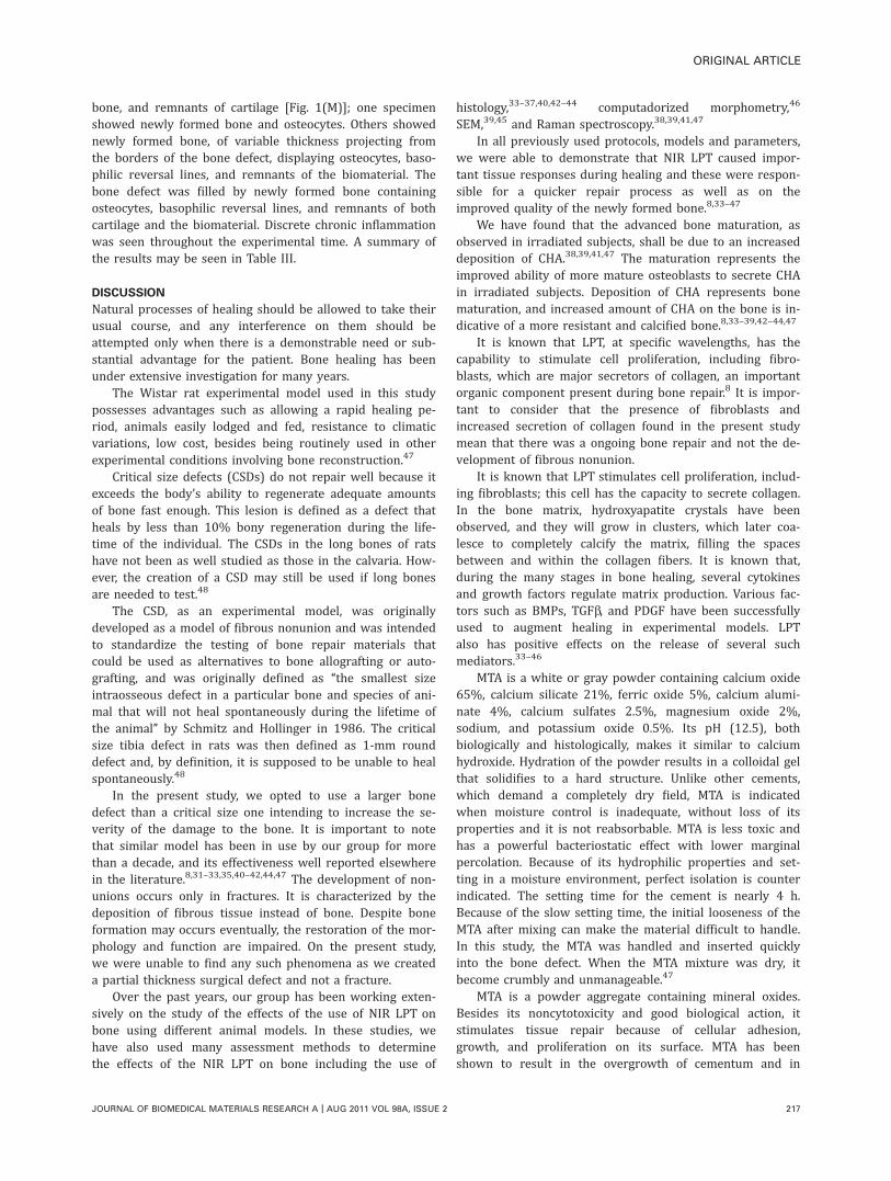
FIGURE 1. (A) Photomicrography of a control specimen on day 21 showing the bone defect completely filled with newly formed extending
down to the medullar tissue and displaying osteocytes nonaligned and basophilic reversal lines. (B) Photomicrography of a specimen grafted
with MTA on day 15 showing the bone spicules amidst bone defect whose superficial area shows bone necrosis. Note inferior margin with
bone walls close to medullar tissue. (C) Photomicrography of a specimen of the MTA þ GBR group on day 21 showing the bone defect filled by
bone spicules of variable size amidst inflammation composed of lymphocytes and macrophages. Note necrosis due to the biomaterial on sur-
face. (D) Photomicrography of specimen of the group MTA þ BMP on day 15 showing the fibrous band with small areas of necrosis and rem-
nant of the biomaterial (seen on top right), followed by the bone defect filled by newly formed bone in the form of interconnecting that displays
osteocytes nonaligned. There were signs of reabsorption. (E) Photomicrography of a specimen of group MTA þ BMP þ GBR on day 15 showing
the bone defect partially filled with bone spicules and trabeculi extending from one cortical side, and the other whose surface displays necrosis,
cartilage remnants, and osteocytes nonaligned are also observed. (F) Photomicrography of group NIR LPT on day 15 showing the bone defect
filled by interconnecting and newly formed bone with nonaligned osteocytes and discrete basophilic reversal lines, amidst medullar tissue. (G)
Photomicrography MTA þ NIR LPT on day 15 showing a fibrous band with remnants of the biomaterial and necrosis followed by irregular trabe-
culi displaying nonaligned osteocytes and some medullar tissue. (H) Photomicrography of specimen of group MTA þ NIR LPT on day 21 show-
ing newly formed bone displaying numerous osteocytes and active multinucleated giant cells amidst fibrous tissue with chronic inflammation.
(I) Photomicrography of a specimen of group MTA þ GBR þ NIR LPT on day 15 showing the bone defect filled by interconnecting and newly
formed bone and displaying nonaligned osteocytes. No signs of reabsorption were seen, and inferior part showed extensive irregular bone
close to medullar tissue. (J) Photomicrography of a specimen of group MTA þ BMP þ NIR LPT on day 15 showing the bone defect displaying
small bone spicules and areas of necrosis. (L) Photomicrography of a specimen of group MTA þ BMP þ NIR LPT on day 15 showing the bone
defect filled by interconnecting trabeculi displaying active osteoblasts, osteocytes nonaligned, and reversal basophilic lines amidst inflamed tis-
sue. Note that neoformation progressed of bone cortical as seen on the bottom. (M) Photomicrography of a specimen of group MTA þ GBR þBMP þ NIR LPT on day 21 showing a fibrous band with remnants of the biomaterial and necrosis followed by extensive displaying osteocytes
nonaligned and some medullar tissue.
ORIGINAL ARTICLE
JOURNAL OF BIOMEDICAL MATERIALS RESEARCH A | AUG 2011 VOL 98A, ISSUE 2 215

deposition. Irregular osteocytes, basophilic reversal lines,and few medullary spaces were also seen. At the surface,besides the presence of remnants of the biomaterial, a bandof fibrotic tissue was also seen as well as evidence of bonereabsorption, and moderate chronic inflammation. At theend of the experimental time, the bone defect was partiallyor completely filled by newly formed bone displaying nona-ligned osteocytes and few medullary spaces. In some cases,at the surface, a band of fibrous tissue and remnants of thebiomaterial were seen. No inflammation was observed atthis stage.
NIR LPTOn day 15, the bone defect was partially filled by newlyformed bone. Interconnecting delicate bone trabeculi, nona-ligned osteocytes, and basophilic reversal lines were seen atthis time. In some specimens, osteoblasts and remnants ofcartilage were seen at the surface [Fig. 1(F)]. The newlyformed bone was dispersed on medullar tissue and no signsof reabsorption could be seen. On day 21, the newly formedbone was regularly distributed and showed no signs ofreabsorption. At the end of the experimental period, thebone defect was mostly filled by newly formed bone thatwas either regular or irregularly distributed and showedfew medullar spaces. No signs of reabsorption were seen.
MTA þ NIR LPTOn day 15, surface necrosis was observed as well as thepresence of an irregularly distributed; either thin or thicknewly formed interconnecting bone trabeculi, with nona-ligned osteocytes and basophilic reversal lines [Fig. 1(G)].The presence of remnants of the biomaterial was also seenat the surface. There was evidence of sites of reabsorptionof the bone. On day 21, the bone filling the bone defect waseither thick or thin and also showed nonaligned osteocytes,basophilic reversal lines, and few medullary spaces. No evi-dence of reabsorption was seen at this stage [Fig. 1(H)]. Atthe end of the experimental period, the bone defect wasfilled by newly formed bone and focal area displaying reab-sorption was seen. Chronic inflammation was seen duringall experimental time.
MTA þ GBR þ NIR LPTOn day 15, the bone defect was either partially or com-pletely filled by irregular bone trabeculi dispersed on
medullar tissue. The bone showed nonaligned osteocytesand active osteoblasts at the periphery of bone trabeculi[Fig. 1(I)]. In a few cases, bony fragments were observed.Foci of necrosis could also be seen at the surface at thisstage. Remnants of the biomaterial were also seen in allcases as well as a band of fibrotic tissue. These remnantswere sometimes observed incorporated to the newly formedbone and they assumed a crystalloid aspect. Despite thepresence of some foci of necrosis close to the bony frag-ments, deeper region (within the bone defect) showednewly formed bone. No evidence of bone reabsorption couldbe seen at this stage, but chronic inflammation was seen.On day 21, the bone defect was mostly filled with intercon-necting bone trabeculi, displaying nonaligned osteocytesand basophilic reversal lines as well as discrete chronicinflammation. No signs of necrosis or reabsorption could beseen at this stage. A delicate mesh of newly formed boneand medullary tissue filled the bone defect. In some cases, aband of connective tissue was also seen at surface. Noinflammation was found at this time.
MTA þ BMP þ NIR LPTOn day 15, the bone defect was partially covered by ne-crotic tissue and showed foci of remnants of the biomaterial[Fig. 1(J)]. Beneath it, bony fragments dispersed within ne-crotic tissue were seen. In some cases, the newly formedbone was thick and showed osteocytes and few medullaryspaces. The inferior margin of the bone defect showed athin bone wall [Fig. 1(L)]. On day 21 thick newly formedbone, containing remnants of the biomaterial filled the bonedefect. In some specimens, a band of fibrosis was presentand usually it projected itself within the medulla. At the endof the experimental period, the bone defect was completelyfilled by a dense newly formed bone sometimes irregular,osteocytes, and basophilic reversal lines. Discrete chronicinflammation was seen during all times.
MTA þ GBR þ BMP þ NIR LPTOn day 15, the bone defect was filled by newly formedbone that was distributed in an irregular manner andshowed both osteocytes and remnants of the biomaterialwithin it. Some specimens showed osteoblasts at the surfaceand signs of bone reabsorption at this time. On day 21, thehistological aspect varied: one specimen showed remnantsof the biomaterial dispersed on granulation tissue, lamellar
TABLE III. Summary of the Histological Analysis
Criterion/Group Bone Reabsorption Bone Neoformation Inflammatory Infiltrate Collagen Deposition
Clot Absent Intense Discrete (chronic) ModerateMTA Absent Moderate Discrete (chronic) ModerateMTA þ GBR Discrete Moderate Discrete (chronic) ModerateMTA þ BMP Moderate Moderate Absent ModerateMTA þ BMP þ GBR Discrete Intense Moderate (chronic) ModerateNIR LPT Absent Intense Absent IntenseMTA þ NIR LPT Discrete Intense Absent IntenseMTA þ GBR þ NIR LPT Absent Intense Moderate (chronic) IntenseMTA þ GBR þ BMP þ NIR LPT Discrete Intense Discrete Intense
216 PINHEIRO ET AL. LASER ON MTA GRAFTS
bone, and remnants of cartilage [Fig. 1(M)]; one specimenshowed newly formed bone and osteocytes. Others showednewly formed bone, of variable thickness projecting fromthe borders of the bone defect, displaying osteocytes, baso-philic reversal lines, and remnants of the biomaterial. Thebone defect was filled by newly formed bone containingosteocytes, basophilic reversal lines, and remnants of bothcartilage and the biomaterial. Discrete chronic inflammationwas seen throughout the experimental time. A summary ofthe results may be seen in Table III.
DISCUSSION
Natural processes of healing should be allowed to take theirusual course, and any interference on them should beattempted only when there is a demonstrable need or sub-stantial advantage for the patient. Bone healing has beenunder extensive investigation for many years.
The Wistar rat experimental model used in this studypossesses advantages such as allowing a rapid healing pe-riod, animals easily lodged and fed, resistance to climaticvariations, low cost, besides being routinely used in otherexperimental conditions involving bone reconstruction.47
Critical size defects (CSDs) do not repair well because itexceeds the body’s ability to regenerate adequate amountsof bone fast enough. This lesion is defined as a defect thatheals by less than 10% bony regeneration during the life-time of the individual. The CSDs in the long bones of ratshave not been as well studied as those in the calvaria. How-ever, the creation of a CSD may still be used if long bonesare needed to test.48
The CSD, as an experimental model, was originallydeveloped as a model of fibrous nonunion and was intendedto standardize the testing of bone repair materials thatcould be used as alternatives to bone allografting or auto-grafting, and was originally defined as ‘‘the smallest sizeintraosseous defect in a particular bone and species of ani-mal that will not heal spontaneously during the lifetime ofthe animal’’ by Schmitz and Hollinger in 1986. The criticalsize tibia defect in rats was then defined as 1-mm rounddefect and, by definition, it is supposed to be unable to healspontaneously.48
In the present study, we opted to use a larger bonedefect than a critical size one intending to increase the se-verity of the damage to the bone. It is important to notethat similar model has been in use by our group for morethan a decade, and its effectiveness well reported elsewherein the literature.8,31–33,35,40–42,44,47 The development of non-unions occurs only in fractures. It is characterized by thedeposition of fibrous tissue instead of bone. Despite boneformation may occurs eventually, the restoration of the mor-phology and function are impaired. On the present study,we were unable to find any such phenomena as we createda partial thickness surgical defect and not a fracture.
Over the past years, our group has been working exten-sively on the study of the effects of the use of NIR LPT onbone using different animal models. In these studies, wehave also used many assessment methods to determinethe effects of the NIR LPT on bone including the use of
histology,33–37,40,42–44 computadorized morphometry,46
SEM,39,45 and Raman spectroscopy.38,39,41,47
In all previously used protocols, models and parameters,we were able to demonstrate that NIR LPT caused impor-tant tissue responses during healing and these were respon-sible for a quicker repair process as well as on theimproved quality of the newly formed bone.8,33–47
We have found that the advanced bone maturation, asobserved in irradiated subjects, shall be due to an increaseddeposition of CHA.38,39,41,47 The maturation represents theimproved ability of more mature osteoblasts to secrete CHAin irradiated subjects. Deposition of CHA represents bonematuration, and increased amount of CHA on the bone is in-dicative of a more resistant and calcified bone.8,33–39,42–44,47
It is known that LPT, at specific wavelengths, has thecapability to stimulate cell proliferation, including fibro-blasts, which are major secretors of collagen, an importantorganic component present during bone repair.8 It is impor-tant to consider that the presence of fibroblasts andincreased secretion of collagen found in the present studymean that there was a ongoing bone repair and not the de-velopment of fibrous nonunion.
It is known that LPT stimulates cell proliferation, includ-ing fibroblasts; this cell has the capacity to secrete collagen.In the bone matrix, hydroxyapatite crystals have beenobserved, and they will grow in clusters, which later coa-lesce to completely calcify the matrix, filling the spacesbetween and within the collagen fibers. It is known that,during the many stages in bone healing, several cytokinesand growth factors regulate matrix production. Various fac-tors such as BMPs, TGFb, and PDGF have been successfullyused to augment healing in experimental models. LPTalso has positive effects on the release of several suchmediators.33–46
MTA is a white or gray powder containing calcium oxide65%, calcium silicate 21%, ferric oxide 5%, calcium alumi-nate 4%, calcium sulfates 2.5%, magnesium oxide 2%,sodium, and potassium oxide 0.5%. Its pH (12.5), bothbiologically and histologically, makes it similar to calciumhydroxide. Hydration of the powder results in a colloidal gelthat solidifies to a hard structure. Unlike other cements,which demand a completely dry field, MTA is indicatedwhen moisture control is inadequate, without loss of itsproperties and it is not reabsorbable. MTA is less toxic andhas a powerful bacteriostatic effect with lower marginalpercolation. Because of its hydrophilic properties and set-ting in a moisture environment, perfect isolation is counterindicated. The setting time for the cement is nearly 4 h.Because of the slow setting time, the initial looseness of theMTA after mixing can make the material difficult to handle.In this study, the MTA was handled and inserted quicklyinto the bone defect. When the MTA mixture was dry, itbecome crumbly and unmanageable.47
MTA is a powder aggregate containing mineral oxides.Besides its noncytotoxicity and good biological action, itstimulates tissue repair because of cellular adhesion,growth, and proliferation on its surface. MTA has beenshown to result in the overgrowth of cementum and in
ORIGINAL ARTICLE
JOURNAL OF BIOMEDICAL MATERIALS RESEARCH A | AUG 2011 VOL 98A, ISSUE 2 217

facilitating regeneration of the periodontal ligament and for-mation of bone.
As far as we are concerned this is the first histologicalreport to describe, histologically, the mechanisms of therepair bone defects treated or not with laser light, MTA,BMPs, and GBR. The comparison of our results with otherprevious reports is difficult. This is the first report of theuse of this model. Our previous experience using othertypes of biomaterials already reported in the literature isalso suggestive that the association of NIR LPT with bioma-terials causes improvement on the repair of bone defects.8
A previous report from our team evaluated the effectlaser light associated to MTA on the alveolar bone repairprocess. We found that laser-irradiated sockets substantialformation of thick interwoven osteocyte-rich trabecularbone, with an evident osteoblastic rimming when comparedto the use of MTA as well as to untreated controls showingthat laser light was the most successful treatment toimprove alveolar bone repair.49
This study was designed to carry out a qualitativeassessment of the repair process, as this is the first reportof its kind and the description of the process considered anappropriated method of assessing the effects of differenttechniques used both isolated or in combination, allowingus to have a qualitative ‘‘picture’’ of the process.
The present study is the histological analysis of thestudy recently published by our team in which Raman spec-troscopy was used as assessment method and showedresults aligned with the histological findings of the presentstudy.47 This aspect is important, as the only use of histo-logical findings could be considered inadequate by anyoneand some authors suggested that this kind of data would bebetter utilized if correlated with other test such as radiolog-ical evaluation or with mechanical tests of the bone.50–52
In the Raman study we found that, laser-irradiated sub-jects, showed significant higher levels of CHA in the earlierperiod of healing. However, these levels were similar to theobserved when the association of MTA þ BMP þ GBR þLaser was used. Lowest levels of deposition were observedwhen MTA þ Laser and MTA þ GBR were used. At the endof the experimental period, all defects were found to besimilar. However, higher levels of CHA were seen in groupMTA þ GBR þ Laser and lower in group MTA þ Laser. Asincreased peaks of CHA are indicative of bone maturation,the association of MTA, GBR, and laser resulted in a moreadvanced repair.47
We found differences among the groups during all ex-perimental timing. It is important to keep in mind that theMTA probably acted as a local irritant even on being consid-ered biocompatible. When the MTA was associated to GBR,no major morphological changes were seen. But, weobserved crystalloid structures that may be attributable tothe MTA as its anhydrous phases involve the formation ofcrystals, an important physiologic event in the formation ofthe bone.53 Again, these changes might be attributed to thepresence of the MTA. The addition of BMPs to the MTA didnot improve the process significantly. When the MTA þBMP graft was associated to GBR, the major observed
change was of irregular osteoid tissue, whose appearancemay be attributed to the GBR technique. The use of NIRLPT caused both early deposition of bone trabeculi and thecessation of the osteoclastic activity,54 which plays a pivotalrole on the remodeling of the bone.55–57 These lines werenot seen on controls but were present on LPT and on MTAþ BMP þ GBR treated subjects. This may be indicative thatthe use alone of the LPT may cause similar results to theuse of the association of conventional techniques alone.
Later on, untreated controls and MTA-grafted subjectsshowed similar healing pattern as seen at early stages. Wealso found remnants of cartilaginous tissue on the controlsthat may be indicative of evolution of the process. The MTAseemed to cause a small delay in the repair process. Theuse of the GBR caused the presence of thick newly formedbone. Increased thickness might have been caused by theGBR technique. Adding BMPs to the biomaterial also causedthe appearance of thick and irregular newly formed bone.However, despite remnants of the graft causing foreign bodyreaction there was no bone reabsorption. The use of theBMPs might have caused more bone deposition during therepair due to its properties.
The use of the GBR to the association of MTA and BMPscaused no major improvement on the repair. The use of theLPT caused the deposition of a regularly distributed newlyformed bone. On the other hand, MTA grafts irradiatedcaused irregular deposition of bone. The association of theLPT with GBR did not improve the repair significantly. TheLPT associated to the MTA, BMPs, and GBR caused variableresponses including the presence of newly formed bone, ofvariable thickness, projecting from the borders of the bonedefect. This was not seen on other groups.
At the end of the experimental period, the controlsshowed the bone defect completely filled by lamellar trabe-cullar bone and few Haversian systems. The use of GBR ornot on MTA-grafted subjects caused a deposition of a lessdense bone. The addition of BMPs caused the appearancenecrotic debris and macrophages. The use of MTA, BMPs,and GBR caused the presence of newly formed bone eitherpartially or completely filling the bone defects. LPT-treatedsubjects showed the bone defect mostly filled regular orirregular bone. The use of MTA caused the appearance ofnewly formed bone of varied thickness. Using NIR LPT ongrafts subjected to GBR did not affect the overall repairmuch. NIR LPT associated to MTA and BMPs caused thecomplete filling of the bone defect by a dense newly formedbone. The laser light associated to MTA, BMPs, and GBRalso resulted in the deposition of newly formed bone andremnants of cartilage. It is possible that the chondrogenicdifferentiation occurred due to mesenchymal stem cellspresent in the bone marrow, and represents the presence ofprogenitor cells capable of differentiating into osteoblasts.Thus, these cartilage remnants could play a key role as scaf-fold in the bone repair.
One may question that it is expected inflammatoryresponse to develop against bone repair materials (i.e., scaf-fold) as these materials, no matter how biocompatible theyare, they are foreign materials. In the present study we
218 PINHEIRO ET AL. LASER ON MTA GRAFTS

found more inflammatory response in clot group, in whichthere is no scaffold applied at all, than the ones in MTA þBMP, NIR LPT, and MTA þ NIR LPT groups. This may beexplained by previous investigations that have indicatedthat, the persistence of the inflammatory response in thelater phases of alveolar bone repair might be a result of aphlogistic activity of residual blood clots.49,58
In terms of bone neoformation, we found more bone for-mation in clot group than on the ones in MTA (in whichthere is osteoconductive scaffold), MTA þ GBR (in whichthere is osteoconductive scaffold and GBR was induced),and MTA þ BMP (in which there is osteoconductive scaffoldand osteoinductive cytokine). Despite the presence of MTAto be expected to facilitate the process of bone deposition,due to its biochemical properties, such as alkalinity andhigh content of calcium phosphate, calcium oxide and silica,which are important inorganic constituents widely requiredfor bone mineralization, this material probably, due to somephlogistic activity promoted by its handling during the fill-ing of the defect, delayed the process. It may be speculatedthat MTA handling within the alveolar wound caused a dis-arrangement of part of the blood clot and altered the boneregeneration process.49
We were not able to find in the literature any previousreports on the association of MTA with BMPs. Despite wehave shown that the use of BMPs may improve the outcomeof bone healing when associated with LPT, in the presentstudy it was not the case. This was probably due to theproperties of the MTA.
It has also been demonstrated by our group that, theuse of GBR, is helpful in the healing of bone defects andthat the association with LPT improves the outcome of thistherapeutic approach as demonstrated previously by ourusing different models.33–47
The results of our study indicate that irradiated boneshowed increased osteoblastic proliferation, collagen deposi-tion, and bone neoformation when compared to nonirradi-ated one. The results of our studies and others indicate thatbone irradiated mostly with IR wavelengths showsincreased osteoblastic proliferation, collagen deposition, andbone neoformation when compared to nonirradiated bone.The effects observed in irradiated subjects might be a resultof positive effects of laser irradiation on the cell membraneand mitochondria. Laser light influences the production ofATP and it is more effective at when used at early stagesand depends on the physiologic status of the cell.33–47
Vascularization is also an important and decisive factorfor the healing of wounds and it is improved after the useof laser light as well as increased release of mediators suchas PGE2 that is also increased after irradiation. There is evi-dence that PGE2 is also produced by osteoblasts and that itseffects may be therapeutic or adverse.33–46
It is known that laser light stimulates cell proliferation,including fibroblasts; this cell has the capacity to secretecollagen. In the bone matrix, hydroxyapatite crystals havebeen observed, and they will grow in clusters, which latercoalesce to completely calcify the matrix, filling the spacesbetween and within the collagen fibers. It is known that,
during the many stages in bone healing, several cytokinesand growth factors regulate matrix production.33–46
Various factors such as BMPs, transforming growth fac-tor-b, and platelet-derived growth factor have been success-fully used to augment healing in experimental models. Laserlight also has positive effects on the release of several suchmediators.33–46
A parameter able to produce any photo biologicalresponse alone does not exist. The photoresponse dependson the conjugation of different parameters. It still remainsuncertain if bone stimulation by laser light is a generaleffect or if the isolate stimulation of osteoblasts is possible.It is possible that laser’s effect on bone regenerationdepends not only on the total dose of irradiation, but alsoon the irradiation time and the irradiation mode. Mostimportantly, the threshold parameter energy density and in-tensity are biologically independent of one another. This in-dependence accounts for the success and the failure of thetreatment.33–46
One may question the advantage of using an in vitroexperiment where the direct influence of LPT on osteoblastsand the MTA-scaffold could be analyzed. In the presentstudy, we opted for using an in vivo model and not an invitro one as the animal model allowed us a more detailedand ‘‘close’’ to human tissue response analysis of the differ-ent treatments carried out as well as the host-responses tothem. The host-response on in vitro model is very limited ornot possible of simulation in many aspects. For instance, de-spite recent study59 have found in vivo adhesion of osteo-blasts to MTA, an in vitro report60 using osteoblasts failedto get cell adhesion to MTA. In the present study, we oftenobserved a close contact of the graft with the bone matrixand possibly inducing the formation of important compo-nents for bone neoformation as a result of the interaction ofthe MTA, the matrix, and the host.61,62 Then, biologicalresponses may be similarly detected using different assess-ment methods.
CONCLUSION
The results showed different tissue response on all groupsduring the experimental time. Major changes were seen inirradiated subjects and included marked deposition of newbone seen in advanced maturation. It is concluded that NIRLPT improved the results of the use of the MTA on bonedefects.
REFERENCES1. Pastori CM, Zorzetto DLG, Toledo Filho JL, Marzola C. Implantes
de bioapatita þ osseobondVR þ membrana reabsorvıvel dentoflex
þ aglutinante dentoflex. [Implants of bioapatite þ osseobondVR þdentoflex reabsorbable membrane þ dentoflex link agent: Surgi-
cal cases report]. Rev Bras Cienc Estomatol 1996;1:51–63.
2. Reddi AH. Initiation of fracture repair by bone morphogenetic
proteins. Clin Orthop Relat 1998;355(Suppl):S66–S72.
3. Park YJ. Enhanced guided bone regeneration by controlled tetra-
cycline release from poly(L-lactide) barrier membranes. J Biomed
Mater Res 2000;51:391–397.
4. Segundo T. Avaliacao dos Enxertos Osseos e Homologos Utiliza-
dos em Implantodontia. [Assessment of bone grafts and similar
used in implantology]. RGO 2000;48:217–220.
ORIGINAL ARTICLE
JOURNAL OF BIOMEDICAL MATERIALS RESEARCH A | AUG 2011 VOL 98A, ISSUE 2 219

5. Restrepo LL, Marzola C, Consolaro A, Pereira AC, Toledo Filho JL,
Andreo JC. Avaliacao de implantes de osso bovino liofilizado
‘‘Osseobond’’VR
e membrana reabsorvıvel de osso bovino liofili-
zado. [Assemment of liophilized bovine bone ‘‘Osseobond’’VR
and
reabsorbable bovine bone membrane]. Available at http://
www.odontologia.com.br/artigos. 02 March 2001.
6. Taga EM, Mulatinho J. Biomateriais para uso em Clınica Medico-
Odontologica. [Biomaterials used on medical-dental clinics].
Available at http://www.dentoflex.com.br. 05 February 2001.
7. Nyman S, Lindhe J, Karring T, Rylander H. New attachment fol-
lowing surgical treatment of human periodontal disease. J Clin
Periodontol 1982;9:290–296.
8. Pinheiro ALB, Gerbi MEMM. Photoengineering of bone repair
processes. Photomed Laser Surg 2006;24:169–178.
9. Torabinejad M, Hong CU, Lee SJ, Monsef M, Pitt Ford TR. Investi-
gation of mineral trioxide aggregate for root end filling in dogs. J
Endod 1995;21:603–608.
10. Torabinejad M, Hong CU, Pitt Ford TR. Physical properties of a
new root end filling material. J Endod 1995;21:349–353.
11. Torabinejad M, Hong CU, Pitt Ford TR, Kariyawasam SP. Tissue
reaction to implanted super-EBA and mineral trioxide aggregate
in the mandible of guinea pigs: A preliminary report. J Endod
1995;21:569–571.
12. Torabinejad M, Hong CU, Pitt Ford TR, Kettering JD. Cytotoxicity
of four root end filling materials. J Endod 1995;21:489–492.
13. Torabinejad M, Pitt Ford TR, Abedi HR, Kariyawasam SP, Tang
HM. Tissue reaction to implanted root-end filling materials in the
tibia and mandible of guinea pigs. J Endod 1998;24:468–471.
14. Torabinejad M, Chivian N. Clinical applications of mineral trioxide
aggregate. J Endod 1999;25:197–205.
15. Schwartz RS, Mauger M, Clement DJ, Walker WAM. Mineral triox-
ide aggregate: A new material for endodontics. J Am Dent Assoc
1999;130:967–975.
16. Zhu Q, Haglund R, Safavi KE, Spangberg LSW. Adhesion of human
osteoblasts on root-end filling materials. J Endod 2000;26:404–406.
17. Regan JD, Gutmann JL, Witherspoon DE. Comparison of Diaket
and MTA when used as rootend filling materials to support
regeneration of the periradicular tissues. Int Endod J 2002;35:
840–847.
18. Economides N, Pantelidou O, Kokkas A, Tziafas D. Short-term
periradicular tissue response to mineral trioxide aggregate (MTA)
as root-end filling material. Int Endod J 2003;36:44–48.
19. Al-Rabeah E, Perinpanayagam H, MacFarland D. Human alveolar
bone cells interact with ProRoot and tooth-colored MTA. J Endod
2006;32:872–875.
20. Oviir T, Pagoria Ibarra G, Geurtsen W. Effects of gray and white
mineral trioxide aggregate on the proliferation of oral keratino-
cytes and cementoblasts. J Endod 2006;32:210–213.
21. Holland R, de Souza V, Nery MJ, Otoboni Filho A, Barnabe PFE,
Dezan E Jr. Reaction of dog’s teeth to root canal filling with min-
eral trioxide aggregate or a glass ionomer sealer. J Endod 1999;
25:728–730.
22. Moretton TR, Brown CE Jr, Legan JJ, Kafrawy AH. Tissue reac-
tions after subcutaneous and intraosseous implantation of min-
eral trioxide aggregate and ethoxybenzoic acid cement. J Biomed
Mater Res 2000;52:528–533.
23. Bystrom A, Claesson R, Sundqvist G. The antibacterial effect of
camphorated paramonochlorophenol, camphorated phenol and
calcium hydroxide in the treatment of infected root canals. Endod
Dent Traumatol 1995;1:170–175.
24. Mitchell PJ, Pitt Ford TR, Torabinejad M, McDonald F. Osteoblast
biocompatibility of mineral trioxide aggregate. Biomaterials 1999;
20:167–173.
25. Tronstad L, Andreasen JO, Hasselgren G, Kristerson L, Riis I. pH
changes in dental tissues after root canal filling with calcium
hydroxide. J Endod 1980;7:17–21.
26. De Moor RJ, DeWitte AM. Periapical lesions accidentally filled
with calcium hydroxide. Int Endod J 2002;35:946–958.
27. De Rossi A, Silva LA, Leonardo MR, Rocha LB, Rossi MA. Effect
of rotary or manual instrumentation, with or without a calcium
hydroxide/1% chlorhexidine intracanal dressing, on the healing of
experimentally induced chronic periapical lesions. Oral Surg Oral
Med Oral Pathol Oral Radiol Endod 2005;99:628–636.
28. Tronstad L. Root resorption-etiology, terminology and clinical
manifestations. Endod Dent Traumatol 1988;4:241–252.
29. Gottlow J, Nyman S, Karring T, Lindhe J. New attachment forma-
tion as the result of controlled tissue regeneration. J Clin Peri-
odontol 1984;11:494–503.
30. Garrett S. Periodontal regeneration around natural teeth. Ann
Periodontol 1996;1:621–666.
31. Stahl SS, Froum S, Tarnow D. Human histologic responses to
guided tissue regenerative techniques in intrabony lesions. Case
reports on 9 sites. J Clin Periodontol 1990;17:191–198.
32. Cortellini P, Clauser C, Prato GP. Histologic assessment of new
attachment following the treatment of a human buccal recession
by means of a guided tissue regeneration procedure. J Periodon-
tol 1993;64:387–391.
33. Pinheiro ALB, Gerbi MEMM, Limeira FA Jr, Ponzi EAC, Marques
AMC, Carvalho CM, Santos RC, Oliveira PC, Noia M, Ramalho
LMP. Bone repair following bone grafting hydroxyapatite guided
bone regeneration and infrared laser photobiomodulation: A his-
tological study in a rodent model. Lasers Med Sci 2009;24:
234–240.
34. Gerbi MEMM, Ponzi EAC, Ramalho LMP, Marques AMC, Carvalho
CM, Oliveira RC, Oliveira PC, Noia M, Pinheiro ALB. Infrared laser
light further improves bone healing when associated with bone
morphogenic proteins: An in vivo study in a rodent model.
Photomed Laser Surg 2008;26:55–60.
35. Pinheiro ALB, Gerbi MEMM, Ponzi EAC, Ramalho LMP, Marques
AMC, Carvalho CM, Oliveira RC, Oliveira PC, Noia M. Infrared
laser light further improves bone healing when associated with
bone morphogenetic proteins and guided bone regeneration: An
in vivo study in a rodent model. Photomed Laser Surg 2008;26:
167–174.
36. Torres CS, Santos JN, Monteiro JSC, Amorim PG, Pinheiro ALB.
Does the use of laser photobiomodulation, bone morphogenetic
proteins, and guided bone regeneration improve the outcome of
autologous bone grafts? An in vivo study in a rodent model.
Photomed Laser Surg 2008;26:371–377.
37. Gerbi MEMM, Pinheiro ALB, Ramalho LMP. Effect of IR laser
photobiomodulation on the repair of bone defects grafted with
organic bovine bone. Lasers Med Sci 2008;23:313–317.
38. Lopes CB, Pacheco MT, Silveira L Jr, Duarte J, Cangussu MC,
Pinheiro ALB. The effect of the association of NIR laser therapy
BMPs, and guided bone regeneration on tibial fractures treated
with wire osteosynthesis: Raman spectroscopy study. J Photo-
chem Photobiol B 2007;89:125–130.
39. Lopes CB, Pinheiro ALB, Sathaiah S, Da Silva NS, Salgado MA.
Infrared laser photobiomodulation (830 nm) on bone tissue
around dental implants: A Raman spectroscopy and canning elec-
tronic microscopy study in rabbits. Photomed Laser Surg 2007;25:
96–101.
40. Weber JBB, Pinheiro ALB, Oliveira MG, Oliveira FAM, Ramalho
LMP. Laser therapy improves healing of bone defects submitted
to autogenos bone graft. Photomed Laser Surg 2006;24:38–44.
41. Lopes CB, Pinheiro ALB, Sathaiah S, Duarte J, Martins MC. Infra-
red laser light reduces loading time of dental implants: A Raman
spectroscopy study. Photomed Laser Surg 2005;23:27–31.
42. Gerbi MEMM, Pinheiro ALB, Marzola C, Limeira Junior FA,
Ramalho LMP, Ponzi EAC, Soares AO, Carvalho LC, Lima HV,
Goncalves TO. Assessment of bone repair associated with the
use of organic bovine bone and membrane irradiated at 830 nm.
Photomed Laser Surg 2005;23:382–388.
43. Pinheiro ALB, Limeira Junior FA, Gerbi MEMM. Effect of low level
laser therapy on the repair of bone defects grafted with inorganic
bovine bone. Braz Dent J 2003;14:177–181.
44. Pinheiro ALB, Limeira Junior FA, Gerbi MEMM, Ramalho LMP,
Marzola C, Ponzi EAC, Carvalho LCB, Lima HCAV, Goncalves TO,
Soares AO. Effect of 830-nm laser light on the repair of bone
defects grafted with inorganic bovine bone and decalcified corti-
cal osseous membrane. J Clin Laser Med Surg 2003;21:383–388.
45. Pinheiro ALB, Oliveira MAM, Martins PPM. Biomodulacao da
cicatrizacao ossea pos-implantar com o uso da laserterapia nao-
cirurgica: Estudo por microscopia eletronica de varredura.
[Biomodulation of Peri-implant bone repair with lasertherapy:
SEM study]. Rev FOUFBA 2001;22:12–19.
220 PINHEIRO ET AL. LASER ON MTA GRAFTS
46. Silva N Jr, Pinheiro ALB, Oliveira MGA, Weissmann R, Ramalho
LMP, Nicolau RA. Computadorized morphometric assessment of
the effect of low-level laser therapy on bone repair: An experi-
mental animal study. J Clin Laser Med Surg 2002;20:83–88.
47. Pinheiro ALB, Aciole GTS, Cangussu MCT, Pacheco MTT, Silveira
L Jr. Effects of laser phototherapy on bone defects grafted with
mineral trioxide aggregate, bone morphogenetic proteins, and
guided bone regeneration: A Raman spectroscopic study. J
Biomed Mater Res A 2010;95:1041–1047.
48. Mooney MP, Siegel MI. Animal models for bone tissue engineer-
ing of critical-sized defects (CSDs), bone pathologies, and
orthopedic disease states. In: Hollinger JO, Einhorn TA, Doll BA,
Sfeir C, editors. Bone Tissue Engineering. Boca Raton, FL: CRC
Press;2005. p217–244.
49. Oliveira EA, Oliveira VGM, Pires JA, Barreto ALS, Ribeiro MAG,
Pinheiro ALP, Marques AMC, Melo CM, Albuquerque Junior RLC.
Effect of low-level laser therapy and mineral trioxide aggregate
on alveolar bone repair. Braz J Oral Sci 2008;27:1657–1661.
50. Liebschner MA. Biomechanical considerations of animal models
used in tissue engineering of bone. Biomaterials 2004;25:
1697–1714.
51. Ebina H, Hatakeyama J, Onodera M, Honma T, Kamakura S,
Shimauchi H, Sasano Y. Micro-CT analysis of alveolar bone
healing using a rat experimental model of critical-size defects.
Oral Dis 2009;15:273–280.
52. Young S, Kretlow JD, Nguyen C, Bashoura AG, Baggett LS,
Jansen JA, Wong M, Mikos AG. Microcomputed tomography
characterization of neovascularization in bone tissue engineering
applications. Tissue Eng Part B Rev 2008;14:295–306.
53. Lee YL, Lee BS, Lin FH, Lin AY, Lan WH, Lin CP. Effects of physio-
logical environments on the hydration behavior of mineral triox-
ide aggregate. Biomaterials 2004;25:787–793.
54. Romano PR, Caton JG, Puzas JE. The reversal line may be a key
modulator of osteoblast function: Observations from an alveolar
bone wound -healing model. J Periodontal Res 1997;32:143–147.
55. Palumbo C, Ferretti M, Ardizzoni A. Osteocyte-osteoclast morpho-
logical relationships and the putative role of osteocytes in bone
remodeling. J Musculoskelet Neuronal Interact 2001;1:327–332.
56. Domon T, Suzuki R, Takata K, Yamazaki Y, Takahashi S, Yama-
moto, Wakita M. The nature and function of mononuclear cells on
the resorbed surfaces of bone in the reversal phase during
remodeling. Ann Anat 2001;83:103–110.
57. Matsuda C, Takagi M, Hattori T, Wakitani S, Yoshida T. Differen-
tiation of human bone marrow mesenchymal stem cells to chon-
drocytes for construction of three-dimensional cartilage tissue.
Cytotechnology 2005;47:11–17.
58. Coneglian PZA. Avaliacao do processo evolutivo do reparo
osseo frente ao sulfato de calcio e a hidroxiapatita. Estudo
microscopico em alveolos dentarios de ratos. [Assessment of
the bone repair following the use of calcium sulphate or
hidroyapatite. Light microscopic study on dental sockts of rats].
M.Sci Dissertation. Bauru (SP), School of Dentistry of Bauru,
Sao Paulo University; 2007. 254 p.
59. Perinpanaygam H. Cellular response to mineral trioxide aggregate
root-end filling materials. J Can Dent Assoc 2009;75:369–372.
60. Perez AL, Spears R, Gutman JL, Opperman LA. Osteoblasts and
MG-63 osteosarcoma cells behave differently when in contact
with ProRoot MTA and White MTA. Int Endod J 2000;36:564–570.
61. Camilleri J, Pitt Ford TR. Mineral trioxide aggregate: A review of
the constituents and biological properties of the material. Int
Endod J 2006;39:747–754.
62. Saidon J, He J, Zhu Q, Safavi K, Spangberg LSW. Cell and tissue
reactions to mineral trioxide aggregate and Portland cement. Oral
Surg Oral Med Oral Pathol Oral Radiol Endod 2003;95:483–489.
ORIGINAL ARTICLE
JOURNAL OF BIOMEDICAL MATERIALS RESEARCH A | AUG 2011 VOL 98A, ISSUE 2 221
Copyright © 2022 FDOKUMEN