Glutathione depletion enhances arsenic trioxide-induced apoptosis in lymphoma cells through...
8
Glutathione Depletion Enhances Arsenic Trioxide-Induced Apoptosis in Lymphoma Cells Through Mitochondrial- Independent Mechanisms Savita Bhalla 1,2,4 , Leo I. Gordon 1,2,4 , Kevin David 1,2,4 , Sheila Prachand 1,2,4 , Amareshwar T.K. Singh 1,2,4 , Shuo Yang 1,2,4 , Jane N. Winter 1,2,4 , Dongsheng Guo 4 , Thomas O’Halloran 3,4 , Leonidas C. Platanias 1,4,5 , and Andrew M. Evens 1,2,4 1 Division of Hematology/Oncology, Chicago, IL 60611 2 Lymphoma Program, Chicago, IL 60611 3 Department of Chemistry, Chicago, IL 60611 4 Northwestern University Feinberg School of Medicine, the Robert H. Lurie Comprehensive Cancer Center of Northwestern University, Chicago, IL 60611 5 Jesse Brown VA Medical Center, Chicago, IL 60611 Keywords non-Hodgkin lymphoma; arsenic trioxide; apoptosis; oxidative stress; ROS Introduction Arsenic trioxide (ATO) is an effective therapeutic agent for acute promyelocytic leukemia (APL) (Evens et al, 2004). In APL, ATO induces differentiation at low concentrations, while inducing apoptosis at higher concentrations (Miller, et al 2002). In addition, ATO- induced apoptosis in APL is mediated through the mitochondrial apoptotic pathway, resulting in part from the production of reactive oxygen species (ROS) such as hydrogen peroxide (Dai, et al 1999, Yi, et al 2002). High intracellular levels of glutathione (GSH) confer resistance to ATO in part through the detoxification of ROS. Compounds that promote ROS and/or deplete protective metabolites such as GSH are able to sensitize tumor cells to oxidative cytolysis. Buthionine sulfoximine (BSO), a selective inhibitor of gamma glutamylcysteine synthetase, is known to effectively deplete cellular GSH (Davison, et al 2003, Gartenhaus, et al 2002). We evaluated herein the cytotoxic activity and cell death pathways induced by ATO alone and combined with BSO in non-Hodgkin’s lymphoma (NHL) cell lines and primary lymphoproliferative cells. Results ATO-induced apoptosis in lymphoma cell lines and primary cells With ATO 10μM, approximately 15–30% apoptosis was seen in Ramos, HF1, and SUDHL4 cell lines (Figure 1A). NHL cell lines were subsequently treated with BSO (100μM) or ATO Corresponding author: Andrew M. Evens, DO, MS, Division of Hematology/Oncology, 676 N. St. Clair Street, Suite 850, Chicago, IL 60611, [email protected]. NIH Public Access Author Manuscript Br J Haematol. Author manuscript; available in PMC 2010 September 1. Published in final edited form as: Br J Haematol. 2010 August ; 150(3): 365–369. doi:10.1111/j.1365-2141.2010.08197.x. NIH-PA Author Manuscript NIH-PA Author Manuscript NIH-PA Author Manuscript
-
Upload
independent -
Category
Documents
-
view
0 -
download
0
Transcript of Glutathione depletion enhances arsenic trioxide-induced apoptosis in lymphoma cells through...

Glutathione Depletion Enhances Arsenic Trioxide-InducedApoptosis in Lymphoma Cells Through Mitochondrial-Independent Mechanisms
Savita Bhalla1,2,4, Leo I. Gordon1,2,4, Kevin David1,2,4, Sheila Prachand1,2,4,Amareshwar T.K. Singh1,2,4, Shuo Yang1,2,4, Jane N. Winter1,2,4, Dongsheng Guo4,Thomas O’Halloran3,4, Leonidas C. Platanias1,4,5, and Andrew M. Evens1,2,41Division of Hematology/Oncology, Chicago, IL 606112Lymphoma Program, Chicago, IL 606113Department of Chemistry, Chicago, IL 606114Northwestern University Feinberg School of Medicine, the Robert H. Lurie ComprehensiveCancer Center of Northwestern University, Chicago, IL 606115Jesse Brown VA Medical Center, Chicago, IL 60611
Keywordsnon-Hodgkin lymphoma; arsenic trioxide; apoptosis; oxidative stress; ROS
IntroductionArsenic trioxide (ATO) is an effective therapeutic agent for acute promyelocytic leukemia(APL) (Evens et al, 2004). In APL, ATO induces differentiation at low concentrations,while inducing apoptosis at higher concentrations (Miller, et al 2002). In addition, ATO-induced apoptosis in APL is mediated through the mitochondrial apoptotic pathway,resulting in part from the production of reactive oxygen species (ROS) such as hydrogenperoxide (Dai, et al 1999, Yi, et al 2002).
High intracellular levels of glutathione (GSH) confer resistance to ATO in part through thedetoxification of ROS. Compounds that promote ROS and/or deplete protective metabolitessuch as GSH are able to sensitize tumor cells to oxidative cytolysis. Buthionine sulfoximine(BSO), a selective inhibitor of gamma glutamylcysteine synthetase, is known to effectivelydeplete cellular GSH (Davison, et al 2003, Gartenhaus, et al 2002). We evaluated herein thecytotoxic activity and cell death pathways induced by ATO alone and combined with BSOin non-Hodgkin’s lymphoma (NHL) cell lines and primary lymphoproliferative cells.
ResultsATO-induced apoptosis in lymphoma cell lines and primary cells
With ATO 10µM, approximately 15–30% apoptosis was seen in Ramos, HF1, and SUDHL4cell lines (Figure 1A). NHL cell lines were subsequently treated with BSO (100µM) or ATO
Corresponding author: Andrew M. Evens, DO, MS, Division of Hematology/Oncology, 676 N. St. Clair Street, Suite 850, Chicago, IL60611, [email protected].
NIH Public AccessAuthor ManuscriptBr J Haematol. Author manuscript; available in PMC 2010 September 1.
Published in final edited form as:Br J Haematol. 2010 August ; 150(3): 365–369. doi:10.1111/j.1365-2141.2010.08197.x.
NIH
-PA Author Manuscript
NIH
-PA Author Manuscript
NIH
-PA Author Manuscript

(2µM) alone or in combination. Minimal apoptosis was seen with BSO or ATO alone, whileBSO combined with ATO was highly synergistic inducing over 75% apoptosis in all celllines (Figure 1B).
We next measured ROS prior to and after ATO and/or BSO. ATO alone induced minimalROS, while ATO/BSO combined resulted in pronounced ROS production. To determineROS-dependence, we co-incubated cells with the antioxidant, N-acetylcysteine (NAC).Pretreatment with NAC significantly reduced ROS levels in cells treated with ATO/BSO(Figure 1C). Furthermore, NAC blocked ATO/BSO-induced apoptosis as well as ATO alone(Figures 1D). Catalase did not inhibit ATO-induced apoptosis, while ATO/BSO-inducedapoptosis was significantly reduced (Figure 1E). These data suggest that ATO-inducedapoptosis is attributed primarily through the depletion of GSH, while ATO/BSO inducedapoptosis is more prominently ROS-mediated.
Primary chronic lymphocytic leukemia (CLL) and follicular lymphoma cells were treatedwith increasing concentrations of ATO +/− BSO (Figure 1F and 1G). Apoptosis in primaryCLL cells was approximately 75% with 5µM of ATO alone. Interestingly, significantly lesscell death was seen compared with the same concentrations (5–10µM ATO) in NHL celllines (Figure 1A). Further, the addition of BSO to ATO in primary CLL or follicularlymphoma cells did not enhance apoptosis compared with ATO alone. We hypothesized thatlow intracellular GSH content might explain the higher sensitivity of primary cells to ATOalone and the lower sensitivity to ATO/BSO. We found that GSH levels in CLL cells were4–5 fold lower compared with levels seen in Ramos or HF1 cells (data not shown).
ATO alone, but not ATO/BSO, requires Bax, Bak, and Δψm to induce cell deathWe investigated whether ATO-induced apoptosis is dependent on Bax translocation. Asshown in Figure 2A, ATO alone redistributed Bax from the cytosol to the mitochondria(evident at 6 hours) with release of cytochrome C in Ramos cells. Of note, treatment of cellswith combined ATO/BSO did not affect Bax translocation or change in mitochondrialmembrane potential (Δψm), suggesting an alternative cell death pathway.
To further examine the role of Bax translocation and Δψm, we used immortalized wild typeand Bax−/−Bak−/− mouse embryonic fibroblasts (MEFs). Treatment of wild type MEFs withATO alone resulted in enhanced apoptosis compared with Bax−/−Bak−/− MEFs, while ATO/BSO induced similar apoptosis in wild type and Bax−/−Bak−/− MEFs (Figure 2B). Further,we stained cells with mitotracker red and Hoechst followed by confocal microscopy.Treatment of cells with ATO alone resulted in Δψm loss as indicated by the loss of red stainin Hoechst-stained blue cells (Figure 2C). We also determined the loss of Δψm bytetramethylrhodamine (TMRE) staining (Figure 2D). Treatment of cells with ATO/BSO didnot show significant Δψm change indicating mitochondrial-independent cell death in thesecells. Moreover, compared with changes in Δψm in wild-type MEFs and the Bax−/−Bak−/−
double knockout MEFs, there was a substantial difference in Δψm in wild type andBax−/−Bak−/− double knockout MEFs following ATO treatment, while ATO/BSO did notshow any difference (Figure 2E). Altogether, these studies implicate the dependence of Baxand loss of Δψm in NHL cells treated with ATO alone, but not with ATO/BSO.
ATO-induced apoptosis is caspase-dependentWith ATO alone, there was increased caspase 9 and caspase 3 cleavage in all lymphoma celllines at 5–10µM (Figure 2F). ATO also induced PARP and BID activation. These findingssuggest involvement of both cell death pathways using ATO alone, although with moreprominent activation of the intrinsic cascade. In contrast, minimal activation of the intrinsiccascade was observed with ATO/BSO (Figure 2G). However, increase in cleaved caspase 7
Bhalla et al. Page 2
Br J Haematol. Author manuscript; available in PMC 2010 September 1.
NIH
-PA Author Manuscript
NIH
-PA Author Manuscript
NIH
-PA Author Manuscript
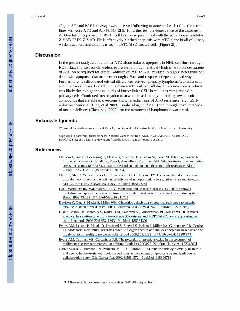
(Figure 5C) and PARP cleavage was observed following treatment of each of the three celllines with both ATO and ATO/BSO (2H). To further test the dependence of the caspases inATO–related apoptosis (+/−BSO), cell lines were pre-treated with the pan-caspase inhibitor,Z-VAD-FMK. Z-VAD–FMK effectively blocked apoptosis with ATO alone in all cell lines,while much less inhibition was seen in ATO/BSO-treated cells (Figure 2I).
DiscussionIn the present study, we found that ATO alone induced apoptosis in NHL cell lines throughROS, Bax, and caspase-dependent pathways, although relatively high in vitro concentrationsof ATO were required for effect. Addition of BSO to ATO resulted in highly synergistic celldeath with apoptosis that occurred through a Bax- and caspase-independent pathway.Furthermore, we discovered critical differences between primary lymphoma/leukemia cellsand in vitro cell lines. BSO did not enhance ATO-related cell death in primary cells, whichwas likely due to higher basal levels of intracellular GSH in cell lines compared withprimary cells. Continued investigation of arsenic-based therapy, including new arsenicalcompounds that are able to overcome known mechanisms of ATO resistance (e.g., GSH-redox mechanisms) (Diaz, et al 2008, Tsimberidou, et al 2009) and through novel methodsof arsenic delivery (Chen, et al 2009), for the treatment of lymphoma is warranted.
AcknowledgmentsWe would like to thank members of Flow Cytometry and cell imaging facility of Northwestern University.
Supported in part from grants from the National Cancer Institute (AME, K23 CA109613-A1 and LCP,R01CA121192 and a Merit review grant from the Department of Veterans Affairs.
ReferencesChandra J, Tracy J, Loegering D, Flatten K, Verstovsek S, Beran M, Gorre M, Estrov Z, Donato N,
Talpaz M, Sawyers C, Bhalla K, Karp J, Sausville E, Kaufmann SH. Adaphostin-induced oxidativestress overcomes BCR/ABL mutation-dependent and -independent imatinib resistance. Blood2006;107:2501–2506. [PubMed: 16291594]
Chen H, Ahn R, Van den Bossche J, Thompson DH, O'Halloran TV. Folate-mediated intracellulardrug delivery increases the anticancer efficacy of nanoparticulate formulation of arsenic trioxide.Mol Cancer Ther 2009;8:1955–1963. [PubMed: 19567824]
Dai J, Weinberg RS, Waxman S, Jing Y. Malignant cells can be sensitized to undergo growthinhibition and apoptosis by arsenic trioxide through modulation of the glutathione redox system.Blood 1999;93:268–277. [PubMed: 9864170]
Davison K, Cote S, Mader S, Miller WH. Glutathione depletion overcomes resistance to arsenictrioxide in arsenic-resistant cell lines. Leukemia 2003;17:931–940. [PubMed: 12750708]
Diaz Z, Mann KK, Marcoux S, Kourelis M, Colombo M, Komarnitsky PB, Miller WH Jr. A novelarsenical has antitumor activity toward As2O3-resistant and MRP1/ABCC1-overexpressing celllines. Leukemia 2008;22:1853–1863. [PubMed: 18633430]
Evens AM, Lecane P, Magda D, Prachand S, Singhal S, Nelson J, Miller RA, Gartenhaus RB, GordonLI. Motexafin gadolinium generates reactive oxygen species and induces apoptosis in sensitive andhighly resistant multiple myeloma cells. Blood 2005;105:1265–1273. [PubMed: 15388578]
Evens AM, Tallman MS, Gartenhaus RB. The potential of arsenic trioxide in the treatment ofmalignant disease: past, present, and future. Leuk Res 2004;28:891–900. [PubMed: 15234563]
Gartenhaus RB, Prachand SN, Paniaqua M, Li Y, Gordon LI. Arsenic trioxide cytotoxicity in steroidand chemotherapy-resistant myeloma cell lines: enhancement of apoptosis by manipulation ofcellular redox state. Clin Cancer Res 2002;8:566–572. [PubMed: 11839678]
Bhalla et al. Page 3
Br J Haematol. Author manuscript; available in PMC 2010 September 1.
NIH
-PA Author Manuscript
NIH
-PA Author Manuscript
NIH
-PA Author Manuscript

Jing Y, Dai J, Chalmers-Redman RM, Tatton WG, Waxman S. Arsenic trioxide selectively inducesacute promyelocytic leukemia cell apoptosis via a hydrogen peroxide-dependent pathway. Blood1999;94:2102–2111. [PubMed: 10477740]
Miller WH Jr, Schipper HM, Lee JS, Singer J, Waxman S. Mechanisms of action of arsenic trioxide.Cancer Res 2002;62:3893–3903. [PubMed: 12124315]
Tsimberidou AM, Camacho LH, Verstovsek S, Ng C, Hong DS, Uehara CK, Gutierrez C, Daring S,Stevens J, Komarnitsky PB, Schwartz B, Kurzrock R. A phase I clinical trial of darinaparsin inpatients with refractory solid tumors. Clin Cancer Res 2009;15:4769–4776. [PubMed: 19584162]
Yi J, Gao F, Shi G, Li H, Wang Z, Shi X, Tang X. The inherent cellular level of reactive oxygenspecies: one of the mechanisms determining apoptotic susceptibility of leukemic cells to arsenictrioxide. Apoptosis 2002;7:209–215. [PubMed: 11997664]
Zhu XH, Shen YL, Jing YK, Cai X, Jia PM, Huang Y, Tang W, Shi GY, Sun YP, Dai J, Wang ZY,Chen SJ, Zhang TD, Waxman S, Chen Z, Chen GQ. Apoptosis and growth inhibition in malignantlymphocytes after treatment with arsenic trioxide at clinically achievable concentrations. J NatlCancer Inst 1999;91:772–778. [PubMed: 10328107]
Bhalla et al. Page 4
Br J Haematol. Author manuscript; available in PMC 2010 September 1.
NIH
-PA Author Manuscript
NIH
-PA Author Manuscript
NIH
-PA Author Manuscript

Figure 1. Arsenic trioxide (ATO)-induced apoptosis in lymphoma cells with or withoutbuthionine sulfoximine (BSO)(A) Ramos, HF1, and SUDHL4 cells were incubated with increasing concentrations of ATO(10µM–100µM) for 18 hours. Percentage apoptosis was measured by annexinV/propidiumiodide (PI) staining and analyzed by flow cytometry as described in Materials and Methods.Dose dependent apoptosis was seen in all cell lines, with effective dose for 50% apoptosis(ED50) between 10–20µM for Ramos and HF-1 and 20– 50µM for SUDHL4. P valuesrepresent respective concentrations compared with control. (B) Ramos, HF1, and SUDHL4cells were treated with 2µM ATO, 100µM BSO alone, or ATO/BSO combined for 48 hours.The percentage of apoptotic cells was determined by AnnexinV/PI staining and analyzed byflow cytometry as described. P values compare ATO + BSO vs ATO and BSO alone. In allthree cell lines, there was >75% apoptosis when BSO was added to ATO. P values representrespective concentrations compared with control and single agent alone.(C) ROS wasmeasured in Ramos, HF1, and SUDHL4 cells. Cells were incubated with ATO alone(10µM) or ATO 2µM combined with BSO 100µM for 16 hours. Increase in ROS wasmeasured by staining with H2DCF-DA and analyzed by flow cytometry as described inMaterials and Methods. Peak shift to the right denotes an increase in ROS production. Pvalue for level of ROS production was <0.05 for control vs ATO/BSO and for ATO/BSO vsATO+BSO+NAC for all three cell lines. (D) N-acetylcysteine (NAC) attenuated ATO- andATO/BSO-induced apoptosis. Ramos, HF1, and SUDHL4 cells were pretreated with NAC10mM for 4 hours followed by incubation with ATO 10µM or ATO 2µM and BSO 100µM,and in combination with NAC for 48 hours. Percentage apoptosis was determined as
Bhalla et al. Page 5
Br J Haematol. Author manuscript; available in PMC 2010 September 1.
NIH
-PA Author Manuscript
NIH
-PA Author Manuscript
NIH
-PA Author Manuscript

previously described. P values compared ATO+NAC vs ATO and ATO+BSO+NAC vsATO+BSO. *P<0.05, **P<0.01, and ***P<0.001. (E) Ramos cells were pre-incubated with1000 units of catalase following treatment with the indicated concentrations of ATO orATO/BSO for 48 hours. Apoptosis was measured by annexinV/PI staining. (F) Peripheralblood mononuclear cells (PBMC) were isolated from the peripheral blood of three CLLpatients and from (G) two patients with follicular lymphoma as described in the Materialsand Methods. Cells were incubated with indicated concentrations of ATO or ATO/BSO for24 hours (all CLL cases), 48 hours (follicular lymphoma patient #1), and 72 hours (follicularlymphoma patient #2). Percentage apoptosis was determined as described before. P valuescompare ATO vs control. *P<0.05, **P<0.01, and ***P<0.001.
Bhalla et al. Page 6
Br J Haematol. Author manuscript; available in PMC 2010 September 1.
NIH
-PA Author Manuscript
NIH
-PA Author Manuscript
NIH
-PA Author Manuscript

Figure 2. ATO, but not ATO/BSO-induced apoptosis, was mediated by Bax translocation,mitochondrial depolarization, and caspase activation(A) Ramos cells were treated with ATO (2µM or 10µM) alone or combined with 100µMBSO for the indicated period of time. Mitochondrial and cytosolic fractions were separatedas described in Materials and Methods and analyzed by Western blotting for pro-apoptoticBax and cytochrome c proteins. Mn-SOD and actin were used as internal controls formitochondrial and cytosolic extracts, respectively. There is release of cytochrome c from themitochondria to the cytosol over time with ATO alone with translocation of Bax to themitochondria (left panel), while in the presence of BSO, there was no change inmitochondrial Bax or cytosolic cytochrome c, suggesting an alternate cell death pathway.(B) Apoptosis was measured in wild type and Bax−/−/Bak−/− MEFs. Cells were plated thenight before the treatment. Cells were treated with the indicated concentrations of ATO andATO/BSO for 24 hours followed by annexinV/PI staining and flow cytometry. (C)SUDHL4 cells were treated with ATO or ATO/BSO for 16 hours followed by incubationwith 200nM mitotracker red and 1ug/ml Hoechst stain for 30 minutes at 37°C followed bywashing with HBSS and fixed in 3.7 % formaldehyde. Fixed cells were used for confocalmicroscopy. (D) Mitochondrial membrane potential was measured in Ramos and SUDHL4cells. Cells were treated with indicated concentrations of ATO vs ATO/BSO for 16 hours,followed by staining with Tetramethylrhodamine (TMRE) as described in materials andmethods. Cells were analyzed by flow cytometry. 3-chlorophenylhydrazone (CCCP,Invitrogen) was used as a positive control. (E) Mitochondrial membrane potential wasmeasured in wild type and Bax−/−/Bak−/− MEFs. Cells were plated the night before the
Bhalla et al. Page 7
Br J Haematol. Author manuscript; available in PMC 2010 September 1.
NIH
-PA Author Manuscript
NIH
-PA Author Manuscript
NIH
-PA Author Manuscript

treatment and treated with the indicated concentrations of ATO and ATO/BSO for 6 hr andstained with TMRE followed by flow cytometry.Unless indicated, p values representcomparison with control. Abbreviations: Hr, hours; Mito-Bax, mitochondrial-related Bax.*P<0.05, **P<0.01, and ***P<0.001, unless otherwise indicated. (F) Ramos, HF1, andSUDHL4 cells were treated for 16 hours with increasing concentrations of ATO or (G)using BSO (100µM) in combination with lower dose ATO (2µM) for 16 hours. Westernblots of whole cell extracts were performed to detect caspase, PARP, and BID proteins.GAPDH was used as internal control. (H) Western blot of cleaved caspase7 in cells treatedwith ATO or ATO?BSO with and without NAC. (I) Ramos, SUDHL4, and HF1 cells werepre-treated with the pan-caspase inhibitor, Z-VAD-FMK (50µM) for 4 hours, followed bytreatment with ATO 10µM or ATO 2µM combined with BSO 100µM for 48 hours.Percentage apoptosis was determined by flow cytometry after staining the cells with annexinV/PI.
Bhalla et al. Page 8
Br J Haematol. Author manuscript; available in PMC 2010 September 1.
NIH
-PA Author Manuscript
NIH
-PA Author Manuscript
NIH
-PA Author Manuscript