Correspondence to
10
ORIGINAL ARTICLE A de novo X;8 translocation creates a PTK2-THOC2 gene fusion with THOC2 expression knockdown in a patient with psychomotor retardation and congenital cerebellar hypoplasia Eleonora Di Gregorio, 1,2 Federico T Bianchi, 3 Alfonso Schiavi, 4,5 Alessandra M A Chiotto, 3 Marco Rolando, 6 Ludovica Verdun di Cantogno, 7 Enrico Grosso, 2 Simona Cavalieri, 2 Alessandro Calcia, 1 Daniela Lacerenza, 1 Orsetta Zuffardi, 8,9 Saverio Francesco Retta, 10 Giovanni Stevanin, 11,12 Cecilia Marelli, 11,12 Alexandra Durr, 11,12 Sylvie Forlani, 11 Jamel Chelly, 13 Francesca Montarolo, 14 Filippo Tempia, 14 Hilary E Beggs, 15 Robin Reed, 16 Stefania Squadrone, 17 Maria C Abete, 17 Alessandro Brussino, 1 Natascia Ventura, 4,5 Ferdinando Di Cunto, 3 Alfredo Brusco 1,2 ▸ Additional material is published online only. To view please visit the journal online (http://dx.doi.org/10.1136/ jmedgenet-2013-101542). For numbered affiliations see end of article. Correspondence to Alfredo Brusco, Department of Medical Sciences, University of Torino, via Santena, 19, Torino 10126, Italy; [email protected]/brusco. [email protected] Received 22 January 2013 Revised 2 May 2013 Accepted 6 May 2013 Published Online First 7 June 2013 To cite: Di Gregorio E, Bianchi FT, Schiavi A, et al. J Med Genet 2013;50: 543–551. ABSTRACT Background and aim We identified a balanced de novo translocation involving chromosomes Xq25 and 8q24 in an eight year-old girl with a non-progressive form of congenital ataxia, cognitive impairment and cerebellar hypoplasia. Methods and Results Breakpoint definition showed that the promoter of the Protein Tyrosine Kinase 2 (PTK2, also known as Focal Adhesion Kinase, FAK) gene on chromosome 8q24.3 is translocated 2 kb upstream of the THO complex subunit 2 (THOC2) gene on chromosome Xq25. PTK2 is a well-known non-receptor tyrosine kinase whereas THOC2 encodes a component of the evolutionarily conserved multiprotein THO complex, involved in mRNA export from nucleus. The translocation generated a sterile fusion transcript under the control of the PTK2 promoter, affecting expression of both PTK2 and THOC2 genes. PTK2 is involved in cell adhesion and, in neurons, plays a role in axonal guidance, and neurite growth and attraction. However, PTK2 haploinsufficiency alone is unlikely to be associated with human disease. Therefore, we studied the role of THOC2 in the CNS using three models: 1) THOC2 ortholog knockout in C.elegans which produced functional defects in specific sensory neurons; 2) Thoc2 knockdown in primary rat hippocampal neurons which increased neurite extension; 3) Thoc2 knockdown in neuronal stem cells (LC1) which increased their in vitro growth rate without modifying apoptosis levels. Conclusion We suggest that THOC2 can play specific roles in neuronal cells and, possibly in combination with PTK2 reduction, may affect normal neural network formation, leading to cognitive impairment and cerebellar congenital hypoplasia. INTRODUCTION Cerebellar malformations are rare developmental dis- orders that comprise many heterogeneous diseases, with both acquired and genetic causes. 1 They can be confined to the cerebellum or variably involve other infratentorial or supratentorial structures, such as the brainstem, the corpus callosum and the cerebral cortex. 2 The cerebellum is usually hypoplastic, at dif- ference from neurodegenerative disorders, in which progressive cerebellar atrophy is observed. Nevertheless, the distinction between cerebellar hypoplasia and cerebellar atrophy is not always clear- cut as secondary atrophy may occur in a hypoplastic cerebellum. Both syndromic and pure forms of cere- bellar hypoplasia are known. The clinical spectrum associated with cerebellar hypoplasia varies according to the aetiology, and includes non-progressive con- genital ataxia (NPCA), abnormal ocular movements and, less frequently, hypotonia. Besides motor deficits, clinical features may include developmental delay, cognitive impairment, deficit of executive functions, language deficits and mood disorders, including autistic-like behaviour. 3 The genetic component has been partially defined in recent years, and autosomal recessive, autosomal dominant or X-linked inherit- ance has been reported (http://neuromuscular.wustl. edu/ataxia/recatax.html#congenital). At least 15 syn- dromes with cerebellar hypoplasia have X-linked inheritance. For some of them causative genes have been identified (eg, OPHN1, DKC1, CASK and CUL4B), 4 5 while two additional loci have been mapped to Xp11.21-Xq24 6 7 and Xq25-q27.1, 8 respectively. Here we report the detailed investigation, start- ing from the abnormal karyotype, in a child affected by cerebellar hypoplasia, NPCA and psychomotor delay. Cytogenetic and breakpoint analyses led to the identification of genes poten- tially involved in the disease, a role corroborated by functional analyses. MATERIALS AND METHODS Subjects, cell lines, and extraction of genomic DNA and total RNA Peripheral blood lymphocytes (PBL) were obtained from the patient and her parents. PBL were used to Di Gregorio E, et al. J Med Genet 2013;50:543–551. doi:10.1136/jmedgenet-2013-101542 543 Chromosomal rearrangements group.bmj.com on August 17, 2013 - Published by jmg.bmj.com Downloaded from
Transcript of Correspondence to

ORIGINAL ARTICLE
A de novo X;8 translocation creates a PTK2-THOC2gene fusion with THOC2 expression knockdownin a patient with psychomotor retardation andcongenital cerebellar hypoplasiaEleonora Di Gregorio,1,2 Federico T Bianchi,3 Alfonso Schiavi,4,5
Alessandra M A Chiotto,3 Marco Rolando,6 Ludovica Verdun di Cantogno,7
Enrico Grosso,2 Simona Cavalieri,2 Alessandro Calcia,1 Daniela Lacerenza,1
Orsetta Zuffardi,8,9 Saverio Francesco Retta,10 Giovanni Stevanin,11,12
Cecilia Marelli,11,12 Alexandra Durr,11,12 Sylvie Forlani,11 Jamel Chelly,13
Francesca Montarolo,14 Filippo Tempia,14 Hilary E Beggs,15 Robin Reed,16
Stefania Squadrone,17 Maria C Abete,17 Alessandro Brussino,1 Natascia Ventura,4,5
Ferdinando Di Cunto,3 Alfredo Brusco1,2
▸ Additional material ispublished online only. To viewplease visit the journal online(http://dx.doi.org/10.1136/jmedgenet-2013-101542).
For numbered affiliations seeend of article.
Correspondence toAlfredo Brusco,Department of MedicalSciences, University of Torino,via Santena, 19, Torino 10126,Italy;[email protected]/[email protected]
Received 22 January 2013Revised 2 May 2013Accepted 6 May 2013Published Online First7 June 2013
To cite: Di Gregorio E,Bianchi FT, Schiavi A, et al.J Med Genet 2013;50:543–551.
ABSTRACTBackground and aim We identified a balancedde novo translocation involving chromosomes Xq25 and8q24 in an eight year-old girl with a non-progressiveform of congenital ataxia, cognitive impairment andcerebellar hypoplasia.Methods and Results Breakpoint definition showedthat the promoter of the Protein Tyrosine Kinase 2 (PTK2,also known as Focal Adhesion Kinase, FAK) gene onchromosome 8q24.3 is translocated 2 kb upstream of theTHO complex subunit 2 (THOC2) gene on chromosomeXq25. PTK2 is a well-known non-receptor tyrosine kinasewhereas THOC2 encodes a component of the evolutionarilyconserved multiprotein THO complex, involved in mRNAexport from nucleus. The translocation generated a sterilefusion transcript under the control of the PTK2 promoter,affecting expression of both PTK2 and THOC2 genes. PTK2is involved in cell adhesion and, in neurons, plays a role inaxonal guidance, and neurite growth and attraction.However, PTK2 haploinsufficiency alone is unlikely to beassociated with human disease. Therefore, we studied therole of THOC2 in the CNS using three models: 1) THOC2ortholog knockout in C.elegans which produced functionaldefects in specific sensory neurons; 2) Thoc2 knockdown inprimary rat hippocampal neurons which increased neuriteextension; 3) Thoc2 knockdown in neuronal stem cells(LC1) which increased their in vitro growth rate withoutmodifying apoptosis levels.Conclusion We suggest that THOC2 can play specificroles in neuronal cells and, possibly in combination withPTK2 reduction, may affect normal neural networkformation, leading to cognitive impairment and cerebellarcongenital hypoplasia.
INTRODUCTIONCerebellar malformations are rare developmental dis-orders that comprise many heterogeneous diseases,with both acquired and genetic causes.1 They can beconfined to the cerebellum or variably involve otherinfratentorial or supratentorial structures, such as the
brainstem, the corpus callosum and the cerebralcortex.2 The cerebellum is usually hypoplastic, at dif-ference from neurodegenerative disorders, in whichprogressive cerebellar atrophy is observed.Nevertheless, the distinction between cerebellar
hypoplasia and cerebellar atrophy is not always clear-cut as secondary atrophy may occur in a hypoplasticcerebellum. Both syndromic and pure forms of cere-bellar hypoplasia are known. The clinical spectrumassociated with cerebellar hypoplasia varies accordingto the aetiology, and includes non-progressive con-genital ataxia (NPCA), abnormal ocular movementsand, less frequently, hypotonia. Besides motor deficits,clinical features may include developmental delay,cognitive impairment, deficit of executive functions,language deficits and mood disorders, includingautistic-like behaviour.3 The genetic component hasbeen partially defined in recent years, and autosomalrecessive, autosomal dominant or X-linked inherit-ance has been reported (http://neuromuscular.wustl.edu/ataxia/recatax.html#congenital). At least 15 syn-dromes with cerebellar hypoplasia have X-linkedinheritance. For some of them causative genes havebeen identified (eg, OPHN1, DKC1, CASK andCUL4B),4 5 while two additional loci have beenmapped to Xp11.21-Xq246 7 and Xq25-q27.1,8
respectively.Here we report the detailed investigation, start-
ing from the abnormal karyotype, in a childaffected by cerebellar hypoplasia, NPCA andpsychomotor delay. Cytogenetic and breakpointanalyses led to the identification of genes poten-tially involved in the disease, a role corroboratedby functional analyses.
MATERIALS AND METHODSSubjects, cell lines, and extraction of genomicDNA and total RNAPeripheral blood lymphocytes (PBL) were obtainedfrom the patient and her parents. PBL were used to
Di Gregorio E, et al. J Med Genet 2013;50:543–551. doi:10.1136/jmedgenet-2013-101542 543
Chromosomal rearrangements
group.bmj.com on August 17, 2013 - Published by jmg.bmj.comDownloaded from
create immortalised lymphoblastoid cell lines (LCL) byEpstein-Barr Virus (EBV) transformation. Fibroblasts wereobtained from dermal biopsy of the patient and three healthygender-matched adult controls. Informed consent was obtainedfor the patient and her parents. Genomic DNA was extractedfrom peripheral blood (Qiagen, Hilden, Germany) following themanufacturer’s instructions. Total RNA was extracted from lym-phoblasts or fibroblasts using an RNeasy Plus Mini Kit(Qiagen), and retro-transcribed using the Transcriptor FirstStrand cDNA Synthesis kit (Roche Diagnostics, Mannheim,Germany).
Cytogenetic, fluorescence in situ Hybridisation (FISH)analyses, array-CGHCytogenetic analysis of PBL from the proband and from bothparents was performed using standard techniques. Array-CGHwas carried out on genomic DNA using a whole genome oligo-nucleotide microarray platform (Human Genome CGHMicroarray 244A Kit; Agilent Technologies, Santa Clara,California, USA) (see online supplement).
Mice tissue collection and RNA isolationThoc2 and Ptk2 mRNA quantification was performed on tissuesobtained from C57BL/6 mice at different ages from embryonicday 14 (E14) to 2 months postnatal (P60), including the day ofbirth (P0) (pools of three individuals per time point) (see onlinesupplement).
In vitro transcription/translationTo obtain the chimeric PTK2-THOC2 transcript, we amplifiedthe patient’s cDNA using a forward primer on exon 1 of thePTK2 gene and a reverse primer on exon 12 of the THOC2gene. Transcription/translation was performed using TnT T7Quick for PCR as described (Promega) (see online supplement).
Caenorhabditis elegans models, nematode strains,maintenance and gene silencingStrains utilised in this work were N2:wild-type Bristol, RB776:kin-32(ok166) I, and VC673: thoc-2(ok961) III/hT2(bli-4(e937)let-?(q782) qIs48) (I;III). We employed standard nematodeculture conditions.9 Strains were maintained at 20°C onNematode Growth Media agar supplemented with Escherichiacoli (OP50 or transformed HT115). Gene silencing was carriedout through the RNA interference feeding technique as previ-ously described10 using bacteria transformed with L4440 emptyvector as control, or L4440 containing dsRNA against kin-32(C30F8.4), or thoc-2 (C16A3.8). Functional assays were per-formed as described in the online supplement.
PTK2 expression inductionPatient’s fibroblast cells (2×10⁴) were plated on 24-well plates,cultured overnight, treated with 50 ng/ml of TNF-α (p.n.T6674, Sigma-Aldrich, St. Louis, Missouri, USA) for 6 h. TotalRNA was collected using the Cells-to-Ct kit (Applied Biosystem)according to the manufacturer’s protocols. Messenger RNAlevels of PTK2, PTK2-THOC2 fusion product and THOC2 weredetermined by real-time RT-PCR as described in the onlinesupplement.
Rat hippocampal neurones and LC1 neuronal precursorsculture and silencingPrimary cultures of rat hippocampal neurones were preparedfrom rat embryonic brains at E17.5 as previously described.9
For morphological analysis in the first stages of development,
5×10⁵ neurones were nucleofected with Amaxa NucleofectionKit (Lonza, Cologne, Germany). Neurones were plated inMEM-Horse medium on poly-L-lysine pre-coated coverslips.After 4 h, medium was changed in N2 medium and coverslipswere flipped upside down. For morphological analysis 2 × 10⁵
neurones were plated.LC1 were plated (typically 2–3× 10⁶ cells into a T75 flask)
on uncoated plastic in Neuromed N medium (Euroclone, Milan,Italy) supplemented with modified N211 and 10 ng/ml of bothEGF and FGF-2 (NS expansion medium). LC1 were detachedwith Accutase (Invitrogen), pelleted in PBS and then split intofresh plates. For transfection 8× 10⁶ cells were nucleofectedwith Amaxa Nucleofection Kit.
For transfection in hippocampal neurones and LC1 we usedtwo efficient Thoc2 siRNA constructs (Sh8137 and Sh2113,Ambion).
Rat hippocampal neurones grown on coverslips were fixedwith 4% paraformaldehyde (PFA)/PBS for 10 min, thenquenched with NH4Cl 50 mM/PBS. Permeabilisation was per-formed with 0.1% TritonX-100/PBS for 3 min and a 5% BSA/PBS saturation was left for 30 min over the coverslips.Following this step, primary antibodies were left for 1 h andappropriate Alexa-conjugated secondary antibodies were usedfor 30 min followed by other three washings with PBS.Coverslips were mounted with Mowiol on cover glasses andanalysed with an inverted fluorescence microscope. All sampleswere examined using Apotome system (Zeiss).
The following primary antibodies were used: mouse mono-clonal anti-SMI 312 (Covance), mouse monoclonal anti-αTubulin (Sigma), counterstaining using Phalloidin (Sigma) foractin and DAPI (Sigma) for nuclei.
Further Materials and Methods are in the online supplement.
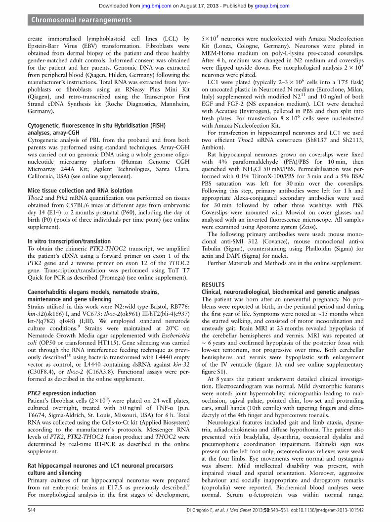
RESULTSClinical, neuroradiological, biochemical and genetic analysesThe patient was born after an uneventful pregnancy. No pro-blems were reported at birth, in the perinatal period and duringthe first year of life. Symptoms were noted at ∼15 months whenshe started walking, and consisted of motor incoordination andunsteady gait. Brain MRI at 23 months revealed hypoplasia ofthe cerebellar hemispheres and vermis. MRI was repeated at∼ 6 years and corfirmed hypoplasia of the posterior fossa withlow-set tentorium, not progressive over time. Both cerebellarhemispheres and vermis were hypoplastic with enlargementof the IV ventricle (figure 1A and see online supplementaryfigure S1).
At 8 years the patient underwent detailed clinical investiga-tion. Electrocardiogram was normal. Mild dysmorphic featureswere noted: joint hypermobility, micrognathia leading to mal-occlusion, ogival palate, pointed chin, low-set and protrudingears, small hands (10th centile) with tapering fingers and clino-dactyly of the 4th finger and hyperconvex toenails.
Neurological features included gait and limb ataxia, dysme-tria, adiadochokinesia and diffuse hypothonia. The patient alsopresented with bradylalia, dysarthria, occasional dyslalia andpneumophonic coordination impairment. Babinski sign waspresent on the left foot only; osteotendinous reflexes were weakat the four limbs. Eye movements were normal and nystagmuswas absent. Mild intellectual disability was present, withimpaired visual and spatial orientation. Moreover, aggressivebehaviour and socially inappropriate and derogatory remarks(coprolalia) were reported. Biochemical blood analyses werenormal. Serum α-fetoprotein was within normal range.
544 Di Gregorio E, et al. J Med Genet 2013;50:543–551. doi:10.1136/jmedgenet-2013-101542
Chromosomal rearrangements
group.bmj.com on August 17, 2013 - Published by jmg.bmj.comDownloaded from
Sialoglycoprotein deficits were ruled out by laboratory testing.Friedreich’s ataxia was excluded by routine genetic testing.
Cytogenetic analysisChromosome analysis found a translocation involving chromo-somes X and 8: 46,X,t(X;8)(q25;q24.3) (figure 1B). Thenormal karyotype of the parents and the segregation of poly-morphic markers (Profiler Plus kit, Applied Biosystems) demon-strated that the translocation was de novo. FISH analysis withprobes painting the X chromosome confirmed the translocation(data not shown), and a subtelomeric 8q probe (Vysis) provedthat a small telomeric 8q region was translocated on the der(X)chromosome. Array-CGH analysis using a 244 K array (AgilentTechnologies) did not reveal genomic deletion/duplicationbesides a few known copy number variants.
X-inactivation was completely skewed in the proband (seeonline supplementary figure S1C), with the active allele inher-ited from the father.
Mapping and characterisation of the breakpoint junctionsWe mapped the breakpoint to a region of ∼37 kb and definedthe translocation between 8q24.3 and Xq25 by FISH (seeonline supplementary figure S1D and S1E).
We used a set of forward and reverse primers to amplify thebreakpoint junctions on the two derivatives. The two break-points were located in nonhomologous regions, involving repeti-tive elements: MER4/AluJ on chromosome 8 and SINE, VNTRand Alu (SVA) element on chromosome X (see online supple-mentary figure S2). A segment of 88 bp was lost on the Xchromosome in the translocation.
The translocation interrupted the PTK2 gene at 8q24.3 in the50-UTR between exons 1 and 2, ∼30 kb from the transcriptionstart site (TSS), whereas no known gene was interrupted atXq25. However, the TSS of the closest gene, THOC2, lay only2 kb downstream of the breakpoint (see scheme in figure 1E).
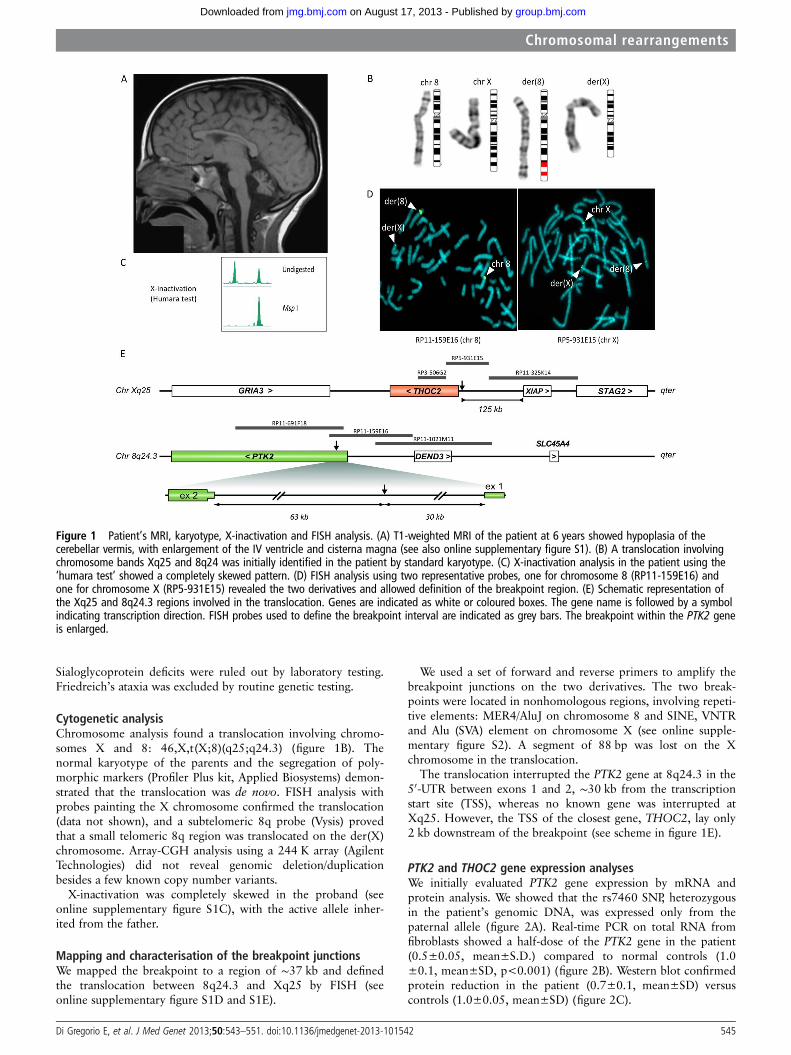
PTK2 and THOC2 gene expression analysesWe initially evaluated PTK2 gene expression by mRNA andprotein analysis. We showed that the rs7460 SNP, heterozygousin the patient’s genomic DNA, was expressed only from thepaternal allele (figure 2A). Real-time PCR on total RNA fromfibroblasts showed a half-dose of the PTK2 gene in the patient(0.5±0.05, mean±S.D.) compared to normal controls (1.0±0.1, mean±SD, p<0.001) (figure 2B). Western blot confirmedprotein reduction in the patient (0.7±0.1, mean±SD) versuscontrols (1.0±0.05, mean±SD) (figure 2C).
Figure 1 Patient’s MRI, karyotype, X-inactivation and FISH analysis. (A) T1-weighted MRI of the patient at 6 years showed hypoplasia of thecerebellar vermis, with enlargement of the IV ventricle and cisterna magna (see also online supplementary figure S1). (B) A translocation involvingchromosome bands Xq25 and 8q24 was initially identified in the patient by standard karyotype. (C) X-inactivation analysis in the patient using the‘humara test’ showed a completely skewed pattern. (D) FISH analysis using two representative probes, one for chromosome 8 (RP11-159E16) andone for chromosome X (RP5-931E15) revealed the two derivatives and allowed definition of the breakpoint region. (E) Schematic representation ofthe Xq25 and 8q24.3 regions involved in the translocation. Genes are indicated as white or coloured boxes. The gene name is followed by a symbolindicating transcription direction. FISH probes used to define the breakpoint interval are indicated as grey bars. The breakpoint within the PTK2 geneis enlarged.
Di Gregorio E, et al. J Med Genet 2013;50:543–551. doi:10.1136/jmedgenet-2013-101542 545
Chromosomal rearrangements
group.bmj.com on August 17, 2013 - Published by jmg.bmj.comDownloaded from
THOC2 showed an apparent mRNA overexpression(patient=2.8±0.025, mean±S.D., controls=1±0.06, p<0.001)with a real-time PCR assay on exons 33–34 from patient’s fibro-blasts (figure 2D). However, using an assay on exons 1–2 of theTHOC2 gene revealed a transcript reduction to about half thedose of controls (patient=0.5±0.035, mean±S.D.; controls=1±0.06, p<0.01) (figure 2D)
THOC2 protein levels were also significantly reduced(patient=0.4±0.1, mean±S.D., controls=1±0.9, p<0.01, two-tailed Student’s t test) (figure 2E).
To test the effect of the translocation on THOC2 flankinggenes, we measured the expression of GRIA3 (patient=0.97±0.04, controls=1±0.05, mean±S.D.) and XIAP (patient 0.93±0.10, controls 1±0.08, mean±S.D.) versus TBP in the patient’sfibroblasts. Expression of both genes was similar to controls.
We reasoned that even if the breakpoint on the der(X) layoutside the THOC2 coding sequence, it could alter THOC2expression by transcriptional interference.
PTK2-THOC2 fusion sterile transcriptThe above results suggested that two different mRNAs wereproduced from the der(X): a less abundant, corresponding to
the wild-type transcript, initiated at the THOC2 TSS, and amore abundant, fusion transcript under the control of the PTK2promoter. The fusion transcript was confirmed by RT-PCR onRNA from the patient’s fibroblasts, using a forward primerwithin PTK2 exon 1, and a reverse primer in THOC2 exon 2(figure 3A). The sequence of the PCR product showed the skip-ping of THOC2 exon 1, likely because the first exon does nothave an acceptor splice site (see scheme in figure 3A). Theexpected sterility of the fusion transcript was confirmed in vitrothrough a coupled transcription/translation assay: as shown infigure 3B, the 45.7 kDa protein produced in the control lanewas absent in the transcribed and translated PTK2-THOC2 con-struct (see also online supplement).
PTK2 promoter transcriptional interference on THOC2To evaluate whether expression of the fusion transcript affectedexpression of the wild-type PTK2 allele, we treated the patient’sfibroblasts with 50 ng/ml TNF-α for 6 h, known to induce PTK2expression.12 By real-time PCR, we measured expression ofwild-type PTK2, wild-type THOC2 (exons 1–2) and the sum ofwild-type THOC2 and the fusion transcript (with an assay onTHOC2 exons 33–34). Under these conditions, wild-type PTK2
Figure 2 Expression analysis of PTK2 and THOC2 genes in patient’s cells. (A) Sequence analysis of patient’s genomic DNA (gDNA) and cDNA atSNP rs7460 in exon 32 of the PTK2 gene. Only the maternal ‘T’ allele was expressed. (B) Real-time PCR on the patient’s cDNA extracted fromfibroblasts showed a reduction of the PTK2 gene measured vs. TBP reference (***p<0.001). (C) Reduction was confirmed at protein level versusβ actin (ACTB) (**p<0.01). (D) Expression analysis of the THOC2 gene (assay on exons 1–2) showed a ∼50% reduction (reference gene TBP).However, using an assay on the 30-end of the gene, THOC2 transcript is apparently increased (***p<0.001, assay on exons 33-34). (E) THOC2reduction was confirmed at protein level versus β actin (ACTB) (**p<0.01). Statistic analysis was performed using a two-tailed Student’s t test.
546 Di Gregorio E, et al. J Med Genet 2013;50:543–551. doi:10.1136/jmedgenet-2013-101542
Chromosomal rearrangements
group.bmj.com on August 17, 2013 - Published by jmg.bmj.comDownloaded from
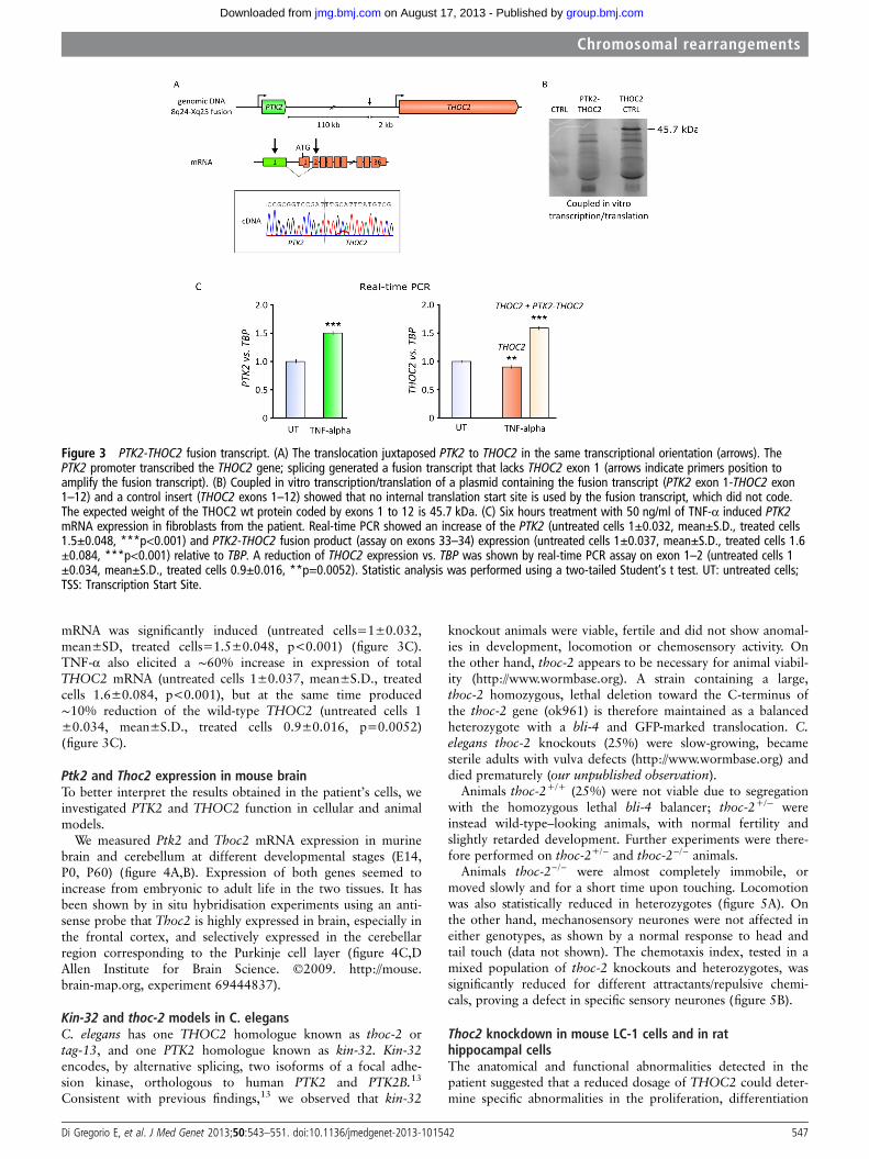
mRNA was significantly induced (untreated cells=1±0.032,mean±SD, treated cells=1.5±0.048, p<0.001) (figure 3C).TNF-α also elicited a ∼60% increase in expression of totalTHOC2 mRNA (untreated cells 1±0.037, mean±S.D., treatedcells 1.6±0.084, p<0.001), but at the same time produced∼10% reduction of the wild-type THOC2 (untreated cells 1±0.034, mean±S.D., treated cells 0.9±0.016, p=0.0052)(figure 3C).
Ptk2 and Thoc2 expression in mouse brainTo better interpret the results obtained in the patient’s cells, weinvestigated PTK2 and THOC2 function in cellular and animalmodels.
We measured Ptk2 and Thoc2 mRNA expression in murinebrain and cerebellum at different developmental stages (E14,P0, P60) (figure 4A,B). Expression of both genes seemed toincrease from embryonic to adult life in the two tissues. It hasbeen shown by in situ hybridisation experiments using an anti-sense probe that Thoc2 is highly expressed in brain, especially inthe frontal cortex, and selectively expressed in the cerebellarregion corresponding to the Purkinje cell layer (figure 4C,DAllen Institute for Brain Science. ©2009. http://mouse.brain-map.org, experiment 69444837).
Kin-32 and thoc-2 models in C. elegansC. elegans has one THOC2 homologue known as thoc-2 ortag-13, and one PTK2 homologue known as kin-32. Kin-32encodes, by alternative splicing, two isoforms of a focal adhe-sion kinase, orthologous to human PTK2 and PTK2B.13
Consistent with previous findings,13 we observed that kin-32
knockout animals were viable, fertile and did not show anomal-ies in development, locomotion or chemosensory activity. Onthe other hand, thoc-2 appears to be necessary for animal viabil-ity (http://www.wormbase.org). A strain containing a large,thoc-2 homozygous, lethal deletion toward the C-terminus ofthe thoc-2 gene (ok961) is therefore maintained as a balancedheterozygote with a bli-4 and GFP-marked translocation. C.elegans thoc-2 knockouts (25%) were slow-growing, becamesterile adults with vulva defects (http://www.wormbase.org) anddied prematurely (our unpublished observation).
Animals thoc-2+/+ (25%) were not viable due to segregationwith the homozygous lethal bli-4 balancer; thoc-2+/− wereinstead wild-type–looking animals, with normal fertility andslightly retarded development. Further experiments were there-fore performed on thoc-2+/− and thoc-2−/− animals.
Animals thoc-2−/− were almost completely immobile, ormoved slowly and for a short time upon touching. Locomotionwas also statistically reduced in heterozygotes (figure 5A). Onthe other hand, mechanosensory neurones were not affected ineither genotypes, as shown by a normal response to head andtail touch (data not shown). The chemotaxis index, tested in amixed population of thoc-2 knockouts and heterozygotes, wassignificantly reduced for different attractants/repulsive chemi-cals, proving a defect in specific sensory neurones (figure 5B).
Thoc2 knockdown in mouse LC-1 cells and in rathippocampal cellsThe anatomical and functional abnormalities detected in thepatient suggested that a reduced dosage of THOC2 could deter-mine specific abnormalities in the proliferation, differentiation
Figure 3 PTK2-THOC2 fusion transcript. (A) The translocation juxtaposed PTK2 to THOC2 in the same transcriptional orientation (arrows). ThePTK2 promoter transcribed the THOC2 gene; splicing generated a fusion transcript that lacks THOC2 exon 1 (arrows indicate primers position toamplify the fusion transcript). (B) Coupled in vitro transcription/translation of a plasmid containing the fusion transcript (PTK2 exon 1-THOC2 exon1–12) and a control insert (THOC2 exons 1–12) showed that no internal translation start site is used by the fusion transcript, which did not code.The expected weight of the THOC2 wt protein coded by exons 1 to 12 is 45.7 kDa. (C) Six hours treatment with 50 ng/ml of TNF-α induced PTK2mRNA expression in fibroblasts from the patient. Real-time PCR showed an increase of the PTK2 (untreated cells 1±0.032, mean±S.D., treated cells1.5±0.048, ***p<0.001) and PTK2-THOC2 fusion product (assay on exons 33–34) expression (untreated cells 1±0.037, mean±S.D., treated cells 1.6±0.084, ***p<0.001) relative to TBP. A reduction of THOC2 expression vs. TBP was shown by real-time PCR assay on exon 1–2 (untreated cells 1±0.034, mean±S.D., treated cells 0.9±0.016, **p=0.0052). Statistic analysis was performed using a two-tailed Student’s t test. UT: untreated cells;TSS: Transcription Start Site.
Di Gregorio E, et al. J Med Genet 2013;50:543–551. doi:10.1136/jmedgenet-2013-101542 547
Chromosomal rearrangements
group.bmj.com on August 17, 2013 - Published by jmg.bmj.comDownloaded from

and/or survival of neuronal precursor cells or in the survival ofdifferentiated neurones. To address these possibilities, we con-ducted knockdown studies in rodent cells transfected withshRNA constructs capable of reducing the Thoc2 orthologexpression by approximately 70% in HeLa cells (data notshown). To analyse the possible effects of Thoc2 knockdown on
the expansion of neuronal precursors we used mouse LC-1 cells,which are positive for the neuronal progenitor marker nestin andrapidly proliferate in presence of FGF2 and EGF, but differenti-ate with high efficiency into neurones and astrocytes when cul-tured without growth factors.11 On the other hand, to addressthe role of Thoc2 in neuronal differentiation, we resorted to rat
Figure 4 Expression analysis of Ptk2 and Thoc2 in mouse brain. (A, B) Expression analysis of Ptk2 and Thoc2 genes versus Hmbs in murine brainor cerebellar lysates by real-time RT-PCR, at different developmental stages (E14, P0, P60). Ptk2 and Thoc2 increased their expression during mousedevelopment (brain from E14 to P60; cerebellum from P0 to P60). (C) Thoc2 in situ hybridisation (above Nissl, and below Thoc2 In SituHybridisation (ISH) expression highlighted) in adult mouse brain shows that the gene is highly expressed, with a prevalence in frontal cortex andcerebellum. In this last tissue, the pattern is compatible with Purkinje neurones (see enlargement in panel D)(Allen Institute for Brain Science.©2009. Available from: http://mouse.brain-map.org).
Figure 5 Characterisation of C.elegans neuronal features. (A)Locomotion activity (body bends) inwild type, thoc-2+/− and thoc-2−/−
1 day-old adult animals, and (B)sensory function (chemotaxis index).The functionality of a specific subset ofanimal sensory neurones (AWA, AWB,AWC, ASE) was assessed byquantifying their attraction or repulsionto specific chemicals (respectively:pyrazine, nonanone, benzaldheide,NH4Acetate). Bars represent mean ofdata coming from two to fourindependent experiments, each timecarried in duplicate from twoindependent operators; error barsrepresent SE of the mean (SEM);*p<0.05, ***p<0.001, two-tailed,unpaired Student’s t test.
548 Di Gregorio E, et al. J Med Genet 2013;50:543–551. doi:10.1136/jmedgenet-2013-101542
Chromosomal rearrangements
group.bmj.com on August 17, 2013 - Published by jmg.bmj.comDownloaded from

hippocampal neurones in primary culture.14 These cells weretransfected with shRNA constructs before plating and allowed todifferentiate for ∼72 h. In both systems, Thoc2 silencing did notincrease significantly the number of apoptotic cells (data notshown), thus excluding that Thoc2 knockdown affected neuronalcell viability. On the contrary, Thoc2-depleted LC-1 cells dis-played a significantly increased proliferation rate, as determinedby the MTT assay (figure 6A). In differentiating primary hippo-campal neurones, the knockdown of Thoc2 produced a signifi-cant increase in the length of neuronal processes (figure 6B,C),without affecting the timing of transitions between the first dif-ferentiation stages (data not shown).
DISCUSSIONChromosomal rearrangements have been instrumental in identi-fying disease-causing genes located across or near the break-points.15 16 Here we describe a de novo reciprocal translocationbetween Xq25-ter and 8q24.3-ter, associated with psychomotor
retardation and congenital cerebellar hypoplasia, likely causedby altered expression of two genes, PTK2 at 8q24.3 andTHOC2 at Xq25.
The 8q24.3 breakpoint occurred within the PTK2 gene. Asexpected, the mRNA and PTK2 protein levels were reduced to50% in the patient’s fibroblasts, compared to healthy controls.The absence of mutation in the transcript expressed by thesecond PTK2 allele does not support the hypothesis of a reces-sive phenotype. PTK2 encodes a cytosolic protein tyrosinekinase involved in focal adhesion formation.17 Its activity elicitsintracellular signal transduction pathways that stimulate theturnover of cell contacts with the extracellular matrix, promot-ing cell migration. PTK2 has been implicated in central nervoussystem development and myelination, and synaptic plasticity inthe mouse hippocampus.18 19 20
Although we cannot exclude that PTK2 haploinsufficiencyplayed some role in our patient’s phenotype, it is worth notingthat: (a) microdeletions and macrodeletions (∼120 kb to 6.9 Mb)
Figure 6 Thoc2 knockdown in LC1 neural precursor cells and rat hippocampal neurones. (A) Thoc2 knockdown stimulated the proliferation of LC1neural precursor cells in culture. Neural precursor LC1 were electroporated with a scramble shRNA (shCTRL) or with specific shRNA expressingplasmid (sh2113) before plating, and allowed to proliferate in culture for 24 h, 48 h and 72 h. The proliferation rate was measured using the MTTassay. Thoc2 knockdown neurones grew faster than control cells (p<0.05, two-tailed, paired Student’s t test). (B, C) Primary rat hippocampalneurones were electroporated before plating with control (shCTRL) or specific shRNA-expressing plasmids (sh8137, sh2113) and allowed todifferentiate for 3 days. The knockdown of Thoc2 gene stimulated neurite outgrowth (*p<0.05, **p<0.01, ***p<0.001, two-tailed, unpairedStudent’s t test). Green signal in the transfected cells is GFP and red signal is the staining for α-Tubulin (C).
Di Gregorio E, et al. J Med Genet 2013;50:543–551. doi:10.1136/jmedgenet-2013-101542 549
Chromosomal rearrangements
group.bmj.com on August 17, 2013 - Published by jmg.bmj.comDownloaded from
containing PTK2 have been reported in at least two healthyparents of children with cognitive impairment, with or withoutmalformations (ref: #255112 Decipher Database; #11255_85Rome, Italian Database of Copy Number Variants, http://dbcnv.oasi.en.it); (b) Ptk2 homozygous knockout is lethal in mice,whereas haploinsufficiency does not result in a pathologicalphenotype.21 Purkinje cells from homozygous conditionalknockout mice show a normal phenotype22; (c) kin-32 (PTK2ortholog) silencing in C. elegans did not affect viability, develop-ment, fertility, locomotion and chemosensory activity.
The Xq25 translocation created a fusion product that main-tained both the PTK2 and THOC2 promoters. We demonstratedthat the transcription of the PTK2—THOC2 fusion gene, underthe PTK2 promoter, downregulates the level of the wild-typeTHOC2 mRNA and protein.
Such a phenomenon, known as transcriptional interfer-ence,23 is responsible of a variety of human diseases, forexample, α-thalassemia (a gain-of-function, regulatory single-nucleotide polymorphism creates a new promoter-like elementthat interferes with normal activation of all downstream α-likeglobin genes24); Lynch syndrome: TACSTD1—a gene upstreamto the Mismatch repair MSH2 gene, transcribed in the samedirection—not rarely shows deletions spanning the last exon,including the polyA signal. The RNA polymerase proceeds,interfering with the transcription of the downstream MSH2promoter, and gives rise to TACSTD1/MSH2 fusiontranscripts.25
THOC2 product is a subunit of a multiprotein complex calledthe THO complex, which is conserved from yeast to humans.26
THO interacts physically and functionally with the mRNA exportfactors Yra1 and Sub2 forming the TREX (TRanscription-EXport)complex.
It has been shown that TREX plays a role in mRNA transportfrom the nucleus to cytoplasm and distinct pathophysiologicalstates are correlated with defective mRNA export mechan-isms.27 Thoc5 deletion in mice causes death in the first 2 weeksafter birth, similar to Thoc1 deletion. Thoc5 conditional knock-out mouse develops acute leukocytopenia and anaemia, suggest-ing that the TREX complex has a key role not only in earlyembryogenesis, but also in differentiation, as previouslyreported with Thoc1 knockout.28 29 Other mRNA export geneshave been linked to human diseases, such as the fragile Xmental retardation protein, FMRP, that regulates mRNA metab-olism and translation of key molecules involved in receptor sig-nalling and spine morphology.30
THOC2 is expressed in murine brain, and its pattern in thecerebellum is compatible with Purkinje cells distribution, con-sistent with our patient’s clinic-pathological phenotype. PTK2 isalso expressed in brain, but with a more diffuse pattern 22. Inthe cerebellum, the generation of the different categories of neu-rones is accomplished through well-defined space and time con-straints, regulated by signals that are only partially known31:downregulation of THOC2, possibly combined with PTK2 hap-loinsufficiency, may lead to aberrant timing of axonal sproutingor altered proliferation of specific neuronal populations, thusdetermining the cerebellar hypoplasia/cognitive impairmentfound in our patient.
Indeed, using fibroblasts from our patient, we demonstratedthat when the PTK2 gene was upregulated by TNF-α, the fusiontranscript was overexpressed, whereas wild-type THOC2 genewas further repressed. We suggest that THOC2 downregulationby transcriptional interference is a hypomorphic mutation andits expression may be further reduced in tissues where the ratioPTK2/THOC2 favours the former.
The possible role of THOC2 in the pathogenesis of ourpatient’s phenotype was supported by our findings in three cel-lular/animal models: (i) thoc-2 gene knockout in C.elegans werealmost completely immobile and thoc-2+/− had impaired loco-motion activity. The function of sensory neurones (AWA, AWB,AWC, ASE) was significantly impaired even in a mixed popula-tion of heterozygotes and thoc2 knockouts. (ii) In LC1 mouseneuronal precursors, downregulated Thoc2 (∼30% of the wildtype) led to a significant increase in the proliferation, and (iii) inrat hippocampal primary neurones an increased length ofneurites.
Overall, these data prove a neuronal defect driven by THOC2suppression and suggest that THOC2 is a dosage-sensitive gene.Thus, THOC2 may represent a further member of nuclearmRNA export factors, whose inappropriate interaction, expres-sion pattern and possibly mRNA-binding specificity areexpected to produce a variety of congenital syndromes, asso-ciated with brain diseases.
In conclusion, we describe a female with a cerebellarhypoplasia-psychomotor delay syndrome in which a chromo-somal translocation alters the expression of two genes, PTK2and THOC2, involved in central nervous system. Our data andthat of the literature suggest that the phenotype in our patient isdue to the decreased expression of THOC2, or to a combinedeffect of THOC2/PTK2 reduction, although no definitive evi-dence exists in support of one of these two hypotheses.
Author affiliations1Department of Medical Sciences, University of Torino, Turin, Italy2S.C.d.U. Medical Genetics, Città della Salute e della Scienza, Torino, Italy3Department of Molecular Biotechnology and Health Sciences, University of Torino,Molecular Biotechnology Centre, Torino, Italy4Department of Biomedicine and Prevention, University of Roma ‘Tor Vergata’,Rome, Italy5Institute of Clinical Chemistry and Laboratory Medicine of the Heinrich HeineUniversity, and the IUF—Leibniz Research Institute for Environmental Medicine,Duesseldorf, Germany6S.C. Neuropsichiatria Infantile, Pinerolo, Italy7S.C.d.U. Anatomia Patologica, Città della Salute e della Scienza, Torino, Italy8Department of Molecular Medicine, University of Pavia, Pavia, Italy9IRCCS ‘National Neurological Institute C. Mondino’ Foundation, Pavia, Italy10Department of Clinical and Biological Sciences, University of Turin, Orbassano,Italy11Centre de Recherche de l’Institut du Cerveau et de la Moelle épinière (INSERM/UPMC Univ. Paris 6, UMR_S975; CNRS 7225, EPHE), Pitié-Salpêtrière Hospital,Paris, France12APHP, Fédération de génétique, Pitié-Salpêtrière Hospital, Paris, France13Université Paris Descartes, Institut Cochin, Hôpital Cochin, Paris, France14University of Torino, Neuroscience Institute Cavalieri Ottolenghi (NICO), Torino,Italy15Department of Ophthalmology, University of California San Francisco,San Francisco, California, USA16Department of Cell Biology, Harvard Medical School, Boston, USA17Istituto Zooprofilattico Sperimentale del Piemonte, Liguria e Valle d’Aosta, CReAA-National Reference Centre for Surveillance and Monitoring of Animal Feed, Lab.Contaminanti Ambientali, Torino, Italy
Acknowledgements We are gratefully indebted to the family who participatedin this study. The funding organisations had no role in study design, datacollection and analysis, decision to publish, or preparation of the manuscript.We thank the clinicians who referred their patients, the technical assistance ofDr P Pappi (S.C.d.U. Medical Genetics Città della Salute e della Scienza, Turin,Italy). Drs. E. Ferrero and Nicola Migone (University of Turin) for their advice andcritical reading of the manuscript, and all members of the DNA and Cell bankteam of the Centre de Recherche de l’Institut du Cerveau et de la Moelle épinière(Paris, France). LC1 neural stem cells were a kind gift from Dr. Luciano Conti(Department of Pharmacological Science, University of Milan, Milan, Italy).
Contributors EDiG designed the study, performed the experiments, analysed andinterpreted data and wrote the manuscript. FTB, AS, AC, LVDiC, ZO, SR, FM, SS,MCA performed the experiments, analysed and interpreted data, and edited the
550 Di Gregorio E, et al. J Med Genet 2013;50:543–551. doi:10.1136/jmedgenet-2013-101542
Chromosomal rearrangements
group.bmj.com on August 17, 2013 - Published by jmg.bmj.comDownloaded from
manuscript. NV, FT, HB, RR, AB, FDC analysed and interpreted data, and edited themanuscript. MR, GS, CM, AD, SF, JC, EG performed clinical evaluation of patients,and provided samples, interpreted data and edited the manuscript. ABruscodesigned the study, analysed and interpreted data, and wrote the manuscript.
Funding National Ataxia Foundation, the VERUM foundation, the FrenchAssociation Connaitre les Syndromes Cérébelleux, the ‘Associazione E.E. Rulfo per laGenetica Medica’, A.I.S.A. Piemonte, NV is recipient of ‘My First Airc Grant’(MFAG11509).’
Competing interests None.
Ethics approval Department internal ethical committee.
Provenance and peer review Not commissioned; externally peer reviewed.
Data sharing statement The authors will share primer sequences used in theexperiments to all academic researchers.
REFERENCES1 Steinlin M. Non-progressive congenital ataxias. Brain Dev 1998;20:199–208.2 Manto MU, Jissendi P. Cerebellum: links between development, developmental
disorders and motor learning. Front Neuroanat 2012;6:1.3 Courchesne E, Townsend J, Akshoomoff NA, Saitoh O, Yeung-Courchesne R,
Lincoln AJ, James HE, Haas RH, Schreibman L, Lau L. Impairment in shiftingattention in autistic and cerebellar patients. Behav Neurosci 1994;108:848–65.
4 Zanni G, Bertini ES. X-linked disorders with cerebellar dysgenesis. Orphanet J RareDis 2011;6:24.
5 Zanni G, Saillour Y, Nagara M, Billuart P, Castelnau L, Moraine C, Faivre L,Bertini E, Durr A, Guichet A, Rodriguez D, des Portes V, Beldjord C, Chelly J.Oligophrenin 1 mutations frequently cause X-linked mental retardation withcerebellar hypoplasia. Neurology 2005;65:1364–9.
6 Illarioshkin SN, Tanaka H, Markova ED, Nikolskaya NN, Ivanova-Smolenskaya IA,Tsuji S. X-linked nonprogressive congenital cerebellar hypoplasia: clinical descriptionand mapping to chromosome Xq. Ann Neurol 1996;40:75–83.
7 Bertini E, des Portes V, Zanni G, Santorelli F, Dionisi-Vici C, Vicari S, Fariello G,Chelly J. X-linked congenital ataxia: a clinical and genetic study. Am J Med Genet2000;92:53–6.
8 Zanni G, Bertini E, Bellcross C, Nedelec B, Froyen G, Neuhauser G, Opitz JM,Chelly J. X-linked congenital ataxia: a new locus maps to Xq25-q27.1. Am J MedGenet A 2008;146A:593–600.
9 Stiernagle T. Maintenance of C. elegans. WormBook 2006:1–11.10 Ventura N, Rea SL, Schiavi A, Torgovnick A, Testi R, Johnson TE. p53/CEP-1
increases or decreases lifespan, depending on level of mitochondrial bioenergeticstress. Aging Cell 2009;8:380–93.
11 Conti L, Pollard SM, Gorba T, Reitano E, Toselli M, Biella G, Sun Y, Sanzone S,Ying QL, Cattaneo E, Smith A. Niche-independent symmetrical self-renewal of amammalian tissue stem cell. PLoS Biol 2005;3:e283.
12 Kurenova E, Xu LH, Yang X, Baldwin AS Jr, Craven RJ, Hanks SK, Liu ZG,Cance WG. Focal adhesion kinase suppresses apoptosis by binding to the deathdomain of receptor-interacting protein. Mol Cell Biol 2004;24:4361–71.
13 Cram EJ, Fontanez KM, Schwarzbauer JE. Functional characterization of KIN-32, theCaenorhabditis elegans homolog of focal adhesion kinase. Dev Dyn2008;237:837–46.
14 Banker G, Goslin K. Developments in neuronal cell culture. Nature 1988;336:185–6.
15 Billuart P, Bienvenu T, Ronce N, des Portes V, Vinet MC, Zemni R, Carrie A,Beldjord C, Kahn A, Moraine C, Chelly J. Oligophrenin 1 encodes a rho-GAPprotein involved in X-linked mental retardation. Pathol Biol (Paris) 1998;46:678.
16 Najm J, Horn D, Wimplinger I, Golden JA, Chizhikov VV, Sudi J, Christian SL,Ullmann R, Kuechler A, Haas CA, Flubacher A, Charnas LR, Uyanik G, Frank U,Klopocki E, Dobyns WB, Kutsche K. Mutations of CASK cause an X-linked brainmalformation phenotype with microcephaly and hypoplasia of the brainstem andcerebellum. Nat Genet 2008;40:1065–7.
17 Andre E, Becker-Andre M. Expression of an N-terminally truncated form of humanfocal adhesion kinase in brain. Biochem Biophys Res Commun 1993;190:140–7.
18 Beggs HE, Schahin-Reed D, Zang K, Goebbels S, Nave KA, Gorski J, Jones KR,Sretavan D, Reichardt LF. FAK deficiency in cells contributing to the basal laminaresults in cortical abnormalities resembling congenital muscular dystrophies. Neuron2003;40:501–14.
19 Rico B, Beggs HE, Schahin-Reed D, Kimes N, Schmidt A, Reichardt LF. Control ofaxonal branching and synapse formation by focal adhesion kinase. Nat Neurosci2004;7:1059–69.
20 Monje FJ, Kim EJ, Pollak DD, Cabatic M, Li L, Baston A, Lubec G. Focal adhesionkinase regulates neuronal growth, synaptic plasticity and hippocampus-dependentspatial learning and memory. Neurosignals 2012;20:1–14.
21 Ilic D, Furuta Y, Kanazawa S, Takeda N, Sobue K, Nakatsuji N, Nomura S,Fujimoto J, Okada M, Yamamoto T. Reduced cell motility and enhanced focaladhesion contact formation in cells from FAK-deficient mice. Nature1995;377:539–44.
22 Watanabe F, Miyazaki T, Takeuchi T, Fukaya M, Nomura T, Noguchi S, Mori H,Sakimura K, Watanabe M, Mishina M. Effects of FAK ablation on cerebellarfoliation, Bergmann glia positioning and climbing fiber territory on Purkinje cells.Eur J Neurosci 2008;27:836–54.
23 Palmer AC, Egan JB, Shearwin KE. Transcriptional interference by RNA polymerasepausing and dislodgement of transcription factors. Transcription 2011;2:9–14.
24 De Gobbi M, Viprakasit V, Hughes JR, Fisher C, Buckle VJ, Ayyub H, Gibbons RJ,Vernimmen D, Yoshinaga Y, de Jong P, Cheng JF, Rubin EM, Wood WG,Bowden D, Higgs DR. A regulatory SNP causes a human genetic disease by creatinga new transcriptional promoter. Science 2006;312:1215–7.
25 Kovacs ME, Papp J, Szentirmay Z, Otto S, Olah E. Deletions removing the last exonof TACSTD1 constitute a distinct class of mutations predisposing to Lynch syndrome.Hum Mutat 2009;30:197–203.
26 Reed R, Cheng H. TREX, SR proteins and export of mRNA. Curr Opin Cell Biol2005;17:269–73.
27 Rondon AG, Jimeno S, Aguilera A. The interface between transcription and mRNPexport: from THO to THSC/TREX-2. Biochim Biophys Acta 2010;1799:533–8.
28 Mancini A, Niemann-Seyde SC, Pankow R, El Bounkari O, Klebba-Farber S, Koch A,Jaworska E, Spooncer E, Gruber AD, Whetton AD, Tamura T. THOC5/FMIP, anmRNA export TREX complex protein, is essential for hematopoietic primitive cellsurvival in vivo. BMC Biol 2010;8:1.
29 Wang X, Chinnam M, Wang J, Wang Y, Zhang X, Marcon E, Moens P,Goodrich DW. Thoc1 deficiency compromises gene expression necessary for normaltestis development in the mouse. Mol Cell Biol 2009;29:2794–803.
30 Bagni C, Tassone F, Neri G, Hagerman R. Fragile X syndrome: causes, diagnosis,mechanisms, and therapeutics. J Clin Invest 2012;122:4314–22.
31 Carletti B, Williams IM, Leto K, Nakajima K, Magrassi L, Rossi F. Time constraintsand positional cues in the developing cerebellum regulate Purkinje cell placement inthe cortical architecture. Dev Biol 2008;317:147–60.
Di Gregorio E, et al. J Med Genet 2013;50:543–551. doi:10.1136/jmedgenet-2013-101542 551
Chromosomal rearrangements
group.bmj.com on August 17, 2013 - Published by jmg.bmj.comDownloaded from

doi: 10.1136/jmedgenet-2013-101542 2013 50: 543-551 originally published online June 7, 2013J Med Genet
Eleonora Di Gregorio, Federico T Bianchi, Alfonso Schiavi, et al. hypoplasiaretardation and congenital cerebellarknockdown in a patient with psychomotor
expressionTHOC2 gene fusion with THOC2-PTK2 X;8 translocation creates a de novoA
http://jmg.bmj.com/content/50/8/543.full.htmlUpdated information and services can be found at:
These include:
Data Supplement http://jmg.bmj.com/content/suppl/2013/06/06/jmedgenet-2013-101542.DC1.html
"Supplementary Data"
References http://jmg.bmj.com/content/50/8/543.full.html#ref-list-1
This article cites 30 articles, 3 of which can be accessed free at:
serviceEmail alerting
the box at the top right corner of the online article.Receive free email alerts when new articles cite this article. Sign up in
CollectionsTopic
(761 articles)Genetic screening / counselling � (56 articles)Memory disorders (psychiatry) �
Articles on similar topics can be found in the following collections
Notes
http://group.bmj.com/group/rights-licensing/permissionsTo request permissions go to:
http://journals.bmj.com/cgi/reprintformTo order reprints go to:
http://group.bmj.com/subscribe/To subscribe to BMJ go to:
group.bmj.com on August 17, 2013 - Published by jmg.bmj.comDownloaded from