adult consent and spousal consent in malaysian gestational ...
Effects of gestational length, gender, postnatal age, and birthorder on visual contrast sensitivity in infants
Karen R. Dobkins,Department of Psychology, University of California, San Diego, La Jolla, California, USA
Rain G. Bosworth, andDepartment of Psychology, University of California, San Diego, La Jolla, California, USA
Joseph P. McCleeryDivision of Developmental Medicine, Children’s Hospital Boston and Harvard Medical School, USA
AbstractTo investigate effects of visual experience versus preprogrammed mechanisms on visualdevelopment, we used multiple regression analysis to determine the extent to which a variety ofvariables (that differ in the extent to which they are tied to visual experience) predict luminance andchromatic (red/green) contrast sensitivity (CS), which are mediated by the magnocellular (M) andparvocellular (P) subcortical pathways, respectively. Our variables included gestational length (GL),birth weight (BW), gender, postnatal age (PNA), and birth order (BO). Two-month-olds (n = 60) and6-month-olds (n = 122) were tested. Results revealed that (1) at 2 months, infants with longer GLhave higher luminance CS; (2) at both ages, CS significantly increases over a ~21-day range of PNA,but this effect is stronger in 2- than 6-month-olds and stronger for chromatic than luminance CS; (3)at 2 months, boys have higher luminance CS than girls; and (4) at 2 months, firstborn infants havehigher CS, while at 6 months, non-firstborn infants have higher CS. The results for PNA/GL areconsistent with the possibility that P pathway development is more influenced by variables tied tovisual experience (PNA), while M pathway development is more influenced by variables unrelatedto visual experience (GL). Other variables, including prenatal environment, are also discussed.
Keywordsinfant vision; contrast sensitivity; magnocellular/parvocellular; magnocellular; parvocellular; visualexperience
IntroductionThe extent to which development is governed by “nature” versus “nurture” is a topic that hasbeen discussed since the time of Aristotle. The nature argument proposes that development isdictated by genetic factors that are unaffected by an animal’s environment, which we refer toas “preprogrammed” development. The nurture argument proposes that experience plays aprominent role in development. With the advance of neuroscience in the last century, muchempirical progress has been made on the nature/nurture debate, with clear evidence for both
© ARVOCorresponding author: Karen R. Dobkins., [email protected]., Address: Department of Psychology, 0109 University of California,San Diego La Jolla, CA 92093, USA.Commercial relationships: none.
NIH Public AccessAuthor ManuscriptJ Vis. Author manuscript; available in PMC 2010 October 8.
Published in final edited form as:J Vis. ; 9(10): 19.1–1921. doi:10.1167/9.10.19.
NIH
-PA Author Manuscript
NIH
-PA Author Manuscript
NIH
-PA Author Manuscript

effects (sometimes referred to as “experience-independent” and “experience-dependent”effects; e.g., Crair, 1999). Perhaps the most progress has been made in the field of visualdevelopment due to the relative ease of manipulating the visual environment of a developinganimal. Vision studies have been conducted in both human and animals, although the bulk ofthe data is from animals, and we generally restrict the discussion to monkeys as their visualsystems are known to be very similar, in both structure and function, to that of humans (e.g.,De Valois, Morgan, Polson, Mead, & Hull, 1974; Golomb, Andersen, Nakayama, MacLeod,& Wong, 1985; Jacobs, 2008; Newsome, Britten, & Movshon, 1989; Newsome & Paré,1988).
To date, most studies investigating whether visual development is governed more bypreprogrammed mechanisms versus visual experience have asked whether visual experienceis necessary, with the assumption that if it is not, preprogrammed mechanisms can solely guidedevelopment. Obviously, one way this can be addressed is to determine whether the newborn’svisual system is adultlike. Although common sense suggests otherwise, it is not unreasonableto speculate that there may be some aspects of visual development that are completed inutero (and thus do not require visual experience). In support of this possibility, cell mitosis anddifferentiation in the anterior segment, retina, and optic nerves are completed by 30 weeks ofgestation in humans (for a review, see Birch & Bosworth, 2004; Birch & O’Connor, 2001),and cortical ocular dominance columns are in place before birth (Horton & Hocking, 1996;Rakic, 1976). Still, these relatively complete aspects of visual development that occur inutero pale in comparison to the multitude of other aspects of visual development that takemonths or years to become adultlike, such as myelination of axons (Stiles, 2008).
Given that the newborn’s visual system is clearly not adultlike, the most common way thenecessity of visual experience has been investigated involves asking whether postnataldevelopment proceeds normally (and reaches the adultlike state) when the system is deprivedof visual input. [The same question of necessity cannot easily be asked for preprogrammeddevelopment as this would require inactivating all the relevant genes, although the knockoutmice model promises to provide insight into the question (for a review, see Welzl, D’Adamo,Wolfer, & Lipp, 2006).] In animals, this has been accomplished by dark rearing (Hendrickson& Boothe, 1976; Regal, Boothe, Teller, & Sackett, 1976) or lid suture (Blakemore & Vital-Durand, 1983; Harwerth, Smith, Boltz, Crawford, & von Noorden, 1983) after birth. Anothervariation of this experiment is to ask whether retinal input is necessary, by silencing retinalneurons using action potential blockers, such as tetrodotoxin (e.g., Shatz & Stryker, 1988). Inhumans, study of individuals who were visually deprived at birth due to congenital cataractsallows the necessity question to be addressed. Although a full review of the results from suchstudies is outside the scope of this Introduction, in brief, deprivation studies reveal differentdegrees of abnormal development depending on the function studied and the length ofdeprivation (for a review, see Boothe, Dobson, & Teller, 1985). Most relevant to the currentstudy, contrast sensitivity is quite abnormal, particularly for medium to high spatialfrequencies, in humans who experience early visual deprivation (e.g., Birch, Stager, Leffler,& Weakley, 1998; Ellemberg, Lewis, Maurer, & Brent, 2000; Ellemberg, Lewis, Maurer, Lui,& Brent, 1999; Maurer & Lewis, 1993; Tytla, Maurer, Lewis, & Brent, 1988). Because contrastsensitivity is thought to be mediated at or before the level of primary visual cortex (V1)(Boynton, Demb, Glover, & Heeger, 1999; Hawken & Parker, 1990; Palmer, Cheng, &Seidemann, 2007; although this may not be the case early in development, see Stavros &Kiorpes, 2008), this suggests that areas at or before the level of V1 are prone to the effects ofvisual deprivation. In sum, the results of deprivation studies demonstrate that visual experienceis necessary, and thus by logical extension, preprogrammed mechanisms are not sufficient, fornormal postnatal development of most aspects of vision.
Dobkins et al. Page 2
J Vis. Author manuscript; available in PMC 2010 October 8.
NIH
-PA Author Manuscript
NIH
-PA Author Manuscript
NIH
-PA Author Manuscript
Obviously, the most parsimonious description of visual development is that bothpreprogrammed mechanisms and visual experience are important. And, of course, it is nowwell accepted that the two forces do not act in isolation but rather interact. Specifically, theenvironment can affect gene expression, and genes can predispose an organism to seek outcertain environments (for reviews, see Gottlieb, 1998; Stiles, 2008). Although this concept ofgene/environment interaction is typically discussed outside the domain of vision, there is avery similar discourse in the vision field regarding how visual experience may guide biologicaldevelopment (for reviews, see Crair, 1999; Feller & Scanziani, 2005; Kiorpes & Movshon,2004; Movshon & Van Sluyters, 1981). The nature of this guidance has been conjectured totake one of two forms. First, visual experience may permit biological development, either bytriggering biological events (such as gene expression) or by allowing them to proceed in theirpreprogrammed fashion. Second, visual experience may instruct biological development,sculpting it to the statistics of the environment. Evidence for an instructive role has been shownin studies that rear animals in an environment with selective visual input, for example, withonly one eye open (e.g., Horton & Hocking, 1997; for a review, see Boothe et al., 1985) orwith both eyes receiving a selective set of visual cues (orientation: Hirsch & Spinelli, 1970;Muir & Mitchell, 1975; Movshon & Van Sluyters, 1981; motion: Cynader & Cmerneko,1976; Kennedy & Orban, 1983; Pasternak, Schumer, Gizzi, & Movshon, 1985; color: Brenner,Cornelissen, & Nuboer, 1990; Sugita, 2004). An example of a naturally occurring type of visualinstruction is the “oblique effect,” i.e., acuity and contrast sensitivity are better for vertical andhorizontal orientations than for oblique orientations (adults: Appelle, 1972; Campbell,Kulikowski, & Levinson, 1966; Mitchell, Freeman, & Westheimer, 1967; infants: Sokol,Moskowitz, & Hansen, 1987, 1989; Teller, Morse, Borton, & Regal, 1974), which is thoughtto be driven by there being greater prevalence of, and thus greater experience with, cardinalorientations in the environment (Baddeley & Hancock, 1991; Bosworth, Bartlett, & Dobkins,2006; Coppola, Purves, McCoy, & Purves, 1998; Keil & Cristobal, 2000; Switkes, Mayer, &Sloan, 1978; van der Schaaf & van Hateren, 1996).
In the current study, we investigated effects of visual experience versus preprogrammeddevelopment, not by removing or manipulating visual experience as other studies have donebut by asking whether a variety of variables (that differ in the extent to which they are tied tovisual experience) predict variance across subjects in visual performance. Using a multipleregression analysis (MRA), our predictor variables were gestational length, birth weight,gender, postnatal age, and birth order, which we chose because they have all been shown topredict visual and non-visual behaviors (see Discussion). Our visual performance measureswere luminance (light/dark) and chromatic (red/green) CS, which are thought to be mediatedby the magnocellular and parvocellular subcortical pathways, respectively (Lee, Pokorny,Smith, Martin, & Valberg, 1990; Shapley, 1990; Smith, Pokorny, Davis, & Yeh, 1995; for anopposing point of view, see Lennie & D’Zmura, 1988; for the possibility that CS is mediatedby the M and P representations at the level of visual cortex, see Discussion section). Thesepathways make up the bulk of the projections from the retina to area V1. There has been someevidence that P pathway development is more dependent on visual experience (i.e., in general,early visual deprivation leads to greater deficits on P than on M pathway tasks: Bradley &Freeman, 1981; Davis et al., 2006; Demirci et al., 2002; Hess & Howell, 1977; Levi &Harwerth, 1977; but see Zele, Pokorny, Lee, & Ireland, 2007), and that M pathway developmentmay be more susceptible in genetic-based developmental disorders (for a review, see Braddick,Atkinson, & Wattam-Bell, 2003). Thus, we hypothesized that predictor variables likely tied tovisual experience (e.g., postnatal age) would account for more variance in our P than our Mpathway measure, while predictor variables not tied to visual experience, and thus more likelytied to preprogrammed development (e.g., birth weight or gestational length), would accountfor more variance in our M than our P pathway measure. We also entertained a third possibility,which is that prenatal environment could contribute to the effects observed in the current study.This is based on a large literature showing that prenatal environmental factors (e.g., maternal
Dobkins et al. Page 3
J Vis. Author manuscript; available in PMC 2010 October 8.
NIH
-PA Author Manuscript
NIH
-PA Author Manuscript
NIH
-PA Author Manuscript
nutrition, immune response, smoking/drug use, teratogens, etc.) affect development in manydomains (for reviews, see Blanchard, 2001; Dalby, 1978; Khera, 1981; Leader, Wong, &Deitel, 1981).
MethodsSubjects
Subjects were recruited via mass mailings of 3,000 to 4,000 letters sent each month to newparents residing in San Diego County. Interested recipients of letters called the laboratory toenroll (which typically occurred when the infant was between 2 and 6 weeks old). Infants withimpairments (neurological, ocular, visual, or hearing), illnesses, or pregnancy/laborcomplications, based upon parent reports, were not tested. Because we employed red/ greenstimuli, also excluded were infants with a greater than 50% chance of colorblindness; forexample, male infants whose paternal grandfather was known to be colorblind. A total of 182infants participated and fell into one of two age ranges: 2-month-olds (n = 60) or 6-month-olds(n = 122). Data from the 2-month-old group were comprised of infants who served as controlinfants in a study investigating contrast sensitivity in premature infants (Bosworth & Dobkins,2008). Data from the 6-month-old group were comprised of infants who served as controlinfants in a study investigating contrast sensitivity in infants at risk for autism spectrumdisorders (see Supplementary materials of McCleery, Allman, Carver, & Dobkins, 2007). Atthe time of enrollment, parents provided the following information about their infant: gender,birth order (BO), birth weight (BW), and due date. Parent report has been shown to be quiteaccurate for BW and due date when the information is obtained soon after birth (Seidman,Slater, Ever-Hadani, & Gale, 1987), which is the case in the current study. With regard toobtaining BO information, we confirmed that non-firstborn infants had older siblings who livedin the same household, and that firstborn infants did not have older step- or half-siblings wholived in the same household.
Gestational length (GL) and postnatal age (PNA) on the first day of testing were restricted bycriteria employed by our laboratory, which was ~±15 days. For PNA, ±15 refers to schedulingthe first day of testing for an infant within ±15 days from the desired month-birthday. For GL(which was calculated, in part, from due date, see below), ±15 refers to including infants whowere born ±15 days from their due date. In both our 2- and 6-month-old samples, both GL andPNA turned out to be normally distributed,1 and PNA and GL also ended up with roughlyequal variances (see below). GL was calculated from the difference between an infant’s birthdate and due date. For example, if an infant was born 3 days “late,” we considered their GL tobe 266 days (which is an estimate of the “standard” GL for infants, based on a 38-week, post-conception, gestation period) + 3 days = 269 days. Note that while the value we chose for the“standard” GL (i.e., 266 days) could be debated, it is inconsequential for our statistical analyses.Also, we are of course aware that some variability in our GL measure will be due to error inpredicted due date, the latter derived based on ultrasound dating, typically within the firsttrimester (86% of our sample) or last menstrual period, LMP (13% of our sample). We modelthe effect this error might have had on our results in Appendix A.
In our final samples, for 2-month-olds, PNA ranged from 54 to 77 days (mean = 63.3 days;SD = 5.1 days; range = 23 days), GL ranged from 251 to 283 days (mean = 264 days, SD = 6.4
1Our GL values were normally distributed because the real-world distribution is normal (see Sladkevicius et al., 2005; Tunon, Eik-Nes,Grottum, Von During, & Kahn, 2000), and because we randomly sampled over a range (±15 days from the due date) that captures a largeextent of the real-world range. We believe the PNA distribution was normal because our lab coordinator, who calls parents for the study,attempts to get an infant in right at the time of their month-birthday. If this is not possible for the parent (or if the day is a weekend), thenthe infant is scheduled for another day that is as close as possible to their month-birthday. The extent to which this “other day” differsfrom their month-birthday varies in a normal distribution.
Dobkins et al. Page 4
J Vis. Author manuscript; available in PMC 2010 October 8.
NIH
-PA Author Manuscript
NIH
-PA Author Manuscript
NIH
-PA Author Manuscript
days; range = 32 days), and mean birth weight was 7.8 lb (SD = 0.88 lb). For 6-month-olds,PNA ranged from 171 to 191 days (mean = 181.9 days; SD = 4.4 days; range = 20 days), GLranged from 252 to 281 days (mean = 264 days, SD = 6.8 days; range = 29 days), and meanbirth weight was 7.9 lb (SD = 0.91 lb). As would be expected, the 2- and 6-month-olds did notdiffer in GL (F(1,179) = 0.1, p = 0.75) or birth weight (F(1,179) = 0.35; p = 0.55). With regardto comparisons between ages, Levene’s statistical test of equal variance revealed no differencebetween 2- and 6-month-olds in PNA variance (F(1,180) = 0.54; p = 0.46) or GL variance (F(1,180) = 1.12; p = 0.29). With regard to comparisons between PNA and GL variances (whichcannot be done statistically, but can be done by inspection of their standard deviations, seeabove), there is roughly equal variance between the two predictor variables in the 2-month-oldgroup. In the 6-month-old group, there was a greater difference between PNA and GL variance,but we think this difference highly unlikely to account for our results since we found theopposite of what might be predicted from this difference; the variable with less variance (PNA)predicted contrast sensitivity, while the variable with more variance (GL) did not.
On a final note, for all of our statistical analyses, GL and PNA values were entered as lineardays, but we also analyzed the data using logged values. The results were nearly identical, andthus we only present the results for linear days. In addition, using linear days is preferredbecause it makes slopes of regression lines easier to interpret.
Apparatus and stimuliLuminance (light/dark) and chromatic (red/green) stimuli were presented on an Iiyama VisionMaster Pro 510 monitor (1024 × 768 pixels, 100 Hz) powered by a Dell Dimension computerand viewed at a distance of 38 cm. Stimuli were horizontally oriented sinusoidal gratings(moving upward or downward) with a spatial frequency of 0.27 cycles/degree and a temporalfrequency of 4.2 Hz. These parameters were chosen because they are near the peak of thecontrast sensitivity functions for young infants (e.g., Atkinson, Braddick, & Moar, 1977; Banks& Salapatek, 1978; Dobkins, Anderson, & Lia, 1999; Hartmann & Banks, 1992; Rasengane,Allen, & Manny, 1997).2 The stimuli subtended 11° × 11° and were centered 15° to the left orright of the middle of the video monitor. The mean chromaticity of the gratings and thebackground was CIE = 0.486, 0.442. The mean luminance of gratings and the background was20 cd/m2 for 2-month-olds and 13 cd/m2 for 6-month-olds; the luminance difference betweenages being due to the fact that the data from 2- and 6-month-olds were obtained during thecourse of different studies (see above). Contrast of stimuli is described in terms of conecontrast, i.e., the amount of response modulation produced in the long-wavelength-selective(L) and medium-wavelength-selective (M) cones in the eye (for methodological details, seeDobkins et al., 1999; Gunther & Dobkins, 2002).
Determining red/green isoluminance—The red/green chromatic stimulus was presentedat the mean isoluminance value obtained from 22 adults, using standard motion photometry(Dobkins & Teller, 1996b; Moreland, 1982; Teller & Lindsey, 1993). In this task, adults fixatedon a small dot in the center of a moving red/green grating and adjusted the luminance contrastin the grating until the percept of motion was least salient. Each adult subject’s isoluminancepoint was determined from the mean of 25 trials. The stimulus conditions for the motionphotometry procedure were identical to those employed in the main experiments (i.e., same
2We chose to use the same spatial frequency in 2- and 6-month-olds because collection of data from 6-month-olds (at 0.27 cycles/degree)had started first and we did not want to go to lower spatial frequency for 2-month-olds. This is because the low end of the contrastsensitivity function is pretty flat (e.g., Peterzell, Chang, & Teller, 2000; Peterzell, Kaplan, & Werner, 1995), making it difficult todetermine which spatial frequency in 2-month-olds is equivalent to 0.27 cycles/degree in 6-month-olds. For this reason, we thought itsafest to use the same spatial frequency at both ages. For temporal frequency, previous data on infant temporal contrast sensitivity functions(e.g., Dobkins et al., 1999; Dobkins, Lia, & Teller, 1997; Rasengane et al., 1997) likewise led us to think it best to use the same temporalfrequency in 2- and 6-months-olds.
Dobkins et al. Page 5
J Vis. Author manuscript; available in PMC 2010 October 8.
NIH
-PA Author Manuscript
NIH
-PA Author Manuscript
NIH
-PA Author Manuscript
size, orientation, spatiotemporal frequency). As previously discussed (e.g., Dobkins & Teller,1996b), the justification for using the adult mean isoluminance value in our infant experimentsis based on previous experiments demonstrating that infant and adult mean isoluminance pointsare highly similar for red/green stimuli (Bieber, Volbrecht, & Werner, 1995; Brown, Lindsey,McSweeney, & Walters, 1995; Dobkins, Anderson, & Kelly, 2001; Maurer, Lewis, Cavanagh,& Anstis, 1989; Morrone, Burr, & Fiorentini, 1993; Pereverzeva, Hui-Lin Chien, Palmer, &Teller, 2002; Teller & Lindsey, 1989). Moreover, Brown and colleagues argue quantitativelythat the variability of isoluminance points across infant subjects is comparable to the variabilityacross adult subjects, when measurement error is taken into account. In previous studies, wehave calculated that the amount of luminance error likely to exist in our red/green stimuli isbelow luminance contrast threshold for infants (see Dobkins & Teller, 1996b).
Obtaining contrast sensitivitiesThe dependent measures in this study were log luminance and log chromatic contrastsensitivities (CS), which were obtained using forced-choice preferential looking (Teller,1979) with the method of constant stimuli, as described in detail previously (see Dobkins &Teller, 1996a, 1996b). Briefly, an adult experimenter held the infant 38 cm away from the frontof the stimulus monitor in the view of a video camera aimed at the infant’s face. On each trial,a grating stimulus appeared on the left or right side of the video monitor (centered at 15°eccentricity), and the experimenter used cues such as the infant’s head turning and gazingbehavior to judge the left versus right location of the stimulus. Chromatic or luminance gratingswere presented randomly across trials, as was one of five contrast levels (luminance = 1.25%–80% cone contrast, chromatic = 1.25%–26% cone contrast). Stimuli remained present on thevideo monitor until the experimenter made the left/right judgment, which was typically lessthan two seconds. The experimenter’s answer was entered into the computer by pressing keyson the keyboard and computer beeps provided feedback as to whether the experimenter wascorrect. Our goal was to obtain 200 total trials per infant (100 trials × 2 conditions) over thecourse of 2 or 3 days within a 1-week period. The mean number of total trials obtained perinfant was 158 (SD = 58) and 267 (SD = 40) for 2- and 6-month-olds, respectively.
For each infant, a psychometric curve was fit to chromatic and luminance data using Weibullfunctions and maximum likelihood analysis (Watson, 1979; Weibull, 1951). Threshold wasdefined as the contrast yielding 75% correct performance, and sensitivity was computed as theinverse of threshold × 100. Sensitivity was then logged since log, but not linear, sensitivitydata conform to normal distributions (Graham, 1989). It is perhaps important to point out thatthere should be no measurement-imposed restriction in the variance of the CS measure (whichif were the case, would lessen the chance of seeing effects of predictor variables on the CSmeasure). This is because there was a large range of possible CS values, and subjects’ CSvalues were neither at ceiling nor floor for our system (see scatter plots of data in Figure 1 andFigure 2). (This is to be contrasted with other sorts of dependent measures, e.g., “number ofwords produced at 10 months,” where the range/variance of the measure would clearly berestricted.)
Multiple regression analyses (MRA)Four separate MRAs were conducted, for two CS types (chromatic and luminance) and twoage groups (2-month-olds and 6-month-olds). For each of the four MRAs, we tested thecontribution of five different predictor variables. Three were continuous: gestational length(GL), postnatal age (PNA), and birth weight (BW). Two were categorical: gender (boy/girl)and birth order (BO, firstborn vs. non-firstborn).3 Note that we refer to these variables as“predictors” because we hypothesize that each might contribute to variance in CS (seeIntroduction). It is also for this reason that we simultaneously entered all predictors in our MRA(as opposed to using a hierarchical approach).
Dobkins et al. Page 6
J Vis. Author manuscript; available in PMC 2010 October 8.
NIH
-PA Author Manuscript
NIH
-PA Author Manuscript
NIH
-PA Author Manuscript

Before conducting the MRA, each variable was examined for acceptable skew, normality ofresiduals, and multicollinearity. Shapiro–Wilkes W tests on dependent measures (luminanceand chromatic CS) and continuous predictor variables (GL, PNA, and BW) indicated that datawere normally distributed. Pearson correlation coefficients (r values) between continuousvariables tested for indications of overly high correlation among variables (for example, GLand BW are likely to be intercorrelated). Pearson correlation coefficients are often the first stepin an MRA study, although unlike the MRA, they cannot control for possible covariates.Pearson r values are presented in Table 1. We also investigated whether continuous predictorvariables (GL, PNA, and BW) differed between categorical predictor variables (gender: boyvs. girl; BO: firstborn vs. non-firstborn). The results revealed gender differences for BW, withboys being heavier as would be expected (p < 0.05), which is controlled for in the MRA. Anunanticipated difference was seen for PNA in 2-month-olds, where the non-firstborn groupwas tested at a slightly, but significantly, older age than the firstborn group (p < 0.05). This isa random skew in the data set, which is non-meaningful and is controlled for in the MRA. Allother variables were balanced across the categories.
Using SPSS, the four MRAs provided the following outputs:
1. The multiple R squared, R2, represents the percentage of variance in the dependentmeasure (luminance and chromatic CS) accounted for by all the predictor variablestogether.
2. The squared semipartial correlation coefficient, sr2, represents the percentage ofvariance in the dependent measure (luminance and chromatic CS) uniquely accountedfor by a given predictor variable, once other variables have been taken into account.The sr2 also represents how much the R2 would decrease if that given variable wasremoved from the model. When the sr2 is statistically significant, that variableaccounts for a significant portion of the variance in the regression model.
3. The unstandardized regression coefficient, B, represents the slope of a givenvariable’s effect while other variables are held constant.
4. The beta weight, β, is a standardized correlation coefficient measure, defined as theamount (in standard deviations) that the dependent measure (luminance and chromaticCS) changes for one standard deviation change in the predictor variable. β is oftenthought of as representing the relative contribution or importance of a given variablein the model, once other variables have been taken into account.
ANCOVAAs explained above, the MRAs were performed separately on the two CS types (luminanceand chromatic CS) and also separately for the two age groups (2-month-olds and 6-month-olds). We therefore performed an analysis of covariance (ANCOVA) on all of the data togetherto investigate effects of age group (2- vs. 6-month-olds) and whether age group interacts withCS type (luminance and chromatic CS). The ANCOVA also allowed us to confirm/ disconfirmeffects that were seen in the MRA (for example gender effects) or effects that were seen in theMRA that could not be tested statistically (for example, a reversal in the effect of BO between2 and 6 months). The ANCOVA was also used to test specific interactions that test hypothesesbased on results from previous studies (Dannemiller, 2004 reported an interaction betweengender and BW on a visual performance measure in 2- to 5-month-olds; see Discussion). In
3Note that because the vast majority of our infants who were not firstborn were secondborn (i.e., only a few were thirdborn or fourthborn),we made this variable categorical: firstborn versus non-firstborn. The number of firstborn versus non-firstborn infants was 41 and 19 for2-month-olds and 73 and 49 for 6-month-olds.
Dobkins et al. Page 7
J Vis. Author manuscript; available in PMC 2010 October 8.
NIH
-PA Author Manuscript
NIH
-PA Author Manuscript
NIH
-PA Author Manuscript
the ANCOVA, CS type was the repeated-subjects variable, Age Group, Gender, and BO werebetween-subjects factors, and PNA, BW, and GL were included as covariates.
ResultsPearson correlation coefficients (r values) for continuous variables
Table 1 presents r for the two age groups (2-month-olds and 6-month-olds). In 2-month-olds,there was a significant correlation between GL and luminance CS and between PNA andchromatic CS. In 6-month-olds, there was a significant correlation between PNA and bothluminance and chromatic CS. As might be expected, BW and GL were correlated (i.e., infantswith shorter GL had lower BW), although this effect was only significant for our sample of 6-month-olds (we think the lack of an effect in 2-month-olds was due to insufficient power). Twounanticipated findings were seen in 2-month-olds. First, there was a correlation between BWand PNA. This is a random skew in the data set, which is non-meaningful and is controlled forin the MRA. Second, there was a correlation between BW and chromatic CS. This effect, whichgoes away in the MRA, is likely driven by a combination of the unanticipated (i.e., spurious)correlation between BW and PNA and a genuine correlation between PNA and chromatic CS.
Luminance and chromatic CS were found to be strongly correlated (2 months: r = 0.66, p <0.0001; 6 months: r = 0.62, p < 0.0001). These correlations are driven by one or both of twofactors. First, it is likely driven by the fact that PNA correlates positively with both types ofCS (in other words, both types of CS are increasing with age). Second, it could be driven by atrue interrelated-ness between luminance and chromatic CS, i.e., by a single source ofvariability for the two. Interestingly, while factor analysis studies in adults have demonstratedindependent sources of variability for the two types of CS (Dobkins, Gunther, & Peterzell,2000; Gunther & Dobkins, 2002, 2003; Peterzell & Teller, 2000), a previous study in 4-month-olds reported a single source of variability (Peterzell et al., 2000). However, as in the currentstudy, it is possible that the Peterzell et al. (2000) finding could have been driven by smallPNA variations in their sample. At the current time, we cannot distinguish between the twopossibilities, PNA-driven correlation versus true intercorrelation between luminance andchromatic CS; however, we feel strongly that luminance and chromatic CS cannot becompletely accounted for by a single source of variance since our MRA results (below) showdifferential effects of various variables on the two types of CS.
Multiple regression analyses (MRA)Table 2 presents the results from four MRAs, for the two age groups (2-month-olds and 6-month-olds), and the two different CS types (luminance and chromatic CS). As described inthe Methods section, the MRA computes the unique contribution of a given predictor variable,represented by sr2, taking all other variables into account. Controlling for the effects of othervariables is especially important in cases where different predictor variables correlate with oneanother, either in an anticipated or unanticipated way (see Table 1). The MRA also computesan R2, which represents the percentage of variance in CS accounted for by all the predictorvariables together (often referred to as the “full model”). Residual variance represents variancein the CS measure that is unaccounted for in the full model. Residual variance can be systematic(i.e., mediated by variable(s) that were not tested in our model) or unsystematic (i.e., due tonoise in the measurement). R2 and residual variance values are shown at the bottom of eachsubtable in Table 2.
In terms of the unique contribution of the different variables, the significant effects were asfollows. In 2-month-olds, both GL and PNA were significant predictors of luminance CS,accounting for 7% and 9% of the variance, respectively. However, only PNA (and not GL)was a significant predictor of chromatic CS, accounting for 21% of the variance. With regard
Dobkins et al. Page 8
J Vis. Author manuscript; available in PMC 2010 October 8.
NIH
-PA Author Manuscript
NIH
-PA Author Manuscript
NIH
-PA Author Manuscript

to gender, boys had higher luminance CS than girls. With regard to BO, firstborn infants hadhigher CS (both luminance and chromatic) than non-firstborn infants. In 6-month-olds, likethe results of 2-month-olds, PNA was a significant predictor of luminance and chromatic CS,accounting for 5% and 4% of the variance, respectively. And, with regard to BO, non-firstborninfants had higher CS than firstborn infants (the effect was significant for luminance CS andmarginally significant for chromatic CS). Interestingly, this BO effect is opposite in 6-month-olds as compared to 2-month-olds, which is further supported by the results of the ANCOVA(below).
Since PNA was found to be a significant predictor of luminance and chromatic CS in both 2-and 6-month-olds, we investigated whether PNA was a stronger predictor in the younger versusolder group. Using a formula for comparing two-sample regression coefficients provided byClogg, Petkova, and Haritou (1995; and see Paternoster, Brame, Mazerolle, & Piquero,1998), PNA was found to be a marginally stronger predictor of chromatic CS in 2- than 6-month-olds (β: 21% vs. 4%; z = 1.80; p = 0.07). Note that the variance of PNA did not differbetween the two age groups (see Methods), so this alone cannot underlie the age-relateddifference in the effect of PNA on chromatic CS. In contrast to the results for chromatic CS,for luminance CS, PNA was found to be an equally strong predictor in 2- versus 6-month-olds(β: 9% vs. 5%; z = 1.06; p = NS). These findings suggest stronger PNA effects in 2- than 6-month-olds, selectively for chromatic CS. We also used Levene’s test of equal variance to askwhether the two age groups differed in residual variances (see Table 2). Residual variance didnot differ between 2- and 6-month-olds, for chromatic CS (F(1,180) = 0.075; p = 0.78) orluminance CS (F(1,180) = 0.19, p = 0.89), which suggests that there are no differences betweenthe two age groups in (1) the amount to which another untested variable accounts for varianceand (2) the measurement error (this is despite the fact that 6-month-olds, on average, weretested with more total trials; see Methods).
Within each age group, we also inspected whether PNA was a stronger predictor of chromaticCS or luminance CS (and compared residual variance between the two CS types). For 2-month-olds, PNA was a stronger predictor of chromatic CS (accounting for 21% of the variance) thanof luminance CS (accounting for 9% of the variance). By contrast, for 6-month-olds, PNA wasan equal predictor of chromatic CS (accounting for 5% of the variance) and luminance CS(accounting for 4% of the variance). At both 2 and 6 months, residual variance was greater forluminance than for chromatic CS (see Table 2). This difference suggests that (1) there are otheruntested variables that account for more variance in luminance than in chromatic CS and/or(2) there is more measurement error for luminance than for chromatic CS (perhaps becauseinfants find the luminance stimulus less interesting). Note, however, that the possibility thatthere is less measurement error for chromatic than for luminance CS should not be taken asthe reason for why stronger PNA effects were found for chromatic CS (at least for 2 months)since for other variables (specifically, gender and GL), stronger effects were found forluminance CS at 2 months.
Scatter PlotsTo visually demonstrate the effects of PNA and GL on contrast sensitivity revealed from theMRA, we present scatter plots of the data in Figure 1 (PNA) and Figure 2 (GL). However, notethat these scatter plots and their regression lines cannot be derived directly from the outputs ofthe MRA but instead are derived from linear regression lines between two variables, asrepresented by the Pearson correlation coefficients (r values) for continuous variables in Table1. Using the individual correlations as an approximation to results from the MRA is reasonablesince, with one exception, the results of the individual correlations and the MRA are in linewith one another. This exception is the effect of PNA on luminance CS in 2-month-olds, which
Dobkins et al. Page 9
J Vis. Author manuscript; available in PMC 2010 October 8.
NIH
-PA Author Manuscript
NIH
-PA Author Manuscript
NIH
-PA Author Manuscript
was significant in the MRA (Table 2) but not in the individual correlation between PNA andluminance CS (Table 1).
ANCOVAAn ANCOVA was conducted to investigate effects that could not be studied directly with theMRAs. This includes investigating differences in contrast sensitivity (CS) between the twoage groups, 2- versus 6-month-olds, and whether age group interacts with CS type (luminanceand chromatic CS). In addition, the ANCOVA allowed us to confirm/disconfirm effects thatwere seen in the MRA (for example, gender effects) or effects that were seen in the MRA thatcould not be tested statistically (for example, a reversal in the effect of BO between 2 and 6months). As would be expected, the results revealed a significant main effect of age group,with higher CS in 6-month-olds than 2-month-olds (F(1,171) = 12.7; p = 0.0005). This effectdid not interact with CS type, suggesting comparable rates of luminance and chromatic CSdevelopment between 2 and 6 months. The ANCOVA also revealed a main effect of gender,with boys having higher CS than girls (F(1,171) = 4.7; p = 0.03). This result is generally inline with the results from all four MRAs, which revealed higher contrast sensitivity in boysthan girls, although in the MRA this was only significant for luminance CS in 2-month-olds.We believe that gender became significant in the ANCOVA as a result of pooling all subjectdata. Finally, the ANCOVA revealed a significant interaction between age group and BO (F(1,171) = 7.04; p = 0.009), which did not create a three-way interaction with CS type. This isin line with the results from the MRAs, showing a reversal in the effect of BO between 2 months(firstborn infants had higher CS) and 6 months (non-firstborn infants had higher CS).
DiscussionThe current study investigated whether luminance and chromatic contrast sensitivity (CS) arepredicted by different variables that vary in the extent to which they are tied to visualexperience. We chose to study luminance and chromatic CS because they are thought to bemediated by the magnocellular (M) and parvocellular (P) subcortical pathways, respectively(Lee et al., 1990; Shapley, 1990; Smith et al., 1995; for an opposing point of view, see Lennie& D’Zmura, 1988), and thus studying the development of these two types of CS should shedlight on the development of the two pathways.4 In addition, studying the effects of thesedifferent variables on M and P pathway development may have clinical implications as the twopathways have been conjectured to be differentially affected by early abnormal visualexperience as well as differentially susceptible in developmental disorders (see Introduction).Although we refer to our luminance and chromatic CS measures as tapping the subcortical Mand P pathways, we should point out that CS measured at the behavioral level is unlikely to becontrolled solely within subcortical (i.e., M and P) pathways. Rather, CS is likely mediated bya combination of contrast gain mechanisms at the cortical and subcortical levels. The fact thatthe current study reveals differential effects of various variables on our intended M and Ppathway measures is consistent with a subcortical contribution, although it is certainly possiblethat the observed effects occur on M and P representations at the level of visual cortex.
In the remainder of the Discussion section, for each variable that was found to predict CS inthe current study, we address the likelihood that this variable is tied to visual experience versuspreprogrammed development. And, when applicable, we address the possibility that prenatalenvironment, although not providing visual experience, could contribute to our findings. Thisis based on a large literature showing that prenatal environmental factors (e.g., maternalnutrition, immune response, smoking/drug use, teratogens, etc.) affect many aspects of
4There is also a third subcortical pathway, referred to as the koniocellular (K) pathway. A lot less is known about this pathway, althoughrecent evidence suggests it is involved in processing blue/yellow information (for reviews, see Dobkins, 2000; Hendry & Reid, 2000).This pathway will not be discussed further here.
Dobkins et al. Page 10
J Vis. Author manuscript; available in PMC 2010 October 8.
NIH
-PA Author Manuscript
NIH
-PA Author Manuscript
NIH
-PA Author Manuscript
development (for reviews, see Blanchard, 2001; Dalby, 1978; Khera, 1981; Leader et al.,1981).
Effects of gestational length (GL)The MRA of the current study revealed GL effects only for luminance CS at 2 months. We arenot surprised by the finding of GL effects at 2 months, but not 6 months, because other studiesin our laboratory have shown a difference in CS between “preterm” infants (with atypicallyshort GL) and “full-term” infants at 2 months but not 6 months (Bosworth & Dobkins, 2008).That is, effects of GL on CS seem to disappear by 6 months of age. But why were effects foundonly for luminance CS, and not chromatic CS, at 2 months? Obviously, it cannot simply bedue to restricted variance in GL since the amount of variance was sufficient to reveal significanteffects on luminance CS. [In addition, note that the GL variance did not differ between 2- and6-month-olds (see Methods), so this cannot account for a lack of GL effect at 6 months.]However, it is possible that we missed significant effects of GL on chromatic CS at 2 monthsdue to noise in the data arising from error in the GL estimate. This type of noise would alsomake the effect of GL on luminance CS at 2 months appear weaker than it actually is. Toaddress this, we conducted a simulation model using the known error in GL estimate (seeAppendix A). The results of this simulation suggest that error in GL estimate is unlikely toaccount for finding a lack of effect of GL on chromatic CS at 2 months.
Clearly, the observed effects of GL on luminance CS are not tied to visual experience, and thuswe can say with certainty that GL is more likely tied to preprogrammed development than tovisual experience. If preprogrammed mechanisms do, in fact, account for the effect of GL onluminance CS, this would mean that genetic/biological factors underlying maturation ofluminance mechanisms in the M pathway are developing at a substantial rate in utero, with theresult that an extra day in utero has implications for luminance CS measured postnatally.However, there is another possibility that must be entertained; perhaps effects of prenatalenvironment underlie variance in both GL and luminance CS at 2 months. One reasonablecandidate is maternal nutrition, which has been shown to correlate with GL (Delgado,Martorell, Brineman, & Klein, 1982; Jacobson et al., 2008; Olsen et al., 2000; Olsen et al.,1992; Rayco-Solon, Fulford, & Prentice, 2005; Rush, Stein, & Susser, 1980; Smuts et al.,2003), as well as with BW (Godfrey, Robinson, Barker, Osmond, & Cox, 1996; Harding,2001; Lou et al., 1994; Mora, Sanchez, de Paredes, & Herrera, 1981). [Note that, in general,studies report effects of maternal nutrition on either GL or BW effects, without attempting toinvestigate whether there are independent effects on BW and GL.] If maternal nutrition is alsocorrelated with visual performance measured postnatally, then maternal nutrition could be the“third variable” (much cautioned against in correlational analyses), which drives the correlationbetween GL and CS observed in the current study. Although we know of no study that hasinvestigated CS specifically, there is evidence that both prenatal maternal nutrition (e.g.,Jacobson et al., 2008; although these results should be interpreted with caution since the studydid not use randomized assignment) and postnatal infant nutrition (e.g., dietarysupplementation of docosahexaenoic acid and arachidonic acid; Birch et al., 2007; Birch,Hoffman, Uauy, Birch, & Prestidge, 1998; Hoffman et al., 2004; Makrides, Neumann, Simmer,Pater, & Gibson, 1995; O’Connor et al., 2001) correlates with visual acuity in infants/children,making this maternal nutrition hypothesis a possible account of our data. Of course, this accountmust be considered in any study that finds a correlation between a measure of physiologicalmaturity at birth (e.g., gestational length, weight, length, or head circumference) and anoutcome measure. In fact, there is a sizeable literature showing that different aspects ofphysiological maturity at birth predict visual performance (visual orienting at 2 to 5 months:Dannemiller, 2004; attention to faces at 4 to 6 months: Camp, Jamieson-Darr, Hansen, &Schmidt, 1990; visual recognition memory from 5 to 12 months: Rose, 1994) and non-visualperformance (language and gross movement at 4 years: Ounsted, Moar, & Scott, 1984; IQ in
Dobkins et al. Page 11
J Vis. Author manuscript; available in PMC 2010 October 8.
NIH
-PA Author Manuscript
NIH
-PA Author Manuscript
NIH
-PA Author Manuscript
childhood: Churchill, 1965; Jefferis, Power, & Hertzman, 2002; Matte, Bresnahan, Begg, &Susser, 2001; Scarr, 1969). Analyses that control for prenatal environment factors will helpdetermine whether correlations observed between physiological measures of maturity at birthand later visual performance (as in the current and previous studies) are driven by these factors.
In any event, the current study’s finding of GL effects on luminance, but not chromatic CS,indicates that M pathway development is more affected by GL than is P pathway development.This could reflect a faster rate of M than of P pathway preprogrammed development inutero. Alternatively, if the effect is tied to prenatal environment, it suggests that the M pathwaymay be more affected by factors like maternal nutrition, possibly because M neurons are largerthan P neurons and therefore have greater metabolic demands (Glovinsky, Quigley, &Dunkelberger, 1991; Perry, Silveira, & Cowey, 1990; Quigley, Sanchez, Dunkelberger,L’Hernault, & Baginski, 1987; but cf. Crawford, Harwerth, Smith, Shen, & Carter-Dawson,2000). At the current time, we cannot distinguish between the two possibilities.
Effects of PNAThe finding from our MRAs that PNA predicts chromatic and luminance contrast sensitivity(CS) in 2-and 6-month-olds is not terribly surprising since both types of CS obviously increasewith age (FPL: Dobkins et al., 1999, 2001; Peterzell et al., 2000; visually evoked potentials(VEP): Allen, Banks, & Norcia, 1993; Crognale, 2002; Kelly, Borchert, & Teller, 1997;Morrone et al., 1993). Thus, given that the range of PNA in a sample is sufficiently large, asignificant age effect will be revealed. In the current study, the range was 23 and 20 days for2- and 6-month-olds, respectively, and thus our results show that significant improvement inluminance and chromatic CS occurs over this relatively short period of time (~3 weeks). Inaddition, the current study shows differences in PNA effects between 2- and 6-month-olds andbetween chromatic and luminance CS. Specifically, the MRA data showed that (1) PNA effectson chromatic CS were stronger in 2- than 6-month-olds, whereas PNA effects on luminanceCS were statistically indistinguishable between 2- and 6-month-olds; and (2) PNA effects in2-month-olds were stronger for chromatic than for luminance CS, while in 6-month-olds, PNAeffects were nearly identical for chromatic and luminance CS.
If we believe that postnatal development of luminance and chromatic CS is preprogrammed,the PNA results of the current study could be explained by supposing that biological/geneticfactors dictate a different developmental trajectory for the M (luminance) and P (chromatic)pathways. Specifically, the P pathway may be programmed to develop quickly early on (~2months) followed by a slowing period (~6 months), while the M pathway may be programmedto develop at a more even pace. If this scenario were true, we would expect longitudinal databetween 2 and 6 months to show a greater compressive non-linearity for chromatic thanluminance CS. However, this possibility is not supported by data from our laboratory, whichshow that developmental trajectories for both chromatic and luminance CS are well describedby log functions, and thus by definition, the two functions have the same compressive non-linearity (Bosworth & Dobkins, 2008). Nonetheless, we cannot rule out that preprogrammeddevelopment dictates a different (possibly subtle) trajectory for M versus P pathwaydevelopment, which could underlie the differential effects of PNA observed in the currentstudy.
Alternatively, if visual experience plays a role in postnatal development and given that a longerpostnatal period affords greater visual experience, the PNA results of the current study couldbe explained by supposing that visual experience has differential impact on the developmentof the M and P pathways. Specifically, visual experience may exert stronger effects on the Ppathway than on the M pathway early on (~2 months) but equivalent effects later on (~6months). There are at least two ways this effect could be mediated. First, it could be that the Ppathway is generally more amenable to being shaped by visual experience than is the M
Dobkins et al. Page 12
J Vis. Author manuscript; available in PMC 2010 October 8.
NIH
-PA Author Manuscript
NIH
-PA Author Manuscript
NIH
-PA Author Manuscript
pathway. In line with this possibility are adult data from our laboratory showing greaterperceptual learning on a P pathway (chromatic CS) than on an M pathway (luminance CS) task(Thurston & Dobkins, 2007, and see Heckman & Engel, 2006). Second, it could be that the Pand M pathways are equally amenable to being shaped by visual experience, but that thestatistics of the visual environment stimulate the P pathway more than the M pathway. Forexample, this could occur if visual scenes contain relatively more frequent or higher contrast,chromatic (red/green) versus luminance information, although this possibility is not supportedby studies measuring the chromatic/ luminance statistics of natural scenes (MacLeod, 2003;Webster & Mollon, 1997). Still, it is possible that other visual properties that favor the Ppathway, such as high spatial frequency/low temporal frequency, are particularly prominentin visual scenes (see Bex & Makous, 2002; Bosworth et al., 2006; Dakin, Mareschal, & Bex,2005). In truth, it is more fitting to ask about the statistics of the infant’s visual environment,where the statistics of scenes that contain toys, mobiles, etc., may be biased to stimulate onevisual pathway more than another (and may change as an infant starts to locomote and explore).Finding a relationship between the statistics of the infants’ visual environment and the strengthof PNA effects on the different visual pathways would at least provide correlational evidencefor effects of visual experience (of the sort used to account for the “oblique effect” being drivenby a preponderance of cardinal orientations in the environment; see Introduction). Futurestudies that analyze the statistics of the infants’ visual environment will be required to helpinform these possibilities.
In sum, if the PNA effects of the current study are, in fact, tied to visual experience, our resultssuggest that early in development (~2 months) visual experience exerts stronger effects on theP pathway than on the M pathway. This scenario is the flip-side of the asymmetry seen for GL(tied to either preprogrammed development or prenatal environment), where stronger effectswere revealed for the M than for the P pathway measure (see above).
Gender effectsThe current study revealed higher CS in boys than girls (ANCOVA results), which was drivenmainly by higher luminance CS in boys at 2 months (MRA results). Previous studiesinvestigating gender differences in visual abilities have yielded mixed results, depending onthe visual function and age tested. In a study of luminance CS, it was reported that girlsoutperformed boys at 6 months (but not at 4 or 8 months; Peterzell et al., 1995), an effect thatwas not seen in the current study (and note that many previous studies of CS did not explicitlyinclude gender as a variable for analysis; e.g., Banks & Salapatek, 1981; Dobkins et al.,1999; Hammarrenger et al., 2003). With respect to other visual abilities, an advantage for girlshas been reported for vernier and stereoacuity (e.g., 3- to 6 months; for reviews, see Gwiazda,Bauer, & Held, 1989; Held, Thorn, Gwiazda, & Bauer, 1996), while an advantage for boys hasbeen reported for visual accommodation (1 to 3 months; Horwood & Riddell, 2008) and mentalrotation (5 months; Moore & Johnson, 2008). One possibility is that gender differences invisual performance arise from biological differences, either in preprogrammed developmentdetermined from genes on the sex chromosomes or in testosterone levels.5 Gender differencesin visual performance have, in fact, been tied to differences in testosterone levels (monkeys:Bachevalier, Hagger, & Bercu, 1989; humans: Held et al., 1996). The other possibility is thatgender differences in visual performance arise from differences in visual experience. It hasbeen shown that by 3 months of age, parents’ socialization habits are different with boys versus
5It is not clear whether testosterone should be thought of as being tied to genes or tied to environment. With regard to reproductive organs,testosterone is produced predominantly in the male testes, with only small amounts produced in the female ovaries. Because the testesdevelop in a preprogrammed way, based on the presence of the Y chromosome, one could argue that testosterone level is, in this respect,under the influence of genes. However, because testosterone is also produced in the adrenal glands (of both genders) and because itsproduction and release are affected by environment (e.g., stress), testosterone levels are not tied entirely to genetic factors.
Dobkins et al. Page 13
J Vis. Author manuscript; available in PMC 2010 October 8.
NIH
-PA Author Manuscript
NIH
-PA Author Manuscript
NIH
-PA Author Manuscript
girls (Donovan, Taylor, & Leavitt, 2007; Stern & Karraker, 1989), which could differentiallyaffect their visual experiences.
In addition to main effects of gender, it is also interesting to consider the possibility that gendermight interact with other variables. Raising this possibility is motivated by reports that therelationship between BW and visual orienting in 2- to 5-month-old infants (Dannemiller,2004)6 and between BW and IQ in 7-year-olds (Matte et al., 2001) is significantly stronger forboys than girls. Given that maternal nutrition has an effect on both BW and later visual/non-visual performance (see above), these larger effects of BW for boys might be accounted for bythe fact that the link between maternal nutrition and BW has been shown to be greater for boys(Mora et al., 1981; possibly because boys grow faster, see discussion in Dannemiller, 2004).In the current study, the MRA did not reveal an effect of BW on our visual measure, i.e., contrastsensitivity. However, based on the results from Dannemiller (2004), we entertained thepossibility that we could have washed out a BW effect by including both girls and boys in theMRA. To address this, we conducted a hypothesis-driven analysis in our ANCOVA,specifically looking for an interaction between BW × Gender. Given the close associationbetween BW and GL and because we did find an effect of GL on our visual measure in theMRA, we also looked for an interaction between GL and gender. Both these interactions cameup non-significant. The difference between the current result (no BW × Gender interaction forcontrast sensitivity) and that of Dannemiller (significant interaction between BW × Gender forvisual orienting) could be due to differences in the visual measure between the two studies ordifferences in power between the two studies. Clearly, more studies are needed to address thisissue.
In sum, the current study observed gender effects, with 2-month-old boys having higherluminance (M pathway) CS than girls. Further studies will be needed to determine whetherthese differences are driven by gender differences in biological factors (testosterone/sexchromosome genes), postnatal experience, or effects of prenatal environment. It is interestingthat the gender effect of the current study was seen in the same condition (i.e., luminance CSat 2 months) that revealed significant effects of GL. If there is, in fact, a common mechanismfor the two effects, we could rule out postnatal experience as a possible mechanism (sincepostnatal experience is not a component of GL). In any event, whatever the mechanism, ourresults suggest that it affects the M more than the P pathway early in development.
Birth order (BO) effectsThe current study revealed significantly higher CS (both luminance and chromatic) in 2-month-olds who were firstborn yet significantly higher CS (both luminance and chromatic) in 6-month-olds who were non-firstborn. Birth order effects have previously been addressed in theliterature, with perhaps the most studied outcome being intelligence (higher birth order isassociated with lower IQ score; Belmont & Marolla, 1973; Bjerkedal, Kristensen, Skjeret, &Brevik, 2006; Boomsma et al., 2008) and sexual orientation (higher fraternal birth order isassociated with homosexuality; for a review, see Blanchard, 2008). In line with the notionsraised earlier in the Discussion section, the IQ/sexual orientation literature discusses twofactors that could contribute to the BO effect. The first is based on a proposed change in theprenatal environment. Specifically, with increasing parity comes an increase in the probabilityof maternal immune attack upon the fetal brain, a hypothesis referred to as the “maternalimmune hypothesis” (see Blanchard, 2001, 2004; Blanchard & Bogaert, 1996; Blanchard &Klassen, 1997; Foster & Archer, 1979). The second factor that could contribute to BO effectsis presence of siblings, i.e., higher birth order children have more siblings. There is, in fact, a
6Dannemiller did not test the effects of gestational length directly; however, he noted that the birth weight effect is likely explained bygestational length as well.
Dobkins et al. Page 14
J Vis. Author manuscript; available in PMC 2010 October 8.
NIH
-PA Author Manuscript
NIH
-PA Author Manuscript
NIH
-PA Author Manuscript
literature that specifically addresses the influence of older siblings on younger siblings in thedomains of social, cognitive, and language development. Some of these studies report a benefitof having an older sibling (Barr & Hayne, 2003; Barr, Hildreth, & Rovee-Collier, 2001; Brody,2004; Hanna & Meltzoff, 1993; McHale & Updegraff, 2001; Oshima-Takane, Goodz, &Derevensky, 1996; Perner, Ruffman, & Leekham, 1994), which is attributed to the youngersibling interacting with (and learning from) the older sibling. Other studies report a detrimentof having an older sibling (Wellen, 1985; Woollett, 1986), which is attributed to less interactionwith parents whose resources are tapped by the older sibling. One way the two factors (prenatalenvironment vs. presence/absence of siblings) have been teased apart has been to studysituations where parity dissociates from presence of siblings. In the field of sexual orientation,Bogaert (2006) reported that biological brothers increase the odds of homosexuality in later-born males, even if they were reared in different households, whereas stepbrothers or adoptivebrothers have no effect on sexual orientation. Thus, the available evidence indicates that theBO effect on homosexuality results from prenatal environment.
Likewise, we must entertain that the effect of BO on CS observed in the current study couldbe due either to prenatal environment or presence/absence of an older sibling. Keeping in mindthat we found a reversal of the BO effect between 2 and 6 months, if prenatal environmentaccounts for our results, we would need to suppose that the direction of this prenatal effectreverses over time, which seems unlikely. If presence versus absence of an older siblingaccounts for our results, we would likewise need to suppose that the direction of the siblingeffect reverses over time. This seems plausible, however. It may be that having an older siblingis a detriment for a 2-month-old, if parent resources are tapped by the older sibling (and siblinginteraction is minimal), yet a benefit for a 6-month-old, if sibling interaction (which presumablyenriches the infant’s visual experience) increases at this time. This latter effect could eitheroverride the “tapped parental resources” effect or could coincide with parents getting better at“juggling two kids” over time. A final possibility is that the reversal in BO effects between 2and 6 months reflects a combination of the two factors. That is, prenatal environment effectsmay persist in both 2- and 6-month-olds, but at 6 months, this effect may be overridden by thebenefits of experience with an older sibling. If the observed BO effects on visual contrastsensitivity are, in fact, tied to sibling experience, we acknowledge that the relevant experientialcue may not necessarily be visual in nature. Instead, the driving force could be some otherfactor, like amount of social experience, which by itself is a fascinating prospect.
SummaryIn conclusion, the results of the current study suggest that the M and P pathways aredifferentially influenced by the different variables we tested. Given that these variables differin the extent to which they could be tied to (1) preprogrammed (genetic) mechanisms, (2)postnatal visual experience, and (3) prenatal environment, the results shed light on the extentto which M and P pathway development in humans is “nature versus nurture.” In general, theresults of the current study suggest that P pathway development is more tied to visualexperience than is M pathway development, which is in line with results from studies of humanswith early visual deprivation show greater detrimental effects on P pathway tasks (seeIntroduction). There are, of course, other ways to investigate the effects of nature versus nurtureon visual development in humans. One way is to study twin pairs. Finding that monozygotictwins are more similar on a visual measure than are dizygotic twins (assuming both have similarenvironments during early infancy) is evidence for a role of nature. To date, there are only ahandful of visual perceptual studies in twin pairs (all in adults), with the results supporting agenetic role in stereopsis (Wilmer & Backus, 2007), color matching (Bimler & Kirkland,2004; Paramei, Bimler, & Mislavskaia, 2004), but not eye vergence (Wilmer & Backus,2008). We are currently taking this same approach by studying luminance and chromatic CSin infant twins. Another way to study nature versus nurture in humans would be to carefully
Dobkins et al. Page 15
J Vis. Author manuscript; available in PMC 2010 October 8.
NIH
-PA Author Manuscript
NIH
-PA Author Manuscript
NIH
-PA Author Manuscript

monitor and quantify the visual statistics of infants’ daily experience (which is now becomingpossible given new head-mounted video techniques for recording an infant’s visual world,Aslin, 2009) and then determine if the visual experience instructs development, e.g., do infantsraised in rooms with heavily saturated colors develop chromatic contrast sensitivity faster thanthose raised in rooms with desaturated colors? We hope to address these questions in futureexperiments. Regardless of the approach one takes, it is important to keep in mind that thestudy of nature versus nurture will always be complicated by what is now an accepted notion;genes (nature) and environment (nurture) interact. Future research should be guided in waysthat address this interaction (see Gottlieb, 1998, 2007).
Appendix A
Simulating errors in GL estimateOur GL estimate was derived from the difference between the due date of the infant, as reportedto us by the parent, and the birth date, with the assumption that the due date is for a 266-daygestation period. However, there are several sources of error in this due date information. Themost likely sources of error are (1) errors in estimate of last menstrual period and (2)measurement errors in ultrasound technology used to calculate the age of the fetus (i.e.,“ultrasound dating”), both employed to predict the due date. (While errors in parent reportsmay also exist, we believe they are minimal, see Methods). Because the vast majority of oursample reported due dates based on ultrasound dating within the first trimester (see Methods),we restrict the discussion to this type of error. Errors in ultrasound dating have been welldocumented in obstetrical studies that compare the known gestational dates of infantsconceived via in vitro fertilization (defined as day from oocyte retrieval in IVF pregnancies)versus the gestational dates using ultrasound technology (which is based on data from a largenumber of spontaneous pregnancies). These studies show that, in the first trimester, ultrasounddating is off from the actual gestational age within a range that has a standard deviation of±2.30 to 2.45 days across studies (see Sladkevicius et al., 2005; Tunon et al., 2000, whichreviewed several other studies, employing large samples of pregnant participants, usingdifferent dating formulae, all of which had fairly good agreement). Thus, error in ultrasounddating necessarily injects error into our GL estimate, which in turn will lower the strength ofGL as a predictor in our MRA model.
Although we could not remove the error from our GL estimates, we instead took an alternativeapproach of asking to what extent errors can disrupt a significant effect. To this end, we ran asimulation model in which we injected the amount of noise known to exist in the GL estimate(standard deviation of ~±2.4) into our PNA values (since PNA was found to have a significantand strong effect on CS in all conditions, see Table 2) and then measured the resulting decreasein PNA effects. Specifically, we randomly added a Gaussian distribution with mean of zeroand a standard deviation of ±2.4 days to the PNA values in our subject sample, repeated 100times, and for each sample, ran the MRA. Each of the 100 runs yielded sr2, β, B, and p values,although for the sake of brevity, we only report the β and p values here. As might be expected,the results of this simulation showed that the degree to which significant effects of PNA wereretained (to a probability of ≤0.05) in the face of adding noise varied as a function of howstrong the effect was without noise. For luminance and chromatic CS in 2-month-olds, theaverage β values (across 100 simulations) were 0.30 (66% of the simulations significant) and0.46 (100% of the simulations significant) compared to the values obtained without noise (seeTable 2), where β values were 0.35 and 0.54 and p values were 0.015 and 0.0001, respectively.For luminance and chromatic CS in 6-month-olds, the average β values were 0.21 (74% of thesimulations significant) and 0.19 (60% of the simulations significant) compared to the valuesobtained without noise (see Table 2), where β values were 0.24 and 0.21 and p values were0.01 and 0.02, respectively. On average, across the four separate MRAs (two CS types and two
Dobkins et al. Page 16
J Vis. Author manuscript; available in PMC 2010 October 8.
NIH
-PA Author Manuscript
NIH
-PA Author Manuscript
NIH
-PA Author Manuscript

age groups), the decrease in β resulting from adding noise to the PNA value was 13%. If wenow work backwards and increase the β obtained for the effects of GL by 13%, for chromaticCS at 2 months, which we found to have a non-significant β value of 0.15 in the originalanalyses (see Table 2), the β value would increase to 0.17, a value that is still far fromsignificant. This simulation result suggests that the lack of a significant effect of GL onchromatic CS at 2 months in our original analyses was unlikely due to too much error in theGL estimate.
AcknowledgmentsThis work was supported by NIH grants R01-EY12153 (KRD) and EY19035 (RGB).
We would like to thank Elizabeth Allman, Fiona Yeh, and Marie Chuldzhyan for assistance with data collection andVanitha Sampath and Jeff Judson for assistance in programming. We would also like to thank the anonymousreviewers, John Wixted, Nicholas Christenfeld, and Mark Appelbaum for many useful discussions about data analysesand James Dannemiller for helpful discussions about this project.
ReferencesAllen D, Banks MS, Norcia AM. Does chromatic sensitivity develop more slowly than luminance
sensitivity? Vision Research 1993;33:2553–2562. [PubMed: 8249334]Appelle S. Perception and discrimination as a function of stimulus orientation: The “oblique effect” in
man and animals. Psychology Bulletin 1972;78:266–278.Aslin RN. How infants view natural scenes gathered from a head-mounted camera. Optometry and Vision
Science 2009;86:561–565. [PubMed: 19417702]Atkinson J, Braddick O, Moar K. Contrast sensitivity of the human infant for moving and static patterns.
Vision Research 1977;17:1045–1047. [PubMed: 595413]Bachevalier J, Hagger C, Bercu B. Gender differences in visual habit formation in 3-month-old rhesus
monkeys. Development Psychobiology 1989;22:585–599.Baddeley RJ, Hancock PJ. A statistical analysis of natural images matches psychophysically derived
orientation tuning curves. Proceedings of the Royal Society of London B: Biological Sciences1991;246:219–223.
Banks MS, Salapatek P. Acuity and contrast sensitivity in 1-, 2-, and 3-month-old human infants.Investigative Ophthalmology & Visual Science 1978;17:361–365. [PubMed: 640783]
Banks MS, Salapatek P. Infant pattern vision: A new approach based on the contrast sensitivity function.Journal of Experimental Child Psychology 1981;31:1–45. [PubMed: 7217886]
Barr R, Hayne H. It’s not what you know, it’s who you know: Older siblings facilitate imitation duringinfancy. International Journal of Early Years Education 2003;11:7–18.
Barr, R.; Hildreth, K.; Rovee-Collier, C. Making the train go: Infants learn from their siblings; Biennialmeeting for the International Conference on Infant Studies; Brighton, UK: 2001.
Belmont L, Marolla FA. Birth order, family size, and intelligence. Science 1973;182:1096–1101.[PubMed: 4750607]
Bex PJ, Makous W. Spatial frequency, phase, and the contrast of natural images. Journal of the OpticalSociety of America A, Optics, Image Science, and Vision 2002;19:1096–1106.
Bieber ML, Volbrecht VJ, Werner JS. Spectral efficiency measured by heterochromatic flickerphotometry is similar in human infants and adults. Vision Research 1995;35:1385–1392. [PubMed:7645267]
Bimler D, Kirkland J. Twins and odd-ones-out: A twin study of genetic contributions to variability inpersonal colour space. Clinical and Experimental Optometry 2004;87:313–321. [PubMed:15378839]
Birch, EE.; Bosworth, RG. Visual evoked potentials in infants and children. In: Aminoff, MJ., editor.Electrodiagnosis in Clinical Neurology. New York: Churchill-Livingstone; 2004. p. 439-450.
Birch EE, Garfield S, Castaneda Y, Hughbanks-Wheaton D, Uauy R, Hoffman D. Visual acuity andcognitive outcomes at 4 years of age in a double-blind, randomized trial of long-chain polyunsaturated
Dobkins et al. Page 17
J Vis. Author manuscript; available in PMC 2010 October 8.
NIH
-PA Author Manuscript
NIH
-PA Author Manuscript
NIH
-PA Author Manuscript

fatty acid-supplemented infant formula. Early Human Development 2007;83:279–284. [PubMed:17240089]
Birch EE, Hoffman DR, Uauy R, Birch DG, Prestidge C. Visual acuity and the essentiality ofdocosahexaenoic acid and arachidonic acid in the diet of term infants. Pediatric Research1998;44:201–209. [PubMed: 9702915]
Birch EE, O’Connor AR. Preterm birth and visual development. Seminars in Neonatology 2001;6:487–497. [PubMed: 12014889]
Birch EE, Stager D, Leffler J, Weakley D. Early treatment of congenital unilateral cataract minimizesunequal competition. Investigative Ophthalmology & Visual Science 1998;39:1560–1566. [PubMed:9699545]
Bjerkedal T, Kristensen P, Skjeret GA, Brevik JI. A follow up of persons who received basic and/orsupplemental benefits in childhood. Tidsskrift for Den Norske Laegeforening 2006;126:436–439.[PubMed: 16477279]
Blakemore C, Vital-Durand F. Visual deprivation prevents the postnatal maturation of spatial resolutionand contrast sensitivity for neurones of the monkey’s striate cortex. The Journal Physiology1983;345:40P.
Blanchard R. Fraternal birth order and the maternal immune hypothesis of male homosexuality.Hormones and Behavior 2001;40:105–114. [PubMed: 11534970]
Blanchard R. Quantitative and theoretical analyses of the relation between older brothers andhomosexuality in men. Journal of Theoretical Biology 2004;230:173–187. [PubMed: 15302549]
Blanchard R. Review and theory of handedness, birth order, and homosexuality in men. Laterality2008;13:51–70. [PubMed: 18050001]
Blanchard R, Bogaert AF. Homosexuality in men and number of older brothers. American Journal ofPsychiatry 1996;153:27–31. [PubMed: 8540587]
Blanchard R, Klassen P. H-Y antigen and homosexuality in men. Journal of Theoretical Biology1997;185:373–378. [PubMed: 9156085]
Bogaert AF. Biological versus nonbiological older brothers and men’s sexual orientation. Proceedingsof the National Academy of Sciences of the United States of America 2006;103:10771–10774.[PubMed: 16807297]
Boomsma DI, van Beijsterveld TCEM, Beem AL, Hoekstra RA, Polderman TJC, Bartels M. Intelligenceand birth order in boys and girls. Intelligence 2008;36:630–634.
Boothe RG, Dobson V, Teller DY. Postnatal development of vision in human and non-human primates.Annual Review of Neuroscience 1985;8:495–545.
Bosworth RG, Bartlett MS, Dobkins KR. Image statistics of American Sign Language: Comparison withfaces and natural scenes. Journal of the Optical Society of America A, Optics, Image Science, andVision 2006;23:2085–2096.
Bosworth, RG.; Dobkins, KR. Chromatic (red/green) and luminance contrast sensitivity in fullterm and“late” preterm infants: Effects of early visual experience on magnocellular and parvocellular pathwayprocessing [Abstract]; Journal of Vision. 2008. p. 4646a, http://journalofvision.org/8/17/46/, doi:10.1167/8.17.46
Boynton GM, Demb JB, Glover GH, Heeger DJ. Neuronal basis of contrast discrimination. VisionResearch 1999;39:257–269. [PubMed: 10326134]
Braddick O, Atkinson J, Wattam-Bell J. Normal and anomalous development of visual motion processing:Motion coherence and ‘dorsal-stream vulnerability’. Neuropsychologia 2003;41:1769–1784.[PubMed: 14527540]
Bradley A, Freeman RD. Contrast sensitivity in anisometropic amblyopia. Investigative Ophthalmology& Visual Science 1981;21:467–476. [PubMed: 7275532]
Brenner E, Cornelissen F, Nuboer W. Striking absence of long-lasting effects of early color deprivationon monkey vision. Developmental Psychobiology 1990;23:441–448. [PubMed: 2253820]
Brody GH. Siblings’ direct and indirect contributions to child development. Current Directions inPsychological Science 2004;13:124–126.
Brown AM, Lindsey DT, McSweeney EM, Walters MM. Infant luminance and chromatic contrastsensitivity: Optokinetic nystagmus data on 3-month-olds. Vision Research 1995;35:3145–3160.[PubMed: 8533349]
Dobkins et al. Page 18
J Vis. Author manuscript; available in PMC 2010 October 8.
NIH
-PA Author Manuscript
NIH
-PA Author Manuscript
NIH
-PA Author Manuscript

Camp BW, Jamieson-Darr K, Hansen R, Schmidt B. Growth parameters and attention to faces at 4 to 6months of age. Journal of Developmental and Behavioral Pediatrics 1990;11:229–233. [PubMed:2258440]
Campbell FW, Kulikowski JJ, Levinson J. The effect of orientation on the visual resolution of gratings.The Journal of Physiology 1966;187:427–436. [PubMed: 5972182]
Churchill JA. The relationship between intelligence and birth weight in twins. Neurology 1965;15:341–347. [PubMed: 14280597]
Clogg CC, Petkova E, Haritou A. Statistical methods for comparing regression coefficients betweenmodels. American Journal of Sociology 1995;100:1261–1293.
Coppola DM, Purves HR, McCoy AN, Purves D. The distribution of oriented contours in the real world.Proceedings of the National Academy of Sciences of the United States of America 1998;95:4002–4006. [PubMed: 9520482]
Crair MC. Neuronal activity during development: Permissive or instructive? Current Opinion inNeurobiology 1999;9:88–93. [PubMed: 10072369]
Crawford ML, Harwerth RS, Smith EL 3rd, Shen F, Carter-Dawson L. Glaucoma in primates:Cytochrome oxidase reactivity in parvo- and magnocellular pathways. Investigative Ophthalmology& Visual Science 2000;41:1791–1802. [PubMed: 10845600]
Crognale, MA. Development, maturation, and aging of chromatic visual pathways: VEP results; Journalof Vision. 2002. p. 2438–450, http://journalofvision.org/2/6/2/, doi:10.1167/2.6.2
Cynader M, Cmerneko G. Abolition of direction selectivity in the visual cortex of the cat. Science1976;193:504–505. [PubMed: 941025]
Dakin, SC.; Mareschal, I.; Bex, PJ. An oblique effect for local motion: Psychophysics and natural moviestatistics; Journal of Vision. 2005. p. 9878–887, http://journalofvision.org/5/10/9/, doi:10.1167/5.10.9
Dalby JT. Environmental effects of prenatal development. Journal of Pediatric Psychology 1978;3:105–109.
Dannemiller JL. Variations in birth weight within the normal range are related to visual orienting ininfancy for boys but not for girls. Infant Behavior & Development 2004;27:204–215.
Davis AR, Sloper JJ, Neveu MM, Hogg CR, Morgan MJ, Holder GE. Differential changes ofmagnocellular and parvocellular visual function in early- and late-onset strabismic amblyopia.Investigative Ophthalmology & Visual Science 2006;47:4836–4841. [PubMed: 17065495]
De Valois RL, Morgan HC, Polson MC, Mead WR, Hull EM. Psychophysical studies of monkey vision—I. Macaque luminosity and color vision tests. Vision Research 1974;14:53–67. [PubMed: 4204837]
Delgado H, Martorell R, Brineman E, Klein RE. Nutrition and length of gestation. Nutrition Research1982;2:117–126.
Demirci H, Gezer A, Sezen F, Ovali T, Demiralp T, Isoglu-Alkoc U. Evaluation of the functions of theparvocellular and magnocellular pathways in strabismic amblyopia. Journal of PediatricOphthalmology and Strabismus 2002;39:215–221. [PubMed: 12148554]
Dobkins KR. Moving colors in the lime light. Neuron 2000;25:15–18. [PubMed: 10707968]Dobkins KR, Anderson CM, Kelly JP. Development of psychophysically-derived detection contours in
L- and M- cone contrast space. Vision Research 2001;41:1791–1807. [PubMed: 11369043]Dobkins KR, Anderson CM, Lia B. Infant temporal contrast sensitivity functions (tCSFs) mature earlier
for luminance than for chromatic stimuli: Evidence for precocious magnocellular development?Vision Research 1999;39:3223–3239. [PubMed: 10615492]
Dobkins KR, Gunther KL, Peterzell DH. What covariance mechanisms underlie green/redequiluminance, chromatic contrast sensitivity and luminance contrast sensitivity? Vision Research2000;40:613–628. [PubMed: 10824265]
Dobkins KR, Lia B, Teller DY. Infant color vision: Temporal contrast sensitivity functions (tCSFs) forchromatic (red/green) stimuli in 3-month-olds. Vision Research 1997;37:2699–2716. [PubMed:9373669]
Dobkins KR, Teller DY. Infant contrast detectors are selective for direction of motion. Vision Research1996a;36:281–294. [PubMed: 8594826]
Dobkins et al. Page 19
J Vis. Author manuscript; available in PMC 2010 October 8.
NIH
-PA Author Manuscript
NIH
-PA Author Manuscript
NIH
-PA Author Manuscript

Dobkins KR, Teller DY. Infant motion: detection (M:D) ratios for chromatic-defined and luminance-defined moving stimuli. Vision Research 1996b;36:3293–3310. [PubMed: 8944288]
Donovan W, Taylor N, Leavitt L. Maternal sensory sensitivity and response bias in detecting change ininfant facial expressions: Maternal self-efficacy and infant gender labeling. Infant Behavior &Development 2007;30:436–452. [PubMed: 17683753]
Ellemberg D, Lewis TL, Maurer D, Brent HP. Influence of monocular deprivation during infancy on thelater development of spatial and temporal vision. Vision Research 2000;40:3283–3295. [PubMed:11008144]
Ellemberg D, Lewis TL, Maurer D, Lui CH, Brent HP. Spatial and temporal vision in patients treated forbilateral congenital cataracts. Vision Research 1999;39:3480–3489. [PubMed: 10615511]
Feller MB, Scanziani M. A precritical period for plasticity in visual cortex. Current Opinion inNeurobiology 2005;15:94–100. [PubMed: 15721750]
Foster JW, Archer SJ. Birth order and intelligence: An immunological interpretation. Perceptual andMotor Skills 1979;48:79–93. [PubMed: 450643]
Glovinsky Y, Quigley HA, Dunkelberger GR. Retinal ganglion cell loss is size dependent in experimentalglaucoma. Investigative Ophthalmology & Visual Science 1991;32:484–491. [PubMed: 2001923]
Godfrey K, Robinson S, Barker DJ, Osmond C, Cox V. Maternal nutrition in early and late pregnancyin relation to placental and fetal growth. British Medical Journal 1996;312:410–414. [PubMed:8601112]
Golomb B, Andersen RA, Nakayama K, MacLeod DI, Wong A. Visual thresholds for shearing motionin monkey and man. Vision Research 1985;25:813–820. [PubMed: 4024479]
Gottlieb G. Normally occurring environmental and behavioral influences on gene activity: From centraldogma to probabilistic epigenesis. Psychology Review 1998;105:792–802.
Gottlieb G. Probabilistic epigenesis. Developmental Science 2007;10:1–11. [PubMed: 17181692]Graham, NVS. Visual pattern analyzers. New York: Oxford University Press; 1989.Gunther KL, Dobkins KR. Individual differences in chromatic (red/green) contrast sensitivity are
constrained by the relative number of L- versus M-cones in the eye. Vision Research 2002;42:1367–1378. [PubMed: 12044743]
Gunther KL, Dobkins KR. Independence of mechanisms tuned along cardinal and non-cardinal axes ofcolor space: Evidence from factor analysis. Vision Research 2003;43:683–696. [PubMed: 12604104]
Gwiazda J, Bauer J, Held R. From visual acuity to hyperacuity: A 10-year update. Canadian Journal ofPsychology 1989;43:109–120. [PubMed: 2486491]
Hammarrenger B, Lepore F, Lippe S, Labrosse M, Guillemot JP, Roy MS. Magnocellular andparvocellular developmental course in infants during the first year of life. DocumentaOphthalmologica 2003;107:225–233. [PubMed: 14711154]
Hanna E, Meltzoff AN. Peer imitation by toddlers in laboratory, home and day-care contexts: Implicationsfor social learning and memory. Developmental Psychology 1993;29:701–710.
Harding JE. The nutritional basis of the fetal origins of adult disease. International Journal ofEpidemiology 2001;30:15–23. [PubMed: 11171842]
Hartmann EE, Banks MS. Temporal contrast sensitivity in human infants. Vision Research1992;32:1163–1168. [PubMed: 1509708]
Harwerth RS, Smith EL 3rd, Boltz RL, Crawford ML, von Noorden GK. Behavioral studies on the effectof abnormal early visual experience in monkeys: Temporal modulation sensitivity. Vision Research1983;23:1511–1517. [PubMed: 6666052]
Hawken, MJ.; Parker, AJ. Detection and discrimination mechanisms in the striate cortex of the old-worldmonkey. In: Blakemore, C., editor. Vision: Coding and efficiency. Cambridge: Cambridge UniversityPress; 1990. p. 103-116.
Heckman, GM.; Engel, SA. Perceptual learning of contrast detection is color selective [Abstract]; Journalof Vision. 2006. p. 160160a, http://journalofvision.org/6/6/160/, doi:10.1167/6.6.160
Held, R.; Thorn, F.; Gwiazda, J.; Bauer, J. Development of binocularity and its sexual differentiation. In:Vital-Durand, F.; Atkinson, J.; Braddick, OJ., editors. Infant Vision. Oxford: Oxford UniversityPress; 1996. p. 265-274.
Dobkins et al. Page 20
J Vis. Author manuscript; available in PMC 2010 October 8.
NIH
-PA Author Manuscript
NIH
-PA Author Manuscript
NIH
-PA Author Manuscript

Hendrickson A, Boothe R. Morphology of the retina and dorsal lateral geniculate nucleus in dark-rearedmonkeys (Macaca nemestrina). Vision Research 1976;16:517–521. [PubMed: 821250]
Hendry SHC, Reid RC. The koniocellular pathway in primate vision. Annual Review of Neuroscience2000;23:127–153.
Hess RF, Howell ER. The threshold contrast sensitivity function in strabismic amblyopia: Evidence fora two type classification. Vision Research 1977;17:1049–1055. [PubMed: 595414]
Hirsch HV, Spinelli DN. Visual experience modifies distribution of horizontally and vertically orientedreceptive fields in cats. Science 1970;168:869–871. [PubMed: 5444065]
Hoffman DR, Theuer RC, Castaneda YS, Wheaton DH, Bosworth RG, O’Connor AR, et al. Maturationof visual acuity is accelerated in breast-fed term infants fed baby food containing DHA-enriched eggyolk. Journal of Nutrition 2004;134:2307–2313. [PubMed: 15333721]
Horton JC, Hocking DR. An adult-like pattern of ocular dominance columns in striate cortex of newbornmonkeys prior to visual experience. Journal of Neuroscience 1996;16:1791–1807. [PubMed:8774447]
Horton JC, Hocking DR. Timing of the critical period for plasticity of ocular dominance columns inmacaque striate cortex. Journal of Neuroscience 1997;17:3684–3709. [PubMed: 9133391]
Horwood AM, Riddell PM. Gender differences in early accommodation and vergence development.Ophthalmic & Physiological Optics 2008;28:115–126. [PubMed: 18339042]
Jacobs GH. Primate color vision: A comparative perspective. Visual Neuroscience 2008;25:619–633.[PubMed: 18983718]
Jacobson JL, Jacobson SW, Muckle G, Kaplan-Estrin M, Ayotte P, Dewailly E. Beneficial effects of apolyunsaturated fatty acid on infant development: Evidence from the Inuit of arctic Quebec. Journalof Pediatrics 2008;152:356–364. [PubMed: 18280840]
Jefferis B, Power C, Hertzman C. Birth weight, child socioeconomic environment, and cognitivedevelopment in the 1985 British birth cohort. British Medical Journal 2002;325:305–310. [PubMed:12169505]
Keil MS, Cristobal G. Separating the chaff from the wheat: Possible origins of the oblique effect. Journalof the Optical Society of America A, Optics, Image Science, and Vision 2000;17:697–710.
Kelly JP, Borchert K, Teller DY. The development of chromatic and achromatic contrast sensitivity ininfancy as tested with the sweep VEP. Vision Research 1997;37:2057–2072. [PubMed: 9327054]
Kennedy H, Orban GA. Response properties of visual cortical neurons in cats reared in strobo-scopicillumination. Journal of Neurophysiology 1983;49:686–704. [PubMed: 6834094]
Khera KS. Common fetal aberrations and their teratologic significance: A review. Fundamental andApplied Toxicology 1981;1:13–18. [PubMed: 6135638]
Kiorpes, L.; Movshon, JA. Neural limitations on visual development in primates. In: Chalupa, LM.;Werner, JS., editors. The Visual Neurosciences. Cambridge, MA: MIT Press; 2004. p. 159-173.
Leader A, Wong KH, Deitel M. Maternal nutrition in pregnancy: Part I. A review. Canadian MedicalAssociation Journal 1981;125:545–549. [PubMed: 7026013]
Lee BB, Pokorny J, Smith VC, Martin PR, Valberg A. Luminance and chromatic modulation sensitivityof macaque ganglion cells and human observers. Journal of the Optical Society of America A,Optics, Image Science, and Vision 1990;7:2223–2236.
Lennie P, D’Zmura M. Mechanisms of color vision. Critical Reviews in Neurobiology 1988;3:333–400.[PubMed: 3048707]
Levi M, Harwerth RS. Spatio-temporal interactions in anisometropic and strabismic amblyopia.Investigative Ophthalmology & Visual Science 1977;16:90–95. [PubMed: 832970]
Lou HC, Hansen D, Nordentoft M, Pryds O, Jensen F, Nim J, Hemmingsen R. Prenatal stressors of humanlife affect fetal brain development. Developmental Medicine and Child Neurology 1994;36:826–832. [PubMed: 7926332]
MacLeod, DIA. Colour discrimination, colour constancy, and natural scene statistics (the Verriestlecture). In: Mollon, JD.; Pokorny, J.; Knoblauch, K., editors. Normal and defective colour vision.London: Oxford University Press; 2003.
Makrides M, Neumann M, Simmer K, Pater J, Gibson R. Are long-chain polyunsaturated fatty acidsessential nutrients in infancy? Lancet 1995;345:1463–1468. [PubMed: 7769900]
Dobkins et al. Page 21
J Vis. Author manuscript; available in PMC 2010 October 8.
NIH
-PA Author Manuscript
NIH
-PA Author Manuscript
NIH
-PA Author Manuscript

Matte TD, Bresnahan M, Begg MD, Susser E. Influence of variation in birth weight within normal rangeand within sibships on IQ at age 7 years: Cohort study. British Medical Journal 2001;323:310–314.[PubMed: 11498487]
Maurer, D.; Lewis, TL. Visual outcomes after infantile cataract. In: Simons, K., editor. Early VisualDevelopment: Normal and Abnormal. New York: Oxford University Press; 1993. p. 454-484.
Maurer D, Lewis TL, Cavanagh P, Anstis S. A new test of luminous efficiency for babies. InvestigativeOphthalmology & Visual Science 1989;30:297–303. [PubMed: 2644178]
McCleery JP, Allman E, Carver LJ, Dobkins KR. Abnormal magnocellular pathway visual processingin infants at risk for autism. Biological Psychiatry 2007;62:1007–1014. [PubMed: 17531206]
McHale SM, Updegraff KA. Sibling influences on gender development in middle childhood and earlyadolescence: A longitudinal study. Developmental Psychology 2001;37:115–125. [PubMed:11206426]
Mitchell DE, Freeman RD, Westheimer G. Effect of orientation on the modulation sensitivity forinterference fringes on the retina. Journal of the Optical Society of America 1967;57:246–249.[PubMed: 6034529]
Moore DS, Johnson SP. Mental rotation in human infants: A sex difference. Psychol Science2008;19:1063–1066.
Mora JO, Sanchez R, de Paredes B, Herrera MG. Sex related effects of nutritional supplementation duringpregnancy on fetal growth. Early Human Development 1981;5:243–251. [PubMed: 7261988]
Moreland JD. Spectral sensitivity measured by motion photometry. Documenta OhthalmologicaProceedings Series 1982;33:61–66.
Morrone MC, Burr DC, Fiorentini A. Development of infant contrast sensitivity to chromatic stimuli.Vision Research 1993;33:2535–2552. [PubMed: 8249333]
Movshon JA, Van Sluyters RC. Visual neural development. Annual Review of Psychology 1981;32:477–522.
Muir DW, Mitchell DE. Behavioral deficits in cats following early selected visual exposure to contoursof a single orientation. Brain Research 1975;85:459–477. [PubMed: 1111849]
Newsome WT, Britten KH, Movshon JA. Neuronal correlates of a perceptual decision. Nature1989;341:52–54. [PubMed: 2770878]
Newsome WT, Paré EB. A selective impairment of motion perception following lesions of the middletemporal visual area (MT). Journal of Neuroscience 1988;8:2201–2211. [PubMed: 3385495]
O’Connor DL, Hall R, Adamkin D, Auestad N, Castillo M, Connor WE, Connor SL, et al. Growth anddevelopment in preterm infants fed long-chain polyunsaturated fatty acids: A prospective,randomized controlled trial. Pediatrics 2001;108:359–371. [PubMed: 11483801]
Olsen SF, Secher NJ, Tabor A, Weber T, Walker JJ, Gluud C. Randomised clinical trials of fish oilsupplementation in high risk pregnancies. Fish oil trials in pregnancy (FOTIP) team. British Journalof Obstetrics and Gynaecology 2000;107:382–395. [PubMed: 10740336]
Olsen SF, Sorensen JD, Secher NJ, Hedegaard M, Henriksen TB, Hansen HS, et al. Randomisedcontrolled trial of effect of fish-oil supplementation on pregnancy duration. Lancet 1992;339:1003–1007. [PubMed: 1349049]
Oshima-Takane Y, Goodz E, Derevensky JL. Birth order effects on early language development: Dosecondborn children learn from overheard speech. Child Development 1996;67:621–634.
Ounsted M, Moar VA, Scott A. Associations between size and development at four years among childrenwho were small-for-dates and large-for-dates at birth. Early Human Development 1984;9:259–268.[PubMed: 6734487]
Palmer C, Cheng SY, Seidemann E. Linking neuronal and behavioral performance in a reaction-timevisual detection task. Journal of Neuroscience 2007;27:8122–8137. [PubMed: 17652603]
Paramei GV, Bimler DL, Mislavskaia NO. Colour perception in twins: Individual variation beyondcommon genetic inheritance. Clinical and Experimental Optometry 2004;87:305–312. [PubMed:15312033]
Pasternak T, Schumer RA, Gizzi MS, Movshon JA. Abolition of visual cortical direction selectivityaffects visual behavior in cats. Experimental Brain Research 1985;61:214–217.
Dobkins et al. Page 22
J Vis. Author manuscript; available in PMC 2010 October 8.
NIH
-PA Author Manuscript
NIH
-PA Author Manuscript
NIH
-PA Author Manuscript

Paternoster R, Brame R, Mazerolle P, Piquero A. Using the correct statistical test for the equality ofregression coefficients. Criminology 1998;36:859–866.
Pereverzeva M, Hui-Lin Chien S, Palmer J, Teller DY. Infant photometry: Are mean adult isoluminancevalues a sufficient approximation to individual infant values? Vision Research 2002;42:1639–1649.[PubMed: 12079792]
Perner J, Ruffman T, Leekham SR. Theory of mind is contagious: You catch it from your sibs. ChildDevelopment 1994;65:1228–1238.
Perry VH, Silveira LC, Cowey A. Pathways mediating resolution in the primate retina. Ciba FoundationSymposium 1990;155:5–14. discussion 14–21. [PubMed: 2088681]
Peterzell DH, Chang SK, Teller DY. Spatial frequency tuned covariance channels for red-green andluminance-modulated gratings: Psychophysical data from human infants. Vision Research2000;40:431–444. [PubMed: 10820623]
Peterzell DH, Kaplan PS, Werner JS. Individual differences in contrast sensitivity functions: Longitudinalstudy of 4-, 6- and 8-month-old human infants. Vision Research 1995;35:961–979. [PubMed:7762153]
Peterzell DH, Teller DY. Spatial frequency tuned covariance channels for red-green and luminance-modulated gratings: Psychophysical data from human adults. Vision Research 2000;40:417–430.[PubMed: 10820622]
Quigley HA, Sanchez RM, Dunkelberger GR, L’Hernault NL, Baginski TA. Chronic glaucomaselectively damages large optic nerve fibers. Investigative Ophthalmology & Visual Science1987;28:913–920. [PubMed: 3583630]
Rakic P. Prenatal genesis of connections subserving ocular dominance in the rhesus monkey. Nature1976;261:467–471. [PubMed: 819835]
Rasengane TA, Allen D, Manny RE. Development of temporal contrast sensitivity in human infants.Vision Research 1997;37:1747–1754. [PubMed: 9274761]
Rayco-Solon P, Fulford AJ, Prentice AM. Maternal preconceptional weight and gestational length.American Journal of Obstetrics and Gynecology 2005;192:1133–1136. [PubMed: 15846192]
Regal DM, Boothe R, Teller DY, Sackett GP. Visual acuity and visual responsiveness in dark-rearedmonkeys (Macaca nemestrina). Vision Research 1976;16:523–530. [PubMed: 821251]
Rose SA. Relation between physical growth and information processing in infants born in India. ChildDevelopment 1994;65:889–902. [PubMed: 8045175]
Rush D, Stein Z, Susser M. A randomized controlled trial of prenatal nutritional supplementation in NewYork City. Pediatrics 1980;65:683–697. [PubMed: 6988785]
Scarr S. Effects of birth weight on later intelligence. Social Biology 1969;16:249–256. [PubMed:5392814]
Seidman DS, Slater PE, Ever-Hadani P, Gale R. Accuracy of mothers’ recall of birthweight andgestational age. British Journal of Obstetrics and Gynaecology 1987;94:731–735. [PubMed:3663531]
Shapley R. Visual sensitivity and parallel retinocortical channels. Annual Review of Psychology1990;41:635–658.
Shatz CJ, Stryker MP. Prenatal tetrodotoxin infusion blocks segregation of retinogeniculate afferents.Science 1988;242:87–89. [PubMed: 3175636]
Sladkevicius P, Saltvedt S, Almstrom H, Kublickas M, Grunewald C, Valentin L. Ultrasound dating at12–14 weeks of gestation. A prospective cross-validation of established dating formulae in in-vitrofertilized pregnancies. Ultrasound Obstetrician Gynecology 2005;26:504–511.
Smith VC, Pokorny J, Davis M, Yeh T. Mechanisms subserving temporal modulation sensitivity in silent-cone substitution. Journal of the Optical Society of America A, Optics, Image Science, and Vision1995;12:241–249.
Smuts CM, Huang M, Mundy D, Plasse T, Major S, Carlson SE. A randomized trial of docosahexaenoicacid supplementation during the third trimester of pregnancy. Obstetrician Gynecology2003;101:469–479.
Sokol S, Moskowitz A, Hansen V. Electro-physiological evidence for the oblique effect in human infants.Investigative Ophthalmology & Visual Science 1987;28:731–735. [PubMed: 3557877]
Dobkins et al. Page 23
J Vis. Author manuscript; available in PMC 2010 October 8.
NIH
-PA Author Manuscript
NIH
-PA Author Manuscript
NIH
-PA Author Manuscript

Sokol S, Moskowitz A, Hansen V. Evoked potential and preferential looking correlates of the obliqueeffect in 3-month-old infants. Document Ophthalmology 1989;71:321–328.
Stavros KA, Kiorpes L. Behavioral measurement of temporal contrast sensitivity development inmacaque monkeys (Macaca nemestrina). Vision Research 2008;48:1335–1344. [PubMed:18406441]
Stern M, Karraker KH. Sex stereotyping of infants: A review of gender labeling studies. Sex Roles1989;20:501–522.
Stiles, J. The Fundamentals of Brain Development: Integrating Nature and Nurture. Cambridge, MA:Harvard University Press; 2008.
Sugita Y. Experience in early infancy is indispensable for color perception. Current Biology2004;14:1267–1271. [PubMed: 15268857]
Switkes E, Mayer MJ, Sloan JA. Spatial frequency analysis of the visual environment: Anisotropy andthe carpentered environment hypothesis. Vision Research 1978;18:1393–1399. [PubMed: 726283]
Teller DY. The forced-choice preferential looking procedure: A psychophysical technique for use withhuman infants. Infant Behavior & Development 1979;2:135–153.
Teller DY, Lindsey DT. Motion nulls for white versus isochromatic gratings in infants and adults. Journalof the Optical Society of America A, Optics and Image Science 1989;6:1945–1954.
Teller DY, Lindsey DT. Motion at isoluminance: Motion dead zones in three-dimensional color space.Journal of the Optical Society of America A, Optics and Image Science 1993;10:1324–1331.
Teller DY, Morse R, Borton R, Regal D. Visual acuity for vertical and diagonal gratings in human infants.Vision Research 1974;14:1433–1439. [PubMed: 4446373]
Thurston, C.; Dobkins, KR. Stimulus-specific perceptual learning for chromatic, but not luminance,contrast detection [Abstract]; Journal of Vision. 2007. p. 469469a,http://journalofvision.org/7/9/469/, doi:10.1167/7.9.469
Tunon K, Eik-Nes SH, Grottum P, Von During V, Kahn JA. Gestational age in pregnancies conceivedafter in vitro fertilization: A comparison between age assessed from oocyte retrieval, crown-rumplength and biparietal diameter. Ultrasound in Obstetrics and Gynecology 2000;15:41–46. [PubMed:10776011]
Tytla ME, Maurer D, Lewis TL, Brent HP. Contrast sensitivity in children treated for congenital cataract.Clinical Vision Science 1988;2:251–264.
van der Schaaf A, van Hateren JH. Modelling the power spectra of natural images: Statistics andinformation. Vision Research 1996;36:2759–2770. [PubMed: 8917763]
Watson AB. Probability summation over time. Vision Research 1979;19:515–522. [PubMed: 483579]Webster MA, Mollon JD. Adaptation and the color statistics of natural images. Vision Research
1997;37:3283–3298. [PubMed: 9425544]Weibull W. A statistical distribution function of wide applicability. Journal of Applied Mechanics
1951;18:292–297.Wellen C. Effects of older siblings on the language young children hear and produce. Journal of Speech
and Hearing Disorders 1985;50:84–99. [PubMed: 3974217]Welzl, H.; D’Adamo, P.; Wolfer, D.; Lipp, H. Mouse models of hereditary mental retardation. In: Fisch,
GS.; Flint, J., editors. Transgenic and Knockout Models of Neuropsychiatric Disorders. Totowa,NJ: Humana Press Inc; 2006. p. 101-125.
Wilmer, JB.; Backus, B. Precision of depth judgment from binocular disparity is heritable [Abstract];Journal of Vision. 2007. p. 831831a, http://journalofvision.org/7/9/831/, doi:10.1167/7.9.831
Wilmer, JB.; Backus, B. Behavioral genetic evidence for plasticity in the oculomotor system [Abstract];Journal of Vision. 2008. p. 633633a, http://journalofvision.org/8/6/633/, doi:10.1167/8.6.633
Woollett A. The influence of older siblings on the language young children hear and produce. Journal ofSpeech and Hearing Disorders 1986;50:84–99.
Zele AJ, Pokorny J, Lee DY, Ireland D. Anisometropic amblyopia: Spatial contrast sensitivity deficitsin inferred magnocellular and parvocellular vision. Investigative Ophthalmology & Visual Science2007;48:3622–3631. [PubMed: 17652732]
Dobkins et al. Page 24
J Vis. Author manuscript; available in PMC 2010 October 8.
NIH
-PA Author Manuscript
NIH
-PA Author Manuscript
NIH
-PA Author Manuscript
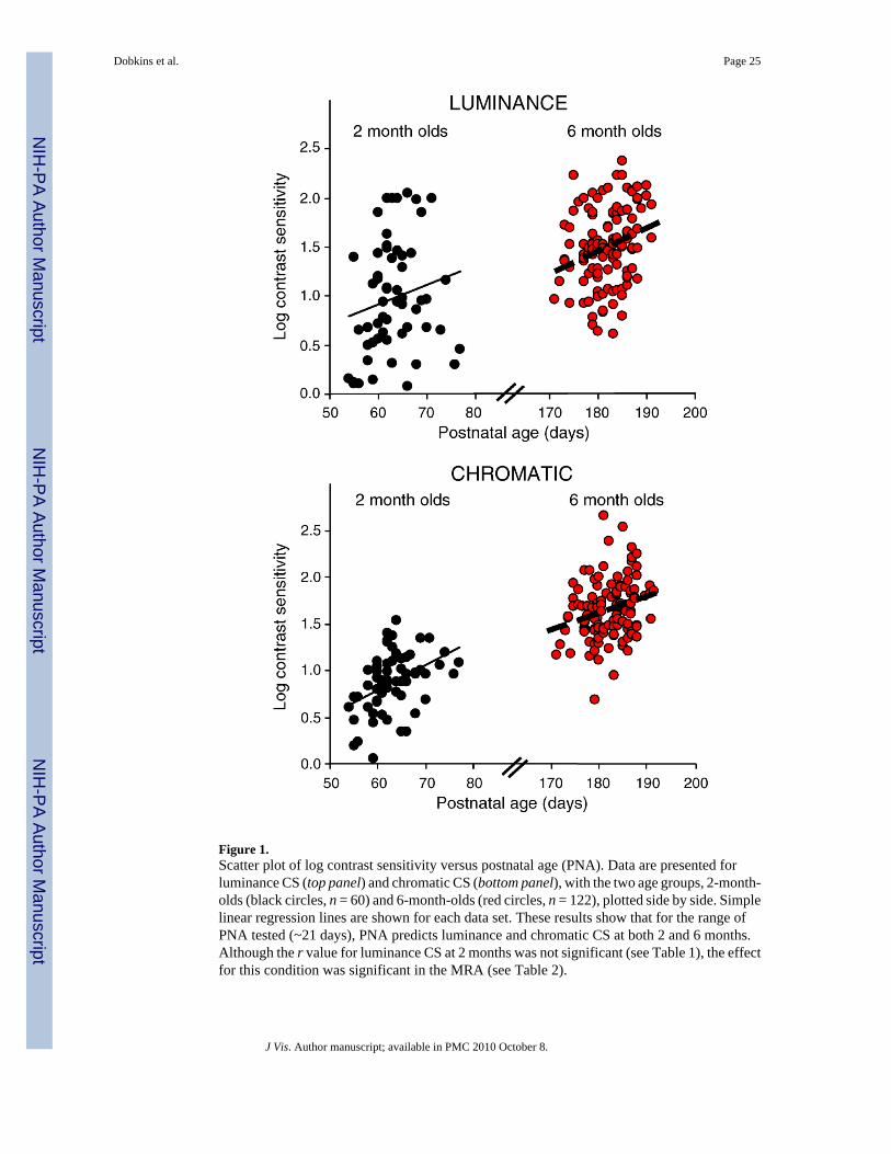
Figure 1.Scatter plot of log contrast sensitivity versus postnatal age (PNA). Data are presented forluminance CS (top panel) and chromatic CS (bottom panel), with the two age groups, 2-month-olds (black circles, n = 60) and 6-month-olds (red circles, n = 122), plotted side by side. Simplelinear regression lines are shown for each data set. These results show that for the range ofPNA tested (~21 days), PNA predicts luminance and chromatic CS at both 2 and 6 months.Although the r value for luminance CS at 2 months was not significant (see Table 1), the effectfor this condition was significant in the MRA (see Table 2).
Dobkins et al. Page 25
J Vis. Author manuscript; available in PMC 2010 October 8.
NIH
-PA Author Manuscript
NIH
-PA Author Manuscript
NIH
-PA Author Manuscript

Figure 2.Scatter plot of log contrast sensitivity versus gestational length (GL). Data are presented forluminance CS (top panel) and chromatic CS (bottom panel), with the two age groups, 2-month-olds (black circles, n = 60) and 6-month-olds (red circles, n = 122), plotted together. Simplelinear regression lines are shown for each set of data. In both the MRA and linear regressionlines, GL was found to be a significant predictor only for luminance CS in 2-month-olds; infantswith longer GL exhibit higher luminance CS.
Dobkins et al. Page 26
J Vis. Author manuscript; available in PMC 2010 October 8.
NIH
-PA Author Manuscript
NIH
-PA Author Manuscript
NIH
-PA Author Manuscript

NIH
-PA Author Manuscript
NIH
-PA Author Manuscript
NIH
-PA Author Manuscript
Dobkins et al. Page 27
Tabl
e 1
Pear
son
corr
elat
ion
coef
ficie
nts (
r val
ues)
for c
ontin
uous
var
iabl
es.
2-m
onth
-old
s (N
= 6
0)6-
mon
th-o
lds (
N =
122
)
PNA
BW
Lum
inan
ce C
SC
hrom
atic
CS
PNA
BW
Lum
inan
ce C
SC
hrom
atic
CS
GL
0.04
0.15
0.27
*0.
18−0
.18
0.21
*−0
.07
−0.1
0
PNA
0.29
*0.
180.
43*
−0.0
30.
27*
0.25
*
BW
0.18
0.27
*−0
.08
−0.1
5
Not
e:
* Sign
ifica
nt v
alue
s at p
< 0
.05.
Not
e th
at r
valu
es fo
r cor
rela
tions
of G
L, P
NA
, and
BW
with
bot
h lu
min
ance
CS
and
chro
mat
ic C
S ar
e pr
esen
ted
with
in th
e sa
me
tabl
e ev
en th
ough
the
two
type
s of C
S w
ere
sepa
rate
d in
the
MR
A.
J Vis. Author manuscript; available in PMC 2010 October 8.
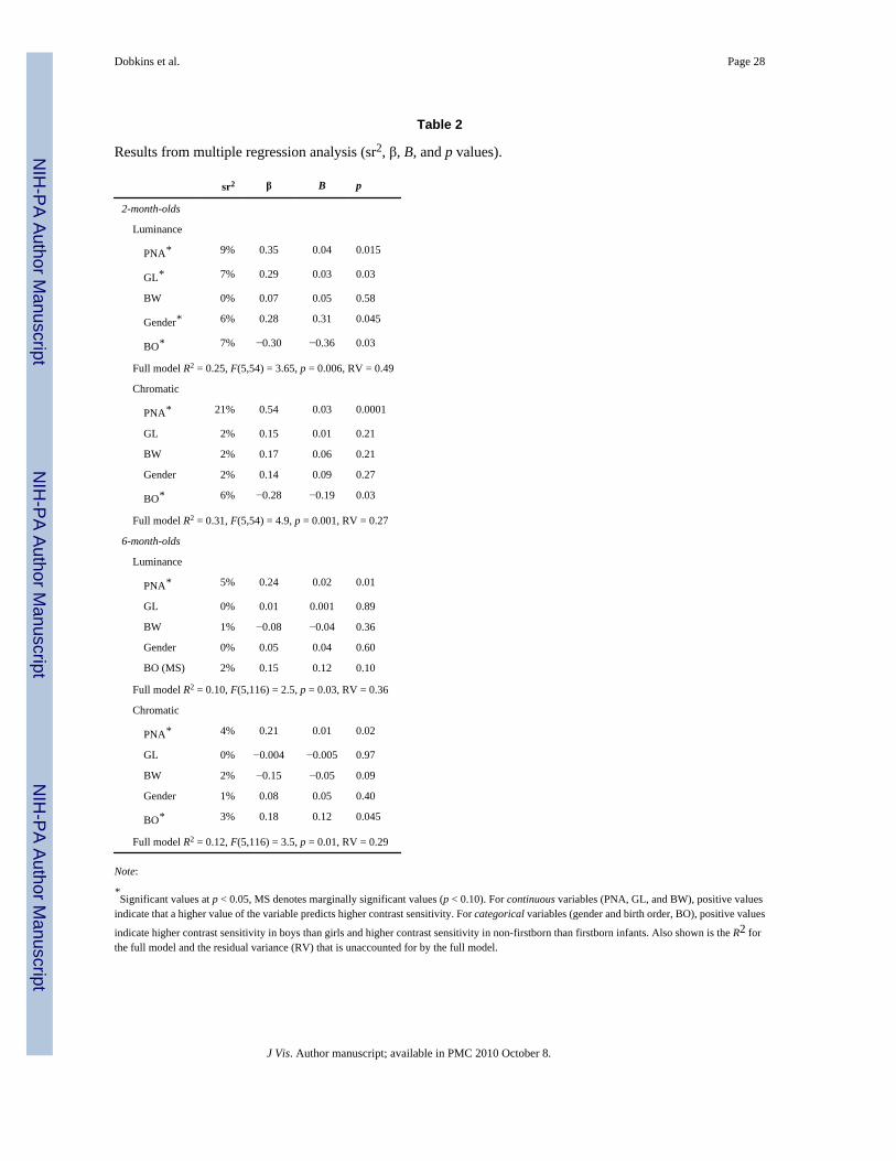
NIH
-PA Author Manuscript
NIH
-PA Author Manuscript
NIH
-PA Author Manuscript
Dobkins et al. Page 28
Table 2
Results from multiple regression analysis (sr2, β, B, and p values).
sr2 β B p
2-month-olds
Luminance
PNA* 9% 0.35 0.04 0.015
GL* 7% 0.29 0.03 0.03
BW 0% 0.07 0.05 0.58
Gender* 6% 0.28 0.31 0.045
BO* 7% −0.30 −0.36 0.03
Full model R2 = 0.25, F(5,54) = 3.65, p = 0.006, RV = 0.49
Chromatic
PNA* 21% 0.54 0.03 0.0001
GL 2% 0.15 0.01 0.21
BW 2% 0.17 0.06 0.21
Gender 2% 0.14 0.09 0.27
BO* 6% −0.28 −0.19 0.03
Full model R2 = 0.31, F(5,54) = 4.9, p = 0.001, RV = 0.27
6-month-olds
Luminance
PNA* 5% 0.24 0.02 0.01
GL 0% 0.01 0.001 0.89
BW 1% −0.08 −0.04 0.36
Gender 0% 0.05 0.04 0.60
BO (MS) 2% 0.15 0.12 0.10
Full model R2 = 0.10, F(5,116) = 2.5, p = 0.03, RV = 0.36
Chromatic
PNA* 4% 0.21 0.01 0.02
GL 0% −0.004 −0.005 0.97
BW 2% −0.15 −0.05 0.09
Gender 1% 0.08 0.05 0.40
BO* 3% 0.18 0.12 0.045
Full model R2 = 0.12, F(5,116) = 3.5, p = 0.01, RV = 0.29
Note:
*Significant values at p < 0.05, MS denotes marginally significant values (p < 0.10). For continuous variables (PNA, GL, and BW), positive values
indicate that a higher value of the variable predicts higher contrast sensitivity. For categorical variables (gender and birth order, BO), positive values
indicate higher contrast sensitivity in boys than girls and higher contrast sensitivity in non-firstborn than firstborn infants. Also shown is the R2 forthe full model and the residual variance (RV) that is unaccounted for by the full model.
J Vis. Author manuscript; available in PMC 2010 October 8.
Copyright © 2022 FDOKUMEN