Effects of an Anticarcinogenic Bowman-Birk Protease Inhibitor on Purified 20S Proteasome and MCF-7...
10
Effects of an Anticarcinogenic Bowman-Birk Protease Inhibitor on Purified 20S Proteasome and MCF-7 Breast Cancer Cells Larissa da Costa Souza 1 , Ricardo Camargo 2 , Marilene Demasi 3 , Jaime Martins Santana 4 , Ce ´ zar Martins de Sa ´ 2,{ , Sonia Maria de Freitas 1 * 1 Laboratory of Biophysics, Department of Cellular Biology, University of Brası ´lia, Brası ´lia, Brazil, 2 Laboratory of Microbiology Department of Cellular Biology, University of Brası ´lia, Brası ´lia, Brazil, 3 Laboratory of Biochemistry and Biophysics, Butantan Institute, Sa ˜o Paulo, Brazil, 4 Laboratory of Pathogen-Host Interface, Department of Cellular Biology, University of Brası ´lia, Brası ´lia, Brazil Abstract Proteasome inhibitors have been described as an important target for cancer therapy due to their potential to regulate the ubiquitin-proteasome system in the degradation pathway of cellular proteins. Here, we reported the effects of a Bowman- Birk-type protease inhibitor, the Black-eyed pea Trypsin/Chymotrypsin Inhibitor (BTCI), on proteasome 20S in MCF-7 breast cancer cells and on catalytic activity of the purified 20S proteasome from horse erythrocytes, as well as the structural analysis of the BTCI-20S proteasome complex. In vitro experiments and confocal microscopy showed that BTCI readily crosses the membrane of the breast cancer cells and co-localizes with the proteasome in cytoplasm and mainly in nucleus. Indeed, as indicated by dynamic light scattering, BTCI and 20S proteasome form a stable complex at temperatures up to 55uC and at neutral and alkaline pHs. In complexed form, BTCI strongly inhibits the proteolytic chymotrypsin-, trypsin- and caspase-like activities of 20S proteasome, indicated by inhibition constants of 10 27 M magnitude order. Besides other mechanisms, this feature can be associated with previously reported cytostatic and cytotoxic effects of BTCI in MCF-7 breast cancer cells by means of apoptosis. Citation: Souza LdC, Camargo R, Demasi M, Santana JM, Sa ´ CMd, et al. (2014) Effects of an Anticarcinogenic Bowman-Birk Protease Inhibitor on Purified 20S Proteasome and MCF-7 Breast Cancer Cells. PLoS ONE 9(1): e86600. doi:10.1371/journal.pone.0086600 Editor: Matthew Bogyo, Stanford University, United States of America Received August 9, 2013; Accepted December 11, 2013; Published January 27, 2014 Copyright: ß 2014 Souza et al. This is an open-access article distributed under the terms of the Creative Commons Attribution License, which permits unrestricted use, distribution, and reproduction in any medium, provided the original author and source are credited. Funding: This work was supported by Coordenac ¸a ˜o de Aperfeic ¸oamento de Pessoal de Nı ´vel Superior (CAPES), Conselho Nacional de Desenvolvimento Cientifico e Tecnolo ´ gico (CNPq), Fundac ¸a ˜ o de Amparo a ` Pesquisa do Distrito Federal (FAP-DF) and Financiadora de Estudos e Projetos (FINEP). The funders had no role in study design, data collection and analysis, decision to publish, or preparation of the manuscript. Competing Interests: The authors have declared that no competing interests exist. * E-mail: [email protected] { Deceased Introduction Proteases are involved in many biological processes such as the hydrolysis of intracellular proteins, transcription, cell cycle, cell invasion and apoptosis [1]. The activity of these proteases can be regulated by proteolytic degradation and inhibitors that display variable degrees of affinity with the enzymes [2,3]. Natural protease inhibitors are classified into about 20 different families [4,5], among which the Bowman-Birk inhibitors (BBI) and Kunitz have been the most studied [6,7]. Bowman-Birk inhibitors are found in mono and dicotyledons, especially in leguminous seeds [8]. Diets rich in these legumes have been associated with low incidence of cancer in human populations, in which protease inhibitors are considered to be responsible for this protective action [9–11]. In addition, BBIs are the most characterized inhibitors for their role as carcinogenesis suppressors [12–16], and they have been studied in a human phase IIa clinical trial [17]. The Black-eyed pea Trypsin/Chymotrypsin Inhibitor (BTCI) is a natural plant protease inhibitor isolated from Vigna unguiculata (Cowpea) seeds, and it belongs to the BBI family. Members of this protease inhibitor family are proteins that inactivate the functions of serine proteases by providing a reactive site, present in the canonical loop connecting the b-hairpin motif, which acts competitively as a pseudo or analogue substrate for the cognate enzyme [2,18,19]. The remarkable complementarities of these inhibitors, in particular BTCI, determine their high affinity for cognate enzymes. The dissociation constants of 10 27 –10 29 M magnitude order for BBIs and BTCI are compatible with their low dissociation process from the S1 active site of the enzymes [3,20,21]. BTCI is a globular protein containing 83 amino acid residues presenting seven disulfide bonds and molecular weight of 9.1 kDa [22–24]. It has two different and independent reactive sites for trypsin (Lys26) and chymotrypsin (Phe53) [23–26]. Its binary and ternary complexes with these proteases were isolated and physicochemically characterized by analytical ultracentrifugation, viscometry and light scattering, which showed the hydrodynamic parameters and high stability of these complexes at pH 7.0 [25]. The binding constants were calculated by enzymatic assays resulting in values of 10 7 –10 9 M 21 magnitude for chymotrypsin and trypsin, respectively [27,28]. Additionally, thermodynamic parameters calculated for the formation of trypsin-BTCI and chymotrypsin-BTCI complexes characterized these associations as PLOS ONE | www.plosone.org 1 January 2014 | Volume 9 | Issue 1 | e86600
-
Upload
independent -
Category
Documents
-
view
1 -
download
0
Transcript of Effects of an Anticarcinogenic Bowman-Birk Protease Inhibitor on Purified 20S Proteasome and MCF-7...

Effects of an Anticarcinogenic Bowman-Birk ProteaseInhibitor on Purified 20S Proteasome and MCF-7 BreastCancer CellsLarissa da Costa Souza1, Ricardo Camargo2, Marilene Demasi3, Jaime Martins Santana4,
Cezar Martins de Sa2,{, Sonia Maria de Freitas1*
1 Laboratory of Biophysics, Department of Cellular Biology, University of Brasılia, Brasılia, Brazil, 2 Laboratory of Microbiology Department of Cellular Biology, University of
Brasılia, Brasılia, Brazil, 3 Laboratory of Biochemistry and Biophysics, Butantan Institute, Sao Paulo, Brazil, 4 Laboratory of Pathogen-Host Interface, Department of Cellular
Biology, University of Brasılia, Brasılia, Brazil
Abstract
Proteasome inhibitors have been described as an important target for cancer therapy due to their potential to regulate theubiquitin-proteasome system in the degradation pathway of cellular proteins. Here, we reported the effects of a Bowman-Birk-type protease inhibitor, the Black-eyed pea Trypsin/Chymotrypsin Inhibitor (BTCI), on proteasome 20S in MCF-7 breastcancer cells and on catalytic activity of the purified 20S proteasome from horse erythrocytes, as well as the structuralanalysis of the BTCI-20S proteasome complex. In vitro experiments and confocal microscopy showed that BTCI readilycrosses the membrane of the breast cancer cells and co-localizes with the proteasome in cytoplasm and mainly in nucleus.Indeed, as indicated by dynamic light scattering, BTCI and 20S proteasome form a stable complex at temperatures up to55uC and at neutral and alkaline pHs. In complexed form, BTCI strongly inhibits the proteolytic chymotrypsin-, trypsin- andcaspase-like activities of 20S proteasome, indicated by inhibition constants of 1027 M magnitude order. Besides othermechanisms, this feature can be associated with previously reported cytostatic and cytotoxic effects of BTCI in MCF-7 breastcancer cells by means of apoptosis.
Citation: Souza LdC, Camargo R, Demasi M, Santana JM, Sa CMd, et al. (2014) Effects of an Anticarcinogenic Bowman-Birk Protease Inhibitor on Purified 20SProteasome and MCF-7 Breast Cancer Cells. PLoS ONE 9(1): e86600. doi:10.1371/journal.pone.0086600
Editor: Matthew Bogyo, Stanford University, United States of America
Received August 9, 2013; Accepted December 11, 2013; Published January 27, 2014
Copyright: � 2014 Souza et al. This is an open-access article distributed under the terms of the Creative Commons Attribution License, which permitsunrestricted use, distribution, and reproduction in any medium, provided the original author and source are credited.
Funding: This work was supported by Coordenacao de Aperfeicoamento de Pessoal de Nıvel Superior (CAPES), Conselho Nacional de DesenvolvimentoCientifico e Tecnologico (CNPq), Fundacao de Amparo a Pesquisa do Distrito Federal (FAP-DF) and Financiadora de Estudos e Projetos (FINEP). The funders had norole in study design, data collection and analysis, decision to publish, or preparation of the manuscript.
Competing Interests: The authors have declared that no competing interests exist.
* E-mail: [email protected]
{ Deceased
Introduction
Proteases are involved in many biological processes such as the
hydrolysis of intracellular proteins, transcription, cell cycle, cell
invasion and apoptosis [1]. The activity of these proteases can be
regulated by proteolytic degradation and inhibitors that display
variable degrees of affinity with the enzymes [2,3]. Natural
protease inhibitors are classified into about 20 different families
[4,5], among which the Bowman-Birk inhibitors (BBI) and Kunitz
have been the most studied [6,7].
Bowman-Birk inhibitors are found in mono and dicotyledons,
especially in leguminous seeds [8]. Diets rich in these legumes have
been associated with low incidence of cancer in human
populations, in which protease inhibitors are considered to be
responsible for this protective action [9–11]. In addition, BBIs are
the most characterized inhibitors for their role as carcinogenesis
suppressors [12–16], and they have been studied in a human phase
IIa clinical trial [17].
The Black-eyed pea Trypsin/Chymotrypsin Inhibitor (BTCI) is
a natural plant protease inhibitor isolated from Vigna unguiculata
(Cowpea) seeds, and it belongs to the BBI family. Members of this
protease inhibitor family are proteins that inactivate the functions
of serine proteases by providing a reactive site, present in the
canonical loop connecting the b-hairpin motif, which acts
competitively as a pseudo or analogue substrate for the cognate
enzyme [2,18,19]. The remarkable complementarities of these
inhibitors, in particular BTCI, determine their high affinity for
cognate enzymes. The dissociation constants of 1027–1029 M
magnitude order for BBIs and BTCI are compatible with their low
dissociation process from the S1 active site of the enzymes
[3,20,21].
BTCI is a globular protein containing 83 amino acid residues
presenting seven disulfide bonds and molecular weight of 9.1 kDa
[22–24]. It has two different and independent reactive sites for
trypsin (Lys26) and chymotrypsin (Phe53) [23–26]. Its binary and
ternary complexes with these proteases were isolated and
physicochemically characterized by analytical ultracentrifugation,
viscometry and light scattering, which showed the hydrodynamic
parameters and high stability of these complexes at pH 7.0 [25].
The binding constants were calculated by enzymatic assays
resulting in values of 107–109 M21 magnitude for chymotrypsin
and trypsin, respectively [27,28]. Additionally, thermodynamic
parameters calculated for the formation of trypsin-BTCI and
chymotrypsin-BTCI complexes characterized these associations as
PLOS ONE | www.plosone.org 1 January 2014 | Volume 9 | Issue 1 | e86600

endothermic, spontaneous and entropy-driven processes [27–28].
In spite of the slow process of peptide bond cleavage in the P1
reactive sites of BTCI and the characteristic reversibility of the
inhibition process, the presence of one disulfide bond flanking each
loop containing the P1 residues prevents the displacement of the
product from the S1 enzyme pocket [24].
The biochemical, biophysical and biotechnological properties of
BTCI have been extensively characterized [14,23,24,27–35].
BTCI is a thermally stable protein that retains 96% of its
inhibitory activity after heating at 95uC for 60 min, as well as
when it is exposed from pH 3 to 10 [30]. BTCI presented in vitro
and in vivo effects on development of the boll weevil (Anthonomus
grandis), a cotton pest. It caused larval growth delay and reduced
the insect population, showing its potential for expression in
transgenic plants engineered for pest resistance [31]. Dose-
response experiments showed that BTCI was capable of inducing
changes in renal functions by enhancing guanylin-induced
natriuresis, a strong but transient diuretic response. Additionally,
BTCI promoted increases in urine flow, in fractional excretion of
Na+ and K+, in perfusion pressure, in glomerular filtration rate
and osmolar clearance [32]. It was the first description of renal
effects induced by a protease inhibitor belonging to the Bowman-
Birk family. Both effects are due to the ability of BTCI to inhibit
chymotrypsin-like protease, thus playing a fundamental role in the
renal metabolism of guanylin in a natriuretic process [32] and
trypsin- and chymotrypsin- like protease from the midguts of
larvae and adult insects from the cotton boll weevil Antonomus
grandis [31]. Furthermore, our previous results indicated that BTCI
induced significant cytostatic and cytotoxic effects on MCF-7
breast cancer cells associated with severe cell morphological
alteration, lysosome membrane permeabilization and apoptosis
[14]. Although BTCI induced a significant reduction in MCF-7
cancer cell viability and proliferation (arrest in S and G2/M
phase), it did not affect the viability of normal MCF-10A breast
cells. BTCI induced severe morphological changes in MCF-7
cancer cells, such as plasma membrane fragmentation, cytoplasm
disorganization, and presence of double-membrane vesicles,
mitochondrial swelling, and an increase in the size of lysosomes.
In addition, meaningful DNA fragmentation, annexin-V+ cell
number increase, mitochondrial membrane potential reduction,
and cytoplasm acidification were also detected [14].
In another study, it was shown that topical applications of BTCI
on the skin of mice significantly reduced the incidence and volume
of pre-malignant lesions during chemical induction of non-
melanoma cancer in this tissue [35]. Reduction was also observed
both in the number of histopathological features as well as in
production of inflammatory mediators involved in tumor pro-
gression. Regarding the preventive effects, BTCI treatments were
able to retard the progression of non-melanoma skin cancer,
probably inducing anti-inflammatory effects [14,35]. Non-mela-
noma skin and breast cancer are of high incidence, and breast
cancer is the most frequent malignancy among women worldwide
[36]; both are leading causes of cancer-related mortality.
The high stability of BTCI in terms of maintaining its inhibitory
activity at temperatures as high as 95uC in a wide range of pHs (3–
11), besides its lack of effect on normal breast cells MCF-10A, are
of fundamental importance in evaluating the reduction of side
effects using, in the future, BTCI in breast cancer treatment.
Therefore, the aforementioned results are relevant in focusing on
BTCI as a potential anticarcinogenic drug, so as to design new
strategies for current breast cancer treatments, as well as skin
cancer prevention.
As is known, cancer is related to dysfunction in the cell cycle in
which the ubiquitin-proteasome system plays a fundamental role
in the proteolysis of intracellular proteins [37,38]. The 20S
proteasome is a catalytic complex mediating caspase-like, trypsin-
like and chymotrypsin-like activities in the b1, b2, and b5 catalytic
subunits, respectively [39], important in ubiquitinated protein
breakdown. This process occurs after recognition of ubiquitinated
proteins by 26S proteasome [40–42], which is formed by 20S
proteasome binding to one or two 19S regulatory caps [43].
Cancer cells are generally associated with increased constitutive
proteasome activity compared to non-malignant cells, due to cell
proliferation, increased oxidative stress, and elevated cytokine
levels, which can also induce the expression of immunoprotea-
somes [44]. The constitutive 20S proteasome is present in all
eukaryotic cells, whereas the immunoproteasome is mainly
expressed in immunocompetent tissues. The b1, b2, and b5
catalytic subunits present in the constitutive proteasome are
replaced by b1i (low-molecular-weight protein-2, LMP2), b2i
(multicatalytic endopeptidase complex–like-1, MECL1) and b5i
(LMP7) in the immunoproteasome [45,46]. Despite both protea-
some diversities, the expression and function of the immunopro-
teasome in different cancer types seem to be discrepant in the
literature and still remain under investigation [47]. Regarding
MCF-7 breast cancer cells, few descriptions of the expression and
activity of the immunoproteasome subunits have been reported
[48–50]. As shown, in MCF-7 cells both b5i/LMP7 and b1i/
LMP2 subunits presented minimum expression levels when
compared to other cancer cells, even with MCF-10A cells,
indicating that immunoproteasomes are almost absent in MCF-7
breast tumor cells [48].
Proteasome inhibition results in cellular homeostasis disruption
[51] and in the induction of apoptosis [52,53]. In this context,
proteasome inhibitors have been characterized as important
compounds for cancer therapy [54]. Among them, Bowman-Birk
inhibitors (BBI) appear as a very promising compound of
anticarcinogenic drugs. As reported in 2005 [55], BBI from
soybean inhibits the chymotrypsin activity of the proteasome in
breast cancer cells, but the molecular mechanism involved in this
process is not fully understood. BBI in MCF-7 cells decreases
proteasome function and results in up-regulation of MAP kinase
phosphatase-1, which suppresses ERK1/2 activity. These results
led to BBI being indicated as a novel mechanism that contributes
to prevention of cancer, as well as a potential chemopreventive
agent [55].
Here, we report the effects of BTCI on the 20S proteasome of
MCF-7 breast cancer cells and on the catalytic activity of the 20S
proteasome purified from horse erythrocytes, as well as the
molecular analysis of the BTCI-proteasome complex. Our findings
also identified BTCI as a proteasome inhibitor, which could be
related to ubiquitin-proteasome pathway inhibition and its pre-
viously reported anticarcinogenic effects [14].
Materials and Methods
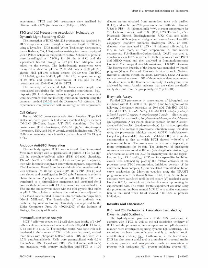
Protein PurificationBTCI was purified as previously described [29] and its purity
was confirmed by mass spectrometry analysis [30]. The 20S
proteasome used in enzymatic assays, BTCI-proteasome associa-
tion and structural stability studies was purified from horse
erythrocytes. The purification was performed by sequential
chromatography in DEAE-Sepharose, sucrose density gradient
ultracentrifugation and chromatography on mono-Q column
using FPLC facility (Pharmacia) [56]. This proteasome source
was chosen considering its well established purification procedure
resulting in a high amount of protein and also the conserved
structural features of the proteasome in all organisms. For all
Effect of a Bowman-Birk Inhibitor on Proteasome
PLOS ONE | www.plosone.org 2 January 2014 | Volume 9 | Issue 1 | e86600
experiments, BTCI and 20S proteasome were sterilized by
filtration with a 0.22 mm membrane (Millipore, USA).
BTCI and 20S Proteasome Association Evaluated byDynamic Light Scattering (DLS)
The interaction of BTCI with 20S proteasome was analyzed by
DLS assays carried out through a laser wavelength of 800 nm,
using a DynaPro - DLS model (Wyatt Technology Corporation,
Santa Barbara, CA, USA) molecular-sizing instrument equipped
with a Peltier system for temperature control. Solutions of proteins
were centrifuged at 15,000 g for 20 min at 4uC, and the
supernatant filtered through a 0.22 mm filter (Millipore) and
added to the cuvette. The hydrodynamic parameters were
measured at different pHs in 20.0 mM buffers (KCl pH 2.0;
glycine HCl pH 3.0; sodium acetate pH 4.0–6.0; Tris-HCl,
pH 7.0–9.0; glycine NaOH, pH 10.0–12.0), temperature range
of 25–60uC and protein concentration of 21.0 nM for 20S
proteasome and 15.0–90.0 mM for BTCI.
The intensity of scattered light from each sample was
normalized considering the buffer scattering contribution. Poly-
dispersity (Pd), hydrodynamic diameter (DH) and molecular weight
were determined from the intensity correlation function using the
cumulant method [57,58] and the Dynamics V.6 software. The
experiments were performed with an average of 100 acquisitions.
Cell CultureHuman MCF-7 breast cancer cells, from American Type Cell
Collection, were grown in Dulbecco’s modified Eagle’s medium
(DMEM) (HyClone, Logan, UT USA) containing 10% fetal
bovine serum (FBS – Invitrogen, USA), 30.0 mg/mL streptomycin
(Invitrogen, USA) and 100.0 mg/mL ampicillin (Invitrogen, USA).
Cells were maintained in a humidified atmosphere of 5% CO2 at
37uC.
Antibody Anti-BTCI PreparationThe antibody against BTCI was obtained from immunized
Swiss mice lineage with 3 applications of purified BTCI (0.1 mg/
mL) in phosphate-buffered saline (PBS) (10 mM phosphate,
137 mM NaCl, 2.7 mM KCl, pH 7.4) and complete adjuvant,
then with incomplete adjuvant and without adjuvant, sequentially.
The blood was collected from the carotid vein after anesthetization
with ketamine (75 ml) and xylazine (120 ml) in PBS (805 ml) and
then clotted and centrifuged at 10,000 g for 7 minutes in order to
obtain the serum. A polyacrylamide gel with 400 mg of BTCI was
transferred to a nitrocellulose membrane and incubated for 2
hours with the serum anti-BTCI. The membrane was washed with
PBS and the antibody was eluted with 0.2 mM glycine-HCl buffer
at pH 2. The solution containing the antibody was adjusted to
pH 7.0 and concentrated in amicon ultra-0.5 mL centrifugal filter
(Merck Millipore). The functionality of the antibody was
confirmed by Western blotting. This study was approved by the
Ethics Committee (Proc. No 47028/2007) of the Institute of
Biology, University of Brasilia.
Immunofluorescence AnalysisMCF-7 cells were seeded on 12-well plates at a density of 56104
cells in culture medium and incubated with 200 mM BTCI for 2,
6, 12 and 24 h at 37uC. The negative control was done with cells
incubated in the absence of BTCI. Cells were harvested, washed
three times with phosphate-buffered saline (PBS) and fixed with
3.7% paraformaldehyde. Cells were permeabilized with 0.2%
Triton-X in PBS, blocked with PBS - 5% of skimmed milk (w/v)
and incubated with primary antibodies: anti-BTCI at 1:100
dilution (serum obtained from immunized mice with purified
BTCI), and rabbit anti-20S proteasome core (Affiniti – Biomol,
USA) in PBS - 1% skimmed milk (w/v), at room temperature for
2 h. Cells were washed with PBST (PBS, 0.2% Tween 20, v/v -
Pharmacia Biotech, Buckinghamshire, UK). Goat anti rabbit
Alexa Fluor 633-conjugated and goat anti mouse Alexa Fluor 488-
conjugated secondary antibodies (Invitrogen, USA), at 1:400
dilutions, were incubated in PBS - 1% skimmed milk (w/v), for
2 h, in dark room, at room temperature. A blue nuclear
counterstain 49,6-diamidino-2-phenylindole (DAPI) was used to
visualize nuclear DNA in fixed cells. Cells were washed with PBST
and MilliQ water, and then analyzed in Immunofluorescence
Confocal Microscopy (Leica Microsystems, TCS SP5 German).
The fluorescence intensity of the images was quantified by ImageJ
program (Wayne Rasband; Research Services Branch, National
Institute of Mental Health, Bethesda, Maryland, USA). All values
were expressed as mean 6 SD of three independent experiments.
The differences in the fluorescence intensity of each group were
analyzed by t-test. Asterisk indicates that the values are signifi-
cantly different from the group analyzed (*, p,0.001).
Enzymatic AssaysPurified 20S proteasome at concentration of 2.0 mg/mL was
incubated with BTCI (2.0 to 30.0 mg/mL) and 62.5 mg/mL of the
following fluorogenic substrates in 20.0 mM Tris-HCl pH 7.5,
1.0 mM EDTA, 1.0 mM NaN3, 1.0 mM DTT: t-butyloxycarbonyl-
L-leucyl-L-arginyl-L-arginine-4-methylcoumaryl-7-amide (Boc-leu-arg-
arg-AMC) for trypsin-like; benzyloxycarbonyl-L-leucyl-L-leucyl-L-gluta-
myl-naphthylamide (Z-leu-leu-glu-bna) for caspase-like and N-succinyl-
Leu-Leu-Val-Tyr-AMC (Suc-leu-leu-val-tyr) for chymotrypsin-like
activities. The control of proteasome inhibition assays was done
using the proteasome inhibitor named MG132 (carbobenzoxyl-
leucyl-leucyl-leucinaI-H, also called Z-LLL-CHO) at a similar
concentration to that at which BTCI showed ,80–95% of
proteases inhibition. The assays were carried out in triplicate, at
room temperature for 60 min. The hydrolysis of fluorogenic
substrates was monitored at 480 nm (wavelength of emission, lem),
after excitation at 380 nm (lexc) for chymotrypsin-like and trypsin-
like, and lexc of 410 and lem of 335 nm for caspase-like. Inhibition
curves were obtained by plotting the relative activities of the
proteases versus BTCI concentration. Inhibition constant of the
enzyme-inhibitor complex, Ki, was calculated from fitted inhibition
curve considering the Morrison equation using the GRAFIT
program version 3 (Erithacus Software Ltd., UK). All inhibition
constants were calculated until the chi-square (x2) reached a value
less than 0.012, compatible with the best fit curves representing the
experimental data. The control for this experiment was done using
the proteasome inhibitor named MG132 at a similar concentra-
tion to that used when BTCI showed ,80–95% of protease
inhibition.
Results and Discussion
BTCI and 20S Proteasome Association Evaluated byDynamic Light Scattering
The hydrodynamic parameters of the 20S proteasome in
complex with BTCI, as well as the self-association tendency of
BTCI and the proteasome, in a temperature and pH dependent
manner, were investigated by using dynamic light scattering. This
technique has been commonly used mainly to analyze protein
crystallization tendency [59]. Furthermore, in the last decade,
DLS has also been a useful tool in a number of structural studies
involving proteins and nanoparticles, such as association of
proteins with surfactants [60], protein unfolding process [61],
Effect of a Bowman-Birk Inhibitor on Proteasome
PLOS ONE | www.plosone.org 3 January 2014 | Volume 9 | Issue 1 | e86600

protein stability [62,63], dimension of nanoparticles [64–66],
protein-protein interaction and protein self-association process
[67–70], in addition to others.
In the present work, the parameters obtained from DLS
measurements indicate that the 20S proteasome appears as
a monomer at 21.4 nM and pH 7.5 with hydrodynamic diameter
of 15 nm (Fig. 1a), in agreement with previously reported data
[71,72]. BTCI forms a trimer at 15 mM (Fig. 1b) and pH 7.5, with
hydrodynamic diameter of 4.5 nm, which is consistent with the
tendency of this inhibitor to form oligomers [33,73]. In contrast, at
pH 2.0 and 4.0 (Fig. 1c) BTCI and 20S proteasome assembled
into oligomeric or aggregate structures. BTCI interacts with the
20S proteasome in the first 15 min and, after that, induces low
conformational changes in the 20S proteasome, as indicated by
the increase in the hydrodynamic diameter of the complex from
21.8 nm to 22.7 nm (Fig. 1d) and concomitant disappearance of
scattering peak corresponding to BTCI.
The proteasome-BTCI complex presented tertiary conforma-
tional changes as indicated by differences in hydrodynamic
diameters under these conditions (Fig. 2a). Indeed, the complex
was characterized as thermally stable at pH 7.5 and up to 55uC,
but dissociated and formed an aggregate above 60uC (Fig 2b).
These structural changes were attributed mainly to the protea-
some, since BTCI was previously reported as a highly thermosta-
ble protein within a wide pH range (3.0–10.0), preserving its
tridimensional structure and inhibitory activity up to 95uC after
incubation for 60 min [30].
The structure of the 20S proteasome is conserved through fungi
to humans [74]. Its physicochemical features, such as sedimenta-
tion and diffusion coefficients, secondary structure content and
amino acid composition, are similar among eukaryotic organisms
[75]. The 20S proteasome was here characterized as a stable
protein complex from pH 6.0 to 12.0, with aggregation tendency
at acid pH, and structural stability up to 60uC, where dissociation
of proteasome ring structures probably occurs. Our finding
corroborates previous data reported for the 20S proteasome from
ostrich skeletal muscle, which presented pH and temperature
optima ranged between 8.0 and 11.0, and 40 to 70uC, respectively,
and stabilities for enzymatic activities between pH 5.0 and 12.0 up
to 60uC [76].
Immunocolocalization of BTCI and Proteasome in MCF-7Breast Cancer Cells
The immunocolocalization of BTCI and the proteasome in
MCF-7 breast cancer cells was investigated by confocal micros-
copy. Cells were treated with 200 mM BTCI from 2 to 24 h. The
negative control was performed in the absence of the inhibitor.
Figure 3a shows that BTCI (green) and the proteasome (red) are
distributed all over the cells after 2 h incubation. The two
molecules co-localize in the cytoplasm and nucleus of cells, as
indicated by the yellow color, corresponding to the overlapping
Alexa Fluor 488 (green) and Alexa Fluor 633 (red) antibody
conjugated fluorophores, respectively. These results indicate that
MCF-7 cellular membranes are very permeable to BTCI, and that
once BTCI is inside cells, it co-localizes in similar regions, such as
the proteasome.
The anti-20S proteasome used in the present study was a general
antibody that is able to detect multiple constitutive (b1, b2, b5)
Figure 1. Dynamic Light Scattering size distribution of BTCI and the proteasome. Hydrodynamic diameter of BTCI as a trimer (15 mM;25 kDa) and the 20S proteasome (21.4 nM; 505,2 kDa) as a monomer at neutral pH (A and B) and as aggregate in acidic condition (C) determined byDynamic Light Scattering. The BTCI and BTCI-proteasome complex (in gray) were shown in panel (D). The conformational change of the complex (inblack) was observed as indicated by differences in scattering intensity, polydispersity and hydrodynamic diameter (from 21.8 nm to 22.7 nm).doi:10.1371/journal.pone.0086600.g001
Effect of a Bowman-Birk Inhibitor on Proteasome
PLOS ONE | www.plosone.org 4 January 2014 | Volume 9 | Issue 1 | e86600

and immunoproteasome (b1i, b2i, b5i) subunits. The co-localiza-
tion observed in the immunofluorescence assay (Figure 3a) using
the anti-20S core antibody probably occurs between BTCI and
the constitutive proteasome. It is supported by the reportedly very
low expression of immunoprotesomes in MCF-7 cells [48–50];
hence, their representation in this assay is negligible. Therefore,
the detection of i20S or BTCI targeting the i20S subunits in MCF-
7 cells could be difficult to verify.
Fluorescence intensity quantification (Fig. 3b–c) was done by
recording the average of the fluorescence intensity of each tagged
protein using the ImageJ program (Wayne Rasband; Research
Services Branch, National Institute of Mental Health, Bethesda,
Maryland, USA). The fluorescence intensity of BTCI (Fig. 3b)
decreased through the first 12 h (p,0.001) and increased at 24 h
(p,0.001), when compared with the first 2 h of incubation.
Figure 2. Dynamic Light Scattering size distribution of BTCI-proteasome complex. A) Hydrodynamic diameter of the BTCI-proteasomecomplex as a function of temperature and B) pH. The complex appears as a stable protein until 55uC but dissociates and forms BTCI as a trimer andthe proteasome as an aggregate up to 60uC. The conformational changes in the complex were observed as a function of pH, as indicated by thedifferences in hydrodynamic diameter.doi:10.1371/journal.pone.0086600.g002
Figure 3. Immunofluorescence of the proteasome and BTCI in the MCF-7 cells by Confocal Microscopy. A) BTCI (green), proteasome(red), and BTCI proteasome colocalization (yellow) with DAPI DNA marked in blue. The anti BTCI specificity was assessed in MCF-7 cells treated with200 mM BTCI for 2, 6, 12 and 24 hours, whereas the control was done without BTCI (black). Fluorescence intensity of BTCI (B) and the proteasome (C)was calculated by ImageJ program as a function of the incubation time of BTCI in MCF-7 cells. Ctr: control; T-test: *p,0.001, compared to 2 hours ofBTCI incubation. Bar = 20 mm.doi:10.1371/journal.pone.0086600.g003
Effect of a Bowman-Birk Inhibitor on Proteasome
PLOS ONE | www.plosone.org 5 January 2014 | Volume 9 | Issue 1 | e86600

Moreover, the fluorescence intensity of the proteasome (Fig. 3c)
decreased significantly in the presence of the inhibitor (p,0.001).
This result suggests that BTCI enters through the MCF-7 cell
(high initial fluorescence intensity with 2 h incubation) compatible
with continuous flux of the inhibitor through the cell, coincident
with cellular membrane disruption and other morphological
alterations in the cell, as previously reported by Joanitti et al.
(2010) [14]. In contrast, the fluorescence intensity corresponding
to the proteasome (Fig. 3c) decreased until 12 h, which may have
occurred as a consequence of the steric hindrance of antibody
binding due to conformational changes in the proteasome caused
by BTCI.
These immunofluorescent assays showed that this inhibitor is
taken up by the MCF-7 cells in a time-dependent manner and is
present in the cells for 24 h. In addition, according to our previous
results, the ultra-structural analysis of MCF-7 cell morphology
indicated a pronounced effect of BTCI on plasma membrane
fragmentation, cytoplasm disorganization, presence of double-
membrane vesicles, and lysosome size increase [14]. The
recognition and internalization processes of BTCI by MCF-7
are unknown. However, according to those results, this process can
be activated after structural and/or functional alterations of
plasma membrane integrity occurred with exposure of phospha-
tidyl serine outside the inner membrane.
BTCI Inhibits the 20S Proteasome Catalytic ActivitiesThree protease activity sites are present in the b subunits of the
20S proteasome, including the caspase-like (b1), trypsin-like (b2)
and chymotrypsin-like (b5) sites [77–79]. In the present work
BTCI was characterized as a novel and potent Bowman-Birk
inhibitor of the 20S proteasome through specific inhibition of
those three protease activities. BTCI presented high affinity to the
20S proteasome, as indicated by inhibition or dissociation
constants (KI or Kd) values of 1.061027 M, 7.061027 M and
14.061027 M for trypsin-like (Fig. 4a), chymotrypsin-like (Fig. 4b)
and caspase-like sites (Fig. 4c), respectively. The calculated KI
magnitude order of 1027 to 1028 M is similar to the previous
estimate for most BBI inhibitors [3,25,80] and also BTCI [27,28].
In addition, BTCI was able to inhibit all proteases in a similar way
to the known proteasome inhibitor MG132 (carbobenzoxyl-leucyl-
leucyl-leucinaI-H), here used as a control of proteasome inhibition
assays (Fig. 5). It can be observed that BTCI was a more potent
inhibitor for trypsin than MG132 and presented a similar
inhibition to MG132 against caspase- and chymotrypsin-like
activities.
MG132 was among the first developed proteasome inhibitors
and the most widely used in research. It is a peptide aldehyde-
based reversible proteasome inhibitor, which inhibits the protea-
some primarily on the chymotrypsin-like site, but also inhibits
trypsin- and caspase-like sites. Although it is a potent proteasome
inhibitor, MG132 is rapidly oxidized into inactive carbonic acid
in vivo and, for this reason, its therapeutic use is usually prevented
[81–83].
BTCI was the first member of the Bowman-Birk family to be
characterized as a potent inhibitor of all three trypsin-like,
chymotrypsin-like and caspase-like proteasomal activities. As
previously reported, BBI, a Bowman-Birk Inhibitor isolated from
soybean, inhibits only chymotrypsin-like activity (inhibition of
70%) of the 26S proteasome in vitro at 40 mM [55]. In contrast,
BTCI inhibited almost 100% of the three enzymatic activities of
the 20S proteasome at 20 mM. This indicates that although BTCI
and BBI present similar structures [24], low differences in primary
and tertiary structure of BTCI, compared to BBI, are essential for
its significant inhibition of three proteasome proteases.
Figure 4. Inhibitory activity of BTCI toward the 20S protea-some. A) Relative activity of proteasome trypsin-like; B) Relative activityof proteasome chymotrypsin-like; C) Relative activity of proteasome
Effect of a Bowman-Birk Inhibitor on Proteasome
PLOS ONE | www.plosone.org 6 January 2014 | Volume 9 | Issue 1 | e86600

Surprisingly, the way that BTCI inhibits the caspase-like
proteasomal activity has never been reported before in the case
of any other Bowman-Birk type inhibitor. This ambiguous feature
is sustained by the structural changes in the 20S proteasome which
occurred when in complex with BTCI (Fig. 1c). The BTCI-20S
proteasome complex probably favors the steric hindrance effects,
where the caspase-like catalytic site may be buried within a large
protein surface, when BTCI binds specifically to chymotrypsin-like
or trypsin-like catalytic sites. These conformational changes reflect
the differences in proteasome enzymatic activities in the presence
of their specific substrates and BTCI. This is in accordance with
previous work reporting the conformational changes in the
proteasome when a particular active site is activated or inhibited
[84].
Studies of proteasome inhibitors and yeast mutants have shown
that chymotrypsin-like activity is a rate-limiting step responsible
for the ubiquitinated protein breakdown [85–89]. In mammalian
cells, the caspase-like and trypsin-like sites also play a significant
role in protein degradation, but the relative importance of the
three active sites depends on the protein being degraded. Indeed,
previous studies show that simultaneous inhibition of the
chymotrypsin-like and the caspase- or trypsin-like sites were
needed to decrease protein degradation by the proteasome [90].
As discussed, the inactivation of one site is not sufficient to
markedly block protein degradation by the proteasome. As
previously reported, BTCI can inhibit the activity of chymotrypsin
and trypsin enzymes independently and simultaneously through its
two specific reactive sites (P1), Lys26 and Phe53, acting as
a substrate analogous for cognate serine proteases by forming
stable ternary or binary complexes with those enzymes [23–
25,27,28,34]. Similarly, BTCI can inhibit the activity of all three
enzymes of the b1, b2, and b5 subunits of the 20S proteasome.
As is known, BBIs from legumes are potent inhibitors of both
trypsin and chymotrypsin, with Ki values within the nanomolar
units [3,21]. These inhibitors, such as BTCI, are well- character-
ized as ‘‘double-headed’’ protease inhibitors, presenting two
conserved reactive sites located in two opposite and independent
domains. The structure of BTCI folds into two similar domains
with three b-strands forming an antiparallel b-sheet stabilized by
disulfide bonds. Each domain contains one reactive site in position
P1, Lys26 and Phe53, located in a loop connecting two strands,
capable of binding to the active site of the trypsin and
chymotrypsin, respectively. The distance between the Ca atoms
of the two reactive sites (32.5 Au) is very similar to that of the BBIs,
which allows the inhibition of two protease molecules simulta-
neously and independently [24,25]. According to our results, the
inhibition of the proteasome by BTCI probably occurs in an
independent manner for all three proteases, since their active sites
are positioned in the N-terminal region of independent b1, b2 and
b5 subunits of the 20S proteasome. In this case, independent
monomers of BTCI are oriented towards each specific active site
of the different b subunits composing the 20S proteasome.
Additionally, it is known that the three b1, b2 and b5 subunits
work independently [91].
The magnitude of dissociation constants was similar to those
obtained for free BBIs with cognate enzymes. These results
indicate that reactive P1 sites of the BTCI bind to the specific S1
pocket of the catalytic region of the subunits b2 (trypsin-like), b5
(chymotrypsin-like) and block the active site of b1 (caspase-like) of
the 20S proteasome [92]. This property strongly indicates that this
protein can act as a potent inhibitor of the proteasome, one of the
most important macromolecular complexes involved in intracel-
lular protein breakdown. These results can partially corroborate
recently reported cytostatic and cytotoxic effects of BTCI on
MCF-7 breast cancer cells [14]. In this case, the induction of
apoptosis cell death by BTCI is associated with severe morpho-
logical alterations in the cell and lysosome membrane permeabi-
lization. These alterations are related to degradation of the
cytoskeleton proteins, structural protein-phospholipid membrane
disruption, chromatin condensation and DNA fragmentation,
where cytoplasmatic caspase proteins play vital roles. These
proteins belong to a group of enzymes known as cysteine proteases
and exist within the cell, differently from the caspase-like protein
of the proteasome, as inactive pro-forms or zymogens. These
zymogens can be cleaved to form active enzymes following the
induction of apoptosis [93].
Currently, there are numerous reports of proteasome inhibitors
which act only on one or two protease-like sites of the proteasome,
but few proteasome inhibitors acting simultaneously on three
proteases-like sites [94]. One of the most investigated proteasome
inhibitors is bortezomib, the first proteasome inhibitor approved
for clinical use by the FDA in 2004, which is a more potent
inhibitor for b5 than b1 subunit. Bortezomib is a reversible
inhibitor of the proteasome in spite of its very slow dissociation
rate [82,83]. This inhibitor has several molecular effects, such as
cell cycle arrest in G2/S phase [95], stabilization of cell cycle
regulatory proteins, inhibition of NF-kB activation, induction of
apoptosis, among others [96,97], mainly involving the inhibition of
subunit b5 (chymotrypsin-like activity) of the proteasome [98].
Like bortezomib, BTCI is a reversible inhibitor that inhibits the
proteases of the b1 and b5 subunits. Moreover, like MG132
(Fig. 5), BTCI inhibits all three chymotrypsin-, trypsin- and
caspase-like activities of the 20S proteasome. Although bortezomib
and MG132 are considered to be potent proteasome inhibitors,
exploratory investigation of others is needed. This is principally
due to the occurrence of bortezomib resistance after clinical use in
multiple myeloma and other diseases, such as malignant lympho-
ma [99], and the fact that the therapeutic use of MG132 is usually
prevented. In these contexts, therefore, the fact that BTCI inhibits
all three chymotrypsin-, trypsin- and caspase-like activities may be
an advantage in its application in cancer therapy, compared to
caspase-like. The dissociation or inhibition constants, calculated fromthe fitted curve by GraFit program (Erithacus software Ltd.), were1.061027 M, 7.061027 M, and 14.061027 M, respectively.doi:10.1371/journal.pone.0086600.g004
Figure 5. Inhibitory activity of BTCI and MG132 (positivecontrol), toward 20S proteasome caspase-like (Casp), chymo-trypsin-like (Chym) and trypsin-like (Tryp). The concentration ofboth inhibitors is 10 mM. As shown, BTCI is a more potent inhibitor fortrypsin than MG132 and presented similar inhibition to MG132 againstcaspase-like and chymotrypsin-like.doi:10.1371/journal.pone.0086600.g005
Effect of a Bowman-Birk Inhibitor on Proteasome
PLOS ONE | www.plosone.org 7 January 2014 | Volume 9 | Issue 1 | e86600

other reported proteasome inhibitors. These inhibitory properties
of BTCI are probably associated with its previously reported
cytostatic and cytotoxic effects on breast cancer cells [14], as
discussed above. Furthermore, as is known, the inhibited
ubiquitin-proteasome pathway can induce cell death. This is one
of the molecular pathways of cell death that need to be
investigated in order to verify the involvement of BTCI.
The mechanism by which BTCI induces apoptosis of the MCF-
7 cells is not yet known. However, as indicated here, BTCI activity
against the 20S proteasome may be related to apoptosis by
inhibiting protein breakdown processes. This mechanism, along
with others promoting cytostatic and cytotoxic effects, may be
responsible for the anticarcinogenic effect of BTCI on MCF-7 cells
by means of apoptosis.
Finally, this work is the first study analyzing the molecular
association of BTCI with the 20S proteasome in breast cancer
pathways. BTCI forms a stable complex with the 20S proteasome,
independently inhibiting its three catalytic activities. Our data also
indicate that due to its inhibitory action on the proteasome, BTCI
may be associated with the anticarcinogenic effects previously
reported in MCF-7 cancer cells [14]. Therefore, BTCI can
become an even more promising anticancer agent in the treatment
and prevention of breast cancer, but other studies should be
undertaken to elucidate the mechanism of BTCI action in breast
cancer cells.
Acknowledgments
We thank the Brazilian Synchrotron Light Laboratory (LNLS) for access to
the Dynamic Light Scattering (DLS) instrument. The authors thank Dr
Jose R. Correa (Microscopy Laboratory), for access to and support for the
Confocal Mycroscopic instrument. We also thank Murielle Taborda
Lotterman for technical assistance.
Author Contributions
Conceived and designed the experiments: LCS CMS SMF. Performed the
experiments: LCS RC MD JMS CMS SMF. Analyzed the data: LCS RC
JMS CMS SMF. Contributed reagents/materials/analysis tools: MD JMS
CMS SMF. Wrote the paper: LCS JMS SMF.
References
1. Kato GJ. (1999) Human genetic diseases of proteolysis. Hum Mutat 13: 87–98.
2. Laskowski M, Kato I. (1980) Protein inhibitors of proteinases. Annu RevBiochem 49: 593–626.
3. Ikenaka T, Norioka S. Bowman-Birk family serine proteinase inhibitors. In:Barrett A. J.; Salvesen, G. (eds). (1986) Proteinase Inhibitors, Wagenigen,
Elsevier Science cap. 9: 361–371.
4. Laskowski M, Qasim MA. (2000) What can the structures of enzyme-inhibitor
complexes tell us about the structures of enzyme substrate complexes? Biochem
Biophys Acta 1477: 324–337.
5. De Leo F, Volpicella M, Licciulli F, Liuni S, Gallerani R, Ceci LR. (2002)
PLANT-PIs: a database for plant protease inhibitors and their genes. Nucl AcidsRes 30: 347–348.
6. Ascenzi P, Bocedi A, Bolognesi M, Spallarossa A, Coletta M, et al. (2003) Thebovine basic pancreatic trypsin inhibitor (Kunitz inhibitor): a milestone protein.
Curr Protein & Peptide Sci 4: 231–251.
7. Joanitti GA, Freitas SM, Silva LP. (2006) Proteinaceous Protease Inhibitors:
Structural Features and Multiple Functional Faces. Current Enzyme Inhibition2(3): 199–217.
8. Mello MO, Tanaka AS, Silva-Filho MC. (2003) Molecular evolution ofBowman-Birk type proteinase inhibitors in flowering plants. Mol Phylogenet
Evol 27: 103–112.
9. Kennedy AR. (1993) Overview: anticarcinogenic activity of protease inhibitors.
In: Troll W.; Kennedy, A. R. (eds). Protease inhibitors as Cancer Chemopre-
ventive agents. New York: Plenum Press p. 9–64.
10. Messina MJ, Persky V, Setchell KDR, Barnes S. (1994) Soy intake and cancer
risk: a review of the in vitro and in vivo data. Nutr Cancer 21: 113–131.
11. Messina M, Barnes S. (1991) The role of soy products in reducing risk of cancer.
J Natl Cancer Inst 83: 541–546.
12. Kennedy AR. (1998) The Bowman-Birk inhibitor from soybean as an
anticarcinogenic agent. Am J Clin Nutr 68: 1406–1412.
13. Kennedy AR. (1995) Prevention of Carcinogenesis by Protease Inhibitors.
Cancer Research (Suppl.) 54: 1999–2005.
14. Joanitti GA, Azevedo RB, Freitas SM. (2010) Apoptosis and lysosome
membrane permeabilization induction on breast cancer cells by an anticarci-nogenic Bowman–Birk protease inhibitor from Vigna unguiculata seeds. Cancer
Letters. 293: 73–81.
15. Clemente A, Sonnante G, Domoney C. (2011) Bowman-Birk inhibitors from
legumes and human gastrointestinal health: current status and perspectives. CurrProtein Pept Sci 12 (5): 358–373.
16. Clemente A, Marın-Manzano MC, Jimenez E, Arques MC, Domoney C. (2012)The anti-proliferative effect of TI1B, a major Bowman-Birk isoinhibitor from
pea (Pisum sativum L.), on HT29 colon cancer cells is mediated through
protease inhibition. Br J Nutr 108, Suppl. 1: 135–144.
17. Armstrong WB, Kennedy AR, Wan XS, Taylor TH, Nguyen QA, et al. (2000)
Clinical modulation of oral Leukoplakia and protease activity by Bowman–Birkinhibitor concentrate in a phase IIa chemoprevention trial. Clin Cancer Res 6:
4684–4691.
18. Richardson M. (1991). Seed storage proteins: the enzyme inhibitors. Meth Plant
Biochem 5: 259–303.
19. Bode W, Huber R. (1992). Natural protein proteinase inhibitors and their
interaction with proteinases. Eur J Biochem 204: 433–451.
20. Chen P, Rose J, Love R, Wei CH, Wang BC. (1992) Reactive Sites of an
Anticarcinogenic Bowman-Birk Proteinase Inhibitor are Similar to OtherTrypsin Inhibitors. The Journal of Biological Chemistry 267: 1990–1994.
21. Clemente A, Marın-Manzano MC, Arques MC, Domoney C. (2013) Bowman-Birk Inhibitors from Legumes: Utilization in Disease Prevention and Therapy,
Bioactive Food Peptides in Health and Disease. Dr. Blanca Hernandez-Ledesma
(Ed.), ISBN: 978–953–51–0964–8, In Tech, DOI: 10.5772/51262. http://www.intechopen.com/books/bioactive-food-peptides-in-health-and-disease/
bowman-birk-inhibitors-from-legumes-utilisation-in-disease-prevention-and-therapy.
22. Morhy L, Ventura MM. (1987) The complete amino acid sequence of the Vigna
unguiculata (L.) WaLP seed trypsin and chymotrypsin inhibitor. An Acad Bras
Cienc 59: 71–81.
23. Freitas SM, De Mello LV, Da Silva MC, Vriend G, Neshich G, et al. (1997)Analysis of the black-eyed pea trypsin and chymotrypsin inhibitor-a-chymo-
trypsin complex. FEBS Lett 409: 121–127.
24. Barbosa JARG, Silva LP, Teles RCL, Esteves GF, Azevedo RB, et al. (2007)
Crystal Structure of the Bowman-Birk Inhibitor from Vigna unguiculata Seeds inComplex with b-Trypsin at 1.55 A Resolution and Its Structural Properties in
Association with Proteinases. Biophysical Journal 92: 1638–1650.
25. Ventura MM, Martin CO, Morhy L. (1975) A trypsin and chymotrypsin
inhibitor from black-eyed pea (Vigna sinensis L.). VI. Isolation and properties of
complexes with trypsin and chymotrypsin. An Acad Brasil Cienc 47: 335–346.
26. Xavier-Filho J, Ventura MM. (1988) Trypsin inhibitors in Cowpea: a review.
Comments Agric Food Chem 1: 239–314.
27. Fachetti HCS, Mizuta K, Ventura MM. (1984) Thermodynamics of the black-
eyed pea trypsin and chymotrypsin inhibitor. An Acad Bras Cienc 56: 311–317.
28. Freitas SM, Ikemoto H, Ventura MM. (1999) Thermodynamics of the binding
of chymotrypsin with the black-eyed pea trypsin and chymotrypsin inhibitor(BTCI). J Protein Chem 18: 307–313.
29. Ventura MM, Xavier-Filho J. (1966) A trypsin and chymotrypsin inhibitor from
black-eyed pea (V. Sinensis L.) I. Purification and partial characterization. AnAcad Bras Cienc 38: 553–566.
30. Silva LP, Leite JRSA, Bloch C, Freitas SM. (2001) Stability of a Black Eyed PeaTrypsin/Chymotrypsin inhibitor (BTCI). Protein and Peptide Lett 8: 33–38.
31. Franco OL, Santos RC, Batista JAN, Mendes ACM, Araujo A, et al. (2003)Effects of black-eyed pea trypsin/chymotrypsin inhibitor on proteolytic activity
and on development of Anthonomus grandis. Phytochemistry 63: 343–349.
32. Carvalho AF, Santos-Neto MS, Monteiro HSA, Freitas SM, Morhy L, et al.
(2008) BTCI enhances guanylin-induced natriuresis and promotes renal
glomerular and tubular effects. Braz J Biol 68 (1): 149–154.
33. Silva LP, Azevedo RB, Morais PC, Ventura MM, Freitas SM. (2005)
Oligomerization States of Bowman-Birk Inhibitor by Atomic Force Microscopyand Computational Approaches. Proteins: Structure, Function, and Bioinfor-
matics 61: 642–648.
34. Esteves GF, Teles RCL, Calvacanti NS, Neves D, Ventura MM, et al. (2007)
Crystallization, data collection and processing of the chymotrypsin–BTCI–
trypsin ternary complex. Acta Cryst F63: 1087–1090.
35. Joanitti GA. Supervisors: Azevedo RB and Freitas SM. (2012) Ph thesis: Efeitos
de extrato bruto de sementes de Vigna unguiculata e do inibidor de proteasesBTCI, encapsulado em nanopartıculas, no tratamento preventivo e terapeutico
de cancer de mama e de pele, in vitro e in vivo. UnB-Depto de Morfologia.
36. Hortobagyi GN, De La Garza Salazar J, Pritchard K, Amadori D, Haidinger R,
et al. (2005) The global breast cancer burden: variations in epidemiology andsurvival. Clin Breast Cancer 6: 391–401.
37. Dou QP, Li B. (1999) Proteasome inhibitors as potential novel anticancer agents.
Drug Resist Updat 2 (4): 215–223.
Effect of a Bowman-Birk Inhibitor on Proteasome
PLOS ONE | www.plosone.org 8 January 2014 | Volume 9 | Issue 1 | e86600

38. Bochtler M, Ditzel L,M, Hartmann C, Huber R. (1999) The proteasome. Annu
Rev Biophys Biomol Struct 28: 295–317.
39. Loidl G, Groll M, Musiol HJ, Huber R, Moroder L. (1999) Bivalency as
a principle for proteasome inhibition. Proc Natl Acad Sci USA 96 (10): 5418–
5422.
40. Glickman MH, Rubin DM, Fried VA, Finley D. (1998) The regulatory particle
of the Saccharomyces cerevisiae proteasome. Mol Cell Biol 18: 3149–3162.
41. Voges D, Zwickl P, Baumeister W. (1999) The 26S proteasome: a molecular
machine designed for controlled proteolysis. Annu Rev Biochem 68: 1015–
10168.
42. Pickart CM, Cohen RE. (2004) Proteasomes and their kin: proteases in the
machine age. Nat Rev Mol Cell Biol 5: 177–187.
43. Holzl H, Kapelari B, Kellermann J, Seemuller E, Sumegi M, et al. (2000) The
regulatory complex of Drosophila melanogaster 26S proteasomes. Subunit
composition and localization of a deubiquitylating enzyme. J Cell Biol 150 (1):
119–130.
44. Bossola M, Muscaritoli M, Costelli P, Grieco G, Bonelli G, et al. (2003)
Increased muscle proteasome activity correlates with disease severity in gastric
cancer patients. Ann Surg 237: 384–389.
45. Groll M, Ditzel L, Lowe J, Stock D, Bochtler M, et al. (1997) Structure of 20S
proteasome from yeast at 2.4 Au resolution. Nature 386: 463–471.
46. Kloetzel PM, Ossendorp F. (2004) Proteasome and peptidase function in MHC-
class-I-mediated antigen presentation. Curr Opin Immunol 16: 76–81.
47. Blackburn C, Gigstad KM, Hales P, Garcia K, Jones M, et al. (2010)
Characterization of a new series of non-covalent proteasome inhibitors with
exquisite potency and selectivity for the 20S b5-subunit.Biochem. J 430: 461–
476.
48. Gavilan E, Sanchez-Aguayo I, Daza P, Ruano D. (2013) GSK-3b signaling
determines autophagy activation in the breast tumor cell line MCF-7 and
inclusion formation in the non-tumor cell line MCF-10A in response to
proteasome inhibition. Cell Death and Disease 4: e572.
49. Wehenkel M, Ban JO, Ho YK, Carmony KC, Hong JT, et al. (2012) A selective
inhibitor of the immunoproteasome subunit LMP2 induces apoptosis in PC-3
cells and suppresses tumour growth in nude mice. Br J Cancer 107: 53–62.
50. Park JE, Ao L, Miller Z, Kim K, Wu Y, et al. (2013) PSMB9 Codon 60
Polymorphisms Have No Impact on the Activity of the Immunoproteasome
Catalytic Subunit B1i Expressed in Multiple Types of Solid Cancer. PLoS One
8(9): e73732.
51. Codony-Servat J, Tapia MA, Bosch M, Oliva C, Domingo-Domenech J, et al.
(2006) Differential cellular and molecular effects of bortezomib, a proteasome
inhibitor, in human breast cancer cells. Mol Cancer Ther 5 (3): 665–675.
52. An B, Goldfarb RH, Siman R, Dou QP. (1998) Novel dipeptidyl proteasome
inhibitors overcome Bcl-2 protective function and selectively accumulate the
cyclin-dependent kinase inhibitor p27 and induce apoptosis in transformed, but
not normal, human fibroblasts. Cell Death Differ 5: 1062–1075.
53. Lopes UG, Erhardt P, Yao R, Cooper GM. (1997) p53-dependent induction of
apoptosis by proteasome inhibitors. J Biol Chem 272 (20): 12893–12896.
54. Voorhees PM, Dees EC, O’Neil B, Orlowski RZ. (2003) The Proteasome as
a Target for Cancer Therapy. Clinical Cancer Research 9: 6316–6325.
55. Chen YW, Huang S, Lin-Shiau S, LIN J. (2005) Bowman–Birk inhibitor abates
proteasome function and suppresses the proliferation of MCF-7 breast cancer
cells through accumulation of MAP kinase phosphatase-1. Carcinogenesis 26 (7):
1296–1306.
56. Demasi M, Shringarpure R, Davies KJ. (2001) Glutathiolation of the
proteasome is enhanced by proteolytic inhibitors. Arch Biochem Biophys 389
(2): 254–263.
57. Frisken BJ. (2001) Revisiting the method of cumulants for the analysis of
dynamic light-scattering data. Applied Optics 40 (24): 4087–4091.
58. Hassan PA and Kulshreshtha SK. (2006) Modification to the cumulant analysis
of polydispersity in quasielastic light scattering data. Journal of Colloid and
Interface Science 300(2): 744–748.
59. Baldwin ET, Crumley KV, Carter CW. (1986) Practical, Rapid Screening of
Protein Crystallization Conditions by Dynamic Light Scattering. Biophys J 49
(1): 47–48.
60. Tardioli S, Bonincontro A, Mesa CL, Muzzalupo R. (2010) Interaction of
bovine serum albumin with gemini surfactants. Journal of Colloid and Interface
Science 347 (1): 96–101.
61. Boehm K, Guddorf J, Albers A, Kamiyama T, Fetzner S, Hinz HJ. (2008)
Thermodynamic Analysis of Denaturant-Induced Unfolding of HodC69S
Protein Supports a Three-State Mechanism. Biochemistry 47: 7116–7126.
62. Carrotta R, Manno M, Giordano FM, Longo A, Portale G, et al. (2009) Protein
stability modulated by a conformational effector: effects of trifluoroethanol on
bovine serum albumin. Phys Chem. Chem Phys 11: 4007–4018.
63. Hanlon AD, Larkin MI, Reddick RM. (2010) Free-Solution, Label-Free Protein-
Protein Interactions Characterized by Dynamic Light Scattering. Biophysical
Journal 98: 297–304.
64. Lacerda SH, Park JJ, Meuse C, Pristinski D, Becker ML, et al. (2010) Interaction
of gold nanoparticles with common human blood proteins. ACS Nano 4 (1):
365–379.
65. Liang L, Yao P, Gong J, Jiang M. (2004) Interaction of apo cytochrome c with
sulfonated polystyrene nanoparticles. Langmuir 20 (8): 3333–3338.
66. Chen L, Tianqing L. (2008) Interaction behaviors between chitosan and
hemoglobin. Int J Biol Macromol 42 (5): 441–446.
67. Libich DS, Hill CM, Bates IR, Hallett FR, Armstrong S, et al. (2003) Interactionof the 18.5-kD isoform of myelin basic protein with Ca2+-calmodulin: effects of
deimination assessed by intrinsic Trp fluorescence spectroscopy, dynamic light
scattering, and circular dichroism. Protein Sci 12 (7): 1507–1521.
68. Gokarn YR, Fesinmeyer RM, Saluja A, CAO S, Dankberg J, et al. (2009) Ion-
specific modulation of protein interactions: Anion-induced, reversible oligomer-ization of a fusion protein. Protein Science 18: 169–179.
69. Shiba K, Niidome T, Katoh E, Xiang H, Han L, et al. (2010) Polydispersity as
a Parameter for Indicating the Thermal Stability of Proteins by Dynamic LightScattering. Analytical Sciences 26: 659.
70. Li S, Xing D, LI J. (2004) Dynamic Light Scattering Application to StudyProtein Interactions in Electrolyte Solutions. Journal of Biological Physics 30:
313–324.
71. Kopp F, Steiner R, Dahlmann B, Kuehn L, Reinauer H. (1986) Size and shapeof the multicatalytic proteinase from rat skeletal muscle. Biochim Biophys Acta
872 (3): 253–260.
72. Yoshimura T, Kameyama K, Takagi T, Ikai A, Tokunaga F, et al. (1993)
Molecular characterization of the ‘‘26S’’ proteasome complex from rat liver.
J Struct Biol 111 (3): 200–211.
73. Ventura MM, Mizuta K, Ikemoto H. (1981) Self-association of the black-eyed
pea trypsin and chymotrypsin inhibitor in solution - a study by light-scattering.An Acad Bras Cienc 53: 195–201.
74. Lupas A, Koster AJ, Baumeister W. (1993) Structural features of 26S e 20S
proteasomes. Enzyme Protein 47: 252–273.
75. Tanaka K, Yoshimura T, Kumatori A, Ichihara A, Ikai A, et al. (1988)
Proteasomes (multi-protease complexes) as 20 S ring-shaped particles in a variety
of eukaryotic cells. J Biol Chem 263 (31): 16209–16217.
76. Thomas AR, Oosthuizen V, Naude RJ, Muramoto K. (2002) Purification and
characterization of the 20S proteasome from ostrich skeletal muscle. Biol Chem383 (7–8): 1267–1270.
77. Griffin TA, Nandi D, Cruz M, Fehling HJ, Kaer LV, et al. (1998)
Immunoproteasome assembly: cooperative incorporation of interferon gamma(IFN-gamma)-inducible subunits. J Exp Med 187: 97–104.
78. Orlowski M, Wilk S. (2000) Catalytic Activities of the 20S proteasome,a multicatalytic proteinase complex. Arch Biochem Biophys 383 (1): 1–16.
79. Groll M, Huber R. (2003) Substrate access and processing by the 20S
proteasome core particle. Internat J Biochem 35: 606–616.
80. McBride JD, and Leatherbarrow RJ. (2001) Synthetic Peptide Mimics of the
Bowman-Birk Inhibitor Protein Current Medicinal Chemistry 8: 909–917.
81. deBettignies G, Coux O. (2010) Proteasome inhibitors: dozens of molecules andstill counting. Biochimie 92: 1530–45.
82. Kisselev AF, van der Linden WA, Overkleeft HS. (2012) Proteasome inhibitors:an expanding army attacking a unique target. Chem Biol 19: 99–115.
83. Borissenko L, Groll M. (2007) 20S proteasome and its inhibitors: crystallo-
graphic knowledge for drug development. Chem Rev 107: 687–717.
84. Kisselev AF, Kaganovich D, Goldberg AL. (2002) Binding of Hydrophobic
Peptides to Several Non-catalytic Sites Promotes Peptide Hydrolysis by AllActive Sites of 20S Proteasomes. J Biol Chem 277: 22260–22270.
85. Rock KL, Gramm C, Rothstein L, Clark K, Stein R, et al. (1994) Inhibitors of
the proteasome block the degradation of most cell proteins and the generation ofpeptides presented on MHC class I molecules. Cell 78: 761–771.
86. Pereira MEF, Chen WE, Li J, Johdo O. (1996) The Antitumor Drug
Aclacinomycin A, Which Inhibits the Degradation of Ubiquitinated Proteins,Shows Selectivity for the Chymotrypsin-like Activity of the Bovine Pituitary 20 S
Proteasome. J Biol Chem 271: 16455–16459.
87. Lee DH, Goldberg AL. (1998) Proteasome inhibitors: valuable new tools for cell
biologists. Trends in Cell Biology 8: 397–403.
88. Craiu A, Gaczynska M, Akopian T, Gramm CF, Fenteany G, et al. (1997)Lactacystin and clasto-lactacystin beta-lactone modify multiple proteasome beta-
subunits and inhibit intracellular protein degradation and major histocompat-ibility complex class I antigen presentation. J Biol Chem 272 (20): 13437–13445.
89. Heinemeyer W, Fischer M, Krimmer T, Stachon U, Wolf DH. (1997) The
active sites of the eukaryotic 20 S proteasome and their involvement in subunitprecursor processing. J Biol Chem 272: 25200–25209.
90. Kisselev AF, Callard A, Goldberg AL. (2006) Importance of the differentproteolytic sites of the proteasome and the efficacy of inhibitors varies with the
protein substrate. J Biol Chem 281 (13): 8582–8590.
91. Schmidtke G, Kraft R, Kostka S, Henklein P, Frommel C, et al. (1996) Analysisof mammalian 20S proteasome biogenesis: the maturation of beta-subunits is an
ordered two-step mechanism involving autocatalysis. The EMBO J 15: 6887–6898.
92. Groll M, Heinemeyer W, Jager S, Ullrich T, Bochtler M, et al. (1999) The
catalytic sites of 20S proteasomes and their role in subunit maturation: Amutational and crystallographic study, Proc Natl Acad Sci USA 96: 10976–
10983.
93. Ghobrial IM, Witzig TE, Adjei AA. (2005) Targeting apoptosis pathways in
cancer therapy. CA Cancer J Clin 55 (3): 178–194.
94. Shen M, Schmitt S, Buac D, Ping Dou Q. (2013) Targeting the ubiquitin–proteasome system for cancer therapy. Expert Opin Ther Targets 17(9): 1091–
1108.
95. Mortenson MM, Schlieman MG, Virudachalam S, Bold RJ. (2004) Effects of theproteasome inhibitor Bortezomib alone and in combination with chemotherapy
in the A549 non-small-cell lung cancer cell line. Cancer Chemo Ther Pharmacol54 (4): 343–353.
Effect of a Bowman-Birk Inhibitor on Proteasome
PLOS ONE | www.plosone.org 9 January 2014 | Volume 9 | Issue 1 | e86600

96. Adams J, Palombella VJ, Sausville EA, Johnson J, Destree A, et al. (1999)
Proteasome inhibitors: a novel class of potent and effective antitumor agents.
Cancer Res 59: 2615–2622.
97. Adams J. (2004) The proteasome: a suitable antineoplastic target. Nat Rev
Cancer 4: 349–360.
98. Crawford LJ, Walker B, Ovaa H, Chauhan D, Anderson KC, et al. (2006)
Comparative selectivity and specificity of the proteasome inhibitors BzLLLCO-CHO, PS-341, and MG-132. Cancer Res 66: 6379–6386.
99. O’Connor OA, Wright J, Moskowitz C, Muzzy J, MacGregor-Cortelli B, et al.
(2005) Phase II clinical experience with the novel proteasome inhibitorbortezomib in patients with indolentnon-Hodgkin’s lymphoma and mantle cell
lymphoma. J Clin Oncol 23: 676–684.
Effect of a Bowman-Birk Inhibitor on Proteasome
PLOS ONE | www.plosone.org 10 January 2014 | Volume 9 | Issue 1 | e86600