
Effects of a bacterial trehalose lipid on phosphatidylglycerol membranes
6
Effects of a bacterial trehalose lipid on phosphatidylglycerol membranes Antonio Ortiz a , José A. Teruel a , Ángeles Manresa b , María J. Espuny b , Ana Marqués b , Francisco J. Aranda a, ⁎ a Departamento de Bioquímica y Biología Molecular-A, Facultad de Veterinaria, Universidad de Murcia, Campus de Espinardo, E-30100 Murcia, Spain b Laboratorio de Microbiología, Facultad de Farmacia, Universidad de Barcelona, Joan XXIII s/n, E-08028 Barcelona, Spain abstract article info Article history: Received 24 February 2011 Accepted 2 May 2011 Available online 10 May 2011 Keywords: Trehalose lipid Phosphatidylglycerol DSC X-ray diffraction FT-IR Fluorescence polarization Bacterial trehalose lipids are biosurfactants with potential application in the biomedical/healthcare industry due to their interesting biological properties. Given the amphiphilic nature of trehalose lipids, the understanding of the molecular mechanism of their biological action requires that the interaction between biosurfactant and membranes is known. In this study we examine the interactions between a trehalose lipid from Rhodococcus sp. and dimyristoylphosphatidylglycerol membranes by means of differential scanning calorimetry, X-ray diffraction, infrared spectroscopy and fluorescence polarization. We report that there are extensive interactions between trehalose lipid and dimyristoylphosphatidylglycerol involving the perturba- tion of the thermotropic gel to liquid-crystalline phase transition of the phospholipid, the increase of fluidity of the phosphatidylglycerol acyl chains and dehydration of the interfacial region of the bilayer, and the modulation of the order of the phospholipid bilayer. The observations are interpreted in terms of structural perturbations affecting the function of the membrane that might underline the biological actions of the trehalose lipid. © 2011 Elsevier B.V. All rights reserved. 1. Introduction Biological surface active compounds are a diverse group of amphiphilic surface-active molecules synthesized by different pro- karyotic and eukaryotic organisms [1]. From the chemical point of view biosurfactants present a wide structural diversity, the com- pounds with the most interesting properties being lipopeptides and glycolipids [2]. Glycolipids of bacterial origin are widespread biosurfactants which contain carbohydrates in combination with long-chain aliphatic or hydroxyl aliphatic acids. An important group of biosurfactant glycolipids includes the rhamnolipids secreted by Pseudomonas aeruginosa, which have been shown to affect the structural properties of membrane phospholipids [3,4]. Another important group of glycolipid biosurfactants is formed by trehalose- containing glycolipids [5]. These trehalose lipids are mainly produced by rhodococci and present such interesting physicochemical and biological properties that a number of different commercial applica- tions have been proposed for them [6]. At present, the main applications are found in the hydrocarbon bioremediation and oil and petroleum industry. A very important emerging field of applica- tion is the biomedical/healthcare industry, since some biosurfactants have been demonstrated to be suitable alternatives to synthetic products as antimicrobial and therapeutic agents [7,8]. Trehalose lipids have been reported to have antiviral properties [9,10]. It has been shown that trehalose lipids have excellent growth inhibition and differentiation-inducing activities against human leukaemia cells [11– 13]. They also inhibit the activity of phospholipids- and calcium- dependent protein kinase C of HL60 cells [14] and show immunomodu- lating activity [15]. Interestingly, some trehalose lipids have shown antibacterial activity [16]. Although the amphiphilic nature of trehalose lipids points to the membrane as their hypothetical site of action, very little is known about the interaction between these biosurfactants and biological membranes. We have found that trehalose lipid permeabilizes phospholipid membranes [17] and induces hemolysis of human red blood cells [18]. In order to get insight into the molecular interaction between these biosurfactants and the lipidic component of biological membranes, we have studied the effect of trehalose lipid on the most important membrane phospholipids. We have shown that trehalose lipid increases the fluidity of phosphatidylcholine membranes forming domains in the fluid state [19], and that it exhibits an important dehydrating effect on the interfacial region of saturated phosphatidyl- ethanolamines and greatly promotes the formation of the inverted hexagonal H II phase in unsaturated phosphatidylethanolamines [20]. Recently we have shown that trehalose lipid was also able to affect the thermotropic membrane phase transition of phosphatidylserine in the absence and presence of calcium [21]. This work focuses on the effects of trehalose lipid on the gel and fluid phases of phosphatidylglycerol, a predominant phospholipid of the cytoplasmic membrane of bacteria. Phosphatidylglycerol has been used extensively as model for acidic phospholipid membranes [22–24]. The physico-chemical properties of phospholipids present in membranes of microorganisms are of interest because it has been suggested that the lipid composition of bacterial Biochimica et Biophysica Acta 1808 (2011) 2067–2072 ⁎ Corresponding author. Tel.: + 34 868884760; fax: + 34 868884147. E-mail address: [email protected] (F.J. Aranda). 0005-2736/$ – see front matter © 2011 Elsevier B.V. All rights reserved. doi:10.1016/j.bbamem.2011.05.003 Contents lists available at ScienceDirect Biochimica et Biophysica Acta journal homepage: www.elsevier.com/locate/bbamem
Transcript of Effects of a bacterial trehalose lipid on phosphatidylglycerol membranes
Biochimica et Biophysica Acta 1808 (2011) 2067–2072
Contents lists available at ScienceDirect
Biochimica et Biophysica Acta
j ourna l homepage: www.e lsev ie r.com/ locate /bbamem
Effects of a bacterial trehalose lipid on phosphatidylglycerol membranes
Antonio Ortiz a, José A. Teruel a, Ángeles Manresa b, María J. Espuny b, Ana Marqués b, Francisco J. Aranda a,⁎a Departamento de Bioquímica y Biología Molecular-A, Facultad de Veterinaria, Universidad de Murcia, Campus de Espinardo, E-30100 Murcia, Spainb Laboratorio de Microbiología, Facultad de Farmacia, Universidad de Barcelona, Joan XXIII s/n, E-08028 Barcelona, Spain
⁎ Corresponding author. Tel.: +34 868884760; fax: +E-mail address: [email protected] (F.J. Aranda).
0005-2736/$ – see front matter © 2011 Elsevier B.V. Aldoi:10.1016/j.bbamem.2011.05.003
a b s t r a c t
a r t i c l e i n f oArticle history:Received 24 February 2011Accepted 2 May 2011Available online 10 May 2011
Keywords:Trehalose lipidPhosphatidylglycerolDSCX-ray diffractionFT-IRFluorescence polarization
Bacterial trehalose lipids are biosurfactants with potential application in the biomedical/healthcare industrydue to their interesting biological properties. Given the amphiphilic nature of trehalose lipids, theunderstanding of the molecular mechanism of their biological action requires that the interaction betweenbiosurfactant and membranes is known. In this study we examine the interactions between a trehalose lipidfrom Rhodococcus sp. and dimyristoylphosphatidylglycerol membranes by means of differential scanningcalorimetry, X-ray diffraction, infrared spectroscopy and fluorescence polarization. We report that there areextensive interactions between trehalose lipid and dimyristoylphosphatidylglycerol involving the perturba-tion of the thermotropic gel to liquid-crystalline phase transition of the phospholipid, the increase of fluidityof the phosphatidylglycerol acyl chains and dehydration of the interfacial region of the bilayer, and themodulation of the order of the phospholipid bilayer. The observations are interpreted in terms of structuralperturbations affecting the function of the membrane that might underline the biological actions of thetrehalose lipid.
34 868884147.
l rights reserved.
© 2011 Elsevier B.V. All rights reserved.
1. Introduction
Biological surface active compounds are a diverse group ofamphiphilic surface-active molecules synthesized by different pro-karyotic and eukaryotic organisms [1]. From the chemical point ofview biosurfactants present a wide structural diversity, the com-pounds with the most interesting properties being lipopeptides andglycolipids [2]. Glycolipids of bacterial origin are widespreadbiosurfactants which contain carbohydrates in combination withlong-chain aliphatic or hydroxyl aliphatic acids. An important groupof biosurfactant glycolipids includes the rhamnolipids secreted byPseudomonas aeruginosa, which have been shown to affect thestructural properties of membrane phospholipids [3,4]. Anotherimportant group of glycolipid biosurfactants is formed by trehalose-containing glycolipids [5]. These trehalose lipids are mainly producedby rhodococci and present such interesting physicochemical andbiological properties that a number of different commercial applica-tions have been proposed for them [6]. At present, the mainapplications are found in the hydrocarbon bioremediation and oiland petroleum industry. A very important emerging field of applica-tion is the biomedical/healthcare industry, since some biosurfactantshave been demonstrated to be suitable alternatives to syntheticproducts as antimicrobial and therapeutic agents [7,8]. Trehalose lipidshave been reported to have antiviral properties [9,10]. It has been
shown that trehalose lipids have excellent growth inhibition anddifferentiation-inducing activities against human leukaemia cells [11–13]. They also inhibit the activity of phospholipids- and calcium-dependent protein kinase C of HL60 cells [14] and show immunomodu-lating activity [15]. Interestingly, some trehalose lipids have shownantibacterial activity [16]. Although the amphiphilic nature of trehaloselipids points to the membrane as their hypothetical site of action, verylittle is known about the interaction between these biosurfactantsand biological membranes. We have found that trehalose lipidpermeabilizes phospholipid membranes [17] and induces hemolysisof human red blood cells [18]. In order to get insight into the molecularinteraction between these biosurfactants and the lipidic component ofbiological membranes, we have studied the effect of trehalose lipid onthe most important membrane phospholipids. We have shown thattrehalose lipid increases thefluidity of phosphatidylcholinemembranesforming domains in the fluid state [19], and that it exhibits an importantdehydrating effect on the interfacial region of saturated phosphatidyl-ethanolamines and greatly promotes the formation of the invertedhexagonal HII phase in unsaturated phosphatidylethanolamines [20].Recently we have shown that trehalose lipid was also able to affect thethermotropic membrane phase transition of phosphatidylserine in theabsence andpresence of calcium [21]. Thiswork focuses on the effects oftrehalose lipid on the gel and fluid phases of phosphatidylglycerol, apredominant phospholipid of the cytoplasmic membrane of bacteria.Phosphatidylglycerol has been used extensively as model for acidicphospholipid membranes [22–24]. The physico-chemical properties ofphospholipids present in membranes of microorganisms are of interestbecause it has been suggested that the lipid composition of bacterial
2068 A. Ortiz et al. / Biochimica et Biophysica Acta 1808 (2011) 2067–2072
membranes plays an important role in the interaction with antimicro-bial compounds [25]. Combining differential scanning calorimetry, X-ray diffraction, infrared spectroscopy and fluorescence polarization, wehave studied the interaction between a purified trehalose lipid (Fig. 1)and dimyristoylphosphatidylglycerol in order to understand theinfluence of this biosurfactant on the thermotropic and structuralproperties of phosphatidylglycerol membranes.
2. Materials and methods
2.1. Materials
Dimyristoylphosphatidylglycerol, sodium salt (DMPG), was pur-chased from Avanti Polar Lipids Inc. (Birmingham, AL). All the otherreagents were of the highest purity available. Purified water wasdeionised in a Milli-Q equipment from Millipore (Bedford, MA), andfiltered through 0.24 μm filters prior to use. Stock solutions of DMPGand the trehalose lipid were prepared in chloroform/methanol (8:1)and stored at −20 °C. Phospholipid concentrations were determinedby phosphorous analysis [26].
2.2. Trehalose lipid production and purification
Strain 51T7 was isolated from an oil-contaminated soil sampleafter culture enrichment with kerosene, and was identified asRhodococcus sp. [27]. This strain was maintained by fortnightlycultures on Trypticase Soy Agar (Pronadisa, Spain) and preserved incryovials at −20 °C. Biosurfactants were produced, purified andstructurally characterized as described before [27,28].
2.3. Differential scanning calorimetry
Samples for DSC were prepared by mixing the appropriateamounts of DMPG and trehalose lipid in chloroform/methanol (8:1).The solvent was gently evaporated under a stream of dry N2 to obtaina thin film at the bottom of a glass tube. Last traces of solvent wereremoved by a further 3 h desiccation under high vacuum. To the drysamples, 2 ml of a buffer containing 100 mM NaCl, 0.1 mM EDTA, and10 mM Hepes pH 7.4 was added, and vesicles were formed byvortexing the mixture, always keeping the temperature above 40 °C.Experiments were performed using a MicroCal MC2 calorimeter(MicroCal, Northampton, USA). The final phospholipid concentrationwas 1 mg ml−1. The heating scan rate was 60 °C h−1. The construc-tion of the partial phase diagram was based on the heating thermo-grams of mixtures of DMPG and trehalose lipid at various trehaloselipid concentrations. The onset and completion temperatures for eachtransition peak were plotted as a function of the mol fraction oftrehalose lipid. These onset and completion temperatures formed thebasis for defining the boundary lines of the partial temperature-composition phase diagram.
2.4. X-ray diffraction
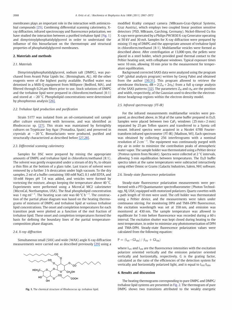
Simultaneous small (SAX) andwide (WAX) angle X-ray diffractionmeasurements were carried out as described previously [29] using a
Fig. 1. The chemical structure of Rhodococcus sp. trehalose lipid.
modified Kratky compact camera (MBraum-Graz-Optical Systems,Graz Austria), which employs two coupled linear position sensitivedetectors (PSD, MBraum, Garching, Germany). Nickel-filtered Cu KαX-rayswere generated by a Philips PW3830X-rayGenerator operatingat 50 kV and 30 mA. Samples for X-ray diffraction were prepared bymixing 15 mg of DMPG and the appropriate amount of trehalose lipidin chloroform/methanol (8:1). Multilamellar vesicles were formed asdescribed above. After centrifugation at 13,000 rpm, the pellets wereplaced in a steel holder, which provided good thermal contact to thePeltier heating unit, with cellophane windows. Typical exposure timeswere 10 min, allowing 10 min prior to the measurement for temper-ature equilibration.
Background corrected SAXS data were analyzed using the programGAP (global analysis program) written by Georg Pabst and obtainedfrom the author [30,31]. This program allowed to retrieve themembrane thickness, dB=2(ZH+2σH) from a full q-range analysisof the SAXS patterns [32]. The parameters ZH and σH are the positionand width, respectively, of the Gaussian used to describe the electron-dense headgroup regions within the electron density model.
2.5. Infrared spectroscopy (FT-IR)
For the infrared measurements multilamellar vesicles were pre-pared, as described above, in 50 μl of the same buffer prepared in D2O.Samples were placed between two CaF2 windows (25 mm×2 mm)separated by 25 μm Teflon spacers and transferred to a Symta cellmount. Infrared spectra were acquired in a Nicolet 6700 Fourier-transform infrared spectrometer (FT-IR) (Madison,WI). Each spectrumwas obtained by collecting 256 interferograms with a nominalresolution of 2 cm−1. The equipment was continuously purged withdry air in order to minimize the contribution peaks of atmosphericwater vapor. The sample holderwas thermostated using a Peltier device(Proteus system from Nicolet). Spectra were collected at 2 °C intervals,allowing 5 min equilibration between temperatures. The D2O bufferspectra taken at the same temperatures were subtracted interactivelyusing either Omnic or Grams (Galactic Industries, Salem, NH) software.
2.6. Steady-state fluorescence polarization
Steady-state fluorescence polarization measurements were per-formed with a PTI Quantamaster spectrofluorometer (Photon Technol-ogy, NJ, USA) equippedwithmotorized polarizers. Quartz cuvettes witha path length of 10 mm were used. The cell holder was thermostatedusing a Peltier device, and the measurements were taken undercontinuous stirring. For monitoring DPH and TMA-DPH fluorescence,the excitation wavelength was set at 358 nm, and emission wasmonitored at 430 nm. The sample temperature was allowed toequilibrate for 5 min before fluorescence was recorded during a 60 sinterval. The excitation shutter was kept closed during heating to thenext temperature, in order to minimize any photoisomerization of DPHand TMA-DPH. Steady-state fluorescence polarization values werecalculated from the following equation:
P = IVV−GIVHð Þ= IVV + GIVHð Þ
where IVV and IVH are the fluorescence intensities with the excitationpolarizer oriented vertically and the emission polarizer orientedvertically and horizontally, respectively. G is the grating factor,calculated as the ratio of the efficiencies of the detection system forvertically and horizontally polarized light, and is equal to IHV/IHH.
4. Results and discussion
The heating thermograms corresponding to pure DMPG and DMPG/trehalose lipid systems are presented in Fig. 2. The thermogram of pureDMPG shows two transitions attributed to the weakly energetic
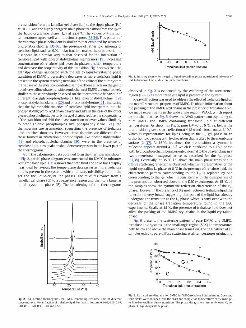
Fig. 3. Enthalpy change for the gel to liquid-crystalline phase transition of mixtures ofDMPG/trehalose lipid at different molar fractions.
2069A. Ortiz et al. / Biochimica et Biophysica Acta 1808 (2011) 2067–2072
pretransition from the lamellar-gel phase (Lβ′) to the ripple phase (Pβ′)at 10.2 °C and the highly energeticmain phase transition from the Pβ′ tothe liquid-crystalline phase (Lα) at 22.4 °C. The values of transitiontemperatures agree well with previous reports [33,34]. This pattern ofthermotropic phase behaviour is similar to that exhibited by saturatedphosphaticylcholines [35,36]. The presence of rather low amounts oftrehalose lipid, such as 0.02 molar fraction, makes the pretransition todisappear, in a similar way to that observed for the interaction oftrehalose lipid with phosphatidylcholine membranes [19]. Increasingconcentrationsof trehalose lipid lower thephase transition temperatureand decrease the cooperativity of this transition. Fig. 3 shows that theenthalpy change associated with the gel to liquid-crystalline phasetransition of DMPG progressively decreases as more trehalose lipid ispresent in the system reaching near 40% of the value of the pure systemin the case of the most concentrated sample. These effects on the gel toliquid-crystalline phase transition endothermof DMPG are qualitativelysimilar to those previously observed on the thermotropic behaviour ofdifferent diacylglycerophospholipids like phosphatidylcholine [19],phosphatidylethanolamine [20] and phosphatidylserine [21], indicatingthat the hydrophobic moieties of trehalose lipid incorporate into thephosphatidylglycerol acyl chain bilayer and, like in the case of the otherglycerophospholipids, perturb the acyl chains, reduce the cooperativityof the transition and shift the phase transition to lower values. Similarlyto other anionic phospholipids like phosphatidylserine [21], thethermograms are asymmetric, suggesting the presence of trehaloselipid enriched domains. However, these domains are different fromthose formed in zwitterionic phospholipids like phosphatidylcholine[19] and phosphatidylethanolamine [20] were, in the presence oftrehalose lipid, newpeaks or shoulderswere present in the lower part ofthe thermograms.
From the calorimetric data obtained form the thermograms shownin Fig. 2, partial phase diagramwas constructed for DMPG in mixtureswith trehalose lipid. Fig. 4 shows that both fluid and solid lines displaynear ideal behaviour, the temperature decreasing as more trehaloselipid is present in the system, which indicates miscibility both in thegel and the liquid-crystalline phases. The mixtures evolve from alamellar gel phase (G) to a coexistence region and then to a lamellarliquid-crystalline phase (F). The broadening of the thermograms
Fig. 2. DSC heating thermograms for DMPG containing trehalose lipid at differentconcentrations. Molar fraction of trehalose lipid from top to bottom: 0, 0.02, 0.05, 0.07,0.10, 0.15, 0.20, 0.30, 0.40 and 0.50.
observed in Fig. 2 is evidenced by the widening of the coexistenceregion (G+F) as more trehalose lipid is present in the system.
X-ray diffractionwas used to address the effect of trehalose lipid onthe overall structural properties of DMPG. To obtain information aboutthe packing of the DMPG acyl chains in the presence of trehalose lipid,we made experiments in the wide angle region (WAX), which reporton the chain lattice. Fig. 5 shows the WAX pattern corresponding topure DMPG and DMPG containing trehalose lipid at differenttemperatures. As shown in Fig. 5, pure DMPG at 6 °C, i.e. below thepretransition, gives a sharp reflectionat4.18 Åandabroadoneat4.10 Å,which is representative for lipids being in the Lβ′ gel phase in anorthorhombic packingwith hydrocarbon chains tilted to themembranesurface [24,37]. At 15 °C, i.e. above the pretransition, a symmetricreflection appears around 4.15 Å which is attributed to a lipid phasewith hydrocarbon chains being oriented normal to the bilayer plane in atwo-dimensional hexagonal lattice as described for the Pβ′ phase[37,38]. Eventually, at 35 °C, i.e. above the main phase transition, adiffuse scattering reflection is observed, which is representative for theliquid-crystalline Lα phase. At 6 °C, in the presence of trehalose lipid, thecharacteristic pattern corresponding to the Lβ′ is replaced by onecorresponding to the Pβ′, which is consistent with the disappearing ofthe pretransition observed above in the DSC experiments. At 15 °C, allthe samples show the symmetric reflection characteristic of the Pβ′phase. However in the presence of 0.2 mol fraction of trehalose lipid thereflection is very broad, suggesting that part of the lipid has alreadyundergone the transition to the Lα phase, which is consistent with thedecrease of the phase transition temperature found in the DSCexperiments. Finally at 35 °C, the presence of trehalose lipid does notaffect the packing of the DMPG acyl chains in the liquid-crystallinephase.
Fig. 6 presents the scattering pattern of pure DMPG and DMPG/trehalose lipid systems in the small angle region (SAX) at temperaturesboth below and above the main phase transition. The SAX pattern of allsamples exhibits pure diffuse scattering at all temperatures originating
Fig. 4. Partial phase diagrams for DMPG in DMPG/trehalose lipid mixtures. Open andsolid circles were obtained from the onset and completion temperatures of themain gelto liquid-crystalline phase transition. The phase designations are as follows: G, gelphase; F, liquid-crystalline phase.

Fig. 5.Wide angle X-ray diffraction profiles of DMPG system containing different concentrations of trehalose lipid at different temperatures. From top to bottom: pure DMPG, DMPGcontaining 0.05 mol fraction trehalose lipid, and DMPG containing 0.20 mol fraction trehalose lipid.
2070 A. Ortiz et al. / Biochimica et Biophysica Acta 1808 (2011) 2067–2072
from positionally uncorrelated bilayers. This can be explained by theoverall negative surface charge that leads to the formation ofpositionally uncorrelated bilayers,most likely large unilamellar vesicles,because of electrostatic repulsion [33,39]. The analysis of the SAXpatternsusing the global analysis program(GAP)allowedus to calculatethemembrane thickness (dB) of the different systems. For pure DMPG amembrane thickness of 50.0 Å was calculated for the gel phase and44.7 Å for the liquid-crystalline phase (these values being in agreementwith previous reports [24,40]), while a decrease ofmembrane thicknessboth in the gel (47.8 Å) and the liquid-crystalline phase (43.0 Å) wasfound for the system containing 0.2 molar fraction of trehalose lipid.
The infrared spectra of glycerophospholipid contain usefulinformation regarding the intermolecular interactions that occur inthe different domains of the molecule. In this way, the CH2 stretchingregion includes information about the conformational disposition ofthe hycrocarbon chains, whereas the C=O stretching region includesinformation about lipid interfacial hydration–hydrogen bondinginteraction. The CH2 symmetric stretching band near 2850 cm−1 is ofremarkable importance because of its sensitivity to changes in themobility and in the conformational disorder of the hydrocarbon chains[41]. Fig. 7 presents the temperature dependence of the frequency at theabsorbance maximum of the symmetric CH2 stretching vibration bandof the infrared spectra of pureDMPGandDMPG/trehalose lipid systems.For pure DMPG the data show a discontinuous increase of about 2 cm−1
in the frequency of the bandmaximum at a temperature that coincideswith the gel to liquid-crystalline phase transition, in agreement withprevious results [34]. This frequency increase is characteristic of thechainmelting transitionof hydratedphospholipids [42] and results fromthe increased conformational disorder in the hydrocarbon as aconsequence of the introduction of a high population of gaucheconformers [43]. In the presence of low concentrations of trehaloselipid the chainmelting phase transition is broadened and starts at lower
Fig. 6. Experimental (open symbols) and best fitting (bold lines) small angle X-ray diffracdifferent temperatures. From top to bottom: pure DMPG, DMPG containing 0.05 mol fractio
temperatures than that of pure DMPG, in accordance with the DSCexperiments shown above. At high trehalose lipid concentration afurther broadening and shift of the transition to lower temperatures isobserved together with an increase of the frequency of the band bothbelow and above the phase transition, the latter indicating and increasein gauche conformers and thus an increase in hydrocarbon chainconformational disorder both in the gel and in the liquid-crystallinephase. A similar increase in hydrocarbon chain conformational disorderwas found in the case of phosphatidylserine [21], and suggests that thedecrease of about 2 Å in the bilayer thickness that we found in the SAXmeasurements shown above could be due to an effective increase in thegauche conformers leading to an increase in conformational disorder.
TheC=Ostretchingbandof pureDMPG is a fairly broadband around1750–1700 cm−1, and seems to be a summation of subcomponentscentered near 1741 and 1727 cm−1[34]. The relative intensities of thesecomponent bands reflect the contribution of subpopulations of dehy-drated and hydrated carbonyl groups [44]. Fig. 8 shows the temperaturedependence of the frequency at the absorbance maximum of the C=Ostretching band of the infrared spectra of pure DMPG and DMPG/trehalose lipid systems. For pure DMPG, the gel to liquid-crystallinephase transition produced a shift of the maximum frequency to lowervalues, reflecting the increase in intensity of the underlying componentband at 1727 cm−1, attributed to a higher amount of hydrogen bondedcarbonyl groups resulting from a phase state-induced increase in thehydration of the polar–apolar interface [34]. The broadening and shift ofthe phase transition to lower temperatures produced by the presence oftrehalose lipid can be also observed following themaximumof the C=Oband depicted in Fig. 8. It is interesting to note that the presence of highconcentration of trehalose lipid produced a shift of the maximum of theC=O band to higher frequencies as compared to pure phospholipid,both in the gel and liquid-crystalline phases. This increase in frequencyindicates an increase in the proportion of dehydrated C=O component
tion profiles of DMPG system containing different concentrations of trehalose lipid atn trehalose lipid, DMPG containing 0.20 mol fraction trehalose lipid.
Fig. 7. Temperature dependence of the maximum of the symmetric CH2 stretchingabsorption band exhibited by pure DMPG (●) and DMPG/trehalose lipid mixtures at0.05 (○) and 0.20 (□) molar fraction.
Fig. 9. Steady state fluorescence polarization as a function of temperature of DPH (A)and TMA-DPH (B) incorporated into membranes composed of pure DMPG (●) andDMPG/trehalose lipid mixtures at 0.05 (○) and 0.20 (□) molar fraction.
2071A. Ortiz et al. / Biochimica et Biophysica Acta 1808 (2011) 2067–2072
at all temperatures and suggests that trehalose lipid interacts with theinterfacial region of the DMPG bilayer, decreasing the hydrogen bondingof the C=O groupswith thewatermolecules of the hydration layer. Thelatter is in line with the dehydrating effect of trehalose lipid observed inother diacylglycerophospholipids systems [19–21] whichwas attributedto the ability of free hydroxyl groups of trehalose lipid to participate inhydrogen bonds, leaving less water molecules available to interact withthe phospholipids. It seems that the presence of trehalose lipid producesdifferent effects on the conformational order of the DMPG molecule: itcauses conformational disorder in the methylene region while de-hydrates the interfacial region, which is correlated with a decrease oflipid mobility [45].
Fluorescence polarization measurements, using DPH and TMA-DPHprobes,were carried out to report changes inmembrane order both at thedeep inner phospholipid bilayer and at the interfacial region of themembrane, respectively. Fig. 9 shows the fluorescence polarization ofDPH (Fig. 9A) and TMA-DPH (Fig. 9B) incorporated into pure DMPGsystemsand those containing trehalose lipid, as a functionof temperature.The sharp drop in polarization values detected in the pure DMPG samplereflects the decrease in membrane order taking place during the gel toliquid-crystalline phase transition. The presence of trehalose lipid causeda broadening and shifting of the transition to lower temperatures, in linewith the evidence shown above. Interestingly, at temperatures below thephase transition,when thephospholipids are in theorderedgel phase, thepresence of trehalose lipid decreases the polarization values of bothprobes indicatingadecrease in themembraneorder, andemphasizing theperturbation of the methylene region exerted by the biosurfactant.However, at temperatures above the phase transition, when thephospholipids are in the disordered liquid-crystalline phase, the presenceof trehalose lipid produces a small increase in the polarization valuesindicating an ordering effect of the fluid phase. This latter effect is moreprominent in the case of the TMA-DPH probe which is located near theaqueous interface, and underlines the influence of the dehydrating effectof trehalose lipid on the fluid DMPG membrane.
Fig. 8. Temperature dependence of the maximum of the carbonyl stretching absorptionband exhibited by pure DMPG (●) and DMPG/trehalose lipid mixtures at 0.05 (○) and0.20 (□) molar fraction.
5. Conclusions
The aim of this work was to characterize the interactions between atrehalose lipid biosurfactant and phosphatidylglycerol membranes. OurDSC data supported that trehalose lipid is able to incorporate intoDMPGmembranes and to intercalate between the phospholipids molecules,where it can reduce the cooperativity and lower the transitiontemperature of the gel to liquid-crystalline phase transition. The partialphase diagram showed good miscibility between trehalose lipid andDMPG, both in the gel and liquid-crystalline phases. X-ray diffractionmeasurements indicated that trehalose lipid did not affect themacroscopic bilayer organization of DMPG, but the presence of thebiosurfactant produced a small decrease of the bilayer thickness.Infrared experiments revealed that the biosurfactant increased thefluidity of the phospholipids acyl chains and decreased the hydration ofthe interfacial region of the membrane. Finally, fluorescence polariza-tion of membrane probes provided evidence that trehalose lipiddisordered the DMPG membrane in the gel phase while producing asmall ordering effect in the liquid-crystalline phase. In view of thecrucial structural and functional importance of phosphatidylglycerol inmembranes, particularly bacterial ones, and the broad diversity ofbiological actions played by trehalose lipids, the results presented herecontribute to the knowledge of the molecular mechanisms underlyingthemembrane-related biological actions of this bacterial trehalose lipidbiosurfactant.
Acknowledgement
This work was supported by project No. CTQ2007-66244 from theSpanish Ministry of Science and Innovation (MCINN).
References
[1] S. Lang, Biological amphiphiles (microbial biosurfactants), Curr. Opin. ColloidInterface Sci. 7 (2002) 12–20.
[2] E.Z. Ron, E. Rosenberg, Natural roles of biosurfactants, Environ. Microbiol. 3(2002) 229–236.
[3] A. Ortiz, J.A. Teruel, M.J. Espuny, A. Marqués, A. Manresa, F.J. Aranda, Effects ofdirhamnolipid on the structural properties of phosphatidylcholine membranes,Int. J. Pharm. 325 (2006) 99–107.
[4] M. Sánchez, J.A. Teruel, M.J. Espuny, A. Marqués, F.J. Aranda, M. Sánchez, J.A.Teruel, M.J. Espuny, A. Marqués, A. Manresa, M. Sánchez, J.A. Teruel, M.J. Espuny, A.Marqués, A. Ortiz, Modulation of the physical properties of dielaidoylphos-phatidylethanolamine membranes by a dirhamnolipid biosurfactant produced byPseudomonas aeruginosa, Chem. Phys. Lipids 142 (2006) 118–127.
2072 A. Ortiz et al. / Biochimica et Biophysica Acta 1808 (2011) 2067–2072
[5] C. Asselineau, J. Asselineau, Trehalose-containing glycolipids, Prog. Chem. FatsOther Lipids 16 (1978) 59–99.
[6] S. Lang, J.C. Philp, Surface-active compounds in rhodococci, Antonie VanLeeuwenhoek 74 (1998) 59–70.
[7] I.M. Banat, R.S. Makkar, S.S. Cameotra, Potential commercial application ofmicrobial surfactant, Appl. Microbiol. Biotechnol. 53 (2000) 495–508.
[8] A. Franzretti, I. Gandolfi, G. Bestetti, T.J.P. Smyth, I.M. Banat, Production andapplicationsof trehalose lipid biosurfactants, Eur. J. Lipid Sci. Technol. 112 (2010) 617–627.
[9] M. Azuma, T. Suzutani, K. Sazaki, I. Yoshida, T. Sakuma, T. Yoshida, Role ofinterferon in the augmented resistance of trehalose 6,6′-dimycolate-treated miceto influenza virus infection, J. Gen. Virol. 68 (1987) 835–843.
[10] Md. Hoq, M. Suzutani, T. Toyoda, T. Horijke, G. Yoshida, I.M. Azuma, Role of γδ TCRlymphocytes in the augmented resistance of trehalose 6,6′-dimycolate-treatedmice to influenza virus infection, J. Gen. Virol. 78 (1997) 1597–1603.
[11] H. Isoda, H. Shinmoto, D. Kitamoto, M. Matsumura, T. Nakahara, Succinoyltrehalose lipid induced differentiation of human monocytoid leukemic cell lineU937 into monocyte-macrophages, Cryotechnology 19 (1996) 79–88.
[12] H. Isoda, H. Shinmoto, D. Kitamoto, M. Matsumura, T. Nakahara, Differentiation ofhuman promyelocytic leukaemia cell line HL60 by microbial extracellularglycolipids, Lipids 32 (1997) 263–271.
[13] T. Sudo, X. Zhao, Y. Wakamatsu, M. Shibahara, N. Nomura, T. Nakahara, A. Suzuki,Y. Kobayashi, C. Jin, T. Murata, K.K. Yokohama, Induction of the differentiation ofhuman HL60 promyelocytic leukaemia cell line by succinoyl trehalose lipids,Cryotechnology 33 (2000) 259–264.
[14] H. Isoda, D. Kitamoto, H. Shinmoto, M. Matsumura, T. Nakahara, Microbialextracellular glycolipid induction of differentiation and inhibition of the proteinkinase C activity of human promyelocytic leukemia cell line HL60, Biosci.Biotechnol. Biochem. 61 (1997) 609–614.
[15] M.S. Kuyukina, I.B. Ivshina, S.V. Gein, T.A. Baeva, V.A. Chereshnev, In vitroimmunomodulating activity of biosurfactant glycolipid complex from Rhodococ-cus rubber, Bull. Exp. Biol. Med. 144 (2007) 326–330.
[16] E. Vollbrecht, U. Ra, S. Lang, Microbial conversion of vegetable oils into surface-active di-, tri-, and tetrasaccharide lipids (biosurfactants) by bacterial strainTsukamurella spec, Lipids 101 (1999) 389–394.
[17] A. Zaragoza, F.J. Aranda, M.J. Espuny, J.A. Teruel, A. Marqués, A. Manresa, A. Ortiz,Mechanism of membrane permeabilization by a bacterial trehalose lipidbiosurfactant produced by Rhodococcus sp. Langmuir 21 (2009) 7892–7898.
[18] A. Zaragoza, F.J. Aranda, M.J. Espuny, J.A. Teruel, A. Marqués, A. Manresa, A. Ortiz,Hemolytic activity of a bacterial trehalose lipid biosurfactant produced by Rhodococcussp.: evidence for a colloid-osmotic mechanism, Langmuir 26 (2010) 8567–8572.
[19] F.J. Aranda, J.A. Teruel, M.J. Espuny, A. Marqués, A. Manresa, E. Palacios-Lidón, A. Ortiz,Domain formation by a Rhodococcus sp. biosurfactant trehalose lipid incorporated intophosphatidylcholine membranes, Biochim. Biophys. Acta 1768 (2007) 2596–2604.
[20] A. Ortiz, J.A. Teruel, M.J. Espuny, A. Marqués, A. Manresa, F.J. Aranda, Interactionsof a Rhodococcus sp. biosurfactant trehalose lipid with phosphatidylethanolaminemembranes, Biochim. Biophys. Acta 1778 (2008) 2806–2813.
[21] A. Ortiz, J.A. Teruel, M.J. Espuny, A. Marqués, A. Manresa, F.J. Aranda, Interactionsof a bacterial biosurfactant trehalose lipid with phosphatidylserine membranes,Chem. Phys. Lipids 158 (2009) 46–53.
[22] K.K. Eklund, P.K. Kinnunen, Effects of polyamines on the thermotropic behaviourof dipalmitoylphosphatidylglycerol, Chem. Phys. Lipids 39 (1986) 109–117.
[23] J.M. Alakoskela, P.K. Kinnunen, Thermal phase behavior of DMPG: the exclusion ofcontinuous network and dense aggregates, Langmuir 10 (2007) 4203–4213.
[24] G. Pabst, S.L. Grage, S. Danner-Pongratz, W. Jing, A.S. Ulrich, A. Watts, K. Lohner, A.Hickel, Membrane thickening by the antimicrobial peptide PGLa, Biophys. J. 95(2008) 5779–5788.
[25] K. Lohner, The role of membrane lipid composition in cell targeting ofantimicrobial peptides, in: K. Lohner (Ed.), Development of Novel AntimicrobialAgents: Emerging Strategies, Horizon Scientific Press, Wymondham, Norfolk, UK,2001, pp. 149–165.
[26] C.J.F. Böttcher, C.M. Van Gent, C. Pries, A rapid and sensitive sub-micro phosphorusdetermination, Anal. Chim. Acta 24 (1961) 203–204.
[27] M.J. Espuny, S. Egido, M.E. Mercadé, A. Manresa, Characterization of trehalosetetraester produced by a waste lube oil degrader Rhodococcus sp 51T7, Toxicol.Environ. Chem. 48 (1995) 83–88.
[28] M.J. Espuny, S. Egido, I. Rodón, A. Manresa, M.E. Mercadé, Nutricional re-quirements of a biosurfactant producing strain Rhodococcus sp 51T7, Biotechnol.Letters 18 (1996) 521–526.
[29] J.A. Teruel, A. Ortiz, F.J. Aranda, Influence of organotin compounds onphosphatidylserine membranes, Appl. Organometal. Chem. 18 (2004) 111–116.
[30] G. Pabst, M. Rappolt, H. Amenitsch, P. Laggner, Structural information frommultilamellar liposomes at full hydration: full q-range fitting with high quality X-ray data, Phys. Rev. E 62 (2000) 4000–4009.
[31] G. Pabst, R. Koschuch, B. Pozo-Navas, M. Rappolt, K. Lohner, P. Laggner, Structuralanalysis of weakly ordered membranes stacks, J. Appl. Crystallogr. 63 (2003)378–1388.
[32] G. Pabst, Global properties of biomimetic membranes on molecular features,Biophys. Rev. Lett. 1 (2006) 57–84.
[33] G. Pabst, S. Danner, S. Karmakar, G. Deutsch, V.A. Raghunathan, On the propensityof phosphatidylglycerols to form interdigitated phases, Biophys. J. 93 (2007)513–525.
[34] Y.-P. Zhang, R.N.A.H. Lewis, R.N. McElhaney, Calorimetric and spectroscopic studies ofthe thermotropic phase behaviour of the n-saturated 1,2-diacylphosphatidylglycerols,Biophys. J. 72 (1997) 779–793.
[35] R.N.A.H. Lewis, N. Mak, R.N. McElhaney, Differential scanning calorimetry of thethermotropic phase behaviour of model membranes composed of phosphatidyl-cholines containing linear saturated fatty acyl chains, Biochem. 26 (1987)6118–6126.
[36] R.N.A.H. Lewis, Y.-P. Zhang, R.N. McElhaney, Calorimetric and spectroscopicstudies of the phase behaviour and organization of lipid bilayer modelmembranes composed of binary mixtures of dimirystoylphosphatidylcholineand dimiristoylphosphatidylglycerol, Biochim. Biophys. Acta 1668 (2005)203–214.
[37] A. Tardieu, V. Luzzati, F.C. Reman, Structure and polymorphism of thehydrocarbon chains of lipids: a study of lecithin-water phases, J. Mol. Biol. 75(1973) 711–733.
[38] K. Lohner, A. Latal, G. Degovics, P. Garidel, Packing characteristics of a modelsystem mimicking cytoplasmic bacterial membranes, Chem. Phys. Lipids 111(2001) 177–192.
[39] H. Hauser, Some aspects of the phase behaviour of charged lipids, Biochim.Biophys. Acta 772 (1984) 37–50.
[40] R.M. Fernandez, K.A. Riske, L.A. Amaral, R. Itri, M.T. Lamy, Influence of salt on thestructure of DMPG studied by SAXS and optical microscopy, Biochim. Biophys.Acta 1778 (2008) 907–916.
[41] R. Mendelson, H.H. Mantsch, Fourier transform infrared studies of lipid–proteininteraction, in: A. Watts, J.J.H.H.M. De Pont (Eds.), Progress in Lipid ProteinInteractions, vol. 2, Elsevier, New York, 1986, pp. 103–146.
[42] H.H. Mantsch, R.N. McElhaney, Phospholipid phase transition in model andbiological membranes as studied by infrared spectroscopy, Chem. Phys. Lipids 57(1991) 213–226.
[43] R.N.A.H. Lewis, R.N. McElhaney, Fourier transform infrared spectroscopy in thestudy of hydrated lipids and lipid bilayer membranes, in: H.H. Mantsch, D.Chapman (Eds.), Infrared Spectroscopy of Biomolecules, Wiley-Liss, New York,1996, pp. 159–202.
[44] A. Blume, W. Hübner, G. Messner, Fourier transform infrared spectroscopy of13C=O-labeled phospholipids hydrogen bonding to carbonyl groups, Biochem-istry 27 (1988) 8239–8249.
[45] A.S. Ulrich, F. Volke, A. Watts, The dependence of phospholipids head-groupmobility on hydration as studied by deuterium-NMR spin-lattice relaxation timemeasurements, Chem. Phys. Lipids 55 (1990) 64–66.


![Fluorescence properties of a potential antitumoral benzothieno[3,2-b]pyrrole in solution and lipid membranes](https://static.fdokumen.com/doc/165x107/63440ec5df19c083b1076b23/fluorescence-properties-of-a-potential-antitumoral-benzothieno32-bpyrrole-in.jpg)

