Effects of 8-week strength training with two models of chest press machines on muscular activity...
10
Effects of 8-week strength training with two models of chest press machines on muscular activity pattern and strength Angelo Cacchio a, * , Romildo Don a , Alberto Ranavolo a , Enrico Guerra b , Steven T. McCaw c , Rita Procaccianti a , Filippo Camerota a , Massimo Frascarelli a , Valter Santilli a a Department of Physical Medicine and Rehabilitation, School of Medicine, ‘‘La Sapienza’’ University of Roma, p.le A. Moro 5, 00185 Roma, Italy b LAMS Laboratory, University of Perugia, Via della Pallotta, Podere S. Pietro, 06100 Perugina, Italy c School of Kinesiology and Recreation, Biomechanics Laboratory, Illinois State University, Normal, IL 61790-5120, USA Received 31 May 2006; received in revised form 22 November 2006; accepted 1 December 2006 Abstract The purposes of this study were to assess: (i) the effects of 8-week training programs with constrained-path and unconstrained-path chest press machines on 1-RM; (ii) the different activity patterns of selected arm and shoulder girdle muscles during push movement performed on the different machines; (iii) the transfer of the training effects from one machine to the other. Twenty healthy, sedentary women (mean ± SD age, 24.8 ± 1.0 yrs), randomized to either the FM or CM strength training protocols were evaluated before and after the strength training program. Muscular activity signals were recorded by surface electromyography (sEMG) from eight muscles while each subject performed the exercise on each machine. Muscle strength was defined by a 1 repetition maximum (1-RM) test for each sub- ject on each machine. Both machines were effective in improving 1-RM, but the 1-RM increased more in the FM than the CM. Adaptive change in the sEMG was observed in all muscles after training on the FM machine, but not within the stabilizers when training on the CM machine. The results suggest that training in an unconstrained condition provides a more effective method for improving inter-mus- cular coordination via adaptation of the motor strategy aimed at optimising muscular efforts. Ó 2007 Elsevier Ltd. All rights reserved. Keywords: Electromyography; Kinematics; Resistance exercise; Muscle; Weight machines; Movement analysis; Biomechanics 1. Introduction Progressive resistance exercise is an effective method for developing muscular strength and is indicated for sports and fitness purposes as well as for injury prevention and rehabilitation (ACSM Position Stand, 1998). The physio- logical modifications resulting from strength training are due not only to muscle hypertrophy, but also to the prac- tice-induced neural adaptations to the kinematic and kinetic modalities of a given exercise (Sale, 1988; Enoka, 1997). Muscle strength has been defined as the maximal vol- untary muscle torque (Enoka, 1988) or, more recently, as the ability of the neuromuscular system to produce force (Siff, 2001). Muscle strength is often assessed by measur- ing a 1 repetition maximum (1-RM) to quantify the greatest resistance that can be lifted in a single concentric action (Simpson et al., 1997; Ploutz-Snyder and Giamis, 2002). Muscle activity patterns and coordination have been investigated by surface electromyography (sEMG) during strength training (Ha ¨kkinen and Komi, 1986), sports activity (Kelly et al., 2002), rehabilitation exercise (Hin- termeister et al., 1998), and more recently to determine whether the neural adaptations to resistance training 1050-6411/$ - see front matter Ó 2007 Elsevier Ltd. All rights reserved. doi:10.1016/j.jelekin.2006.12.007 * Corresponding author. Tel.: +39 06 491672/347 6753669. E-mail address: [email protected] (A. Cacchio). Available online at www.sciencedirect.com Journal of Electromyography and Kinesiology 18 (2008) 618–627 www.elsevier.com/locate/jelekin
-
Upload
independent -
Category
Documents
-
view
3 -
download
0
Transcript of Effects of 8-week strength training with two models of chest press machines on muscular activity...

Available online at www.sciencedirect.com
Journal of Electromyography and Kinesiology 18 (2008) 618–627
www.elsevier.com/locate/jelekin
Effects of 8-week strength training with two models of chestpress machines on muscular activity pattern and strength
Angelo Cacchio a,*, Romildo Don a, Alberto Ranavolo a, Enrico Guerra b,Steven T. McCaw c, Rita Procaccianti a, Filippo Camerota a, Massimo Frascarelli a,
Valter Santilli a
a Department of Physical Medicine and Rehabilitation, School of Medicine, ‘‘La Sapienza’’ University of Roma, p.le A. Moro 5, 00185 Roma, Italyb LAMS Laboratory, University of Perugia, Via della Pallotta, Podere S. Pietro, 06100 Perugina, Italy
c School of Kinesiology and Recreation, Biomechanics Laboratory, Illinois State University, Normal, IL 61790-5120, USA
Received 31 May 2006; received in revised form 22 November 2006; accepted 1 December 2006
Abstract
The purposes of this study were to assess: (i) the effects of 8-week training programs with constrained-path and unconstrained-pathchest press machines on 1-RM; (ii) the different activity patterns of selected arm and shoulder girdle muscles during push movementperformed on the different machines; (iii) the transfer of the training effects from one machine to the other. Twenty healthy, sedentarywomen (mean ± SD age, 24.8 ± 1.0 yrs), randomized to either the FM or CM strength training protocols were evaluated before and afterthe strength training program. Muscular activity signals were recorded by surface electromyography (sEMG) from eight muscles whileeach subject performed the exercise on each machine. Muscle strength was defined by a 1 repetition maximum (1-RM) test for each sub-ject on each machine. Both machines were effective in improving 1-RM, but the 1-RM increased more in the FM than the CM. Adaptivechange in the sEMG was observed in all muscles after training on the FM machine, but not within the stabilizers when training on theCM machine. The results suggest that training in an unconstrained condition provides a more effective method for improving inter-mus-cular coordination via adaptation of the motor strategy aimed at optimising muscular efforts.� 2007 Elsevier Ltd. All rights reserved.
Keywords: Electromyography; Kinematics; Resistance exercise; Muscle; Weight machines; Movement analysis; Biomechanics
1. Introduction
Progressive resistance exercise is an effective method fordeveloping muscular strength and is indicated for sportsand fitness purposes as well as for injury prevention andrehabilitation (ACSM Position Stand, 1998). The physio-logical modifications resulting from strength training aredue not only to muscle hypertrophy, but also to the prac-tice-induced neural adaptations to the kinematic andkinetic modalities of a given exercise (Sale, 1988; Enoka,1997).
1050-6411/$ - see front matter � 2007 Elsevier Ltd. All rights reserved.
doi:10.1016/j.jelekin.2006.12.007
* Corresponding author. Tel.: +39 06 491672/347 6753669.E-mail address: [email protected] (A. Cacchio).
Muscle strength has been defined as the maximal vol-untary muscle torque (Enoka, 1988) or, more recently, asthe ability of the neuromuscular system to produce force(Siff, 2001). Muscle strength is often assessed by measur-ing a 1 repetition maximum (1-RM) to quantify thegreatest resistance that can be lifted in a single concentricaction (Simpson et al., 1997; Ploutz-Snyder and Giamis,2002).
Muscle activity patterns and coordination have beeninvestigated by surface electromyography (sEMG) duringstrength training (Hakkinen and Komi, 1986), sportsactivity (Kelly et al., 2002), rehabilitation exercise (Hin-termeister et al., 1998), and more recently to determinewhether the neural adaptations to resistance training
1 Freemotion Fitness�, ICON Health & Fitness SpA, Via GiacomoPuccini 220, 06077 Ponte Felcino (PG), Italy. Tel.: +39 075 5910180; fax:+39 075 5910101. www.freemotionfitness.it.
A. Cacchio et al. / Journal of Electromyography and Kinesiology 18 (2008) 618–627 619
occur to a greater extent at cortical or subcortical sites inthe CNS, comparing the effects of resistance training onthe electromyographic (EMG) responses to transcranialmagnetic (TMS) and electrical (TES) stimulation (Carrollet al., 2002). Despite the absence of a linear correlationbetween the sEMG signal and force output in dynamicconditions (Farina et al., 2004), it is generally acceptedthat muscle force increases with an increase in sEMGamplitude (Hintermeister et al., 1998). Therefore, thesEMG can be used to assess the involvement of eachmuscle in the execution of a motor task to qualify theneuromuscular adaptation following a period of training(Sale, 1988; Enoka, 1997). The effects of strength trainingprograms on muscular strength and neuromuscular coor-dination are influenced by variables such as the character-istics of the instrumentation and the complexity of theexercise (Wilson, 1994).
Weight machines are considered a safe, effective andeasy-to-learn alternative to free weights (ACSM PositionStand, 2002), and are available in a variety of mechanicalmodels that allow the joint loads and muscular involve-ment to be adjusted to the objectives of the training pro-gram (Wilson, 1994).
Multi-joint exercises are considered to be more effectivethan mono-articular exercises for increasing muscularstrength by allowing different muscle groups to be recruitedand heavier loads to be lifted (ACSM Position Stand,1998). Multi-joint movements have numerous degrees offreedom (DOF) and can be performed using different kine-matic patterns, thereby varying the role of each musclegroup in the force output (Bernstein, 1967; Galloway andKoshland, 2002). The presence of machine constraintsreducing the DOF in multi-joint exercises can conceivablyinfluence muscle activity patterns by altering the muscleforces required for movement execution and stabilization,which affects the specificity and effectiveness of strengthtraining.
The chest press machine is a multi-joint exerciser thatactivates the pectoralis major, the anterior portion of thedeltoideus and the triceps brachii muscles to performshoulder horizontal adduction and elbow extension againstresistance. While some studies have assessed the effects ofbase stability on chest press exercise performance (Ander-son and Behm, 2004), and other studies have comparedexercises performed with the bench press machine andthose performed with free weights (Wilson et al., 1989;McCaw and Friday, 1994) no research has yet comparedthe characteristics of the chest press exercise performedwith constrained-path and unconstrained-path chest pressmachines.
The purposes of this study were to assess: (i) the effectsof 8-week training programs with constrained-path andunconstrained-path chest press machines on 1-RM; (ii)the activity levels of selected arm and shoulder girdle mus-cles during the push movement performed on the differentmachines; (iii) the transfer of the training effects from onemachine to the other.
2. Materials and methods
2.1. Subjects
Twenty sedentary novice-lifter healthy women served as sub-jects. No subject reported either a history of previous upperextremity injury or the presence of a systemic or neurologicaldisease. This study was approved by our local ethics committee,and written informed consent was obtained from each subjectbefore participation. Subjects were randomly assigned, by meansof a computer-based 1:1 randomization scheme, to either theunconstrained-path chest press group (UC group, n = 10) or aconstrained-path chest press group (CC group, n = 10). The age,height, and body mass of the women in the UC group were,respectively, (mean ± SD) 25.0 ± 0.8 yrs, 166.5 ± 5 cm and55.6 ± 4.2 kg, while those of the women in the CC group were,respectively, 24.6 ± 1.1 yrs, 168.2 ± 4.6 cm and 57.4 ± 3.7 kg. Allsubjects completed the training program.
2.2. Instrumentation
2.2.1. Training machines
Two models of chest press machines were used in this study.One was a chest press with a constrained handle path (TraditionalChest Press*,1 CM, Fig. 1a), while the other was a chest press withan unconstrained handle path (Freemotion Chest Press*,1 FM,Fig. 1b). Resistance on both machines is provided by a cable-weight stack assembly; one end of the cable is attached to a stackof 15 five kilogram plates and the other end to a lever arm, withthe load transmitted by a system of pulleys. On the FM machine,the handles are connected directly to the cable, and the user is freeto move the handles along an unconstrained path. By contrast, onthe CM machine the handles are connected to the cable through arigid connection system and the user must move the handles alonga fixed path constrained by the connection system.
2.2.2. Electromyographic, kinematic and video recordings
The sEMG signals were acquired with a sampling rate of1000 Hz using a 12-bit acquisition board, and amplified using aneight-channel telemetric transmission surface electromyograph(TELEMG System, BTS, Milano, Italy). The Common ModeReaction Ratio was 100 dB.
Kinematic data were acquired using an optoelectronic motionanalysis system (ELITE System, BTS, Milano, Italy) consisting ofeight infra-red ray cameras (100 Hz sampling rate) to detect themotion of retro-reflective markers placed on skin over the spinousprocess of the seventh cervical vertebrae, and, bilaterally, over theacromion process, lateral epicondyle of the elbow and the ulnarstyloid. For subsequent analysis of upper limb movement, Bio-mech software (BTS S.p.a., Milan, Italy) was used to reconstructthe marker 3D co-ordinates using the biomechanical model pro-posed by Rab et al. (2002).
2.2.3. 1-RM determination
A 1-RM test and a 6-repetition trial with kinematic and sEMGrecordings were performed by each subject 1 week before the startand 1 week after the end of the training period. The 1-RM wasdetermined on both machines using a protocol suggested by
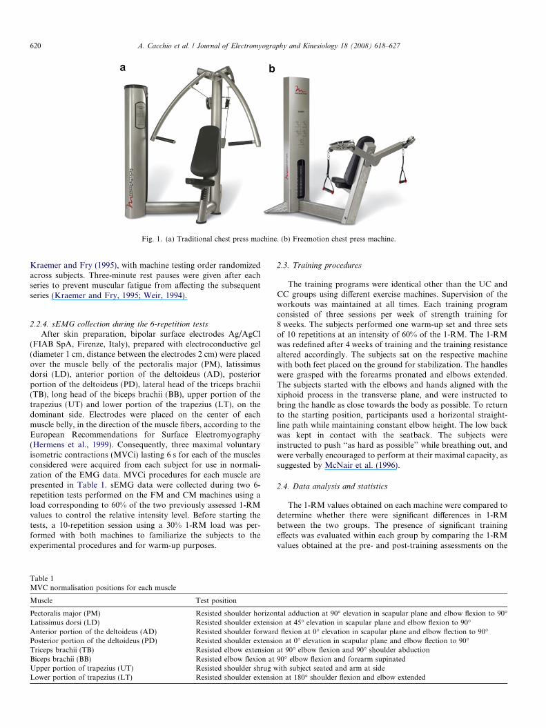
Fig. 1. (a) Traditional chest press machine. (b) Freemotion chest press machine.
620 A. Cacchio et al. / Journal of Electromyography and Kinesiology 18 (2008) 618–627
Kraemer and Fry (1995), with machine testing order randomizedacross subjects. Three-minute rest pauses were given after eachseries to prevent muscular fatigue from affecting the subsequentseries (Kraemer and Fry, 1995; Weir, 1994).
2.2.4. sEMG collection during the 6-repetition tests
After skin preparation, bipolar surface electrodes Ag/AgCl(FIAB SpA, Firenze, Italy), prepared with electroconductive gel(diameter 1 cm, distance between the electrodes 2 cm) were placedover the muscle belly of the pectoralis major (PM), latissimusdorsi (LD), anterior portion of the deltoideus (AD), posteriorportion of the deltoideus (PD), lateral head of the triceps brachii(TB), long head of the biceps brachii (BB), upper portion of thetrapezius (UT) and lower portion of the trapezius (LT), on thedominant side. Electrodes were placed on the center of eachmuscle belly, in the direction of the muscle fibers, according to theEuropean Recommendations for Surface Electromyography(Hermens et al., 1999). Consequently, three maximal voluntaryisometric contractions (MVCi) lasting 6 s for each of the musclesconsidered were acquired from each subject for use in normali-zation of the EMG data. MVCi procedures for each muscle arepresented in Table 1. sEMG data were collected during two 6-repetition tests performed on the FM and CM machines using aload corresponding to 60% of the two previously assessed 1-RMvalues to control the relative intensity level. Before starting thetests, a 10-repetition session using a 30% 1-RM load was per-formed with both machines to familiarize the subjects to theexperimental procedures and for warm-up purposes.
Table 1MVC normalisation positions for each muscle
Muscle Test position
Pectoralis major (PM) Resisted shoulder horizonLatissimus dorsi (LD) Resisted shoulder extensiAnterior portion of the deltoideus (AD) Resisted shoulder forwarPosterior portion of the deltoideus (PD) Resisted shoulder extensiTriceps brachii (TB) Resisted elbow extensionBiceps brachii (BB) Resisted elbow flexion atUpper portion of trapezius (UT) Resisted shoulder shrug wLower portion of trapezius (LT) Resisted shoulder extensi
2.3. Training procedures
The training programs were identical other than the UC andCC groups using different exercise machines. Supervision of theworkouts was maintained at all times. Each training programconsisted of three sessions per week of strength training for8 weeks. The subjects performed one warm-up set and three setsof 10 repetitions at an intensity of 60% of the 1-RM. The 1-RMwas redefined after 4 weeks of training and the training resistancealtered accordingly. The subjects sat on the respective machinewith both feet placed on the ground for stabilization. The handleswere grasped with the forearms pronated and elbows extended.The subjects started with the elbows and hands aligned with thexiphoid process in the transverse plane, and were instructed tobring the handle as close towards the body as possible. To returnto the starting position, participants used a horizontal straight-line path while maintaining constant elbow height. The low backwas kept in contact with the seatback. The subjects wereinstructed to push ‘‘as hard as possible’’ while breathing out, andwere verbally encouraged to perform at their maximal capacity, assuggested by McNair et al. (1996).
2.4. Data analysis and statistics
The 1-RM values obtained on each machine were compared todetermine whether there were significant differences in 1-RMbetween the two groups. The presence of significant trainingeffects was evaluated within each group by comparing the 1-RMvalues obtained at the pre- and post-training assessments on the
tal adduction at 90� elevation in scapular plane and elbow flexion to 90�on at 45� elevation in scapular plane and elbow flexion to 90�d flexion at 0� elevation in scapular plane and elbow flection to 90�on at 0� elevation in scapular plane and elbow flection to 90�at 90� elbow flexion and 90� shoulder abduction90� elbow flexion and forearm supinatedith subject seated and arm at side
on at 180� shoulder flexion and elbow extended
A. Cacchio et al. / Journal of Electromyography and Kinesiology 18 (2008) 618–627 621
machine used(u) for training (1-RMupre vs. 1-RMu
postÞ, i.e. FMmachine for the UC group and CM machine for the CC group.The training effects were calculated as the percentage variation ofthe 1-RM value (D%1-RMu ¼ ð1-RMu
post � 1-RMupreÞ * 100/
1-RMupreÞ.
The significance of the transfer of the training effects wasevaluated within each group by comparing the 1-RM valuesobtained on the machine not used(nu) for training at the pre- andpost-training assessment ð1-RMnu
pre vs. 1-RMnupostÞ, i.e. CM machine
for the UC group and FM machine for the CC group. The degreeof transfer was calculated as the ratio between the percentagevariations of 1-RM values observed on the machine not used fortraining (D%1-RMnu) and those on the machine used for training(D%1-RMu), i.e. D% Ratio = D%1-RMnu/D%1-RMu. Transfer ofthe training effects between groups was defined as the D% Ratiovalues in the UC group and CC group.
The presence of significant differences in the effectiveness of themovement executed on the two machines within each group wasascertained at each assessment by comparing the 1-RM valuesobtained on the FM and CM machines (1-RMFM vs. 1-RMCM).Any difference in effectiveness was determined by calculating theratio between the 1-RM values obtained on the FM machine andthose obtained on the CM machine (1-RM Ratio=1-RMFM/1-RMCM). The presence of any significant variation in the 1-RMRatio between the pre- and post-training assessments within eachgroup, as well as the presence of significant differences in the 1-RM Ratio between groups at each assessment were alsoinvestigated.
2.4.1. Kinematic and EMG evaluation during 6-repetition tests
For the kinematic and sEMG assessment of the 6-repetitiontests, the first and sixth repetitions were discarded to eliminatemovement variability due to initiation and termination of theexercise.
The movements of the ulnar styloid marker (USm) were ana-lyzed to identify the push phases of the exercise. We defined thepush phase onset as the time when the USm exceeded thethreshold velocity of 0.025 m/s in the direction of the pushmovement for three consecutive samples. The push phase wasdefined to end when the USm velocity passed from positive tonegative values.
The range of shoulder horizontal adduction and elbowextension in each push phase was calculated according to themethod of Rab (Rab et al., 2002), using YZ 0Y
00as the shoulder
rotation sequence; coefficients of variation (CV) of the height ofthe USm and lateral epicondyle of the elbow marker (LEm) werecalculated to ensure that a participant utilized a similar overallmovement pattern cross trials. The duration of the push phasewas also calculated for the individual repetitions. Post-processingof the sEMG signals was performed by means of Myolab (BTSSpA, Milan, Italy). sEMG signals were filtered through a Ham-ming filter with lower and upper cut-off frequencies of 10 Hz and400 Hz, respectively. The root mean square (RMS) values of thesEMG signals recorded during the push phases were calculatedfor consecutive periods of 50 ms, and averaged across the pushphase. In order to allow comparisons within- and between-sub-jects, data from each muscle were expressed as a percentage of themaximum value obtained during the MVCi. Muscle activity wascategorized using four groups of percentages, as proposed byKelly et al. (2002). sEMG data in the range of 0–20% were con-sidered to indicate minimal activity, 21–40% moderate activity,41–60% high activity, and 61–100% very high activity.
2.4.2. Statistics
Statistical analysis was performed using the SAS 8.2 (SASInstitute Inc., Cary, NC, USA). Data normality was verified bymeans of the Shapiro–Wilk test. The mean value ± standarddeviation (SD) of each parameter was calculated in each group.For the 1-RM assessments, we used the paired and unpaired t testto evaluate within- and between-group differences, respectively.To investigate the presence of significant differences within eachgroup concerning the kinematic parameters and the normalizedRMS of each muscle, a two-way analysis of variance (ANOVA)with the test (FM vs. CM) as the within-assessment factor andtime (pre- vs. post-training) as the within-test factor was used. ATukey post hoc comparison was used to determine significantdifferences between mean values when significant main effects andinteraction were found. A significant a value of .05 was used forall the analyses.
3. Results
3.1. 1-RM training effects
At the pre-training assessment, there were no differ-ences in the 1-RMFM and 1-RMCM values between thetwo groups (Table 2). At the post-training assessment,the 1-RMFM and 1-RMCM values of the UC group werehigher than those of the CC group (1-RMFM:47.5 ± 4.9 kg vs. 25.5 ± 3.7 kg, p < 0.001; 1-RMCM:49.0 ± 6.1 kg vs. 41.0 ± 6.1 kg, p < 0.01). In the UCgroup, the 1-RMFM value increased significantly(143.6 ± 23.1%, p < 0.001) during the 8-week training pro-gram. In the CC group, the 1-RMCM value increased sig-nificantly (49.1 ± 9.2%, p < 0.001) during the 8-weektraining program (Table 2).
3.2. sEMG during 6-repetition tests
No significant differences in the kinematic and temporalcharacteristics of the push phases during the 6-repetitiontests performed on the two machines were identified bythe repeated measure ANOVA in either group. The UCgroup (range of shoulder horizontal adduction, p = .76;range of elbow extension, p = .59; push phase duration,p = .82) or CC group (range of shoulder horizontal adduc-tion, p = .68; range of elbow extension, p = .70; push phaseduration, p = .83). The CV values of the USm and LEmheights were less than 2% in both groups when tested oneach machine at each assessment, and no differences dueto time (pre- vs. post-training) or testing machine (FMvs. CM) were found within either group.
No significant differences in the maximum sEMG valuesobtained during the MVCi were observed for any musclebetween the pre- and post-training assessments. In theUC group, ANOVA revealed significant differences in themean values of the sEMG activity for all muscles(p < 0.001). At the pre-training assessment, activity in allthe muscles was higher in the FM test than in the CM test(Fig. 2a and b). At the post-training assessment, the onlymuscles in which activity was greater in the FM test than
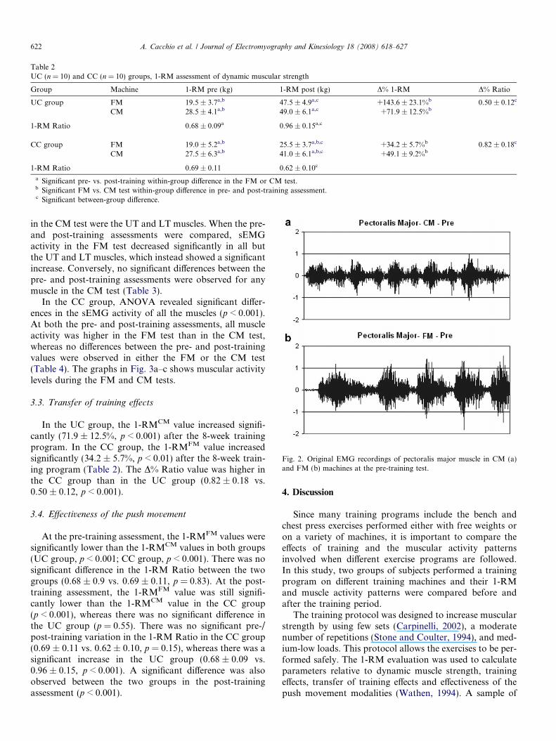
Table 2UC (n = 10) and CC (n = 10) groups, 1-RM assessment of dynamic muscular strength
Group Machine 1-RM pre (kg) 1-RM post (kg) D% 1-RM D% Ratio
UC group FM 19.5 ± 3.7a,b 47.5 ± 4.9a,c +143.6 ± 23.1%b 0.50 ± 0.12c
CM 28.5 ± 4.1a,b 49.0 ± 6.1a,c +71.9 ± 12.5%b
1-RM Ratio 0.68 ± 0.09a 0.96 ± 0.15a,c
CC group FM 19.0 ± 5.2a,b 25.5 ± 3.7a,b,c +34.2 ± 5.7%b 0.82 ± 0.18c
CM 27.5 ± 6.3a,b 41.0 ± 6.1a,b,c +49.1 ± 9.2%b
1-RM Ratio 0.69 ± 0.11 0.62 ± 0.10c
a Significant pre- vs. post-training within-group difference in the FM or CM test.b Significant FM vs. CM test within-group difference in pre- and post-training assessment.c Significant between-group difference.
Fig. 2. Original EMG recordings of pectoralis major muscle in CM (a)and FM (b) machines at the pre-training test.
622 A. Cacchio et al. / Journal of Electromyography and Kinesiology 18 (2008) 618–627
in the CM test were the UT and LT muscles. When the pre-and post-training assessments were compared, sEMGactivity in the FM test decreased significantly in all butthe UT and LT muscles, which instead showed a significantincrease. Conversely, no significant differences between thepre- and post-training assessments were observed for anymuscle in the CM test (Table 3).
In the CC group, ANOVA revealed significant differ-ences in the sEMG activity of all the muscles (p < 0.001).At both the pre- and post-training assessments, all muscleactivity was higher in the FM test than in the CM test,whereas no differences between the pre- and post-trainingvalues were observed in either the FM or the CM test(Table 4). The graphs in Fig. 3a–c shows muscular activitylevels during the FM and CM tests.
3.3. Transfer of training effects
In the UC group, the 1-RMCM value increased signifi-cantly (71.9 ± 12.5%, p < 0.001) after the 8-week trainingprogram. In the CC group, the 1-RMFM value increasedsignificantly (34.2 ± 5.7%, p < 0.01) after the 8-week train-ing program (Table 2). The D% Ratio value was higher inthe CC group than in the UC group (0.82 ± 0.18 vs.0.50 ± 0.12, p < 0.001).
3.4. Effectiveness of the push movement
At the pre-training assessment, the 1-RMFM values weresignificantly lower than the 1-RMCM values in both groups(UC group, p < 0.001; CC group, p < 0.001). There was nosignificant difference in the 1-RM Ratio between the twogroups (0.68 ± 0.9 vs. 0.69 ± 0.11, p = 0.83). At the post-training assessment, the 1-RMFM value was still signifi-cantly lower than the 1-RMCM value in the CC group(p < 0.001), whereas there was no significant difference inthe UC group (p = 0.55). There was no significant pre-/post-training variation in the 1-RM Ratio in the CC group(0.69 ± 0.11 vs. 0.62 ± 0.10, p = 0.15), whereas there was asignificant increase in the UC group (0.68 ± 0.09 vs.0.96 ± 0.15, p < 0.001). A significant difference was alsoobserved between the two groups in the post-trainingassessment (p < 0.001).
4. Discussion
Since many training programs include the bench andchest press exercises performed either with free weights oron a variety of machines, it is important to compare theeffects of training and the muscular activity patternsinvolved when different exercise programs are followed.In this study, two groups of subjects performed a trainingprogram on different training machines and their 1-RMand muscle activity patterns were compared before andafter the training period.
The training protocol was designed to increase muscularstrength by using few sets (Carpinelli, 2002), a moderatenumber of repetitions (Stone and Coulter, 1994), and med-ium-low loads. This protocol allows the exercises to be per-formed safely. The 1-RM evaluation was used to calculateparameters relative to dynamic muscle strength, trainingeffects, transfer of training effects and effectiveness of thepush movement modalities (Wathen, 1994). A sample of
Table 3UC group (n = 10), muscle mean EMG activity (%MVC) during 6-repetition tests
Test PM LD AD PD TB BB UT LT
Pre-training assessment
FM test 70.2 ± 12.3 43.5 ± 8.3 83.3 ± 16.4 59.5 ± 11.4 77.5 ± 17.8 41.4 ± 7.8 31.5 ± 6.8 29.3 ± 5.5CM test 54.1 ± 13.1 17.2 ± 3.8 58.2 ± 12.3 14.4 ± 3.7 48.2 ± 9.8 12.6 ± 3.7 19.3 ± 4.9 16.8 ± 4.2p* 0.011 <0.001 0.001 <0.001 <0.001 <0.001 <0.001 <0.001
Post-training assessment
FM test 53.1 ± 10.3 16.7 ± 3.9 59.4 ± 11.7 13.1 ± 3.2 47.3 ± 10.6 13.3 ± 3.5 69.2 ± 13.3 69.1 ± 13.8CM test 52.6 ± 9.8 15.6 ± 4.3 56.4 ± 12.9 12.2 ± 2.9 45.8 ± 10.1 11.1 ± 2.9 17.7 ± 3.3 15.4 ± 3.3p* 0.912 0.556 0.592 0.518 0.749 0.143 <0.001 <0.001
Pre- vs. post-training assessment
FM test p* 0.003 <0.001 0.002 <0.001 <0.001 <0.001 <0.001 <0.001
CM test p* 0.775 0.389 0.753 0.156 0.596 0.326 0.403 0.412
* Post-hoc Tukey Test. Bold values indicate statistical significance.
Table 4CC group (n = 10), muscle EMG activity (%MVC) during 6-repetition tests
Test PM LD AD PD TB BB UT LT
Pre-training assessment
FM test 69.3 ± 14.0 42.1 ± 10.3 84.2 ± 16.8 56.2 ± 11.3 78.2 ± 15.4 43.4 ± 8.6 30.9 ± 5.6 31.0 ± 5.6CM test 56.2 ± 9.1 16.3 ± 4.4 58.5 ± 13.0 15.3 ± 3.9 46.3 ± 10.2 15.4 ± 3.6 20.5 ± 4.9 13.9 ± 3.5p* 0.023 <0.001 0.001 <0.001 <0.001 <0.001 <0.001 <0.001
Post-training assessment
FM test 71.3 ± 15.0 42.4 ± 8.3 85.3 ± 17.8 56.6 ± 10.3 79.8 ± 16.8 45.8 ± 9.5 34.1 ± 7.2 34.1 ± 6.9CM test 53.9 ± 11.1 15.8 ± 4.2 58.1 ± 11.2 14.2 ± 3.2 44.6 ± 9.8 14.2 ± 3.6 20.2 ± 4.9 12.1 ± 2.7p* <0.001 <0.001 <0.001 <0.001 <0.001 <0.001 <0.001 <0.001
Pre- vs. post-training assessment
FM test p* 0.761 0.943 0.888 0.935 0.826 0.561 0.281 0.284CM test p* 0.618 0.797 0.942 0.499 0.708 0.465 0.892 0.214
* Post-hoc Tukey Test. Bold values indicate statistical significance.
A. Cacchio et al. / Journal of Electromyography and Kinesiology 18 (2008) 618–627 623
healthy female novice-lifters was used to prevent previouslylearned motor skills from affecting motor performances.
At the pre-training assessment, as expected, no differ-ences were observed between the two groups in 1-RM.By contrast, significant differences were observed at theend of the study, when 1-RM values were significantlyhigher in the UC group than in the CC group. The signif-icant difference observed at the post-training assessmentsuggests that different training effects may be attributedto the two machines used for training.
The results from previous studies regarding the presenceof a transfer of training from one machine to the otherhave been controversial. According to the concept of train-ing specificity (Behm et al., 2002), significant 1-RMincreases should be expected exclusively on the machineused for training (Rutherford and Jones, 1986; Sale,1988; Stone et al., 2000; Behm et al., 2002). However, somestudies reported that a transfer of the effects of trainingfrom one machine to another is possible (Wilson and Mur-phy, 1996), and that the transfer is more likely from freeweights to weight machines than viceversa (Boyer, 1990).
In our study, the effects of training were found to bereciprocally transferred from one machine to the other,though the transfer was unexpectedly greater from the con-strained to the unconstrained machine than viceversa. A
possible explanation for this finding is that the 1-RMincrease in the UC group was due to both strength gainand motor strategy improvement, with only the strengthgain transferable to the CM machine as this machine doesnot require fine motor strategy. By contrast, the 1-RMincrease in the CC group might be due exclusively to astrength gain that could be fully transferred to the FMmachine. Thus, the D% Ratio was lower in the UC groupthan in the CC group because the overall training effectswere greater in the former than in the latter.
Different dynamic conditions have been reported to pro-duce different force output levels, with unstable conditionsassociated with lower force outputs (Vera-Garcia et al.,2000; Anderson and Behm, 2004). For example, isometricpeak force attained on an unstable chest press was around60% lower than that attained on a stable chest press(Anderson and Behm, 2004). This suggests that motorstrategy during an exercise executed under unstabledynamic conditions may lack effectiveness because muscu-lar activity is partly utilized for stabilization of undesirablemovements (Korneki and Zschorlich, 1994).
Our data results showed that the 1-RMFM values weresignificantly lower than the 1-RMCM values at the pre-train-ing assessments for both groups (1-RM Ratio of�0.7), sug-gesting use of a recruitment strategy on the FM machine
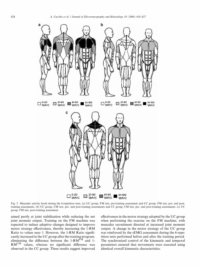
Fig. 3. Muscular activity levels during the 6-repetition tests. (a) UC group, FM test, pre-training assessment and CC group, FM test, pre- and post-training assessments. (b) UC group, CM test, pre- and post-training assessments and CC group, CM test, pre- and post-training assessments. (c) UCgroup, FM test, post-training assessment.
624 A. Cacchio et al. / Journal of Electromyography and Kinesiology 18 (2008) 618–627
aimed partly at joint stabilization while reducing the netjoint moment output. Training on the FM machine wasexpected to induce adaptive changes designed to improvemotor strategy effectiveness, thereby increasing the 1-RMRatio to values near 1. However, the 1-RM Ratio signifi-cantly increased in the UC group after the training program,eliminating the difference between the 1-RMFM and 1-RMCM values, whereas no significant difference wasobserved in the CC group. These results suggest improved
effectiveness in the motor strategy adopted by the UC groupwhen performing the exercise on the FM machine, withmuscular recruitment directed at increased joint momentoutput. A change in the motor strategy of the UC groupwas reinforced by the sEMG assessment during the 6-repe-tition tests performed before and after the training period.The synchronized control of the kinematic and temporalparameters ensured that movements were executed usingidentical overall kinematic characteristics.
A. Cacchio et al. / Journal of Electromyography and Kinesiology 18 (2008) 618–627 625
In the push movement, characterized by shoulder hori-zontal adduction and elbow extension, the PM, DA andTB muscles act as agonists, the LD, DP and BB musclesact as antagonists, and the UT and LT act as stabilizers(McCaw and Friday, 1994; Lee and An, 2002). When seg-ment motion is constrained, by the CM machine, the ago-nists may be highly activated while the activity in thestabilizers and antagonists may be decreased since thereis less need to maintain control of the kinematic patternof the movement (Sale, 1988). In an unconstrained condi-tion, such as on the FM machine, control of the movementmay demand greater muscular co-contraction betweenpairs of agonist/antagonist muscles, such as the PM/LD,DA/DP and TB/BB, reducing the net joint momentrequired to perform the movement against the externalresistance (Lander et al., 1985; McCaw and Friday, 1994).
Therefore, performance of the pressing movementwould require higher levels of muscle activity on the FMmachine than on the CM machine (Lander et al., 1985;Korneki and Zschorlich, 1994; Anderson and Behm,2004). However, as agonist/antagonist co-activity typicallydecreases with motor learning and training (Carolan andCaffarelli, 1992; Osu et al., 2002; Gribble et al., 2003), achange in muscular pattern, due to an improvement ininter-muscular coordination, was to be expected in theUC group after the training period (Rutherford and Jones,1986).
Our results not only confirm these hypotheses, but alsohelp explain 1-RM findings. Indeed, all the muscles weremore highly activated in the FM test than in the CM testin both groups at the pre-training assessment. In particular,activity of the agonists (PM, AD and TB muscles) was veryhigh in the FM test and high in the CM test, activity of theantagonists (LD, PD and BB muscles) was high in the FMtest and minimal in the CM test, and activity of the stabi-lizers (UT and LT muscles) was moderate in the FM testand minimal in the CM test (Fig. 3). These results are inaccordance with those of other researchers (McCaw andFriday, 1994), who have reported higher agonist andantagonist EMG activity during free-weight bench pressmovement against low external resistance (e.g. 60% of the1-RM) than during machine bench press movement. Thegreater activity observed in the FM test can be attributedto the greater effort required of the stabilizers and antago-nists to control the movement, and therefore to the needfor a higher agonist activity level to obtain an equivalentnet force output (Lander et al., 1985; Korneki and Zschor-lich, 1994; Anderson and Behm, 2004). The absence of anysignificant difference between pre- and post-training assess-ments observed in the CC group indicates that the motorstrategy was unaffected by the training modality. Whentested on the FM machine after the training period, theCC group still displayed a marked involvement of the ago-nist/antagonist muscles in stabilization of the movement.This finding explains, in part, why the motor strategyadopted by the CC group was less effective on the FMmachine than on the CM machine, even at the post-train-
ing assessment. By contrast, a series of adaptive changeswere observed in the UC group. At the post-training assess-ment, agonist and antagonist activity in the FM testdecreased to a high and minimal level, respectively, i.e.the same levels as in the CM test. Stabilizer activity insteadincreased to a very high level (Fig. 3), thereby indicatingthat a greater effort was required of these muscles. Thesechanges suggest that motor strategy was re-organized tooptimise the muscular effort, by maximally directing ago-nist activity to generate movement against the external loadin the direction of the trajectory requested, whereas it wasabove all the stabilizer activity that opposed the externalload in all other directions to stabilize the movement.Therefore, decreased antagonist activity allowed for areduction in the agonist’s activity level. This rearrangedmotor strategy adopted by the UC group on the FMmachine increased the effectiveness of the push movementto a level similar to that displayed on the CM machine,thereby attaining the same level of %1-RM on bothmachines. It is noteworthy that the increase in stabilizeractivity observed in the UC group during the FM testwas not observed during the CM test (Fig. 3). As the needfor stabilization on the CM machine is relatively low, anunnecessary increase in muscular activity would determinean increase in energy expenditure with no mechanicaladvantages. Thus, the UC group not only optimised themotor strategy adopted on the FM machine, but alsoadapted it to dynamic conditions that differed from thoseexperienced during training.
5. Perspectives
Our findings indicate that, in addition to muscularstrength, inter-muscular coordination plays a significantrole in force output during a motor task by more appropri-ately distributing efforts among different muscles. Ourresults support the proposal by other researchers that train-ing in unconstrained dynamic conditions represents a moreeffective way of improving inter-muscular coordination inthe execution of a motor task, as such conditions requiregreater stabilization as well as an adaptation of the motorstrategy to optimise muscular efforts (Rutherford andJones, 1986; Anderson and Behm, 2004).
As the force production and stabilization required byunconstrained multi-joint movements are more similar tosports and daily living activity movements than thoserequired by constrained multi-joint movements (McCawand Friday, 1994), unconstrained machines should be usedwhen the aim of the training program is not only strengthdevelopment, but also improved inter-muscular coordina-tion. Further research is warranted to understand whetherthe enhanced strength development obtained with an 8-week training program with an unconstrained machine,as opposed to a constrained machine, persists even whenthe motor strategy has been optimised, thereby minimizingthe demands made upon the agonist muscles, as observedin our study at the end of the training period.
626 A. Cacchio et al. / Journal of Electromyography and Kinesiology 18 (2008) 618–627
References
American College of Sports Medicine. Position stand: the recommendedquantity and quality of exercise for developing and maintainingcardiorespiratory and muscular fitness, and flexibility in healthy adults.Med Sci Sports Exerc 1998;30:975–91.
American College of Sports Medicine. Kraemer WJ, Writing GroupChairman. Position stand: progression models in resistance trainingfor healthy adults. Med Sci Sports Exerc 2002;34:364–80.
Anderson KG, Behm DG. Maintenance of EMG activity and loss of forceoutput with instability. J Strength Cond Res 2004;18:637–40.
Behm DG, Anderson KG, Curnew S. Muscle force and neuromuscularactivation under stable and unstable conditions. J Strength Cond Res2002;16:416–22.
Bernstein NA. The co-ordination and regulation of move-ment. Oxford: Pergamon Press; 1967.
Boyer BT. A comparison of three strength training programs on women. JAppl Sports Sci Res 1990;4:88–94.
Carolan B, Caffarelli E. Adaptation in coactivation after isometricresistance training. J Appl Physiol 1992;73:911–7.
Carpinelli RN. Berger in retrospect: effect of varied weight trainingprogrammes on strength. Br J Sports Med 2002;36:319–24.
Carroll TJ, Riek S, Carson RG. The sites of neural adaptation induced byresistance training in humans. J Physiol 2002;544:641–52.
Enoka RM. Muscle strength and its development. Sports Med1988;6:146–68.
Enoka RM. Neural adaptations with chronic physical activity. J Biomech1997;30:447–55.
Farina D, Merletti R, Enoka RM. The extraction of neural strategies fromthe surface EMG. J Appl Physiol 2004;96:1486–95.
Galloway JC, Koshland GF. General coordination of shoulder, elbow andwrist dynamics during multijoint arm movements. Exp Brain Res2002;142:163–80.
Gribble PL, Mullin LI, Cothros N, Mattarl A. Role of cocontraction inarm movement accuracy. J Neurophysiol 2003;89:2396–405.
Hakkinen K, Komi P. Training-induced changes in neuromuscularperformance under voluntary and reflex conditions. Eur J ApplPhysiol 1986;55:147–55.
Hermens HJ, Freriks B, Merletti R, Hagg G, Stegeman D, Block J, et al.,editors. SENIAM: European recommendations for surface electromy-ography. ISBN: 90-75452-15-2: Roessingh Research and Developmentbv, 1999.
Hintermeister RA, Lange GW, Schulteis JM, Bey MJ, Hawkins RJ.Electromyographic activity and applied load during shoulder rehabil-itation exercises using elastic resistance. Am J Sports Med1998;26:210–9.
Kelly BT, Backus SI, Warren RD, Williems RJ. Electromyographicanalysis and phase definition of the overhead football throw. Am JSports Med 2002;30:837–47.
Korneki S, Zschorlich V. The nature of the stabilizing functions of skeletalmuscles. J Biomech 1994;27:215–25.
Kraemer WJ, Fry AC. Strength testing: development and evaluation ofmethodology. In: Maud P, Foster C, editors. Physiological assessmentof human fitness. Champaign, IL: Human Kinetics; 1995. p. 115–38.
Lander JE, Bates BT, Sawhill JA, Hamill J. A comparison between free-weight and isokinetic bench pressing. Med Sci Sports Exerc 1985;17:344–353.
Lee SB, An KN. Dynamic glenohumeral stability provided by three headsof the deltoid muscle. Clin Orthop Relat Res 2002;400:40–7.
McCaw ST, Friday J. A comparison of muscle activity between a freeweight and machine bench press. J Strength Cond Res 1994;8:259–64.
McNair PJ, Depledge J, Brettkelly M. Verbal encouragement: effects onmaximum effort voluntary muscle action. Br J Sports Med 1996;30:28–35.
Osu R, Franklin DW, Kato H, Gomi H, Domen K, Yoshioka T,et al.. Short- and long-term changes in joint co-contraction
associated with motor learning as revealed from surface EMG. JNeurophysiol 2002;88:991–1004.
Ploutz-Snyder LL, Giamis EL. Orientation and familiarization to 1-RMstrength testing in old and young women. J Strength Cond Res2002;15:519–23.
Rab G, Petuskey K, Bagley A. A method for determination of upperextremity kinematics. Gait & Posture 2002;15:113–9.
Rutherford OM, Jones DA. The role of learning and coordination instrength training. Eur J Appl Physiol 1986;55:100–5.
Sale DG. Neural adaptations to resistance training. Med Sci Sport Exerc1988;20:S135–45.
Siff M. Biomechanical foundations of strength and power training. In:Zatsiorky V, editor. Biomechanics in sports. London: Blackwell SciLtd; 2001. p. 103–39.
Simpson SR, Rozenck R, Garhammer J, Lacourse M. Comparison of onerepetition maximum between free weight and universal machineexercises. J Strength Cond Res 1997;11:103–6.
Stone WJ, Coulter SP. Strength/endurance effects from three resistancetraining protocols with women. J Strength Condition Res 1994;8:23–234.
Stone MH, Collins DC, Plisk S, Haff G, Stone ME. Training principles:evaluation of modes and methods of resistance training. StrengthCondition 2000;22:65–76.
Vera-Garcia FJ, Greiner SG, McGill SM. Abdominal muscle responseduring curl-ups on both stable and labile surfaces. Phys Ther2000;80:564–9.
Wathen D. Load assignment. In: Baechle TR, editor. Essentials ofstrength training and conditioning. NSCA. Champaign, IL: HumanKinetics; 1994.
Weir JP. The effect of rest interval length on repeated maximal benchpresses. J Strength Condition Res 1994;8:58–60.
Wilson GJ. Strength and power in sport. In: Bloomfield J, Ackland TR,Elliott BC, editors. Applied anatomy and biomechanics in sport. Cam-bridge, MA: Blackwell Scientific Publications; 1994. p. 110–208.
Wilson GJ, Murphy AJ. The use of isometric tests of muscular function inathletic assessment. Sports Med 1996;22:19–37.
Wilson GJ, Elliott BC, Kerr GK. Bar path and force profile characteristicsfor maximal and submaximal loads in the bench press. Int J SportBiomech 1989;5:390–402.
Angelo Cacchio received his BSc degree in Physi-cal Education from Superior Institute of PhysicalEducation of L’Aquila, his BSc degree in PhysicalTherapy from School of Physical Therapy ofL’Aquila, and his MD degree from School ofMedicine of University of L’Aquila, in 1990,1993, and 2003, respectively. Since 2003 he hasbeen in practice at the Specialization School ofPhysical Medicine and Rehabilitation of theUniversity of Roma ‘‘La Sapienza’’. He is Con-tract Professor of Kinesiology at School of
Physical Therapy of the University of L’Aquila. His major researchinterests include sport biomechanics, movement analysis, physical medi-
cine and rehabilitation.Romildo Don received his MD degree from Schoolof Medicine of Univeristy of Roma ‘‘LaSapienza’’ in 2003. Since the same year he hasbeen in practice at the Specialization School ofPhysical Medicine and Rehabilitation at the sameUniversity. His major research interests includesport biomechanics, movement analysis andrehabilitation.
A. Cacchio et al. / Journal of Electromyography and Kinesiology 18 (2008) 618–627 627
Alberto Ranavolo graduated in Electronic Engi-neering from University of Napoli ‘‘Federico II’’,in 2002, and he is currently a PhD candidate inBiomechanics at the ‘‘La Sapienza’’ Univeristy ofRoma, where he works. His major researchinterests include biomechanics, movement analy-sis, and surface electromyography.
Enrico Guerra received his MSc degree in Motor
Science from the University of Roma ‘‘TorVergata’’, and his specialization in Biomechanicsof Physical and Sporting Education from theUniversity Institute of Motor Sciences of Roma.He is Contract Professor at the School of Motorand Sporting Sciences of the University of Peru-gia. His major research interests include biome-chanics, and sport exercise.Steven T. McCaw received his BPHE degree from
the Lakehead University of Thunder Bay, in1989, and is MA (Biomechanics) from McGillUniversity of Montreal, in 1985 and his PhD inBiomechanics and Sports Medicine from theUniversity of Oregon in 1989. Currently he isProfessor of Biomechanics, the Director of Bio-mechanics Laboratory and the Coordinator ofthe Graduate Program at the School of Kinesi-ology and Recreation at the Illinois State Uni-versity. His major research interests includebiomechanics of strength exercises and energy absorption by the lowerextremities.
Rita Procaccianti received her MD degree fromthe Catholic University of Roma ‘‘Sacro Cuore’’,in 1995, DDS degree in Physical Medicine andRehabilitation from the University of Roma ‘‘TorVergata’’, in 2000, and PhD in Physical Medicineand Rehabilitation from the same University, in2004. Currently, she is Physiatrist in the Depart-ment of Physical Medicine and Rehabilitation atthe University of Roma ‘‘La Sapienza’’. Hermajor research interests include area of physicalmedicine and rehabilitation.
Filippo Camerota received his MD degree fromthe University of Roma ‘‘La Sapienza’’, in 1990,and his DDS degree in Physical Medicine andRehabilitation from the same University, in 1994.Currently he is a Physiatrist in the Department ofPhysical Medicine and Rehabilitation at theUniversity of Roma ‘‘La Sapienza’’. His mainresearch interests are in the areas of segmentalmuscle vibration applied to spasticity in cerebralpalsy and others neurological diseases.
Massimo Frascarelli received his MD degree from
the University of Roma ‘‘La Sapienza’’, and hisDDS degree in Childish Neuropsychiatry and inPhysical Medicine and Rehabilitation from thesame University. Currently he is a Physiatrist andAdjunct Professor in the Department of PhysicalMedicine and Rehabilitation, and Chairman ofSchool of Occupational Therapy at the Universityof Roma ‘‘La Sapienza’’. His main researchinterests are in the areas of paediatric neurologi-cal diseases, electromyography and movementanalysis.
Valter Santilli received his MD degree from theUniversity of Roma ‘‘La Sapienza’’, and his DDSdegree in Neurology and in Physical Medicineand Rehabilitation from the same University.Currently he is Chairman of the Department ofPhysical Medicine and Rehabilitation at theUniversity of Roma ‘‘La Sapienza’’ and Directorof the Specialization School of Physical Medicineand Rehabilitation at the same University. He hasbeen coordinator and tutor of many PhD pro-grams in Physical Medicine and Rehabilitation.
His main activities relates to Physical Medicine and Rehabilitation clinicalresearch, neurological diseases and movement analysis.