Effect of Hydrophobic Surfactant Proteins SP-B and SPC on the Permeability of Phospholipid Membranes
15
326 Biophysical Journal Volume 84 January 2003 326–340 Effect of Hydrophobic Surfactant Proteins SP-B and SP-C on Binary Phospholipid Monolayers II. Infrared External Reflectance-Absorption Spectroscopy Jennifer M. Brockman,* Zhengdong Wang, y Robert H. Notter, y and Richard A. Dluhy* *Department of Chemistry, University of Georgia, Athens, Georgia 30602; and y Department of Pediatrics, University of Rochester, Rochester, New York 14642 ABSTRACT In situ external reflection infrared spectroscopy at the air-water interface was used to study the influence on phospholipid structure of an endogenous mixture of the two hydrophobic surfactant proteins, SP-B and SP-C, which are thought to play pivotal roles in the adsorption and function of pulmonary surfactant. Mixtures studied were 1:1, 2:1, and 7:1 (mol:mol) DPPC-d 62 :DPPG, and 7:1 DPPC-d 62 :DOPG, alone and in the presence of 0.5–10 wt % mixed SP-B/C purified chromatographically from calf lung surfactant extract. Perdeuteration of DPPC produced a shift in vibrational frequencies so that it could be differentiated spectroscopically from the phosphoglycerol component in the surface monolayer. CH 2 antisymmetric and symmetric stretching bands (;2920 and 2852 cm ÿ1 ) along with the analogous CD 2 stretching bands (;2194 and 2089 cm ÿ1 ) were analyzed, and band heights and peak wavenumber positions were assessed as a function of monolayer surface pressure. Small, near-physiological contents of 1–2 wt % SP-B/C typically produced the maximum observed spectroscopic effects, which were abolished at high protein contents of 10 wt %. Analysis of CH 2 and CD 2 stretching bands and C-H/C-D band height ratios indicated that SP-B/C affected PC and PG lipids differently within the surface monolayer. SP-B/C had preferential interactions with DPPG in 1:1, 2:1, and 7:1 DPPC-d 62 :DPPG films that increased its acyl chain order. SP-B/C also interacted specifically with DOPG in 7:1 DPPC-d 62 :DOPG monolayers, but in this case an increase in CH 2 band heights and peak wavenumber positions indicated a further disordering of the already fluid DOPG acyl chains. CD 2 band height and peak wavenumber analysis indicated that SP-B/C had no significant effect on the structure of DPPC-d 62 chains in 7:1 films with DPPG or DOPG, and had only a slight tendency to increase the acyl chain order in 1:1 films of DPPC-d 62 :DPPG. SP-B/C had no significant effect on DPPC-d 62 structure in films with DOPG. Infrared results also indicated that interactions involving SP-B/C and lipids led to exclusion of PC and PG lipids from the compressed interfacial monolayer, in agreement with our previous report on the phase morphology of lipid monolayers containing 1 wt % SP-B/C. INTRODUCTION Pulmonary surfactant is a highly specialized substance found in all mammalian lungs that is known to promote lung expansion on inspiration and to prevent lung collapse upon expiration. Surfactant is necessary for normal breathing, and the lack of surfactant in the underdeveloped lungs of pre- mature infants is the root cause of Respiratory Distress Syndrome (Avery and Mead, 1959). Disruption of surfactant activity is also implicated as contributing to the pathophysi- ology of clinical acute lung injury and Acute Respiratory Distress Syndrome (Pison et al., 1989; Lewis and Jobe, 1993; Notter and Wang, 1997). Lung surfactant composition varies relatively little across a range of animal species, differing most significantly dur- ing development or when lung disease or injury is present (Notter, 2000). Lavaged mammalian surfactant contains ;85% phospholipid, 7–10% protein, and 4–8% neutral lipid (Creuwels et al., 1997; Notter, 2000). The most abundant phospholipid class is phosphocholine, comprising ;80% of the phospholipid fraction. The single disaturated phospho- lipid 1,2-dipalmitoyl-sn-glycero-3-phosphocholine (DPPC) accounts for about one-third or slightly more of total lung surfactant phospholipid (Hunt et al., 1991; Kahn et al., 1995; Holm et al., 1996). Anionic phospholipids including phosphatidylglycerols make up ;10–15% of lung surfactant phospholipids. Lung surfactant also contains four apopro- teins, three of which are active in biophysical function: SP-A, SP-B, and SP-C. The largest of these, SP-A, is a relatively hydrophilic glycoprotein that also participates in surfactant biology and host defense (Crouch, 1998; Mason et al., 1998). SP-B and SP-C are small hydrophobic proteins known to have extensive interactions with phospholipids to enhance their adsorption and dynamic film behavior (Hawgood and Schiffer, 1991; Johansson et al., 1994b; Creuwels et al., 1997; Notter, 2000). It is the effect of these hydrophobic surfactant proteins on lipid structure in surface films that is probed in the current study. From the time that lung surfactant extracts were first studied at the surface of a Langmuir-Wilhelmy film bal- ance (Clements, 1957), researchers have used insoluble, Submitted January 25, 2002, and accepted for publication August 14, 2002. Address reprint requests to Richard A. Dluhy. Tel.: 706-542-1950; Fax: 706-542-9454; E-mail: [email protected]. Abbreviations used: A/W, air-water; DOPG, 1,2-dioleoyl-sn-glycero-3- phosphoglycerol; DPPC, 1,2-dipalmitoyl-sn-glycero-3-phosphocholine. DPPC-d 62 , acyl chain perdeuterated 1,2-dipalmitoyl-sn-glycero-3-phospho- choline; DPPG, 1,2-dipalmitoyl-sn-glycero-3-phosphoglycerol; LE, LC, liquid expanded, liquid condensed monolayer phase; PC, PG, phophocho- line, phosphoglycerol; SP-B/C, column isolated bovine hydrophobic surfactant proteins SP-B and -C; IRRAS, infrared external reflection- absorption spectroscopy. Ó 2003 by the Biophysical Society 0006-3495/03/01/326/15 $2.00
-
Upload
independent -
Category
Documents
-
view
4 -
download
0
Transcript of Effect of Hydrophobic Surfactant Proteins SP-B and SPC on the Permeability of Phospholipid Membranes

326 Biophysical Journal Volume 84 January 2003 326–340
Effect of Hydrophobic Surfactant Proteins SP-B and SP-C on BinaryPhospholipid MonolayersII. Infrared External Reflectance-Absorption Spectroscopy
Jennifer M. Brockman,* Zhengdong Wang,y Robert H. Notter,y and Richard A. Dluhy**Department of Chemistry, University of Georgia, Athens, Georgia 30602; and yDepartment of Pediatrics, University of Rochester,Rochester, New York 14642
ABSTRACT In situ external reflection infrared spectroscopy at the air-water interface was used to study the influence onphospholipid structure of an endogenous mixture of the two hydrophobic surfactant proteins, SP-B and SP-C, which are thoughtto play pivotal roles in the adsorption and function of pulmonary surfactant. Mixtures studied were 1:1, 2:1, and 7:1 (mol:mol)DPPC-d62:DPPG, and 7:1 DPPC-d62:DOPG, alone and in the presence of 0.5–10 wt % mixed SP-B/C purifiedchromatographically from calf lung surfactant extract. Perdeuteration of DPPC produced a shift in vibrational frequencies sothat it could be differentiated spectroscopically from the phosphoglycerol component in the surface monolayer. CH2
antisymmetric and symmetric stretching bands (;2920 and 2852 cm�1) along with the analogous CD2 stretching bands (;2194and 2089 cm�1) were analyzed, and band heights and peak wavenumber positions were assessed as a function of monolayersurface pressure. Small, near-physiological contents of 1–2 wt % SP-B/C typically produced the maximum observedspectroscopic effects, which were abolished at high protein contents of 10 wt %. Analysis of CH2 and CD2 stretching bands andC-H/C-D band height ratios indicated that SP-B/C affected PC and PG lipids differently within the surface monolayer. SP-B/Chad preferential interactions with DPPG in 1:1, 2:1, and 7:1 DPPC-d62:DPPG films that increased its acyl chain order. SP-B/Calso interacted specifically with DOPG in 7:1 DPPC-d62:DOPG monolayers, but in this case an increase in CH2 band heightsand peak wavenumber positions indicated a further disordering of the already fluid DOPG acyl chains. CD2 band height andpeak wavenumber analysis indicated that SP-B/C had no significant effect on the structure of DPPC-d62 chains in 7:1 films withDPPG or DOPG, and had only a slight tendency to increase the acyl chain order in 1:1 films of DPPC-d62:DPPG. SP-B/C hadno significant effect on DPPC-d62 structure in films with DOPG. Infrared results also indicated that interactions involving SP-B/Cand lipids led to exclusion of PC and PG lipids from the compressed interfacial monolayer, in agreement with our previous reporton the phase morphology of lipid monolayers containing 1 wt % SP-B/C.
INTRODUCTION
Pulmonary surfactant is a highly specialized substance found
in all mammalian lungs that is known to promote lung
expansion on inspiration and to prevent lung collapse upon
expiration. Surfactant is necessary for normal breathing, and
the lack of surfactant in the underdeveloped lungs of pre-
mature infants is the root cause of Respiratory Distress
Syndrome (Avery and Mead, 1959). Disruption of surfactant
activity is also implicated as contributing to the pathophysi-
ology of clinical acute lung injury and Acute Respiratory
Distress Syndrome (Pison et al., 1989; Lewis and Jobe,
1993; Notter and Wang, 1997).
Lung surfactant composition varies relatively little across
a range of animal species, differing most significantly dur-
ing development or when lung disease or injury is present
(Notter, 2000). Lavaged mammalian surfactant contains
;85% phospholipid, 7–10% protein, and 4–8% neutral lipid
(Creuwels et al., 1997; Notter, 2000). The most abundant
phospholipid class is phosphocholine, comprising ;80% of
the phospholipid fraction. The single disaturated phospho-
lipid 1,2-dipalmitoyl-sn-glycero-3-phosphocholine (DPPC)
accounts for about one-third or slightly more of total lung
surfactant phospholipid (Hunt et al., 1991; Kahn et al., 1995;
Holm et al., 1996). Anionic phospholipids including
phosphatidylglycerols make up ;10–15% of lung surfactant
phospholipids. Lung surfactant also contains four apopro-
teins, three of which are active in biophysical function:
SP-A, SP-B, and SP-C. The largest of these, SP-A, is a
relatively hydrophilic glycoprotein that also participates in
surfactant biology and host defense (Crouch, 1998; Mason
et al., 1998). SP-B and SP-C are small hydrophobic
proteins known to have extensive interactions with
phospholipids to enhance their adsorption and dynamic
film behavior (Hawgood and Schiffer, 1991; Johansson et
al., 1994b; Creuwels et al., 1997; Notter, 2000). It is the
effect of these hydrophobic surfactant proteins on lipid
structure in surface films that is probed in the current study.
From the time that lung surfactant extracts were first
studied at the surface of a Langmuir-Wilhelmy film bal-
ance (Clements, 1957), researchers have used insoluble,
Submitted January 25, 2002, and accepted for publication August 14,
2002.
Address reprint requests to Richard A. Dluhy. Tel.: 706-542-1950; Fax:
706-542-9454; E-mail: [email protected].
Abbreviations used: A/W, air-water; DOPG, 1,2-dioleoyl-sn-glycero-3-
phosphoglycerol; DPPC, 1,2-dipalmitoyl-sn-glycero-3-phosphocholine.
DPPC-d62, acyl chain perdeuterated 1,2-dipalmitoyl-sn-glycero-3-phospho-
choline; DPPG, 1,2-dipalmitoyl-sn-glycero-3-phosphoglycerol; LE, LC,
liquid expanded, liquid condensed monolayer phase; PC, PG, phophocho-
line, phosphoglycerol; SP-B/C, column isolated bovine hydrophobic
surfactant proteins SP-B and -C; IRRAS, infrared external reflection-
absorption spectroscopy.
� 2003 by the Biophysical Society
0006-3495/03/01/326/15 $2.00
monomolecular films spread at the A/W interface as models
for pulmonary surfactant (Notter, 1984; Notter, 2000).
Surface balance techniques have been used to study the
monolayer properties of the hydrophobic proteins SP-B and
SP-C and their mixtures with lipids at the A/W interface
(Oosterlaken-Dijksterhuis et al., 1991a; Taneva and Keough,
1994c; Taneva and Keough, 1994b; Taneva and Keough,
1994a; Wang et al., 1995; Wang et al., 1996). In addition,
microscopic techniques such as electron (Tchoreloff et al.,
1991), Brewster angle (Discher et al., 1996; Lipp et al.,
1997b; Discher et al., 1999), fluorescence (Kruger et al., 1999;
Ding et al., 2001; Lipp et al., 1996; Nag et al., 1996a; Nag
et al., 1997; Takamoto et al., 2001), and near-field
(Kramer et al., 2000), as well as scanning probe methods
(Panaiotov et al., 1996; von Nahmen et al., 1997; Krol
et al., 2000a) have been used to study surfactant preparations
and model monomolecular films relevant for pulmonary
surfactant. Whereas these microscopic techniques provide
important biophysical information, they cannot give the
same detailed molecular-level information about lipid-
protein interactions that can be obtained using vibrational
spectroscopic methods.
The use of external reflection Fourier transform infrared
spectroscopy to study the structure of monomolecular films
directly at the A/W interface was originally developed in the
mid-1980s and progress in this field has recently been
reviewed (Mendelsohn et al., 1995a; Dluhy, 2000). It has
been shown that external reflectance infrared allows for the
detection of the CH2 stretching vibrations of the lipid acyl
chains that can be used to monitor, in situ, the phase
transitions that take place in monolayer films. In addition, the
amide I vibration can be observed in pure or highly enriched
lipid-protein films, and protein structural information can be
obtained. This reflection IR technique has been applied to the
study of monolayer films of extracted lung preparations
(Dluhy et al., 1989) and more recently, to investigate the
roles that SP-B and SP-C play in the function of lung
surfactant (Pastrana-Rios et al., 1995; Gericke et al., 1997;
Flach et al., 1999).
In the present investigation, ternary mixtures were
constructed containing two phospholipids and column-
isolated bovine hydrophobic surfactant proteins (SP-B/C);
these mixtures were then studied in interfacial films using in
situ external reflectance IR absorption spectroscopy. Phos-
pholipids examined were DPPC plus either 1,2-dipalmitoyl-
sn-glycero-3-phosphoglycerol (DPPG) or 1,2-dioleoyl-sn-
glycero-3-phosphoglycerol (DOPG). The acyl chains of the
DPPC component were selectively deuterated, resulting in a
shift in the vibrational frequencies to lower wavenumber
values for DPPC. This enabled the effects of the surfactant
protein on each of the phospholipid components to be
monitored individually. SP-B/C were incorporated into
phospholipid films at 0.5–10% by weight, a range extending
above and below the physiologically relevant content of 1–2
wt % hydrophobic proteins in alveolar surfactant. Because
protein concentrations were too low to directly detect the
protein amide bonds spectroscopically, the focus of study
was on the influence of SP-B/C on the molecular behavior of
phospholipids and on preferential lipid-protein associations
in the surface film.
MATERIALS AND METHODS
Materials
The synthetic phospholipids 1,2-dipalmitoyl-sn-glycero-3-phosphoglycerol
(DPPG), 1,2-dioleyol-sn-glycero-3-phosphoglycerol (DOPG), and 1,2-
dipalmitoyl-d62-sn-glycero-3-phosphocholine (DPPC-d62) were purchased
from Avanti Polar Lipids (Alabaster, AL) and were used as received. ACS
grade NaCl and HPLC grade methanol and chloroform were obtained from
J. T. Baker. Subphase H2O was obtained in-lab from a Barnstead (Dubuque,
IA) ROpure/NANOpure reverse osmosis/deionization system having a
nominal resistivity of 18.3 MV cm.
Isolation and purification of surfactanthydrophobic protein fraction
Mixed hydrophobic surfactant proteins, SP-B/C, were obtained from calf
lung surfactant extract (CLSE). Whole lung surfactant was obtained by
repeated saline lavage of lungs from freshly killed calves followed by
centrifugation (250 3 g for 10 min) to remove cells and debris. The
resulting supernatant was centrifuged at a higher speed (12,500 3 g for 30
min) to pellet large surface-active aggregates as described previously (Notter
et al., 1983; Whitsett et al., 1986; Notter and Shapiro, 1987). The whole
surfactant pellets were then resuspended, and the hydrophobic constituents
extracted in CHCl3:MeOH (Bligh and Dyer, 1959) to obtain CLSE.
Separation of SP-B/C from lipids in CLSE was performed by gel permeation
column chromatography through an LH-20 column (Pharmacia-LKB
Biotechnology, Piscataway, NJ) using 2:1 CHCl3:MeOH as the elution
solvent (Hall et al., 1994). Two column passes were required to completely
eliminate lipids, and the final pooled protein fraction contained no
phospholipids as measured by phosphorus assay (Ames, 1966). Purity was
assessed by SDS-PAGE and by N-terminal amino acid sequencing. The
latter technique indicated that the mixed protein preparation contained only
sequences specific for SP-B and SP-C. The total content of SP-B/C in CLSE
was 1.5% based on the Folin phenol reagent assay (Lowry et al., 1951)
modified by the addition of 15% SDS to allow for quantitative protein
determination in the presence of lipid. ELISA testing based on an antibody
to bovine SP-B (rabbit-derived) indicated an SP-B content of ;0.9 wt % in
CLSE (Notter et al., 2002). Our previous results have shown that the
column-purified SP-B/C mixture used here can be combined with purified
calf lung surfactant lipids to give overall surface and physiological activities
very similar to CLSE (Hall et al., 1994; Wang et al., 1995), indicating that
the purified proteins represent the closest physiological analogy feasible for
such material. The final mixed SP-B/C isolate was dissolved in 1:1
CHCl3:MeOH at a concentration of 0.18 mg/ml and stored at low
temperature until use.
Preparation of samples
Stock solutions of DPPC-d62 (;1 mg/ml in CHCl3), DPPG and DOPG (;1
mg/ml in 4:1 CHCl3:MeOH) were prepared and concentrations verified by
inorganic phosphorus assay (Chen et al., 1956). Solutions of the desired
DPPC-d62:DPPG and DPPC-d62:DOPG mole ratios (1:1, 2:1, or 7:1)
containing between 0 and 10 wt % SP-B/C were prepared by mixing the
appropriate amounts of the above phospholipid stock solutions and the 0.18
mg/ml stock solution of SP-B/C in 1:1 CHCl3:MeOH. The subphase used for
all experiments was 150 mM NaCl in deionized H2O (pH 5.6).
IR Studies of SP-B/C Interaction with Mixed Lipid Monolayers 327
Biophysical Journal 84(1) 326–340

FTIR external reflectance measurements
Infrared external reflection-absorbance spectra of monolayers at the A/W
interface were collected using a PerkinElmer Spectrum 2000 FTIR
spectrometer equipped with an external sample beam. A 608, gold-coated,
off-axis parabolic mirror (Janos Technology, Townshend, VT) reflected the
beam coming from the spectrometer onto the surface of a Nima 601M film
balance (Coventry, England) at an incidence angle of 308 to the surface
normal. The beam reflects off the subphase, sampling the film, and a second
parabolic mirror collects the beam and directs it into a collection mirror and
then onto the sensing chip of a liquid N2-cooled HgCdTe detector. The film
balance, optical components, and detector are housed in a sealed, Plexiglas
chamber that allows humidity control of the local trough environment, thus
improving water vapor background subtraction.
The subphase was first cleaned by aspiration and a single beam spectrum
was collected for use as the IR background spectrum. The subphase
temperature was held constant at 22 6 18C by flowing thermostatted water
through the hollow body of the trough. The temperature in the enclosed
chamber was typically 248C and the relative humidity remained fairly
constant at 70%. Typically 5–10 ml of sample was spread via syringe onto
the trough surface resulting in an initial surface pressure of 3–6 mN/m. The
film was allowed to equilibrate for a period of 30 min and then was
compressed intermittently and spectra collected over a range of surface
pressures from ;5 mN/m to a maximum of 45–65 mN/m depending on the
nature of the film.
Parameters for IR spectra collection included 1024 coadded scans at 4
cm�1 resolution. Interferograms were apodized using a Norton-Beer (strong)
function, and Fourier transformed with one level of zero filling. Monolayer
IR spectra are presented as IRRAS spectra, i.e., �logðR/R0Þ, where R is IR
reflectivity of the monolayer surface and Ro is the IR reflectivity of the bare
water subphase background. The reflectance IR spectra were baseline
corrected before determination of peak positions and band intensities using
the GRAMS/32 (Thermo Galactic, Salem, NH) software package. Other-
wise, the spectra have not been smoothed or further processed. Vibrational
wavenumber peak positions and band heights were calculated using a five-
point center-of-gravity algorithm (Cameron et al., 1982), written in our
laboratory for the Grams/32 environment.
RESULTS
IRRAS spectra of lipid-protein monolayer films
Infrared external reflectance-absorption spectra obtained at
the A/W interface for a monolayer film of 7:1 DPPC-
d62:DPPG plus 5 wt % SP-B/C are shown in Fig. 1. IR
spectra were acquired while the monolayer was held at spe-
cific surface pressure values from 6.0 mN/m to 60.0 mN/m
during stepwise compression. The overlaid data in Fig. 1 are
monolayer IRRAS absorbance spectra that show the C-H
stretching region (Fig. 1 A, 3000–2800 cm�1) and analogous
C-D stretching region (Fig. 1 B, 2250–2050 cm�1) at various
surface pressures. Clearly evident in the Fig. 1A spectra of the
C-H stretching region are the CH2 antisymmetric stretching
band at 2920 cm�1, the CH2 symmetric stretching bands at
2850 cm�1, and the CH3 antisymmetric stretching bands at
2960 cm�1, which is seen as a shoulder on the antisymmetric
CH2 band (Snyder et al., 1982). Similarly, in Fig. 1 B, the
spectra of the C-D stretching region show the CD2 anti-
symmetric stretching band at 2194 cm�1, the CD2 symmetric
stretching bands at 2090 cm�1, and the CD3 antisymmetric
stretching bands at 2218 cm�1 (Bunow and Levin, 1977).
As seen in Fig. 1, both C-H and C-D vibrational bands
grow in intensity as the surface pressure increases and the
surface density of the lipid molecules increases. As the
average surface area per molecule is reduced, hydrophobic
interactions predominate and the lipids begin to adopt a
moreorderedconfiguration.Thus, inhomogeneous linebroad-
ening is reduced, the peaks become sharper, and the peak
maxima are shifted to lower frequency values (see, e.g.,
Mendelsohn et al., 1995b or Dluhy, 2000). The measured
peak wavenumber position therefore reflects the intra-
molecular conformation of the phospholipid acyl chains,
whereby a higher wavenumber value is indicative of a
disordered chain, whereas a lower wavenumber value
reflects an ordered, trans segment of the hydrocarbon tails
(Snyder et al., 1978). This shift in peak position is seen
most dramatically for the CH2 antisymmetric stretching
band contributed by the DPPG lipid component of the
DPPG:DPPC-d62 binary mixture in Fig. 1 A.
Deuteration of the DPPC acyl chains produces a shift in
the vibrational bands to lower wavenumber values, thereby
allowing the two phospholipid species to be individually
identified in monolayer IRRAS spectra (Dluhy et al., 1985;
Cameron and Dluhy, 1986). As seen in Fig. 1, substitution of
deuterium for hydrogen in the lipid acyl chains results in a
wavenumber shift offfiffiffi
2p
for the perdeuterated C-D bands, as
predicted by the harmonic oscillator model. Therefore, the
new C-D vibrations are located in the spectral range 2000–
2300 cm�1, a spectral region free from other vibrational
modes and easily observed spectroscopically using IRRAS.
The intensity and wavenumber values of the peaks provide
information not only concerning chain conformations, but
also about the number density of the individual phospholipid
components present at the film surface (Rana et al., 1993b).
Isotopic substitution not only causes wavenumber shifts in
IR bands, but also influences their integrated intensities
(Pinchas and Laulicht, 1971). We have previously empiri-
cally determined that the ratio of integrated intensities of the
C-H to C-D vibrations for a 1:1 mol:mol DPPC-d62:DPPG
monolayer film was 0.95 (Rana et al., 1993a). Assuming a
linear relationship between mole fraction and integrated
intensity, one would therefore expect the band height ratio of
C-H to C-D bands in a 7:1 mol:mol DPPC-d62:DPPG film to
be ;0.24. Instead, calculations based on the spectra in Fig. 1
indicate that the apparent band height ratio for this film (a 7:1
DPPC-d62:DPPG monolayer with 5 wt % SP-B/C added) is
;0.6. The difference seen in these band height ratios reflects
a preferential interaction of SP-B/C with DPPG in the binary
lipid film, as explained further below.
Effect of SP-B/C proteins onDPPC-d62:DPPG monolayers
In Fig. 2, the CH2 antisymmetric stretching vibration
wavenumber and band heights are plotted as a function of
measured surface pressure for monomolecular films of 7:1
328 Brockman et al.
Biophysical Journal 84(1) 326–340

DPPC-d62:DPPG with and without added SP-B/C. These
plots provide information about the structure of the DPPG
component in the film. The data in Fig. 2 were obtained from
the CH2 antisymmetric band at ;2920 cm�1, which was
used in analysis because of its larger intensity and higher
signal-to-noise ratio compared to the corresponding sym-
metric bands. This allowed molecular behavior to be probed
in the film at low surface pressures where average molecular
areas are high and peak intensities are inherently weak. It
also proved crucial for the analysis of films containing a low
percentage of protiated lipid (i.e., the monolayer contains
only ;12% PG in 7:1 DPPC-d62:DPPG and DPPC-
d62:DOPG films) and consequently exhibited C-H bands
that were extremely weak. Due to the very low band
intensities on which these analyses are based, there is a fair
amount of scatter in some of the data sets. Lines are thus
drawn through the individual points in Fig. 2 and subsequent
figures to help point out trends. These trend lines are for
illustrative purposes only, and are not intended to be a
rigorous mathematical model of the relationship among the
given variables. In addition, representative error bars
illustrating the level of uncertainty for a specific point within
a data set are included (error bars for each point are not
included out of concern for legibility). The center-of-gravity
algorithm used here to process IRRAS spectra defined
wavenumber peak positions to better than 60.01 cm�1
precision (Cameron et al., 1982). The observed scatter is thus
due solely to inherent data fluctuations in the monolayer
sample. For the IRRAS data here, typical levels of
uncertainty were ;1.0 cm�1 in peak positions and 10% in
peak heights for strong bands in the IR spectra of repetitive
data sets.
FIGURE 1 IR external reflection-absorption spec-
tra of a 7:1 DPPC-d62:DPPG þ 5 wt % SP-B/C mono-
layer film collected at surface pressures from 6.0 to
60.0 mN/m. (A) CH2 stretching region between 3000–
2800 cm�1. (B) CD2 stretching region between 2250–
2050 cm�1.
IR Studies of SP-B/C Interaction with Mixed Lipid Monolayers 329
Biophysical Journal 84(1) 326–340

For the model mixtures shown in Fig. 2 A, the wave-
number values observed for the CH2 antisymmetric stretch
decreased by as much as 6.1 cm�1 as surface pressure
increased, reflecting a more condensed, ordered state for the
DPPG component in the monolayer. A close study of the
data reveals that there are differences in the extent of this
ordering that depend upon the amount of SP-B/C incorpo-
rated into the film, with the largest effect seen upon addition
of the smallest amount of protein. The greater shift in
wavenumber values between 10 mN/m and 30 mN/m in
films of 7:1 DPPC-d62:DPPG plus 1 wt % SP-B/C indicates
the greatest extent of acyl chain ordering. Wavenumber
values also decreased relative to the lipids alone as the
amount of protein was increased to 2 and then 5 wt %, but
not as much as for the 1 wt % mixture. Further increase in the
amount of surfactant protein to 10 wt % resulted in
wavenumber shifts for the DPPG component that were
indistinguishable from the pure binary lipid mixture itself. A
consideration of the uncertainties associated with these data
indicate that only the monolayers containing 1 and 2 wt % of
SP-B/C protein in Fig. 2 produced wavenumber shifts
statistically different from the lipid-only monolayer.
Measurement of the height of the CH2 asymmetric
vibration in these lipid-protein monolayers also reflects the
influence of the SP-B/C proteins on lipid structure (Fig. 2 B).
All of the model mixtures in Fig. 2 B show an increase in
band height with an increase in the measured surface
pressure; this increased band height results from both an
increase in surface density of molecules sampled by the
infrared beam as well as increased ordering and reorientation
of the DPPG acyl chains. Fig. 2 B shows a significant
difference in the band height versus surface pressure curves
for the different model mixtures. At any given surface
pressure, an incremental increase in band height occurred for
SP-B/C contents of 1 wt %, 2 wt %, and 5 wt % in the mixed
monolayer. In contrast, addition of 10 wt % SP-B/C to the
monolayer resulted in band height values that were lower
than those seen for the pure lipid film alone. Hence, small
amounts of surfactant protein appear to interact with the
DPPG in the monolayer, causing an increase in acyl chain
ordering/reorientation, but higher amounts of SP-B/C (10 wt
%) actually caused a reduction in the perturbation to both
wavenumber shift and band height.
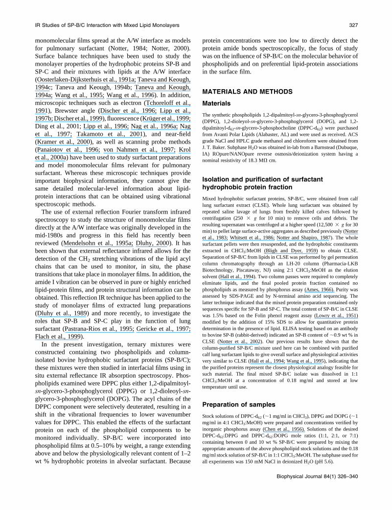
Fig. 3, A and B, are the same type of plots shown in Fig. 2,
A and B, the difference being that the data are obtained from
the CD2 antisymmetric stretching band of the DPPC-d62
component for these same 7:1 DPPC-d62:DPPG films. The
CD2 vibrational modes in Fig. 3 exhibited similar behavior to
the CH2 modes in Fig. 2: the wavenumber peak positions
decreased and band heights grew larger as surface pressure
increased. The overall magnitude of change in the band
parameters of the perdeuterated DPPC was smaller than that
of its protiated counterpart (4.4 cm�1 vs. 6.1 cm�1). The
smaller wavenumber shift for perdeuterated acyl chains
relative to protiated chains is well known and is related to the
thermodynamic properties of deuterated phospholipids
(Dluhy et al., 1985). Most important in Fig. 3, however,
was that unlike the CH2 bands from the DPPG in this model
mixture, the addition of differing amounts of SP-B/C had
relatively little influence on the wavenumber or band height
of the antisymmetric CD2 stretching bands compared to the
pure lipid mixture. The surface pressure-induced wave-
number shifts for all lipid-protein monolayers essentially
overlapped (Fig. 3 A). Similarly, the measured band heights
of the CD2 antisymmetric vibration were only minimally
affected by addition of SP-B/C at all of the protein
concentrations studied (Fig. 3 B). These results indicate that
no statistically significant preferential interaction involving
wavenumber shifts or band height changes occurred between
the SP-B/C proteins and the DPPC-d62 component of the 7:1
DPPC-d62:DPPG binary mixture.
The differential effect of the SP-B/C surfactant proteins on
each of the two lipid components in this 7:1 DPPC-
FIGURE 2 IR external reflection-absorption C-H spectral band parame-
ters for 7:1 DPPC-d62: DPPG binary monolayer films with and without SP-
B/C added. C-H band parameters reflect changes in structure for the DPPG
component in the monolayer. Variations in wavenumber (A) and band
height (B) for the CH2 antisymmetric stretching band are shown plotted as a
function of surface pressure. See key in A for symbols.
330 Brockman et al.
Biophysical Journal 84(1) 326–340
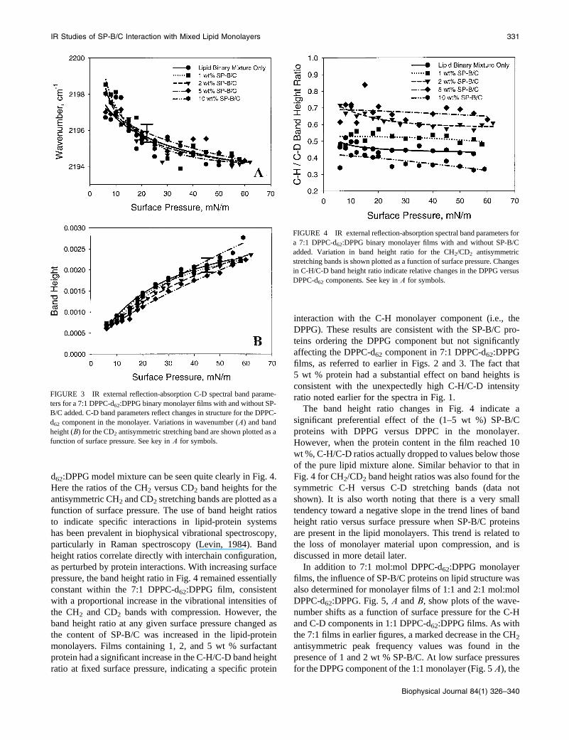
d62:DPPG model mixture can be seen quite clearly in Fig. 4.
Here the ratios of the CH2 versus CD2 band heights for the
antisymmetric CH2 and CD2 stretching bands are plotted as a
function of surface pressure. The use of band height ratios
to indicate specific interactions in lipid-protein systems
has been prevalent in biophysical vibrational spectroscopy,
particularly in Raman spectroscopy (Levin, 1984). Band
height ratios correlate directly with interchain configuration,
as perturbed by protein interactions. With increasing surface
pressure, the band height ratio in Fig. 4 remained essentially
constant within the 7:1 DPPC-d62:DPPG film, consistent
with a proportional increase in the vibrational intensities of
the CH2 and CD2 bands with compression. However, the
band height ratio at any given surface pressure changed as
the content of SP-B/C was increased in the lipid-protein
monolayers. Films containing 1, 2, and 5 wt % surfactant
protein had a significant increase in the C-H/C-D band height
ratio at fixed surface pressure, indicating a specific protein
interaction with the C-H monolayer component (i.e., the
DPPG). These results are consistent with the SP-B/C pro-
teins ordering the DPPG component but not significantly
affecting the DPPC-d62 component in 7:1 DPPC-d62:DPPG
films, as referred to earlier in Figs. 2 and 3. The fact that
5 wt % protein had a substantial effect on band heights is
consistent with the unexpectedly high C-H/C-D intensity
ratio noted earlier for the spectra in Fig. 1.
The band height ratio changes in Fig. 4 indicate a
significant preferential effect of the (1–5 wt %) SP-B/C
proteins with DPPG versus DPPC in the monolayer.
However, when the protein content in the film reached 10
wt %, C-H/C-D ratios actually dropped to values below those
of the pure lipid mixture alone. Similar behavior to that in
Fig. 4 for CH2/CD2 band height ratios was also found for the
symmetric C-H versus C-D stretching bands (data not
shown). It is also worth noting that there is a very small
tendency toward a negative slope in the trend lines of band
height ratio versus surface pressure when SP-B/C proteins
are present in the lipid monolayers. This trend is related to
the loss of monolayer material upon compression, and is
discussed in more detail later.
In addition to 7:1 mol:mol DPPC-d62:DPPG monolayer
films, the influence of SP-B/C proteins on lipid structure was
also determined for monolayer films of 1:1 and 2:1 mol:mol
DPPC-d62:DPPG. Fig. 5, A and B, show plots of the wave-
number shifts as a function of surface pressure for the C-H
and C-D components in 1:1 DPPC-d62:DPPG films. As with
the 7:1 films in earlier figures, a marked decrease in the CH2
antisymmetric peak frequency values was found in the
presence of 1 and 2 wt % SP-B/C. At low surface pressures
for the DPPG component of the 1:1 monolayer (Fig. 5 A), the
FIGURE 3 IR external reflection-absorption C-D spectral band parame-
ters for a 7:1 DPPC-d62:DPPG binary monolayer films with and without SP-
B/C added. C-D band parameters reflect changes in structure for the DPPC-
d62 component in the monolayer. Variations in wavenumber (A) and band
height (B) for the CD2 antisymmetric stretching band are shown plotted as a
function of surface pressure. See key in A for symbols.
FIGURE 4 IR external reflection-absorption spectral band parameters for
a 7:1 DPPC-d62:DPPG binary monolayer films with and without SP-B/C
added. Variation in band height ratio for the CH2/CD2 antisymmetric
stretching bands is shown plotted as a function of surface pressure. Changes
in C-H/C-D band height ratio indicate relative changes in the DPPG versus
DPPC-d62 components. See key in A for symbols.
IR Studies of SP-B/C Interaction with Mixed Lipid Monolayers 331
Biophysical Journal 84(1) 326–340

drop in wavenumber of ;3 cm�1 upon addition of 1 wt %
protein and ;5 cm�1 upon addition of 2 wt % protein is
consistent with a statistically significant increase in the
ordering of the DPPG acyl chains. For the DPPC-d62
component of the 1:1 monolayer, the shift in the CD2
antisymmetric band peak position to lower values (Fig. 5 B)
indicates that the addition of SP-B/C caused a small but
statistically significant increase in ordering of the DPPC-d62
chains, whereas these proteins had no significant effect on
the deuterated bands in the 7:1 monolayer (Fig. 3 A).
Band height ratios for the C-H versus C-D antisymmetric
bands were calculated for the 1:1 DPPC-d62:DPPG model
mixtures in the presence and absence of surfactant protein;
these ratios are presented in Fig. 6 A. Trends in the the C-H
versus C-D band height ratios for the 1:1 films were similar,
but not identical, to those found for 7:1 DPPC-d62:DPPG
films. When 1 wt % protein was present in the 1:1 DPPC-
d62:DPPG monolayer, there was a significant increase in the
C-H/C-D ratio over the pressure range 10–25 mN/m (Fig. 6
A). However, when the protein content in the film was raised
to 2 wt %, this effect was not present.
Band height ratios are plotted in Fig. 6 B as a function of
surface pressure for 2:1 mol:mol DPPC-d62:DPPG mono-
layers with 0, 1, and 2 wt % added SP-B/C. The results for
the 2:1 DPPC-d62:DPPG films are very similar to those of the
7:1 monolayers. At SP-B/C contents of both 1 and 2 wt %,
the surfactant proteins interacted preferentially with the
DPPG component causing the band height ratio to increase.
Analysis of the antisymmetric CH2 band parameters
indicated a slight ordering of the DPPG acyl chains with
increasing surface pressure in the 2:1 system based on an
increase in band height and a decrease in peak frequency
values in the presence of protein (data not shown). How-
FIGURE 5 IR external reflection-absorption C-H and C-D spectral band
parameters for 1:1 DPPC-d62:DPPG binary monolayer films with and
without SP-B/C added. C-H band parameters reflect changes in structure for
the DPPG component in the monolayer whereas C-D band parameters reflect
changes in structure for the DPPC-d62 component. Variation in wavenumber
for the CH2 (A) and CD2 (B) antisymmetric stretching bands is shown
plotted as a function of surface pressure. See key in A for symbols.
FIGURE 6 IR external reflection-absorption spectral band parameters for
(A) 1:1 DPPC-d62:DPPG and (B) 2:1 DPPC-d62:DPPG binary monolayer
films with and without SP-B/C added. Variation in band height ratio for the
CH2/CD2 antisymmetric stretching bands is shown plotted as a function of
surface pressure. Changes in C-H/C-D band height ratio indicate relative
changes in the DPPG versus DPPC-d62 components. See key in A for
symbols.
332 Brockman et al.
Biophysical Journal 84(1) 326–340

ever, there did not appear to be any effect of SP-B/C on
the DPPC-d62 component, as no significant difference in the
antisymmetric CD2 band parameters (wavenumber or band
height) was apparent in the presence of protein in the 2:1
DPPC-d62:DPPG monolayer system.
In summary, the major differences in the data for the three
different mole ratios of DPPC-d62:DPPG appeared to be the
interaction of the protein with the phosphoglycerol constit-
uent. In all of the mixtures, SP-B/C acted to order the DPPG
component in the film. In terms of the PC component, the
effects of SP-B/C depended on film composition. When the
content of DPPC-d62 in the film was high (;88 mol % in the
7:1 mixtures and 66 mol % in the 2:1 mixture), SP-B/C had
little effect on the PC component. When the proportion of
DPPC-d62 was lowered to 50 mol %, (in the 1:1 mixture),
SP-B/C appeared to have only a slight ordering effect on this
film component.
Effect of SP-B/C proteins on DPPC-d62:DOPGmonolayer films
A final set of studies examined films containing the
unsaturated phosphoglycerol DOPG in place of the DPPG.
Spectra were collected at various surface pressures for 7:1
DPPC-d62:DOPG mixtures containing 0, 0.5, 1, and 2 wt %
SP-B/C, and trends in the resulting CH2 antisymmetric
stretching band parameters are shown plotted in Fig. 7, Aand B. The wavenumber values obtained at fixed surface
pressures for 7:1 DPPC-d62:DOPG films were on average
much higher than those found for the corresponding films
with DPPG. This is due to the inherent thermodynamic
properties of the DOPG molecule. The acyl chains of DOPG
pack together less efficiently because of the 9-cis oleoyl
double bond, causing a shift in peak frequencies to higher
values. However, the overall extent of ordering of the acyl
chains does increase as surface pressure is increased just as in
the case of films containing DPPG. Addition of SP-B/C is
seen to affect the packing of the DOPG component, but in
a different way than in the DPPG films. The presence of
SP-B/C at 1 and 2 wt % in 7:1 DPPC-d62:DOPG films
increased CH2 band peak wavenumber values (Fig. 7 A), and
also led to small increases in band height (Fig. 7 B). When
compared to the lipid binary monolayer alone, the wave-
number shifts for the 1 and 2 wt % monolayers showed a
significant difference at all surface pressures. Low amounts
of SP-B/C (0.5 wt %) increased peak wavenumber values
only at surface pressures ,20 mN/m, but gave the largest
increase in band heights. These results suggest that SP-B/C
specifically interacted with DOPG, thereby increasing the
effective surface density of CH2 groups in the IR sample
beam. At the same time, this lipid-protein interaction caused
further disorder in the acyl chain.
The effects of SP-B/C on the deuterated PC component in
7:1 DPPC-d62:DOPG films are shown in Fig. 8, A and B. SP-
B/C appeared to have relatively little influence on the chain
conformation of the DPPC-d62 component, inasmuch as
there was no statistically significant difference in the values
of the CD2 antisymmetric peak frequencies between the
lipid-only and surfactant added films (Fig. 8 A). As was the
case with the DOPG component, a large increase in
measured band heights was found when SP-B/C is added.
This may again be explained as occurring as a result of a
general condensing effect of the surfactant proteins on the
phospholipids in the films, although no specific structural
change to the DPPC-d62 component in 7:1 DPPC-d62:DOPG
films was noted in the presence of SP-B/C.
The data for 7:1 DPPC-d62:DOPG monolayers indicate a
specific interaction of SP-B/C with the phosphoglycerol
component as found earlier with films containing DPPG. Fig.
9 shows substantial increases in the C-H/C-D band height
ratios with the addition of 0.5–2 wt % SP-B/C in the 7:1
DPPC-d62:DOPG film. The increases in C-H/C-D band
height ratios are not as large as found in DPPG-containing
FIGURE 7 IR external reflection-absorption C-H spectral band parame-
ters for a 7:1 DPPC-d62:DOPG binary monolayer films with and without SP-
B/C added. C-H band parameters reflect changes in structure for the DOPG
component in the monolayer. Variations in wavenumber (A) and band
height (B) for the CH2 antisymmetric stretching band are shown plotted as a
function of surface pressure. See key in A for symbols.
IR Studies of SP-B/C Interaction with Mixed Lipid Monolayers 333
Biophysical Journal 84(1) 326–340

films in Fig. 4 (note the expanded scale in Fig. 9), but they
represent a significant change compared to the lipid-only
mixture. Again, negative slopes were found in the trend lines
in all plots of band height ratio versus surface pressure in Fig.
9, although their magnitudes were lower than in Fig. 4 for
films containing DPPG. As discussed in the next section, this
negative slope likely results from the loss of DOPG from the
film upon compression, as unsaturated lipid components
have been inferred by in situ IR analysis to be squeezed out
of films of DPPC-d62 (Pastrana-Rios et al., 1994).
DISCUSSION
Using in situ IR spectroscopy, we have investigated the
influence of differing levels of the hydrophobic lung
surfactant proteins SP-B/C on phospholipid structure in
surface monolayers containing deuterated DPPC plus either
DPPG or DOPG. The results indicate a specific interaction
between the SP-B/C proteins and negatively charged anionic
PG headgroups in the binary PC/PG mixture, leading to the
exclusion of a lipid-protein complex from the monolayer.
The addition of SP-B/C caused a disproportionate increase in
the C-H band height, consistent with increased acyl chain
order in DPPG. SP-B/C also interacted specifically with
DOPG to increase its surface density and to further disorder
acyl chains. SP-B/C had no significant effect on the structure
of DPPC-d62 chains in 7:1 films with DPPG or DOPG, and
had only a slight tendency to increase the acyl chain order in
1:1 films of DPPC-d62:DPPG. Also, an important result of
this work is that the effects of the SP-B/C surfactant proteins
are highly concentration dependent, with the maximum
effect occurring at 1–2 wt %, the physiologically relevant
concentration. Indeed, higher amounts of protein were seen
to abolish the measured spectroscopic changes, and to induce
no discernable structural differences when compared to the
lipid mixture alone. These IR spectroscopic results provide a
structural complement to our previous optical microscopy
images concerning the influence of the SP-B/C proteins on
the phase morphology of PC/PG monolayers (Kruger et al.,
1999).
Comments on the use of physiologically relevantamounts of protein
The current study examined the effects of a chromato-
graphically purified mixture of SP-B/C that presumptively
maintained the endogenous ratio of the two hydrophobic
proteins. A number of prior studies have addressed the
biophysical roles played by SP-B and SP-C in the function of
pulmonary surfactant. Most of these studies, however, have
not utilized the naturally occurring combination of both
proteins. Moreover, it has not been uncommon in previous
FIGURE 9 IR external reflection-absorption spectra band parameters for
a 7:1 DPPC-d62:DOPG binary monolayer films with and without SP-B/C
added. Variation in band height ratio for the CH2/CD2 antisymmetric
stretching bands is shown plotted as a function of surface pressure. Changes
in C-H/C-D band height ratio indicate relative changes in the DPPG versus
DPPC-d62 components. See key in A for symbols.
FIGURE 8 IR external reflection-absorption C-D spectral band parame-
ters for a 7:1 DPPC-d62:DOPG binary monolayer films with and without SP-
B/C added. C-D band parameters reflect changes in structure for the DPPC-
d62 component in the monolayer. Variations in wavenumber (A) and band
height (B) for the CD2 antisymmetric stretching band are shown plotted as a
function of surface pressure. See key in A for symbols.
334 Brockman et al.
Biophysical Journal 84(1) 326–340

work to study protein contents much higher than those
encountered in vivo. The current work examined a range of
protein contents, but with a focus on physiologically relevant
levels (1–2 wt %) in investigating interactions with lipids in
the surface monolayer. Using an endogenous mixture of SP-
B/C at physiologically relevant contents in lipid mixtures has
the advantage that it more closely mimics true lung
surfactant composition, but at the cost of being unable to
differentiate the effects of SP-B from those of SP-C. In
addition, the use of low physiological levels of SP-B/C also
makes it difficult or impossible to directly study protein
vibrational bands.
Some studies have examined the effects of physiologically
relevant levels of surfactant proteins in lipid mixtures. Dluhy
et al. (1989), used IR spectroscopy to study molecular
behavior in films of bovine lung surfactant containing
endogenous ratios of protein and lipid components. In
addition, several recent investigations have used near-field
and scanning probe techniques to visualize transferred
monolayer films containing SP-B and SP-C at levels in the
neighborhood of 2 wt % (Kramer et al., 2000; Krol et al.,
2000b). However, other IR studies of SP-B and SP-C in lipid
monolayers at the A/W interface or in bulk phase vesicles
have incorporated these proteins at between 3 and 80 wt %
(Pastrana-Rios et al., 1995; Gericke et al., 1997; Gordon
et al., 2000). Similarly, optical microscopy studies of mono-
layers containing SP-C or synthetic peptides based on the
truncated N-terminus of SP-B (i.e., SP-B1-25) have used
protein amounts that span a wide range between 2 and 28 wt
% (Lipp et al., 1996; Nag et al., 1996a; Lipp et al., 1997a;
Nag et al., 1997; Ding et al., 2001; Takamoto et al., 2001).
One study (Perez-Gil et al., 1992) found that SP-C affected
lipid monolayer domains in two different ways depending
upon the concentration, and stated that a precise determi-
nation of the amounts of SP-B and SP-C present in surfactant
may be critical. Other authors (Pastrana-Rios et al., 1995)
have also commented on the use of above-normal amounts of
protein in model systems. The influence of protein concen-
tration on experimental results was particularly apparent in
this study where trends in data were often reversed upon
addition of higher amounts of SP-B/C (e.g., 10 wt %).
The use of high, nonphysiological levels of exogenous
protein in surfactant biophysical studies is sometimes
necessary due to the limitations of methodology or to
specifically study protein monolayers. Nevertheless, suffi-
ciently high protein concentrations inevitably lead to
aggregation and lipid-protein interactions driven by non-
physiological energetics. Our prior optical microscopy data
indicate a distinct difference in monolayer morphology upon
addition of higher amounts of protein (Kruger et al., 1999).
From the data presented here on the anomalous effects of 10
wt % SP-B/C in films, it appears that a nonphysiological
response occurs at smaller protein concentrations than
previously reported. This fact must be considered when
trying to draw conclusions concerning physiological re-
sponses from model systems containing artificially high
protein levels.
Comments on data error and significance
One factor in interpreting our results on interactions between
SP-B/C and phospholipids concerns the degree of variability
or scatter present in the IR data. This scatter is due to the very
small IR intensities inherent in the monolayer IRRAS
experiment (;10�3 to 10�4 AU) as well as the very small
amounts of protein used in these experiments, which caused
concomitantly small shifts in the measured IR band
parameters. Despite this expected data variability, the IR
band parameters showed clear and consistent patterns
indicating the relative effects of different amounts of SP-
B/C protein on lipid molecular packing and interfacial order.
In addition, specific interactions of these proteins with DPPG
or DOPG relative to DPPC in the binary films are easily
discernable.
Several methods of data analysis were employed to en-
hance interpretations. Representative error bars were utilized
on standard wavenumber versus surface pressure or band
intensity versus surface pressure plots to illustrate the typical
uncertainty for a particular data set. Because wavenumber
peak positions were calculated to better than 60.01 cm�1
precision (see Methods), the scatter in these data represented
inherent fluctuations in the monolayer. Our results indicated
an uncertainty of ;1.0 cm�1 in peak positions and 10% in
peak heights for the measured monolayer IRRAS spectra.
Interpretations on lipid-protein interactions here were based
on data that were statistically significant relative to
monolayers of lipids alone, as noted in the Results section.
A further refinement used band height ratio analyses in
addition to absolute shifts in band parameters to enhance
insights about interactions between specific film components
and surfactant proteins. Band height ratios have been widely
used in biophysical vibrational spectroscopy to indicate
specific interactions in lipid-protein systems. Band height
ratios correlate directly with interchain configuration, as
perturbed by protein interactions (Levin, 1984). Therefore,
C-H/C-D ratios reflect acyl chain conformation and config-
uration, similar to wavenumber shifts. Band height ratios
were necessary in the present study because of the very small
protein concentrations used and the very small shifts in the
wavenumber and intensity parameters observed. Analysis
based on band heights ratios in Figs. 4, 6, and 9 provided
important added evidence for specific lipid-protein inter-
actions to enhance interpretations based on wavenumber
shifts and intensity differences observed in the other Figures.
Relationship of the current study to previoussurfactant research
In terms of lipid components, the studies here utilized well-
defined mixtures containing only two phospholipids: DPPC
IR Studies of SP-B/C Interaction with Mixed Lipid Monolayers 335
Biophysical Journal 84(1) 326–340

plus either DPPG or DOPG. Studies on simple lipid mixtures
such as these greatly facilitate the ability of spectroscopic
assessments to discern molecular effects. At the same time,
such studies clearly oversimplify the complex lipid compo-
sition of pulmonary surfactant. The results here show a
dependence of apoprotein-induced effects on lipid compo-
sition even in simple binary mixtures. Endogenous surfactant
contains multiple saturated and unsaturated phosphatidyl-
cholines plus a broad distribution of fatty chain derivatives in
the anionic phospholipid classes that are present (PG,
phosphatidylinositol, and phosphatidylserine). It is very
likely that specific molecular activities observed here for SP-
B/C in binary lipid mixtures will be modified to some extent
as a result of multicomponent associations and interactions
in the biological material. Nonetheless, the findings here are
relevant in elucidating the potential range of molecular
actions of SP-B/C.
The ability of a naturally derived mixture of SP-B/C to
interact preferentially with the net negatively charged head-
groups of DPPG and DOPG was clearly demonstrated in the
current IR results. In all of the lipid binary systems studied,
the addition of SP-B/C caused a disproportionate increase in
the band heights due to the phosphoglycerol component as
seen in the CH2/CD2 band height ratio plots of Figs. 4, 6, and
9. This effect of the SP-B/C proteins on the phosphoglycerol
headgroups appears analogous to the effect that divalent
cations in the subphase have on PG (Williams et al., 1995).
Differential interactions of SP-B/C with DPPC versus DPPG
must reflect the influence of headgroup structure, inasmuch as
the acyl chains of the lipids are identical in length and
saturation. In films of 1:1, 2:1, and 7:1 DPPC-d62:DPPG, the
interaction of SP-B/C with DPPG headgroups was accom-
panied by an ordering of the lipid acyl chains indicated by a
shift in peak frequencies to lower values. In DPPC-d62:DOPG
films, differential interactions of SP-B/C with the two lipids
may reflect differences in both headgroups and chains,
because the oleoyl acyl chains in DOPG are two carbon atoms
longer than DPPC and contain a 9-cis unsaturated double
bond. Addition of SP-B/C in 7:1 DPPC-d62:DOPG films
caused an interaction with the DOPG headgroup, but with-
out the accompanying increase in acyl chain ordering.
Instead, the DOPG acyl chains became increasingly dis-
ordered with the addition of the mixed surfactant proteins.
Our study could not separate the individual contributions
of SP-B and SP-C in spectroscopic assessments, but both
likely contributed to the molecular biophysical changes
observed. The structural and molecular properties of SP-B
and SP-C have been examined in a variety of studies (for
reviews see Hawgood and Schiffer, 1991; Johansson et al.,
1994a; Creuwels et al., 1997; Notter, 2000). SP-B has a
highly conserved sequence yielding an amphipathic structure
that is particularly suited to interact with phospholipid
headgroups as well as chains. The fully processed form of
human SP-B is a highly charged protein composed of 79
amino acids. Although many of these amino acids are
hydrophobic, SP-B also contains 10 basic Lys/Arg residues
and two acidic Glu/Asp residues and has a net positive
charge at neutral pH. The protein exists in both monomer and
oligomeric forms, including a presumptively active homo-
dimer in most species. Amphipathic a-helical segments are
thought to locate SP-B relatively peripherally in phospho-
lipid bilayers, rather than as a transmembrane (or trans-
monolayer) protein. Using polarized ATR Fourier transform
IR spectroscopy, the hydrophobic domains of SP-B were
reported to be associated with the phospholipid headgroups
with the short a-helical portions located only slightly inside
the bilayer (Vandenbussche et al., 1992). Fluorescence
anisotropy was applied to the problem and also determined
that SP-B was not a transmembrane protein, but was asso-
ciated with the membrane surface (Baatz et al., 1990).
Tryptophan fluorescence was used to locate SP-B-like
peptides incorporated into phospholipid films and found
them to be present mostly at the polar interfacial area of the
lipid monolayers (Cochrane and Revak, 1991).
Experimental evidence also indicates the existence of a
specific interaction of SP-B with anionic phospholipids.
Fluorescence microscopy studies (Lipp et al., 1996) indicate
that positively charged SP-B protein segments complex with
the negatively charged lipid, palmitic acid. Incorporation of
a charged peptide of SP-B into monolayer films of palmitic
acid induced a change in the appearance of the isotherm
whereas an uncharged mutant of the same synthetic SP-B
peptide did not (Longo et al., 1993). It was therefore con-
cluded that a specific charge interaction between the cationic
peptide and the anionic lipid must be present. Our previous
optical microscopy studies with SP-B/C proteins clearly
implied that electrostatic interactions are critical for lung
surfactant function (Kruger et al., 1999). At low subphase pH
(;1.9), there was no change in the observed fluorescence
phase morphology throughout the liquid expanded/liquid
condensed (LE/LC) phase transition of model lipid mixtures
regardless of whether the SP-B/C proteins were present in the
monolayer. Only when the subphase pH was raised to 6.2
(which is a value above the pK of the PG headgroup), did the
monolayer exhibit a distinct new protein-induced morpho-
logy. These data suggest that an electrostatic component is
required for the formation of physiologically relevant
complexes.
SP-C as well as SP-B also is known to interact
significantly with lipids as a result of its structure and
properties (Hawgood and Schiffer, 1991; Johansson et al.,
1994a; Creuwels et al., 1997; Notter, 2000). Fully processed
human SP-C consists of 35 amino acids, most of which are
very hydrophobic. The sequence and overall extreme
hydrophobicity of SP-C is highly conserved among animal
species. Cysteine residues at positions 5 and 6 of SP-C in
humans and many other species are linked by thioester bonds
to palmitic acid moieties (see e.g., Curstedt et al., 1990).
SP-C is primarily a-helical with a length able to span a
phospholipid bilayer. Although the majority of its inter-
336 Brockman et al.
Biophysical Journal 84(1) 326–340

actions with lipids are hydrophobic, it does contain some
hydrophilic residues including positively charged Arg/Lys
residues at positions 11 and 12 in most species. Hydrophilic
residues near the N-terminus of SP-C have the potential to
interact with charged groups or ions in the plane of the
phospholipid headgroups. A number of studies has been
done on the effects of SP-C on the phospholipid components
in monomolecular films. Fluorescence microscopy suggests
that SP-C enhances the adsorption of DPPC to the monolayer
surface from vesicles (Nag et al., 1996b), an effect thought to
involve a perturbation of the packing arrangements of the
phospholipid molecules. The incorporation of SP-C (and to a
smaller extent SP-B) into monolayers reduces the size of
condensed phases in these films (Perez-Gil et al., 1992; Nag
et al., 1996a; Nag et al., 1997). As noted, the interaction of
SP-C with monolayer phospholipids is theorized to be
primarily hydrophobic in nature. Isotherm evidence was first
used to predict that SP-C has its a-helical axis oriented
parallel to the A/W interface (Oosterlaken-Dijksterhuis et al.,
1991a). Recent dichroic FTIR investigations of SP-C in the
liquid expanded monolayer phase of DPPC showed an
orientation angle of the SP-C helices of ;308 from the sur-
face, whereas at higher surface pressure, an orientation of
the helix closer to the surface normal was reported (Gericke
et al., 1997). Recent results from our laboratory (Kruger et al.,
2002) have argued that SP-C acts analogously to a molecular
machine, or loaded spring, in which the stored energy of the
hydrophobic rigid a-helix is released upon reorientation
around its flexible, more hydrophilic N-terminus, which is
anchored at the A/W interface. If such a reorientation does in
fact occur, it would appear from the present study to be
associated with a decreased phospholipid packing density
and lowering of band heights for the DPPC-d62 component,
although still maintaining an ordered PC acyl chain
conformation.
The current study did not attempt to address the functional
importance to surface activity of the hydrophobic surfactant
protein-induced effects found here in spectroscopic experi-
ments. Both SP-B and SP-C are known to enhance the sur-
face properties of phospholipids, but SP-B is more effective
in doing so on either a weight or molar basis (Curstedt et al.,
1987; Revak et al., 1988; Yu and Possmayer, 1988; Sarin
et al., 1990; Oosterlaken-Dijksterhuis et al., 1991a; Ooster-
laken-Dijksterhuis et al., 1991b; Seeger et al., 1992; Wang
et al., 1996; Johansson et al., 1998). SP-B has been shown to
be more active than SP-C in increasing both adsorption and
dynamic surface tension lowering when combined with
phospholipids. These activity findings correlate with the fact
that SP-B has a fourfold higher capacity than SP-C on a
weight basis for binding lipid vesicles to interfacial films
(Oosterlaken-Dijksterhuis et al., 1991a; Oosterlaken-
Dijksterhuis et al., 1991b). Mixtures of phospholipids with
SP-B versus SP-C also have an improved ability to resist
inhibition by serum albumin (Seeger et al., 1992; Wang et al.,
1996). Physiological studies have shown that instilled
exogenous surfactants containing lipids plus SP-B have
higher activity in improving lung mechanics and function in
animals relative to corresponding mixtures containing SP-C
(Rider et al., 1993). Supplementation with SP-B or related
synthetic peptides has also been found to improve the surface
and physiological activity of the clinical exogenous surfactant
Survanta, which contains SP-C but has only minimal levels of
SP-B (Mizuno et al., 1995; Walther et al., 1997).
A previous study examined the adsorption, dynamic film
behavior, and physiological activity of naturally derived lung
surfactant mixtures with and without anionic phospholipids
(Wang et al., 1997). Gel permeation chromatography was
used to obtain the complete mix of zwitterionic and anionic
calf lung surfactant phospholipids, as well as the subfraction
containing only the zwitterionic components. These phos-
pholipid mixtures were then studied for activity when com-
bined with column-isolated SP-B/C. Depletion in anionic
phospholipids led to only small decreases in adsorption and
dynamic surface tension lowering ability, and no significant
detriment in physiological activity in restoring pressure-
volume mechanics in lavaged rat lungs. The relevance of
these findings on complex multicomponent mixtures of lung
surfactant phospholipids to the present studies on spread
binary films of synthetic phospholipids is uncertain. Addi-
tional studies need to address the functional importance of
specific molecular interactions between anionic phospho-
lipids and SP-B/C in lung surfactant activity.
Implications for surface structure reorganization
The results of the present study not only addressed the effects
of SP-B/C on molecular order within the surface film, but
also provide insights about film refining or squeeze-out
during cycling. This is the process whereby some compo-
nents of pulmonary surfactant are ejected from the alveolar
surface film during compression and reinserted back into it
during expansion in the breathing cycle (Notter, 1984, 2000).
IR spectroscopic evidence has shown that both unsaturated
lipids (Pastrana-Rios et al., 1994) and SP-B (Pastrana-Rios
et al., 1995) are excluded from monolayer films as the surface
pressure is increased. More recently, it has been established
using optical microscopy techniques that SP-C is incorpo-
rated in exclusion particles that adhere to the surface
structure after transfer to solid substrates (Kramer et al.,
2000). In this study, the expulsion of DOPG from the films
can be inferred from the negative slopes seen in the band
height ratio data plotted in Fig. 9. These data are consistent
with the interpretation that mixed SP-B/C interacts strongly
with the charged headgroup of the DOPG so that a lipid-
protein complex is excluded from monomolecular films.
This is also supported by the PC data in Fig. 8. The
presence of SP-B/C did not affect peak wavenumber values
but did significantly increase band heights for DPPC-d62 in
the 7:1 DPPC-d62:DOPG film. This increase in band height
could potentially be attributed to the formation of three-
IR Studies of SP-B/C Interaction with Mixed Lipid Monolayers 337
Biophysical Journal 84(1) 326–340

dimensional exclusion particles that begin with specific SP-B/
C:PG interactions, but also incorporate a significant amount
of PC lipid.
The ability of SP-B/C to influence squeeze-out from lipid-
rich monolayers is also supported by our recent fluorescence
and dark-field microscopy studies (Kruger et al., 1999).
These studies have directly shown by dark-field microscopy
that the addition of SP-B/C (or the synthetic equivalent of
SP-C) induces the formation of a new monolayer phase of
different morphology than either the LC or LE phases that
form in pure lipid mixtures. Material is excluded from the
lipid-protein surface film upon compression and forms three-
dimensional, surface-associated structures of micron dimen-
sions. Such exclusion bodies form with SP-B/C peptides at
the physiological levels (1 wt %) used here, or in the pres-
ence of synthetic SP-C at higher (10 wt %) concentrations
(Kruger et al., 2002).
Our current IR results and previous microscopic studies
would be consistent with the following conceptual model.
Anionic components such as phosphoglycerols in phospho-
lipid films preferentially associate with SP-B and/or SP-C.
The positively charged, amphiphatic structure of SP-B has
multiple regions where such interactions could occur,
whereas the SP-C peptide has the potential for association
with anionic lipids in the vicinity of its N-terminus where
several hydrophilic amino acid residues are present. Upon
monolayer compression, the overall energy of the system
rises continuously, leading to a driving force for surface
film reorganization. Lipid molecules associated through
electrostatic and/or hydrophobic contacts with SP-B and/or
SP-C are lifted from the monolayer surface and inverted to
form a localized bilayer structure, possibly serving as a
nucleation site for additional material including PC lipids,
thereby leading to the formation of larger-scale interfacial
structures visible via dark-field microscopy (Kruger et al.,
1999). The formation of subinterfacial domains of lipid-
protein material ejected from the film is also possible. This
model would be consistent with the IR wavenumber shifts
and band height changes seen in the C-H region (e.g., Figs.
2 and 5) as preferential interaction of the SP-B/C proteins
with the phosphoglycerol lipids. Also, the slope of the band
height ratios (Figs. 4 and 9) can be interpreted as evidence
for the formation of three-dimensional, surface-associated
structures. A more detailed explanation of the energetics of
how SP-C might individually interact with anionic
phospholipids in surface films is presented elsewhere
(Kruger et al., 2002).
CONCLUSIONS
The primary conclusions drawn from these in situ IR
measurements on surface monolayers containing DPPC-
d62:DPPG or DPPC-d62:DOPG plus chromatographically
isolated bovine SP-B/C are as follows.
Bovine SP-B/C in the endogenous ratio had molecular
interactions with both PC and PG components in binary lipid
films to influence acyl chain order and lipid packing density
during stepwise compression from surface pressures of 6 to
60 mN/m. Specific effects depended upon the lipid
component, protein content, and monolayer composition.
Of particular note was the observation that maximal
spectroscopic effects were found for hydrophobic protein
contents in the physiological range of 1–2 wt %, and these
effects were abolished at artificially high protein levels (10
wt %).
Although SP-B/C interacted with both PC and PG in
binary lipid films, analysis of C-H/C-D spectroscopic signals
indicated preferential interactions with the anionic phospho-
lipid component. The mixed proteins ordered the acyl chains
of DPPG in 7:1, 2:1, and 1:1 monolayers of DPPC-
d62:DPPG, and disordered the acyl chains of DOPG in 7:1
films of DPPC-d62DOPG. SP-B/C had no significant effect
on DPPC acyl chain structure in 7:1 DPPC-d62:DPPG or
DPPC-d62DOPG monolayers, with only slight ordering
effects noted for the PC acyl chains in 1:1 DPPC-d62:DPPG
monolayers.
IR spectroscopic data were consistent with ejection of PC
and PG lipids from the surface monolayer during compres-
sion to high surface pressures, consistent with previous near-
field, fluorescence, and Brewster-angle microscopic studies
indicating phase morphology changes and surface aggregate
formation in compressed lung surfactant films.
The work described here was supported by the U.S. Public Health Service
through National Institutes of Health grants GM40117 (R.A.D) and
HL56176 (R.H.N., Z.W.)
REFERENCES
Ames, B. 1966. Assay of inorganic phosphate, total phosphate, andphosphatases. Methods Enzymol. 8:115–118.
Avery, M. E., and J. Mead. 1959. Surface properties in relation toatelectasis and hyaline membrane disease. Am. J. Dis. Child. 97:517–523.
Baatz, J. E., B. Elledge, and J. A. Whitsett. 1990. Surfactant protein SP-Binduces ordering at the surface of model membrane bilayers. Bio-chemistry. 29:6714–6720.
Bligh, E. G., and W. J. Dyer. 1959. A rapid method of total lipid extractionand purification. Can. J. Biochem. Physiol. 37:911–917.
Bunow, M. R., and I. W. Levin. 1977. Raman spectra and vibrationalassignments for deuterated membrane lipids. 1,2-Dipalmitoyl phospha-tidylcholine-d9 and -d62. Biochim. Biophys. Acta. 489:191–206.
Cameron, D. G., and R. A. Dluhy. 1986. FT-IR studies of molecular con-formation in biological membranes. In Spectroscopy in the BiomedicalSciences. R. M. Gendreau, editor. CRC Press, Boca Raton, FL. 53–86.
Cameron, D. G., J. K. Kauppinen, and D. Moffatt. 1982. Precision in con-densed phase vibrational spectroscopy. Appl. Spectrosc. 36:245–250.
Chen, P. S., T. Y. Toriba, and H. Warner. 1956. Microdetermination ofphosphorous. Anal. Chem. 28:1756–1758.
Clements, J. A. 1957. Surface tension of lung extracts. Proc. Soc. Exp.Biol. Med. 95:170–172.
Cochrane, C. G., and S. D. Revak. 1991. Pulmonary surfactant protein B(SP-B): structure-function relationships. Science. 254:566–568.
338 Brockman et al.
Biophysical Journal 84(1) 326–340

Creuwels, L. A. J. M., L. M. G. van Golde, and H. P. Haagsman. 1997.The pulmonary surfactant system: biochemical and clinical aspects.Lung. 175:1–39.
Crouch, E. C. 1998. Collectins and pulmonary host defense. Am. J. Respir.Cell Mol. Biol. 19:177–201.
Curstedt, T., J. Johansson, P. Persson, A. Eklund, B. Robertson, B.Lowenadler, and H. Jornvall. 1990. Hydrophobic surfactant-associatedpolypeptides: SP-C is a lipopeptide with two palmitoylated cysteineresidues, whereas SP-B lacks covalently linked fatty acyl groups. Proc.Natl. Acad. Sci. USA. 87:2985–2990.
Curstedt, T., H. Jornvall, B. Robertson, T. Bergman, and P. Berggren.1987. Two hydrophobic low-molecular-mass protein fractions ofpulmonary surfactant: characterization and biophysical activity. Eur. J.Biochem. 168:255–262.
Ding, J., D. Y. Takamoto, A. von Nahmen, M. M. Lipp, K. Y. C. Lee, A.Waring, and J. A. Zasadzinski. 2001. Effects of lung surfactant proteins,SP-B and SP-C, and palmitic acid on monolayer stability. Biophys. J.80:2262–2272.
Discher, B. M., K. M. Maloney, J. W. R. Schief, D. W. Grainger, V. Vogel,and S. B. Hall. 1996. Lateral phase separation in interfacial films ofpulmonary surfactant. Biophys. J. 71:2583–2590.
Discher, B. M., W. R. Schief, V. Vogel, and S. B. Hall. 1999. Phase sep-aration in monolayers of pulmonary surfactant phospholipids at the air-water interface: composition and structure. Biophys. J. 77:2051–2061.
Dluhy, R. A. 2000. Infrared spectroscopy of biophysical monolayer films atinterfaces. Theory and applications. In Physical Chemistry of BiologicalInterfaces. A. Baszkin and W. Norde, editors. Marcel Deker, New York.711–747.
Dluhy, R. A., D. Moffatt, D. G. Cameron, R. Mendelsohn, and H. H.Mantsch. 1985. Characterization of cooperative conformational tran-sitions by Fourier transform infrared spectroscopy: application tophospholipid binary mixtures. Can. J. Chem. 63:1925–1932.
Dluhy, R. A., K. E. Reilly, R. D. Hunt, M. L. Mitchell, A. J. Mautone, andR. Mendelsohn. 1989. Infrared spectroscopic investigations of pulmo-nary surfactant. Surface film transitions at the air-water interface and bulkphase thermotropism. Biophys. J. 56:1173–1181.
Flach, C. R., A. Gericke, K. M. W. Keough, and R. Mendelsohn. 1999.Palmitoylation of lung surfactant protein SP-C alters surface thermody-namics, but not protein secondary structure or orientation in 1,2-dipalmitoylphosphatidylcholine Langmuir films. Biochim. Biophys.Acta. 1416:11–20.
Gericke, A., C. R. Flach, and R. Mendelsohn. 1997. Structure andorientation of lung surfactant SP-C and L-a-dipalmitoylphosphatidylcho-line in aqueous monolayers. Biophys. J. 73:492–499.
Gordon, L. M., K. Y. C. Lee, M. M. Lipp, J. A. Zasadzinski, F. J. Walther,M. A. Sherman, and A. J. Waring. 2000. Conformational mapping of theN-terminal segment of surfactant protein B in lipid using C-13-enhancedFourier transform infrared spectroscopy. J. Pept. Res. 55:330–347.
Hall, S. B., Z. Wang, and R. H. Notter. 1994. Separation of subfractions ofthe hydrophobic components of calf lung surfactant. J. Lipid Res.35:1386–1394.
Hawgood, S., and K. Schiffer. 1991. Structures and properties of thesurfactant-associated proteins. Annu. Rev. Physiol. 53:375–394.
Holm, B. A., Z. Wang, E. A. Egan, and R. H. Notter. 1996. Content ofdipalmitoyl phosphatidylcholine in lung surfactant: ramifications forsurface activity. Pediatr. Res. 39:805–811.
Hunt, A. N., F. J. Kelly, and A. D. Postle. 1991. Developmental variation inwhole human lung phosphatidylcholine molecular species: a comparisonof guinea pig and rat. Early Hum. Dev. 25:157–171.
Johansson, J., T. Curstedt, and B. Robertson. 1994a. The proteins of thesurfactant system. Eur. Respir. J. 7:372–391.
Johansson, J., M. Gustafsson, S. Zaltash, B. Robertson, and T. Curstedt.1998. Synthetic surfactant protein analogs. Biol. Neonate. 74:9–14.
Johansson, J., T. Szyperski, T. Curstedt, and K. Wuthrich. 1994b. TheNMR structure of the pulmonary surfactant-associated polypeptide SP-Cin an apolar solvent contains a valyl-rich a-helix. Biochemistry.33:6015–6023.
Kahn, M. C., G. J. Anderson, W. R. Anyan, and S. B. Hall. 1995.Phosphotidylcholine molecular species of calf lung surfactant. Am. J.Physiol. 13:L567–L573.
Kramer, A., A. Wintergalen, M. Sieber, H. J. Galla, M. Amrein, and R.Guckenberger. 2000. Distribution of the surfactant-associated protein Cwithin a lung surfactant model film investigated by near-field opticalmicroscopy. Biophys. J. 78:458–465.
Krol, S., A. Janshoff, M. Ross, and H. J. Galla. 2000a. Structure andfunction of surfactant protein B and C in lipid monolayers: a scanningforce microscopy study. Phys. Chem. Chem. Phys. 2:4586–4593.
Krol, S., M. Ross, M. Sieber, S. Kunneke, H. J. Galla, and A. Janshoff.2000b. Formation of three-dimensional protein-lipid aggregates inmonolayer films induced by surfactant protein B.Biophys. J. 79:904–918.
Kruger, P., J. E. Baatz, R. A. Dluhy, and M. Losche. 2002. Effect of thehydrophobic peptide SP-C on binary phospholipid monolayers. Molec-ular machinery at the air/water interface. Biophys. Chem. 99:209–228.
Kruger, P., M. Schalke, Z. Wang, R. H. Notter, R. A. Dluhy, and M.Losche. 1999. Effect of hydrophobic surfactant peptides SP-B and SP-Con Binary Phospholipid Monolayers. I. Fluorescence and dark-fieldmicroscopy. Biophys. J. 77:903–914.
Levin, I. W. 1984. Vibrational spectroscopy of membrane assemblies. InAdvances in Infrared and Raman Spectroscopy. R. J. H. Clark and R. E.Hester, editors. Wiley, Heyden, The Netherlands. 1–48.
Lewis, J. F., and A. H. Jobe. 1993. Surfactant and the adult respiratorydistress syndrome. Am. Rev. Respir. Dis. 147:218–233.
Lipp, M. M., K. Y. C. Lee, A. Waring, and J. Zasadzinski. 1997a.Fluorescence, polarized fluorescence, and Brewster angle microscopy ofpalmitic acid and lung surfactant protein B monolayers. Biophys. J.72:2783–2790.
Lipp, M. M., K. Y. C. Lee, J. A. Zasadzinski, and A. J. Waring. 1996.Phase and morphology changes in lipid monolayers induced by SP-Bprotein and its amino-terminal peptide. Science. 273:1196–1199.
Lipp, M. M., K. Y. C. Lee, J. A. Zasadzinski, and A. J. Waring. 1997b.Design and performance of an integrated fluorescence, polarizedfluorescence, and Brewster angle microscope/Langmuir trough assemblyfor the study of lung surfactant monolayers. Rev. Sci. Instrum. 68:2574–2582.
Longo, M. L., A. M. Bisagno, J. A. N. Zasadzinski, R. Bruni, and A. J.Waring. 1993. A function of lung surfactant protein SP-B. Science.261:453–456.
Lowry, O. H., N. J. Rosebrough, A. L. Farr, and R. J. Randall. 1951.Protein measurement with the Folin phenol reagent. J. Biol. Chem.132:265–275.
Mason, R. J., K. Greene, and D. R. Voelker. 1998. Surfactant protein A andsurfactant protein D in health and disease. Am. J. Physiol. 275:L1–L13.
Mendelsohn, R., J. W. Brauner, and A. Gericke. 1995a. External infraredreflection-absorption spectrometry. Monolayer films at the air-waterinterface. Annu. Rev. Phys. Chem. 46:305–334.
Mendelsohn, R., G. L. Liang, H. L. Strauss, and R. G. Snyder. 1995b. IRspectroscopic determination of gel state miscibility in long-chainphosphatidylcholine mixtures. Biophys. J. 69:1987–1998.
Mizuno, K., M. Ikegami, C. M. Chen, T. Ueda, and A. H. Jobe. 1995.Surfactant protein-B supplementation improves in vivo function of amodified natural surfactant. Pediatr. Res. 37:271–276.
Nag, K., J. Perez-Gil, A. Cruz, and K. M. W. Keough. 1996a. Fluorescentlylabeled pulmonary surfactant protein C in spread phospholipid mono-layers. Biophys. J. 71:246–256.
Nag, K., J. Perez-Gil, A. Cruz, N. H. Rich, and K. M. W. Keough. 1996b.Spontaneous formation of interfacial lipid-protein monolayers duringadsorption from vesicles. Biophys. J. 71:1356–1363.
Nag, K., S. G. Taneva, J. Perez-Gil, A. Cruz, and K. M. W. Keough. 1997.Combinations of fluorescently labeled pulmonary surfactant proteinsSP-B and SP-C in phospholipid films. Biophys. J. 72:2638–2650.
Notter, R. H. 1984. Surface chemistry of pulmonary surfactant: The role ofindividual components. In Pulmonary Surfactant. B. Robertson, L. G. M.van Golde, and J. J. Battenburg, editors. Elsevier, Amsterdam. 17–53.
Notter, R. H. 2000. Lung Surfactants: Basic Science and Clinical Appli-cations. Marcel Dekker, New York.
IR Studies of SP-B/C Interaction with Mixed Lipid Monolayers 339
Biophysical Journal 84(1) 326–340

Notter, R. H., and D. L. Shapiro. 1987. Lung surfactants for replacementtherapy: biochemical, biophysical, and clinical aspects. Clin. Perinatol.14:433–479.
Notter, R. H., R. D. Taubold, and J. N. Finkelstein. 1983. Comparativeadsorption of natural lung surfactant, extracted phospholipids, andartificial phospholipid mixtures to the air-liquid interface. Chem. Phys.Lipids. 33:67–80.
Notter, R. H., and Z. Wang. 1997. Pulmonary surfactant: physicalchemistry, physiology and replacement. Rev. Chem. Eng. 13:1–118.
Notter, R. H., Z. Wang, E. A. Egan, and B. A. Holm. 2002. Component-specific surface and physiological activity in bovine-derived lungsurfactants. Chem. Phys. Lipids. 114:21–34.
Oosterlaken-Dijksterhuis, M. A., H. P. Haagsman, L. M. van Golde, and R.A. Demel. 1991a. Characterization of lipid insertion into monomolecularlayers mediated by lung surfactant proteins SP-B and SP-C. Biochem-istry. 30:10965–10971.
Oosterlaken-Dijksterhuis, M. A., H. P. Haagsman, L. M. van Golde, and R.A. Demel. 1991b. Interaction of lipid vesicles with monomolecular layerscontaining lung surfactant proteins SP-B or SP-C. Biochemistry.30:8276–8281.
Panaiotov, I., T. Ivanova, J. Proust, F. Boury, B. Denizot, K. Keough, andS. Taneva. 1996. Effect of hydrophobic protein SP-C on structure anddilational properties of the model monolayers of pulmonary surfactant.Coll. Surf. B. 6:243–260.
Pastrana-Rios, B., C. R. Flach, J. W. Brauner, A. J. Mautone, and R.Mendelsohn. 1994. A direct test of the "squeeze-out" hypothesis of lungsurfactant function. External reflection FT-IR at the air/water interface.Biochemistry. 33:5121–5127.
Pastrana-Rios, B., S. Taneva, K. M. W. Keough, A. J. Mautone, and R.Mendelsohn. 1995. External reflection absorption infrared spectroscopystudy of lung surfactant proteins SP-B and SP-C in phospholipidmonolayers at the air/water interface. Biophys. J. 69:2531–2540.
Perez-Gil, J., K. Nag, S. Taneva, and K. Keough. 1992. Pulmonarysurfactant protein SP-C causes packing rearrangements of dipalmitoyl-phosphatidylcholine in spread monolayers. Biophys. J. 63:197–204.
Pinchas, S., and I. Laulicht. 1971. Infrared Spectra of Labelled Com-pounds. Academic Press, London.
Pison, U., W. Seeger, R. Buchhorn, T. Joka, M. Brand, U. Obertacke, H.Neuhof, and K. P. Schmit-Nauerburg. 1989. Surfactant abnormalities inpatients with respiratory failure after multiple trauma. Am. Rev. Respir.Dis. 140:1033–1039.
Rana, F. R., A. J. Mautone, and R. A. Dluhy. 1993a. A combined infraredand 31P NMR spectroscopic method for determining the fractionalcomposition in Langmuir-Blodgett films of binary phospholipidmixtures. Appl. Spectrosc. 47:1015–1023.
Rana, F. R., A. J. Mautone, and R. A. Dluhy. 1993b. Surface chemistry ofbinary mixtures of phospholipids in monolayers. Infrared studies ofsurface composition at varying surface pressures in a pulmonarysurfactant model system. Biochemistry. 32:3169–3177.
Revak, S. D., T. A. Merritt, E. Degryse, L. Stefani, M. Courtney, M.Hallman, and C. G. Cochrane. 1988. Use of human low molecular weightapoprotein in the reconstitution of surfactant biological activity. J. Clin.Invest. 3:826–833.
Rider, E., M. Ikegami, J. Whitsett, W. Hull, D. Absolom, and A. H. Jobe.1993. Treatment responses to surfactants containing natural surfactantproteins in preterm rabbits. Am. Rev. Respir. Dis. 147:669–676.
Sarin, V. K., S. Gupta, T. K. Leung, V. E. Taylor, B. L. Ohning, J. A.Whitsett, and J. L. Fox. 1990. Biophysical and biological activity of asynthetic 8.78 kDa hydrophobic pulmonary surfactant protein SP-B.Proc. Natl. Acad. Sci. USA. 87:2633–2637.
Seeger, W. A., A. Gunther, and C. Thede. 1992. Differential sensitivity tofibrinogen inhibition of SP-C vs. SP-B based surfactants. Am. J.Physiol. 261:L286–L291.
Snyder, R. G., S. L. Hsu, and S. Krimm. 1978. Vibrational spectra in thecarbon-hydrogen stretching region and the structure of the poly-methylene chain. Spectrochim. Acta. 34A:395–406.
Snyder, R. G., H. L. Strauss, and C. A. Elliger. 1982. Carbon-hydrogenstretching modes and the structure of n-alkyl chains. 1. Long, disorderedchains. J. Phys. Chem. 86:5145–5150.
Takamoto, D. Y., M. M. Lipp, A. von Nahmen, K. Y. C. Lee, A. Waring,and J. A. Zasadzinski. 2001. Interaction of lung surfactant proteins withanionic phospholipids. Biophys. J. 81:153–169.
Taneva, S., and K. M. W. Keough. 1994a. Pulmonary surfactant proteinsSP-B and SP-C in spread monolayers at the air-water interface: I.Monolayers of pulmonary surfactant protein SP-B and phospholipids.Biophys. J. 66:1137–1148.
Taneva, S., and K. M. W. Keough. 1994b. Pulmonary surfactant proteinsSP-B and SP-C in spread monolayers at the air-water interface: II.Monolayers of pulmonary surfactant protein SP-C and phospholipids.Biophys. J. 66:1149–1157.
Taneva, S., and K. M. W. Keough. 1994c. Pulmonary surfactant proteinsSP-B and SP-C in spread monolayers at the air-water interface: III.Proteins SP-B plus SP-C with phospholipids in spread monolayers.Biophys. J. 66:1158–1166.
Tchoreloff, P., A. Gulik, B. Denizot, J. E. Proust, and F. Puisieux. 1991. Astructural study of interfacial phospholipid and lung surfactant layers bytransmission electron microscopy after Blodgett sampling: influence ofsurface pressure and temperature. Chem. Phys. Lipids. 59:151–165.
Vandenbussche, G., A. Clerx, M. Clerx, T. Curstedt, J. Johansson, H.Jornvall, and J. Ruysschaert. 1992. Secondary structure and orientationof the surfactant protein SP-B in a lipid environment: a Fourier transforminfrared spectroscopy study. Biochemistry. 31:9169–9176.
von Nahmen, A., M. Schenk, M. Sieber, and M. Amrein. 1997. Thestructure of a model pulmonary surfactant as revealed by scanning forcemicroscopy. Biophys. J. 72:463–469.
Walther, F. J., J. Hernandez-Juviel, R. Bruni, and A. Waring. 1997. SpikingSurvanta with synthetic surfactant peptides improves oxygenation insurfactant-deficient rats. Am. J. Respir. Crit. Care Med. 156:855–861.
Wang, Z., O. Gurel, J. E. Baatz, and R. H. Notter. 1996. Differentialactivity and lack of synergy of lung surfactant proteins SP-B and SP-C ininteractions with phospholipids. J. Lipid Res. 37:1749–1760.
Wang, Z., O. Gurel, S. Weinbach, and R. H. Notter. 1997. Primaryimportance of zwitterionic over anionic phospholipids in the surfaceactive function of calf lung surfactant extract. Am. J. Respir. Crit. CareMed. 156:1049–1057.
Wang, Z., S. B. Hall, and R. H. Notter. 1995. Dynamic surface activity offilms of lung surfactant phospholipids, hydrophobic proteins, and neutrallipids. J. Lipid Res. 36:1283–1293.
Whitsett, J. A., B. L. Ohning, G. Ross, J. Meuth, T. Weaver, B. A. Holm,D. L. Shapiro, and R. H. Notter. 1986. Hydrophobic surfactant-associated protein in whole lung surfactant and its importance forbiophysical activity in lung surfactant extracts used for replacementtherapy. Pediatr. Res. 20:460–467.
Williams, A. D., J. M. Wilkin, and R. A. Dluhy. 1995. An investigation ofmiscibility in monolayer films of phosphocholine-phosphoglycerolbinary mixtures. Coll. Surf. A. 102:231–245.
Yu, S. H., and F. Possmayer. 1988. Comparative studies on the biophysicalactivities of the low molecular weight hydrophobic proteins purifiedfrom bovine pulmonary surfactant. Biochim. Biophys. Acta. 961:337–350.
340 Brockman et al.
Biophysical Journal 84(1) 326–340