The hydrophobic effect and its role in cold denaturation
10
This article appeared in a journal published by Elsevier. The attached copy is furnished to the author for internal non-commercial research and education use, including for instruction at the authors institution and sharing with colleagues. Other uses, including reproduction and distribution, or selling or licensing copies, or posting to personal, institutional or third party websites are prohibited. In most cases authors are permitted to post their version of the article (e.g. in Word or Tex form) to their personal website or institutional repository. Authors requiring further information regarding Elsevier’s archiving and manuscript policies are encouraged to visit: http://www.elsevier.com/copyright
-
Upload
independent -
Category
Documents
-
view
4 -
download
0
Transcript of The hydrophobic effect and its role in cold denaturation

This article appeared in a journal published by Elsevier. The attachedcopy is furnished to the author for internal non-commercial researchand education use, including for instruction at the authors institution
and sharing with colleagues.
Other uses, including reproduction and distribution, or selling orlicensing copies, or posting to personal, institutional or third party
websites are prohibited.
In most cases authors are permitted to post their version of thearticle (e.g. in Word or Tex form) to their personal website orinstitutional repository. Authors requiring further information
regarding Elsevier’s archiving and manuscript policies areencouraged to visit:
http://www.elsevier.com/copyright

Author's personal copy
The hydrophobic effect and its role in cold denaturation q
Cristiano L. Dias a,*, Tapio Ala-Nissila b,c, Jirasak Wong-ekkabut a, Ilpo Vattulainen c,d,e,Martin Grant f, Mikko Karttunen a
a Department of Applied Mathematics, The University of Western Ontario, Middlesex College, 1151 Richmond St. N., London, Ont., Canada N6A 5B7b Department of Physics, Brown University, Providence, RI 02912-1843, USAc COMP Center of Excellence and Department of Applied Physics, Helsinki University of Technology, P.O. Box 1100, FI-02015 TKK, Espoo, Finlandd Institute of Physics, Tampere University of Technology, P.O. Box 692, FI-33101 Tampere, Finlande MEMPHYS-Center for Biomembrane Physics, University of Southern Denmark, Denmarkf Physics Department, Rutherford Building, McGill University, 3600 rue University, Montreal, Que., Canada H3A 2T8
a r t i c l e i n f o
Article history:Received 2 July 2009Accepted 14 July 2009Available online 17 July 2009
Keywords:Hydrophobic effectThermodynamicsClathrate cagesHydrate cagesCold denaturationProteins
a b s t r a c t
The hydrophobic effect is considered the main driving force for protein folding and plays an importantrole in the stability of those biomolecules. Cold denaturation, where the native state of the protein losesits stability upon cooling, is also attributed to this effect. It is therefore not surprising that a lot of efforthas been spent in understanding this phenomenon. Despite these efforts, many unresolved fundamentalaspects remain. In this paper we review and summarize the thermodynamics of proteins, the hydropho-bic effect and cold denaturation. We start by accounting for these phenomena macroscopically then moveto their atomic-level description. We hope this review will help the reader gain insights into the roleplayed by the hydrophobic effect in cold denaturation.
� 2009 Elsevier Inc. All rights reserved.
Introduction
Using cold temperatures in biology and medicine has its originsin ancient Egypt where they were used for healing already around2500 BC. The birth of modern cryobiology is often associated withJames Arnott (1797–1883) who applied cold temperatures to de-stroy cancerous tumors [20]. The temperatures he reached werenot extreme by today’s standards, only to �24 �C, but his workhas been the inspiration for using cold temperatures as a cheapand efficient method for certain surgical operations as well as forpreservation of biological matter. In today’s cryotherapy, liquidnitrogen temperatures are typically applied.
At a more microscopic level, temperature is one of the mostimportant parameters in defining proteins’ behavior in living mat-ter. It is now well established that proteins denature at both high(typically �60 �C) and low (typically ��20 �C) temperatures[40,58,57]. The term ‘denaturation’ typically refers to the wellestablished phenomenon of heat denaturation, whereas the latteris often called ‘cold denaturation’ and it is the focus of this article.Denaturation can also be induced by pressure [34,46].
Denaturation, by heat or cooling, refers to the loss of the uniquethree-dimensional structure [3] a protein has under physiologicalconditions. When a protein experiences this structural instabilityit also loses its functionality. Although cold denaturation has beenexperimentally established [57,59] and even suggested to be a uni-versal mechanism present in most proteins [42,52,57], it is muchharder to study experimentally due to the necessary sub-zero tem-peratures. When considering cold denaturation as a universal mech-anism, two notable exceptions should be noticed. First, despiteseveral studies, there is only one report of cold denaturation inhyperthermophile organisms [16]. Second, the so-called ‘intrinsi-cally disordered proteins’ are known to be resistant against heatdenaturation and experiments have now shown that to be the casefor their low temperature behavior as well although the kineticmechanisms may be different [1]. Historically, cold denaturation(in the presence of urea) was first suggested by Hopkins in 1930 [33].
Non-covalent bonding (i.e., hydrogen bonds, electrostatic inter-actions and hydrophobicity) plays a crucial role in the structuralstability of proteins. Hydrogen bonding is important for the forma-tion of secondary structures, while electrostatic and hydrophobicinteractions are needed for stabilizing the tertiary structure of pro-teins. The subtle variation in the relative strengths of these interac-tions lies in the heart of denaturation. Nowadays it is clear thatunderstanding hydrophobicity [15,24] and the role of entropy vs.enthalpy [23] are key for a better understanding. However, funda-mental questions remain unanswered despite several theoretical
0011-2240/$ - see front matter � 2009 Elsevier Inc. All rights reserved.doi:10.1016/j.cryobiol.2009.07.005
q This work was supported by the Natural Sciences and Engineering ResearchCouncil of Canada, and le Fonds Québécois de la recherche sur la nature et lestechnologies. T.A.-N. and M.K. wishes to thank support from the Academy of Finlandthrough its COMP Center of Excellence and TransPoly grants.
* Corresponding author.E-mail address: [email protected] (C.L. Dias).
Cryobiology 60 (2010) 91–99
Contents lists available at ScienceDirect
Cryobiology
journal homepage: www.elsevier .com/locate /ycryo

Author's personal copy
modeling and computer simulations at lattice and even atomisticlevels [10,12,17,19,23,45,47,54,61,70].
In our earlier work [23], we introduced a microscopic model todescribe heat and cold denaturation within the same framework.Clathrate cages (particular order structures of water molecules)around non-polar residues were identified as the crucial structureleading to both types of denaturation. Their high entropic cost ac-counts for the folding of the protein at ambient temperature whiletheir low enthalpy is responsible for the unfolding of the protein atlow temperature. This explains why cold denaturation proceedswith heat release as opposed to heat absorption seen during heatdenaturation. Notice that a literal interpretation of the word ‘‘cage”calls for caution since a complete hydrate cage around the solute islikely to form at only low temperature while at ambient tempera-ture only incomplete cages survive for a reasonable amount oftime. In this paper we summarize some of the theoretical effortsto understand microscopically the hydrophobic effect and the roleit plays in cold denaturation. We proceed as follow: in the next sec-tion we describe the thermodynamics of proteins and identify thehydrophobic effect as the main mechanism behind cold denatur-ation. This effect is discussed in ‘‘Hydrophobic effect”. Thermody-namical and atomic models accounting for cold denaturation areintroduced in ‘‘Cold denaturation”, providing an explanation forthe phenomena. A conclusion is given at the end.
Thermodynamics of proteins
In their natural environment proteins exist in a variety of con-figurations, which can be mapped into folded and unfolded statesof the protein [21]. In equilibrium, the stability of a configurationassociated with the folded structure is given by the relative free en-ergy of these two states [40,58,71]: DG ¼ Gu � Gf . The folded stateis stable if DG > 0, and this stability increases with increasing DG.If DG < 0, then the system unfolds spontaneously, releasing energyto the environment. Thermodynamics provides a framework fordescribing the temperature dependence of DG [6]. This depen-dence is obtained by expanding the enthalpy and the entropyaround the transition point by means of the heat capacity as de-scribed below.
At the transition temperature Tc , both the folded and unfoldedconfigurations have the same energy:
DGðTcÞ ¼ DHðTcÞ � TcDSðTcÞ ¼ 0; ð1Þ
such that
DSðTcÞ ¼ DHðTcÞ=Tc; ð2Þ
where DS is the change in entropy and DH is the change in enthal-py. The enthalpy of transition can be written in terms of the heatcapacity at constant pressure:
DHðTÞ ¼ DHðTcÞ þZ T
Tc
dT DCPðTÞ; ð3Þ
since DCp � ðoDHðTÞ=oTÞP . Whenever DCPðTÞ can be considered tobe constant, this equation reduces to:
DHðTÞ ¼ DHðTcÞ þ ðT � TcÞDCP: ð4Þ
For the entropy of transition, we have
DSðTÞ ¼ DSðTcÞ þZ T
Tc
dT oDSðTÞ=oTð Þ
¼ DHðTcÞTc
þZ T
Tc
dðln TÞDCPðTÞ ¼DHðTcÞ
Tcþ DCP ln
TTc
� �; ð5Þ
where the last equality is obtained by assuming that DCp isconstant.
Now, by considering Eqs. (4) and (5) together, we obtain theGibbs energy of unfolding:
DGðTÞ ¼ DHðTÞ � TDSðTÞ
¼ ðTc � TÞTc
DHðTcÞ þ ðT � TcÞDCP � TDCp lnTTc
� �: ð6Þ
This equation brings about that the temperature dependence of DGis determined by Tc; DHðTcÞ; and DCP . These quantities can be ob-tained experimentally from calorimetry experiments [57], whichhave shown typical numbers for real proteins to be [63]:Tc = 60 �C, DHðTcÞ ¼ 500 kJ mol�1, and DCP ¼ 10 kJ mol�1 K�1.
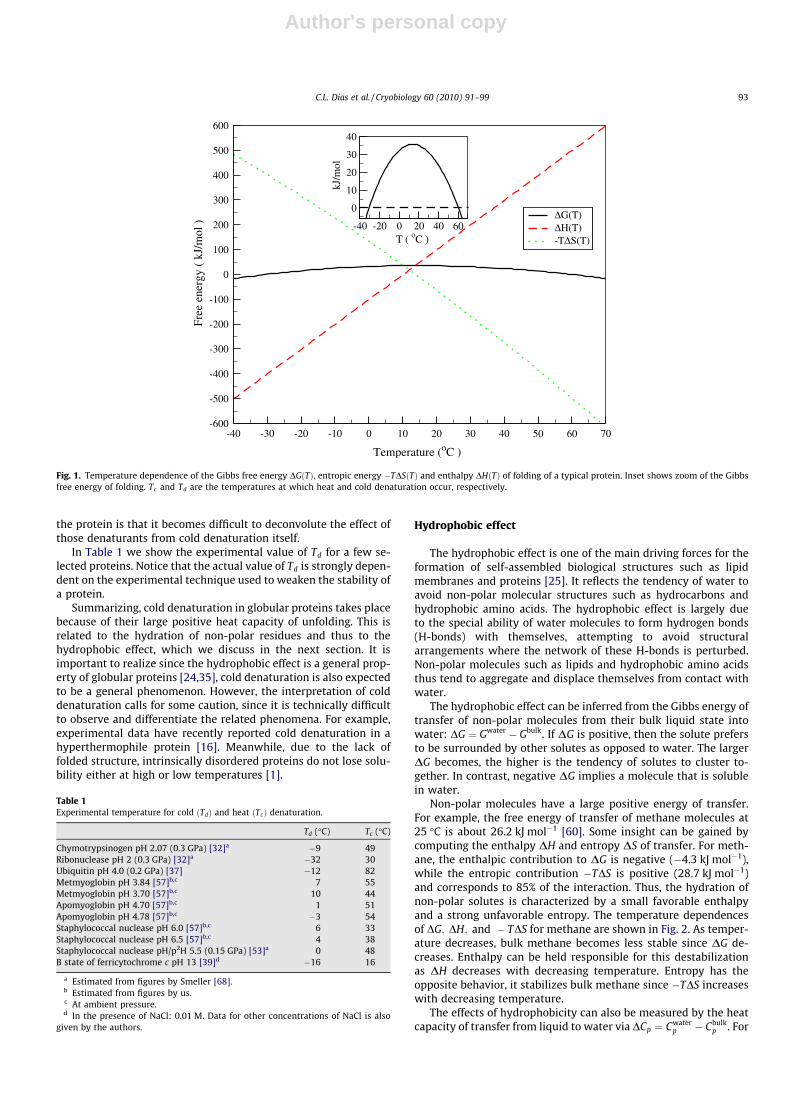
Fig. 1 shows the temperature dependence of DG for a typicalprotein. This quantity has a convex shape, indicating the presenceof two phase transitions. These transitions take place wheneverDGðTÞ ¼ 0. The transitions correspond to heat denaturation atT ¼ Tc , and to cold denaturation at T ’ �30 �C. At intermediatetemperatures, between T ’ �30 �C and Tc , the folded configura-tion is thermodynamically stable, with maximal stability occurringat about 17 �C. It is interesting to note that even under most stableconditions, very little energy is required to unfold the protein:DGðT ¼ 17 �CÞ ’ 32 kJ mol�1. Thus, while nature requires thefolded protein to be stable in order to function, the stability is mar-ginal, with 32 kJ mol�1 being only a minor fraction (about 5%) ofthe interaction energy of a single covalent bond between two car-bon atoms.
In this work, we are mainly interested in the low temperaturetransition to the denatured state, i.e., cold denaturation. Thermo-dynamically this transition results from the convex curvature ofthe Gibbs energy. This curvature can be computed from Eq. (6):
o2DGðTÞoT2 ¼ �DCP
T; ð7Þ
and it becomes convex, i.e., o2DGðTÞoT2 < 0, whenever DCp > 0. Forglobular proteins DCp is positive [57] and increases with the lengthof the protein [43]. This feature is mostly attributed to the hydra-tion of non-polar amino acids which have a distinguishable positiveDCp, as opposed to polar amino acids that contribute negatively tothe heat capacity of proteins [36,56].
The temperature Td at which cold denaturation occurs can beobtained by solving DGðTdÞ ¼ 0. This task becomes a simple analyt-ical exercise when the logarithmic term in Eq. (6) is approximatedby its second order Taylor expansion:
lnTc
T
� �’ Tc � T
T
� �� Tc � T
2T2 : ð8Þ
The temperature at which cold denaturation takes place then reads[40]:
T2d ’
T2c DCp
2DHðTcÞ þ TcDCp: ð9Þ
This equation shows clearly that cold denaturation becomes acces-sible at higher temperatures for proteins with larger DCp and Tc .Notice that DHðTcÞ has the opposite effect on Td.
For a typical protein, the temperature at which cold denatur-ation occurs is below the freezing point of water, see Fig. 1. Thisundesirable feature for experimental studies is usually overcomeby weakening the folded protein through pressure [40,46,59,68]or chemical denaturants [37,57]. Qualitatively, the weakeningcan be seen as shifting of the convex Gibbs energy (shown inFig. 1) downwards, thereby increasing Td above freezing, anddecreasing Tc . Pressure has the additional effect of decreasing thefreezing point of water to �22 �C (at 200 MPa) such that experi-ments can be performed within a wider range of temperatures be-low 0 �C. The drawback of using pressure or chemistry to weaken
92 C.L. Dias et al. / Cryobiology 60 (2010) 91–99
Author's personal copy
the protein is that it becomes difficult to deconvolute the effect ofthose denaturants from cold denaturation itself.
In Table 1 we show the experimental value of Td for a few se-lected proteins. Notice that the actual value of Td is strongly depen-dent on the experimental technique used to weaken the stability ofa protein.
Summarizing, cold denaturation in globular proteins takes placebecause of their large positive heat capacity of unfolding. This isrelated to the hydration of non-polar residues and thus to thehydrophobic effect, which we discuss in the next section. It isimportant to realize since the hydrophobic effect is a general prop-erty of globular proteins [24,35], cold denaturation is also expectedto be a general phenomenon. However, the interpretation of colddenaturation calls for some caution, since it is technically difficultto observe and differentiate the related phenomena. For example,experimental data have recently reported cold denaturation in ahyperthermophile protein [16]. Meanwhile, due to the lack offolded structure, intrinsically disordered proteins do not lose solu-bility either at high or low temperatures [1].
Hydrophobic effect
The hydrophobic effect is one of the main driving forces for theformation of self-assembled biological structures such as lipidmembranes and proteins [25]. It reflects the tendency of water toavoid non-polar molecular structures such as hydrocarbons andhydrophobic amino acids. The hydrophobic effect is largely dueto the special ability of water molecules to form hydrogen bonds(H-bonds) with themselves, attempting to avoid structuralarrangements where the network of these H-bonds is perturbed.Non-polar molecules such as lipids and hydrophobic amino acidsthus tend to aggregate and displace themselves from contact withwater.
The hydrophobic effect can be inferred from the Gibbs energy oftransfer of non-polar molecules from their bulk liquid state intowater: DG ¼ Gwater � Gbulk. If DG is positive, then the solute prefersto be surrounded by other solutes as opposed to water. The largerDG becomes, the higher is the tendency of solutes to cluster to-gether. In contrast, negative DG implies a molecule that is solublein water.
Non-polar molecules have a large positive energy of transfer.For example, the free energy of transfer of methane molecules at25 �C is about 26.2 kJ mol�1 [60]. Some insight can be gained bycomputing the enthalpy DH and entropy DS of transfer. For meth-ane, the enthalpic contribution to DG is negative (�4.3 kJ mol�1),while the entropic contribution �TDS is positive (28.7 kJ mol�1)and corresponds to 85% of the interaction. Thus, the hydration ofnon-polar solutes is characterized by a small favorable enthalpyand a strong unfavorable entropy. The temperature dependencesof DG; DH; and � TDS for methane are shown in Fig. 2. As temper-ature decreases, bulk methane becomes less stable since DG de-creases. Enthalpy can be held responsible for this destabilizationas DH decreases with decreasing temperature. Entropy has theopposite behavior, it stabilizes bulk methane since �TDS increaseswith decreasing temperature.
The effects of hydrophobicity can also be measured by the heatcapacity of transfer from liquid to water via DCp ¼ Cwater
p � Cbulkp . For
-40 -30 -20 -10 0 10 20 30 40 50 60 70
Temperature (oC )
-600
-500
-400
-300
-200
-100
0
100
200
300
400
500
600
Free
ene
rgy
( kJ
/mol
)
ΔG(T)ΔH(T)-TΔS(T)
-40 -20 0 20 40 60T ( oC )
0
10
20
30
40
kJ/m
ol
Fig. 1. Temperature dependence of the Gibbs free energy DGðTÞ, entropic energy �TDSðTÞ and enthalpy DHðTÞ of folding of a typical protein. Inset shows zoom of the Gibbsfree energy of folding. Tc and Td are the temperatures at which heat and cold denaturation occur, respectively.
Table 1Experimental temperature for cold ðTdÞ and heat ðTcÞ denaturation.
Td (�C) Tc (�C)
Chymotrypsinogen pH 2.07 (0.3 GPa) [32]a �9 49Ribonuclease pH 2 (0.3 GPa) [32]a �32 30Ubiquitin pH 4.0 (0.2 GPa) [37] �12 82Metmyoglobin pH 3.84 [57]b,c 7 55Metmyoglobin pH 3.70 [57]b,c 10 44Apomyoglobin pH 4.70 [57]b,c 1 51Apomyoglobin pH 4.78 [57]b,c �3 54Staphylococcal nuclease pH 6.0 [57]b,c 6 33Staphylococcal nuclease pH 6.5 [57]b,c 4 38Staphylococcal nuclease pH/p2H 5.5 (0.15 GPa) [53]a 0 48B state of ferricytochrome c pH 13 [39]d �16 16
a Estimated from figures by Smeller [68].b Estimated from figures by us.c At ambient pressure.d In the presence of NaCl: 0.01 M. Data for other concentrations of NaCl is also
given by the authors.
C.L. Dias et al. / Cryobiology 60 (2010) 91–99 93

Author's personal copy
simple solutions DCp is small, while for non-polar solutes it is largeand positive [27]. This large heat capacity of transfer has beenshown to be proportional to the surface area around non-polar sol-utes accessible to water [44], and thus proportional to the numberof solvent molecules around the solute [31].
Thermodynamics
As mentioned above the hydration of non-polar solutes in waterhas a large entropic cost and a small favorable enthalpy. This pecu-liar partition of the free energy imposes constraints on models forthe hydrophobic effect. In this section we discuss a model intro-duced by Muller [31,41,50] that displays the correct partition ofthe free energy and provides insights into the microscopic natureof the phenomena due to its simplicity.
The focus of Muller’s model is in the H-bond between watermolecules. Those are assumed to exist in two states in mutualequilibrium:
HbondðintactÞ�HbondðbrokenÞ; ð10Þ
where the equilibrium constant K is given by:
K � f1� f
¼ expð�DG�=RTÞ: ð11Þ
In this equation, f is the fraction of broken H-bonds, R is the gas con-stant and DG� is the difference in Gibbs free energy between brokenand intact states ðDG� ¼ Gbroken � GintactÞ. Based on the assumptionthat the energy of the system is determined by H-bonds alone,the enthalpy and entropy are given by:
H ¼ fHbroken þ ð1� f ÞHintact; ð12ÞS ¼ fSbroken þ ð1� f ÞSintact; ð13Þ
and the specific heat is given by:
cp �oHoT
� �P¼ of
oT
� �PDH�: ð14Þ
Here, DH� � Hbroken � Hintact and it is assumed to be temperatureindependent. The dependence of f on temperature is obtained fromEq. (11):
ofoT
� �P¼ DH�
f ð1� f ÞRT2 ; ð15Þ
such that the specific heat [31] is given by:
cp ¼ DH�ð Þ2f ð1� f Þ=RT2: ð16Þ
Eqs. (12), (13) and (16) account for the enthalpy, entropy and spe-cific heat of the H-bond network of water. Now, Muller assumesthat hydration energies of non-polar solutes are related to rear-rangements in this network alone. Thus hydration energies arecomputed as the difference in energies between the disturbed andundisturbed networks. Notice that the H-bond network is only per-turbed locally by the solute, i.e., in the first hydrated shell, and onlythese molecules need to be taken into account for the hydrationenergies. This implies that f, DH� and DS� are different for bulkand first-shell water and we use b and s subscripts to distinguishthem. Therefore the enthalpy and entropy upon hydration are givenby:
DH ¼ n ð1� fbÞDH�b � ð1� fsÞDH�s� �
; ð17ÞDS ¼ n ð1� fbÞDS�b � ð1� fsÞDS�s � RDF
� �; ð18Þ
where DF � Fs � Fb with Fb � fb ln fb þ ð1� fbÞ lnð1� fbÞ and simi-larly for Fs. These are the ‘‘mixing” entropies characteristic of mix-ture models. In these equations, n is the number of H-bonds in thefirst-shell and it is related to number N of water molecules byn ¼ 3N=2.1 In the same line of thought the heat capacity of hydrationis given by:
DChydrationp ¼ n cs
p � cbp
h i; ð19Þ
where csp and cb
p are the specific heat, given in Eq. (16), of first-shelland bulk water.
Actual values for the parameters of the model are given in Fig. 3.The entropy predicted with those values for propane, butane, andisobutane at 25 �C are �88.4 (�75.32), �101.4 (�93.20) and�98.9 (�89.14), respectively. Units are in J/K mol�1 and experi-
0 10 20 30 40 50 60
Temperature (oC )
-20
-10
0
10
20
30
40
50
Ene
rgy
(kJ
/mol
)
G H-T S
ΔΔ
Δ
Fig. 2. Experimental data [60] of methane’s free energy (circles), entropy (triangles) and enthalpy (squares).
1 To obtain Eq. (17), Muller assumes that the enthalpies of a broken H-bond in thefirst-shell and in the bulk are the same [41]. The same assumption is also made for theentropy of a broken H-bond in Eq. (18).
94 C.L. Dias et al. / Cryobiology 60 (2010) 91–99

Author's personal copy
mental results are given in parentheses – showing a good agree-ment. The temperature dependence of the specific heat of hydra-tion, Eq. (19), is characteristic of two-state models. Itsatisfactorily reproduces the positive DChydration for non-polar sol-ute and the observed decrease in DChydration with increasing T [31].
Muller’s model has been refined [41]. However, the original ver-sion of the model already shows that the energetic states of H-bonds are predominantly responsible for the thermodynamicalfeatures of the hydrophobic effect. The drawback of the model isthat it requires many parameters and does not explain how the dif-ferent energetic states of H-bonds correlate with the atomic struc-ture of water.
Atomic description
The first ‘‘pictorial representation” of the hydrophobic effectcame from Frank and Evans in 1945 [30]. While studying non-polarmolecules in liquids, they computed anomalous negative entropyof mixing for aqueous solution. This implied that hydration ofnon-polar molecules increased the amount of order in the system.
Also the expected positive heat of mixing related to an increase inthe number of broken H-bonds in shell water was absent. Thus,they concluded that non-polar solutes perturbed water towardsits crystalline state, locally ordering water and increasing theamount of H-bonds. Those ordered regions were named ‘‘iceberg”,although it was noted that the name should not be taken literally.
The structure of water molecules in the iceberg region is still aquestion of debate. It is clear that icebergs account for much lessorder than ice and thus, the suggested name is misleading. Thiscan be seen [35] by comparing the entropy released during freez-ing and during hydration per water molecule. Those are 22.17 J/K mol�1 and 4.18 J/K mol�1, respectively. The latter is much smal-ler indicating that freezing brings water molecules to a much moreordered state then iceberg water. On the other hand, the amount oforder in the icebergs is greater than in bulk (by 4.18 J/K mol�1 perwater molecule).
Insights into the ‘‘iceberg” structure can be gained by looking atthe distribution of H-bonds around the solute. At room tempera-ture, water saturates 3–3.5 H-bonds with neighboring molecules.When a non-polar solute is inserted in water, the number of satu-rated H-bonds should be much smaller for the molecules in thefirst hydrated shell around the solute. This can see by fixing the po-sition of a shell-water and rotating it in all possible orientations. Inmost of those orientations shell-water has at least one H-bondpointing towards the inert solute, therefore being non-saturated.On the other hand, a few orientations exist where all the H-bondsare saturated. We will refer to these two types of situations, asnon-saturated and saturated ones.
Energy wise, the saturated orientations have a lower (morefavorable) enthalpy than the many non-saturated ones. However,they also have lower (less favorable) entropy since there are notmany of them. This shows a balance between enthalpy and entropywhen the system switches from saturated to non-saturated orien-tations. This balance does not even out and the free energy of thesystem is smaller whenever shell-water are constrained to satu-rated orientations. In other words, the surface of the solute, whichis tiled with water molecules, has saturated orientations as its til-ing motif. Different types of tilings are possible. Those are calledclathrate cages [9,29] and the simplest of them is represented bya dodecahedron with water molecules sitting on each of its vertex(see Fig. 4). It is likely that a perfect cage only occurs at low tem-perature and at room temperature only incomplete cages survivefor a reasonable amount of time.
Thus, the formation of clathrate cages around the solute mini-mizes the free energy of shell water. However, even in those con-figurations shell-water has a higher free energy than bulk water,i.e., the hydration free energy is positive. Therefore, when morethan one solute is inserted in water they tend to cluster to reducethe amount of shell water in the system. This occurs through theoverlap of iceberg regions. The tendency of non-polar solutes tocluster is knows as the hydrophobic interaction. Since shell waterare more ordered and form more H-bonds than bulk water, theclustering of solutes increases the entropy and enthalpy of the sys-tem. Therefore, the hydrophobic interaction is stabilized by entro-py and destabilized by enthalpy. It is said to be entropically driven.
The behavior of the hydrophobic interaction upon cooling is ofsignificance to this paper. For most materials, the average strengthof the interaction between atoms increases as thermal energydecreases. In contrast, the strength of the hydrophobic interactiondecreases with decreasing temperature. This non-intuitive behav-ior is related to the complex interplay between entropy and enthal-py – see Fig. 2. Upon cooling, both the stabilizing effect of entropyand the destabilizing effect of enthalpy increase. This indicates thatthe differentiation between shell and bulk water increases withdecreasing temperature, with shell water becoming more orderedand forming more H-bonds than bulk water. However, the entropic
. 0621obS
. 6327osS
mol
J
molK
J
9. 08obH
696.10osH
Broken H-bond
Intact H-bond
Δ
Δ
Δ
Δ
=
=
=
=
.
Fig. 3. Parameters for Muller’s model. For the number N of water molecules in thefirst-shell Muller uses [31]: 25 for propane, 28 for butane and isobutane.
Fig. 4. Schematic representation of a clathrate hydrate caging a non-polar solute.Water molecules are in the vertices of the dodecahedron.
C.L. Dias et al. / Cryobiology 60 (2010) 91–99 95

Author's personal copy
and enthalpic terms do not change at the same rate. The enthalpicpenalty increases faster upon cooling, thus accounting for a weak-ening of the hydrophobic interaction. It should be noted that theinterplay between enthalpy and entropy is not clearly understoodfrom a microscopic point of view.
Cold denaturation
In the previous section we have discussed the thermodynamicsof the hydrophobic effect and how it arises from the atomic struc-ture of water. This effect has been shown to be the dominant driv-ing force for protein folding and is responsible for the stability ofthe protein core [24,35,51]. It has been incorporated in simplemodels of proteins where the solvent is describe implicitly – anexample is the well known Hydrophobic-Polar model [14]. An im-plicit description is unlikely to account for both cold and heatdenaturation – unless the parameters in the model are made tem-perature dependent [17]. When the solvent is described explicitlyboth heat and cold denaturation are recovered naturally[4,10,11,13,18,19,23,54,55,61,62,65]. In the proceeding paragraphsan overview of two approaches used to mimic the hydrophobic ef-fect in proteins through an explicit solvent are summarized. Explic-itly, a thermodynamical approach based on Muller’s model forwater and a molecular dynamics approach based on a simple mod-el that accounts for the relevant structure of shell water.
Thermodynamical model
One of the first models to account for cold denaturation in thephysics literature was proposed by De Los Rios and Caldarelli[62]. In this model, the protein corresponds to a self-avoiding ran-dom walk where each amino acid occupies a site in a lattice. Allother nodes i are occupied by water molecules which can be inthe bulk or form the first shell around the polymer. The bulk stateis considered to be q times degenerate and for simplicity the en-ergy of this state is set to zero. The first shell can be in an orderedor a disordered state [50]. The disordered state is considered to beq� 1 times degenerate while the ordered state is not degenerate.This model is therefore described by three parameters: J, K and q
– where J and K are the energies of the ordered and disorderedshell states, respectively. If s describes the state of first shell watermolecules such that s ¼ 0 represents the ordered state ands ¼ 1; . . . ; q� 1 corresponds to the disordered states, then theHamiltonian is given by:
H ¼X
j
�Jdsj ;0 þ Kð1� dsj ;0Þ� �
; ð20Þ
where the sum is over all water molecules that are nearest neigh-bors of some hydrophobic monomer. Therefore, for each conforma-tion C of the polymer its energy can be computed. The partitionfunction of the system can be cast in the form: ZN ¼
PCZNðCÞ,
where the partition function of a given conformation C is given by:
ZNðCÞ ¼ qnbðCÞ expðbJÞ þ ðq� 1Þexpð�bKÞ½ �nsðCÞ; ð21Þ
where ns and nb are the number of water molecules in the first shelland bulk, respectively, and b is reciprocal of the thermal energy.
It is possible to classify the polymer according to ns. For self-avoiding random walks of size N, the fraction of configurations ofperimeter ns is well approximated by a Poisson distribution:
PNðnsÞ � edðN�1Þ dðN � 1Þ½ �2Nþ2�ns
ð2N þ 2� nsÞ!; ð22Þ
with d � 0:75. Using this distribution and Eq. (21), the partitionfunction of the system reads:
ZNðbÞ ¼X2Nþ2
nmin
PNðnÞq2Nþ2�n expðbJÞ þ ðq� 1Þ expð�bKÞ½ �n; ð23Þ
where the smallest perimeter nmin ¼ 2ffiffiffiffiffiffiffipNp
, assuming that in thisconfiguration the system has a circular compact shape. The maxi-mum number of water sites in contact with the polymer is2N þ 2. The heat capacity, computed as:
Cv ¼ b2 o2 ln Z=ob2 ; ð24Þ
is shown in Fig. 5. Three peaks appear in the heat capacity. Fromzero temperature to the first peak, the polymer is swollen – inagreement with cold denaturation. As the temperature is raisedabove the first peak, the polymer folds and the number of first shellwater is approximately 2
ffiffiffiffiffiffiffipNp
. As temperature increases above the
0 1 2 3 4
Temperature
0
100
200
300
400
500
600
700
Cv N = 100
N = 50N = 25
Swollen
Folded
Molten globule
Swollen
Fig. 5. Heat capacity, Eq. (24), for different polymer sizes. Here, K/J = 2 and q ¼ 103.
96 C.L. Dias et al. / Cryobiology 60 (2010) 91–99

Author's personal copy
second peak, the polymer occupies globule-molten states. Beyondthe third peak, the polymer reopens.
Consistent with experiments, the free energy of the model has aconvex curvature which is correctly partitioned in entropy and en-thalpy [62]. The model has been extended successfully to study thepresence of kosmotropes and chaotropes cosolvent [48,49]. How-ever, as in the case of Muller’s model, it does not provide much in-sight into the structure of water around the solute. In the nextsection we describe an attempt to fill this gap.
Atomic description
Recently, an effort to understand the physical mechanism be-hind cold denaturation of biopolymers was undertaken by con-structing a minimal microscopic model for the protein–watersystem [23]. For computational efficiency, this study was per-formed in 2D. The water phase was modeled using the 2D Merce-des-Benz (MB) model [5,7,25] where H2O molecules arerepresented as 2D disks, with three H-bonding arms resemblingthe famous Mercedes-Benz symbol. This model has now been ex-tended to 3D and the first results look very promising [8,22]. Thissimple model (2D) reproduces many important thermodynamicproperties of water, such as the density anomaly, the minimumin the isothermal compressibility as a function of temperature,the large heat capacity, and the experimental trends for the ther-modynamic properties of hydration of non-polar solutes [25,67].In the study, the parameters of the MB model were chosen to bethose used by Silverstein et al. [66].
To model the protein, the simple bead-spring model of poly-mers [26] was used: monomers which are adjacent along the back-bone of the protein are connected to each other by harmonicsprings, and non-adjacent monomers are connected by a shiftedLennard-Jones potential. The interaction between monomers andwater molecules was also given by a shifted Lennard-Jones poten-tial with the same binding energy as between the water molecules.
To study the process of cold denaturation at constant pressure,molecular dynamics simulations were performed on the water–protein model system in the isothermal-isobaric ensemble[2,28,38]. The pressure in the system was set such that the MBmodel reproduces water-like anomalies seen at ambient pressure
[25] and hydrates non-polar molecules in a realistic manner [69].Typically, the simulation box contained 512 molecules comprisedof a 10-monomer long protein and 502 water molecules. To inducedenaturation, simulations were performed at various different(effective) temperatures.
The main result for the study can be seen in Fig. 6, where theequilibrium distribution of the size of the protein, as measuredby its radius of gyration RG [26], is shown at three different temper-atures. In ‘‘hot” water (referring to the largest values of T shownhere), proteins favor more compact configurations with decreasingtemperature. However, a further decrease of temperature results inreversal of this trend: as the temperature decreases further, thepeak shifts to a larger value indicating that in ‘‘cold” water proteinsbecome less compact for decreasing temperature. This behaviorcan be seen systematically in the inset of Fig. 6, which depictsthe temperature dependence of the protein size. This type of non-monotonic behavior is characteristic to denaturation of real pro-teins and in line with previous studies [40,54,61].
Characteristic configurations of the protein at different temper-atures are shown in Fig. 7. In cold water (upper panels), the mono-mers are surrounded by an ordered layer of ‘‘shell” watermolecules. Molecules forming this cage are strongly H-bonded toeach other and therefore have a low energy. At T ¼ 0:21, the pro-tein favors compact configurations. Water molecules close to theprotein have at least one non-saturated H-bond which is pointingtowards the protein. When the temperature is increased toT ¼ 0:25, most monomers are in contact with the solvent. In Ref.[23], these observations were further quantified by computingthe average H-bond energy per water molecule for shell and bulkwater.
The molecular dynamics simulations of the MB model provide asimple microscopic picture for cold denaturation in terms ofchanges in hydration: at low temperatures water molecules infil-trate the folded protein in order to passivate the ‘‘dangling”water–water H-bonds found in shell water. At the same time,hydrophobic contacts are destabilized and an ordered layer of‘‘shell” water molecules forms around the protein monomers suchthat they become separated by a layer of solvent in the cold dena-tured state. Solvent layers around the monomer pairs are highly or-dered such that their formation decreases the total entropy of the
Fig. 6. Normalized distribution of the radius of gyration of the protein ðRGÞ at three effective temperatures: T ¼ 0:25 (‘‘hot” water), T ¼ 0:21 (intermediate temperature) andT ¼ 0:17 (‘‘cold” water). Inset shows the detailed temperature dependence of the size of the protein [23].
C.L. Dias et al. / Cryobiology 60 (2010) 91–99 97

Author's personal copy
system. The existence of such low entropic states for shell water atlow T explains why cold denaturation proceeds with heat releaseas opposed to heat absorption seen during heat denaturation.
Conclusion
In this paper we reviewed and summarized some of the effortsto model and understand the hydrophobic effect and the role itplays in the thermodynamics of proteins. This review is not in-tended to be exhaustive but to focus on the recent advances inphysics and chemistry, and especially on the efforts in computa-tional modeling and theory. Thermodynamical models for thehydration of non-polar solutes are successful in reproducing exper-imental data accurately but they rely on many parameters thatneed to be adjusted (see ‘‘Thermodynamics”). They provide a basisfor inferring the molecular structure of water around those soluteswhich is responsible for the hydrophobic effect (see ‘‘Atomicdescription” in ‘‘Thermodynamics of proteins”). When those ther-modynamical models are coupled to a coarse-grained structureof proteins (see ‘‘Thermodynamical model”), non-trivial but realis-tic phases of these biomolecules are found to coexist. Simulationsof simple models have been performed revealing a potential mech-anism for cold denaturation of proteins which is consistent withthe thermodynamical models (see ‘‘Atomic description” in ‘‘Colddenaturation”). Finally, when comparing with experiments, thereare also subtleties which should be considered. For example, recentresults have shown that replacing water with deuterium, as iscommon practise, may lead to changes in the physical properties[64].
Acknowledgments
C.L.D. thank Janet Elliott for suggesting and motivating thispaper.
References
[1] Agnes Tantos, P.T. Peter Friedrich, Cold stability of intrinsically disorderedproteins, FEBS Lett. 583 (2009) 465.
[2] H.C. Andersen, Molecular dynamics simulations at constant pressure and/ortemperature, J. Chem. Phys. 72 (1980) 2384.
[3] C.B. Anfinsen, Principles that govern the folding of protein chains, Science 181(4096) (1973) 223–230.
[4] E. Ascolese, G. Graziano, On the cold denaturation of globular proteins, Chem.Phys. Lett. 467 (2008) 150–153.
[5] J.-P. Becker, O. Collet, Mercedes-Benz model of neutral amino-acid side chains,J. Mol. Struct.: THEOCHEM 774 (2006) 23–28.
[6] W.J. Becktel, J.A. Schellman, Protein stability curves, Biopolymers 26 (1987)1859.
[7] A. Ben-Naim, Statistical mechanics of waterlike particles in two dimensions. I.Physical model and application of the percus yevick equation, J. Chem. Phys. 54(1971) 3682.
[8] A. Bizjak, T. Urbic, V. Vlachy, K. Dill, The three-dimensional Mercedes-Benzmodel of water, Acta Chim. Slov. 54 (2007) 532–537.
[9] D.T. Bowron, A. Filipponi, M.A. Roberts, J.L. Finney, Hydrophobic hydration andthe formation of a clathrate hydrate, Phys. Rev. Lett. 81 (1998) 4164–4167.
[10] P. Bruscolini, L. Casetti, Lattice model for cold and warm swelling of polymersin water, Phys. Rev. E 61 (2000) R2208.
[11] P. Bruscolini, L. Casetti, Modeling hydration water and its role in polymerfolding, J. Biol. Phys. 27 (2001) 243–256.
[12] S.V. Buldyrev, P. Kumar, H.E. Stanley, A physical mechanism underlying theincrease of aqueous solubility of nonpolar compounds and the denaturation ofproteins upon cooling, Conden. Mater. (2007). cond-mat/0701485.
[13] C. Buzano, E.D. Stefanis, M. Pretti, Low-temperature-induced swelling of ahydrophobic polymer: a lattice approach, J. Chem. Phys. 126 (7) (2007)074904.
[14] H.S. Chan, K.A. Dill, Compact polymers, Macromolecules 22 (1989) 4559–4573.[15] D. Chandler, Interfaces and the driving force of hydrophobic assembly, Nature
437 (2005) 640.[16] S.K. Chandrayan, P. Guptasarma, Partial destabilization of native structure by a
combination of heat and denaturant facilitates cold denaturation in ahyperthermophile protein, Proteins 72 (2008) 539.
[17] O. Collet, Warm and cold denaturation in the phase diagram of a protein latticemodel, Europhys. Lett. 53 (2001) 93–99.
[18] O. Collet, Four-states phase diagram of proteins, Europhys. Lett. 72 (2005)301–307.
[19] O. Collet, Folding kinetics of proteins and cold denaturation, J. Chem. Phys. 129(15) (2008) 155101.
[20] S. Cooper, R. Dawber, History of cryosurgery, J. Roy. Soc. Med. 94 (2001) 196.[21] T.E. Creighton, Protein folding, Biochem. J. 270 (1) (1990) 1–16.[22] C. Dias, T. Ala-Nissila, M. Grant, M. Karttunen, Three-dimensional Mercedes-
Benz model for water, J. Chem. Phys. 131 (2009) doi:10.1063/1.3183935.[23] C.L. Dias, T. Ala-Nissila, M. Karttunen, I. Vattulainen, M. Grant, Microscopic
mechanism for cold denaturation, Phys. Rev. Lett. 100 (2008) 118101.[24] K.A. Dill, Dominant forces in protein folding, Biochemistry 29 (1990) 7133.[25] K.A. Dill, T.M. Truskett, V. Vlachy, Hribar-Lee, Modeling water, the
hydrophobic effect, and ion solvation, Annu. Rev. Biophys. Biomol. Struct. 34(2005) 173.
[26] M. Doi, S.F. Edwards, The Theory of Polymer Dynamics, Oxford UniversityPress, New York, 1988.
[27] J.T. Edsall, Apparent molal heat capacities of amino acids and other organiccompounds, J. Am. Chem. Soc. 54 (1935) 1506–1507.
[28] S.E. Feller, Y. Zhang, R.W. Pastor, B.R. Brooks, Constant pressure molecular dynamicssimulation: the langevin piston method, J. Chem. Phys. 103 (1995) 4613.
Fig. 7. Characteristic configurations of a protein in cold water ðT ¼ 0:15 and T ¼ 0:17Þ, at an intermediate temperature ðT ¼ 0:21Þ, and in hot water ðT ¼ 0:25Þ. The ‘‘shell”water molecules close to the protein are highlighted here. In cold water, the monomers are typically surrounded by clathrate-like cages [23].
98 C.L. Dias et al. / Cryobiology 60 (2010) 91–99

Author's personal copy
[29] A. Filipponi, D.T. Bowron, C. Lobban, J.L. Finney, Structural determination of thehydrophobic hydration shell of Kr, Phys. Rev. Lett. 79 (1997) 1293–1296.
[30] H.S. Frank, M.W. Evans, Free volume and entropy in condensed systems: III.Entropy in binary liquid mixtures; partial molal entropy in dilute solutions;structure and thermodynamics in aqueous electrolytes, J. Chem. Phys. 13(1945) 507.
[31] S.J. Gill, S.F. Dec, G. Olofsson, I. Wadsoe, Anomalous heat capacity ofhydrophobic solvation, J. Phys. Chem. 89 (1985) 3758.
[32] S.A. Hawley, Reversible pressure–temperature denaturation of chymotrypsinogen,Biochemistry 10 (1971) 2436–2442.
[33] F.G. Hopkins, Denaturation of proteins by urea and related substances, Nature126 (1930) 383.
[34] G. Hummer, S. Garde, A.E. Garcia, M.E. Paulaitis, The pressure dependence ofhydrophobic interactions is consistent with the observed pressuredenaturation of proteins, Proc. Natl. Acad. Sci. USA 95 (1998) 1552.
[35] W. Kauzmann, Adv. Protein Chem. 14 (1959) 1.[36] M. Kinoshita, T. Yoshidome, Molecular origin of the negative heat capacity of
hydrophilic hydration, J. Chem. Phys. 130 (2009) 144705.[37] R. Kitahara, A. Okuno, M. Kato, Y. Taniguchi, S. Yokoyama, K. Akasaka, Cold
denaturation of ubiquitin at high pressure, Magn. Reson. Chem. 44 (2006)S108–S113.
[38] A. Kolb, B. Dünweg, Optimized constant pressure stochastic dynamics, J. Chem.Phys. 111 (1999) 4453.
[39] R. Kumar, N. Prakash Prabhu, K. Krishna Rao, K. Bhuyan, Abani, The alkalimolten globule state of horse ferricytochrome c: observation of colddenaturation, J. Mol. Biol. 364 (2006) 483.
[40] S. Kunugi, N. Tanaka, Cold denaturation of proteins under high pressure,Biochim. Biophys. Acta 1595 (2002) 329–344.
[41] B. Lee, G. Graziano, A two-state model of hydrophobic hydration that producescompensation enthalpy and entropy changes, J. Am. Chem. Soc. 118 (22)(1996) 5163.
[42] Y. Li, B. Shan, D. Raleigh, The cold denatured state is compact but expands atlow temperatures: hydrodynamic properties of the cold denatured state of thec-terminal domain of l9, J. Mol. Biol. 368 (2007) 256.
[43] J. Liang, K.A. Dill, Are proteins well-packed?, Biophys J. 81 (2001) 751.[44] J.R. Livingstone, R.S. Spolar, M.T. Record, Contribution to the thermodynamics
of protein folding from the reduction in water-accessible nonpolar surfacearea, Biochemistry 30 (1991) 4237.
[45] M.I. Marques, J.M. Borreguero, H.E. Stanley, N.V. Dokholyan, Possiblemechanism for cold denaturation of proteins at high pressure, Phys. Rev.Lett. 91 (13) (2003) 138103.
[46] F. Meersman, C.M. Dobson, K. Heremans, Protein unfolding, amyloid fibrilformation and configurational energy landscapes under high pressureconditions, Chem. Soc. Rev. 35 (2006) 908–917.
[47] S. Melchionna, G. Briganti, P. Londei, P. Cammarano, Water induced effects onthe thermal response of a protein, Phys. Rev. Lett. 92 (15) (2004) 158101.
[48] S. Moelbert, B. Normand, P.D.L. Rios, Kosmotropes and chaotropes: modellingpreferential exclusion, binding and aggregate stability, Biophys. Chem. 112(2004) 45.
[49] S. Moelbert, P.D.L. Rios, Chaotropic effect and preferential binding in ahydrophobic interaction model, J. Chem. Phys 119 (15) (2003) 7988–8001.
[50] N. Muller, Search for a realistic view of hydrophobic effects, Acc. Chem. Res. 23(1990) 23–28.
[51] A. Nicholls, K.A. Sharp, B. Honig, Protein folding and association: insights fromthe interfacial and thermodynamic properties of hydrocarbons, Proteins 11(1991) 281.
[52] C.N. Pace, C. Tanford, Thermodynamics of the unfolding of b-lactoglobulin ain aqueous urea solutions between 5 and 55 �C, Biochemistry 7 (1968)198.
[53] G. Panick, G.J.A. Vidugiris, R. Malessa, G. Rapp, R. Winter, C. Royer, Exploringthe temperature–pressure phase diagram of staphylococcal nuclease,Biochemistry 38 (1999) 4157–4164.
[54] D. Paschek, S. Nonn, A. Geiger, Low-temperature and high-pressure inducedswelling of a hydrophobic polymer-chain in aqueous solution, Phys. Chem.Chem. Phys. 7 (2005) 2780–2786.
[55] B.A. Patel, P.G. Debenedetti, F.H. Stillinger, P.J. Rossky, The effect of sequenceon the conformational stability of a model heteropolymer in explicit water, J.Chem. Phys. 128 (2008) 175102.
[56] N.V. Prabhu, K.A. Sharp, Heat capacity in proteins, Annu. Rev. Phys. Chem. 56(2005) 521.
[57] P.L. Privalov, Thermodynamics of protein folding, J. Chem. Thermodyn. 29(1997) 447–474.
[58] P.L. Privalov, Y.V. Griko, S.Y. Venyaminov, Cold denaturation of myoglobin, J.Mol. Biol. 190 (1986) 487–498.
[59] R. Ravindra, R. Winter, On the temperature–pressure free-energy landscape ofproteins, ChemPhysChem 4 (2003) 359–365.
[60] T.R. Rettich, Y.P. Handa, R. Battino, E. Wilhelm, Solubility of gases in liquids. 13.High-precision determination of Henry’s constants for methane and ethane inliquid water at 275–328 K, J. Phys. Chem. 85 (1981) 3230.
[61] P.D.L. Rios, G. Caldarelli, Putting proteins back into water, Phys. Rev. E 62(2000) 8449.
[62] P.D.L. Rios, G. Caldarelli, Cold and warm swelling of hydrophobic polymers,Phys. Rev. E 63 (2001) 031802.
[63] A.D. Robertson, K.P. Murphy, Protein structure and the energetics of proteinstability, Chem. Rev. 97 (1997) 1251.
[64] T. Róg, K. Murzyn, J. Milhaud, M. Karttunen, M. Pasenkiewicz-Gierula, Waterisotope effect on the bilayer properties: a molecular dynamics simulationstudy, J. Phys. Chem. B 113 (2009) 97.
[65] G. Salvi, S. Molbert, P.D.L. Rios, Design of lattice proteins with explicit solvent,Phys. Rev. E 66 (2002) 061911.
[66] K.A.T. Silverstein, A.D.J. Haymet, K.A. Dill, A simple model of water and thehydrophobic effect, J. Am. Chem. Soc. 120 (13) (1998) 3166–3175.
[67] K.A.T. Silverstein, A.D.J. Haymet, K.A. Dill, Molecular model of hydrophobicsolvation, J. Chem. Phys. 111 (17) (1999) 8000–8009.
[68] L. Smeller, Pressure–temperature phase diagrams of biomolecules, Biochim.Biophys. Acta 1595 (2002) 11–29.
[69] N.T. Southall, K.A. Dill, Potential of mean force between two hydrophobicsolutes in water, Biophys. Chem. 101–102 (2002) 295–307.
[70] T. Yoshidome, M. Kinoshita, Hydrophobicity at low temperatures and colddenaturation of a protein, Phys. Rev. E 79 (3) (2009) 030905.
[71] A. Zipp, W. Kauzmann, Pressure denaturation of metmyoglobin, Biochemistry12 (1973) 4217–4228.
C.L. Dias et al. / Cryobiology 60 (2010) 91–99 99