Solvent Extraction Research and Development, Japan, Vol. 23 ...
Upload
khangminh22Category
view
2download
0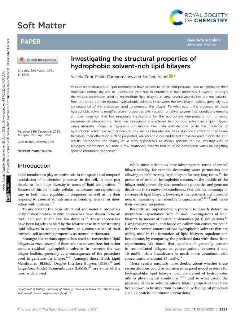
This journal is © The Royal Society of Chemistry 2021 Soft Matter, 2021, 17, 5329–5335 | 5329
Cite this: Soft Matter, 2021,
17, 5329
Investigating the structural properties ofhydrophobic solvent-rich lipid bilayers
Valeria Zoni, Pablo Campomanes and Stefano Vanni *
In vitro reconstitutions of lipid membranes have proven to be an indispensable tool to rationalize their
molecular complexity and to understand their role in countless cellular processes. However, amongst
the various techniques used to reconstitute lipid bilayers in vitro, several approaches are not solvent-
free, but rather contain residual hydrophobic solvents in between the two bilayer leaflets, generally as a
consequence of the procedure used to generate the bilayer. To what extent the presence of these
hydrophobic solvents modifies bilayer properties with respect to native, solvent-free, conditions remains
an open question that has important implications for the appropriate interpretation of numerous
experimental observations. Here, we thorouhgly characterize hydrophobic solvent-rich lipid bilayers
using atomistic molecular dynamics simulations. Our data indicate that while the presence of
hydrophobic solvents at high concentrations, such as hexadecane, has a significant effect on membrane
thickness, their effects on surface properties, membrane order and lateral stress are quite moderate. Our
results corroborate the validity of in vitro approaches as model systems for the investigations of
biological membranes but raise a few cautionary aspects that must be considered when investigating
specific membrane properties.
Introduction
Lipid membranes play an active role in the spatial and temporalmodulation of biochemical processes in the cell, in large partthanks to their huge diversity in terms of lipid composition.1–3
Because of this complexity, cellular membranes can significantlyvary in both their equilibrium properties as well as in theirresponse to external stimuli such as bending, tension or inter-action with proteins.1–3
To understand the basic structural and material propertiesof lipid membranes, in vitro approaches have shown to be aninvaluable tool in the last few decades.4,5 These approacheshave been largely enabled by the relative ease in reconstitutinglipid bilayers in aqueous medium, as a consequence of theirintrinsic self-assembly properties as natural surfactants.
Amongst the various approaches used to reconstitute lipidbilayers in vitro, several of them are not solvent-free, but rathercontain residual hydrophobic solvents in between the twobilayer leaflets, generally as a consequence of the procedureused to generate the bilayer.6–11 Amongst those, Black LipidMembranes (BLMs),9 Droplet Interface Bilayers (DIBs),10 andLarge-Area Model Biomembranes (LAMBs)11 are some of themost widely used.
While these techniques have advantages in terms of overallbilayer stability, for example decreasing water permeation andallowing to stabilize very large bilayers for very long times,11 thepresence of residual hydrophobic solvents in the middle of thebilayer could potentially alter membrane properties and generatedeviations from native-like conditions. One distinct advantage ofsolvent-rich lipid bilayers, however, is the relative straightforward-ness in measuring their membrane capacitance,8,12,13 and hencetheir electrical properties.
Recently, we implemented a protocol to directly determinemembrane capacitance from in silico investigations of lipidbilayers by means of molecular dynamics (MD) simulations.14
Using this approach, and based on calibration curves, we couldinfer the correct amount of two hydrophobic solvents that arewidely used in the formation of lipid bilayers, squalene andhexadecane, by comparing the predicted data with those fromexperiments. We found that squalene is generally presentin reconstituted bilayers at concentrations between 5 and10 mol%, while hexadecane is much more abundant, withconcentrations around 35 mol%.14
These results naturally raise doubts about whether thesereconstitutions could be considered as good model systems forbiological-like lipid bilayers, that are devoid of hydrophobicoils in physiological conditions,15,16 and to what extent thepresence of these solvents affects bilayer properties that havebeen shown to be important to rationalize biological processessuch as protein-membrane interactions.
Department of Biology, University of Fribourg, Chemin du Musee 10, 1700 Fribourg,
Switzerland. E-mail: [email protected]
Received 28th December 2020,Accepted 27th April 2021
DOI: 10.1039/d0sm02270e
rsc.li/soft-matter-journal
Soft Matter
PAPER
Ope
n A
cces
s A
rtic
le. P
ublis
hed
on 2
8 A
pril
2021
. Dow
nloa
ded
on 4
/17/
2022
3:1
7:37
AM
. T
his
artic
le is
lice
nsed
und
er a
Cre
ativ
e C
omm
ons
Attr
ibut
ion-
Non
Com
mer
cial
3.0
Unp
orte
d L
icen
ce.
View Article OnlineView Journal | View Issue
5330 | Soft Matter, 2021, 17, 5329–5335 This journal is © The Royal Society of Chemistry 2021
To investigate this issue, here we use atomistic MD simula-tions, the method of choice to investigate structural anddynamical properties of lipid assemblies with atomisticresolution.17–21 With this methodology, we thoroughly analyzebilayer properties in solvent-rich lipid bilayers, and we compareit to solvent-free bilayers.
We found that while the presence of high amounts of oils,such as hexadecane, has a significant effect on membranethickness, its effect on surface properties, membrane orderand lateral stress is quite moderate. Our results corroborate thevalidity of such in vitro approaches as model systems for theinvestigations of biological membranes but raise a few cau-tionary aspects that must be considered when investigatingspecific membrane properties.
Materials and methodsMolecular dynamics simulations
Atomistic MD simulations of 100 mol% 1,2-dioleoyl-sn-glycero-3-phosphocholine (DOPC), DOPC/Squalene (90 : 10 mol%) andDOPC/Hexadecane (68 : 32 mol%) were carried out using thesoftware GROMACS22 as previously described in.14 In short, aDOPC bilayer containing 128 lipids was generated using theCHARMM-GUI membrane builder.23 Addition of hexadecane orsqualene in the middle of the bilayer was achieved by displa-cing the two DOPC monolayers along the z-axis and by sub-sequent random insertion of the oil molecules, exclusively in aposition parallel to the membrane, between the two mono-layers. The systems were equilibrated for 375 ps while slowlyremoving constraints using the Membrane Builder six-stepprocess.24 For all systems, the CHARMM36m force field25 wasused to describe lipids while the TIP3P26 model was employedfor water. Long-range electrostatic interactions were taken intoaccount by means of the particle mesh Ewald (PME)27 algo-rithm with a Fourier grid space of 0.12 nm and a real spacecutoff of 1.2 nm. The van der Waals interactions were truncatedusing the same cutoff value, and a standard smoothing func-tion for the tail region (1.0–1.2 nm) was employed. The bondsinvolving hydrogen atoms were constrained using the LINCS28
and SETTLE29 algorithms, thus allowing to use of an integra-tion time step of 2 fs. The temperature (298.15 K) and pressure(1 atm) were controlled by coupling the systems to a Nose–Hoover chain thermostat30 with a time constant of 1 ps�1 and aParrinello–Rahman barostat31 with a coupling constant of5 ps�1, respectively.
All systems (containing 128 DOPC lipids and about 77000water molecules, plus 15 squalene or 60 hexadecane molecules)were run for at least 450 ns to collect statistics. Analyses werecarried out excluding the first 50 ns, unless otherwise explicitlystated, and data points for analyses were collected every 10 ps. Arepresentative snapshot of the molecular systems used in thisstudy, together with the chemical structure of the moleculesinvestigated, is shown in Fig. 1. All images were rendered usingVMD32 and all plots were created using python scripts.
Bilayer analyses
Area per lipid. Area per lipid was computed by dividing thetotal area of the bilayer in the xy plane by the number of lipidsin one leaflet.
Lateral density profiles. Lateral density profiles were calcu-lated using the GROMACS tool ‘‘gmx density’’. Density profileswere calculated along the z axis, dividing the systems in 250slices (around 0.04 nm per slice).
Lateral lipid diffusion. Phospholipid (PL) and oil diffusionwas computed by linear regression of the mean squared dis-placement of selected atoms (phosphorus atom (P) for DOPCand the central carbons (C8 for HEXD and C15 for SQL) for theoils). Fitting was computed after removal of the first and last10% of the trajectory.
Oil molecular orientation. The orientation of hexadecaneand squalene in the bilayer was calculated, for each oil mole-cule at every configuration along the trajectory, as the acuteangle between the bilayer normal and the line passing throughits first and last carbon atoms. The corresponding probabilitydensity distributions were computed using the kernel densityestimation (KDE) method.
Fig. 1 Chemical systems investigated in this study. (A) Chemical struc-tures of DOPC, hexadecane and squalene molecules. In the figures,hexadecane is referred as ‘‘hexd’’, squalene as ‘‘sql’’. (B) Representativesnapshot of a solvent-rich lipid bilayer investigated in this study. Colorlegend: DOPC headgroups (gray), DOPC tails (yellow), oil molecules (red)and water molecules (cyan). The same color code is used in all the figures.
Paper Soft Matter
Ope
n A
cces
s A
rtic
le. P
ublis
hed
on 2
8 A
pril
2021
. Dow
nloa
ded
on 4
/17/
2022
3:1
7:37
AM
. T
his
artic
le is
lice
nsed
und
er a
Cre
ativ
e C
omm
ons
Attr
ibut
ion-
Non
Com
mer
cial
3.0
Unp
orte
d L
icen
ce.
View Article Online
This journal is © The Royal Society of Chemistry 2021 Soft Matter, 2021, 17, 5329–5335 | 5331
Lateral pressure profiles. Lateral Pressure Profiles (LPP) werecalculated using the formula
p(z) = PL(z) � PN
where PL(z) is the lateral component of the pressure tensor(PL(z) = 1/2(Pxx(z) + Pyy(z))) and PN is the normal component(PN = Pzz).
For the calculation of LPPs, the program GROMACS-LS, witha grid of 0.1 nm along the z direction and a cut-off of 2.2 nm forelectrostatics, was used33 (https://www.mdstress.org). For pureDOPC, the LPP was calculated over the last 200 ns of produc-tion, while, for the systems containing oils, configurationsextracted from the last 300 ns of production were used forLPP calculations. The raw data have been processed through aGaussian filter with standard deviation 1.0 using the utility‘‘tensortools’’ of the GROMACS_LS program.
Order parameters. Order parameters were calculated usingthe formula
3
2cos2 y� 1
2
� ���������
where y is the average angle between the C–H vector and thebilayer normal. The calculation was performed using a tcl script(https://user.eng.umd.edu/Bjbklauda/wiki/doku.php?id=scd).
Lipid-packing defects. Lipid-packing defects were calculatedusing a modified version of PackMem,34,35 as previouslydescribed,36 that takes into account the presence of oil mole-cules in membranes. The cartesian algorithm used by theprogram maps the membrane surface to a grid parallel to thebilayer and assigns values to each grid depending on the type ofthe first atom encountered (aliphatic vs. polar). Lipid packingdefects are defined as regions where lipids are loosely packedand therefore aliphatic atoms are exposed to the surface. Forthe definition of the central atom (or ‘‘glycerol atom’’, asnamed in the manual) of the oil molecules, the central carbonwas used (C8 for hexadecane and C15 for squalene).
Results and discussionHydrophobic solvents increase bilayer thickness and depletephospholipids from the centre of the bilayer
The most direct effect of the presence of residual solvents in thebilayer is an effective increase in its thickness.14 This isexpected, as the membrane capacitance, that is inversely pro-portional to the membrane thickness,37 can be significantlylower than that of solvent free lipid bilayers when the mem-branes are prepared with hydrophobic oils.38 In detail, thedistance between the phosphate groups of the two monolayersincreases by approximately 5% (from 3.89 to 4.07 nm) in thepresence of squalene and by 13% (from 3.89 to 4.38 nm) in thepresence of hexadecane.14 To put this difference into somebiological context, in the case of hexadecane this correspondsto roughly one entire a-helical turn (3.6 residues, 0.54 nm), avery common secondary structure conformation of transmem-brane (TM) proteins. Interestingly, this difference is within therange of the observed difference between Golgi and Plasma
Membrane proteins for vertebrates39 or in the range of what issufficient to alter the localization of synthetic TM peptides inthe yeast ER.40
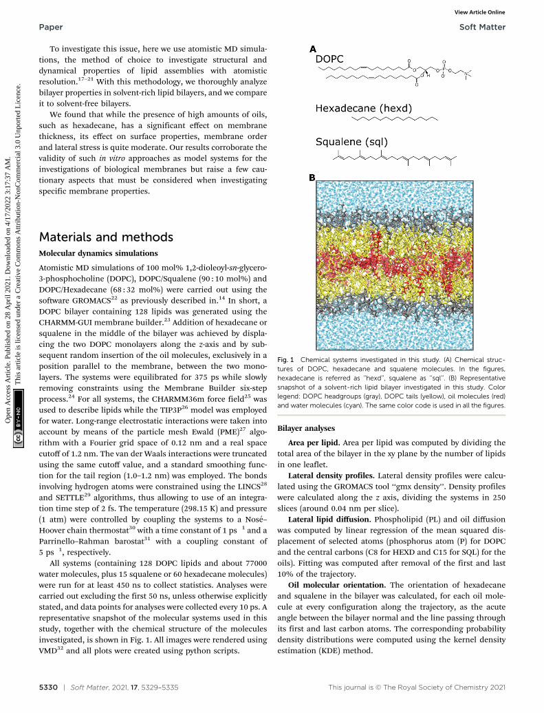
To further investigate the localization of hydrophobic sol-vents in the bilayer, we first compared the lateral densityprofiles of all non-water molecules in the bilayers. This corre-sponds to only DOPC molecules in the pure PL system and ofDOPC + oil in the PLs + squalene and PLs + hexadecane systems(Fig. 2A). Interestingly, in the presence of squalene, the overallbilayer density profile (Fig. 2A) is quite similar to that of 100%DOPC. On the other hand, the presence of hexadecane signifi-cantly affects the density profile at almost all depths, with thesole exception of the middle of the bilayer (Fig. 2A).
Next, we computed the lateral density profiles of DOPClipids and of the two oils (squalene, hexadecane) separately(Fig. 2B). This analysis indicates that, as expected, the two oilsoccupy predominantly the middle of the bilayer, i.e. the spacein between the PL acyl chains (Fig. 2B). Interestingly, bothsqualene and hexadecane display broad tails that extend up toalmost 2 nm from the centre of the bilayer (Fig. 2B).
To further characterize the molecular behaviour of oilmolecules in the bilayer, we next computed their lateral diffu-sion and their molecular orientation. Interestingly, thepresence of both squalene and hexadecane doesn’t alter the2D diffusion of PLs in the bilayer, with DOPC diffusion coeffi-cient being of 5.9 � 1.6 mm2 s�1 in pure DOPC bilayers and of6.2 � 1.8 mm2 s�1 and 5.9 � 4.7 mm2 s�1 in the presence of
Fig. 2 Density profiles of DOPC bilayers with and without hydrophobicsolvents. (A) Lateral density profile of all non-water molecules for pureDOPC (black), DOPC/sql (blue) and DOPC/hexd (red) systems. (B) Indivi-dual lateral density profiles of DOPC lipids and hydrophobic solvents(squalene, hexadecane) in the three systems.
Soft Matter Paper
Ope
n A
cces
s A
rtic
le. P
ublis
hed
on 2
8 A
pril
2021
. Dow
nloa
ded
on 4
/17/
2022
3:1
7:37
AM
. T
his
artic
le is
lice
nsed
und
er a
Cre
ativ
e C
omm
ons
Attr
ibut
ion-
Non
Com
mer
cial
3.0
Unp
orte
d L
icen
ce.
View Article Online
5332 | Soft Matter, 2021, 17, 5329–5335 This journal is © The Royal Society of Chemistry 2021
squalene and hexadecane, respectively. On the other hand, oilmolecules diffuse much faster than PLs, with squalene andhexadecane having diffusion coefficients of 17.6 � 12.1 mm2 s�1
and 114.1 � 24.3 mm2 s�1, respectively. Of note, these resultsare consistent with the recent experimental observation that oilmolecules diffuse faster than PLs in reconstituted solvent-richlipid bilayers.41 On the other hand, our results seem in contrastwith those recently obtained by Roke and coworkers in recon-stituted lipid bilayers, who reported diffusion constants ofsqualene and hexadecane comparable to those of PLs.42 How-ever, their measurements were made on large oil droplets thatcan be imaged with white-light microscopy, rather than onindividual molecules as in our case. As the diffusion coefficientis generally inversely proportional to molecular size (e.g. inStokes–Einstein equation), our observation that individual oilmolecules diffuse much faster than individual PLs is hence inagreement with the experimental measurement of Roke andcoworkers.42
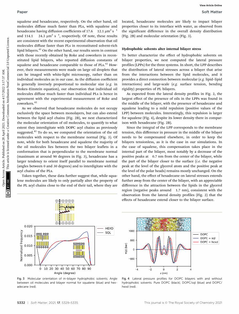
As we observed that hexadecane molecules do not occupyexclusively the space between monolayers, but can also extendbetween the lipid acyl chains (Fig. 2B), we next characterizedthe molecular orientation of oil molecules, to quantify to whatextent they interdigitate with DOPC acyl chains as previouslysuggested.42 To do so, we computed the orientation of the oilmolecules with respect to the membrane normal (Fig. 3). Ofnote, while for both hexadecane and squalene the majority ofthe oil molecules lies between the two bilayer leaflets in aconformation that is perpendicular to the membrane normal(maximum at around 90 degrees in Fig. 3), hexadecane has alarger tendency to orient itself parallel to membrane normal(angles between 0 and 30 degrees) and to interdigitate with theacyl chains of the PLs.
Taken together, these data further suggest that, while squa-lene molecules are likely to only partially alter the property ofthe PL acyl chains close to the end of their tail, where they are
located, hexadecane molecules are likely to impact bilayerproperties closer to its interface with water, as observed fromthe significant difference in the overall density distribution(Fig. 2B) and molecular orientation (Fig. 3).
Hydrophobic solvents alter internal bilayer stress
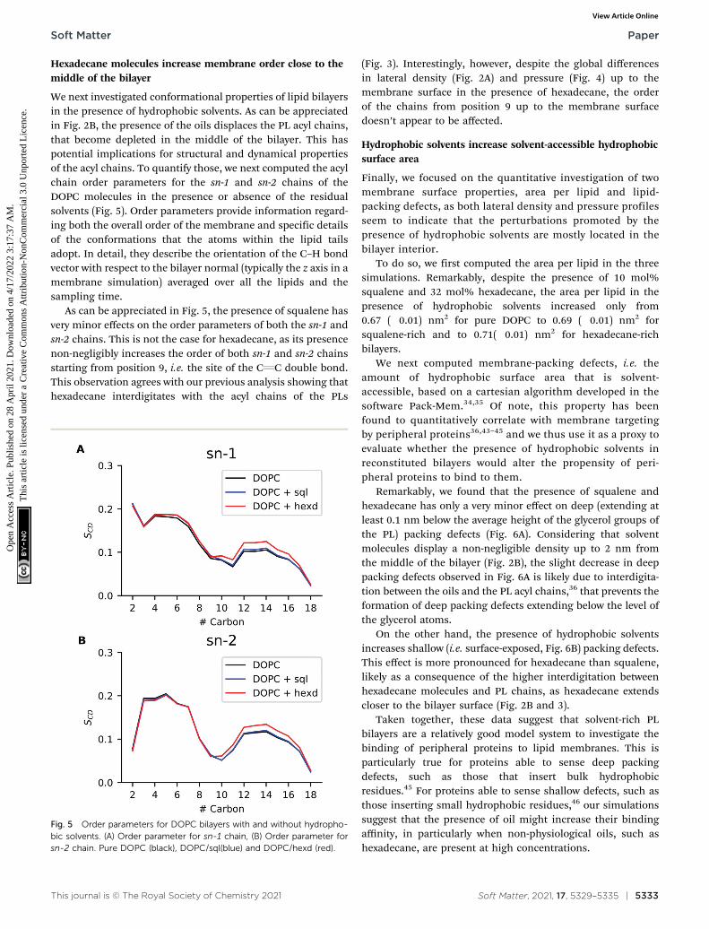
To better characterize the effect of hydrophobic solvents onbilayer properties, we next computed the lateral pressureprofiles (LPPs) for the three systems. In short, the LPP describesthe distribution of lateral stresses across a bilayer that arisefrom the interactions between the lipid molecules, and itprovides a direct connection between molecular (e.g. lipid–lipidinteractions) and large-scale (e.g. surface tension, bendingrigidity) properties of PL bilayers.
As expected from the lateral density profiles in Fig. 2, thelargest effect of the presence of oils in the LPPs was found inthe middle of the bilayer, with the presence of hexadecane andsqualene leading to a mild repulsion (positive values of theLPP) between molecules. Interestingly, this repulsion is largerfor squalene (Fig. 4), despite its lower density there in compar-ison with hexadecane (Fig. 2B).
Since the integral of the LPP corresponds to the membranetension, this difference in pressure in the middle of the bilayerneeds to be compensated elsewhere, in order to keep thebilayers tensionless, as it is the case in our simulations. Inthe case of squalene, this compensation takes place in theinternal part of the bilayer, most notably by a decrease of thepositive peaks at �0.7 nm from the center of the bilayer, whilethe part of the bilayer closer to the surface (i.e. the negativepeak at the level of the glycerol atom and the positive peak atthe level of the polar heads) remains mostly unchanged. On theother hand, the effect of hexadecane on lateral stresses extendsfurther away from the center of the bilayer, with an appreciabledifference in the attraction between the lipids in the glycerolregion (negative peaks around �1.7 nm), consistent with theobservation from the lateral density profiles (Fig. 2) that theeffects of hexadecane extend closer to the bilayer surface.
Fig. 3 Molecular orientation of in-bilayer hydrophobic solvents. Anglebetween oil molecules and bilayer normal for squalene (blue) and hex-adecane (red).
Fig. 4 Lateral pressure profiles for DOPC bilayers with and withouthydrophobic solvents. Pure DOPC (black), DOPC/sql (blue) and DOPC/hexd (red).
Paper Soft Matter
Ope
n A
cces
s A
rtic
le. P
ublis
hed
on 2
8 A
pril
2021
. Dow
nloa
ded
on 4
/17/
2022
3:1
7:37
AM
. T
his
artic
le is
lice
nsed
und
er a
Cre
ativ
e C
omm
ons
Attr
ibut
ion-
Non
Com
mer
cial
3.0
Unp
orte
d L
icen
ce.
View Article Online
This journal is © The Royal Society of Chemistry 2021 Soft Matter, 2021, 17, 5329–5335 | 5333
Hexadecane molecules increase membrane order close to themiddle of the bilayer
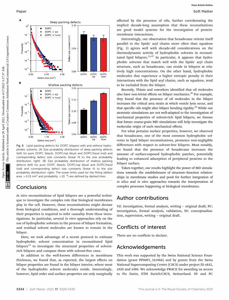
We next investigated conformational properties of lipid bilayersin the presence of hydrophobic solvents. As can be appreciatedin Fig. 2B, the presence of the oils displaces the PL acyl chains,that become depleted in the middle of the bilayer. This haspotential implications for structural and dynamical propertiesof the acyl chains. To quantify those, we next computed the acylchain order parameters for the sn-1 and sn-2 chains of theDOPC molecules in the presence or absence of the residualsolvents (Fig. 5). Order parameters provide information regard-ing both the overall order of the membrane and specific detailsof the conformations that the atoms within the lipid tailsadopt. In detail, they describe the orientation of the C–H bondvector with respect to the bilayer normal (typically the z axis in amembrane simulation) averaged over all the lipids and thesampling time.
As can be appreciated in Fig. 5, the presence of squalene hasvery minor effects on the order parameters of both the sn-1 andsn-2 chains. This is not the case for hexadecane, as its presencenon-negligibly increases the order of both sn-1 and sn-2 chainsstarting from position 9, i.e. the site of the CQC double bond.This observation agrees with our previous analysis showing thathexadecane interdigitates with the acyl chains of the PLs
(Fig. 3). Interestingly, however, despite the global differencesin lateral density (Fig. 2A) and pressure (Fig. 4) up to themembrane surface in the presence of hexadecane, the orderof the chains from position 9 up to the membrane surfacedoesn’t appear to be affected.
Hydrophobic solvents increase solvent-accessible hydrophobicsurface area
Finally, we focused on the quantitative investigation of twomembrane surface properties, area per lipid and lipid-packing defects, as both lateral density and pressure profilesseem to indicate that the perturbations promoted by thepresence of hydrophobic solvents are mostly located in thebilayer interior.
To do so, we first computed the area per lipid in the threesimulations. Remarkably, despite the presence of 10 mol%squalene and 32 mol% hexadecane, the area per lipid in thepresence of hydrophobic solvents increased only from0.67 (�0.01) nm2 for pure DOPC to 0.69 (�0.01) nm2 forsqualene-rich and to 0.71(�0.01) nm2 for hexadecane-richbilayers.
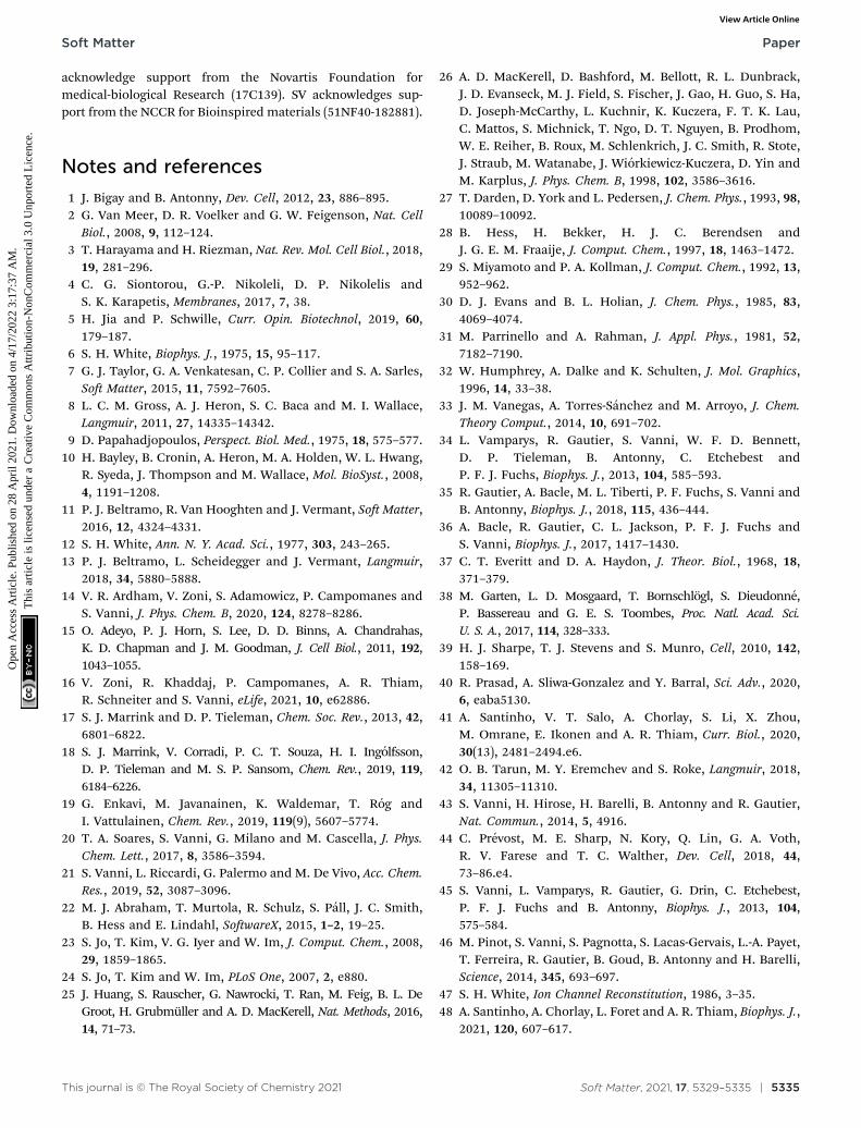
We next computed membrane-packing defects, i.e. theamount of hydrophobic surface area that is solvent-accessible, based on a cartesian algorithm developed in thesoftware Pack-Mem.34,35 Of note, this property has beenfound to quantitatively correlate with membrane targetingby peripheral proteins36,43–45 and we thus use it as a proxy toevaluate whether the presence of hydrophobic solvents inreconstituted bilayers would alter the propensity of peri-pheral proteins to bind to them.
Remarkably, we found that the presence of squalene andhexadecane has only a very minor effect on deep (extending atleast 0.1 nm below the average height of the glycerol groups ofthe PL) packing defects (Fig. 6A). Considering that solventmolecules display a non-negligible density up to 2 nm fromthe middle of the bilayer (Fig. 2B), the slight decrease in deeppacking defects observed in Fig. 6A is likely due to interdigita-tion between the oils and the PL acyl chains,36 that prevents theformation of deep packing defects extending below the level ofthe glycerol atoms.
On the other hand, the presence of hydrophobic solventsincreases shallow (i.e. surface-exposed, Fig. 6B) packing defects.This effect is more pronounced for hexadecane than squalene,likely as a consequence of the higher interdigitation betweenhexadecane molecules and PL chains, as hexadecane extendscloser to the bilayer surface (Fig. 2B and 3).
Taken together, these data suggest that solvent-rich PLbilayers are a relatively good model system to investigate thebinding of peripheral proteins to lipid membranes. This isparticularly true for proteins able to sense deep packingdefects, such as those that insert bulk hydrophobicresidues.45 For proteins able to sense shallow defects, such asthose inserting small hydrophobic residues,46 our simulationssuggest that the presence of oil might increase their bindingaffinity, in particularly when non-physiological oils, such ashexadecane, are present at high concentrations.
Fig. 5 Order parameters for DOPC bilayers with and without hydropho-bic solvents. (A) Order parameter for sn-1 chain, (B) Order parameter forsn-2 chain. Pure DOPC (black), DOPC/sql(blue) and DOPC/hexd (red).
Soft Matter Paper
Ope
n A
cces
s A
rtic
le. P
ublis
hed
on 2
8 A
pril
2021
. Dow
nloa
ded
on 4
/17/
2022
3:1
7:37
AM
. T
his
artic
le is
lice
nsed
und
er a
Cre
ativ
e C
omm
ons
Attr
ibut
ion-
Non
Com
mer
cial
3.0
Unp
orte
d L
icen
ce.
View Article Online
5334 | Soft Matter, 2021, 17, 5329–5335 This journal is © The Royal Society of Chemistry 2021
Conclusions
In vitro reconstitutions of lipid bilayers are a powerful techni-que to investigate the complex role that biological membranesplay in the cell. However, these reconstitutions might deviatefrom biological conditions, and a thorough understanding oftheir properties is required to infer causality from these inves-tigations. In particular, several in vitro approaches rely on theuse of hydrophobic solvents in the process of bilayer formation,and residual solvent molecules are known to remain in thebilayer.
Here, we took advantage of a recent protocol to estimatehydrophobic solvent concentration in reconstituted lipidbilayers14 to investigate the structural properties of solvent-rich bilayers and compare them with solvent-free ones.
In addition to the well-known differences in membranethickness, we found that, as expected, the largest effects onbilayer properties are found in the bilayer interior, where mostof the hydrophobic solvent molecules reside. Interestingly,however, lipid order and surface properties are only marginally
affected by the presence of oils, further corroborating theimplicit decade-long assumption that these reconstitutionsare good model systems for the investigation of protein-membrane interactions.
Interestingly, our observation that hexadecane orients itselfparallel to the lipids’ acyl chains more often than squalene(Fig. 3) agrees well with decade-old considerations on thethermodynamic activity of hydrophobic solvents in reconsti-tuted lipid bilayers.12,47 In particular, it appears that hydro-phobic solvents that match well with the lipids’ acyl chainstructure, such as hexadecane, can reside in bilayers at rela-tively high concentrations. On the other hand, hydrophobicmolecules that experience a higher entropic penalty in theirinteractions with the lipid acyl chains, such as squalene, tendto be excluded from the bilayer.
Recently, Thiam and coworkers identified that oil moleculesalso have non-trivial effects on bilayer mechanics.48 For example,they found that the presence of oil molecules in the bilayerincreases the critical area strain at which vesicle lysis occur, andthat specific oils might alter bilayer bending rigidity.48 While ouratomistic simulations are not well-adapted to the investigation ofmechanical properties of solvent-rich lipid bilayers, we foreseethat future coarse-grain MD simulations will help investigate themolecular origin of such mechanical effects.
For what pertains surface properties, however, we observedthat hexadecane, one of the most common hydrophobic sol-vents in lipid bilayer reconstitutions, promotes non-negligibledifferences with respect to solvent-free bilayers. Most notably,we found that the presence of hexadecane increases theamount of surface-exposed hydrophobic patches, potentiallyleading to enhanced adsorption of peripheral proteins to thebilayer surface.
Taken together, our results highlight the power of MD simula-tions towards the establishment of structure–function relation-ships in membrane studies and posit for further integration ofin silico and in vitro approaches towards the interpretation ofcomplex processes happening at biological membranes.
Author contributions
VZ: investigation, formal analysis, writing – original draft; PC:investigation, formal analysis, validation, SV: conceptualiza-tion, supervision, writing – original draft.
Conflicts of interest
There are no conflicts to declare.
Acknowledgements
This work was supported by the Swiss National Science Foun-dation (grant PP00P3_163966) and by grants from the SwissNational Supercomputing Centre (CSCS) under project ID s842,s920 and s980. We acknowledge PRACE for awarding us accessto Piz Daint, ETH Zurich/CSCS, Switzerland. SV and PC
Fig. 6 Lipid-packing defects for DOPC bilayers with and without hydro-phobic solvents. (A) Size probability distribution of deep packing defects(left) for pure DOPC (black), DOPC/sql (blue) and DOPC/hexd (red) andcorresponding defect size constants (linear fit to the size probabilitydistribution, right). (B) Size probability distribution of shallow packingdefects (left) for pure DOPC (black), DOPC/sql (blue) and DOPC/hexd(red) and corresponding defect size constants (linear fit to the sizeprobability distribution, right). The lower limits used for the fitting (defectarea 40.15 nm2 and probability 410�4) are defined by dashed lines.
Paper Soft Matter
Ope
n A
cces
s A
rtic
le. P
ublis
hed
on 2
8 A
pril
2021
. Dow
nloa
ded
on 4
/17/
2022
3:1
7:37
AM
. T
his
artic
le is
lice
nsed
und
er a
Cre
ativ
e C
omm
ons
Attr
ibut
ion-
Non
Com
mer
cial
3.0
Unp
orte
d L
icen
ce.
View Article Online
This journal is © The Royal Society of Chemistry 2021 Soft Matter, 2021, 17, 5329–5335 | 5335
acknowledge support from the Novartis Foundation formedical-biological Research (17C139). SV acknowledges sup-port from the NCCR for Bioinspired materials (51NF40-182881).
Notes and references
1 J. Bigay and B. Antonny, Dev. Cell, 2012, 23, 886–895.2 G. Van Meer, D. R. Voelker and G. W. Feigenson, Nat. Cell
Biol., 2008, 9, 112–124.3 T. Harayama and H. Riezman, Nat. Rev. Mol. Cell Biol., 2018,
19, 281–296.4 C. G. Siontorou, G.-P. Nikoleli, D. P. Nikolelis and
S. K. Karapetis, Membranes, 2017, 7, 38.5 H. Jia and P. Schwille, Curr. Opin. Biotechnol, 2019, 60,
179–187.6 S. H. White, Biophys. J., 1975, 15, 95–117.7 G. J. Taylor, G. A. Venkatesan, C. P. Collier and S. A. Sarles,
Soft Matter, 2015, 11, 7592–7605.8 L. C. M. Gross, A. J. Heron, S. C. Baca and M. I. Wallace,
Langmuir, 2011, 27, 14335–14342.9 D. Papahadjopoulos, Perspect. Biol. Med., 1975, 18, 575–577.
10 H. Bayley, B. Cronin, A. Heron, M. A. Holden, W. L. Hwang,R. Syeda, J. Thompson and M. Wallace, Mol. BioSyst., 2008,4, 1191–1208.
11 P. J. Beltramo, R. Van Hooghten and J. Vermant, Soft Matter,2016, 12, 4324–4331.
12 S. H. White, Ann. N. Y. Acad. Sci., 1977, 303, 243–265.13 P. J. Beltramo, L. Scheidegger and J. Vermant, Langmuir,
2018, 34, 5880–5888.14 V. R. Ardham, V. Zoni, S. Adamowicz, P. Campomanes and
S. Vanni, J. Phys. Chem. B, 2020, 124, 8278–8286.15 O. Adeyo, P. J. Horn, S. Lee, D. D. Binns, A. Chandrahas,
K. D. Chapman and J. M. Goodman, J. Cell Biol., 2011, 192,1043–1055.
16 V. Zoni, R. Khaddaj, P. Campomanes, A. R. Thiam,R. Schneiter and S. Vanni, eLife, 2021, 10, e62886.
17 S. J. Marrink and D. P. Tieleman, Chem. Soc. Rev., 2013, 42,6801–6822.
18 S. J. Marrink, V. Corradi, P. C. T. Souza, H. I. Ingolfsson,D. P. Tieleman and M. S. P. Sansom, Chem. Rev., 2019, 119,6184–6226.
19 G. Enkavi, M. Javanainen, K. Waldemar, T. Rog andI. Vattulainen, Chem. Rev., 2019, 119(9), 5607–5774.
20 T. A. Soares, S. Vanni, G. Milano and M. Cascella, J. Phys.Chem. Lett., 2017, 8, 3586–3594.
21 S. Vanni, L. Riccardi, G. Palermo and M. De Vivo, Acc. Chem.Res., 2019, 52, 3087–3096.
22 M. J. Abraham, T. Murtola, R. Schulz, S. Pall, J. C. Smith,B. Hess and E. Lindahl, SoftwareX, 2015, 1–2, 19–25.
23 S. Jo, T. Kim, V. G. Iyer and W. Im, J. Comput. Chem., 2008,29, 1859–1865.
24 S. Jo, T. Kim and W. Im, PLoS One, 2007, 2, e880.25 J. Huang, S. Rauscher, G. Nawrocki, T. Ran, M. Feig, B. L. De
Groot, H. Grubmuller and A. D. MacKerell, Nat. Methods, 2016,14, 71–73.
26 A. D. MacKerell, D. Bashford, M. Bellott, R. L. Dunbrack,J. D. Evanseck, M. J. Field, S. Fischer, J. Gao, H. Guo, S. Ha,D. Joseph-McCarthy, L. Kuchnir, K. Kuczera, F. T. K. Lau,C. Mattos, S. Michnick, T. Ngo, D. T. Nguyen, B. Prodhom,W. E. Reiher, B. Roux, M. Schlenkrich, J. C. Smith, R. Stote,J. Straub, M. Watanabe, J. Wiorkiewicz-Kuczera, D. Yin andM. Karplus, J. Phys. Chem. B, 1998, 102, 3586–3616.
27 T. Darden, D. York and L. Pedersen, J. Chem. Phys., 1993, 98,10089–10092.
28 B. Hess, H. Bekker, H. J. C. Berendsen andJ. G. E. M. Fraaije, J. Comput. Chem., 1997, 18, 1463–1472.
29 S. Miyamoto and P. A. Kollman, J. Comput. Chem., 1992, 13,952–962.
30 D. J. Evans and B. L. Holian, J. Chem. Phys., 1985, 83,4069–4074.
31 M. Parrinello and A. Rahman, J. Appl. Phys., 1981, 52,7182–7190.
32 W. Humphrey, A. Dalke and K. Schulten, J. Mol. Graphics,1996, 14, 33–38.
33 J. M. Vanegas, A. Torres-Sanchez and M. Arroyo, J. Chem.Theory Comput., 2014, 10, 691–702.
34 L. Vamparys, R. Gautier, S. Vanni, W. F. D. Bennett,D. P. Tieleman, B. Antonny, C. Etchebest andP. F. J. Fuchs, Biophys. J., 2013, 104, 585–593.
35 R. Gautier, A. Bacle, M. L. Tiberti, P. F. Fuchs, S. Vanni andB. Antonny, Biophys. J., 2018, 115, 436–444.
36 A. Bacle, R. Gautier, C. L. Jackson, P. F. J. Fuchs andS. Vanni, Biophys. J., 2017, 1417–1430.
37 C. T. Everitt and D. A. Haydon, J. Theor. Biol., 1968, 18,371–379.
38 M. Garten, L. D. Mosgaard, T. Bornschlogl, S. Dieudonne,P. Bassereau and G. E. S. Toombes, Proc. Natl. Acad. Sci.U. S. A., 2017, 114, 328–333.
39 H. J. Sharpe, T. J. Stevens and S. Munro, Cell, 2010, 142,158–169.
40 R. Prasad, A. Sliwa-Gonzalez and Y. Barral, Sci. Adv., 2020,6, eaba5130.
41 A. Santinho, V. T. Salo, A. Chorlay, S. Li, X. Zhou,M. Omrane, E. Ikonen and A. R. Thiam, Curr. Biol., 2020,30(13), 2481–2494.e6.
42 O. B. Tarun, M. Y. Eremchev and S. Roke, Langmuir, 2018,34, 11305–11310.
43 S. Vanni, H. Hirose, H. Barelli, B. Antonny and R. Gautier,Nat. Commun., 2014, 5, 4916.
44 C. Prevost, M. E. Sharp, N. Kory, Q. Lin, G. A. Voth,R. V. Farese and T. C. Walther, Dev. Cell, 2018, 44,73–86.e4.
45 S. Vanni, L. Vamparys, R. Gautier, G. Drin, C. Etchebest,P. F. J. Fuchs and B. Antonny, Biophys. J., 2013, 104,575–584.
46 M. Pinot, S. Vanni, S. Pagnotta, S. Lacas-Gervais, L.-A. Payet,T. Ferreira, R. Gautier, B. Goud, B. Antonny and H. Barelli,Science, 2014, 345, 693–697.
47 S. H. White, Ion Channel Reconstitution, 1986, 3–35.48 A. Santinho, A. Chorlay, L. Foret and A. R. Thiam, Biophys. J.,
2021, 120, 607–617.
Soft Matter Paper
Ope
n A
cces
s A
rtic
le. P
ublis
hed
on 2
8 A
pril
2021
. Dow
nloa
ded
on 4
/17/
2022
3:1
7:37
AM
. T
his
artic
le is
lice
nsed
und
er a
Cre
ativ
e C
omm
ons
Attr
ibut
ion-
Non
Com
mer
cial
3.0
Unp
orte
d L
icen
ce.
View Article Online
Copyright © 2022 FDOKUMEN