Effect of Cavity Disinfectants on Adhesion to Primary Teeth—A ...
17
International Journal of Molecular Sciences Review Effect of Cavity Disinfectants on Adhesion to Primary Teeth—A Systematic Review Ana Coelho 1,2,3, * ,† , Inês Amaro 1,† , Ana Apolónio 1 , Anabela Paula 1,2,3 , José Saraiva 1 , Manuel Marques Ferreira 2,3,4 , Carlos Miguel Marto 2,3,5,6 and Eunice Carrilho 1,2,3 Citation: Coelho, A.; Amaro, I.; Apolónio, A.; Paula, A.; Saraiva, J.; Ferreira, M.M.; Marto, C.M.; Carrilho, E. Effect of Cavity Disinfectants on Adhesion to Primary Teeth—A Systematic Review. Int. J. Mol. Sci. 2021, 22, 4398. https://doi.org/ 10.3390/ijms22094398 Academic Editors: Sotiris Hadjikakou, Christina N. Banti and Andreas K. Rossos Received: 31 March 2021 Accepted: 22 April 2021 Published: 22 April 2021 Publisher’s Note: MDPI stays neutral with regard to jurisdictional claims in published maps and institutional affil- iations. Copyright: © 2021 by the authors. Licensee MDPI, Basel, Switzerland. This article is an open access article distributed under the terms and conditions of the Creative Commons Attribution (CC BY) license (https:// creativecommons.org/licenses/by/ 4.0/). 1 Faculty of Medicine, Institute of Integrated Clinical Practice, University of Coimbra, 3000-075 Coimbra, Portugal; [email protected] (I.A.); [email protected] (A.A.); [email protected] (A.P.); [email protected] (J.S.); [email protected] (E.C.) 2 Area of Environment Genetics and Oncobiology (CIMAGO), Faculty of Medicine, Coimbra Institute for Clinical and Biomedical Research (iCBR), University of Coimbra, 3000-548 Coimbra, Portugal; [email protected] (M.M.F.); [email protected] (C.M.M.) 3 Clinical Academic Center of Coimbra (CACC), 3004-561 Coimbra, Portugal 4 Faculty of Medicine, Institute of Endodontics, University of Coimbra, 3000-075 Coimbra, Portugal 5 Faculty of Medicine, Institute of Biophysics, University of Coimbra, 3004-548 Coimbra, Portugal 6 Faculty of Medicine, Institute of Experimental Pathology, University of Coimbra, 3004-548 Coimbra, Portugal * Correspondence: anasofi[email protected] † These authors contributed equally to this work. Abstract: Some authors have been proposing the use of cavity disinfectants in order to reduce, or even eliminate, the effect of the microorganisms present in a dental cavity before a restoration is placed. The aim of this study was to evaluate the effect of different cavity disinfectants on bond strength and clinical success of composite and glass ionomer restorations on primary teeth. The research was conducted using Cochrane Library, PubMed/MEDLINE, SCOPUS, and Web of Science for articles published up to February 2021. The search was performed according to the PICO strategy. The evaluation of the methodological quality of each in vitro study was assessed using the CONSORT checklist for reporting in vitro studies on dental materials. Sixteen in vitro studies and one in situ study fulfilled the inclusion criteria and were analyzed. Chlorhexidine was the most studied cavity disinfectant, and its use does not compromise dentin bonding. Sodium hypochlorite is a promising alternative, but more research on its use is required to clearly state that it can safely be used as a cavity disinfectant for primary teeth. Although other disinfectants were studied, there is a low-level evidence attesting their effects on adhesion, therefore their use should be avoided. Keywords: cavity disinfectants; primary teeth; adhesion; bond strength 1. Introduction Dental caries has a high prevalence worldwide, affecting more than 2.4 thousand million adults and 620 thousand children with primary teeth [1]. It can be defined as a multifactorial pathology arising from the interaction between dental structure and microbial biofilm, due to an imbalance between remineralization and demineralization, with the last one prevailing [2,3]. Although complete removal of the decayed and necrotic tissue is directly related to restorations’ clinical success, cariogenic bacteria can be pushed deep inside the dentinal tubules while removing the carious tissue and remain viable for a long time. In fact, the remaining of cariogenic bacteria in the cavity can be associated with the development of secondary caries [4,5]. According to Dalkilic et al. [6], fermenting microorganisms can remain viable for 139 days in a restored cavity. Moreover, bacteria present in the smear layer may remain viable and proliferate, allowing their metabolism products to reach and to cause inflam- Int. J. Mol. Sci. 2021, 22, 4398. https://doi.org/10.3390/ijms22094398 https://www.mdpi.com/journal/ijms
-
Upload
khangminh22 -
Category
Documents
-
view
1 -
download
0
Transcript of Effect of Cavity Disinfectants on Adhesion to Primary Teeth—A ...

International Journal of
Molecular Sciences
Review
Effect of Cavity Disinfectants on Adhesion to Primary Teeth—ASystematic Review
Ana Coelho 1,2,3,*,† , Inês Amaro 1,†, Ana Apolónio 1, Anabela Paula 1,2,3 , José Saraiva 1,Manuel Marques Ferreira 2,3,4 , Carlos Miguel Marto 2,3,5,6 and Eunice Carrilho 1,2,3
�����������������
Citation: Coelho, A.; Amaro, I.;
Apolónio, A.; Paula, A.; Saraiva, J.;
Ferreira, M.M.; Marto, C.M.; Carrilho,
E. Effect of Cavity Disinfectants on
Adhesion to Primary Teeth—A
Systematic Review. Int. J. Mol. Sci.
2021, 22, 4398. https://doi.org/
10.3390/ijms22094398
Academic Editors: Sotiris Hadjikakou,
Christina N. Banti and Andreas
K. Rossos
Received: 31 March 2021
Accepted: 22 April 2021
Published: 22 April 2021
Publisher’s Note: MDPI stays neutral
with regard to jurisdictional claims in
published maps and institutional affil-
iations.
Copyright: © 2021 by the authors.
Licensee MDPI, Basel, Switzerland.
This article is an open access article
distributed under the terms and
conditions of the Creative Commons
Attribution (CC BY) license (https://
creativecommons.org/licenses/by/
4.0/).
1 Faculty of Medicine, Institute of Integrated Clinical Practice, University of Coimbra,3000-075 Coimbra, Portugal; [email protected] (I.A.); [email protected] (A.A.);[email protected] (A.P.); [email protected] (J.S.); [email protected] (E.C.)
2 Area of Environment Genetics and Oncobiology (CIMAGO), Faculty of Medicine, Coimbra Institute forClinical and Biomedical Research (iCBR), University of Coimbra, 3000-548 Coimbra, Portugal;[email protected] (M.M.F.); [email protected] (C.M.M.)
3 Clinical Academic Center of Coimbra (CACC), 3004-561 Coimbra, Portugal4 Faculty of Medicine, Institute of Endodontics, University of Coimbra, 3000-075 Coimbra, Portugal5 Faculty of Medicine, Institute of Biophysics, University of Coimbra, 3004-548 Coimbra, Portugal6 Faculty of Medicine, Institute of Experimental Pathology, University of Coimbra, 3004-548 Coimbra, Portugal* Correspondence: [email protected]† These authors contributed equally to this work.
Abstract: Some authors have been proposing the use of cavity disinfectants in order to reduce, oreven eliminate, the effect of the microorganisms present in a dental cavity before a restoration isplaced. The aim of this study was to evaluate the effect of different cavity disinfectants on bondstrength and clinical success of composite and glass ionomer restorations on primary teeth. Theresearch was conducted using Cochrane Library, PubMed/MEDLINE, SCOPUS, and Web of Sciencefor articles published up to February 2021. The search was performed according to the PICO strategy.The evaluation of the methodological quality of each in vitro study was assessed using the CONSORTchecklist for reporting in vitro studies on dental materials. Sixteen in vitro studies and one in situstudy fulfilled the inclusion criteria and were analyzed. Chlorhexidine was the most studied cavitydisinfectant, and its use does not compromise dentin bonding. Sodium hypochlorite is a promisingalternative, but more research on its use is required to clearly state that it can safely be used as acavity disinfectant for primary teeth. Although other disinfectants were studied, there is a low-levelevidence attesting their effects on adhesion, therefore their use should be avoided.
Keywords: cavity disinfectants; primary teeth; adhesion; bond strength
1. Introduction
Dental caries has a high prevalence worldwide, affecting more than 2.4 thousandmillion adults and 620 thousand children with primary teeth [1]. It can be defined as amultifactorial pathology arising from the interaction between dental structure and microbialbiofilm, due to an imbalance between remineralization and demineralization, with the lastone prevailing [2,3].
Although complete removal of the decayed and necrotic tissue is directly related torestorations’ clinical success, cariogenic bacteria can be pushed deep inside the dentinaltubules while removing the carious tissue and remain viable for a long time. In fact, theremaining of cariogenic bacteria in the cavity can be associated with the development ofsecondary caries [4,5].
According to Dalkilic et al. [6], fermenting microorganisms can remain viable for139 days in a restored cavity. Moreover, bacteria present in the smear layer may remainviable and proliferate, allowing their metabolism products to reach and to cause inflam-
Int. J. Mol. Sci. 2021, 22, 4398. https://doi.org/10.3390/ijms22094398 https://www.mdpi.com/journal/ijms
Int. J. Mol. Sci. 2021, 22, 4398 2 of 17
matory changes in the dental pulp. Bacteria penetration through restoration and teethinterface (microinfiltration) can also explain restorations’ failure [7–9].
As so, some authors have been proposing the use of cavity disinfectants in order toreduce, or even eliminate, the effect of the microorganisms present in a dental cavity beforea restoration is placed [8–10].
Among the available disinfectants, chlorhexidine, sodium hypochlorite, and fluori-dated solutions are the most used. Despite their benefits, their effect on adhesion to dentin,especially that of primary teeth, is still unknown [7,11,12].
Among the pediatric population, dental caries treatment is the most common proce-dure to be performed in a dental appointment [12]. Restorations’ success rate is associatedwith dentist’s experience and patient’s collaboration. However, one of the most commoncauses of failure is the development of secondary caries [13–15].
Thereby, the aim of this systematic review was to evaluate the effect of the applicationof different cavity disinfectants on bond strength and clinical success of composite andglass ionomer restorations on primary teeth.
2. Results
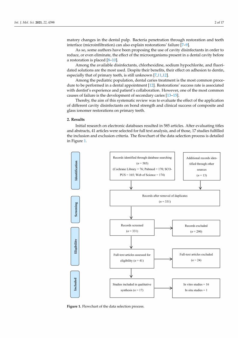
Initial research on electronic databases resulted in 585 articles. After evaluating titlesand abstracts, 41 articles were selected for full text analysis, and of those, 17 studies fulfilledthe inclusion and exclusion criteria. The flowchart of the data selection process is detailedin Figure 1.
Int. J. Mol. Sci. 2021, 22, 4398 3 of 16
Figure 1. Flowchart of the data selection process.
Most of the authors reported the use of 2% chlorhexidine [12,18–20,22,23,25,28,29] as a cavity disinfectant. A few studies reported results on the application of sodium hypo-chlorite [16,24,27], Er:YAG laser [17,21,26], KTP laser [25], ozone [25], doxycycline [29], ethylenediaminetetraacetic acid (EDTA) [29], propolis [25], and Aqua-prep™ (Bisco, Schaumburg, IL, USA) [26].
Except for Vieira et al. [12], all of the authors studying the effect of 2% chlorhexidine as a cavity disinfectant [18–20,22,23,25,28,29] reported positive results, allowing for maintenance or a statistically significant increase in bond strength values. These values ranged from 7.58 ± 3.18 MPa [25] to 66.45 ± 8.3 MPa [28] in resin specimens and from 7.1 ± 5.2 MPa to 14.4 ± 6.6 MPa [18] in caries-affected dentin in glass ionomer specimens. Vieira et al. [12] were the only authors applying chlorhexidine before etching the speci-mens with phosphoric acid.
The authors evaluating the effect of the application of sodium hypochlorite tested different concentrations, ranging from 2.5% [27] to 10% [16]. Regardless of the concentra-tion, all authors [16,24,27] reported positive results, allowing for maintenance or even a statistically significant increase in bond strength values. These values ranged from 9.9 ± 0.2 MPa [16] to 18.45 ± 2.30 MPa [24].
Records identified through database searching
(n = 585)
(Cochrane Library = 76; Pubmed = 170; SCO-
PUS = 165; Web of Science = 174)
Full-text articles excluded
(n = 24)
Records screened
(n = 331) Records excluded
(n = 290)
Records after removal of duplicates
(n = 331)
Full-text articles assessed for
eligibility (n = 41)
Studies included in qualitative
synthesis (n = 17)
Additional records iden-
tified through other
sources
(n = 13)
In vitro studies = 16
In situ studies = 1
Figure 1. Flowchart of the data selection process.

Int. J. Mol. Sci. 2021, 22, 4398 3 of 17
Sixteen in vitro studies [12,16–30] were included in this systematic review.The earliest study was published in 2003 [12], and the most recent one in 2020 [29].Most authors used primary molars [12,16,18–28,30], but Monghini et al. [17] evaluated
canines, and Mohammadi et al. [29] used anterior teeth. Sample size varied from 2 [25] to20 [28,31] teeth per group.
Even though all authors studied healthy dentin, Ersin et al. [18] additionally evaluatedcarious dentin, and Lenzi et al. [22,23] also evaluated demineralized dentin (artificiallyinduced lesions).
After extraction, teeth were stored in thymol [12,18,28], chloramine [16,22,23,27,30],distilled water [16,20,22–25,27,30,32], saline solution [17,26], or sodium azide [17,21].Ricci et al. [19] and Mohammadi [29] did not report data on the storage medium used afterteeth extraction.
All authors used water to store the specimens after adhesive experiments and beforebond strength evaluation.
All authors reported results on adhesion to composite resin. Only Ersin et al. [18] alsoreported results on adhesion to glass ionomer materials.
Most of the authors reported the use of 2% chlorhexidine [12,18–20,22,23,25,28,29] asa cavity disinfectant. A few studies reported results on the application of sodium hypochlo-rite [16,24,27], Er:YAG laser [17,21,26], KTP laser [25], ozone [25], doxycycline [29], ethylene-diaminetetraacetic acid (EDTA) [29], propolis [25], and Aqua-prep™ (Bisco, Schaumburg,IL, USA) [26].
Except for Vieira et al. [12], all of the authors studying the effect of 2% chlorhexi-dine as a cavity disinfectant [18–20,22,23,25,28,29] reported positive results, allowing formaintenance or a statistically significant increase in bond strength values. These valuesranged from 7.58 ± 3.18 MPa [25] to 66.45 ± 8.3 MPa [28] in resin specimens and from7.1 ± 5.2 MPa to 14.4 ± 6.6 MPa [18] in caries-affected dentin in glass ionomer specimens.Vieira et al. [12] were the only authors applying chlorhexidine before etching the specimenswith phosphoric acid.
The authors evaluating the effect of the application of sodium hypochlorite testeddifferent concentrations, ranging from 2.5% [27] to 10% [16]. Regardless of the concen-tration, all authors [16,24,27] reported positive results, allowing for maintenance or evena statistically significant increase in bond strength values. These values ranged from9.9 ± 0.2 MPa [16] to 18.45 ± 2.30 MPa [24].
The Er:YAG laser was evaluated by three studies [17,21,26]. Monghini et al. [17]reported statistically significant negative results when testing the laser with three differentworking parameters. However, Scatena et al. [21] did not find statistically significant differ-ences regarding bond strength results for different focal distances (mm), and Yildiz et al. [26]even reported a statistically significant increase in bond strength values. The bond strengthvalues of these studies ranged from 5.07 ± 2.62 MPa [21] to 20.57 ± 9.02 MPa [26].
Oznurhan et al. [25] assessed the use of a KTP laser as a cavity disinfectant and foundno statistically significant differences when comparing its results to the ones of the controlgroup (9.58 ± 2.92 and 6.38 ± 2.47 MPa, respectively).
Gaseous ozone and ozonated water [25] were also tested as cavity disinfectants. Theauthors reported a maintenance of the bond strength values when using gaseous ozone(5.84 ± 2.62 MPa vs. 6.38 ± 2.47 MPa for the control group) and a statistically significantincrease of the bond strength values when using ozonated water (11.12 ± 2.41 MPa vs.6.38 ± 2.47 MPa for the control group).
Aqua-prep™ [26], an aqueous solution of fluoride and hydroxyethyl methacrylate(HEMA), 2% Doxycycline [29], 17% EDTA [29], and 30% propolis [25] were all evaluated inonly one study each, and no statistically significant differences were found between testand control groups.
Relevant information on each in vitro study is summarized in Table 1.

Int. J. Mol. Sci. 2021, 22, 4398 4 of 17
Table 1. Characteristics of the in vitro studies included in the systematic review.
Authors, Year Groups (n) Teeth Storage Materials Results (MPa)
Vieira et al., 2003[12]
G1—37% phosphoric acid +adhesive (10) + resinG2—2% CHX + 37%
phosphoric acid + adhesive(10) + resin
Molars 0.1% Thymol
Adhesive: 3MSingle Bond (3M,
USA)Resin: FiltekTM
Z250 (3M, USA)
G1: 19.88 ± 1.04G2: 17.99 ± 1.15
G1*/G2
Correr et al., 2004[16]
G1—35% phosphoric acid +adhesive 1 (15)
G2—35% phosphoric acid +10% NaOCl + adhesive 1 (15)G3—37% phosphoric acid +
adhesive 2 (15)G4—37% phosphoric acid +
10% NaOCl + adhesive 2 (15)G5–Adhesive 3 (15)
G6—10% NaOCl + adhesive 3(15)
+ resin
Molars 0.5%Chloramine
Adhesive: 1–3MSingle Bond;
2–Prime & Bond2.1® (Dentsply,
Brazil);3–ClearfillTM SEBond (Kuraray,
Houston, TX, USA)Resin: FiltekTM
Z250 (3M, USA)
G1: 15.8 ± 1.9G2: 14.6 ± 1.3G3: 10.2 ± 0.7G4: 9.9 ± 0.2G5: 13.3 ± 1.2G6: 10.7 ± 1.0
G1*/G3
Monghini et al.,2004 [17]
G1—None (12)G2—Laser Er;YAG
60 mJ/2 Hz (12)G3—Laser Er;YAG
80 mJ/2 Hz (12)G4—Laser Er;YAG100 mJ/2 Hz (12)
+ 35% phosphoric acid +adhesive + resin
Canines
0.9% Salinesolution with0.4% sodium
azide
Adhesive: 3MSingle Bond
Laser: Kavo KeyLaser 2 (Kavo
Dental, Germany)Resin: FiltekTM
Z250
G1:17.89 ± 4.75G2:12.34 ± 4.85G3:10.30 ± 3.67G4:10.41 ± 4.20G1*/G2;G3;G4
Ersin et al., 2009[18]
G1—25% polyacrlylic acid +2% CHX + GIC 1 (sound
dentin) (3)G2—25% polyacrlylic acid +
2% CHX + GIC 1 (cariousdentin) (3)
G3—25% polyacrlylic acid +GIC 1 (sound dentin) (3)
G4—25% polyacrlylic acid +GIC 1 (carious dentin) (3)
G5—2% CHX + GIC 2 (sounddentin) (3)
G6—2% CHX + GIC 2 (cariousdentin) (3)
G7—GIC 2 (sound dentin) (3)G8—GIC 2 (carious dentin) (3)
G9—37% phosphoric acid +2% CHX + adhesive + resin
(sound dentin) (3)G10–37% phosphoric acid +2% CHX + adhesive + resin
(carious dentin) (3)G11—37% phosphoric acid +
adhesive + resin (sounddentin) (3)
G12—37% phosphoric acid +adhesive + resin (carious
dentin) (3)
Molars 0.1% Thymol
Adhesive: Prime &Bond®;
GIC: 1–KetacTM
Molar (3M,Germany);
2–VitremerTM (3M,USA)
Resin–SurefilTM
(Dentsply, USA)
G1: 8.7 ± 4.3G2: 7.1 ± 5.2G3: 9.2 ± 5.2
G4: 10.3 ± 6.6G5: 12.4 ± 5.7G6: 14.4 ± 6.6G7: 11.2 ± 4.8G8: 13.8 ± 4.9G9: 22.9 ± 6.9G10: 23.2 ± 6.2G11: 20.2 ± 5.8G12: 22.1 ± 6.2
G9*/G1;G2;G3;G4;G5;G6;G7;G8
G10*/G1;G2;G3;G4;G5;G6;G7;G8
G11*/G1;G2;G3;G4;G5;G6;G7;G8
G12*/G1;G2;G3;G4;G5;G6;G7;G8

Int. J. Mol. Sci. 2021, 22, 4398 5 of 17
Table 1. Cont.
Authors, Year Groups (n) Teeth Storage Materials Results (MPa)
Ricci et al., 2010[19]
35% phosphoric acid +G1—2% CHX + adhesive 1 (4)
G2—deionized water +adhesive 1 (4)
G3—2% CHX + adhesive 2 (4)G4—deionized water +
adhesive 2 (4)G5—2% CHX + adhesive 3 (4)
G6—deionized water +adhesive 3 (4)
+ resin
Molars NA
Adhesive:1–AdperTM SingleBond (3M, USA);2–Prime & BondNT® (Dentsply,
USA);3–Excite® DSC
(Ivoclar,Liechtenstein)
Resin: FiltekTM
Z250
G1: 47.4 ± 9.5G2: 41.4 ± 11.9G3: 48.0 ± 9.8
G4: 40.8 ± 13.4G5: 45.2 ± 9.2
G6: 43.4 ± 12.0G1*/G2; G3*/G4
Leitune et al.,2011 [20]
37% phosphoric acid +G1—Adhesive (24 h) (10)
G2—Adhesive (6 months) (10)G3—2% CHX + Adhesive
(24 h) (10)G4—2% CHX + Adhesive
(6 months) (10)
Molars Distilled water
Adhesive:AdperTM
ScotchbondTM
Multi Purpose (3M,USA)
Resin: FiltekTM
Z250
G1: 22.37 ± 3.69G2: 19.93 ± 2.05G3: 22.30 ± 3.66G4: 24.48 ± 2.24
G2*/G4
Scatena et al.,2011 [21]
G1–None (10)G2—Laser Er:YAG (80 mJ,
11 mm) (10)G3—Laser Er:YAG (80 mJ,
12 mm) (10)G4—Laser Er:YAG (80 mJ,
16 mm) (10)G5—Laser Er:YAG (80 mJ,
17 mm) (10)G6—Laser Er:YAG (80 mJ,
20 mm) (10)+ 37% phosphoric acid +
adhesive + resin
Molars 0.4% Sodiumazide
Laser: Kavo KeyLaser 2
Adhesive: 3MSingle Bond
Resin: FiltekTM
Z250
G1: 7.32 ± 3.83G2: 5.07 ± 2.62G3: 6.49 ± 1.64G4: 7.71 ± 0.66G5: 7.33 ± 0.02G6: 9.65 ± 2.41
G2*/G4;G6
Manfro et al.,2012 [30]
37% phosphoric acid +G1—water + adhesive (7)
G2—water + adhesive(12 months) (7)
G3—0.5% CHX + adhesive (7)G4—0.5% CHX + adhesive
(12 months) (7)G5—2% CHX + adhesive (7)
G6—2% CHX + adhesive(12 months) (7)
+ resin
Molars 0.5%Chloramine
Adhesive: 3MSingle Bond
Resin: FiltekTM
Z250
G1: 50.8 ± 12.8G2: 20.4 ± 3.7G3: 49.3 ± 2.6G4: 32.3 ± 7.9G5: 44.0 ± 8.7G6: 34.6 ± 5.1
G1*/G2;G2*/G4;G6;
G3*/G4; G5*/G6
Lenzi et al., 2012[22]
35% phosphoric acid +G1—distilled water +
adhesive (sound dentin) (5)G2—2% CHX + adhesive
(sound dentin) (5)G3—distilled water +
adhesive (artificial caries) (5)G4—2% CHX + adhesive
(artificial caries) (5)
Molars 0.5%Chloramine
Adhesive:AdperTM Single
Bond 2Resin: FiltekTM
Z250
G1: 30.8 ± 2.2G2: 32.8 ± 3.8G3: 24.5 ± 3.8G4: 25.6 ± 3.6
G1*/G3;G4;G2*/G3;G4

Int. J. Mol. Sci. 2021, 22, 4398 6 of 17
Table 1. Cont.
Authors, Year Groups (n) Teeth Storage Materials Results (MPa)
Aras et al., 2013[24]
G1—37% phosphoric acid (10)G2—37% phosphoric acid +
5% NaOCl (10)G3—5% NaOCl + 37%phosphoric acid (10)+ adhesive + resin
Molars Distilled water
Adhesive: Gluma®
Confort Bond(Herause-Kulzer,
Germany)Resin: Charisma®
(Herause-Kulzer,Germany)
G1: 14.51 ± 2.89G2: 18.45 ± 2.30G3: 17.06 ± 2.99
G1*/G2
Lenzi et al., 2014[23]
35% phosphoric acid +G1—distilled water +
adhesive (sound dentin) (5)G2—distilled water +
adhesive (sound dentin)(6 months) (5)
G3—2% CHX (withoutrinsing) + adhesive (sound
dentin) (5)G4—2% CHX (without
rinsing) + adhesive (sounddentin) (6 months) (5)G5—distilled water +
adhesive (artificial lesion) (5)G6—distilled water +
adhesive (artificial lesion)(6 months) (5)
G7—2% CHX (withoutrinsing) + adhesive (artificial
lesion) (5)G8—2% CHX (without
rinsing) + adhesive (artificiallesion) (6 months) (5)
Molars Distilled water
Adhesive:AdperTM Single
BondResin: FiltekTM
Z250
G1: 30.7 ± 2.2G2: 25.9 ± 5.7G3: 32.8 ± 3.8G4: 31.3 ± 2.6G5: 26.2 ± 5.4G6: 20.0 ± 3.9G7: 28.3 ± 3.4G8: 26.9 ± 5.9
G1*/G5;G7;G2*/G6;G8G3*/G5;G7G4*/G6;G8
Oznurhan et al.,2015 [25]
G1—2% CHX (2)G2—30% propolis (2)
G3—Gaseous ozone (2)G4—Ozonated water (2)
G5—Laser KTP (2)G6—None (2)
+ adhesive + resin
Molars Distilled water
Adhesive:AdperTM Prime &
Bond NT®
Resin: Tetric®
N-Ceram (IvoclarVivadent,
Liechenstein)Laser: Smartlite D
(Deka, Italy)
G1: 7.58 ± 3.18G2: 7.42 ± 2.28G3: 5.84 ± 2.62G4: 11.12 ± 2.41G5: 9.58 ± 2.92G6: 6.38 ± 2.47
G3*/G5;G4*/G1/G2/G3/G6
Yildiz et al., 2015[26]
G1—37% phosphoric acid (3)G2—37% phosphoric acid +
Aqua-Prep™ (without rinsing)(3)
G3—Laser Er:YAG (10 Hz,8 mm) (3)
+ adhesive + resin
Molars Saline solution
Adhesive:AdperTM Single
Bond 2Resin: FiltekTM
Z250Laser: Fidelis Plus
III (Fotona,Slovenia)
Aqua-PrepTM
(Bisco, USA)
G1: 14.28 ± 5.22G2: 18.35 ± 7.94G3: 20.57 ± 9.02
G1*/G3
Bahrololoomiet al., 2017 [27]
35% phosphoric acid +G1–none (14)
G2–2.5% NaOCl (14)G3–5.25% NaOCl (14)
+ adhesive + resin
Molars 0.5%Chloramine
Adhesive:One-Step® Plus
(Bisco, USA)Resin: AELITE(Bisco, USA)
G1: 13.56 ± 3.36G2: 13.53 ± 3.64G3: 14.36 ± 3.64

Int. J. Mol. Sci. 2021, 22, 4398 7 of 17
Table 1. Cont.
Authors, Year Groups (n) Teeth Storage Materials Results (MPa)
Ebrahimi et al.,2018 [28]
G1—37% phosphoric acid +adhesive 1 (20)
G2—37% phosphoric acid +adhesive 1 (3 months) (20)
G3—37% phosphoric acid +adhesive 1 + 2% CHX(without rinsing) (20)
G4—37% phosphoric acid +adhesive 1 + 2% CHX
(without rinsing) (3 months)(20)
G5—Adhesive 2 (20)G6—Adhesive 2 (3 months)
(20)G7—Adhesive 2 (Primer) + 2%
CHX (without rinsing) +adhesive 2 (bond) (20)
G8—Adhesive 2 (primer) + 2%CHX (without rinsing) +
adhesive 2 (bond) (3months)(20)
Molars 0.1% Thymol +water
Adhesive:1–AdperTM Single
Bond2–ClearfilTM SE
BondResin: FiltekTM
Z250
G1: 25.43 ± 12.94G2: 39.96 ± 21.75
G3: 66.45 ± 8.3G4: 39.02 ± 23.29G5: 47.83 ± 19.83G6: 53.36 ± 18.05G7: 46.25 ± 9.34G8: 56.4 ± 22.18
G1*/G3
Mohammadiet al., 2020 [29]
37% phosphoric acid +G1–PBS (15)
G2—2% CHX (withoutrinsing) (15)
G3—2% Doxycycline (withoutrinsing) (15)
G4—17% EDTA (15)+ adhesive
Anteriorteeth -
Adhesive:AdperTM Single
Bond 2Resin: FiltekTM
Z250
G1: 6.20 ± 2.11G2: 5.60 ± 2.69G3: 8.82 ± 3.29G4: 7.50 ± 3.94
G2*/G3
CHX–Chlorhexidine; EDTA–Ethylenediaminetetraacetic Acid; GIC–Glass Ionomer Cement; NaOCl–Sodium hypochlorite; *–Statisticallysignificant difference (p < 0.05).
No clinical studies were identified, and only one in situ study regarding the use of acavity disinfectant in primary teeth was evaluated. Ricci et al. [31] developed a split-mouthexperimental protocol that included children aged between 8 and 11 years with at leasttwo contralateral primary molars with small carious lesions. Chlorhexidine was used as acavity disinfectant after enamel and dentin were etched with 35% phosphoric acid. Thesolution was removed with absorbent papers, and the cavities were restored with Prime& Bond NT® (Dentsply, York, PA, USA) and Filtek™ Z250 (3M, Saint Paul, MN, USA).All the procedures were done under rubber dam, and the teeth were collected later, afterexfoliation. The teeth were grouped according to the time of oral function after restoration:up to 30 days, 1 to 5 months, 10 to 12 months, and 18 to 20 months. A progressive decreasein bond strength values was reported for control and experimental groups as the time inoral function increased. However, a statistically significant decrease was reported soonerfor the control group (it started after 1–5 months, while for the experimental group itstarted after 10–12 months). Also, significantly higher bond strength values were reportedfor the experimental group after 1–5 and 18–20 months.
Quality Assessment
Methodological quality assessment outcomes are presented in Table 2. All studiespresented accurate information regarding each item from 1 to 10. However, none of themprovided results with confidence intervals. In addition, only two studies [26,29] reportedstudy limitations and sources of potential bias (item 12).

Int. J. Mol. Sci. 2021, 22, 4398 8 of 17
Table 2. Modified CONSORT checklist for reporting in vitro studies of dental materials.
Studies
Item
1Abstract
2aIntroduction(Background)
2bIntroduction(Objectives)
3Methods
(Intervention)
4Methods
(Outcomes)
10Methods
(StatisticalMethods)
11Results
(Outcomes andEstimation)
12Discussion
(Limitations)
13Other
Information(Funding)
14Other
Information(Protocol)
Vieiraet al.,
2003 [12]Yes Yes Yes Yes Yes Yes Yes a No No No
Correret al.,
2004 [16]Yes Yes Yes Yes Yes Yes Yes a No No No
Monghiniet al.,
2004 [17]Yes Yes Yes Yes Yes Yes Yes a No No No
Ersinet al.,
2009 [18]Yes Yes Yes Yes Yes Yes Yes a No No No
Ricciet al.,
2010 [19]Yes Yes Yes Yes Yes Yes Yes a No Yes No
Leituneet al.,
2011 [20]Yes Yes Yes Yes Yes Yes Yes a No No No
Scatenaet al.,
2011 [21]Yes Yes Yes Yes Yes Yes Yes a No No No
Manfroet al.,
2012 [30]Yes Yes Yes Yes Yes Yes Yes a No No No
Lenziet al.,
2012 [22]Yes Yes Yes Yes Yes Yes Yes a No Yes No

Int. J. Mol. Sci. 2021, 22, 4398 9 of 17
Table 2. Cont.
Studies
Item
1Abstract
2aIntroduction(Background)
2bIntroduction(Objectives)
3Methods
(Intervention)
4Methods
(Outcomes)
10Methods
(StatisticalMethods)
11Results
(Outcomes andEstimation)
12Discussion
(Limitations)
13Other
Information(Funding)
14Other
Information(Protocol)
Araset al.,
2013 [24]Yes Yes Yes Yes Yes Yes Yes a No Yes No
Lenziet al.,
2014 [23]Yes Yes Yes Yes Yes Yes Yes a No Yes No
Oznurhanet al.,
2015 [25]Yes Yes Yes Yes Yes Yes Yes a No Yes No
Yildizet al.,
2015 [26]Yes Yes Yes Yes Yes Yes Yes a Yes Yes No
Bahrololoomiet al.,
2017 [27]Yes Yes Yes Yes Yes Yes Yes a No Yes No
Ebrahimiet al.,
2018 [28]Yes Yes Yes Yes Yes Yes Yes a No Yes No
Mohammadiet al.,
2020 [29]Yes Yes Yes Yes Yes Yes Yes a Yes Yes No
a No confidence interval.

Int. J. Mol. Sci. 2021, 22, 4398 10 of 17
3. Discussion
A cavity disinfectant must not only have a strong antimicrobial effect but also notcompromise the adhesion of the restorative material to the dental substrates [7,32]. Themajority of the studies on this topic reports results on permanent teeth, but the structuraland mechanical properties of the primary teeth make it necessary to carry out experimentalprotocols testing this type of teeth [33,34]. Compared to permanent teeth, primary teethhave thinner enamel and dentin, are less mineralized due to their lower concentration ofcalcium and potassium ions, have a hybrid layer more prone to be degraded [35], and theirdentin has a lower tubule density [18,36,37]. This may explain why bond strength valuesof composite materials in primary teeth are lower than those of permanent teeth [38].
Dental adhesion may be affected not only by the cavity disinfectant used but alsoby the dental substrate. In order to minimize its effect, it is recommended to performadhesion tests in the superficial dentin of healthy teeth, ideally without restorations [39].Deep dentin is mainly composed of dentinal tubules and a small percentage of intertubulardentin. Superficial dentin has a higher percentage of organic components (collagen) and ofintertubular dentin and a lower number of dentinal tubules [40–42].
The differences between healthy and caries-affected dentin should also be underlined.The caries-affected dentin is more porous and softer due to its partial demineralization,which leads to a less effective adhesion [43–45]. In fact, some of the articles included inthis systematic review evaluated the effect of a cavity disinfectant in healthy and affecteddentin [18,22,23], and Lenzi et al. [22,23] reported significant lower bond strength valuesfor the affected-dentin groups.
Besides dentin’s quality (superficial/deep dentin, permanent/primary teeth, healthy/carious dentin, amount of collagen and number, diameter, orientation, and size of dentinaltubules), moisture, contaminants, adhesive systems, solvents, and phosphoric acid/acidicprimers are all factors affecting bond strength to dentin [46–49]. As so, the inclusion ofat least one control group per study was mandatory for a study to be included in thissystematic review.
All of the studies reported the use of a storage medium before the samples weresubmitted to the experimental protocol. The ISO/TS 11405/2015 (Dentistry–Testing ofadhesion to tooth structure) [39] provides guidance for testing adhesion between dentalsubstrates and restorative materials. This ISO/TS recommends the use of a 0.5% chloraminesolution or of distilled water as a storage medium for the extracted teeth. If chloramineis chosen, it should be replaced by distilled water after one week. Despite these recom-mendations, some authors used other solutions, such as thymol [12,18,28]. The use ofother solutions is not recommended by the ISO/TS 11405/2015, since it may affect dentin’smechanical properties. In fact, Santana et al. [50] reported that the use of thymol as astorage medium led to impaired adhesion.
After the restorations were made, all authors stated that the samples were kept inwater, which is exactly the recommendation of the ISO/TS 11405/2015 (ISO 3696:1987,grade 3) [51].
Almost all authors reported results on adhesion to molars, which is also in line withthe recommendations of the ISO/TS 11405/201545. However, Monghini et al. [17] andMohammadi et al. [29] used anterior teeth.
Most authors [12,18–20,22,23,25,28,29] evaluated the effect of chlorhexidine as a cav-ity disinfectant. Chlorhexidine has been widely used in dentistry, mainly because of itsantimicrobial properties, including against Streptococcus mutans, and of its antiplaqueeffect [52–55]. Chlorhexidine is also well known for its ability to inhibit matrix metal-loproteinases due to its strong collagenolytic activity, reducing the degradation of thehybrid layer [56,57], which may justify the positive results reported by almost all authors.Although only Ersin et al. [18] evaluated the effect of chlorhexidine on the adhesion to aglass ionomer material, the authors also reported positive results.
Similar results were previously reported for permanent teeth [58], which makeschlorhexidine the most consensual cavity disinfectant to be used in clinical practice.

Int. J. Mol. Sci. 2021, 22, 4398 11 of 17
Not only adhesion to dentin is not only adequate after its use but, as stated by someauthors [59,60], it can even be enhanced. As so, chlorhexidine presents as a safe andeffective product to be used as a cavity disinfectant.
Sodium hypochlorite is commonly used as a cavity disinfectant due to its favorableproperties: antibacterial action against aerobic bacteria, such as S. mutans, wettability, anddeproteinization [61–65]. Although all authors studying the effect of the use of sodiumhypochlorite as a cavity disinfectant in primary teeth reported positive results, only threearticles [16,24,27] were identified. Since there are just a few studies reporting results onprimary teeth and that the use of sodium hypochlorite as a cavity disinfectant in permanentteeth is still a matter of discussion [58], caution is required when choosing this product as acavity disinfectant.
Initially presented as an alternative to the use of burs for cavity preparation, theErbium:Ytrium (Er:YAG) laser was first introduced in 1989 by Hibst and Keller [66]. Fromthen on, lasers have been used in numerous dentistry fields such as oral surgery, periodon-tics, endodontics, and prosthodontics [67]. However, similarly to what was reported forpermanent teeth [58], there is no consensus regarding the use of lasers as cavity disinfec-tants, with some authors reporting an impairment of adhesion [17], and others reportingmaintenance or even an enhancement of the bond strength values [21,26]. Moreover, eventhough some authors did not report secondary side effects [66,68,69], lasers may leadto overheating of the dental structures, which may induce pulp injuries, hydroxyapatitechanges, and excessive dentin dehydration [17,70–76]. Given the results, the use of lasersas a cavity disinfection method should be avoided.
Both gaseous ozone and ozonated water have been recently introduced as alternativesto cavity disinfection due to their known antimicrobial and strong antioxidant properties.Polydorou et al. [77] reported that gaseous ozone eliminated 99.9% of the microorganismsin carious lesions in 20 s. In addition to its great antimicrobial activity (including againstS. mutans), ozone also has antifungal and antiviral properties [78]. Authors analyzing theeffect of either ozonated water or gaseous ozone on adhesion reported positive results [25],which may be justified by the opening of the dentinal tubules caused by oxygen [79–83].Although there is limited information about the use of ozone as a cavity disinfectant inprimary teeth, it looks like a promising alternative.
EDTA is an organic compound responsible for chelating calcium and potassium ionsand for selective removal of hydroxyapatite crystals, which allows for the maintenanceof the collagen matrix [84,85]. It is widely used in endodontics to improve shaping of theentire root canal system and to dissolve the inorganic components of the smear layer [86].Although the reported results were positive (no differences on bond strength values afterusing it as a cavity disinfectant), only one study [29] evaluated it. A few articles onpermanent teeth [58] also showed that EDTA presents as a promising alternative, but thereis a clear need for further research.
Aqua-prepTM [26], 2% doxycycline [29], and 30% propolis [25] were all evaluated bystudies included in this review, and the reported results were positive, but only one articlewas included for each product. Given the limited scientific evidence associated with theseproducts (even in permanent teeth [58]), their use as cavity disinfectants should be avoided.
The limitations of this systematic review mainly reflect the shortcomings of the in-cluded articles. No clinical studies on the topic were identified, and such studies areessential to analyze the effects of the different cavity disinfectants when applied in theoral cavity. In addition, there is no information on the best application time and on thedurability of bond interfaces over time. Also, there are several studies reporting results ondifferent adhesive systems (total etch, self-etch, universal) but given the different methodsapplied, it is impossible to draw conclusions regarding this matter.
Further studies with standardized protocols should be developed to allow solidconclusions and recommendations concerning this issue. The effect of the incorporationof cavity disinfectants into adhesive systems must also be evaluated, since it may reduceclinical steps, which is of great importance in pediatric dentistry.

Int. J. Mol. Sci. 2021, 22, 4398 12 of 17
4. Materials and Methods
The present systematic review was registered on the International Prospective Registerof Systematic Reviews (PROSPERO) platform (ID CRD42020199614) and followed thePRISMA protocol (Preferred Reporting Items for Systematic Reviews and Meta-AnalysesProtocols) [87].
The research questions were developed according to the PICO (Population, Interven-tion, Comparison, Outcome) methodology, as described in Table 3.
Table 3. Problem, Intervention, Comparison, Outcome (PICO) strategy.
Parameter In Vitro Studies Clinical/In Situ Studies
P (Population) Primary teeth / dentin discs Children in need of a restoration
I (Intervention) Restoration with prior application of a cavity disinfectant
C (Comparison) Conventional restoration
O (Outcome) Effect of cavity disinfection on dentin bond strength Effect of cavity disinfection on clinical success
The inclusion and exclusion criteria are presented in Table 4.
Table 4. Inclusion and Exclusion Criteria.
Inclusion Criteria
Primary teeth evaluation
Bond strength/clinical success evaluation
Existence of a control group
Evaluation of commercially available adhesive systems and composite resins or glass ionomer
Application of only one cavity disinfectant per experimental group
Report of results as mean and standard deviation
Exclusion Criteria
Permanent teeth evaluation
Evaluation of teeth with endodontic treatment
Evaluation of adhesion of cements, posts, sealants, or brackets
Use of experimental adhesive systems or of mixtures of adhesives with disinfectants
Revisions, animal or cell studies, letters, abstracts, comments, and clinical cases
An electronic research was conducted in Cochrane Library (www.cochranelibrary.com), PubMed/MEDLINE (pubmed.ncbi.nlm.nih.gov), SCOPUS (www.scopus.com), andWeb of Science (webofknowledge.com). The research keys used in each database can befound in Table 5.
The search was limited to articles published until 14 February 2021, with no restrictionson region, language, or year of publication. A manual search for other references in reviewsand in the included articles was performed.
Duplicate articles were removed with Endnote 20 (Clarivate™, Boston, MA, USA).Two independent reviewers analyzed titles, abstracts, and full texts, and a third one’sopinion was obtained when necessary.
Selected articles were read by the same two independent authors, who collected thefollowing data on the in vitro studies: authors and year of publication, number of elementsper group (n), materials used (cavity disinfectant, type of adhesive system, and restorativematerial), storage, and bond strength results.
Regarding the clinical/in situ studies, the following data were acquired: authors andyear of publication, type of teeth, number and ages of children per group (n), materialsused (cavity disinfectant, type of adhesive system, and restorative material), and results(pigmentation, marginal gaps, or existence of carious lesions).

Int. J. Mol. Sci. 2021, 22, 4398 13 of 17
Table 5. Search keys used in the different databases.
Database Search keys
CochraneLibrary
#1 MeSH descriptor: [Dentin] explode all trees#2 dentin#3 cavity
#4 MeSH descriptor: [Disinfection] explode all trees#5 disinfect*
#6 antibacteria*#7 MeSH descriptor: [Anti-Bacterial Agents] explode all trees
#8 chlorhexidine#9 MeSH descriptor: [Chlorhexidine] explode all trees
#10 “sodium hypochlorite”#11 MeSH descriptor: [Sodium Hypochlorite] explode all trees
#12 laser#13 MeSH descriptor: [Lasers] explode all trees
#14 ozone#15 MeSH descriptor: [Ozone] explode all trees
#16 “aloe vera”#17 MeSH descriptor: [Aloe] explode all trees
#18 ethanol#19 MeSH descriptor: [Ethanol] explode all trees
#20 EDTA#21 MeSH descriptor: [Edetic Acid] explode all trees
#22 “green tea”#23 EGCG
#24 “bond strength”#25 adhesion#26 adhesive
#27 MeSH descriptor: [Dental Cements] explode all trees#28 primary
#29 deciduous#30 MeSH descriptor: [Tooth, Deciduous] explode all trees
#31 temporary#32 #1 OR #2 OR #3
#33 #4 OR #5 OR #6 OR #7 OR #8 OR #9 OR #10 OR #11 OR #12 OR #13 OR #14 OR #15 OR #16 OR #17 OR #18 OR#19 OR #20 OR #21 OR #22 OR #23
#34 #24 OR #25 OR #26 OR #27#35 #28 OR #29 OR #30 OR #31
#36 #32 AND #33 AND #34 AND #35
PubMed
(dentin[MeSH Terms] OR dentin OR cavity) AND (disinfection[MeSH Terms] OR disinfect* OR antibacteria* ORagents, antibacterial[MeSH Terms] OR chlorhexidine[MeSH Terms] OR chlorhexidine OR “sodium hypochlorite”OR sodium hypochlorite[MeSH Terms] OR laser OR lasers[MeSH Terms] OR ozone OR ozone[MeSH Terms] OR
“aloe vera” OR aloe[MeSH Terms] OR ethanol OR ethanol[MeSH Terms] OR EDTA OR Edetic acid[MeSH Terms]OR “green tea” OR EGCG) AND (“bond strength” OR adhesion OR adhesive OR adhesives[MeSH Terms]) AND
(deciduous tooth[MeSH Terms] OR deciduous OR primary OR temporary)
SCOPUS
TITLE-ABS-KEY (dentin OR cavity) AND TITLE-ABS-KEY (disinfect* OR antibacterial* OR chlorhexidine OR“sodium hypochlorite” OR laser OR ozone OR “aloe vera” OR ethanol OR EDTA OR “green tea” OR EGCG) AND
TITLE-ABS-KEY (“bond strength” OR adhesion OR adhesive) AND TITLE-ABS-KEY (primary OR deciduousOR temporary)
Web ofScience
TS= ((dentin[MeSH Terms] OR dentin OR cavity) AND (disinfect* OR antibacteria* OR chlorhexidine OR “sodiumhypochlorite” OR laser OR ozone OR “aloe vera” OR ethanol OR EDTA OR “green tea” OR EGCG) AND (“bond
strength” OR adhesion or adhesive) AND (primary OR deciduous OR temporary))
Quality Assessment
The evaluation of the methodological quality of each in vitro study was assessed usingthe modified Consolidated Standards of Reporting Trials (CONSORT) checklist [88] forreporting in vitro studies on dental materials. When applying this checklist, items 5 to9 could not be evaluated, since these are designed to evaluate sample standardization.

Int. J. Mol. Sci. 2021, 22, 4398 14 of 17
Two authors assessed the risk of bias independently, and any disagreement was solvedby consensus.
5. Conclusions
Chlorhexidine is the most studied cavity disinfectant, and according to the results, itsuse does not compromise adhesion to primary dentin. Sodium hypochlorite is a promisingalternative, but more research on its effects on adhesion is required to clearly state that itcan be safely used as a cavity disinfectant for primary teeth. Although other disinfectantswere studied, there is a low-level evidence attesting their effects on adhesion; therefore,their use should be avoided.
There is a clear need for researchers to conduct well-designed in vitro and clini-cal studies so more options can be identified, and the long-term effect on adhesion canbe evaluated.
Author Contributions: Conceptualization, A.C., I.A., and E.C.; methodology, A.C., I.A., A.P., C.M.M.;software, A.C., I.A.; validation, A.C., I.A., A.P., C.M.M., M.M.F., E.C.; data extraction and analysis,A.C., I.A., A.A., J.S., E.C.; writing-original draft preparation, A.C., I.A., A.A., J.S.; writing-review andediting, A.C., I.A., A.P., C.M.M., M.M.F., E.C.; supervision, A.C., I.A., E.C. All authors have read andagreed to the published version of the manuscript.
Funding: This research received no external funding.
Institutional Review Board Statement: Not applicable.
Informed Consent Statement: Not applicable.
Conflicts of Interest: The authors declare no conflict of interest.
References1. Kassebaum, N.J.; Bernabé, E.; Dahiya, M.; Bhandari, B.; Murray, C.J.L.; Marcenes, W. Global burden of untreated caries: A
systematic review and metaregression. J. Dent. Res. 2015, 94, 650–658. [CrossRef] [PubMed]2. Pitts, N.B.; Zero, D.T.; Marsh, P.D.; Ekstrand, K.; Weintraub, J.A.; Ramos-Gomez, F.; Tagami, J.; Twetman, S.; Tsakos, G.; Ismail, A.
Dental caries. Nat. Rev. Dis. Primers 2017, 3, 17030. [CrossRef] [PubMed]3. Askar, H.; Krois, J.; Göstemeyer, G.; Bottenberg, P.; Zero, D.; Banerjee, A.; Schwendicke, F. Secondary caries: What is it, and how it
can be controlled, detected, and managed? Clin. Oral Investig. 2020, 24, 1869–1876. [CrossRef] [PubMed]4. Singhal, D.K.; Acharya, S.; Thakur, A.S. Microbiological analysis after complete or partial removal of carious dentin using two
different techniques in primary teeth: A randomized clinical trial. Dent. Res. J. 2016, 13, 30–37. [CrossRef] [PubMed]5. Anderson, M.H.; Loesche, W.J.; Charbeneau, G.T. Bacteriologic study of a basic fuchsin caries-disclosing dye. J. Prosthet. Dent.
1985, 54, 51–55. [CrossRef]6. Dalkilic, E.E.; Arisu, H.D.; Kivanc, B.H.; Uctasli, M.B.; Omurlu, H. Effect of different disinfectant methods on the initial
microtensile bond strength of a self-etch adhesive to dentin. Lasers Med. Sci. 2012, 27, 819–825. [CrossRef]7. Elkassas, D.W.; Fawzi, E.M.; Zohairy, A. The effect of cavity disinfectants on the micro-shear bond strength of dentin adhesives.
Eur. J. Dent. 2014, 8, 184–190. [CrossRef] [PubMed]8. Say, E.C.; Koray, F.; Tarim, B.; Soyman, M.; Gülmez, T. In vitro effect of cavity disinfectants on the bond strength of dentin bonding
systems. Quintessence Int. 2004, 35, 56–60.9. Hiraishi, N.; Yiu, C.K.Y.; King, N.M.; Tay, F.R. Effect of 2% chlorhexidine on dentin microtensile bond strengths and nanoleakage
of luting cements. J. Dent. 2009, 37, 440–448. [CrossRef]10. Colares, V.; Franca, C.; Filho, H.A.A. O tratamento restaurador atraumático nas dentições decídua e permanente. Rev. Port.
Estomatol. Med. Dentária Cir. Maxilofac. 2009, 50, 35–41. [CrossRef]11. Suma, N.K.; Shashibhushan, K.K. Effect of Dentin Disinfection with 2% Chlorhexidine Gluconate and 0.3% Iodine on Dentin
Bond Strength: An in vitro Study. Int. J. Clin. Pediatric Dent. 2017, 10, 223–228. [CrossRef] [PubMed]12. Vieira, R.S.; Silva, I.A. Bond strength to primary tooth dentin following disinfection with a chlorhexidine solution: An in vitro
study. Pediatric Dent. 2003, 25, 49–52.13. Franzon, R.; Opdam, N.J.; Guimarães, L.F.; Demarco, F.F.; Casagrande, L.; Haas, A.N.; Araújo, F.B. Randomized controlled clinical
trial of the 24-months survival of composite resin restorations after one-step incomplete and complete excavation on primaryteeth. J. Dent. 2015, 43, 235–1241. [CrossRef] [PubMed]
14. Demarco, F.F.; Corrêa, M.B.; Cenci, M.S.; Moraes, R.R.; Opdam, N.J.M. Longevity of posterior composite restorations: Not only amatter of materials. Dent. Mater. 2012, 28, 87–101. [CrossRef] [PubMed]
15. Sande, F.H.; Collares, K.; Correa, M.B.; Cenci, M.S.; Demarco, F.F.; Opdam, N. Restoration Survival: Revisiting Patients’ RiskFactors through a Systematic Literature Review. Oper. Dent. 2016, 41, S7–S26. [CrossRef] [PubMed]

Int. J. Mol. Sci. 2021, 22, 4398 15 of 17
16. Correr, G.M.; Puppin-Rontani, R.M.; Correr-Sobrinho, L.; Sinhoret, M.A.C.; Consani, S. Effect of sodium hypochlorite on dentinbonding in primary teeth. J. Adhes. Dent. 2004, 6, 307–312. [PubMed]
17. Monghini, E.M.; Wanderley, R.L.; Pécora, J.D.; Dibb, P.R.G.; Corona, S.A.M.; Borsatto, M.C. Bond Strength to Dentin of PrimaryTeeth Irradiated with Varying Er: YAG Laser Energies and SEM Examination of the Surface Morphology. Lasers Surg. Med. 2004,34, 254–259. [CrossRef] [PubMed]
18. Ersin, N.K.; Candan, U.; Aykut, A.; Eronat, C.; Belli, S. No Adverse Effect to Bonding Following Caries. J. Dent. Child. 2009, 76,20–27.
19. Ricci, H.A.; Sanabe, M.E.; Costa, C.A.S.; Hebling, J. Effect of chlorhexidine on bond strength of two-step etch-and-rinse adhesivesystems to dentin of primary and permanent teeth. Am. J. Dent. 2010, 23, 128–132.
20. Leitune, V.C.B.; Portella, F.F.; Bohn, P.V.; Collares, F.M.; Samuel, S.M.W. Influence of chlorhexidine application on longitudinaladhesive bond strength in deciduous teeth. Braz. Oral Res. 2011, 25, 388–392. [CrossRef]
21. Scatena, C.; Torres, C.P.; Gomes-Silva, J.M.; Contente, M.; Pécora, J.D.; Palma-Dibb, R.G.; Borsatto, M.C. Shear strength of thebond to primary dentin: Influence of Er: YAG laser irradiation distance. Lasers Med. Sci. 2011, 26, 293–297. [CrossRef]
22. Lenzi, T.L.; Tedesco, T.K.; Soares, F.Z.M.; Loguercio, A.D.; Rocha, R.O. Chlorhexidine does not increase immediate bond strengthof etch-and-rinse adhesive to caries-affected dentin of primary and permanent teeth. Braz. Dent. J. 2012, 23, 438–442. [CrossRef]
23. Lenzi, T.L.; Tedesco, T.K.; Soares, F.Z.M.; Loguercio, A.D.; Rocha, R.O. Chlorhexidine application for bond strength preservationin artificially-created caries-affected primary dentin. Int. J. Adhes. Adhes. 2014, 54, 51–56. [CrossRef]
24. Aras, S.; Küçükeçmen, H.C.; Öaroglu, S.I. Deproteinization treatment on bond strengths of primary, mature and immaturepermanent tooth enamel. J. Clin. Pediatric Dent. 2013, 37, 275–280. [CrossRef]
25. Oznurhan, F.; Ozturk, C.; Ekci, E.S. Effects of different cavity-disinfectants and potassium titanyl phosphate laser on microtensilebond strength to primary dentin. Niger. J. Clin. Pract. 2015, 18, 400–404. [CrossRef] [PubMed]
26. Yildiz, E.; Karaarslan, E.S.; Simsek, M.; Cebe, F.; Ozsevik, A.S.; Ozturk, B. Effect of a re-wetting agent on bond strength of anadhesive to primary and permanent teeth dentin after different etching techniques. Niger. J. Clin. Pract. 2015, 18, 364–370.[CrossRef] [PubMed]
27. Bahrololoomi, Z.; Dadkhah, A.; Alemrajabi, M. The Effect of Er: YAG laser irradiation and different concentrations of sodiumhypochlorite on shear bond strength of composite to primary teeth’s dentin. J. Lasers Med. Sci. 2017, 8, 29–35. [CrossRef][PubMed]
28. Ebrahimi, M.; Naseh, A.; Abdollahi, M.; Shirazi, A.S. Can chlorhexidine enhance the bond strength of self-etch and etch-and-rinsesystems to primary teeth dentin? J. Contemp. Dent. Pract. 2018, 19, 404–408.
29. Mohammadi, N.; Parsaie, Z.; Jafarpour, D.; Bizolm, F. Effect of different matrix metalloproteinase inhibitors on shear bondstrength of composite attached to primary teeth dentin. Eur. J. Gen. Dent. 2020, 9, 147–151. [CrossRef]
30. Manfro, A.R.G.; Reis, A.; Loguercio, A.D.; Imparato, J.C.P.; Raggio, D.P. Effect of different concentrations of chlorhexidine onbond strength of primary dentin. Pediatric Dent. 2012, 34, 11E–15E.
31. Ricci, H.A.; Sanabe, M.E.; Costa, C.A.S.; Pashley, D.H.; Hebling, J. Chlorhexidine increases the longevity of in vivo resin-dentinbonds. Eur. J. Oral Sci. 2010, 118, 411–416. [CrossRef] [PubMed]
32. Jowkar, Z.; Farpour, N.; Koohpeima, F.; Mokhtari, M.J.; Shafiei, F. Effect of silver nanoparticles, zinc oxide nanoparticles andtitanium dioxide nanoparticles on microshear bond strength to enamel and dentin. J. Contemp. Dent. Pract. 2018, 19, 1405–1412.
33. Koutsi, V.; Noonan, R.G.; Horner, J.A.; Simpson, M.D.; Matthews, W.G.; Pashley, D.H. The effect of dentin depth on thepermeability and ultrastructure of primary molars. Pediatric Dent. 1994, 16, 29–35.
34. Dourda, A.O.; Moule, A.J.; Young, W.G. A morphometric analysis of the cross-sectional area of dentine occupied by dentinaltubules in human third molar teeth. Int. Endod. J. 1994, 27, 184–189. [CrossRef] [PubMed]
35. Hashimoto, M.; Ohno, H.; Endo, K.; Kaga, M.; Sano, H.; Oguchi, H. The effect of hybrid layer thickness on bond strength:Demineralized dentin zone of the hybrid layer. Dent. Mater. 2000, 16, 406–411. [CrossRef]
36. Angker, L.; Nockolds, C.; Swain, M.V.; Kilpatrick, N. Quantitative analysis of the mineral content of sound and carious primarydentine using BSE imaging. Arch. Oral Biol. 2004, 49, 99–107. [CrossRef]
37. Nor, J.; Dennison, J.; Edwardsa, C.; Feigal, R. Dentin bonding: SEM comparison teeth. Am. Acad. Pediatric Dent. 1997, 19, 246–252.38. Sung, E.C.; Chenard, T.; Caputo, A.A.; Amodeo, M.; Chung, E.M.; Rizoiu, I.M. Composite resin bond strength to primary dentin
prepared with ER, CR: YSSG laser. J. Clin. Pediatric Dent. 2005, 30, 45–50. [CrossRef]39. ISO/TS 11405:2015 Dental Materials—Testing of Adhesion to Tooth Structure; International Organisation for Standardization: Geneva,
Switzerland, 2015.40. Uceda-Gómez, N.; Reis, A.; Carrilho, M.R.O.; Loguercio, A.D.; Filho, L.E.R. Effect of sodium hypochlorite on the bond strength of
an adhesive system to superficial and deep dentin. J. Appl. Oral Sci. 2003, 11, 223–228. [CrossRef]41. Ramos, R.P.; Chimello, D.T.; Chinelatti, M.A.; Nonaka, T.; Pécora, J.D.; Dibb, R.G.P. Effect of Er: YAG laser on bond strength to
dentin of a self-etching primer and two single-bottle adhesive systems. Lasers Surg. Med. 2002, 31, 164–170. [CrossRef] [PubMed]42. Yu, H.H.; Zhang, L.; Yu, F.; Li, F.; Liu, Z.Y.; Chen, J.H. Epigallocatechin-3-gallate and Epigallocatechin-3-O-(3-O-methyl)-gallate
Enhance the Bonding Stability of an Etch-and-Rinse Adhesive to Dentin. Materials 2017, 10, 183. [CrossRef] [PubMed]43. Pashley, D.H.; Carvalho, R.M. Dentine permeability and dentine adhesion. J. Dent. 1997, 25, 355–372. [CrossRef]44. Pashley, E.L.; Talman, R.; Horner, J.A.; Pashley, D.H. Permeability of normal versus carious dentin. Dent. Traumatol. 1991, 7,
207–211. [CrossRef] [PubMed]

Int. J. Mol. Sci. 2021, 22, 4398 16 of 17
45. Swift, E.J.J. Dentin/enamel adhesives: Review of the literature. Pediatric Dent. 2002, 24, 456–461.46. Powers, J.M.; O’Keefe, K.L.; Pinzon, L.M. Factores affecting in vitro bond strength of bonding agents to human dentin. Odontology
2003, 91, 1–6. [CrossRef] [PubMed]47. Carvalho, R.M.; Mendonça, J.S.; Santiago, S.L.; Silveira, R.R.; Garcia, F.C.P.; Tay, F.R.; Pashley, D.H. Effects of HEMA/Solvent
combinations on bond strength to dentin. J. Dent. Res. 2003, 82, 597–601. [CrossRef] [PubMed]48. Guo, J.; Wang, L.; Zhu, J.; Yang, J.; Zhu, H. Impact of dentinal tubule orientation on dentin bond strength. Curr. Med. Sci. 2018, 38,
721–726. [CrossRef]49. Lima, D.M.; Candido, M.S.M. Effect of dentin on the shear bond strength of different adhesive systems. Rev. Gaúcha Odontol.
2012, 60, 149–161.50. Santana, F.R.; Pereira, J.C.; Pereira, C.A.; Neto, F.A.J.; Soares, C.J. Influence of method and period of storage on the microtensile
bond strength of indirect composite resin restorations to dentine. Braz. Oral Res. 2008, 22, 352–357. [CrossRef]51. ISO 3696:1987—Water for Analytical Laboratory Use—Specification and Test Methods; International Organisation for Standardization:
Geneva, Switzerland, 1987.52. Kang, H.J.; Moon, H.J.; Shin, D.H. Effect of different chlorhexidine application times on microtensile bond strength to dentin in
Class I cavities. Restor. Dent. Endod. 2012, 37, 9. [CrossRef]53. Coelho, A.; Paula, A.; Carrilho, T.; Silva, M.J.; Botelho, M.F.; Carrilho, E. Chlorhexidine mouthwash as an anticaries agent: A
systematic review. Quintessence Int. 2017, 48, 585–591.54. Haydari, M.; Bardakci, A.G.; Koldsland, O.C.; Aass, A.M.; Sandvik, L.; Preus, H.R. Comparing the effect of 0.06%, 0.12% and
0.2% Chlorhexidine on plaque, bleeding and side effects in an experimental gingivitis model: A parallel group, double maskedrandomized clinical trial. BMC Oral Health 2017, 17, 1–8. [CrossRef]
55. Kandaswamy, S.K.; Sharath, A.; Priya, P.G. Comparison of the Effectiveness of Probiotic, Chlorhexidine-based Mouthwashes, andOil Pulling Therapy on Plaque Accumulation and Gingival Inflammation in 10- to 12-year-old Schoolchildren: A RandomizedControlled Trial. Int. J. Clin. Pediatric Dent. 2018, 11, 66–70. [CrossRef]
56. Hebling, J.; Pashley, D.H.; Tjäderhane, L.; Tay, F.R. Chlorhexidine arrests subclinical degradation of dentin hybrid layers in vivo.J. Dent. Res. 2005, 84, 741–746. [CrossRef] [PubMed]
57. Pashley, D.H.; Tay, F.R.; Yiu, C.; Hashimoto, M.; Breschi, L.; Carvalho, R.M.; Ito, S. Collagen Degradation by Host-derivedEnzymes during Aging. J. Dent. Res. 2004, 83, 216–221. [CrossRef]
58. Coelho, A.; Amaro, I.; Rascão, B.; Marcelino, I.; Paula, A.; Saraiva, J.; Spagnuolo, G.; Ferreira, M.M.; Marto, C.M.; Carrilho, E.Effect of Cavity Disinfectants on Dentin Bond. Strength and Clinical Success of Composite Restorations—A Systematic Review ofIn Vitro, In Situ and Clinical Studies. Int. J. Mol. Sci. 2020, 22, 353. [CrossRef]
59. Pappas, M.; Burns, D.R.; Moon, P.C.; Coffey, J.P. Influence of a 3-step tooth disinfection procedure on dentin bond strength.J. Prosthet. Dent. 2005, 93, 545–550. [CrossRef] [PubMed]
60. Carrilho, M.R.O.; Carvalho, R.M.; Goes, M.F.; Hipólito, V.; Geraldeli, S.; Tay, F.R.; Pashley, D.H.; Tjäderhane, L. Chlorhexidinepreserves dentin bond in vitro. J. Dent. Res. 2007, 86, 90–94. [CrossRef] [PubMed]
61. Salles, M.M.; Badaró, M.M.; Arruda, C.N.F.; Leite, V.; Silva, C.; Watanabe, E.; Oliveira, V.; Paranhos, H. Antimicrobial activity ofcomplete denture cleanser solutions based on sodium hypochlorite and Ricinus communis—A randomized clinical study. J. Appl.Oral Sci. 2015, 23, 637–6342. [CrossRef] [PubMed]
62. Estrela, C.; Estrela, C.R.A.; Decurcio, D.A.; Hollanda, A.C.B.; Silva, J.A. Antimicrobial efficacy of ozonated water, gaseous ozone,sodium hypochlorite and chlorhexidine in infected human root canals. Int. Endod. J. 2007, 40, 85–93. [CrossRef] [PubMed]
63. Arslan, S.; Ozbilge, H.; Kaya, E.G.; Er, O. In vitro antimicrobial activity of propolis, BioPure MTAD, sodium hypochlorite, andchlorhexidine on Enterococcus faecalis and Candida albicans. Saudi Med. J. 2011, 32, 479–483.
64. Cha, H.S.; Shin, D.H. Antibacterial capacity of cavity disinfectants against Streptococcus mutans and their effects on shear bondstrength of a self-etch adhesive. Dent. Mater. J. 2016, 35, 147–152. [CrossRef] [PubMed]
65. Ahuja, B.; Yeluri, R.; Baliga, S.; Munshi, A.K. Enamel deproteinization before acid etching–a scanning electron microscopicobservation. J. Clin. Pediatric Dent. 2010, 35, 169–172. [CrossRef] [PubMed]
66. Hibst, R.; Keller, U. Experimental studies of the application of the Er: YAG laser on dental hard substances: I. Measurement of theablation rate. Lasers Surg. Med. 1989, 9, 338–344. [CrossRef] [PubMed]
67. Franke, M.; Taylor, A.W.; Lago, A.; Fredel, M.C. Influence of Nd: YAG laser irradiation on an adhesive restorative procedure.Oper. Dent. 2006, 31, 604–609. [CrossRef] [PubMed]
68. Keller, U.; Hibst, R. Experimental studies of the application of the Er: YAG laser on dental hard substances: II. Light microscopicand SEM investigations. Lasers Surg. Med. 1989, 9, 345–351. [CrossRef]
69. Tokonabe, H.; Kouji, R.; Watanabe, H.; Nakamura, Y.; Matsumoto, K. Morphological changes of human teeth with Er: YAG laserirradiation. J. Clin. Laser Med. Surg. 1999, 17, 7–12. [CrossRef]
70. Nelson, D.G.; Jongebloed, W.L.; Featherstone, J.D. Laser irradiation of human dental enamel and dentine. N. Z. Dent. J. 1986, 82,74–77.
71. Hossain, M.; Nakamura, Y.; Yamada, Y.; Kimura, Y.; Nakamura, G.; Matsumoto, K. Ablation depths and morphological changesin human enamel and dentin after Er: YAG laser irradiation with or without water mist. J. Clin. Laser Med. Surg. 1999, 17, 105–109.[CrossRef]

Int. J. Mol. Sci. 2021, 22, 4398 17 of 17
72. Armengol, V.; Jean, A.; Rohanizadeh, R.; Hamel, H. Scanning electron microscopic analysis of diseased and healthy dental hardtissues after Er: YAG laser irradiation: In vitro study. J. Endod. 1999, 25, 543–546. [CrossRef]
73. Gonçalves, M.; Corona, S.A.M.; Palma-Dibb, R.G.; Pécora, J.D. Influence of pulse repetition rate of Er: YAG laser and dentindepth on tensile bond strength of dentin-resin interface. J. Biomed. Mater. Res. 2008, 86, 477–482. [CrossRef]
74. Ferreira, L.S.; Apel, C.; Francci, C.; Simoes, A.; Eduardo, C.P.; Gutknecht, N. Influence of etching time on bond strength in dentinirradiated with erbium lasers. Lasers Med. Sci. 2010, 25, 849–854. [CrossRef]
75. Martínez-Insua, A.; Dominguez, L.S.; Rivera, F.G.; Santana-Penín, U.A. Differences in bonding to acid-etched or Er: YAG-laser-treated enamel and dentin surfaces. J. Prosthet. Dent. 2000, 84, 280–288. [CrossRef] [PubMed]
76. Munck, J.; Meerbeek, B.; Yudhira, R.; Lambrechts, P.; Vanherle, G. Micro-tensile bond strength of two adhesives to Erbium:YAG-lased vs. bur-cut enamel and dentin. Eur. J. Oral Sci. 2002, 110, 322–329. [CrossRef] [PubMed]
77. Polydorou, O.; Pelz, K.; Hahn, P. Antibacterial effect of an ozone device and its comparison with two dentin-bonding systems.Eur. J. Oral Sci. 2006, 114, 349–353. [CrossRef] [PubMed]
78. Bocci, V.A. Scientific and medical aspects of ozone therapy. State of the art. Arch. Med. Res. 2006, 37, 425–435. [CrossRef]79. Nagayoshi, M.; Kitamura, C.; Fukuizumi, T.; Nishihara, T.; Terashita, M. Antimicrobial effect of ozonated water on bacteria
invading dentinal tubules. J. Endod. 2004, 30, 778–781. [CrossRef]80. Baysan, A.; Lynch, E. Effect of ozone on the oral microbiota and clinical severity of primary root caries. Am. J. Dent. 2004, 17,
56–60. [PubMed]81. Baysan, A.; Whiley, R.A.; Lynch, E. Antimicrobial effect of a novel ozone-generating device on micro-organisms associated with
primary root carious lesions in vitro. Caries Res. 2000, 34, 498–501. [CrossRef]82. Castillo, A.; Galindo-Moreno, P.; Avila, G.; Valderrama, M.; Liébana, J.; Baca, P. In vitro reduction of mutans streptococci by
means of ozone gas application. Quintessence Int. 2008, 39, 827–831.83. Fagrell, T.G.; Dietz, W.; Lingström, P.; Steiniger, F.; Norén, J.G. Effect of ozone treatment on different cariogenic microorganisms
in vitro. Swed. Dent. J. 2008, 32, 139–147. [PubMed]84. Wang, J.; Song, W.; Zhu, L.; Wei, X. A comparative study of the microtensile bond strength and microstructural differences
between sclerotic and Normal dentine after surface pretreatment. BMC Oral Health 2019, 19, 1–10. [CrossRef]85. Thompson, J.M.; Agee, K.; Sidow, S.; McNally, K.; Lindsey, K.; Borke, J.; Elsalanty, M.; Tay, F.R. Inhibition of endogenous dentin
matrix metalloproteinases by ethylenediaminetetraacetic acid. J. Endod. 2012, 38, 62–65. [CrossRef] [PubMed]86. Youm, S.H.; Jung, K.H.; Son, S.A.; Kwon, Y.H.; Park, J.K. Effect of dentin pretreatment and curing mode on the microtensile bond
strength of self-adhesive resin cements. J. Adv. Prosthodont. 2015, 7, 317–322. [CrossRef] [PubMed]87. Liberati, A.; Altman, D.G.; Tetzlaff, J.; Mulrow, C.; Gøtzsche, P.C.; Ioannidis, J.P.A.; Clarke, M.; Devereaux, P.J.; Kleijnen, J.; Moher,
D. The PRISMA statement for reporting systematic reviews and meta-analyses of studies that evaluate health care interventions:Explanation and elaboration. PLoS Med. 2009, 62, 1–34.
88. Faggion, C.M. Guidelines for reporting pre-clinical in vitro studies on dental materials. J. Evid. Based Dent. Pract. 2012, 12,182–189. [CrossRef] [PubMed]