Ecological implications of changes in cell size and photosynthetic capacity of marine...
12
Vol. 187: 89-100,1999 MARINE ECOLOGY PROGRESS SERIES Mar Ecol Prog Ser Published October 14 Ecological implications of changes in cell size and photosynthetic capacity of marine Prymnesio- phyceae induced by ultraviolet-B radiation Behzad ~ostajir'~', Telesphore Sime-~gando', Serge Demersl, Claude Belzilel, Suzanne ~ o y ' , Michel Gosselinl, Jean-Pierre Chanutl, Stephen de Moral, Juliette ~auchot', Francesca ~idussi', Maurice ~ e v a s s e u r ~ 'Groupe de Recherche en Environnement Cotier, Institut des Sciences de la Mer de Rimouski, Universite du Quebec a Rimouski, 310 Allee des Ursulines. Rimouski, Quebec G5L 3A1, Canada 'Laboratoire de Biologie Comparee des Protistes. UPRES-A CNRS 6023, Universite Blaise Pascal (Clermont-Ferrand 11), 63177 Aubiere Cedex. France 31nstitut Maurice-Lamontagne. Ministere des Peches et des Oceans, CP 1000, Mont-Joli, Quebec G5H 324, Canada ABSTRACT A natural planktonic assemblage of oiganisms (<240 pm) was studied in inesocosm exper- iments for 7 d under varying conditions of ultraviolet-B radiation (UVB 280 to 320 nm) UVB excluded natural radiation and UVB enhanced at 2 ditferent levels Spec~fically the changes in Prymnesio- phyceae abundance and light scatter properties attr~buted to cell size (CS) were investigated by flow cytometry during the flrst 72 h with 4 h ten~poral resolution and were thereafter examined dunng the 4 following days with tw~ce dally sampllng In add~tion the specific rate of photosynthesis (P"") of phyto- plankton <5 pm, crhate abundance (predator 15 to 35 pm) and dynam~cs of nutrients were monitored Prymnesiophyceae ranged in size between 2 7 and 4 and dominated the phytoplanktonic commu- nity <5 pm (more than 94%) Prymnesiophyceae exhibited maiked die1 vanabillty with synchronized cell division CS increased during the day and d~min~shed at nlghttirne, indicating cell d~vision Short- term UVB exposules during the flrst 3 d of the experiment did not affect CS, probably due to vertlcal water mixlng in the mesocosms moderating harmful UVB exposure In contrast long-term UVB treat- ments (3 to 7 d) induced progressive CS Increases in Prymnesiophyceae as a function of increasing UVB doses The successive inhibition of PCe" of phytoplankton <5 pm was also observed as a function of increasing UVB doses The results suggest that enhanced UVB provokes the retardation of cell divi- sion and inhibltlon of PC*" whlch causes Prymnesiophyceae CS enlargement CS enlargement and probably a change In the food qual~ty of Prymnesiophyceae could lesult In food llm~tation for the cili- ate population, although cil~ates seem to be d~rectly affected by UVB enhancement This study h~gh- lights the ecolog~cal ~mpllcat~ons of CS changes dnd photo~ynthet~c capac~ty ot phytoplankton, with respect to predator-prey interactions In response to UVB enhancement KEY WORDS: Ultraviolet-B. Prymnesiophyceae - Cell size. Photosynthetic capacity - Trophic Interactions INTRODUCTION The discovery of large-scale ozone depletion in Antarctica during springtime has prompted investiga- tions on the effects of the resulting enhanced ultravio- let-B (UVB) radiation (280 to 320 nm) on natural water communities. Notable ozone depletion has now been observed in both polar regions, as ozone loss rates in the Arctic region in recent years have reached values comparable to those recorded over the Antarctic (Rex et al. 1997 and references therein). Moreover, Arctic ozone values in 1996 were briefly about 30% below normal, while during the spring of 1997 they were as much as 45% below normal over the high Arctic and about 7 % below normal over the mid-latitude regions of Canada (Tarasick & Fioletov 1997). O Inter-Research 1999 Resale of full article not permitted
Transcript of Ecological implications of changes in cell size and photosynthetic capacity of marine...
Vol. 187: 89-100,1999 MARINE ECOLOGY PROGRESS SERIES
Mar Ecol Prog Ser Published October 14
Ecological implications of changes in cell size and photosynthetic capacity of marine Prymnesio-
phyceae induced by ultraviolet-B radiation
Behzad ~ o s t a j i r ' ~ ' , Telesphore Sime-~gando', Serge Demersl, Claude Belzilel, Suzanne ~ o y ' , Michel Gosselinl, Jean-Pierre Chanutl, Stephen de Moral,
Juliette ~auchot' , Francesca ~ i d u s s i ' , Maurice ~ e v a s s e u r ~
'Groupe de Recherche en Environnement Cotier, Institut des Sciences de la Mer de Rimouski, Universite du Quebec a Rimouski, 310 Allee des Ursulines. Rimouski, Quebec G5L 3A1, Canada
'Laboratoire de Biologie Comparee des Protistes. UPRES-A CNRS 6023, Universite Blaise Pascal (Clermont-Ferrand 11), 63177 Aubiere Cedex. France
31nstitut Maurice-Lamontagne. Ministere des Peches et des Oceans, CP 1000, Mont-Joli, Quebec G5H 324, Canada
ABSTRACT A natural planktonic assemblage of oiganisms (<240 pm) was studied in inesocosm exper- iments for 7 d under varying conditions of ultraviolet-B radiation (UVB 280 to 320 nm) UVB excluded natural radiation and UVB enhanced at 2 ditferent levels Spec~fically the changes in Prymnesio- phyceae abundance and light scatter properties a t t r~buted to cell size (CS) were investigated by flow cytometry during the flrst 72 h with 4 h ten~poral resolution and were thereafter examined dunng the 4 following days with t w ~ c e dally sampllng In add~ t ion the specific rate of photosynthesis (P"") of phyto- plankton < 5 pm, crhate abundance (predator 15 to 35 pm) and dynam~cs of nutrients were monitored Prymnesiophyceae ranged in size between 2 7 and 4 and dominated the phytoplanktonic commu- nity <5 pm (more than 94%) Prymnesiophyceae exhibited maiked die1 vanabillty with synchronized cell division CS increased during the day and d ~ m i n ~ s h e d at nlghttirne, indicating cell d~vision Short- term UVB exposules during the flrst 3 d of the experiment did not affect CS, probably due to vertlcal water mixlng in the mesocosms moderating harmful UVB exposure In contrast long-term UVB treat- ments (3 to 7 d) induced progressive CS Increases in Prymnesiophyceae as a function of increasing
UVB doses The successive inhibition of PCe" of phytoplankton <5 pm was also observed as a function of increasing UVB doses The results suggest that enhanced UVB provokes the retardation of cell divi- sion and inhibltlon of PC*" whlch causes Prymnesiophyceae CS enlargement CS enlargement and probably a change In the food qual~ty of Prymnesiophyceae could lesult In food l lm~tation for the cili- ate population, although cil~ates seem to be d~rectly affected by UVB enhancement This study h ~ g h - lights the ecolog~cal ~mpl l ca t~ons of CS changes dnd pho to~yn the t~c capac~ty ot phytoplankton, with respect to predator-prey interactions In response to UVB enhancement
KEY WORDS: Ultraviolet-B. Prymnesiophyceae - Cell size. Photosynthetic capacity - Trophic Interactions
INTRODUCTION
The discovery of large-scale ozone depletion in Antarctica during springtime has prompted investiga- tions on the effects of the resulting enhanced ultravio- let-B (UVB) radiation (280 to 320 nm) on natural water communities. Notable ozone depletion has now been
observed in both polar regions, as ozone loss rates in the Arctic region in recent years have reached values comparable to those recorded over the Antarctic (Rex et al. 1997 and references therein). Moreover, Arctic ozone values in 1996 were briefly about 30% below normal, while during the spring of 1997 they were as much as 4 5 % below normal over the high Arctic and about 7 % below normal over the mid-latitude regions of Canada (Tarasick & Fioletov 1997).
O Inter-Research 1999 Resale of full article not permitted
90 Mar Ecol Prog Ser 187: 89-100, 1999
Numerous investigations on the influence of en- ciliate Protozoa (Mostajir et al. 1999), were described hanced UVB have focused on phytoplankton because in order to elucidate the role of predator-prey interac- of their importance as primary producers. Phytoplank- tion in response to Prymnesiophyceae reactions to ton are sensitive to UVB, which reduces the rate of UVB enhancement. primary production (Smith et al. 1980, Cullen & Lesser 1991, Smith et al. 1992, Vincent & Roy 1993), inhibits growth, photosynthesis and cell division (Calkins & MATERIALS AND METHODS Thordardottier 1980, Jokiel & York 1984, Karentz et al. 1991), and induces changes in the algal species Experimental setup. The mesocosm experiments composition (Neale et al. 1994, Davidson et al. 1996). were performed on the south shore of the lower St. In addition, with respect to the physiological response Lawrence Estuary (Quebec, Canada, 48.6" N, 68.2" W) of phytoplankton to enhanced UVB, several scientists from July 17 to 23, 1996. A time-series experiment was have shown that UVB can lead to an increase in carried out for 7 d using 8 land-based mesocosms phytoplanktonic cell volume. For example, in the (depth 2.25 m, Fig. 1) each containing 1500 1 of St. presence of ambient UVB over a 20 d period, the cell Lawrence Estuary surface water, previously passed volumes of the estuarine diatom Phaeodactylum tn - through a 240 pm Nitex screen. The experimental set- cornutum were found to be greater than in the up, including natural and experimental light regimes, absence of UVB radiation (Behrenfeld et al. 1992). has been described in detail elsewhere (Belzile et al. Van Donk & Hessen (1995) found that the green alga 1998). Briefly, 4 tanks each containing pairs of meso- Selenastrum capncornutum under UVB stress can cosms were submitted to 4 UVB treatments: (1) nat- double its cell volume within 48 h. Similar increases ural UVB (NUVB) as control, (2) low UVB (LUVB) en- in both cell volume and chloroplast content have been hancement, (3) High UVB (HUVB) enhancement and reported for a marine diatom community (Buma et al. (4) without UVB (WUVB). The UVB intensities were 1996). increased using 2 (LUVB) and 3 (HUVB) lamps (model
Although the finding that the size of phytoplankton XX15B, Spectronics Corporation) with an emission cells changes under enhanced UVB is not novel, its peak at 312 nm, which were turned on from 09:OO to ecological implications have rarely been elucidated. 17:30 h. The shorter wavelengths, not encountered in This is particualrly true for predator-prey interactions. nature but emitted by the lamps, were eliminated by The microbial food web is governed by a complex covering the lamps with aged (for 1 h at 1 cm distance web of nutrient competition and trophic interactions from the lamps) 0.13 mm cellulose acetate sheets that between heterotrophic bacteria, small eukaryotic were changed daily. In the WUVB treatment, natural algae, cyanobacteria and Protozoa. The physiological UVB radiation was removed by a 0.13 mm Mylar'@ D changes of each component under environmental sheet. To ensure the same shading conditions in each stress, such as enhanced UVB, certainly influence the treatment, 3 dummies (wooden lamp replicate) were other compartments. The nature of pelagic ecosystem installed over the NUVB mesocosms and 1 dummy was responses to increased UVB radiation, via its effect added to the lamps over the LUVB mesocosms. No on trophic interactions, is to date the most elusive eco- dummies were installed over the WUVB mesocosms logical question related to UVB impact studies. For because the ~~lar@' sheet already somewhat reduced instance, recent investigations have either confirmed UVA (320 to 400 nm) and PAR (photosynthetically or rejected the influence of UVB radiati.on on the rela- available radiation, 400 to 700 nm). Incident irradiance tionship between predator and prey (for details see was recorded every 10 min during the experiment Mostajir et al. 1999 and references therein). using an IL l700 radiometer with SUDO33/PAR/
To elucidate the effect of UVB on the trophic interac- QNDSlniV (PAR), SUD033/UV-AN (UVA), SUD240/ tions at the base of a temperate microbial food web, a SPS300/T/W (UVB) sensors which provided a cosine- mesocosm approach was adopted. Surface water from corrected irradiance (International Light Inc.). UV irra- the lower St. Lawrence Estuary was subjected to vary- diance through the water column was measured using ing regimes of UVB radiation with continuous vertical a PUV 500 profiling radiometer (Biospherical Instru- mixing of the water column. This paper describes both ments) and an Optronic Laboratories OL 752 spec- short-term (die1 cycles) and long-term (1 wk) UVB- troradiometer. The PUV 500 radiometer provided a induced changes in cell size (CS) and the specific rate measure of cosine-corrected downwelling irradiance of photosynthesis (PCd) of phytoplankton c5 pm, largely at 305, 320, 340, 380 nm and PAR. A correction factor dominated by Prymnesiophyceae, which represented of 2.6 was applied to the absolute irradiances at more than 94 % of the cell numbers of the phytoplank- 305 nm measured with the PUV 500 to compensate for tonic community <5 pm during this experiment. In the underestimation caused by the lamp calibration addition, the dynamics of the presumed predators, i.e. method (Kirk et al. 1994).
Mostajir et a1 . Effect of UVB on Prymneslophyceae 91
A
UVB lamos
Double \\all - (cool~ng syslem)
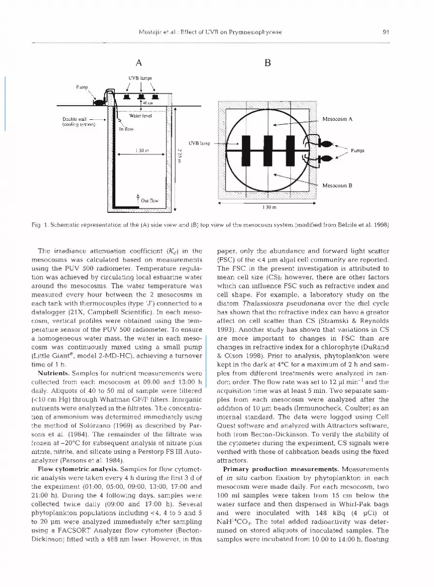
Fig 1. Schematic representation of the (A) side view and (B) top view of the mesocosm system (moddied from Belzile et al. 1998)
The irradiance attenuation coefficient (Kd) in the mesocosms was calculated based on measurements using the PUV 500 radiometer. Temperature regula- tion was achieved by circulating local estuarine water around the mesocosms. The water temperature was measured every hour between the 2 mesocosms in each tank with thermocouples (type 'J') connected to a datalogger (21X, Campbell Scientific). In each meso- cosm, vertical profiles were obtained using the tem- perature sensor of the PUV 500 radiometer. To ensure a homogeneous water mass, the water in each meso- cosm was continuously mixed using a small pump (Little Giant', model 2-MD-HC), achieving a turnover time of 1 h.
Nutrients. Samples for nutrient measurements were collected from each mesocosm at 09:OO and 13:OO h daily. Aliquots of 40 to 50 m1 of sample were filtered ( < l 0 cm Hg) through Whatman GF/F filters. Inorganic nutrients were analyzed in the filtrates. The concentra- tion of ammonium was determined immediately using the method of Solorzano (1969) as described by Par- sons et al. (1984). The remainder of the filtrate was frozen at -20°C for subsequent analysis of nitrate plus nitnte, nitrite, and silicate using a Perstorp FS 111 Auto- analyzer (Parsons et al. 1984).
Flow cytometric analysis. Samples for flow cytomet- ric analysis were taken every 4 h during the first 3 d of the experiment (01:00, 05:00, 09:00, 13:00, 17:OO and 21:OO h). During the 4 following days, samples were collected twice daily (09:OO and 17:OO h) . Several phytoplankton populations including <4, 4 to 5 and 5 to 20 pm were analyzed immediately after sampling using a FACSORT Analyzer flow cytometer (Becton- Dickinson) fitted with a 488 nm laser. However, in this
paper, only the abundance and forward light scatter (FSC) of the < 4 pm algal cell community are reported. The FSC in the present investigation is attributed to mean cell size (CS); however, there are other factors which can influence FSC such as refractive index and cell shape. For example, a laboratory study on the diatom Thalassioslra pseudonana over the die1 cycle has shown that the refractive index can have a greater affect on cell scatter than CS (Stramski & Reynolds 1993). Another study has shown that variations in CS are more important to changes in FSC than are changes in refractive index for a chlorophyte (DuRand & Olson 1998). Prior to analysis, phytoplankton were kept in the dark at 4°C for a maximum of 2 h and sam- ples from different treatments were analyzed in ran- dom order. The flow rate was set to 12 p1 min-' and the acquisition time was at least 5 min. Two separate sam- ples from each mesocosm were analyzed after the addition of 10 pm beads (Immunocheck, Coulter) as a n internal standard. The data were logged using Cell Quest software and analyzed with Attractors software, both from Becton-Diclunson, To verify the stability of the cytometer during the experiment, CS signals were verified with those of calibration beads using the fixed attractors.
Primary production measurements. Measurements of in sltu carbon fixation by phytoplankton in each mesocosm were made daily. For each mesocosm, two 100 m1 samples were taken from 15 cm below the water surface and then dispensed in Whirl-Pak bags and were inoculated with 148 kBq (4 pCi) of NaH14C03. The total added radioactivity was deter- mined on stored aliquots of inoculated samples. The samples were incubated from 10:OO to 14:OO h, floating
92 Mar Ecol Prog Ser 187: 89-100, 1999
on the water surface, and were exposed to above- water radiation intensities. At the end of the incuba- tion, the samples were fractionated on Whatman GF/F filters (total production) and 5 pm polycarbonate filters (Poretics) (production of large algae > 5 pm). The filters were subsequently put into scintillation vials, acidified with 1 m1 of 0.5 N HC1 and then shaken in a hood to expel inorganic I4C (Lean & Burnison 1979). The Ecol- ume scintillation cocktail was added to the samples, which were counted using a Beckman LS5801 scintil- lation counter. The counts were dark corrected. Car- bon fixation rates were computed according to Parsons et al. (1984). The primary production of phytoplankton <5 pm was calculated by subtracting the primary pro- duction of algae > 5 pm from the total (GF/F) primary production. The phytoplankton < 5 pm size fraction was largely dominated by Prymnesiophyceae (see below). The specific rate of photosynthesis (PCe") for phytoplankton < 5 pm was calculated by dividing the primary production of phytoplankton < 5 pm by their abundance estimated by flow cytometry.
Microscopic phytoplankton identification. A combi- nation of DAPI-fluorescence, Nomarski differential interference contrast optics, and Utermohl inverted microscopy was used for the identification of the acid Lugol fixed <5 pm phytoplankton community. This combined optical system has been shown to encom- pass the advantages of 3 methods: Utermohl sedimen- tation for minimizing cellular damage, auto- and fluo- rochrome fluorescence for locating and characterizing cells, and Nomarski contrast for highlighting taxo- nomic features (Lovejoy et al. 1993).
Predator populations. Several predator populations, including heterotrophic flagellates (2 to 10 pm), ciliates (15 to 35 pm) and tintinnids, were identified and moni- tored during the weeklong experiment. However, as it has already been shown that ciliates play a major role in controlling bacteria, heterotrophic flagellates and c5 pm phytoplankton population dynamics (Mostajir et al. 1999), only ciliate abundances at 09:OO h are pre- sented here. Estimates of ciliate abundance were made with a Zeiss inverted microscope using 100 m1 water samples that had been preserved with acid Lugol (0.4 % final conc.] and sedimented for 24 h.
Statistical analysis. A l-way analysis of variance (ANOVA) with UVB treatment as the grouping factor was performed on each data set for each sampling time. When significant differences occurred, Bonfer- roni adjusted pairwise comparisons were used, as well as the Dunnett's test with NUVB treatment acting as the control group. For the PCe" for phytoplankton <5 pm, an ANOVA followed by a Tukey test (HSD: honestly significant difference) were performed. The probability values (p) given hereafter are indicative of significant results for these statistical tests.
RESULTS
Temperature, irradiance and nutrients
During the 7 d of the experiment, the water temper- ature varied from 8.5 to 11.3"C with no significant dif- ferences between the tanks (Belzile et al. 1998). During the experiment, 3 successive periods were distin- guished according to the incident irradiance: 2 sunny days, followed by 3 cloudy days, and finally 2 more sunny days (Fig. 2A,B,C). Only about 40% of the inci- dent irradiance reached the water surface of the meso- cosms due to shading effects (see Belzile et al. 1998 for details). The UVB lamps provided daily unweighted increased UVB doses of 40.5 kJ m-' (LUVB) and 59.2 kJ m-2 (HUVB] at the water surface of the meso- cosms. Fig. 2D shows the average UV radiation inten- sities just below the water surface calculated for a sunny day at noon. Biological weighting functions (BWFs) were applied because the experimental lamps and the sun have different spectral shapes with respect to UVB. The results from application of some well- known BWFs to the spectral irradiance presented in Fig. 2D are illustrated in Table 1 (Cullen et al. 1992, Behrenfeld et al. 1993, Boucher & Prezelin 1996). For a sunny day at noon, the UV irradiance in LUVB and HUVB treatments was 1.10- and 1.15-fold higher,
Table 1 Relative irradiance increases provided by the lamps, for low (LUVB) and high (HUVB) ultraviolet-B enhancements, just below the water surface on a sunny day around noon compared to relative increases of incident irradiance associ- ated with ozone depletion over Antarctica. These increases have been calculated from data presented in Fig. 2D. Spectral irradiances were weighted with the biological weighting function (BWF) for inhib~tion of photosynthesis in the temper- ate latitude diatom Phaeodactylum sp. (Cullen et al. 1992), the 'best fit' action spectrum for photoinhibition (Behrenfeld et al. 1993) and the daily averaged BWF for the in situ inhibition of primary production in a natural community of Antarctic di- atoms (Boucher & Prezelin 1996). The data for McMurdo Station are from Cu.Uen & Neale (1997) and correspond to a
decrease of ozone thickness from 350 to 175 DU
Incident solar LUVB HUVB irradiance at
McMurdo
Unweighted UV 1.06 1.10 1.15
BWF for inhibition of 1.24 1.75 2.10 photosynthesis (Cullen et al. 1992)
BCVF for photoinhibition 4.31 (Behrenfeld et al. 1993)
BWF for in situ inhibition 4.81 of photosynthesis (Boucher & Prezelin 1996)
Mostajir et a1 Effect of UVB on Prymnesiophyceae 93
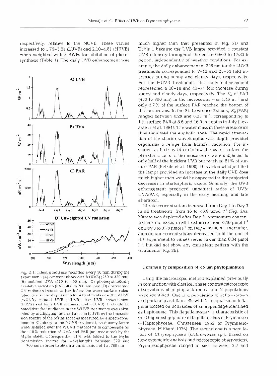
respectively, relative to the NUVB. These values increased to 1.75-3.61 (LUVB) and 2.10-4.81 (HUVB) when weighted with 3 BWFs for inhibition of photo- synthesis (Table 1). The daily UVB enhancement was
A) UVB
B) UVA
davl day2 day 3 day 4 day S day 6 day 7
50 -7 D) Unweighted UV radiation
280 300 320 340 360 380 400
Wavelength (nm)
Fig. 2 Incident irradiance recorded every 10 min during the experiment. (A) Amb~en t ultrav~olet-B (UVB) (280 to 320 nm), (B) ambient UVA (320 to 400 nm), (C) photosynthetically available radiation (PAR: 400 to 700 nm) and (D) unweighted UV radiation i n t e n s ~ t ~ e s just below the water surface calcu- lated for a sunny day at noon for 4 treatments of without UVB (WUVB). natural UVB (NUVB), low UVB enhancement (LUVB) and hlgh UVB enhancement (HUVB). It should be noted that the irradiance in the WUVB treatments was calcu- lated by multiplying the Irradiance in NUVB by the transmis- sion spectra of the Mylar sheet as measured by a spectropho- tometer Contrary to the NUVB treatment, no dummy lamps were installed over the WUVB mesocosms to compensate for the -10% reduction of UVA and PAR (not measured) by the Mylar sheet. Consequently, 11% was added to the Mylar transmission spectra for wavelengths between 320 and
700 nm in order to obtain a transmission of 1 at 700 nm
much higher than that presented in Fig. 2D and Table 1 because the UVB lamps provided a constant UVB intensity throughout the entire 09:00 to 17:30 h period, independently of weather conditions. For ex- ample, the daily enhancement at 305 nm for the LUVB treatments corresponded to 7-13 and 28-51 fold in- creases during sunny and cloudy days, respectively. For the HUVB treatments, this daily enhancement represented a 10-18 and 40-74 fold increase during sunny and cloudy days, respectively. The K, of PAR (400 to 700 nm) in the mesocosms was 1.46 m-' and only 3.7% of the surface PAR reached the bottom of the mesocosms. In the St. Lawrence Estuary, K, (PAR) ranged between 0.29 and 0.53 m-', corresponding to l % surface PAR at 8.6 and 16.0 m depths in July (Lev- asseur et al. 1984). The water mass in these mesocosms thus simulated the euphotic zone. The rapid attenua- tion of the shorter wavelengths with depth provided organisms a refuge from harmful radiation. For in- stance, as little as 14 cm below the water surface the planktonic cells in the mesocosms were subjected to only half of the incident UVB but received 81 % of sur- face PAR (Belzile et al. 1998). It is acknowledged that the lamps provided an increase in the daily UVB dose much higher than would be expected for the projected decreases in stratospheric ozone. Similarly, the UVB enhancement produced unnatural ratios of UVB: UVA:PAR, especially in the early morning and late afternoon.
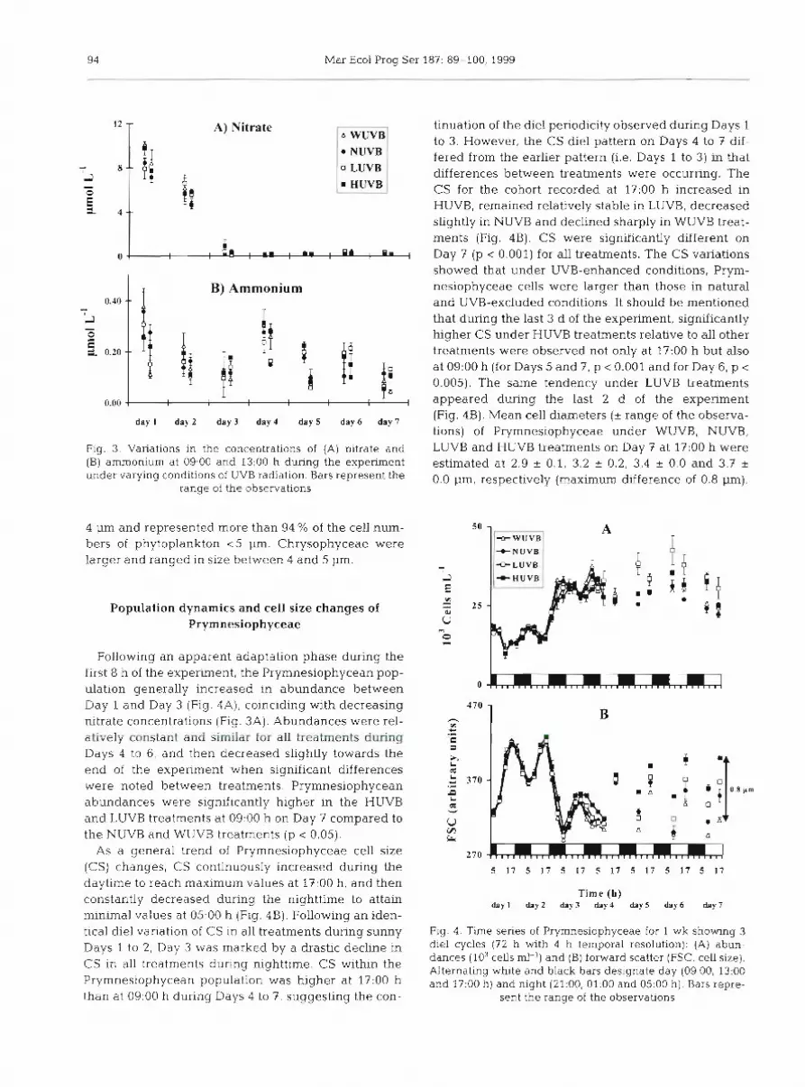
Nitrate concentration decreased from Day 1 to Day 3 in all treatments, from 10 to <0.9 pm01 1-' (Fig. 3A). Nitrate was depleted after Day 3. Amnlonium concen- trations increased in all treatments from 0.10 pm01 1-' on Day 3 to 0.28 pm01 l-l on Day 4 (09:00 h ) . Thereafter, ammonium concentrations decreased until the end of the experiment to values never lower than 0.04 pm01 1-l, but did not show any consistent pattern with the treatments (Fig. 3B).
Community composition of < 5 pm phytoplankton
Using the microscopic method explained previously in conjunction with classical phase contrast microscopic observations of phytoplankton <5 pm, 2 populations were identified. One is a population of yellow-brown and parietal plastidian cells with 2 unequal smooth fla- gella located on both sides of an appendage identified as haptonema. This flagella system is characteristic of the Oligomastigophorean flagellate class of Prymnesea (=Haptophyceae, Christensen 1962 or Prymnesio- phyceae, Hibberd 1976). The second one is a popula- tion of Chrysophyceae (Ochromonas sp.) . Based on flow cytometric analysis and microscopic observations, Prymnesiophyceae ranged in size between 2.7 and
94 Mar Ecol Prog Ser 187: 89-100, 1999
A) Nitrate A WUVB
NUVB 0 LUVB
HUVB 7 B) Ammonium
day I d a y 2 day 3 day 4 d a y 5 day 6 day 7
Fig. 3. Variations in the concentrations of (A) nitrate and (B] ammonium at 09:OO and 13:00 h during the experiment under varying conditions of UVB radiation. Bars represent the
range of the observations
4 pm and represented more than 94 % of the cell num- bers of phytoplankton < 5 pm. Chrysophyceae were larger and ranged in size between 4 and 5 pm.
Population dynamics and cell size changes of Prymnesiophyceae
Following an apparent adaptation phase during the first 8 h of the experiment, the Prymnesiophycean pop- ulation generally increased in abundance between Day 1 and Day 3 (Fig. 4A), coinciding with decreasing nitrate concentrations (Fig. 3A). Abundances were rel- atively constant and similar for all treatments during Days 4 to 6, and then decreased slightly towards the end of the experiment when significant differences were noted between treatments. Pryrnnesiophycean abundances were significantly higher in the HUVB and LUVB treatments at 09:OO h on Day 7 compared to the NUVB and WUVB treatments (p < 0.05).
As a general trend of Prymnesiophyceae cell size (CS) changes, CS continuously increased during the daytime to reach maximum values at 17:OO h, and then constantly decreased during the nighttime to attain minimal values at 05:OO h (Fig. 4B). Following an iden- tical diel variation of CS in all treatments during sunny Days 1 to 2, Day 3 was marked by a drastic decline in CS in all treatments during nighttime. CS within the Prymnesiophycean population was higher at 17:OO h than at 09:OO h during Days 4 to 7, suggesting the con-
tinuation of the diel periodicity observed during Days 1 to 3. However, the CS diel pattern on Days 4 to 7 dif- fered from the earlier pattern (i.e. Days 1 to 3) in that differences between treatments were occurring. The CS for the cohort recorded at 17:00 h increased in HUVB, remained relatively stable in LUVB, decreased slightly in NUVB and declined sharply in WUVB treat- ments (Fig. 4B). CS were significantly different on Day 7 (p < 0.001) for all treatments. The CS variations showed that under UVB-enhanced conditions, Prym- nesiophyceae cells were larger than those in natural and UVB-excluded conditions. It should be mentioned that during the last 3 d of the experiment, significantly higher CS under HUVB treatments relative to all other treatments were observed not only at 17:00 h but also at 09:OO h (for Days 5 and 7, p < 0.001 and for Day 6, p < 0.005). The same tendency under LUVB treatments appeared during the last 2 d of the experiment (Fig. 4B). Mean cell diameters (+ range of the observa- tions) of Prymnesiophyceae under WUVB, NUVB, LUVB and HUVB treatments on Day 7 at 17:00 h were estimated at 2.9 +- 0.1, 3.2 & 0.2, 3.4 + 0.0 and 3.7 + 0.0 pm, respectively (maximum difference of 0.8 pm).
Time (h) day 1 day2 day3 day4 day5 day6 day 7
Fig. 4. Time series of Pryrnnesiophyceae for 1 wk showing 3
d e l cycles (72 h with 4 h temporal resolution): (A) abun- dances (103 cells ml-l) and (B) forward scatter (FSC, cell size). Alternating white and black bars designate day (09:00, 13:OO and 17:OO h ) and night (21:00, 01-00 and 05:OO h) Bars repre-
sent the range of the observations
Mostajir et al.: Effect of UVB on Prymnesiophyceae 95
Assuming a spherical cell shape, these diameters cor- respond to volumes of 12.8, 17.2, 20.6 and 26.5 pm3 for WUVB, NUVB, L W B and HUVB treatments, respec- tively.
Specific rate of photosynthesis (PCe") of phytoplankton <5 pm
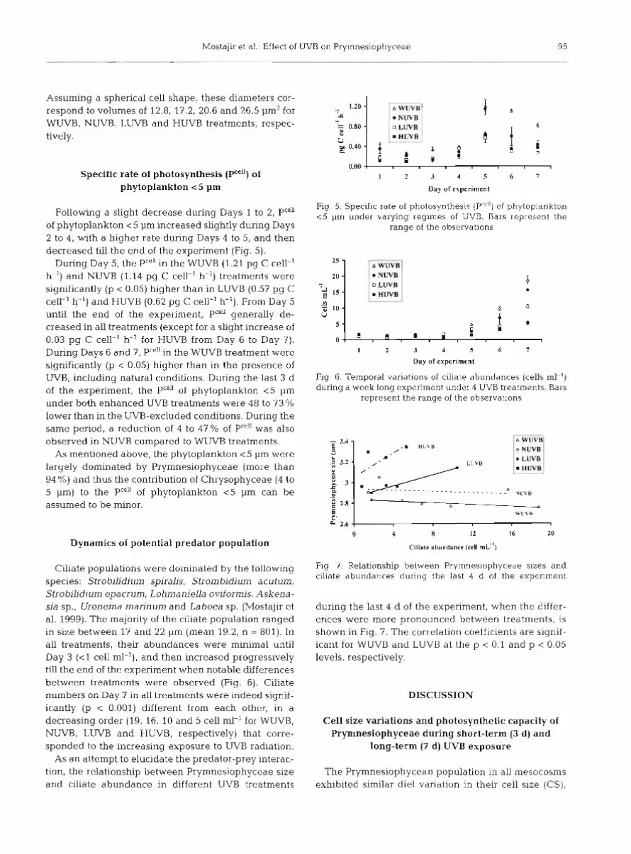
Following a slight decrease during Days 1 to 2, Pceu of phytoplankton <5 pm increased slightly during Days 2 to 4, with a higher rate during Days 4 to 5, and then decreased till the end of the experiment (Fig. 5).
During Day 5, the PCe"n the WUVB (1.21 pg C cell-' h-') and NUVB (1.14 pg C cell-' h-') treatments were significantly (p < 0.05) higher than in LUVB (0.57 pg C cell-' h-') and HUVB (0.62 pg C cell-' h-'). From Day 5 until the end of the experiment, PCe\enerally de- creased in all treatments (except for a slight increase of 0.03 pg C cell-' h-' for HUVB from Day 6 to Day 7). During Days 6 and 7, Pce"in the WUVB treatment were significantly (p < 0.05) higher than in the presence of W B , including natural conditions. During the last 3 d of the experiment, the PCe" of phytoplankton <5 pm under both enhanced UVB treatments were 48 to 73 % lower than in the UVB-excluded conditions. During the same period, a reduction of 4 to 47 % of PCe" was also observed in NUVB compared to WUVB treatments.
As mentioned above, the phytoplankton < 5 pm were largely dominated by Prymnesiophyceae (more than 94 %) and thus the contribution of Chrysophyceae (4 to 5 pm) to the Pce" of phytoplankton <5 pm can be assumed to be minor.
Dynamics of potential predator population
Ciliate populations were dominated by the following species: Strobilidium spiralis, Strombidium acutum, Strobilidium epacrum, Lohmaniella oviformis, Askena- sia sp., Uronema marinum and Laboea sp. (Mostajir et al. 1999). The majority of the ciliate population ranged in size between 17 and 22 pm (mean 19.2, n = 801). In all treatments, their abundances were minimal until Day 3 (<l cell ml-l), and then increased progressively till the end of the experiment when notable differences between treatments were observed (Fig. 6). Ciliate numbers on Day 7 in all treatments were indeed signif- icantly (p < 0.001) different from each other, in a decreasing order (19, 16, 10 and 5 cell ml-' for WUVB, NUVB, LWB and HUVB, respectively) that corre- sponded to the increasing exposure to UVB radiation.
As an attempt to elucidate the predator-prey interac- tion, the relationship between Prymnesiophyceae size and ciliate abundance in different UVB treatments
NUVB ;::::l ;F; , l , ; , , ; U
HUVB 00 0.40 C
P 0.00
1 2 3 4 5 6 7
Day of experiment
Fig. 5. Specific rate of photosynthesis (P"") of phytoplankton <5 pm under varying regimes of UVB. Bars represent the
range of the observations
A WUVB NUVB
0 LUVB
l 2 3 4 5 6 7
Day of experiment
Fig. 6. Temporal variations of ciliate abundances (cells ml-') during a week long experiment under 4 UVB treatments. Bars
represent the range of the observations
Ciliate abundance (cell m ~ " )
- 3.4 - 5 U
.B 3.2 - Y (5 U 3 -
C a 0
.- g 2.8 - E L 2.6 1
Fig. 3. Relationship between Prymnesiophyceae sizes and ciliate abundances during the last 4 d of the experiment
. / .L HUVB
m' LUVB
. . - . - . - . .O VL!VB p
0 d , WLIVR
l
during the last 4 d of the experiment, when the differ- ences were more pronounced between treatments, is shown in Fig. 7. The correlation coefficients are signif- icant for W W B and LUVB at the p < 0.1 and p < 0.05 levels, respectively.
0 4 8 I 2 16 20
DISCUSSION
Cell size variations and photosynthetic capacity of Prymnesiophyceae during short-term (3 d) and
long-term (7 d) UVB exposure
The Prymnesiophycean population in all mesocosms exhibited similar die1 variation in their cell size (CS),

96 Mar Ecol Prog Ser 187: 89-100, 1999
with maxima at 1?:00 h and minima at 05:OO h, during the first 3 d of the experiment (short-term) character- ized by a high sampling resolution (every 4 h) . It would be difficult to identify with certainty any particular environmental cause for a very large decrease in CS on Day 3. However, as an alternative cause, a large decrease in incident irradiance during Day 3 (Fig. 2) coupled with a well-mixed mesocosm would result in the subsaturation of photosynthesis over most of the day. By the end of the day, the cells would likely not have sufficient photosynthate to create 2 large cells during nocturnal cell division, but may be able to cre- ate 2 smaller cells (M. Behrenfeld, as one of the 4 reviewers). The Prymnesiophyceae population during Days 3 to 7 (long-term) always exhibited highest CS in the late afternoon (17:OO h) and lowest CS in the early morning (09:OO h) in all treatments, despite the low sampling resolution (09:OO and 17:00 h) and the occur- rence of differences between treatments. These obser- vations indicate the occurrence of synchronic cell divi- sion within the Prymnesiophycean population during the nighttime and the relative resistance of this appar- ent endogenous rhythmicity, frequent in phytoplank- ton communities, not only to a short-term (3 d) but also to a long-term (7 d) exposure to UVB radiation. Syn- chronized cell division is well known in the aquatic environment for both phytoplankton (Pollingher & Ser- ruya 1976, Heller 1977, Taylor 1980, Frempong 1982) and bacterioplankton (Sime-Ngando et al. 1991).
Prymnesiophyceae CS was significantly larger in the enhanced UVB than in the natural and UVB excluded treatments after 7 d of exposure. This increase in CS for a natural planktonic community agrees with results from earlier laboratory studies (Behrenfeld et al. 1992, Van Donk & Hessen 1995, Buma et al. 1996).
One potential cause of cell enlargement in this experiment could be due to the perturbation of the cell cycle under UVB enhancement. The die1 pattern in CS contrasted with the circadian variability in cell abun- dance that was generally higher during the night than during daytime (from 05:OO to 17:OO h, Fig. 4). This clearly means that during this experiment, UVB appar- ently affected the number of divisions but not the fre- quency or rythmicity. To elucidate the change in the cell division rate of Prymnesiophyceae, the rate of change for this population (reflecting mortality, graz- ing and division rates) was calculated. The results (not shown) do not show any consistent pattern with treatment as this population was grazed under differ- ent pressures (see below: 'Predator-prey interactions under UVB exposure'). Prymnesiophyceae such as Emiliana huxleyi are sensitive to UVB radiation, prob- ably due to its effects on the cell cycle related to nuclear DNA damage (Gieskes & Buma 1997). The DNA damage can generally cause inhibition of DNA
replication which is a precondition for cell division (Buma et al. 1996). Karentz et al. (1991) found an inter- specific decrease in UV sensitivity with increasing cell volume and suggested that increased cell volume may protect DNA by increasing the pathlength for UVB radiation through the cell. However, Laurion & Vincent (1998) have recently demonstrated that CS is not a good ind.ex of UV sensitivity. UVB radiation may inhibit steps in the cell cycle, notably the retardation of the entry of DNA replication in the S phase (Setlow et al. 1963, Buma et al. 1996), resulting in a continued cell growth without division (see Behrenfeld et al. 1992). The same process was reported for Cyclotella sp. (Buma et al. 1997), supporting the hypothesis that DNA is one of the primary targets of UVB radiation (Karentz et al. 1991, Buma et al. 1995, 1996). At the end of the experiment, the higher Prymnesiophycean CS under UVB enhancement compared to natural condi- tions at both dawn and dusk (Fig. 4B) may indicate that a portion of the population was not dividing. This por- tion of cells grows to a larger size in preparation for division, but then does not divide. Although there are no direct measurements of DNA damage in this exper- iment, the findings, illustrating Prymnesiophyceae cell enlargement under UVB stress, could similarly reflect the retardation of the cell cycles in the S phase. Buma et al. (1996) have shown for the diatom Cyclotella sp. that decreased division rates under UVB enhancement were typically accompanied by an increase in CS. For a long time, it has been known that certain types of UV damage could decouple cell growth and division pro- cesses, resulting in the enlargement of cells of some protistan taxa. For example, it has been shown that exposure to UV radiation perturbed cell division in a laboratory culture of the ciliate Paramecium caudatum (Hinrichs 1928, Karentz et al. 1994 and references therein). This provoked the formation of giant or mon- ster cells, partly due to the induction of adducts involved in DNA repair mechanisms or due to the accumulation of substances that could no longer be discharged from the cell.
The second potential cause of cell enlargement in this experiment could be due to the change in photo- synthetic capacity. Although the impact of UVB radia- tion on the photosynthetic capacity is not discussed in detail in the present paper, these data are used firstly to explain Prymnesiophyceae cell enlargement and secondly to elucidate the ecological implications of the photosynthetic capacity of Prymnesiophyceae on predator-prey interactions. A decrease in photosyn- thetic rates of Prymnesiophyceae could cause a decrease in cell division rates and changes in CS. A higher PC'" of smaller Prymnesiophyceae relative to all other conditions at the end of the experiment is illus- trated in Fig. 5. Comparable results about the reduc-
Mostajir et al.: Effect of U\ 'B on Prymnesiophyceae 93
tion of primary production have already demonstrated the sensitivity of phytoplankton (including Prymnesio- phyceae) to UVB radiation, which is known to reduce the rate of primary production (Smith et al. 1980, Cullen & Lesser 1991, Smith e t al. 1992, Vincent & Roy 1993). The results on the reduction of the PCe" under natural and enhanced UVB presented here come from the samples that were incubated at the water surface of the mesocosms, where the UVB doses were higher than those received by organisms present in the water column. Due to the rapid attenuation of the shorter wavelengths with depth in the mesocosms, the UVB doses diminished by 50% at 14 cm below the water surface in the mesocosms. At l m depth, planktonic cells received 23% of surface PAR, but only 0.6% of surface UVB at 305 nm. Given such optical characteris- tics and that the water was continuously mixed, the water column of the mesocosm simulated the euphotic zone. Planktonic organisms would have been able to repair UVB damage in the deeper waters of the meso- cosms, as well as undertake nighttime repair pro- cesses. Therefore, results of PC"" reduction from incu- bations a t the water surface in the presence of UVB are overestimated compared to the average situation in the mesocosm. Compared to the WUVB treatment, these rates clearly demonstrate the sensitivity of phyto- plankton < 5 pm to UVB and reflect their physiological condition under different UVB doses. This highlights the role of vertical mixing in UVB enhancement stud- ies. Vertical mixing in natural aquatic systems trans- ports organisms to deeper water, permitting then? to repair UVB damage. This could also explain the non- significant effect of UVB treatments for the synchro- nized diel variability observed during this experiment.
Nutnent limitation can also play a role in CS changes. During our experiment, a parallel study by Fauchot (1998) showed that there was no significant difference between treatments for nitrate and ammo- nium concentrations, although ammonium and urea uptake became important after nitrate exhaustion on Day 3. Although there is no measurement of other nutrients (Pod3- and trace elements) that could limit algal growth, the work of Fauchot (1998) suggests that Prymnesiophyceae were not limited by nitrates, but were growing on regenerated supplies such as ammo- nium. Hence, CS changes observed for Prymnesio- phyceae were not due to nutrient limitation but rather to the direct effect of UVB radiation.
Predator-prey interactions under UVB exposure
In aquatic systems, the link between primary pro- duction, bacteria and higher trophic levels is generally mediated by microzooplankton (Azam et al. 1983).
Among Protozoa, ciliates are able to play a pivotal role in marine food webs. Through predation, ciliates can regulate the populations of autotrophic picoplankton (52 pm) (Rassoulzadegan et al. 1988, Bernard & Ras- soulzadegan 1990) and bacterioplankton (Fenchel 1980, Rivier et al. 1985, Sherr & Sherr 1987). They can also transfer the b.ulk of both matter and energy fluxes from lower trophic levels, i.e. pico- and nanoplankton (0.2 to 20 pm), to higher ones such as metazoan zoo- plankton (Stoecker & Egloff 1987, Wiadnyana & Ras- soulzadegan 1989, Jonsson & Tiselius 1990, Dolan 1991, Hartmann et al. 1993, Sime-Ngando et al. 1995), coral (Ferrier-Pages et al. 1998), oyster (Le Gall et al. 1997) and fish larvae (De Mendiola 1974).
Although the negative impact of UVB on Prymnesio- phycean cell division was obvious (see above), their abundances paradoxically increased significantly with increasing exposure to UVB (Day 7, 09:OO h, p < 0.05). This contrasts with the pattern in ciliate density which decreased with increasing exposure to UVB (66% lower under HUVB relative to NUVB, Fig. 6) , indicat- ing the prevalence of the predation pressure from cili- ates on Prymnesiophyceae growth during this experi- ment.
The negative effect of UVB enhancement on prey (phytoplankton) can therefore be masked by trophic relationships such as predator-prey interactions. The negative effect of UVB on predator populations (cili- ates) induces a positive feedback between enhanced UVB and prey abundances. Hence, in studies concern- ing the effects of UVB on aquatic systems, trophic interactions should be involved with the other studied parameters, as suggested previously by Mostajir et al. (1999).
Another aspect of predator-prey interactions is related to the preferential prey size. The main feature of the particle-size model developed by Sheldon et al. (1972) and modified by Azam et al. (1983) is that organisms tend to utilize particles 1 order of magni- tude smaller than themselves. Generally, nanociliates (< 20 pm), which dominated the ciliate community dur- ing this study, selectively remove the largest bacteria (Bernard & Rassoulzadegan 1990, Christaki et al. 1998) and picophytoplankton (including cyanobacte- ria) ranging in size around 1 pm (Mostajir et al. 1998). A significant relationship between the diel vertical dis- tribution of planktonic ciliates and zeaxanthin, a pig- ment bioindicator of cyanobacteria, was reported in the surface layer of the NW Mediterranean (Perez et al. in press). In our experiment, the cyanobacteria were absent and ciliates, ranging in size between 17 and 22 pm (mean diameter = 19.2 pm, n = 801), likely grazed on heterotrophic bacteria (< l pm), small phyto- plankton (<5 pm) and heterotrophic nanoflagellates (2 to 10 pm) (Mostajir et al. 1999). Fig. 7 shows that
98 Mar Ecol Prog Ser 187: 89-100, 1999
in UVB-excluded treatments where the highest ciliate abundances were recorded, the Prymnesiophyceae population was exclusively represented by the small- est cells. Apart from a probable direct UVB negative effect on ciliate abundance, one of the reasons for the higher ciliate abundance under WUVB and NUVB conditions (relative to enhanced UVB treatments) could be the relatively greater availability and quality of the Prymnesiophyceae prey (dividing more rapidly and producing more carbon) in the preferred size range. It is also possible that a change in food quality of the Prymnesiophyceae, especially the fatty acid com- position, might have been affected by UVB exposure (Goes et al. 1994, De Lange & Van Donk 1997). The negative significant slope (p < 0.1) of the predator and prey-size relationship in WUVB conditions suggests that smaller Prymnesiophyceae were likely adequate for ciliate development (Fig. 7). The change in slope of this relationship from negative to positive as a function of increased UVB doses demonstrates the importance of prey size for the development of ciliates.
In summary, the results of this study suggest that enhanced UVB can provoke the retardation of cell division and inhibition of the PCe" of phytoplankton, which causes Prymnesiophyceae CS enlargement. The ciliate population decreased significantly in number under UVB enhancement. The reduction of ciliate abundance can be attributed to a direct or an indirect (via prey) effect of enhanced UVB exposure. CS enlargement, as well as probably a change in food quality (fatty acid composition) of prey, could result in food limitation for the ciliate population and thus par- tially explain their decrease in abundance. The ciliates transfer the major flux of matter and energy from the lower trophic levels to higher ones and a slight pertur- bation in such transfers will have an important rarnifi- cation throughout the marine food web with consider- able ecological consequences. We conclude that in order to predict the effect of enhanced UVB at the ecosystem level, trophic interactions between different trophic levels should definitely be considered.
Acknowledgements. We thank J. Bouchard for her help in cytometric analyses, D. Bourget and N. Lafontaine for nutri- ent analyses and L. Zudaire for data visualization. We wish to thank M. Behrenfeld as well as 3 other anonymous reviewers for useful suggestions. This work was supported by NSERC of Canada, Fonds FCAR of Quebec and FODAR (University of Quebec). This investigation is a contribution to the research programs of the Groupe de Recherche en Environnement Cdtier.
LITERATURE CITED
Azam F, Fenchel T, Field JG, Gray JS, Meyer-Reil LA, Thingstad F (1983) The ecological role of water-column
m~crobes in the sea. Mar Ecol Prog Ser 10:257-263 Behrenfeld MJ, Hardy JT, Lee H 11 (1992) Chronic effects of
ultraviolet-B radiation on growth and cell volume of Phaeodactylum tricornutum (Bacillariophyceae). J Phycol 28:725-760
Behrenfeld MJ, Chapman JW, Hardy JT. Lee I1 H (1993) Is there a common response to ultraviolet-B radiation by marine phytoplankton? Mar Ecol Prog Ser 102:59-68
Belzile C, Demers S, Lean DRS, Mostajir B, Roy S, de Mora S, Gosselin M, Bird D, Chanut JP, Levasseur M (1998) An experimental tool for the study of the effects of ultraviolet radiation on planktonic communities: a mesocosm ap- proach Environ Technol 19:667-682
Bernal-d C, Rassoulzadegan F (1990) Bactena or rnicroflagel- lates as a major food source for marine c~hates: possible implications for the microzooplankton. Mar Ecol Prog Ser 64.147-155
Boucher NP, Prezelin B (1996) An in situ biological weighting function for UV inhibition of phytoplankton carbon fixa- tion in the Southern Ocean. Mar Ecol Prog Ser 144: 223-236
Buma AGJ, Van Hannen EJ, Veldhuis MJW, Roza L, Gieskes \ W C (1995) Monitoring UV-B induced DNA damage in individual diatom cells by imrnunofluorescent thyrnine dimer detection. J Phycol31:314-321
Buma AGJ, Zemmelink HJ, Sjollema K, Gieskes VMYC (1996) UVB radiation modifies protein and photosynthetic pig- ment content, volume and ultrastructure of marine diatoms. Mar Ecol Prog Ser 142:47-54
Buma AGJ, Engelen AH, Gieskes WWC (1997) Wavelength- dependent induction of thyrnine dlmers and growth rate reduction in the marine diatom Cyclotella sp. exposed to ultraviolet radiation. Mar Ecol Prog Ser 153:91-97
Calkins J , Thordardotti.er T (1980) The ecological significance of solar UV radiation on aquatic organisms Nature 283: 563-566
Christaki U, Dolan JR, Pelegri S, Rassoulzadegan F (1998) Consumption of picoplankton-size particles by marine cil- i a t e ~ : effects of physiological state of the ciliate and parti- cle quality. Limnol Oceanogr 43:458-464
Christensen T (1962) Alger. In: Bocher T W et al. (eds) Botanik. Vol2. Munksgaard, Copenhagen
Cullen JJ, Lesser MP (1991) Inhibition of photosynthesis by ultraviolet radiation as a function of dose and dosage rate: results for a marine diatom. Mar Biol 11 1:183-190
Cullen JJ , Neale PJ (1997) Biological weighting function for describing the effects of ultraviolet radiation on aquatic systems. In: Hdder DP (ed) The effects of ozone deple- tion on aquatic ecosystems. Landes RG, Georgetown, p 97-118
Cullen JJ , Neale PJ, Lesser MP (1992) Biological weighting function for the inhibition of phytoplankton photosynt.he- sis by ultraviolet radiation. Science 258:646-650
Davidson AT, Marchant HJ, de la Mare WK (1996) Natural UVB exposure changes the species composition of Antarc- tic phytoplankton in mixed culture. Aquat Microb Ecol 10: 299-305
De Lange HJ, Van Donk E (1997) Effects of UVB irradiated algae on Me history traits of Daphnia pulex. Freshw Biol 38:711-720
De Mendiola BR (1974) Food of the larval Anchoveta EngrauLis ringens. In: Blaxter JHS (ed) The early life his- tory of fish. Springer-Verlag, Berlin, p 277-285
Dolan JR (1991) Microphagous ciliates In mesohaline Chesa- peake Bay waters: estimates of growth rates and con- sumption by copepods. Mar Biol 11 1:303-309
DuRand MD, Olson RJ (1998) Die1 patterns in optical proper-
Mostajir et al.: Effect of UVB on Pryinnesiophyceae 99
ties of chlorophyte Nannochlons sp.: relating indi- v~dual-cell to bulk measurements. Limnol Oceanogr 43: 1107-1118
Fauchot J (1998) Influence du rayonnement ultraviolet-B sur l'utilisation de l'azote par un assemblage nature1 phyto- planctonique. MSc thesis. Universite du Quebec a Rimouski, Quebec
Fenchel T (1980) Relation between particle size selection and clearance in suspension-feeding ciliates. Limnol Oceanogr 25:733-738
Ferrier-Pages C, Allemand D. Gattuso JP, Jaubert J , Ras- soulzadegan F (1998) Microheterotrophy in the zooxan- thellate coral Stylophora pistillata: effects of light and cili- ate density. Limnol Oceanogr 43:1639-1648
Frempong E (1982) The space-time resolution of phased cell divlsion in natural populations of the freshwater dinofla- gellates Ceratium hlrundinella. Int Rev Ges Hydrobiol 67: 323-339
Gieskes WWC, Buma GJ (1997) UV damage to plant life in a photobiologically dynamlcs environment: the case of manne phytoplankton Plant Ecol 128:16-25
Goes JI, Handa N, Taguchi S, Hama T (1994) Effect of UV-B radiation on the fatty acid composition of the marine phy- toplankter Tetraselmis sp.. relationship to cellular pig- ments. Mar Ecol Prog Ser 114:259-274
Hartmann HJ, Hassan T, Aleya L, Lair N (1993) Predation on ciliates by suspension-feeding calanoid copepod Acanthodiaptornus denticornis. Can J Fish Aquat Sci 50: 1382-1393
Heller MD (1977) The phased division of the freshwater dinoflagellate Ceratiwn hirundinella and its use as a method of assessing growth in natural populations. Freshw Biol7:527-533
Hibberd DJ (1976) The ultrastructure and taxonomy of the Chrysophyceae and Prymnesiophyceae (Haptophyceae): a survey with some new observations on the ultrastructure of the Chrysophyceae. Bot J Linn 7255-80
Hinrichs MA (1928) Ultra-violet radiation and division in Paramecium caudaturn. Physiol Zol 1 :394-415
Jokiel PL, York RH (1984) Importance of ultraviolet radiation in photoinhibition of microalgal growth. Limnol Oceanogr 29:192-199
Jonsson PR, Tiselius P (1990) Feeding behaviour, prey detec- tion, and capture efficiency of the copepod Acartia tonsa feeding on planktonic ciliates. Mar Ecol Prog Ser 60:35-44
Karentz D, Cleaver JE, Mitchell DL (1991) Cell survival characteristics and molecular responses of Antarctic phytoplankton to ultraviolet-B radiation. J Phycol 27: 326-34 1
Karentz D, Bothwell ML, Coffin RB, Hanson A, Herndl GJ, k lham SS, Lesser MP, Lindell M, Moeller RE, Morris DP, Neale PJ, Sanders RW, Weiler CS, Wetzel RG (1994) Impact of UV-B radiation on pelagic freshwater ecosys- tems: report of working group on bacteria and phyto- plankton. In: Williamson CE, Zagarese HE (eds) Impact of UV-B radiation on pelagic freshwater ecosystems. Arch Hydrobiol Beih Ergebn Limnol 43:31-69
Kirk JTO, Hargreaves BR, Morris DP, Coffin RB, David B, Fredrickson D, Karentz D, Lean DRS, Lesser MP, Madronich S, Morrow JH. Nelson NB, Scully NM (1994) Measurements of UV-B radiation in two freshwater lakes: an instrument intercomparison. In: Williamson CE, Zagarese HE (eds) Impact of UV-B radiation on pelagic freshwater ecosystems. Arch Hydrobiol Beih Ergebn Lim- no1 43:71-99
Laurion I , Vincent FV (1998) Cell size versus taxonomic composition as determinants of UV-sensitivity in nat-
ural phytoplankton communities. Limnol Oceanogr 43: 1774-1779
Lean DRS. Burnison BK (1979) An evaluation of errors in the I4C method of primary production measurement. Limnol Oceanogr 24:917-928
Le Gall S, Be1 Hassen M, Le Gall P (1997) Ingestion of a bac- teri.irorous ciliate by the oyster Crassostrea gigs: protozoa as a trophic link between picoplankton and benthic sus- pension-feeders. Mar Ecol Prog Ser 152:301-306
Levasseur M. Therriault JC, Legendre L (1984) Hierarchical control of phytoplankton succession by physical factors. Mar Ecol Prog Ser 19:211-222
Lovejoy C, Vincent W, Frenette JJ, Dodson JJ (1993) Micro- bial gradients in a turbid estuary: application of a new method for protozoan community analysis. Limnol Oceanogr 38:1295-1303
Mostajir B, Bustillos-Guzmdn J, Claustre H, Rassoulzadegan F (1998) Pigment dynamics associated with the grazing of a ciliate and a flagellate feeding on a cyanobacteria. Ocean01 Acta 21:581-588
Mostalu B, Demers S, de Mora S, Belzile C, Chanut JP, Gos- selin M, Roy S, Fauchot J, Villegas PZ, Bouchard J , Bird D, Monfort P, Levasseur M (1999) Experimental test of the effect of ultraviolet-B radiation in a planktonic community. Limnol Oceanogr 44586-596
Neale PJ, Lesser MP, Cullen JJ (1994) Effects of ultraviolet radiation on the photosynthesis of phytoplankton in the vicinity of McMurdo station, Antarctica. In: Weiler CS, Penhale PA (eds) Ultraviolet radiation in Antarctica: mea- surements and biological effects. Antarctic Research Series Vol 62. Am Geophys Union, Washington, DC, p 125-142
Parsons TR, Maita Y, Lalli CM (1984) A manual of chemical and biological methods for seawater analysis. Pergamon Press, Toronto
Perez MT, Dolan JR. Vidussi F, Fukai E (in press) Die1 vertical distribution of planktonic ciliates within the surface layer of the NW Mediterranean (May 1995). Deep-Sea Res
Pollingher U, Serruya C (1976) Phased division of Pendiniwn cinctum f . westii and the development of the bloom in Lake Kmneret (Israel). J Phycol 12:162-170
Rassoulzadegan F, Laval-Peuto M, Sheldon RW (1988) Parti- tioning of the food ration of marine ciliates between pico- and nanoplankton. Hydrobiolog~a 159:75-88
Rex M, Harris NRP, von der Gathen P, Lehmann R, Braathen GO, Relrner E, Beck A, Chipperfield MP, Alfier R, Allaart M, O'Connor F, Dier H, Dorokhov V, Fast H, Gill M, Kyro E, Litynska Z. Mikkelsen IbS, Molyneux MG, Nakane H, Notholt J , Rummukainen M, Viatte P, Wenger J (1997) Prolonged stratospheric ozone loss in the 1995-96 Arctic winter. Nature 389:835-838
Rivier A. Brownlee DC, Sheldon RW, Rassoulzadegan F (1985) Growth of microzooplankton: a comparative study of bactivorous zooflagellates and ciliates. Mar Microb Food Webs 1 5 - 6 0
Setlow RB, Swenson PA, Carrier WL (1963) Thymine dimers and inhibition of DNA synthesis by ultraviolet irradiation of cells. Science 142:1464-1465
Sheldon RW, Prakash A, Sutcliffe WH (1972) The size distrib- ution of particles in the ocean. Limnol Oceanogr 17: 327-340
Sherr EB, Sherr BF (1987) High rates of consumption of bacte- ria by pelagic ciliates Nature 325:710-711
Sime-Ngando T, Bourdier G, Amblard C, Pinel-Alloul B (1991) Short-term vanations in specific biovolume of dif- ferent bacterial forms in aquatic ecosystems. Microb Ecol 21:211-226
100 Mar Ecol Prog Ser 187: 89-100, 1999
Sime-Ngando T, Gosselin M, Roy S, Chanut JP (1995) Signifi- cance of planktonic ciliated protozoa in the lower St. Lawrence Estuary: comparison with bacterial, phyto- plankton, and particulate organic carbon. Aquat Microb Eco19:243-258
Smith RC, Baker KS, Holm-Hansen 0, Olson R (1980) Photo- inhibition of photosynthesis in natural waters. Photochern Photobiol 31:585-592
Smith RC, Prezelin BB, Baker KS, Bidigare RR, Boucher NP. Coley T. Karentz D, Macintyre S, Matlick HA, Menzies D, Ondrusek M, Wan Z, Waters K J (1992) Ozone depletion: ultraviolet radiation and phytoplankton biology in Antarc- tic waters. Science 255:952-959
Solorzano L (1969) Determination of ammonia in natural waters by the phenolhypochlorite method. Limnol Oce- anogr l 4 :799-801
Stoecker DK, Egloff DA (1987) Predation by Acartia tonsa Dana on planktonic ciliates and rotifers. J Exp Mar Biol Ecol 110:53-68
Stramski D, Reynolds RA (1993) Die1 variations in the optical
Editorial responsibility. Fereidoun Rassoulzadegan (Contributing Editor), V ~ l l e f r n c h e - S - e r , France
properties of a marine dlatom. Limnol Oceanogr 38: 1347-1364
Tarasick DW. Fioletov VE (1997) The distribution of ozone and ozone-depleting substances in the atmosphere and observed changes. In: Wardle DI, Kerr JB, McElroy CT, Francis DR (eds) Ozone science: a Canadian perspective on the changing ozone layer Environment Canada. Uni- versity ot Toronto Press, Toronto, p 9-14
Taylor FJR (1980) Basic biological features of phytoplankton cells. In: Morris I (ed) The physiological ecology of phyto- plankton. Blackwell, Oxford, p 3-55
Van Donk E. Hessen DO (1995) Reduced digestibility of UV-B stressed and nutrient-limited algae by Daphnia magna. Hydrobiologia 307:147-151
Vincent WF, Roy S (1993) Solar ultraviolet-B rad~ati.on and aquatic pnmary product~on: damage, protection, and recovery. Environ Rev 1:l-12
Wiadnyana NN, Rassoulzadegan F (1989) Selective feeding of Acartia clausi and Centropages typicus on microzoo- plankton. Mar Ecol Prog Ser 53:37-45
.Submitted: January 4, 1999; Accepted: May 31, 1999 Proofs rece~ved from author(s): September 17, 1999