Early Versus Late Functional Outcome After Successful Percutaneous Pulmonary Valve Implantation
8
Early Versus Late Functional Outcome After Successful Percutaneous Pulmonary Valve Implantation Are the Acute Effects of Altered Right Ventricular Loading All We Can Expect? Philipp Lurz, MD,*†‡ Johannes Nordmeyer, MD,*†§ Alessandro Giardini, MD, PHD,† Sachin Khambadkone, MD,† Vivek Muthurangu, MD,* Silvia Schievano, PHD,* Jean-Benoit Thambo, MD,† Fiona Walker,¶ Seamus Cullen,¶ Graham Derrick,† Andrew M. Taylor, MD,*† Philipp Bonhoeffer, MD* London, United Kingdom; Leipzig and Berlin, Germany; and Pessac, France Objectives The purpose of this study was to assess the potential of late positive functional remodeling after percutaneous pulmonary valve implantation (PPVI) in right ventricular outflow tract dysfunction. Background PPVI has been shown to impact acutely on biventricular function and exercise performance, but the potential for further late functional remodeling remains unknown. Methods Sixty-five patients with sustained hemodynamic effects of PPVI at 1 year were included. Patients were divided into 2 subgroups based on pre-procedural predominant pulmonary stenosis (PS) (n 35) or predominant pulmonary regurgitation (PR) (n 30). Data from magnetic resonance imaging and cardiopulmonary exercise testing were compared at 3 time points: before PPVI, within 1 month (early) and at 12 months (late) after PPVI. Results There was a significant decrease in right ventricle end-diastolic volume early after PPVI in both subgroups of pa- tients. Right ventricle ejection fraction improved early only in the PS group (51 11% vs. 58 11% and 51 12% vs. 50 11%, p 0.001 for PS, p 0.13 for PR). Late after intervention, there were no further changes in magnetic resonance parameters in either group (right ventricle ejection fraction, 58 11% in the PS group and 52 11% in the PR group, p 1.00 and p 0.13, respectively). In the PS group at cardiopulmonary exer- cise testing, there was a significant improvement in peak oxygen uptake early (24 8 ml/kg/min vs. 27 9 ml/kg/min, p 0.008), with no further significant change late (27 9 ml/kg/min, p 1.00). In the PR group, no significant changes in peak oxygen uptake from early to late could be demonstrated(25 8 ml/kg/min vs. 25 8 ml/kg/min vs. 26 9 ml/kg/min, p 0.48). Conclusions In patients with a sustained hemodynamic result 1 year after PPVI, a prolonged phase of maintained cardiac function is observed. However, there is no evidence for further positive functional remodeling beyond the acute effects of PPVI. (J Am Coll Cardiol 2011;57:724–31) © 2011 by the American College of Cardiology Foundation The acute physiological responses to percutaneous pulmo- nary valve implantation (PPVI) are different in patients with predominant right ventricular outflow tract (RVOT) ob- struction compared with those with significant pulmonary regurgitation (PR). After reduction of RV afterload, pa- tients showed an immediate improvement in right ventricle (RV) function and maximal exercise capacity (1–3); how- ever, this is not seen in patients after restoration of PR (3,4). Data on late physiological outcome after percutaneous restoration of RVOT function are scarce. It is unknown whether percutaneous restoration of RVOT function can promote cardiac remodeling beyond the early period after From the *Cardiovascular Unit, University College London Institute of Child Health, London, United Kingdom; †Cardiorespiratory Unit, Great Ormond Street Hospital for Children, London, United Kingdom; ‡Department of Internal Medicine/ Cardiology and Grown Up Congenital Heart Disease, University of Leipzig-Heart Center, Leipzig, Germany; §Department of Congenital Heart Disease and Pediatric Cardiology, German Heart Institute Berlin, Berlin, Germany; Hopital Cardi- ologique du Haut Leveque, Pessac, France; and the ¶Grown Up Congenital Heart Unit, The Heart Hospital NHS Trust, London, United Kingdom. Dr. Bonhoeffer is a consultant to Medtronic and NuMed and has received honoraria and royalties for the device described. Dr. Taylor has received speaker honoraria for Medtronic and has a research agreement with Siemens. All other authors have reported that they have no relationships to disclose. Drs. Lurz and Nordmeyer contributed equally to this work. Manuscript received March 25, 2010; revised manuscript received June 16, 2010, accepted July 17, 2010. Journal of the American College of Cardiology Vol. 57, No. 6, 2011 © 2011 by the American College of Cardiology Foundation ISSN 0735-1097/$36.00 Published by Elsevier Inc. doi:10.1016/j.jacc.2010.07.056
Transcript of Early Versus Late Functional Outcome After Successful Percutaneous Pulmonary Valve Implantation
Journal of the American College of Cardiology Vol. 57, No. 6, 2011© 2011 by the American College of Cardiology Foundation ISSN 0735-1097/$36.00
Early Versus Late Functional Outcome AfterSuccessful Percutaneous Pulmonary Valve ImplantationAre the Acute Effects of AlteredRight Ventricular Loading All We Can Expect?
Philipp Lurz, MD,*†‡ Johannes Nordmeyer, MD,*†§ Alessandro Giardini, MD, PHD,†Sachin Khambadkone, MD,† Vivek Muthurangu, MD,* Silvia Schievano, PHD,*Jean-Benoit Thambo, MD,†� Fiona Walker,¶ Seamus Cullen,¶ Graham Derrick,†Andrew M. Taylor, MD,*† Philipp Bonhoeffer, MD*
London, United Kingdom; Leipzig and Berlin, Germany; and Pessac, France
Objectives The purpose of this study was to assess the potential of late positive functional remodeling after percutaneouspulmonary valve implantation (PPVI) in right ventricular outflow tract dysfunction.
Background PPVI has been shown to impact acutely on biventricular function and exercise performance, but the potential forfurther late functional remodeling remains unknown.
Methods Sixty-five patients with sustained hemodynamic effects of PPVI at 1 year were included. Patients were dividedinto 2 subgroups based on pre-procedural predominant pulmonary stenosis (PS) (n � 35) or predominant pulmonaryregurgitation (PR) (n � 30). Data from magnetic resonance imaging and cardiopulmonary exercise testing werecompared at 3 time points: before PPVI, within 1 month (early) and at 12 months (late) after PPVI.
Results There was a significant decrease in right ventricle end-diastolic volume early after PPVI in both subgroups of pa-tients. Right ventricle ejection fraction improved early only in the PS group (51 � 11% vs. 58 � 11% and 51 �
12% vs. 50 � 11%, p � 0.001 for PS, p � 0.13 for PR). Late after intervention, there were no further changesin magnetic resonance parameters in either group (right ventricle ejection fraction, 58 � 11% in the PS groupand 52 � 11% in the PR group, p � 1.00 and p � 0.13, respectively). In the PS group at cardiopulmonary exer-cise testing, there was a significant improvement in peak oxygen uptake early (24 � 8 ml/kg/min vs. 27 � 9ml/kg/min, p � 0.008), with no further significant change late (27 � 9 ml/kg/min, p � 1.00). In the PR group,no significant changes in peak oxygen uptake from early to late could be demonstrated (25 � 8 ml/kg/min vs.25 � 8 ml/kg/min vs. 26 � 9 ml/kg/min, p � 0.48).
Conclusions In patients with a sustained hemodynamic result 1 year after PPVI, a prolonged phase of maintained cardiacfunction is observed. However, there is no evidence for further positive functional remodeling beyond the acuteeffects of PPVI. (J Am Coll Cardiol 2011;57:724–31) © 2011 by the American College of CardiologyFoundation
Published by Elsevier Inc. doi:10.1016/j.jacc.2010.07.056
The acute physiological responses to percutaneous pulmo-nary valve implantation (PPVI) are different in patients withpredominant right ventricular outflow tract (RVOT) ob-struction compared with those with significant pulmonaryregurgitation (PR). After reduction of RV afterload, pa-tients showed an immediate improvement in right ventricle
From the *Cardiovascular Unit, University College London Institute of Child Health,London, United Kingdom; †Cardiorespiratory Unit, Great Ormond Street Hospitalfor Children, London, United Kingdom; ‡Department of Internal Medicine/Cardiology and Grown Up Congenital Heart Disease, University of Leipzig-HeartCenter, Leipzig, Germany; §Department of Congenital Heart Disease and Pediatric
Cardiology, German Heart Institute Berlin, Berlin, Germany; �Hopital Cardi-ologique du Haut Leveque, Pessac, France; and the ¶Grown Up Congenital(RV) function and maximal exercise capacity (1–3); how-ever, this is not seen in patients after restoration of PR (3,4).Data on late physiological outcome after percutaneousrestoration of RVOT function are scarce. It is unknownwhether percutaneous restoration of RVOT function canpromote cardiac remodeling beyond the early period after
Heart Unit, The Heart Hospital NHS Trust, London, United Kingdom. Dr. Bonhoefferis a consultant to Medtronic and NuMed and has received honoraria and royalties for thedevice described. Dr. Taylor has received speaker honoraria for Medtronic and has aresearch agreement with Siemens. All other authors have reported that they have norelationships to disclose. Drs. Lurz and Nordmeyer contributed equally to this work.
Manuscript received March 25, 2010; revised manuscript received June 16, 2010,accepted July 17, 2010.

725JACC Vol. 57, No. 6, 2011 Lurz et al.February 8, 2011:724–31 Functional Outcome Post-PPVI
intervention, thus improving cardiac function and exercisecapacity in the midterm, or whether it simply interrupts thedownward spiral of worsening cardiac function, leading to astabilization of biventricular performance and exercisetolerance.
The aim of this study was to analyze the immediate and1-year physiological responses to PPVI. In particular, wesought to analyze whether further changes in biventricularfunction and exercise performance (functional remodeling)occurred after the immediate post-interventional period.For this purpose, we enrolled patients into a prospectivestudy protocol that included magnetic resonance (MR)imaging and cardiopulmonary exercise (CPEX) testing. Toavoid a significant confounding effect of restenosis oroccurrence of PR, only patients with a sustained hemody-namic result 1 year after PPVI were included in this study.
Methods
Patients and study protocol. To assess the acute and lateeffects of PPVI on biventricular function and exerciseperformance, MR imaging and CPEX testing were per-formed at 3 time points: first, within 1 month before PPVI(before PPVI); second, within 1 month after PPVI (earlyafter PPVI); and third, 12 months after PPVI (late afterPPVI).
Patients underwent PPVI according to the clinical andmorphological criteria that have been published previously(5–7) and were enrolled in this study from May 2004through June 2008. For initial inclusion into the study,patients had to fulfill the following criteria: no contraindi-cations to MR imaging or CPEX testing, adequate MRimage quality for assessment of ventricular volumes andgreat vessel blood flow, and symptom-limited maximalCPEX results with a respiratory exchange ratio �1.09. Toavoid confounding of the data related to nonsustainedefficacy of the procedure, patients had to show a sustainedhemodynamic result 1 year after PPVI as assessed byechocardiography (no increase in peak RVOT gradient �15mm Hg and no increase in PR). Only patients with acomplete MR imaging and CPEX testing data set at all 3assessment stages were analyzed. In total, 65 of 107screened patients met these inclusion criteria. Figure 1summarizes reasons for exclusion from the study.
Patients were divided into 2 groups according to PRfraction measured on MR imaging to separate patientswith predominant pulmonary stenosis (PR fraction�25%, n � 35) from those with predominant PR (PRfraction �25%, n � 30). The New York Heart Association(NYHA) functional class was assessed at the same clinicalcontacts in all patients.
Written informed consent was obtained from patientsand parents as appropriate. The ethics committees at the 2contributing institutions approved the study protocol (GreatOrmond Street Hospital for Children and The Heart
Hospital, London, United Kingdom).PPVI. The protocol for valveimplantation (Melody, Med-tronic, Inc., Minneapolis, Min-nesota) has been reported before(5,8–10) and is summarized herebriefly for convenience. All im-plants were performed undergeneral anesthesia. Vascular ac-cess was achieved through thefemoral vein and artery. Standardright heart catheterization, in-cluding pressure measurementsand RVOT angiography, wasundertaken. Invasive systemicpressures were monitored. Aorticroot angiography was performedroutinely to assess the proximityof the coronary arteries to theRVOT to avoid possible coro-nary compression resulting fromPPVI. Simultaneous balloon in-flation in the RVOT and coronary angiography was per-formed in patients at risk for coronary obstruction (5,11–13). Very stenotic, tortuous conduits were pre-dilated withhigh-pressure Mullins balloons (NuMed, Hopkinton, NewYork) or pre-stented with IntraStent Max LD stents (ev3Intravascular, Plymouth, Minnesota). After PPVI, pressuremeasurements and RVOT angiography were performed.Post-dilatation with high-pressure Mullins balloons wasperformed after PPVI where appropriate.Echocardiography. All transthoracic echocardiographicstudies were performed on a Vivid 7 GE machine (Ving-med, Milwaukee, Wisconsin). As an estimate of RV systolicpressure, the RV-to-right atrium pressure gradient wascalculated from the tricuspid regurgitant jet (without addi-tion of right atrial pressure). The peak RVOT gradient wascalculated from the continuous-wave Doppler velocityacross the RVOT (14). PR was defined qualitatively bycolor flow Doppler (15).CPEX testing. CPEX testing was performed on a bicycleergometer. The work rate increased with a ramp protocol of10 to 20 W/min to reach exhaustion after approximately 10min of exercise. Tests were considered to be maximal whenthey were interrupted because of symptoms like fatigue ordyspnea and a respiratory exchange ratio �1.09 wasachieved. A 12-lead electrocardiography test was monitoredcontinuously and blood pressures were recorded every 2 minduring CPEX testing. Breath-by-breath respiratory gasexchange measurements were recorded throughout the test.Peak oxygen uptake was defined as the average of the valuesobtained in the last 20 s of exercise. Anaerobic thresholdwas determined by the modified V-slope method (16).Ventilatory efficiency, defined as the slope between minuteventilation and carbon dioxide elimination (ventilatory ef-
Abbreviationsand Acronyms
CPEX � cardiopulmonaryexercise
EDV � end-diastolic volume
EF � ejection fraction
ESV � end-systolic volume
MR � magnetic resonance
NYHA � New York HeartAssociation
PPVI � percutaneouspulmonary valveimplantation
PR � pulmonaryregurgitation
PS � pulmonary stenosis
RV � right ventricle
RVOT � right ventricularoutflow tract
ficiency slope, or VE/VCO2 slope)
was obtained by linear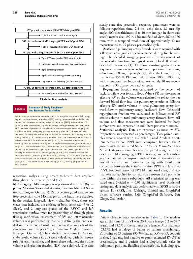
tvPD
R
Pay(Fip
726 Lurz et al. JACC Vol. 57, No. 6, 2011Functional Outcome Post-PPVI February 8, 2011:724–31
regression analysis using breath-to-breath data acquiredthroughout the exercise period (17).MR imaging. MR imaging was performed at 1.5-T (Sym-phony Maestro Series and Avanto, Siemens Medical Solu-tions, Erlangen, Germany). Retrospective gated steady-statefree-precession cine MR images of the heart were acquiredin the vertical long-axis view, 4-chamber view, short-axisviews that included the entirety of both ventricles (9 to 12slices), and 2 long-axis planes of the RVOT and leftventricular outflow tract for positioning of through-planeflow quantification. Assessment of RV and left ventricularvolumes was performed by manually defining the endocar-dial outline at end-diastole and end-systole in each of theshort-axis cine images (Argus, Siemens Medical Systems,Erlangen, Germany). The end-diastolic volume (EDV) andend-systolic volume (ESV) were calculated with Simpson’srule for each ventricle, and from these volumes, the stroke
Figure 1 Summary of Study Enrollmentand Reasons for Exclusion
Initial inclusion criteria (no contraindication to magnetic resonance [MR] imag-ing and cardiopulmonary exercise [CPEX] testing; adequate MR and CPX databefore percutaneous pulmonary valve implantation [PPVI]) were met by 107patients after intervention; 3 patients did not undergo assessment early afterPPVI because of procedural complications and rescue open-heart surgery. Ofthe 104 patients undergoing assessment early after PPVI, 4 were excludedbecause of inadequate MR data (n � 2) and submaximal CPEX testing (n � 2).During follow-up, 30 patients were excluded from the study because of: secondvalve-in-valve PPVI for early restenosis (n � 2), sudden death presumablyresulting from arrhythmia (n � 1), device explantation resulting from endocardi-tis (n � 1) and mechanical aortic valve failure (n � 1); relevant restenosis asdefined by an increase in right ventricular outflow tract (RVOT) gradient �15mm Hg as assessed on echocardiography (n � 8); and missed assessmentlate after PPVI in overseas patients (n � 12). Finally, of 70 patients who under-went assessment late after PPVI, 5 were excluded because of inadequate MRdata (n � 2) and submaximal CPEX testing (n � 3), leaving 65 patients forfinal analysis.
volume and ejection fraction (EF) were derived. The cine p
steady-state free-precession sequence parameters were asfollows: repetition time, 2.4 ms; echo time, 1.1 ms; flipangle, 60°; slice thickness, 8 to 10 mm (no gap in short-axisstack); matrix size, 192 � 156; and field of view, 280 to 380mm, with a temporal resolution of approximately 40 msreconstructed to 25 phases per cardiac cycle.
Aortic and pulmonary artery flow data were acquired witha flow-sensitive gradient echo sequence during free breath-ing. The detailed imaging protocols for assessment ofbiventricular function and great vessel blood flow weredescribed previously (1). The flow sensitive gradient echosequence parameters were as follows: repetition time, 8 ms;echo time, 3.8 ms; flip angle 30°; slice thickness, 5 mm;matrix size 256 � 192; and field of view, 280 to 380 mm,with a temporal resolution of approximately 30 ms recon-structed to 30 phases per cardiac cycle.
Regurgitant fraction was calculated as the percent ofbackward flow over forward flow. Where PR was present, aneffective RV stroke volume was calculated to reflect the netforward blood flow into the pulmonary arteries as follows:effective RV stroke volume � total pulmonary artery for-ward flow � pulmonary artery backward flow. Tricuspidregurgitation fraction was calculated as follows: total RVstroke volume � total pulmonary artery forward flow. Allvolume and flow measurements were indexed for bodysurface area and expressed in milliliters per square meter.Statistical analysis. Data are expressed as mean � SD.Proportions are expressed as percentages. Two-paired sam-ples were analyzed with a 2-tailed paired Student t test.Parameters before PPVI were compared between the 2groups with the unpaired Student t test or Mann-WhitneyU test. Categorical variables were compared using the Fisherexact test. MR imaging, CPEX testing, and echocardio-graphic data were compared with repeated-measures anal-ysis of variance and post-hoc testing with Bonferronicorrection between the states early after PPVI and late afterPPVI. For comparison of NYHA functional class, a Fried-man test was applied for comparison between the 3 points intime within the same subgroups. All statistical testing wasbased on a 2-sided � � 0.05 significance level. Statisticalesting and data analysis was performed with SPSS softwareersion 11 (SPSS, Inc., Chicago, Illinois) and GraphPadrism software version 5.0b (GraphPad Software, Saniego, California).
esults
atient characteristics are shown in Table 1. The mediange at the time of PPVI was 20.4 years (range 5.2 to 57.7ears), and 38.5% of the patients were female. Most patients63.1%) had tetralogy of Fallot or variant morphology.ifty-nine of 65 patients (90.7%) had an RV-to-PA conduit
n situ, 5 patients had a native or patch-extended RVOT atresentation, and 1 patient had a bioprosthetic valve in
ulmonary position. Baseline characteristics, including age,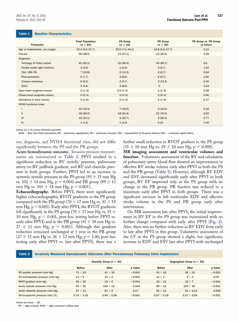
mEhcmf
; TGA �
727JACC Vol. 57, No. 6, 2011 Lurz et al.February 8, 2011:724–31 Functional Outcome Post-PPVI
sex, diagnosis, and NYHA functional class, did not differsignificantly between the PS and the PR groups.Acute hemodynamic outcome. Invasive pressure measure-ments are summarized in Table 2. PPVI resulted in asignificant reduction in RV systolic pressure, pulmonaryartery-to-RV pullback gradient, and RV end-diastolic pres-sure in both groups. Further, PPVI led to an increase insystemic systolic pressure in the PS group (93 � 15 mm Hgvs. 102 � 14 mm Hg, p � 0.004) and PR group (89 � 13
m Hg vs. 103 � 18 mm Hg, p � 0.001).chocardiography. Before PPVI, there were significantlyigher echocardiographic RVOT gradients in the PS groupompared with the PR group (70 � 17 mm Hg vs. 41 � 18m Hg, p � 0.001). Early after PPVI, the RVOT gradients
ell significantly in the PS group (70 � 17 mm Hg vs. 35 �10 mm Hg, p � 0.001, post-hoc testing before PPVI vs.early after PPVI) and in the PR group (41 � 18 mm Hg vs.27 � 11 mm Hg, p � 0.001). Although this gradientreduction remained unchanged at 1 year in the PR group(27 � 11 mm Hg vs. 26 � 12 mm Hg; p � 1.00, post-hoctesting early after PPVI vs. late after PPVI), there was a
Baseline CharacteristicsTable 1 Baseline Characteristics
ParameterTotal Population
(n � 65)
Age at implantation, yrs (range) 20.4 (5.2–57.7) 2
Female 25 (38.5)
Diagnosis
Tetralogy of Fallot variant 41 (63.1)
Double outlet right ventricle 3 (4.6)
TGA, VSD, PS 7 (10.8)
Ross procedure 5 (7.7)
Truncus arteriosus 6 (9.2)
Other 3 (4.6)
Open heart surgeries (mean) 2 (1–5)
Closed heart surgeries (mean) 0 (0–3)
Operations in total (mean) 2 (1–6)
NYHA functional class
I 10 (15.4)
II 39 (60.0)
III 15 (23.1)
IV 1 (1.5)
Values are n (%) unless otherwise specified.NYHA � New York Heart Association; PR � pulmonary regurgitation; PS � pulmonary stenosis
Invasively Measured Hemodynamic Outcomes After Percutaneous PTable 2 Invasively Measured Hemodynamic Outcomes After Pe
Stenotic Group (n �
Before After
RV systolic pressure (mm Hg) 71 � 23 41 � 10
RV end-diastolic pressure (mm Hg) 13 � 5 10 � 4
RVOT gradient (mm Hg) 45 � 23 15 � 6
Aortic systolic pressure (mm Hg) 93 � 15 102 � 14
Aortic diastolic pressure (mm Hg) 57 � 11 61 � 9
RV-to-systemic pressure ratio (%) 0.74 � 0.16 0.40 � 0.08
Values are mean � SD.RV � right ventricle; RVOT � right ventricular outflow tract.
further small reduction in RVOT gradient in the PS group(35 � 10 mm Hg vs. 29 � 10 mm Hg, p � 0.008).MR imaging assessment and ventricular volumes andfunction. Volumetric assessment of the RV and calculationof pulmonary artery blood flow showed an improvement ineffective RV stroke volume early after PPVI in both the PSand the PR group (Table 3). However, although RV EDVand ESV decreased significantly early after PPVI in bothgroups, RV EF improved only in the PS group with nochange in the PR group. PR fraction was reduced to aminimum early after PPVI in both groups. There was asignificant increase in left ventricular EDV and effectivestroke volume in the PS and PR group early afterintervention.
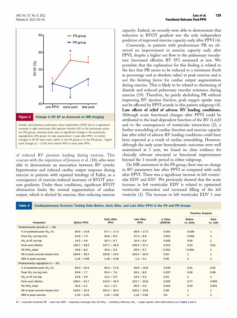
On MR assessment late after PPVI, the initial improve-ment in RV EF in the PS group was maintained with nofurther change compared with early after PPVI (Fig. 2).Also, there was no further reduction in RV EDV from earlyto late after PPVI in this group. Volumetric assessment ofthe LV in the PS group showed a slight, but significant,increase in EDV and ESV late after PPVI with unchanged
Group35)
PR Group(n � 30)
PS Group vs. PR Group(p Value)
.0–44.1) 19.8 (5.2–57.7) 0.15
7.1) 12 (40.0) 1.00
0.0) 20 (66.7) 0.6
.9) 2 (6.7) 1.00
4.3) 2 (6.7) 0.44
.6) 2 (6.7) 1.00
.7) 4 (13.3) 0.40
.6) 0 0.24
–5) 1 (1–5) 0.08
–2) 0 (0–3) 0.40
–6) 2 (1–6) 0.37
9.0) 3 (10.0) 0.32
1.4) 21 (70.0) 0.20
5.7) 6 (20.0) 0.77
.9) 0 (0) 1.00
transposition of the great arteries; VSD � ventricular septal defect.
nary Valve Implantationneous Pulmonary Valve Implantation
Regurgitant Group (n � 30)
p Value Before After p Value
�0.001 51 � 16 39 � 10 �0.001
�0.001 11 � 4 9 � 3 0.02
�0.001 22 � 12 12 � 7 �0.001
0.004 89 � 13 103 � 18 �0.001
0.17 54 � 11 61 � 11.6 0.009
�0.001 0.57 � 0.19 0.37 � 0.09 �0.001
PS(n �
2.0 (7
13 (3
21 (6
1 (2
5 (1
3 (8
2 (5
3 (8
2.0 (1
0 (0
2 (1
7 (1
18 (5
9 (2
1 (2
ulmorcuta
35)

sFccct
D
IcmfbnipATis
tive; ES
728 Lurz et al. JACC Vol. 57, No. 6, 2011Functional Outcome Post-PPVI February 8, 2011:724–31
LV EF compared with early after PPVI. In the PR group,the MR findings were maintained with no significantchange in any MR parameters late after PPVI as comparedwith early after PPVI (Fig. 2).
Before PPVI, there were no patients with severe tricuspidregurgitation. The median tricuspid regurgitation fractionwas 0.0% (range 0% to 33%), with only 2 patients having atricuspid regurgitation fraction �20% (24% and 33%).CPEX testing. The results of CPEX testing are shown inTable 4. In the PS group, there was a significant improvementin absolute measures of peak and percent of predicted peakoxygen uptake, peak workload, oxygen uptake at anaerobicthreshold, and ventilatory efficiency slope early after PPVI.This early improvement in cardiopulmonary exercise capacitywas maintained late after PPVI, with no further change in anyof these parameters compared with early after PPVI.
In the PR group, no improvement in peak oxygen uptakeand percent of predicted peak oxygen uptake, peak work-load, oxygen uptake at anaerobic threshold, or ventilatoryefficiency slope could be demonstrated early after PPVI. Onrepeated assessment of exercise capacity late after PPVI,there was a significant increase in peak workload andventilatory efficiency slope as compared with parametersbefore PPVI. No significant change in peak oxygen uptake,percent of predicted peak oxygen uptake, and oxygen uptake
Magnetic Resonance Imaging Data Before, Early After, and LateAfter PPVI in the Pulmonary Stenosis and Pulmonary RegurgitatTable 3 Magnetic Resonance Imaging Data Before, Early AftAfter PPVI in the Pulmonary Stenosis and Pulmonar
Parameter Before PPVIEarly Aft
PPVI
Predominantly stenotic (n � 35)
RV EDV indexed (ml/m2) 97.4 � 30.4 86.9 � 2
RV ESV indexed (ml/m2) 50.9 � 26.3 40.6 � 2
RV SV indexed (ml/m2) 46.4 � 9.1 47.2 � 7
RV SV eff indexed (ml/m2) 38.8 � 7.9 45.1 � 7
RV EF (%) 50.7 � 10.7 57.7 � 1
PR fraction (%) 11.4 � 8.6 1.4 � 2
LV EDV indexed (ml/m2) 68.7 � 14.3 73.9 � 1
LV ESV indexed (ml/m2) 27.1 � 12.0 27.0 � 1
LV SV eff indexed (ml/m2) 39.9 � 7.7 46.1 � 6
LV EF (%) 62.3 � 9.6 64.6 � 8
Predominantly regurgitant (n � 30)
RV EDV indexed (ml/m2) 118.2 � 32.3 101.1 � 3
RV ESV indexed (ml/m2) 60.5 � 31.0 53.9 � 2
RV SV indexed (ml/m2) 57.5 � 13.4 47.4 � 7
RV SV eff indexed (ml/m2) 34.9 � 7.3 44.3 � 6
RV EF (%) 50.9 � 11.9 49.5 � 1
PR fraction (%) 35.0 � 10.5 3.1 � 4
LV EDV indexed (ml/m2) 69.7 � 18.4 79.5 � 1
LV ESV indexed (ml/m2) 30.9 � 15.0 32.8 � 1
LV SV eff indexed (ml/m2) 37.2 � 7.2 45.4 � 6
LV EF (%) 56.9 � 10.7 60.1 � 9
ANOVA � analysis of variance; EDV � end-diastolic volume; EF � ejection fraction; eff � effecSV � systolic volume; other abbreviation as in Table 1.
at anaerobic threshold was seen late after PPVI. Peak heart c
rate and respiratory exchange ratio did not change early orlate after PPVI in either group.Functional outcome (NYHA functional class). The func-tional class fell from a median of NYHA II to NYHA Iearly after PPVI (p � 0.001, Friedman test), with no furthertatistically significant change late after PPVI (NYHA I).or this categorical variable, 25% of patients were in NYHAlass III or IV before PPVI, whereas 100% were in NYHAlass I or II at 1 year. These improvements in functionallass also were present when separate analyses for the PS orhe PR groups were made.
iscussion
n this study, we showed that the beneficial physiologicalonsequences of PPVI seen in the acute setting (1–3) areaintained at 1 year in those patients with preserved device
unction. However, although there was no worsening ofiventricular function or exercise capacity after 1 year, we haveo evidence that the process of RV remodeling and functional
mprovement extends beyond the 1-month period in thisatient group, as shown by MR imaging and CPEX results.cute effects of relief of adverse RV loading conditions.he present results confirm our previous findings that PPVI
n patients with predominant PS is associated with an earlyignificant improvement in RV systolic function and exer-
roupsnd Lateurgitation Groups
Late AfterPPVI
p Value(ANOVA)
Post-Hoc Analysis
Beforevs. Early
Earlyvs. Late
85.9 � 20.3 �0.001 0.001 1.00
38.2 � 17.5 �0.001 �0.001 0.90
47.8 � 7.7 0.70
45.7 � 8.0 �0.001 �0.001 1.00
57.8 � 10.5 �0.001 �0.001 1.00
1.9 � 3.5 �0.001 �0.001 1.00
77.5 � 14.2 �0.001 0.002 0.04
30.1 � 12.1 0.008 1.00 0.03
46.2 � 7.5 �0.001 �0.001 1.00
62.3 � 9.0 0.02 0.0441 0.10
97.9 � 30.9 �0.001 �0.001 0.60
49.5 � 29.5 �0.001 0.016 0.145
48.4 � 9.1 �0.001 �0.001 1.00
46.1 � 9.2 �0.001 �0.001 0.50
52.3 � 11.2 0.127
2.0 � 3.5 �0.001 �0.001 0.60
82.3 � 25.6 �0.001 �0.001 0.71
35.2 � 21.6 0.064
46.3 � 9.3 �0.001 �0.001 1.00
60.2 � 10.9 0.037 0.11 1.00
V � end-systolic volume; PPVI � percutaneous pulmonary valve implantation; RV � right ventricle;
ion Ger, ay Reg
er
6.3
3.8
.1
.5
0.5
.6
3.9
0.9
.5
.0
1.2
9.5
.4
.5
0.8
.9
8.1
5.8
.5
.5
ise capacity (4). This observation is thought to be the result
LAaaflbamcb
iauivv
729JACC Vol. 57, No. 6, 2011 Lurz et al.February 8, 2011:724–31 Functional Outcome Post-PPVI
of reduced RV pressure loading during exercise. Thisconcurs with the experience of Jonsson et al. (18), who wereable to demonstrate an association between RV systolichypertension and reduced cardiac output response duringexercise in patients with repaired tetralogy of Fallot, as aconsequence of exercise-induced increases of RVOT pres-sure gradients. Under these conditions, significant RVOTobstruction limits the normal augmentation of cardiacoutput, which is elicited by exercise, thus reducing exercise
Figure 2 Change in RV EF as Assessed on MR Imaging
Initially, percutaneous pulmonary valve implantation (PPVI) led to a significantincrease in right ventricular (RV) ejection fraction (EF) in the pulmonary steno-sis (PS) group, whereas there was no significant change in the pulmonaryregurgitation (PR) group. On late assessment 1 year after PPVI, no furtherchanges in RV EF were seen, either in the PS group or in the PR group. *Signif-icant change (p � 0.05) from before PPVI to early after PPVI.
Cardiopulmonary Exercise Testing Data Before, Early After, and LaTable 4 Cardiopulmonary Exercise Testing Data Before, Early A
Parameter Before PPVIEarly Afte
PPVI
Predominantly stenotic (n � 35)
% of predicted peak VO2 (%) 59.9 � 14.8 67.7 � 17
Peak VO2 (ml/kg/min) 23.8 � 7.9 26.8 � 8.
VO2 at AT (ml/kg) 14.5 � 4.6 16.3 � 5.
Peak work (Watts) 136.7 � 52.5 147.7 � 54
VE/VCO2 slope 34.8 � 8.2 30.0 � 6.
HR at peak exercise (beats/min) 166.8 � 25.9 163.8 � 24
RER at peak exercise 1.18 � 0.08 1.18 � 0.
Predominantly regurgitant (n � 30)
% of predicted peak VO2 (%) 65.0 � 16.4 66.9 � 17
Peak VO2 (ml/kg/min) 24.8 � 7.7 25.4 � 7.
VO2 at AT (ml/kg) 13.8 � 5.9 14.1 � 5.
Peak work (Watts) 108.3 � 41.7 110.9 � 42
VE/VCO2 slope 32.6 � 6.1 31.1 � 5.
HR at peak exercise (beats/min) 164.6 � 22.4 161.3 � 22
RER at peak exercise 1.16 � 0.05 1.16 � 0.
AT � anaerobic threshold; HR � heart rate; RER � respiratory exchange ratio; VE/VCO2 � ventilatory ef
capacity. Indeed, we recently were able to demonstrate thatreduction in RVOT gradient was the only independentpredictor of improved exercise capacity early after PPVI (4).
Conversely, in patients with predominant PR we ob-served no improvement in exercise capacity early afterPPVI, despite a higher net flow to the pulmonary vascula-ture (increased effective RV SV) measured at rest. Wepostulate that the explanation for this finding is related tothe fact that PR seems to be reduced to a minimum (bothas percentage and as absolute value) at peak exercise and isnot the limiting factor for cardiac output augmentationduring exercise. This is likely to be related to shortening ofdiastole and reduced pulmonary vascular resistance duringexercise (19). Therefore, by purely abolishing PR withoutimproving RV ejection fraction, peak oxygen uptake maynot be affected by PPVI acutely in this patient subgroup (4).
ate effects of relief of adverse RV loading conditions.lthough acute functional changes after PPVI could be
ttributed to the load-dependent function of the RV (1,4,8)nd to the consequences of ventricular interaction (2), aurther remodeling of cardiac function and exercise capacityate after relief of adverse RV loading conditions could haveeen expected as a result of cardiac remodeling. However,lthough the early acute hemodynamic outcomes were wellaintained at 1 year, we found no clear evidence for
linically relevant structural or functional improvementeyond the 1-month period in either subgroup.On MR assessment in the PS group, there was no change
n RV parameters late after PPVI as compared with earlyfter PPVI. There was a significant increase in left ventric-lar EDV and ESV. We previously showed that the acutencrease in left ventricular EDV is related to optimizedentricular interaction and increased filling of the leftentricle (2). The increase in left ventricular EDV 1 year
er PPVI in the PS and PR Groupsand Late After PPVI in the PS and PR Groups
Late AfterPPVI
p Value(ANOVA)
Post-Hoc Analysis
Beforevs. Early
Earlyvs. Late
68.9 � 17.3 0.001 0.008 1
27.2 � 8.8 0.001 0.008 1
16.4 � 5.6 0.042 0.04 1
155.0 � 57.1 0.013 0.23 0.61
29.9 � 5.7 �0.001 �0.001 1
164.0 � 20.9 0.52 1 1
1.2 � 0.1 0.99 1 1
69.8 � 19.8 0.036 0.41 0.52
26.1 � 8.5 0.057 0.69 0.48
14.2 � 6.2 0.43 1 1
123.7 � 43.8 0.002 0.77 0.009
29.5 � 5.0 0.001 0.24 0.051
162.3 � 19.8 0.58 1 1
1.16 � 0.06 0.6 1 1
te Aftfter,
r
.3
9
7
.5
2
.0
08
.6
6
0
.0
7
.3
05
ficiency; VO2 � oxygen uptake; other abbreviations as in Tables 1 and 3.

730 Lurz et al. JACC Vol. 57, No. 6, 2011Functional Outcome Post-PPVI February 8, 2011:724–31
after PPVI may be a reflection of late remodeling as aresponse to the acute improvement in filling over time;however, this cannot be answered in this study.
In the PR group, we found a nonsignificant tendencytoward further reduction in RV EDV and improved RV EF1 year after PPVI. Further studies have to clarify whetherthese changes can be attributed to late RV remodeling.Similar to the MR data, there was a tendency towardimproved peak oxygen uptake and ventilatory efficiency onCPEX testing, but these subtle changes are unlikely to be ofclinical relevance. Further, we found a significant increase inpeak workload in the PR group. However, this finding isdifficult to interpret as a follow-up parameter in growingchildren, because the studied parameter is not indexed forbody weight. Assessment of patients’ functional class re-vealed a further but nonsignificant reduction in the totalnumber of patients in NYHA functional class II late afterPPVI. Importantly, these 1-year results after PPVI predom-inantly for PR are in keeping with studies on surgicalpulmonary valve replacement for PR, in which no signifi-cant improvement in peak oxygen uptake 1 year aftersurgery could be demonstrated (20,21).
In our opinion, with the current technique and indica-tions for PPVI, the acute changes after PPVI are main-tained 1 year after the intervention. However, no furtherrelevant positive RV remodeling or functional improvementis seen as compared with early after PPVI.
The following factors may have limited positive lateremodeling in the present study:
1. The hemodynamic result achieved by PPVI might nothave been sufficient to allow for positive remodeling.Because only patients with sustained hemodynamic re-sults were included in this study, the initial hemody-namic improvement was well maintained in the PRgroup with even further reduction in RVOT gradients inthe PS group over time. Although this certainly shouldhave limited the confounding effects of worsening RVloading conditions over time, some degree of residualRVOT obstruction was present in most patients. Thishemodynamic finding was present in the PS and also inthe PR group, where the impact of even low-pressuregradients is unknown.
2. PPVI might have been performed too late. Previousstudies in the surgical literature that assessed functionaloutcome after pulmonary valve replacement have sug-gested that patients with severe PR often may be treatedtoo late (20,22). This conclusion is based on the lack ofimprovement in RV function after pulmonary valvereplacement. Similarly, in our study we also did notobserve improvement in RV EF or exercise performancein patients with predominant PR, either immediatelyafter PPVI or at 1 year. However, it remains unknownwhether earlier performance of PPVI in this subgroup of
patients would have resulted in improved outcomes.3. Patients might not have been followed up for longenough. Adverse RV remodeling in these chronically illpatients may be reversible only in the long term.
4. RVOT dysfunction and adverse RV loading conditionsrepresent only one of many possible contributors tofunctional impairment in these patients. The deleteriouseffects of neonatal hypoxia and cyanosis, high pulmonarypressures, multiple cardiopulmonary bypasses with longischemic time and transient inflammation, and ventric-ular scaring resulting from surgery—all seen in patientswith CHD involving the RVOT—could be affected onlymildly by improved RV loading conditions after PPVI.Although certainly multifactorial, the contribution ofadverse RV loading on impaired functional outcome inthese patients may be less relevant than commonlybelieved. Further studies have to clarify the role ofpulmonary valve replacement in patients with RVOTdysfunction for long-term patient management andoutcome.
Study limitations. Although we saw no significant remod-eling in our patients at 1 year, our study period may be tooshort and remodeling may occur over longer periods of time.The outcome parameters used in this study represent onlyselected proxies. Hard end point, such as mortality orarrhythmia burden, which are rare in studies on patientswith congenital heart disease for well-known reasons, arestill missing when procedural success of RVOT interven-tions is assessed. Further, the true onset of RVOT dysfunc-tion could not be determined for each individual patient;thus, the data set does not allow for definite conclusionsabout the timing of intervention. Finally, the relatively smallsample size represents a limitation of this study. Therefore,the findings need to be confirmed in larger cohorts.
Conclusions
With the current technique and indications for PPVI,patients can benefit from a prolonged phase of maintainedcardiac function; however, they cannot expect significantfurther improvement in biventricular function and objectiveexercise performance 1 year after intervention. These resultsunderline the need for further studies to address the ques-tion of when to intervene and how to achieve optimalhemodynamic acute and long-term results after RVOTintervention. At present, we suggest an aggressive approachto any acute post-procedural residual RVOT gradient.Importantly, in patients with chronic volume overload, evensmall gradients may have significant detrimental effects onRV function.
In patients with predominant PS, despite the lack of furtherremodeling at 1 year, the initial marked improvement isencouraging. In contrast, we observed no improvement in RVsystolic function or exercise capacity in the PR group, whichmay suggest that in patients with severe PR, interventionsshould be considered before reduction in RV function or
deterioration in exercise performance are observed.
731JACC Vol. 57, No. 6, 2011 Lurz et al.February 8, 2011:724–31 Functional Outcome Post-PPVI
Reprints requests and correspondence: Dr. Philipp Lurz, De-partment of Internal Medicine/Cardiology & Grown Up Congen-ital Heart Disease, University of Leipzig–Heart Center, Struem-pellstr. 39, 04289 Leipzig, Germany. E-mail: [email protected].
REFERENCES
1. Coats L, Khambadkone S, Derrick G, et al. Physiological and clinicalconsequences of relief of right ventricular outflow tract obstruction lateafter repair of congenital heart defects. Circulation 2006;113:2037–44.
2. Lurz P, Puranik R, Nordmeyer J, et al. Improvement in left ventricularfilling properties after relief of right ventricle to pulmonary arteryconduit obstruction: contribution of septal motion and interventricularmechanical delay. Eur Heart J 2009;30:2266–74.
3. Lurz P, Giardini A, Taylor AM, et al. Effect of altering pathologicright ventricular loading conditions by percutaneous pulmonary valveimplantation on exercise capacity. Am J Cardiol 2010;105:721–6.
4. Coats L, Khambadkone S, Derrick G, et al. Physiological conse-quences of percutaneous pulmonary valve implantation: the differentbehaviour of volume- and pressure-overloaded ventricles. Eur Heart J2007;28:1886–93.
5. Lurz P, Coats L, Khambadkone S, et al. Percutaneous pulmonaryvalve implantation: impact of evolving technology and learning curveon clinical outcome. Circulation 2008;117:1964–72.
6. Lurz P, Gaudin R, Taylor AM, Bonhoeffer P. Percutaneous pulmo-nary valve implantation. Semin Thorac Cardiovasc Surg Pediatr CardSurg Annu 2009:112–7.
7. Lurz P, Bonhoeffer P, Taylor AM. Percutaneous pulmonary valveimplantation: an update. Expert Rev Cardiovasc Ther 2009;7:823–33.
8. Lurz P, Nordmeyer J, Muthurangu V, et al. Comparison of bare metalstenting and percutaneous pulmonary valve implantation for treatmentof right ventricular outflow tract obstruction: use of an x-ray/magneticresonance hybrid laboratory for acute physiological assessment. Circu-lation 2009;119:2995–3001.
9. Bonhoeffer P, Boudjemline Y, Saliba Z, et al. Percutaneous replace-ment of pulmonary valve in a right-ventricle to pulmonary-arteryprosthetic conduit with valve dysfunction. Lancet 2000;356:1403–5.
10. Khambadkone S, Coats L, Taylor A, et al. Percutaneous pulmonaryvalve implantation in humans: results in 59 consecutive patients.
Circulation 2005;112:1189–97.11. Maheshwari S, Bruckheimer E, Nehgme RA, Fahey JT, KholwadwalaD, Hellenbrand WE. Single coronary artery complicating stent im-plantation for homograft stenosis in tetralogy of Fallot. CathetCardiovasc Diagn 1997;42:405–7.
12. Peng LF, McElhinney DB, Nugent AW, et al. Endovascular stentingof obstructed right ventricle-to-pulmonary artery conduits: a 15-yearexperience. Circulation 2006;113:2598–605.
13. Sridharan S, Coats L, Khambadkone S, Taylor AM, Bonhoeffer P.Images in cardiovascular medicine. Transcatheter right ventricularoutflow tract intervention: the risk to the coronary circulation. Circu-lation 2006;113:e934–5.
14. Yock PG, Popp RL. Noninvasive estimation of right ventricularsystolic pressure by Doppler ultrasound in patients with tricuspidregurgitation. Circulation 1984;70:657–62.
15. Joyce JJ, Hwang EY, Wiles HB, Kline CH, Bradley SM, Crawford FAJr. Reliability of intraoperative transesophageal echocardiography dur-ing tetralogy of Fallot repair. Echocardiography 2000;17:319–27.
16. Wasserman K, Sue DY. Principles of Exercise Testing and Interpre-tation. Philadelphia, PA: Lea & Febiger, 1994.
17. Dimopoulos K, Okonko DO, Diller GP, et al. Abnormal ventilatoryresponse to exercise in adults with congenital heart disease relates tocyanosis and predicts survival. Circulation 2006;113:2796–802.
18. Jonsson H, Ivert T, Jonasson R, Wahlgren H, Holmgren A, BjorkVO. Pulmonary function thirteen to twenty-six years after repair oftetralogy of Fallot. J Thorac Cardiovasc Surg 1994;108:1002–9.
19. Roest AA, Helbing WA, Kunz P, et al. Exercise MR imaging in theassessment of pulmonary regurgitation and biventricular function inpatients after tetralogy of Fallot repair. Radiology 2002;223:204–11.
20. Frigiola A, Tsang V, Bull C, et al. Biventricular response after pulmonaryvalve replacement for right ventricular outflow tract dysfunction: is age apredictor of outcome? Circulation 2008;118:S182–90.
21. Ghez O, Tsang VT, Frigiola A, et al. Right ventricular outflow tractreconstruction for pulmonary regurgitation after repair of tetralogy ofFallot. Preliminary results. Eur J Cardiothorac Surg 2007;31:654–8.
22. Therrien J, Siu SC, McLaughlin PR, Liu PP, Williams WG, WebbGD. Pulmonary valve replacement in adults late after repair oftetralogy of Fallot: are we operating too late? J Am Coll Cardiol2000;36:1670–5.
Key Words: cardiopulmonary exercise testing y congenital heart diseasey magnetic resonance imaging y percutaneous pulmonary valve
implantation y right ventricular outflow tract dysfunction.