Early Rehabilitative Exercise Training in the Recovery from Pediatric Burn
29
. . . Published ahead of Print Early Rehabilitative Exercise Training in the Recovery from Pediatric Burn Justin P. Hardee 1 , Craig Porter 2,3,5 , Labros S. Sidossis 2,4,5 , Elisabet Børsheim 2,3,5 , James A. Carson 1 , David N. Herndon 2,3,5 , and Oscar E. Suman 2,3,5 1 Department of Exercise Science, University of South Carolina, Columbia, SC 2 Metabolism Unit, Shriners Hospitals for Children, Galveston, TX 3 Department of Surgery, University of Texas Medical Branch, Galveston, TX 4 Department of Internal Medicine, University of Texas Medical Branch, Galveston, TX 5 Shriners Hospitals for Children-Galveston, TX Accepted for Publication: 25 January 2014 Medicine & Science in Sports & Exercise® Published ahead of Print contains articles in unedited manuscript form that have been peer reviewed and accepted for publication. This manuscript will undergo copyediting, page composition, and review of the resulting proof before it is published in its final form. Please note that during the production process errors may be discovered that could affect the content. Copyright © 2014 American College of Sports Medicine ACCEPTED
Transcript of Early Rehabilitative Exercise Training in the Recovery from Pediatric Burn

. . . Published ahead of Print
Early Rehabilitative Exercise Training in the Recovery from Pediatric Burn
Justin P. Hardee1, Craig Porter2,3,5, Labros S. Sidossis2,4,5, Elisabet Børsheim2,3,5,James A. Carson1, David N. Herndon2,3,5, and Oscar E. Suman2,3,5
1Department of Exercise Science, University of South Carolina, Columbia, SC2Metabolism Unit, Shriners Hospitals for Children, Galveston, TX
3Department of Surgery, University of Texas Medical Branch, Galveston, TX4Department of Internal Medicine, University of Texas Medical Branch, Galveston, TX
5Shriners Hospitals for Children-Galveston, TX
Accepted for Publication: 25 January 2014
Medicine & Science in Sports & Exercise® Published ahead of Print contains articles in unedited manuscript form that have been peer reviewed and accepted for publication. This manuscript will undergo copyediting, page composition, and review of the resulting proof before it is published in its final form. Please note that during the production process errors may be discovered that could affect the content.
Copyright © 2014 American College of Sports Medicine
ACCEPTED

Early Rehabilitative Exercise Training in the Recovery from Pediatric Burn
Justin P. Hardee1, Craig Porter2,3,5, Labros S. Sidossis2,4,5, Elisabet Børsheim2,3,5, James A.
Carson1, David N. Herndon2,3,5, and Oscar E. Suman2,3,5
1Department of Exercise Science, University of South Carolina, Columbia, SC
2Metabolism Unit, Shriners Hospitals for Children, Galveston, TX
3Department of Surgery, University of Texas Medical Branch, Galveston, TX
4Department of Internal Medicine, University of Texas Medical Branch, Galveston, TX
5Shriners Hospitals for Children-Galveston, TX
Running Title: Rehabilitative Exercise and Burn
Conflicts of Interest and Funding Sources: The authors declare no conflicts of interest. This
research was supported by National Institute for Disabilities and Rehabilitation Research grant
H133A120091; National Institutes of Health grants P50 GM060388, R01 HD049471, R01
GM056687; Shriner grants 71006, 71008, 71009, 84080, 84090. JPH was supported by Leon
Hess Professorship Funds. EB was a faculty member and scientific staff at UTMB and SHC, and
is presently at the University of Arkansas for Medical Sciences, Little Rock, AR. This is a
registered clinical trial at Clincaltrials.gov NCT00675714.
Corresponding Author: Oscar E. Suman, PhD, FACSM, Leon Hess Professor for Burn Injuries
Research, University of Texas Medical Branch; Director of Children's Wellness Center, Shriners
Hospitals for Children, 815 Market Street, Galveston, TX 77550; Email: [email protected];
Phone: 409.770.6557; Fax: 409.770.6919
Medicine & Science in Sports & Exercise, Publish Ahead of PrintDOI: 10.1249/MSS.0000000000000296
ACCEPTED
Copyright © 2014 by the American College of Sports Medicine. Unauthorized reproduction of this article is prohibited.

ABSTRACT
PURPOSE: The purpose of this study was to determine the effects of early outpatient exercise
on muscle mass, function, and fractional synthetic rate in severely burned children.
METHODS: Forty-seven children with ≥40 % total body surface area burn performed 12-
weeks standard of care rehabilitation (SOC: N=23) or rehabilitative exercise training (RET:
N=24) immediately following hospital discharge. Dual-energy X-ray absorptiometry was used
to assess lean body mass (LBM) at discharge, post-treatment, and 12 months post-burn. Muscle
function was evaluated with a Biodex Isokinetic Dynamometer and peak aerobic fitness
(VO2peak) measured using a modified Bruce treadmill protocol post-treatment. Stable isotope
infusion studies were performed in a subset of patients (SOC: N=13; RET: N=11) at discharge
and post-treatment to determine mixed-muscle fractional synthetic rate.
RESULTS: Relative peak torque (RET: 138 ± 9 N · m · kg-1 vs SOC: 106 ± 9 N · m · kg-1) and
VO2peak (RET: 32 ± 1 ml · kg-1 · min-1 vs SOC: 28 ± 1 ml · kg-1 · min-1) was greater post-
treatment with RET compared to SOC. In addition, RET increased whole-body (9 ± 2%) and leg
(17 ± 3%) LBM compared to SOC. Furthermore, the percentage change in whole-body (18 ±
3%) and leg (31 ± 4%) LBM from discharge to 12 months post-burn was greater with RET
compared to SOC. Muscle fractional synthetic rate decreased from discharge to post-treatment
in both groups (6.9 ± 1.1% · d-1 vs 3.4 ± 0.4% · d-1); however no differences were observed
between treatment groups at each time-point.
CONCLUSIONS: Early outpatient exercise training implemented at hospital discharge
represents an effective intervention to improve muscle mass and function following severe burn
injury.
KEYWORDS: Severe Burn, Lean Body Mass, Muscle Strength, Cardiorespiratory Fitness,
Fractional Synthetic Rate
ACCEPTED
Copyright © 2014 by the American College of Sports Medicine. Unauthorized reproduction of this article is prohibited.
INTRODUCTION
In the United States, it has been estimated approximately 450,000 people are admitted for
burn treatment each year, with 30% of cases involving individuals less than 16 years of age (1).
The risk of injury is influenced largely by socioeconomic status and geographic location (26).
Major advances in critical care and treatment within the past two decades have improved
survival rates (32). Despite these developments, survivors are often faced with long-term
functional impairments that can impede a proper return to society (20). Consequently, the
restoration of normal function and quality of life following severe burn is of clinical and social
significance.
The pathophysiological response to major burn injury is characterized by elevated resting
energy expenditure, systemic inflammation, and skeletal muscle catabolism (13, 17). In healthy
adults, skeletal muscle comprises 70% of total lean body mass and represents the largest protein
reservoir in humans. Thus, muscle protein is a major source of amino acids to support survival
and recovery post-burn. Indeed, the release of amino acids from skeletal muscle is thought to
facilitate wound healing, the production of inflammatory and acute phase proteins, and provide
substrate for hepatic gluconeogenesis during the recovery from burn (13, 35, 36). This
hypermetabolic and persistent catabolic response can persist for up to 2 years post burn injury
(13, 18). Subsequently, the continuous muscle catabolism contributes to reduced muscle mass
and strength, resulting in an impaired return to functional mobility. Therefore, strategies which
promote the maintenance of skeletal muscle mass and function may provide long-term benefits
in patients during the recovery from severe burn.
Skeletal muscle is a highly plastic tissue, in which the size is tightly regulated by the rates
of protein synthesis and degradation. Net protein balance is responsive to several factors such as
ACCEPTED
Copyright © 2014 by the American College of Sports Medicine. Unauthorized reproduction of this article is prohibited.
mechanical load, nutrient status/energy balance and hormone availability (34). Severe burn
trauma increases both muscle protein and degradation, however favors an overall negative net
protein balance (12, 25). Pharmacological treatments such as growth hormone (10), insulin (28),
and oxandrolone (33) increase mixed-muscle fractional synthetic rate and results in the accretion
of lean body mass (LBM) in severely burned patients. Therefore, the identification of additional
treatments that regulate muscle mass and increase physical function is needed to continually
improve standard of care rehabilitation. Exercise training is a non-pharmacological treatment
that attenuates muscle catabolism associated with aging and chronic disease (7, 21). We have
previously shown that rehabilitative exercise, initiated 6 months post-burn (typically 2-3 months
after discharge), results in significant improvements to LBM, muscular strength and
cardiorespiratory fitness in severely burned children (27, 30). However, the determination of
fractional synthetic rate in these patients is needed in order to determine the impact of
rehabilitative exercise on skeletal muscle protein metabolism. It is currently unknown if these
benefits can be achieved if exercise is started immediately following hospital discharge, a time at
which there is still pronounced post-burn hypermetabolism and hypercatabolism.
Despite significant improvements in muscle mass and function with exercise training,
mechanistic insight as to how these adaptations occur is currently lacking. Therefore, the
purpose of this clinical trial was to determine the effects of early outpatient exercise on muscle
mass, function, and fractional synthetic rate in severely burned children. It was hypothesized
that early rehabilitative exercise training in severely burned patients would increase muscle
mass, muscular strength, and cardiorespiratory fitness when compared to standard of care. In
addition, there would be no differences in basal muscle fractional synthetic rate between standard
of care and rehabilitative exercise training.
ACCEPTED
Copyright © 2014 by the American College of Sports Medicine. Unauthorized reproduction of this article is prohibited.

METHODS
Patients.
Forty-seven children with burns encompassing at least 40% of their total body surface area that
were admitted for acute burn treatment at Shriners Hospital for Children-Galveston participated
in the study. Patients received similar standard medical care and treatment from the time of
admission until the time of discharge (see Patient Standard of Care). Patients received
medications that are standard in the Burn Intensive Care Unit during acute burn treatment, which
consists of pressors, diuretics, antibiotics or anesthetics. None of these drugs are known to be
anabolic, and at no time did patients receive any investigational drugs. Total burn surface area
was assessed by the “rule of nines” method during excisional surgery immediately following
hospital admittance (15). Informed written consent was obtained from each patient’s guardian
prior to enrollment. All procedures were approved by the University of Texas Medical Branch
Institutional Review Board. After consent was obtained, patients were randomly assigned to
standard of care (SOC) or rehabilitative exercise training (RET). The RET group participated in
a 12-week in-hospital physiotherapy program (see Patient Standard of Care) supplemented with
an individualized and supervised rehabilitative exercise training program (N=24). In contrast,
the SOC group participated in the in-hospital physiotherapy program (N=23), but was not
supplemented with the rehabilitative exercise training program. LBM assessment and 5-hour
stable isotope studies were conducted at discharge and following the 12-week treatment
intervention. In addition, muscle strength and cardiorespiratory fitness was assessed at post-
treatment.ACCEPTED
Copyright © 2014 by the American College of Sports Medicine. Unauthorized reproduction of this article is prohibited.

Patient Standard of Care
All patients received similar standard of care treatment during the acute phase of injury as
previously described (14). Briefly, total fluid resuscitation needs were administered within the
first 24 hours according the Galveston formula (5000 mL/m2 total body surface area burned plus
2000 mL/m2 total body surface area lactated Ringer solution). Early burn wound excision and
placement of autograft or allograft was performed within 48 hours of admission, with excision
and grafting procedures continued weekly until all burn sites were 95% healed (22). Following 4
days of bed rest, patients were allowed to ambulate daily between surgeries. Patients were fed a
standardized Vivonex TEN enteral nutrition (3% fat, 82% carbohydrate, 15% protein) via a
nasoduodenal or nasogastric tube. During the first week of hospitalization intake was
determined using the formula 1500 kcal/m2 total body surface area plus 1500 kcal/m2 total body
surface area burned (16, 23). During the remainder of hospitalization intake was then modified
to 1.4 times the weekly measured resting energy expenditure. Prior to hospital discharge, each
patient underwent nutrient counseling with a dietician in which the patient was instructed to
consume 1.4 to 1.6 times their resting energy expenditure.
In addition to the aforementioned acute burn treatment, both treatment groups
participated in a conventional physiotherapy program administered by an occupational therapist
or physical therapy twice daily for one hour. The treatment program included positioning and
splinting, range of motion and strengthening activities, and scar management techniques. In
addition, caregiver education was also provided as part of the program. This therapy program is
designed to improve overall range of motion and minimized deformities and contractures (6). At
time of discharge, caregivers were given a home treatment program to follow during the
treatment period. Patients were discharged when 95% of wounds were considered closed.
ACCEPTED
Copyright © 2014 by the American College of Sports Medicine. Unauthorized reproduction of this article is prohibited.

Lean Body Mass Assessment
Whole body LBM was assessed at discharge, post-treatment, and 12 months post-burn by
dual-energy x-ray absorptiometry (DEXA) using QDR 4500A software (Hologic Inc, Waltham,
MA). This methodology has been previously described by our group in detail (24, 27). Scans
were taken with the patient lying supine on the scanning table, and pediatric analysis software
was used to calculate LBM, bone mineral content, and fat mass. Whole-body and regional
(trunk, leg, arm) LBM is reported in kilograms.
Functional Testing
Due to medical limitations such as impaired mobility and incomplete wound closure at
the time of discharge, the assessment of muscle strength and cardiorespiratory fitness was
conducted following the 12-week intervention. Muscle strength was assessed using the Biodex
System-3 Dynamometer (Shirley, NY). The isokinetic test was performed at an angular velocity
of 150° · s-1 on the dominant leg extensors. This speed has been shown to be well tolerated by
burned children (3, 30, 31). Patients were seated upright with the anatomic axis of the knee joint
aligned with the mechanical axis of the dynamometer. The patient’s position was stabilized with
restraining straps over the mid-thigh, pelvis, and trunk. Patients were familiarized to the
machine and testing procedures with visual and verbal explanations prior to testing. Following
three submaximal repetitions without load, 10 maximal voluntary muscle contractions (full
extension and flexion) were performed consecutively without rest between repetitions. Values of
peak torque, total work, and average power were calculated by the Biodex software system.
Peak torque was corrected for gravitational moments of the lower leg and the lever arm. The
ACCEPTED
Copyright © 2014 by the American College of Sports Medicine. Unauthorized reproduction of this article is prohibited.
highest peak torque for the entire 10 repetitions was selected for analysis. Relative peak torque
is presented to correct for differences in body weight.
Following muscular strength assessment, patients were given a period of rest between 30
minutes to an hour before cardiorespiratory fitness testing. Patients underwent a standardized
treadmill exercise test using the modified Bruce protocol (9, 19). Heart rate and peak oxygen
consumption (VO2peak) were measured throughout the exercise testing. Breath-by-breath
analysis of expired gases, flow, and volume were made by using a Medgraphics CardiO2 system
(St. Paul, MN) during rest and exercise. The exercise test began with the treadmill speed and
elevation at 1.7 miles per hour and 0%, respectively. Thereafter, the speed and elevation
increased every 3 minutes per the modified Bruce protocol. The patients were instructed and
encouraged to complete 3-minutes stages, and the test was terminated once volitional fatigue was
achieved. The heart rate and VO2peak achieved during testing were used to determine exercise
intensities used throughout the 12-week training period for the RET group.
Three-Repetition Maximum Test
The first and second sessions of actual training were spent with patients randomized to
the RET group being tested for a three repetition maximum to establish training loads that
would be used during the training period as previously described (30). The exercises performed
included bench press, leg press, shoulder press, leg extension, bicep curl, leg curl, and triceps
curl. After an instruction period on correct lifting technique the patient performed a warm-up
with the lever arm or bar to become familiar with the movement. Next, the weight was
progressively increased to which the patient could successfully perform four repetitions with
correct technique. If four repetitions were performed with correct technique a one minutes rest
ACCEPTED
Copyright © 2014 by the American College of Sports Medicine. Unauthorized reproduction of this article is prohibited.
was given and the weight was increased. This was repeated until the patient could perform three
repetitions, with the fourth repetition not volitionally possible with correct technique, and the test
was terminated. The amount of weight lifted from the successful lift was recorded as the
individuals three repetition maximum. From this information, the basic 3 sets of 8-12 repetitions
RM’s were established as described below.
Rehabilitative Exercise Training Program
Rehabilitative exercise training was performed as previously described (30). Each
exercise training session consisted of resistance and aerobic exercise under the supervision of a
certified exercise physiologist. Eight basic resistance exercises were used, incorporating bench
press, leg press, shoulder press, leg extension, biceps curl, leg curl, triceps curl, and toe raises.
All exercises were performed using free weights and variable-resistance machines.
Modifications to exercises were made when appropriate depending on the patient injury
characteristics. During the first week of training, patients were familiarized to the equipment and
instructed on proper weight lifting technique. Thereafter, the load was gradually increased from
50-60% of 3RM at the beginning of the program up to 80-85% of 3RM at the end of the
program. Additionally, each exercise training session included aerobic exercise on a treadmill or
cycle ergometer. Patients performed 20-40 minutes aerobic exercise 3-5 days · wk -1 at 70-85%
of VO2peak. All exercise sessions were preceded by a 5-minute warm-up at <50% VO2peak. No
strength training activities were permitted outside the supervised training session; however, both
groups were encouraged to maintain normal daily activities.ACCEPTED
Copyright © 2014 by the American College of Sports Medicine. Unauthorized reproduction of this article is prohibited.

Stable Isotope Infusion Study
Five hour stable isotope infusion studies were conducted in a subset of patients (SOC:
N=13; RET: N=11) to determine mixed-muscle fractional synthetic rate. Stable isotope studies
were performed following an overnight fast (10-12 hours) in the post-absorptive state. Catheters
were placed in the antecubital vein of each arm under sedation for the purpose of stable isotope
infusions and blood sampling. A baseline blood sample was taken before a primed (3.2 umol ·
kg-1) and constant (0.08 umol · kg-1 · min-1) infusion of L-[ring-2H5]-phenylalanine (Cambridge
Isotopes Laboratories, Inc., Andover, MA) was initiated. Biopsies of the vastus lateralis were
taken at hours 2 and 5 with a 5-mm Bergstrom needle (Stille, Stockholm, Sweden) for the
determination of phenylalanine enrichment in bound and intracellular muscle proteins. Tissue
was immediately frozen in liquid nitrogen and stored at -80°C until further analysis.
Skeletal Muscle Analysis
Frozen muscle tissue (20-30 mg) was weighed and homogenized in 10% perchloric acid.
The sample was centrifuged (3000 rpm for 10 min at 4°C) and the supernatant was recovered.
The supernatant represents the free intracellular amino acid pool and was used to determine the
intracellular phenylalanine concentration. The remaining pellet was washed in 2% perchloric
acid, followed by ethanol, and ethyl ether before overnight incubation at 50°C. The next day, the
dried muscle pellets were hydrolyzed in 6N hydrochloric acid at 100°C for 12 hours. The bound
muscle hydrolysate and intracellular portion were then passed over a cation exchange column
(Bio-Rad, Hercules, CA). The bound and intracellular phenylalanine enrichment (tracer/trace
ratio) was determined in tert-butyldimethylsilyl derivatives by gas chromatography-mass
spectrometry (Agilent, Santa Clara, CA) as previously described (25).
ACCEPTED
Copyright © 2014 by the American College of Sports Medicine. Unauthorized reproduction of this article is prohibited.

Mixed-Muscle Fractional Synthetic Rate Calculations
Mixed-muscle fractional synthetic rate was calculated using the precursor-product
method as previously described (5, 25). The precursor is the mean enrichment of the
intracellular free amino acid compartment and the product is the difference in the enrichment of
the bound muscle protein between the two biopsies. Values were multiplied by 24 to express
fractional synthetic rate as the percent per day (% · d-1) (25).
Statistical Analysis
Data are presented as means ± standard error. A repeated two-way ANOVA with factors
of treatment and time was performed with Tukey’s post-hoc correction was used to assess
differences in height, weight, LBM and muscle fractional synthetic rate. Unpaired students t-test
was performed to assess differences in patient characteristics, functional assessments and the
percentage change in LBM. Significance was set at P < 0.05 for all analysis. Statistical analysis
was performed using SigmaStat version 3.5 (Systat Software Inc., Richmond, CA).
RESULTS
Patient Characteristics
Forty-seven children with ≥40% total burn surface area were enrolled in the current
investigation. Baseline characteristics are presented in Table 1. There were no significant
differences between groups in age, height, body weight, sex, length of stay, and percent total
burn surface area burned at the time of discharge. The average patient age and total burn surface
area were 13.0 ± 0.5 years and 60 ± 2%, respectively. There were no differences in height from
discharge to post-treatment (P = 0.14). In contrast, both treatment groups increased body weight
ACCEPTED
Copyright © 2014 by the American College of Sports Medicine. Unauthorized reproduction of this article is prohibited.
from discharge to post-treatment (7.5 ± 1.2%; Main effect of time: P < 0.001), however there
were no differences between treatment groups.
Functional Testing
Muscle strength and cardiorespiratory fitness was assessed following the 12-week
treatment (Figure 1). There was a trend towards an increase in absolute peak torque with RET
when compared to SOC (Figure 1A; SOC: 52 ± 7.5 N · m vs RET: 72 ± 8.2 N · m; P = 0.08).
When corrected for body weight, the RET group had a significantly greater relative peak torque
compared to SOC (Figure 1B; SOC: 106 ± 8.7 N · m · kg -1 vs RET: 138 ± 8.6 N · m · kg-1; P =
0.01). Furthermore, VO2peak was greater with RET when compared to SOC (Figure 1C; SOC:
28 ± 1.3 ml · kg-1 · min-1 vs RET: 32.1 ± 1.3 ml · kg-1 · min-1; P = 0.04). Collectively, these data
demonstrate that burn children in the RET group had higher levels of muscle strength and
aerobic capacity after exercise training when compared to standard rehabilitation.
Whole-body and Regional LBM
Whole-body and regional LBM are presented in Table 2. There were no differences
between groups in whole-body and regional LBM at the time of discharge. There were no
changes in LBM from discharge to post-treatment in the SOC group. In contrast, there was a
significant increase in whole-body and regional LBM from discharge to post-treatment with RET
(Table 2). In addition, the percent change in whole-body and leg LBM from discharge to post-
treatment was greater with RET compared to SOC (Figure 2A). There was a trend towards an
increase in arm LBM with RET compared to SOC (P = 0.06). To determine if early
rehabilitative exercise training resulted in long-term muscle adaptations, LBM was examined 12
ACCEPTED
Copyright © 2014 by the American College of Sports Medicine. Unauthorized reproduction of this article is prohibited.
months post-burn injury. Similarly, the percentage change in whole-body and leg LBM from
discharge to 12 months post-burn were greater with RET compared to SOC (Figure 2B; RET: 18
± 3%, 31 ± 4%, respectively; P < 0.05). These results demonstrate that RET improved whole-
body and regional LBM with training compared to SOC, and the positive changes in LBM were
maintained for up to 12 months post-burn.
Mixed-Muscle Fractional Synthetic Rate
In a subset of patients (SOC: N=13; RET: N=11), 5-hour stable isotope infusion studies
were conducted to determine mixed-muscle fractional synthetic rate (Figure 3). No differences
in muscle fractional synthetic rate were observed at the time of discharge (SOC: 6.1 ± 1.4% · d -1
vs RET: 7.9 ± 1.7% · d-1). Muscle fractional synthetic rate decreased from discharge to post-
treatment (6.9 ± 1.1% · d-1 to 3.4 ± 0.4% · d-1; Main effect of time: P < 0.01), however no
differences in muscle fractional synthetic rate were observed between SOC and RET post-
treatment (SOC: 3.2 ± 0.4% · d-1 vs RET: 3.6 ± 0.9% · d-1). Lastly, no differences in the
magnitude of change from discharge to post-treatment were observed between treatment groups
(SOC: -2.9 ± 1.5% · d-1 vs RET: -4.3 ± 1.8% · d-1).
DISCUSSION
Severe burn is a debilitating condition that results in prolonged metabolic and physical
dysfunction. We have previously shown rehabilitative exercise training initiated 2-3 months
following hospital discharge to have positive benefits on muscle mass and function in pediatric
burn patients (24, 27, 29-31). However, many patients may return home following discharge to
an environment with poor rehabilitative support which could hinder subsequent training-induced
ACCEPTED
Copyright © 2014 by the American College of Sports Medicine. Unauthorized reproduction of this article is prohibited.

adaptations. In addition, in some circumstances patients do not return for exercise training.
Thus, implementing early exercise is of interest to enhance rehabilitative efforts and increase
exercise training compliance. To our knowledge, we are the first to report that early outpatient
exercise training initiated immediately following hospital discharge represents an effective
intervention to augment muscle mass and function following severe burn injury. Early
rehabilitative exercise improved whole-body and regional LBM, and resulted in greater relative
strength and VO2peak when compared to the standard of care rehabilitation. Interestingly, the
changes in muscle mass and function were independent of changes in basal muscle fractional
synthetic rate. These training-induced adaptations are similar, and for some parameters more
robust, than improvements manifested by pharmacological agents such as oxandrolone (27) and
growth hormone (31).
We have reported that a similar RET program, but implemented later in the continuum of
burn injury improves LBM, muscle strength and cardiorespiratory fitness (24, 27, 29-31). The
intent of the current study was to assess if implementing RET immediately after hospital
discharge would offer benefits in these clinically and functionally relevant outcomes. Our results
demonstrate that improvements to these clinically significant outcomes are attainable. More
importantly, the early accretion of LBM was maintained 12 months post-burn despite the
cessation of the rehabilitative exercise training program. Although direct comparisons between
early and late implementation of RET during convalescence was not the intent of this study,
there are similarities in the magnitude of changes (24, 29, 30). Collectively, these studies
support the beneficial effects of rehabilitative exercise in the recovery from burn. However, an
important consideration is that early rehabilitative exercise improves muscle mass and function
during a period when severe hypermetabolism and hypercatabolism have been shown to be
ACCEPTED
Copyright © 2014 by the American College of Sports Medicine. Unauthorized reproduction of this article is prohibited.

present (13, 17). Therefore, patients can continue their rehabilitative exercise and occupational
therapy programs uninterrupted and in a hospital setting, as opposed to the current standard care
of following guidelines and written instructions at home (6). It should also be noted that
compliance to the RET program was nearly 100%, as outpatients were transported each weekday
to and from the hospital for the training and therapy sessions. These findings support a positive
role for early rehabilitation exercise on immediate and long-term improvements to muscle mass
and function in pediatric burn survivors.
Skeletal muscle mass is regulated by the rate of protein turnover, which is the net balance
between protein synthesis and degradation. Severe burn trauma increases protein catabolism
resulting in the redistribution of skeletal muscle amino acids to support the hypermetabolic
response (13, 35, 36). Thus, interventions targeting muscle protein turnover are of clinical
interest. Increased intracellular free amino acids are thought to stimulate protein synthesis, and
therefore muscle fractional synthetic rate may reflect overall protein turnover (4, 25). The
current study examined basal fractional synthetic rate using stable isotope infusion studies
performed at least 48 hours after the last exercise session in the resting state, and therefore any
changes with exercise training reflect alterations in basal muscle protein synthesis. In agreement
with others, we report that pediatric burn survivors basal muscle fractional synthetic rate is
elevated at hospital discharge and then decreases during convalescence (4, 8, 11, 25), which is
consistent with a decrease in hypermetabolism. With regards to exercise training, we were
unable to detect differences in basal fractional synthetic rate between treatment groups at each
time-point despite significant improvements in muscle mass and function. These findings further
demonstrate that exercise training does not negatively affect hypermetabolism in burn patients
(2). Future studies are warranted to determine if improved muscle mass and function with
ACCEPTED
Copyright © 2014 by the American College of Sports Medicine. Unauthorized reproduction of this article is prohibited.

rehabilitative exercise in pediatric burn survivors is manifested through acute post-exercise
increases in protein synthesis (0-24 hours), and if this post-exercise response is altered by
exercise training.
The current clinical trial has several limitations that should be addressed. The primary
limitation to this study is that nutrition and physical activity was not assessed during acute burn
care treatment and the 12-week intervention. There is the potential that nutrition and physical
activity during acute burn treatment may affect treatment interventions upon discharge.
However, patients were given a standard nutrition based on resting energy expenditure during
acute care, and we did not observe differences in clinically relevant variables such as total burn
surface area and length of stay between treatment groups at discharge. Although these
assessments during acute burn treatment were not a primary analysis of the current study, we are
now focusing on future studies which will include patients wearing an accelerometer or
pedometer during this time. Further, despite patients participating in nutritional counseling prior
to hospital discharge, patients engaging in regular exercise training may have been more likely to
adhere to dietary recommendations. Future studies are needed to determine the importance of a
positive energy balance during the early stages of convalescence. Another important limitation
is that the data was collected at a single burn hospital and the study population consisted
primarily of Hispanic children that reside outside of the United States, which may limit the
generalization of the current findings. Multicenter, randomized control trials are currently being
discussed and would increase the consistency of standard of care and rehabilitative exercise
training programs between burn care facilities. Lastly, we examined muscle fractional synthetic
rate using stable isotope infusion studies in a subset of patients. In addition to the children being
severely burned, these are highly complex studies and could not be completed in all patients.
ACCEPTED
Copyright © 2014 by the American College of Sports Medicine. Unauthorized reproduction of this article is prohibited.
Nevertheless, our results yield important and robust information on potential mechanisms on
how rehabilitative exercise might affect muscle protein metabolism in burned children.
In conclusion, a 12-week rehabilitative exercise training program following hospital
discharge improved muscle mass and function in severely burned children. The changes were
not accompanied with perturbations in hypermetabolism, further suggesting a positive role for
exercise training on long-term improvements in the recovery from burn. Based on these results
we continue to advocate on the importance of implementing RET immediately after discharge,
with future efforts to determine optimal strategies to improve clinically and functionally relevant
outcomes.
ACKNOWLEDGEMENTS
The authors thank the study participants and their families for their time and effort, and
the clinical and research staff of Shriners Hospitals for Children, Galveston, TX for their help
with conducting the stable isotope infusion studies, sample processing and GC-MS analyses.
This study was supported by National Institute for Disabilities and Rehabilitation Research grant
H133A120091; National Institutes of Health grants P50 GM060388, R01 HD049471, R01
GM056687; Shriner grants 71006, 71008, 71009, 84080, 84090. JPH was supported by Leon
Hess Professorship Funds. EB was a faculty member and scientific staff at UTMB and SHC, and
is presently at the University of Arkansas for Medical Sciences, Little Rock, AR. The results of
the present study do not constitute endorsement by the American College of Sports Medicine.
CONFLICTS OF INTERESTS
The authors declare no conflicts of interest.
ACCEPTED
Copyright © 2014 by the American College of Sports Medicine. Unauthorized reproduction of this article is prohibited.
REFERENCES
1. American Burn Association Web site [Internet]. Burn Incidence and Treatment in the
United States: 2012 Fact Sheet. Sources: National Electric Injury Surveillance System-
All Injury Project (NEISS-AIP); National Emegency Department Survey (HCUP-NEDS)
(2010 Data); National Ambulatory Medical Care Survey. [cited 2013 Dec 21]. Available
from: http://www.ameriburn.org/resources_factsheet.php.
2. Al-Mousawi AM, Williams FN, Mlcak RP, Jeschke MG, Herndon DN, Suman OE.
Effects of exercise training on resting energy expenditure and lean mass during pediatric
burn rehabilitation. J Burn Care Res. 2010;31(3):400-8.
3. Alloju SM, Herndon DN, McEntire SJ, Suman OE. Assessment of muscle function in
severely burned children. Burns. 2008;34(4):452-9.
4. Biolo G, Fleming RY, Maggi SP, Nguyen TT, Herndon DN, Wolfe RR. Inverse
regulation of protein turnover and amino acid transport in skeletal muscle of
hypercatabolic patients. J Clin Endocrinol Metab. 2002;87(7):3378-84.
5. Biolo G, Fleming RY, Maggi SP, Wolfe RR. Transmembrane transport and intracellular
kinetics of amino acids in human skeletal muscle. Am J Physiol. 1995;268(1 Pt 1):E75-
84.
6. Celis MM, Suman OE, Huang TT, Yen P, Herndon DN. Effect of a supervised exercise
and physiotherapy program on surgical interventions in children with thermal injury. J
Burn Care Rehabil. 2003;24(1):57-61.
7. Charette SL, McEvoy L, Pyka G, et al. Muscle hypertrophy response to resistance
training in older women. J Appl Physiol (1985). 1991;70(5):1912-6.
ACCEPTED
Copyright © 2014 by the American College of Sports Medicine. Unauthorized reproduction of this article is prohibited.

8. Ferrando AA, Chinkes DL, Wolf SE, Matin S, Herndon DN, Wolfe RR. A submaximal
dose of insulin promotes net skeletal muscle protein synthesis in patients with severe
burns. Ann Surg. 1999;229(1):11-8.
9. Froelicher V, Quaglietti S. Handbook of Exercise Testing. Boston: Little, Brown and
Company; 1996. 16 p.
10. Gore DC, Honeycutt D, Jahoor F, Wolfe RR, Herndon DN. Effect of exogenous growth
hormone on whole-body and isolated-limb protein kinetics in burned patients. Arch Surg.
1991;126(1):38-43.
11. Gore DC, Wolf SE, Sanford AP, Herndon DN, Wolfe RR. Extremity hyperinsulinemia
stimulates muscle protein synthesis in severely injured patients. Am J Physiol Endocrinol
Metab. 2004;286(4):E529-34.
12. Hart DW, Wolf SE, Chinkes DL, et al. Determinants of skeletal muscle catabolism after
severe burn. Ann Surg. 2000;232(4):455-65.
13. Hart DW, Wolf SE, Mlcak R, et al. Persistence of muscle catabolism after severe burn.
Surgery. 2000;128(2):312-9.
14. Herndon DN, Rodriguez NA, Diaz EC, et al. Long-term propranolol use in severely
burned pediatric patients: a randomized controlled study. Ann Surg. 2012;256(3):402-11.
15. Herndon DN, Rutan RL, Rutan TC. Management of the pediatric patient with burns. J
Burn Care Rehabil. 1993;14(1):3-8.
16. Hildreth MA, Herndon DN, Desai MH, Duke MA. Caloric needs of adolescent patients
with burns. J Burn Care Rehabil. 1989;10(6):523-6.
ACCEPTED
Copyright © 2014 by the American College of Sports Medicine. Unauthorized reproduction of this article is prohibited.

17. Jeschke MG, Chinkes DL, Finnerty CC, et al. Pathophysiologic response to severe burn
injury. Ann Surg. 2008;248(3):387-401.
18. Jeschke MG, Gauglitz GG, Kulp GA, et al. Long-term persistance of the
pathophysiologic response to severe burn injury. PLoS One. 2011;6(7):e21245.
19. Jones NL. Clinical Exercise Testing. Philadelphia: W.B. Saunders; 1997.
20. Kimmo T, Jyrki V, Sirpa AS. Health status after recovery from burn injury. Burns.
1998;24(4):293-8.
21. LaStayo PC, Marcus RL, Dibble LE, Smith SB, Beck SL. Eccentric exercise versus
usual-care with older cancer survivors: the impact on muscle and mobility--an
exploratory pilot study. BMC Geriatr. 2011;11:5.
22. Muller MJ, Herndon DN. The challenge of burns. Lancet. 1994;343(8891):216-20.
23. Nguyen TT, Gilpin DA, Meyer NA, Herndon DN. Current treatment of severely burned
patients. Ann Surg. 1996;223(1):14-25.
24. Porro LJ, Al-Mousawi AM, Williams F, Herndon DN, Mlcak RP, Suman OE. Effects of
propranolol and exercise training in children with severe burns. J Pediatr.
2013;162(4):799-803 e1.
25. Porter C, Cotter M, Diaz EC, Jennings K, Herndon DN, Borsheim E. Amino acid
infusion fails to stimulate skeletal muscle protein synthesis up to 1 year after injury in
children with severe burns. J Trauma Acute Care Surg. 2013;74(6):1480-5.
26. Pruitt Jr BA, Wolf SE, Mason Jr AD. Epidemiological, demographic and outcome
characteristics of burn injury. In: Herndon DN, editor Total Burn Care. Edinburgh:
Elsevier; 2012. p. 19-23.
ACCEPTED
Copyright © 2014 by the American College of Sports Medicine. Unauthorized reproduction of this article is prohibited.

27. Przkora R, Herndon DN, Suman OE. The effects of oxandrolone and exercise on muscle
mass and function in children with severe burns. Pediatrics. 2007;119(1):e109-16.
28. Sakurai Y, Aarsland A, Herndon DN, et al. Stimulation of muscle protein synthesis by
long-term insulin infusion in severely burned patients. Ann Surg. 1995;222(3):283-94;
94-7.
29. Suman OE, Herndon DN. Effects of cessation of a structured and supervised exercise
conditioning program on lean mass and muscle strength in severely burned children.
Arch Phys Med Rehabil. 2007;88(12 Suppl 2):S24-9.
30. Suman OE, Spies RJ, Celis MM, Mlcak RP, Herndon DN. Effects of a 12-wk resistance
exercise program on skeletal muscle strength in children with burn injuries. J Appl
Physiol (1985). 2001;91(3):1168-75.
31. Suman OE, Thomas SJ, Wilkins JP, Mlcak RP, Herndon DN. Effect of exogenous growth
hormone and exercise on lean mass and muscle function in children with burns. J Appl
Physiol (1985). 2003;94(6):2273-81.
32. Wolf SE, Rose JK, Desai MH, Mileski JP, Barrow RE, Herndon DN. Mortality
determinants in massive pediatric burns. An analysis of 103 children with > or = 80%
TBSA burns (> or = 70% full-thickness). Ann Surg. 1997;225(5):554-65.
33. Wolf SE, Thomas SJ, Dasu MR, et al. Improved net protein balance, lean mass, and gene
expression changes with oxandrolone treatment in the severely burned. Ann Surg.
2003;237(6):801-11.
34. Wolfe RR. The underappreciated role of muscle in health and disease. Am J Clin Nutr.
2006;84(3):475-82.
ACCEPTED
Copyright © 2014 by the American College of Sports Medicine. Unauthorized reproduction of this article is prohibited.

35. Wolfe RR, Herndon DN, Jahoor F, Miyoshi H, Wolfe M. Effect of severe burn injury on
substrate cycling by glucose and fatty acids. N Engl J Med. 1987;317(7):403-8.
36. Wolfe RR, Jahoor F, Herndon DN, Miyoshi H. Isotopic evaluation of the metabolism of
pyruvate and related substrates in normal adult volunteers and severely burned children:
effect of dichloroacetate and glucose infusion. Surgery. 1991;110(1):54-67.
ACCEPTED
Copyright © 2014 by the American College of Sports Medicine. Unauthorized reproduction of this article is prohibited.
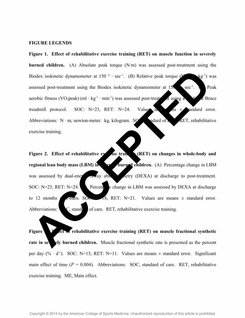
FIGURE LEGENDS
Figure 1. Effect of rehabilitative exercise training (RET) on muscle function in severely
burned children. (A) Absolute peak torque (N·m) was assessed post-treatment using the
Biodex isokinetic dynamometer at 150 ° · sec-1. (B) Relative peak torque (N · m · kg-1) was
assessed post-treatment using the Biodex isokinetic dynamometer at 150 ° · sec-1. (C) Peak
aerobic fitness (VO2peak) (ml · kg-1 · min-1) was assessed post-treatment using a modified Bruce
treadmill protocol. SOC: N=23, RET: N=24. Values are means ± standard error.
Abbreviations: N · m, newton-meter. kg, kilogram. SOC, standard of care. RET, rehabilitative
exercise training.
Figure 2. Effect of rehabilitative exercise training (RET) on changes in whole-body and
regional lean body mass (LBM) in severely burned children. (A) Percentage change in LBM
was assessed by dual-energy X-ray absorptiometry (DEXA) at discharge to post-treatment.
SOC: N=23, RET: N=24. (B) Percentage change in LBM was assessed by DEXA at discharge
to 12 months post-burn. SOC: N=18, RET: N=21. Values are means ± standard error.
Abbreviations: SOC, standard of care. RET, rehabilitative exercise training.
Figure 3. Effect of rehabilitative exercise training (RET) on muscle fractional synthetic
rate in severely burned children. Muscle fractional synthetic rate is presented as the percent
per day (% · d-1). SOC: N=13; RET: N=11. Values are means ± standard error. Significant
main effect of time (P = 0.004). Abbreviations: SOC, standard of care. RET, rehabilitative
exercise training. ME, Main effect.
ACCEPTED
Copyright © 2014 by the American College of Sports Medicine. Unauthorized reproduction of this article is prohibited.

Figure 1
ACCEPTED
Copyright © 2014 by the American College of Sports Medicine. Unauthorized reproduction of this article is prohibited.
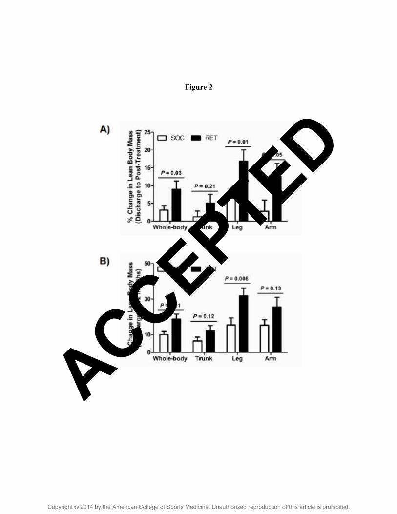
Figure 2
ACCEPTED
Copyright © 2014 by the American College of Sports Medicine. Unauthorized reproduction of this article is prohibited.

Figure 3
ACCEPTED
Copyright © 2014 by the American College of Sports Medicine. Unauthorized reproduction of this article is prohibited.

Table 1. Patient characteristics at hospital discharge.
SOC (N=23) RET (N=24) P valueAge, yr 13 ± 1 13 ± 1 0.58
Height, cm 156 ± 4 152 ± 4 0.50Weight, kg 47 ± 5 46 ± 3 0.87TBSA, % 60 ± 3 59 ± 2 0.89
3rd, % 49 ± 4 46 ± 4 0.62LOS, days 38 ± 7 34 ± 4 0.96
F:M 5:18 4:20 0.72
Values are means ± standard error.
Abbreviations: SOC, standard of care. RET, rehabilitative exercise training. N, number of patients. yr, year. cm, centimeters. kg,
kilogram. TBSA, total burn surface area determined at admittance. 3rd, third degree burn. F:M, number of female and male patients.
LOS, length of hospital stay during acute burn care treatment.
ACCEPTED
Copyright © 2014 by the American College of Sports Medicine. Unauthorized reproduction of this article is prohibited.
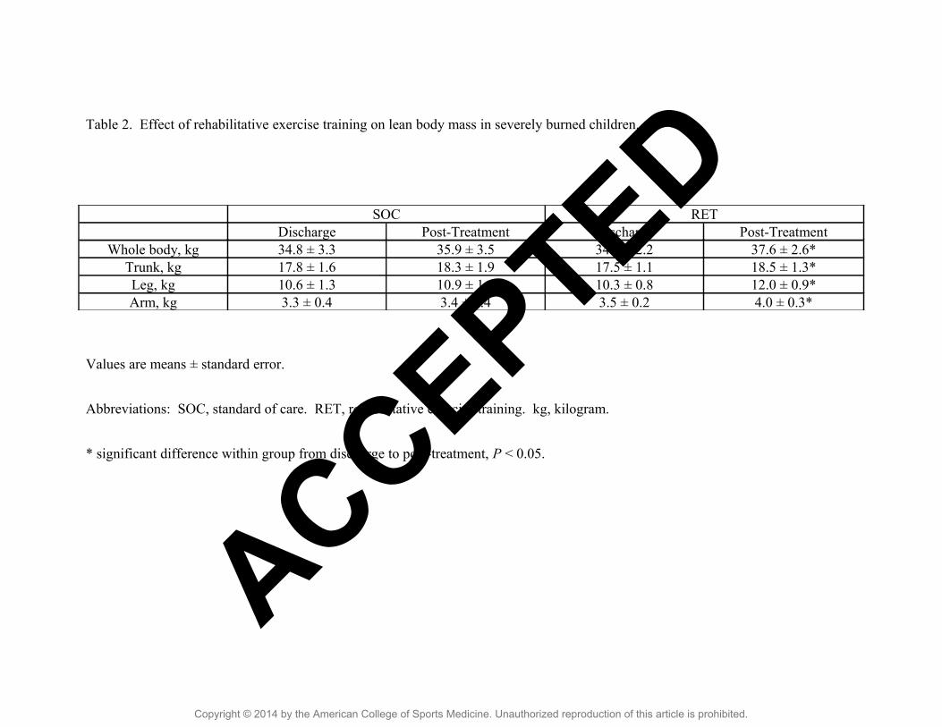
Table 2. Effect of rehabilitative exercise training on lean body mass in severely burned children.
SOC RETDischarge Post-Treatment Discharge Post-Treatment
Whole body, kg 34.8 ± 3.3 35.9 ± 3.5 34.3 ± 2.2 37.6 ± 2.6*Trunk, kg 17.8 ± 1.6 18.3 ± 1.9 17.5 ± 1.1 18.5 ± 1.3*Leg, kg 10.6 ± 1.3 10.9 ± 1.1 10.3 ± 0.8 12.0 ± 0.9*Arm, kg 3.3 ± 0.4 3.4 ± 0.4 3.5 ± 0.2 4.0 ± 0.3*
Values are means ± standard error.
Abbreviations: SOC, standard of care. RET, rehabilitative exercise training. kg, kilogram.
* significant difference within group from discharge to post-treatment, P < 0.05.
ACCEPTED
Copyright © 2014 by the American College of Sports Medicine. Unauthorized reproduction of this article is prohibited.