Predictors of re-epithelialization in pediatric burn
8
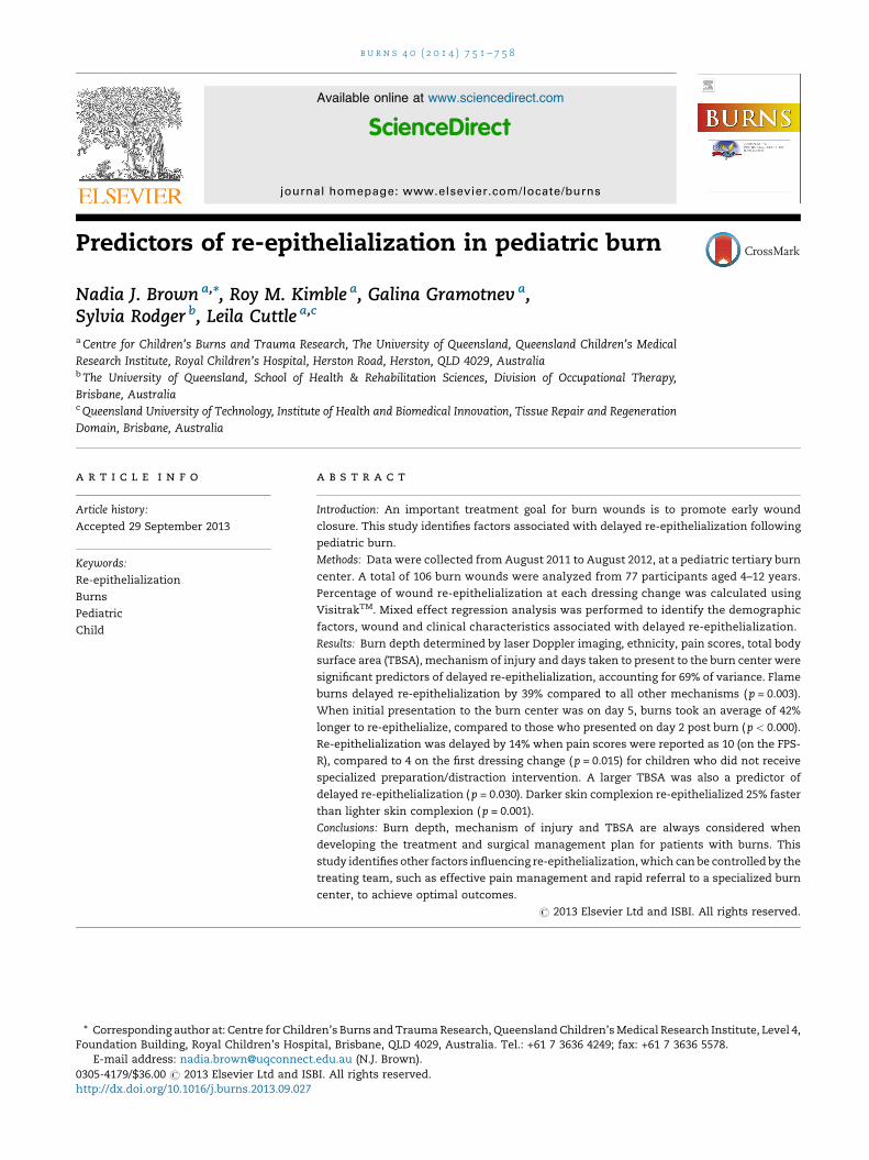
Predictors of re-epithelialization in pediatric burn Nadia J. Brown a, *, Roy M. Kimble a , Galina Gramotnev a , Sylvia Rodger b , Leila Cuttle a,c a Centre for Children’s Burns and Trauma Research, The University of Queensland, Queensland Children’s Medical Research Institute, Royal Children’s Hospital, Herston Road, Herston, QLD 4029, Australia b The University of Queensland, School of Health & Rehabilitation Sciences, Division of Occupational Therapy, Brisbane, Australia c Queensland University of Technology, Institute of Health and Biomedical Innovation, Tissue Repair and Regeneration Domain, Brisbane, Australia b u r n s 4 0 ( 2 0 1 4 ) 7 5 1 – 7 5 8 a r t i c l e i n f o Article history: Accepted 29 September 2013 Keywords: Re-epithelialization Burns Pediatric Child a b s t r a c t Introduction: An important treatment goal for burn wounds is to promote early wound closure. This study identifies factors associated with delayed re-epithelialization following pediatric burn. Methods: Data were collected from August 2011 to August 2012, at a pediatric tertiary burn center. A total of 106 burn wounds were analyzed from 77 participants aged 4–12 years. Percentage of wound re-epithelialization at each dressing change was calculated using Visitrak TM . Mixed effect regression analysis was performed to identify the demographic factors, wound and clinical characteristics associated with delayed re-epithelialization. Results: Burn depth determined by laser Doppler imaging, ethnicity, pain scores, total body surface area (TBSA), mechanism of injury and days taken to present to the burn center were significant predictors of delayed re-epithelialization, accounting for 69% of variance. Flame burns delayed re-epithelialization by 39% compared to all other mechanisms ( p = 0.003). When initial presentation to the burn center was on day 5, burns took an average of 42% longer to re-epithelialize, compared to those who presented on day 2 post burn ( p < 0.000). Re-epithelialization was delayed by 14% when pain scores were reported as 10 (on the FPS- R), compared to 4 on the first dressing change ( p = 0.015) for children who did not receive specialized preparation/distraction intervention. A larger TBSA was also a predictor of delayed re-epithelialization ( p = 0.030). Darker skin complexion re-epithelialized 25% faster than lighter skin complexion ( p = 0.001). Conclusions: Burn depth, mechanism of injury and TBSA are always considered when developing the treatment and surgical management plan for patients with burns. This study identifies other factors influencing re-epithelialization, which can be controlled by the treating team, such as effective pain management and rapid referral to a specialized burn center, to achieve optimal outcomes. # 2013 Elsevier Ltd and ISBI. All rights reserved. * Corresponding author at: Centre for Children’s Burns and Trauma Research, Queensland Children’s Medical Research Institute, Level 4, Foundation Building, Royal Children’s Hospital, Brisbane, QLD 4029, Australia. Tel.: +61 7 3636 4249; fax: +61 7 3636 5578. E-mail address: [email protected] (N.J. Brown). Available online at www.sciencedirect.com ScienceDirect journal homepage: www.elsevier.com/locate/burns 0305-4179/$36.00 # 2013 Elsevier Ltd and ISBI. All rights reserved. http://dx.doi.org/10.1016/j.burns.2013.09.027
Transcript of Predictors of re-epithelialization in pediatric burn
Predictors of re-epithelialization in pediatric burn
Nadia J. Brown a,*, Roy M. Kimble a, Galina Gramotnev a,Sylvia Rodger b, Leila Cuttle a,c
aCentre for Children’s Burns and Trauma Research, The University of Queensland, Queensland Children’s Medical
Research Institute, Royal Children’s Hospital, Herston Road, Herston, QLD 4029, AustraliabThe University of Queensland, School of Health & Rehabilitation Sciences, Division of Occupational Therapy,
Brisbane, AustraliacQueensland University of Technology, Institute of Health and Biomedical Innovation, Tissue Repair and Regeneration
Domain, Brisbane, Australia
b u r n s 4 0 ( 2 0 1 4 ) 7 5 1 – 7 5 8
a r t i c l e i n f o
Article history:
Accepted 29 September 2013
Keywords:
Re-epithelialization
Burns
Pediatric
Child
a b s t r a c t
Introduction: An important treatment goal for burn wounds is to promote early wound
closure. This study identifies factors associated with delayed re-epithelialization following
pediatric burn.
Methods: Data were collected from August 2011 to August 2012, at a pediatric tertiary burn
center. A total of 106 burn wounds were analyzed from 77 participants aged 4–12 years.
Percentage of wound re-epithelialization at each dressing change was calculated using
VisitrakTM. Mixed effect regression analysis was performed to identify the demographic
factors, wound and clinical characteristics associated with delayed re-epithelialization.
Results: Burn depth determined by laser Doppler imaging, ethnicity, pain scores, total body
surface area (TBSA), mechanism of injury and days taken to present to the burn center were
significant predictors of delayed re-epithelialization, accounting for 69% of variance. Flame
burns delayed re-epithelialization by 39% compared to all other mechanisms ( p = 0.003).
When initial presentation to the burn center was on day 5, burns took an average of 42%
longer to re-epithelialize, compared to those who presented on day 2 post burn ( p < 0.000).
Re-epithelialization was delayed by 14% when pain scores were reported as 10 (on the FPS-
R), compared to 4 on the first dressing change ( p = 0.015) for children who did not receive
specialized preparation/distraction intervention. A larger TBSA was also a predictor of
delayed re-epithelialization ( p = 0.030). Darker skin complexion re-epithelialized 25% faster
than lighter skin complexion ( p = 0.001).
Conclusions: Burn depth, mechanism of injury and TBSA are always considered when
developing the treatment and surgical management plan for patients with burns. This
study identifies other factors influencing re-epithelialization, which can be controlled by the
treating team, such as effective pain management and rapid referral to a specialized burn
center, to achieve optimal outcomes.
# 2013 Elsevier Ltd and ISBI. All rights reserved.
* Corresponding author at: Centre for Children’s Burns and Trauma Research, Queensland Children’s Medical Research Institute, Level 4,Foundation Building, Royal Children’s Hospital, Brisbane, QLD 4029, Australia. Tel.: +61 7 3636 4249; fax: +61 7 3636 5578.
Available online at www.sciencedirect.com
ScienceDirect
journal homepage: www.elsevier.com/locate/burns
E-mail address: [email protected] (N.J. Brown).
0305-4179/$36.00 # 2013 Elsevier Ltd and ISBI. All rights reserved.http://dx.doi.org/10.1016/j.burns.2013.09.027

b u r n s 4 0 ( 2 0 1 4 ) 7 5 1 – 7 5 8752
1. Introduction
The past decade has seen much advancement in pediatric
burn treatments and outcomes. The focus in developed
countries has shifted from mortality to concerns around
morbidity; pain reduction and non-pharmacological interven-
tions; cosmetic outcomes; rehabilitation; quality of life and
psychosocial well-being. However, limited research has been
conducted on factors influencing rate of re-epithelialization
since Deitch et al. [1] first identified the importance of re-
epithelialization within 21 days post injury, to minimize the
likelihood of hypertrophic scarring.
Burn depth by laser Doppler imaging (LDI) remains the only
confirmed predictor of wound re-epithelialization to date [2,3].
Several studies have investigated risk factors for hypertrophic
scarring [1,4–7], reporting depth, days to re-epithelialization,
anatomical location of the burn, ethnicity and age as
predictors of hypertrophic scarring. Psychological factors
influencing burn re-epithelialization were investigated in a
retrospective study by Wilson et al. [8], who reported negative
burn perceptions held by the patient were a significant
predictor of re-epithelialization once burn and demographic
characteristics had been controlled for. Other than the depth
of a burn, risk factors for healing potential have not been
validated or investigated in a prospective study. Little is
known about whether risk factors for delayed re-epithelializa-
tion are the same as those identified and documented for
hypertrophic scarring.
This prospective study on burn re-epithelialization inves-
tigates multiple demographic and burn wound clinical
characteristics to identify factors influencing burn wound
re-epithelialization in children. Furthermore, links between
risk factors for delayed healing identified from this study will
be compared and contrasted to risk factors for hypertrophic
scar development reported in the literature. We hypothesize
there are multiple factors which can delay re-epithelialization;
including patient demographics and wound characteristics,
and the clinical management of patients and their wounds.
2. Methods
2.1. Design
Factors predicting delayed burn wound re-epithelialization
were investigated in this prospective longitudinal study by
analyzing data from a randomized controlled trial (RCT) on
burn re-epithelialization [9]. The RCT was registered with the
Australian New Zealand Clinical Trials Registry
(ACTRN12611000913976), and approved by the Queensland
Children’s Health Services (Royal Children’s Hospital) Human
Research Ethics Committee and The University of Queensland
Ethics Committee.
2.2. Setting and participants
Data were collected from the Stuart Pegg Pediatric Burn Center
(SPPBC) at the Royal Children’s Hospital, Brisbane, Australia
from August 2011 to August 2012. Inclusion criteria were
children aged 4 years to 12 years 11 months who presented
with a burn of less than 15% total body surface area (TBSA).
Exclusion criteria were; (1) non-English speaking, (2) children
with a diagnosed condition/illness/developmental delay in
addition to a burns injury, (3) children with a prior history of
suspected child abuse and neglect, and (4) burns requiring
grafting. Data collection commenced at the first dressing
change, and did not alter medical treatment received. Partici-
pants were randomly assigned by administrative staff using a
portable computerized random number generator to one of two
groups: to receive a computerized procedural preparation and
distraction intervention; or to receive standard preparation and
distraction including television, videos, toys, and/or parental
soothing. Data were collected at every dressing change until
complete burn re-epithelialization [9].
2.3. Dressing change procedures
Participants received pain relief in the form of narcotic
analgesia (oxycodone opioid, 0.1–0.2 mg/kg); narcotic combined
(oxycodone and either paracetamol or ibuprofen; or codeine
and paracetamol), or non-narcotic analgesia (paracetamol and/
or ibuprofen). Administration of pain medication was deter-
mined by nursing pain assessments. Participants primarily
received narcotic pain relief on the first change of dressing.
All burns were debrided at the first change of dressing
(blisters were de-roofed and dead skin wiped away with gauze)
and Chlorhexidine solution was used to clean the burn at
every dressing change. Dressings were applied based on the
consultant’s assessment and consisted of Acticoat3TM (dress-
ing changes every three days) or ActicoatTM (dressing changes
every seven days) with or without MepitelTM.
2.4. Data measurements
A VisitrakTM grid (Smith + Nephew, London, UK) was used at
every dressing change to calculate the re-epithelized area of the
wound. The number of days post-injury before burns reached
�95% re-epithelialization (based on VisitrakTM), was used to
calculate the dependent variable ‘‘days to re-epithelialize.’’
The MoorLDI2-BI2 laser Doppler imager (LDI, Moor Instru-
ments Limited, Devon, UK), was used at the first dressing
change to determine burn depth. Scanning distances ranged
between 40 and 70 cm, with the scanner head positioned
approximately 358 off perpendicular and set on fast scanning
resolution. The onboard software package (moorBDA v2.4) was
used to define regions of interest for each burn area, to calculate
the average perfusion units (PU) and minimum value PU of each
burn area, enabling comparison of average and minimum
values across burn wounds. TBSA of the burn injured area was
calculated using the Lund and Browder chart [10].
Pain, distress and anxiety scores were recorded before,
during and after wound care procedures by: (a) participant
self-report on the Faces Pain Scale-Revised (FPS-R); (b) the
nurse’s behavioral/observational rating on the Face, Legs,
Arms, Cry, Consolability (FLACC) scale; (c) and children 8 years
and above rated anxiety on the Visual Analog Scale-Anxiety
(VAS-A). Heart rate as a physiological indicator of distress and
anxiety was monitored and recorded every two minutes
during wound care procedures. Additionally, children six

Table 1 – Participant demographics.
Mean (SD)
Age (months) 96.96 (29.67)
N (%)
Gender Male 46 (60)
Ethnicity Lighter skin complexion 68 (88)
SES Low (SEIFA � 6) 24 (32)
High (SEIFA � 7) 52 (68)
Medication (COD1) Narcotic analgesia 42 (55)
Narcotic combined analgesia 20 (26)
Non-narcotic analgesia 4 (5)
Nil 11 (14)
TBSA, total body surface area; SES, socio-economic status; COD,
change of dressing.
Table 2 – Wound clinical characteristics.
Mean (SD)
TBSA (%) 2.15 (1.95)
LDI Mean (PU) 1107.35 (282.44)
LDI Minimum (PU) 591.12 (164.04)
N (%)
Depth (clinical judgment) Superficial partial thickness 75 (70.8)
Deep partial thickness 30 (28.3)
Full thickness 1 (0.9)
Mechanism Scald 61 (57.6)
Contact 31 (29.3)
Flame 5 (4.72)
Friction 8 (7.6)
Chemical 1 (0.9)
Anatomical site Axilla/upper & lower arm 42 (39.6)
Upper & lower leg/foot 35 (33)
Chest/torso/back 22 (20.8)
Head/face 4 (3.8)
Genitals/buttocks 3 (2.8)
TBSA: total body surface area; LDI: laser Doppler imaging; PU:
perfusion units.
b u r n s 4 0 ( 2 0 1 4 ) 7 5 1 – 7 5 8 753
years and above completed the 10-item Child Trauma
Screening Questionnaire (CTSQ) [11] within the first seven
days post injury and then repeated the CTSQ via mail at three
months post re-epithelialization, to identify symptoms of
Post-Traumatic Stress Disorder (PTSD).
2.5. Statistical methodology
The statistical analyses were conducted using the Stata
statistical software package [12]. The goal of the analysis
was to determine and understand the dependences between
number of days from injury to �95% re-epithelialization (the
dependent variable) and the following independent variables:
burn depth (LDI), TBSA, participant’s self-report of pain (FPS-
R), nurse’s rating of pain/distress (FLACC), participant’s self-
report anxiety scores (VAS-A), ethnicity, mechanism of injury,
days post-injury to present to the burn center, age, gender,
family income, mother’s highest education level, Socio-
Economic Indexes for Areas (SEIFA), site of injury, first aid,
maximum heart rate during the wound care procedures, and
nursing time taken to complete wound care procedures. Due
to a specialized preparation/distraction intervention being
randomly used for some of the patients during wound care
procedures, the categorical variable ‘‘group’’ (reflecting the
application of the distraction technique) was added to the
considered model. The Shapiro–Wilk test for normality
indicated ‘‘days to re-epithelialize’’ was not distributed
normally ( p < 0.001) and this dependent variable was loga-
rithmically transformed to achieve a normal distribution.
As there were multiple (�4) burns per patient, we
considered clustering of burns with respect to each patient,
which gives rise to the two-level random-effects linear
regression with random intercepts to assess the effect of
each independent variable on burn re-epithelialization. The
two levels within the model are: (1) individual burn, and (2) the
patient on which this burn was located. The random effects
within the model are random intercepts for the respective
regression lines of days to re-epithelialize versus the consid-
ered independent variables for each patient.
It was shown that only independent variables burn depth,
TBSA, pain intensity, group, ethnicity, mechanism of injury,
and days taken to present to burns center had statistically
significant impacts on days to 95% re-epithelialization. For all
other considered independent variables: p > 0.1, which means
that these variables have insignificant impact on re-epitheli-
alization time. The test for the presence of non-linear higher
order terms in the dependence of days to re-epithelize on the
continuous and statistically significant variables revealed the
presence of statistically significant terms up to the power of
three for the LDI variable. All possible interactions between
the independent variables were considered, with only the
interaction term of the group and pain (FPS-R) variables found
to be significant.
Using the likelihood ratio test to compare the fit of the
random intercept model to that of the multiple regression
model without clustering, gives p = 0.018, which confirms the
need for using the random intercept model. At the same time,
comparing the random slope and random intercept models
did not show statistically significant difference ( p > 0.45).
Thus, we limit our model to only the consideration of random
intercepts. The assumption of normality for the random
intercepts was confirmed by the Shapiro–Wilk test (giving
p = 0.6) and the quantile–quantile plot.
3. Results
Data were collected on a total of 77 participants. The median
age was 8 years 1 month, with a range from 4 years 1 month to
12 years 9 months, (see Table 1 for demographic character-
istics). A total of 106 burns were analyzed, which were
primarily superficial partial thickness burns resulting from
scald injuries (see Table 2 for burn characteristics). According
to the Australian Bureau of Statistics Socioeconomic Indexes
for Areas (SEIFA) [13,14], participants were from primarily high
socio-economic status postcode areas (SEIFA � 7), and from
parental report total annual family income was greater than
$67,500 for 59% of participants. Laser Doppler Imaging (LDI)
was not always possible to perform due to the hectic nature of
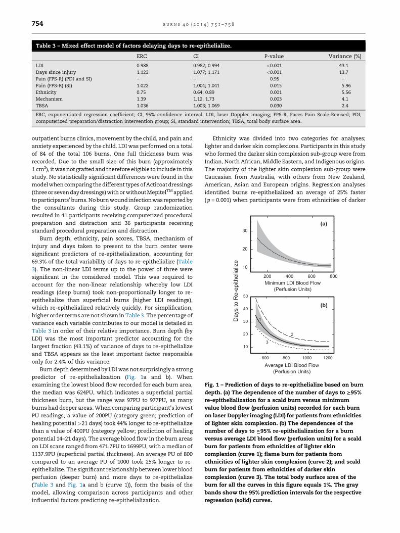
Table 3 – Mixed effect model of factors delaying days to re-epithelialize.
ERC CI P-value Variance (%)
LDI 0.988 0.982; 0.994 <0.001 43.1
Days since injury 1.123 1.077; 1.171 <0.001 13.7
Pain (FPS-R) (PDI and SI) – – 0.95 –
Pain (FPS-R) (SI) 1.022 1.004; 1.041 0.015 5.96
Ethnicity 0.75 0.64; 0.89 0.001 5.56
Mechanism 1.39 1.12; 1.73 0.003 4.1
TBSA 1.036 1.003; 1.069 0.030 2.4
ERC, exponentiated regression coefficient; CI, 95% confidence interval; LDI, laser Doppler imaging; FPS-R, Faces Pain Scale-Revised; PDI,
computerized preparation/distraction intervention group; SI, standard intervention; TBSA, total body surface area.
10
20
30
40
50
600 800 1000 1200
Day
s to
Re-
epith
elia
lize
Average LDI Blood Flow(Perfusion Units)
2
3
10
20
30
200 400 600 800Minimum LDI Blood Flow
(Perfusion Units)
(a)
(b)
1
Fig. 1 – Prediction of days to re-epithelialize based on burn
depth. (a) The dependence of the number of days to I95%
re-epithelialization for a scald burn versus minimum
value blood flow (perfusion units) recorded for each burn
on laser Doppler imaging (LDI) for patients from ethnicities
of lighter skin complexion. (b) The dependences of the
number of days to I95% re-epithelialization for a burn
versus average LDI blood flow (perfusion units) for a scald
burn for patients from ethnicities of lighter skin
complexion (curve 1); flame burn for patients from
ethnicities of lighter skin complexion (curve 2); and scald
burn for patients from ethnicities of darker skin
complexion (curve 3). The total body surface area of the
burn for all the curves in this figure equals 1%. The gray
bands show the 95% prediction intervals for the respective
regression (solid) curves.
b u r n s 4 0 ( 2 0 1 4 ) 7 5 1 – 7 5 8754
outpatient burns clinics, movement by the child, and pain and
anxiety experienced by the child. LDI was performed on a total
of 84 of the total 106 burns. One full thickness burn was
recorded. Due to the small size of this burn (approximately
1 cm2), it was not grafted and therefore eligible to include in this
study. No statistically significant differences were found in the
model when comparing the different types of Acticoat dressings
(three or seven day dressings) with or without MepitelTM applied
to participants’ burns. No burn wound infection was reported by
the consultants during this study. Group randomization
resulted in 41 participants receiving computerized procedural
preparation and distraction and 36 participants receiving
standard procedural preparation and distraction.
Burn depth, ethnicity, pain scores, TBSA, mechanism of
injury and days taken to present to the burn center were
significant predictors of re-epithelialization, accounting for
69.3% of the total variability of days to re-epithelialize (Table
3). The non-linear LDI terms up to the power of three were
significant in the considered model. This was required to
account for the non-linear relationship whereby low LDI
readings (deep burns) took non-proportionally longer to re-
epithelialize than superficial burns (higher LDI readings),
which re-epithelialized relatively quickly. For simplification,
higher order terms are not shown in Table 3. The percentage of
variance each variable contributes to our model is detailed in
Table 3 in order of their relative importance. Burn depth (by
LDI) was the most important predictor accounting for the
largest fraction (43.1%) of variance of days to re-epithelialize
and TBSA appears as the least important factor responsible
only for 2.4% of this variance.
Burn depth determined by LDI was not surprisingly a strong
predictor of re-epithelialization (Fig. 1a and b). When
examining the lowest blood flow recorded for each burn area,
the median was 624PU, which indicates a superficial partial
thickness burn, but the range was 97PU to 977PU, as many
burns had deeper areas. When comparing participant’s lowest
PU readings, a value of 200PU (category green; prediction of
healing potential >21 days) took 44% longer to re-epithelialize
than a value of 400PU (category yellow; prediction of healing
potential 14–21 days). The average blood flow in the burn areas
on LDI scans ranged from 471.7PU to 1699PU, with a median of
1137.9PU (superficial partial thickness). An average PU of 800
compared to an average PU of 1000 took 25% longer to re-
epithelialize. The significant relationship between lower blood
perfusion (deeper burn) and more days to re-epithelialize
(Table 3 and Fig. 1a and b (curve 1)), form the basis of the
model, allowing comparison across participants and other
influential factors predicting re-epithelialization.
Ethnicity was divided into two categories for analyses;
lighter and darker skin complexions. Participants in this study
who formed the darker skin complexion sub-group were from
Indian, North African, Middle Eastern, and Indigenous origins.
The majority of the lighter skin complexion sub-group were
Caucasian from Australia, with others from New Zealand,
American, Asian and European origins. Regression analyses
identified burns re-epithelialized an average of 25% faster
( p = 0.001) when participants were from ethnicities of darker
5
10
15
20
25
0 2 4 6 8
Day
s to
Re-
epith
elia
lize
Days since Injury
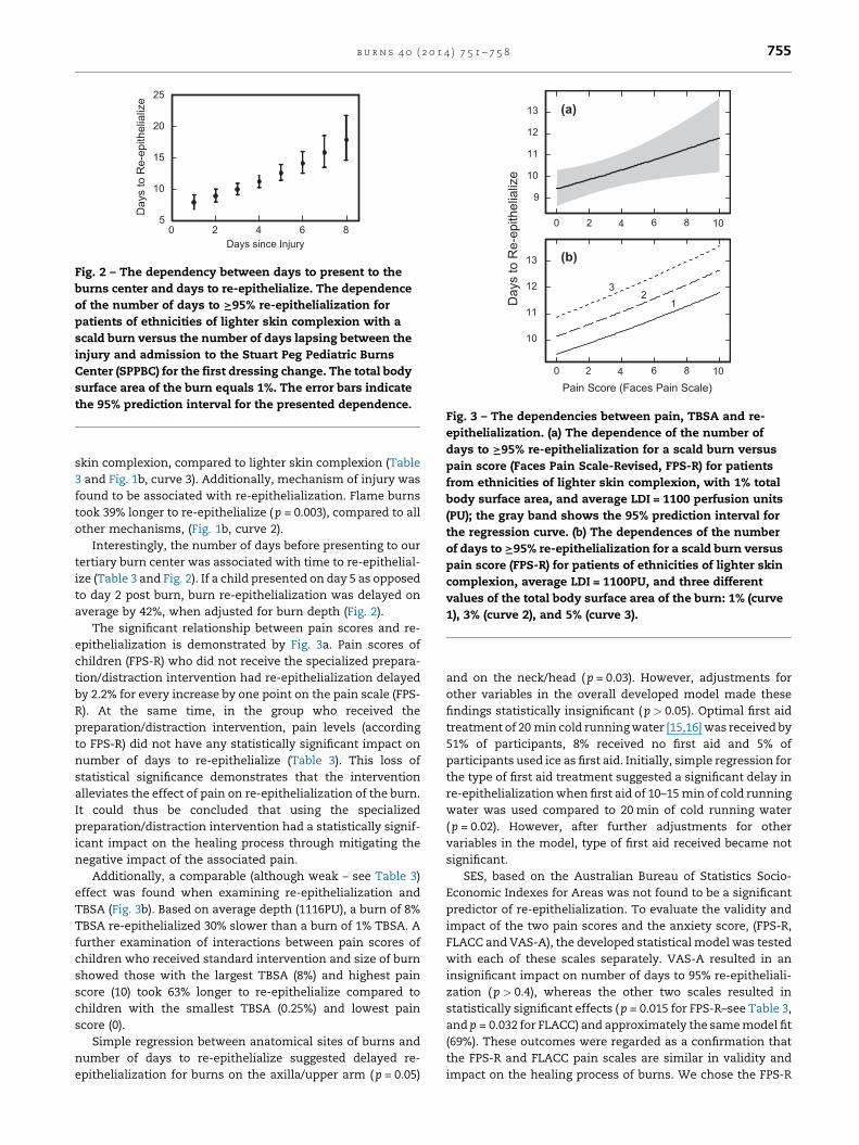
Fig. 2 – The dependency between days to present to the
burns center and days to re-epithelialize. The dependence
of the number of days to I95% re-epithelialization for
patients of ethnicities of lighter skin complexion with a
scald burn versus the number of days lapsing between the
injury and admission to the Stuart Peg Pediatric Burns
Center (SPPBC) for the first dressing change. The total body
surface area of the burn equals 1%. The error bars indicate
the 95% prediction interval for the presented dependence.
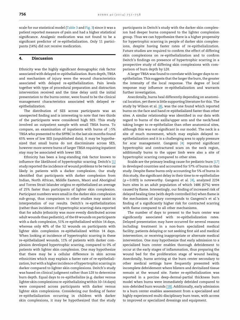
9
10
11
12
13
Day
s to
Re-
epith
elia
lize
Pain Score (Faces Pain Scale)
0 2 4 6 8 10
10
11
12
13
0 2 4 6 8 10
12
3
(a)
(b)
Fig. 3 – The dependencies between pain, TBSA and re-
epithelialization. (a) The dependence of the number of
days to I95% re-epithelialization for a scald burn versus
pain score (Faces Pain Scale-Revised, FPS-R) for patients
from ethnicities of lighter skin complexion, with 1% total
body surface area, and average LDI = 1100 perfusion units
(PU); the gray band shows the 95% prediction interval for
the regression curve. (b) The dependences of the number
of days to I95% re-epithelialization for a scald burn versus
pain score (FPS-R) for patients of ethnicities of lighter skin
complexion, average LDI = 1100PU, and three different
values of the total body surface area of the burn: 1% (curve
1), 3% (curve 2), and 5% (curve 3).
b u r n s 4 0 ( 2 0 1 4 ) 7 5 1 – 7 5 8 755
skin complexion, compared to lighter skin complexion (Table
3 and Fig. 1b, curve 3). Additionally, mechanism of injury was
found to be associated with re-epithelialization. Flame burns
took 39% longer to re-epithelialize ( p = 0.003), compared to all
other mechanisms, (Fig. 1b, curve 2).
Interestingly, the number of days before presenting to our
tertiary burn center was associated with time to re-epithelial-
ize (Table 3 and Fig. 2). If a child presented on day 5 as opposed
to day 2 post burn, burn re-epithelialization was delayed on
average by 42%, when adjusted for burn depth (Fig. 2).
The significant relationship between pain scores and re-
epithelialization is demonstrated by Fig. 3a. Pain scores of
children (FPS-R) who did not receive the specialized prepara-
tion/distraction intervention had re-epithelialization delayed
by 2.2% for every increase by one point on the pain scale (FPS-
R). At the same time, in the group who received the
preparation/distraction intervention, pain levels (according
to FPS-R) did not have any statistically significant impact on
number of days to re-epithelialize (Table 3). This loss of
statistical significance demonstrates that the intervention
alleviates the effect of pain on re-epithelialization of the burn.
It could thus be concluded that using the specialized
preparation/distraction intervention had a statistically signif-
icant impact on the healing process through mitigating the
negative impact of the associated pain.
Additionally, a comparable (although weak – see Table 3)
effect was found when examining re-epithelialization and
TBSA (Fig. 3b). Based on average depth (1116PU), a burn of 8%
TBSA re-epithelialized 30% slower than a burn of 1% TBSA. A
further examination of interactions between pain scores of
children who received standard intervention and size of burn
showed those with the largest TBSA (8%) and highest pain
score (10) took 63% longer to re-epithelialize compared to
children with the smallest TBSA (0.25%) and lowest pain
score (0).
Simple regression between anatomical sites of burns and
number of days to re-epithelialize suggested delayed re-
epithelialization for burns on the axilla/upper arm ( p = 0.05)
and on the neck/head ( p = 0.03). However, adjustments for
other variables in the overall developed model made these
findings statistically insignificant ( p > 0.05). Optimal first aid
treatment of 20 min cold running water [15,16] was received by
51% of participants, 8% received no first aid and 5% of
participants used ice as first aid. Initially, simple regression for
the type of first aid treatment suggested a significant delay in
re-epithelialization when first aid of 10–15 min of cold running
water was used compared to 20 min of cold running water
( p = 0.02). However, after further adjustments for other
variables in the model, type of first aid received became not
significant.
SES, based on the Australian Bureau of Statistics Socio-
Economic Indexes for Areas was not found to be a significant
predictor of re-epithelialization. To evaluate the validity and
impact of the two pain scores and the anxiety score, (FPS-R,
FLACC and VAS-A), the developed statistical model was tested
with each of these scales separately. VAS-A resulted in an
insignificant impact on number of days to 95% re-epitheliali-
zation ( p > 0.4), whereas the other two scales resulted in
statistically significant effects ( p = 0.015 for FPS-R–see Table 3,
and p = 0.032 for FLACC) and approximately the same model fit
(69%). These outcomes were regarded as a confirmation that
the FPS-R and FLACC pain scales are similar in validity and
impact on the healing process of burns. We chose the FPS-R
b u r n s 4 0 ( 2 0 1 4 ) 7 5 1 – 7 5 8756
scale for our statistical model (Table 3 and Fig. 3) since it was a
patient reported measure of pain and had a higher statistical
significance. Analgesic medication was not found to be a
significant predictor of re-epithelialization. Only 11 partici-
pants (14%) did not receive medication.
4. Discussion
Ethnicity was the highly significant demographic risk factor
associated with delayed re-epithelialization. Burn depth, TBSA
and mechanism of injury were the wound characteristics
associated with delayed re-epithelialization. Pain levels
together with type of procedural preparation and distraction
intervention received and the time delay until the initial
presentation to the burns center, were found to be the clinical
management characteristics associated with delayed re-
epithelialization.
The distribution of SES across participants was an
unexpected finding and is interesting to note that two thirds
of the participants were considered high SES. This study
involved an outpatient sample with small TBSA burns. To
compare, an examination of inpatients with burns of �5%
TBSA who presented to the SPPBC in the last six months found
65% were of low SES (unpublished data). It may be hypothe-
sized that small burns do not discriminate across SES,
however more severe burns of larger TBSA requiring inpatient
stay may be associated with lower SES.
Ethnicity has been a long-standing risk factor known to
influence the likelihood of hypertrophic scarring. Deitch’s [1]
study reported the incidence of wound problems to be twice as
likely in patients with a darker complexion. Our study
identified that participants with darker complexion from
Indian, North African, Middle Eastern, Maori and Aboriginal
and Torres Strait Islander origins re-epithelialized an average
of 25% faster than participants of lighter skin complexion.
Participant numbers were small in the darker skin complexion
sub-group, thus comparison to other studies may assist in
interpretation of our results. Deitch’s re-epithelialization
(rather than incidence of hypertrophic scarring) data reported
that for adults (ethnicity was more evenly distributed across
adult wounds than pediatric), of the 69 wounds on participants
with a dark complexion, 51% re-epithelialized within 14 days,
whereas only 40% of the 52 wounds on participants with
lighter skin complexion re-epithelialized within 14 days.
When looking at incidence of hypertrophic scarring in these
re-epithelialized wounds, 11% of patients with darker com-
plexion developed hypertrophic scarring, compared to 0% of
patients with lighter skin complexion. One may hypothesize
that there may be a cellular difference in skin across
ethnicities which may explain a faster rate of re-epitheliali-
zation, but with a higher incidence of hypertrophic scarring in
darker compared to lighter skin complexions. Deitch’s study
was based on clinical judgment rather than LDI to determine
burn depth. Equal days to re-epithelialize (e.g. darker versus
lighter skin complexions re-epithelializing within 10–14 days)
were compared across participants with darker versus
lighter skin complexions. Considering our finding of faster
re-epithelialization occurring in children with darker
skin complexions, it may be hypothesized that the study
participants in Deitch’s study with the darker skin complex-
ion had deeper burns compared to the lighter complexion
group. Thus we can hypothesize there is a higher propensity
for hypertrophic scarring in people of darker skin complex-
ions, despite having faster rates of re-epithelialization.
Future studies are required to confirm the effect of differing
skin complexions on re-epithelialization and to confirm
Deitch’s findings on presence of hypertrophic scarring in a
prospective study of differing skin complexions with com-
parison of burn depth by LDI.
A larger TBSA was found to correlate with longer days to re-
epithelialize. This suggests that the larger the burn, the greater
the intensity of the local response. The degree of local
response may influence re-epithelialization and warrants
further investigation.
Anecdotally, burns heal differently depending on anatomi-
cal location, yet there is little supporting literature for this. The
study by Wilson et al. [8], was the one found which reported
burns on the face and hand re-epithelialized faster than other
sites. A similar relationship was identified in our data with
regard to burns of the axilla/upper arm and the neck/head
taking longer to re-epithelialize than other anatomical sites,
although this was not significant in our model. The neck is a
site of much movement, which may explain delayed re-
epithelialization and it is a challenging area to apply pressure
for scar management. Gangemi [4] reported significant
hypertrophic and contractured scars on the neck region,
additionally burns to the upper limb were also a site of
hypertrophic scarring compared to other sites.
Scalds are the primary leading cause for pediatric burn [17]
in developed countries and accounted for 57% of burns in this
study. Despite flame burns only accounting for 5% of burns in
this study, the significant delay in their time to re-epithelialize
( p = 0.003) is noteworthy. Gangemi et al. [4], analyzed 2234
burn sites in an adult population of which 1486 (67%) were
caused by flame. Interestingly, our finding of increased risk of
delayed healing time (with depth controlled for) when flame is
the mechanism of injury corresponds to Gangemi’s et al.’s
finding of a significantly higher risk for contracted scarring
with flame compared to all other mechanisms.
The number of days to present to the burn center was
significantly associated with re-epithelialization rates.
Delayed presentation may have occurred for several reasons
including: treatment in a non-burn specialized medical
facility; patients delaying or not seeking first aid and medical
intervention; or receiving inappropriate or alternate medical
intervention. One may hypothesize that early admission to a
specialized burn center enables thorough debridement to
occur at the early stages of inflammation, thus preparing the
wound bed for the proliferation stage of wound healing.
Anecdotally, burns arriving at the burn center secondary to
other medical settings have frequently presented with
incomplete debridement where blisters and devitalized tissue
remain at the wound site. Faster re-epithelialization was
reported in a porcine deep-dermal-partial thickness burn
model when burns were immediately debrided compared to
non-debrided burn wounds [18]. Additionally, early admission
to a burn center enables assessment from a specialized and
highly experienced multi-disciplinary burn team, with access
to improved or specialized dressings and equipment.
b u r n s 4 0 ( 2 0 1 4 ) 7 5 1 – 7 5 8 757
Pain was found to be a significant predictor of re-
epithelialization when examining type of procedural prepara-
tion and distraction intervention received. Lack of access to a
novel computerized burn procedural preparation and distrac-
tion device during wound care procedures was associated with
delayed re-epithelialization. Previously, specifically tailored
computerized procedural preparation and distraction as a
non-pharmacological intervention during burn dressing
changes has been demonstrated to reduce pain and distress
compared to standard off the shelf computerized technologies
[19–21]. Procedural preparation alone has proven effective and
is a key component in reducing pain and distress in children
during burn wound care procedures [19]. Heightened feelings
of anxiety and anticipatory pain have also been reported as
predictors of pain intensity during dressing changes in older
subjects with chronic wounds [22]. Our findings identified an
association between a tailor-made computerized preparation/
distraction intervention and re-epithelialization, which fur-
ther supports the preliminary finding of an association
between use of this computerized intervention and a two
day reduction in burn re-epithelialization [19].
The effect pain has on initiating and mediating a myriad of
cellular events involved in re-epithelialization and wound
healing is still not well understood. Links between pain mediators
such as Substance P (SP) and 5-HT and their effects on nerves and
tissues during wound healing continues to be the focus of much
research [23]. Altered SP has been associated with impaired
healing in rats [24], and has been linked to impaired cutaneous
wound healing in association with diabetes mellitus or hyper-
trophic scar formation [25]. Further studies are required before
conclusions can be drawn on how reductions in pain transmis-
sion act on a cellular level to improve re-epithelialization.
A limitation of this study is the narrow patient sample with
regards to burn depth, as 591 PU was the average deepest area of
each burn and the majority of the burns in this study were
superficial partial thickness. It would be interesting to repeat
this study in a pediatric population of primarily deep partial
thickness burns. Furthermore, future studies should examine
demographic, wound and clinical management characteristics
in a population of burns which received surgical intervention, to
determine if the same predictors of wound healing are apparent.
In summary, clinical management characteristics influen-
tial in re-epithelialization and under the control of the burns
team include pain levels and time taken to present to a burn
center. Historically, pediatric pain has been under-treated [26–
29]. Higher pain levels experienced during burn wound care
procedures places patients at an increased risk of adverse
healing outcomes. Our findings highlight the necessity for
repeated pain assessments, use of specialized non-pharma-
cological intervention in addition to medication and improved
pain management in the area of pediatric burns to achieve
optimal outcomes. Additionally, it is vital that patients access
quality care in a tertiary burns center quickly in order to
optimize healing outcomes.
Conflict of interest
This prospective study received financial support by a grant
given to the Royal Children’s Hospital, Brisbane, by Diversionary
Therapy Technologies (DTT). Despite this financial support,
DTT had no part in the study design and data collection of this
project, nor will they have any involvement in the analysis or
publication of results. One of the supervisors of the trial, Roy
Kimble, holds options with DTT, however will not stand to lose
or gain financially or personally from the results during the
clinical trial period and time of submission. The principal
researcher has no financial interest in the DittoTM device or the
DTT Company and remains an employee of the Royal Children’s
Hospital, Brisbane.
Authors’ contributions
NJB, LC, RMK, and SR all made substantial contributions to the
design of this trial. GG has made substantial contributions to
the statistical design and wrote the statistical methodology in
this manuscript. NJB wrote the draft manuscript with input
from LC. All authors provided critical review of the article and
approved the final manuscript.
Acknowledgements
The authors would like to thank all the children and families
who participated in this study and acknowledge all the staff at
the Stuart Pegg Pediatric Burns Center at Royal Children’s
Hospital, Brisbane, Australia for their support and assistance
throughout data collection.
r e f e r e n c e s
[1] Deitch EA, Wheelahan TM, Rose MP, Clothier J, Cotter J.Hypertrophic burn scars: analysis of variables. J Trauma1983;23:895.
[2] Monstrey S, Hoeksema H, Verbelen J, Pirayesh A, BlondeelP. Assessment of burn depth and burn wound healingpotential. Burns 2008;34:761–9.
[3] Mill J, Cuttle L, Harkin DG, Kravchuk O, Kimble RM. LaserDoppler imaging in a paediatric burns population. Burns2009;35:824–31.
[4] Gangemi EN, Gregori D, Berchialla P, Zingarelli E, Cairo M,Bollero D, et al. Epidemiology and risk factors for pathologicscarring after burn wounds. Arch Facial Plast Surg2008;10:93–102.
[5] van der Wal MBA, Vloemans JFPM, Tuinebreijer WE, van deVen P, van Unen E, van Zuijlen PPM, et al. Outcome afterburns: an observational study on burn scar maturation andpredictors for severe scarring. Wound Repair Regen2012;20:676–87.
[6] Cubison T, Pape SA, Parkhouse N. Evidence for the linkbetween healing time and the development of hypertrophicscars (HTS) in paediatric burns due to scald injury. Burns2006;32:992–9.
[7] Wang X-Q, Mill J, Kravchuk O, Kimble RM. Ultrasoundassessed thickness of burn scars in association with laserDoppler imaging determined depth of burns in paediatricpatients. Burns 2010;36:1254–62.
[8] Wilson ERH, Wisely JA, Wearden AJ, Dunn KW, Edwards J,Tarrier N. Do illness perceptions and mood predict healingtime for burn wounds? A prospective, preliminary study. JPsychosom Res 2011;71:364–6.

b u r n s 4 0 ( 2 0 1 4 ) 7 5 1 – 7 5 8758
[9] Brown NJ, Rodger S, Ware RS, Kimble RM, Cuttle L. Efficacyof a children’s procedural preparation and distractiondevice on healing in acute burn wound care procedures:study protocol for a randomized controlled trial. Trials2012;13:238.
[10] Lund CC, Browder NC. The estimation of areas of burns.Surg Gynecol Obstet 1944;79:352–8.
[11] Kenardy JA, Spence SH, Macleod AC. Screening forposttraumatic stress disorder in children after accidentalinjury. Pediatrics 2006;118:1002–9.
[12] StataCorp.. Stata Statistical Software: Release 12. CollegeStation, TX: StataCorp LP; 2011.
[13] Ryan C, Whelan S. Locational disadvantage, socio-economic status and mobility behavior-evidence fromAustralia. Research School of Economics, AustralianNational University; 2010.
[14] Adhikari P. Socio-economic indexes for areas: introduction,use and future directions. Canberra: Australian Bureau ofStatistics; 2006: 1–37.
[15] Cuttle L, Kempf M, Liu P-Y, Kravchuk O, Kimble RM. Theoptimal duration and delay of first aid treatment fordeep partial thickness burn injuries. Burns 2010;36:673–9.
[16] Bartlett N, Yuan J, Holland AJ, Harvey JG, Martin HC, La HeiER, et al. Optimal duration of cooling for an acute scaldcontact burn injury in a porcine model. J Burn Care Res2008;29:828–34.
[17] McKenna K, Harrison JE. Hospital separations due to injuryand poisoning, Australia 2008–09. Australian Institute ofHealth and Welfare; 2012.
[18] Wang XQ, Kempf M, Liu PY, Cuttle L, Chang HE, KravchukO, et al. Conservative surgical debridement as a burn
treatment: supporting evidence from a porcine burn model.Wound Repair Regen 2008;16:774–83.
[19] Miller K, Rodger S, Kipping B, Kimble RM. A noveltechnology approach to pain management in children withburns: a prospective randomized controlled trial. Burns2011;37:395–405.
[20] Miller K, Rodger S, Bucolo S, Greer R, Kimble RM. Multi-modal distraction. Using technology to combat pain inyoung children with burn injuries. Burns 2010;36:647–58.
[21] Kipping B, Rodger S, Miller K, Kimble RM. Virtual reality foracute pain reduction in adolescents undergoing burnwound care: a prospective randomized controlled trial.Burns 2012;38:650–7.
[22] Woo KY. Exploring the effects of pain and stress on woundhealing. Adv Skin Wound Care 2012;25:38–44. quiz 5-6.
[23] Widgerow AD, Kalaria S. Pain mediators and woundhealing—establishing the connection. Burns 2012;38:951–9.
[24] Delgado AV, McManus AT, Chambers JP. Exogenousadministration of substance P enhances wound healing ina novel skin-injury model. Exp Biol Med 2005;230:271–80.
[25] Scott JR, Muangman P, Gibran NS. Making sense ofhypertrophic scar: a role for nerves. Wound Repair Regen2007;15:S27–31.
[26] Cummings EA, Reid GJ, Finley GA, McGrath PJ, Ritchie JA.Prevalence and source of pain in pediatric inpatients. Pain1996;68:25–31.
[27] Young K. Pediatric procedural pain. Ann Emerg Med2005;45:160–71.
[28] Melzack R. The tragedy of needless pain. Sci Am1990;262:27–33.
[29] Schechter N. The undertreatment of pain in children: anoverview. Pediatr Clin North Am 1989;36:781.