Case Review Series - Pediatric Imaging
405
-
Upload
khangminh22 -
Category
Documents
-
view
0 -
download
0
Transcript of Case Review Series - Pediatric Imaging


CASE REVIEWPediatric Imaging

Series EditorDavid M. Yousem, MD, MBA
Professor of Radiology
Director of Neuroradiology
The Russell H. Morgan Department of Radiology and Radiological ScienceJohns Hopkins Medical Institutions
Baltimore, Maryland
Other Volumes in the CASE REVIEW SeriesBrain Imaging
Breast Imaging
Cardiac ImagingEmergency Radiology
Gastrointestinal Imaging
General and Vascular Ultrasound
Genitourinary ImagingHead and Neck Imaging
Musculoskeletal Imaging
Nuclear Medicine
OB/GYN UltrasoundSpine Imaging
Thoracic Imaging
Vascular and Interventional Imaging

Thierry A.G.M. Huisman, MD, EQNR, FICISMedical DirectorDivision of Pediatric Radiology;Professor of RadiologyThe Russell H. Morgan Department of Radiology
and Radiological ScienceJohns Hopkins HospitalBaltimore, Maryland
Jane Benson, MDAssistant Professor of Radiology and PediatricsDivision of Pediatric RadiologyThe Russell H. Morgan Department of Radiology
and Radiological ScienceJohns Hopkins HospitalBaltimore, Maryland
Melissa Spevak, MDAssistant Professor of RadiologyDivision of Pediatric RadiologyThe Russell H. Morgan Department of Radiology
and Radiological ScienceJohns Hopkins HospitalBaltimore, Maryland
Renee Flax-Goldenberg, MDClinical AssociateDivision of Pediatric RadiologyThe Russell H. Morgan Department of Radiology
and Radiological ScienceJohns Hopkins HospitalBaltimore, Maryland
Aylin Tekes, MDAssistant Professor of RadiologyDivision of Pediatric RadiologyThe Russell H. Morgan Department of Radiology
and Radiological ScienceJohns Hopkins HospitalBaltimore, Maryland
CASE REV I EW
Pediatric Imaging
SECOND EDITIONCASE REV IEW SER IES

1600 John F. Kennedy Blvd.Ste 1800Philadelphia, PA 19103-2899
PEDIATRIC IMAGING: CASE REVIEW ISBN: 978-0-323-06698-3
Copyright # 2011, 2007, 2005 by Mosby, Inc., an affiliate of Elsevier Inc.
No part of this publication may be reproduced or transmitted in any form or by any means, electronic ormechanical, including photocopying, recording, or any information storage and retrieval system, withoutpermission in writing from the publisher. Details on how to seek permission, further information aboutthe Publisher’s permissions policies and our arrangements with organizations such as the CopyrightClearance Center and the Copyright Licensing Agency can be found at our website: www.elsevier.com/permissions.
This book and the individual contributions contained in it are protected under copyright by the Publisher(other than as may be noted herein).
Notices
Knowledge and best practice in this field are constantly changing. As new research and experiencebroaden our understanding, changes in research methods, professional practices, or medicaltreatment may become necessary.
Practitioners and researchers must always rely on their own experience and knowledge inevaluating and using any information, methods, compounds, or experiments described herein. Inusing such information or methods, they should be mindful of their own safety and the safety ofothers, including parties for whom they have a professional responsibility.
With respect to any drug or pharmaceutical products identified, readers are advised to check themost current information provided (i) on procedures featured or (ii) by the manufacturer of eachproduct to be administered, to verify the recommended dose or formula, the method and duration ofadministration, and contraindications. It is the responsibility of practitioners, relying on their ownexperience and knowledge of their patients, to make diagnoses, to determine dosages and the besttreatment for each individual patient, and to take all appropriate safety precautions.
To the fullest extent of the law, neither the Publisher nor the authors, contributors, or editorsassume any liability for any injury and/or damage to persons or property as a matter of productsliability, negligence or otherwise, or from any use or operation of any methods, products, instructions,or ideas contained in the material herein.
Library of Congress Cataloging-in-Publication DataCase review : pediatric imaging. – 2nd ed. / Thierry A.G.M. Huisman . . . [et al.].
p. ; cm. – (Case review series)Other title: Pediatric imagingRev. ed. of: Pediatric imaging : case review / Robert J. Ward, Hans Blickman. c2007.Includes bibliographical references and index.ISBN: 978-0-323-06698-3 (pbk. : alk. paper) 1. Pediatric diagnostic imaging–Case studies.
I. Huisman, Thierry A.G.M. II. Ward, Robert J., MD. Pediatric imaging. III. Title: Pediatric imaging.IV. Series: Case review series.[DNLM: 1. Diagnostic Imaging–methods–Case Reports. 2. Diagnostic Imaging–methods–Problems and
Exercises. 3. Child. 4. Infant. 5. Pediatrics–methods–Case Reports. 6. Pediatrics–methods–Problemsand Exercises. WN 18.2 C337 2011]RJ51.R3W37 2011618.92’00754–dc22
2010021008
Acquisitions Editor: Rebecca GaertnerEditorial Assistant: David Mack
Publishing Services Manager: Anne AltepeterSenior Project Manager: Doug TurnerDesigner: Steve StavePrinted in the United States of America
Last digit is the print number: 9 8 7 6 5 4 3 2 1

INTRODUCTION
This is a new collection of cases for the pediatric radiology Case Review Series. I was pleased to
collect and arrange these cases upon the friendly invitation by Dr. David Yousem. The collec-
tion will be part of the second edition of the Requisite Series of Pediatric Radiology. The pur-
pose of the Case Review Series is a didactic one. It allows readers to explore, deepen, and
further develop their knowledge in a most fascinating area of imaging—pediatric radiology.
The cases match the daily routine practice and will stimulate readers to further
diagnostic investigation using textbooks, journals, and the Internet. Studying these cases shouldbe fun.
The creation of an attractive collection of cases was only possible with the help of my gifted
and dedicated colleagues from the pediatric radiology medical staff: Drs. Jane Benson, Renee
Flax-Goldenberg, Melissa Spevak, and Aylin Tekes. They all contributed from their fields of inter-
est and expertise. We tried to cover the spectrum of pediatric radiology to the best of our
abilities. I thank all my staff pediatric radiologists for their contributions, help, and patience.
Another essential factor in completing this case review is the fact that we are supported and
intellectually challenged by brilliant pediatric physicians at the Johns Hopkins Hospital andUniversity, with their professional requests and stimulating discussions at our daily joint confer-
ences. This interdisciplinary culture has its roots in Johns Hopkins’ four core values: (1) excel-
lence and discovery, (2) leadership and integrity, (3) diversity and inclusion, and (4) respect and
collegiality. These are as valid today as they were at the founding of our hospital and our school
of medicine in the late nineteenth century. Our clinical colleagues are aware of the value and
expert use of our imaging tools in the diagnosis and treatment of their patients. Our thanks goes
to both: to our colleagues and to their patients who sought help at our institution and provided
us with their imaging data.I am thankful to my most supportive and dedicated secretary, Iris Bellamy, for her effort in
arranging all the text and illustration material.
Last but not least, I would like to express my gratitude to my wife Charlotte, especially for her
patience, support, and encouragement. And to our wonderful children, Max, Laura, and Emily,
who are the source of my daily inspiration. They remind me that the goal of our professional
work with children is to strive for the betterment of our common future.
I hope that studying this case collection will be as enjoyable for readers as its preparation was
for its authors.Thierry A.G.M. Huisman
May 2010
v


Opening Round

C A S E 1
Tractography
1. Summarize all imaging findings seen on this neonatal magnetic resonance image (MRI).
2. What is your diagnosis?
3. In which order does the corpus callosum (CC) develop?
4. In which malformation is the posterior CC developed without an anterior part?
3

A N S W E R S
C A S E 1
Diagnosis: Corpus Callosum Agenesis
1. Complete lack of the CC, radiating appearance of the
medial brain sulci, no inversion of the cingulategyrus, trident shape of the ventricles on coronal
imaging, malrotated hippocampi, high-riding third
ventricle, prominent adhesion interthalamica,
colpocephaly, parallel course of the lateral ventricles
on axial images, Probst bundle (tractography) that
runs in the anteroposterior (AP) direction without
left-right crossing, mild ventriculomegaly.
2. Complete agenesis of the CC.
3. Genu, truncus, splenium, and rostrum.
4. Lobar and semilobar holoprosencephaly.
ReferenceHetts SW, et al: Anomalies of the corpus callosum: an
MR analysis of the phenotypic spectrum of associated
malformations, AJR Am J Roentgenol 187:1343–1348,
2006.
Cross-ReferenceBlickman JG, Parker BR, Barnes PD: Pediatric radiol-
ogy—the requisites, ed 3, Philadelphia, 2009, Mosby,
p 222.
CommentThe CC is the largest commissure (bundle of white
matter tracts) connecting both cerebral hemispheres.
Additional hemispheric connections are the anterior
commissure and the hippocampal commissure. TheCC has a complex, programmed anterior-to-posterior
development starting with the genu and followed by
the truncus and splenium. The rostrum of the CC is the
final segment to develop. Agenesis of the CC is observed
on imaging with multiple, characteristic anatomic
sequelae. Most of the classical sequelae are demon-
strated in this case. The lack of the CC is usually evident
in the midline, sagittal slice. In addition, the sulci alongthe medial surface of both cerebral hemispheres show
a typical radiating appearance converging to the third
ventricle. The third ventricle may be enlarged and
extend interhemispherically. In rare cases the third ven-
tricle may reach the vertex, or an associated interhemi-
spheric cyst may be revealed. On axial imaging the
lateral ventricles reveal a parallel course because the
CC is lacking. In addition, frequently the occipital hornsof the ventricles are enlarged (colpocephaly). The fibers
that cannot cross the midline usually realign along the
medial contour of the lateral ventricles and run in an
anterior-to-posterior direction. These fibers are known
as Probst bundles and can easily be recognized on trac-
tography reconstructions using diffusion tensor imaging
4
data. On coronal imaging the combination of the sepa-
rated lateral ventricles, the medial impression of these
ventricles by the Probst bundles, and the shape of the
adjacent third ventricle mimic a trident or Texas long-
horn cow. Because the CC is one part of the commis-
sures connecting both hemispheres, the remainder ofthe commissures should be studied for additional mal-
formations. The hippocampi may be malrotated; the
anterior commissure may be lacking. In 50% of children
a CC agenesis is part of a more extensive malformation
(e.g., Dandy-Walker malformation, Arnold-Chiari II mal-
formation, septooptic dysplasia). In addition, migra-
tional abnormalities are frequently encountered. Ruling
out additional malformations is essential; doing so willdetermine a functional and cognitive prognosis. Clini-
cally, an isolated CC agenesis may be an incidental
finding on an MRI. If additional malformations are pres-
ent, then seizures, a developmental delay, and a hypo-
thalamic-pituitary dysfunction may result. CC agenesis
should be differentiated from secondary injury of the
CC. For example, a severe atrophy of the CC resulting
from an extensive periventricular leukomalacia shouldnot be confused with a primary CC agenesis. In addi-
tion, it is important to remember that the only excep-
tion to the anterior-to-posterior rule of development is
a semilobar or lobar holoprosencephaly. In these malfor-
mations the posterior CC may be present without the
genu or anterior trunk of the CC.

C A S E 2
1. What are the imaging findings in this 13-month-old child with a “barking” cough?
2. If you watched this child breathe under fluoroscopy, what would you notice?
3. What other things would the fluoroscopy help you to exclude if the plain films were not conclusive?
C A S E 3
1. What are the findings in this young adult?
2. How will the patient present?
3. What age group is most likely to be affected?
4. Are there any special considerations in the imaging of this kind of patient?
5

A N S W E R S
C A S E 2
Diagnosis: Croup
1. Subglottic narrowing, normal epiglottis, and
aryepiglottic folds.
2. Pharynx overdistends on inspiration; subglottic
narrowing more obvious on expiration (as normal
lower trachea distends against the obstruction);
narrowed segment appears rigid.
3. Congenital subglottic stenosis or subglottic
hemangioma (narrowing might be more rigid or
asymmetric); airway or esophageal foreign body(subtle radiodensity or tracheal shift may be evident
on an oblique or magnified view); false-positive plain
films (forceful inspiration can cause subglottic
collapse; observation of respiratory cycles shows
normal tracheal wall mobility).
ReferenceKuhn JP, Slovis TL, Haller JO: Caffey’s pediatric diag-
nostic imaging, ed 10, Philadelphia, 2004, Mosby,
p 814.
Cross-ReferenceBlickman JG, Parker BR, Barnes PD: Pediatric radiol-
ogy—the requisites, ed 3, Philadelphia, 2009, Mosby.
CommentSymptoms connected with viral laryngotracheobronchi-
tis peak in the 3-month to 3-year age range. Usually inter-
current upper respiratory infection exists. Since the
advent of vaccines against Haemophilus influenzae, epi-
glottitis has ceased to be the main clinical masquerader.
Pulmonary edema is a rare acute complication.
6
C A S E 3
Diagnosis: Epiglottitis
1. The epiglottis is enlarged; the swelling extends to the
aryepiglottic folds. The vallecular airspace is not seenbecause of the extensive pharyngeal swelling.
2. Usually high fever, drooling, dysphasia, and
respiratory distress.
3. Between 3 and 6 years old.
4. If epiglottitis is present, a possibility exists that the
patient will need urgent intubation or tracheostomy.
A radiograph should be obtained (ideally by
personnel nearby who can do so portably in the
patient care area).
ReferenceKuhn JP, Slovis TL, Haller JO: Caffey’s pediatric diagnos-
tic imaging, ed 10, Philadelphia, 2004, Mosby, p 811.
Cross-ReferenceBlickman JG, Parker BR, Barnes PD: Pediatric radiol-
ogy—the requisites, ed 3, Philadelphia, 2009, Mosby,
pp 10–12.
CommentCommon infectious causes of upper respiratory symp-
toms in childhood include epiglottitis, croup and/or tra-
cheitis, and retropharyngeal abscess. Epiglottitis is
usually caused by Haemophilus influenzae type B
(HIB), which is much less common now that immuniza-
tion against HIB is routine. Differentiation from the
other causes of upper respiratory infection in part isdetermined by the clinical setting. Croup is often seen
in a younger patient, 6 months to 3 years old; it is a viral
illness and fever is usually not as high as in epiglottitis.
Tracheitis is less common and has more extensive
involvement of the trachea compared with croup. It is
usually of bacterial origin and is seen in the same age
group as croup. Retropharyngeal abscess may be a com-
plication of bacterial tonsillitis, and clinical findings oftonsillar infection may be present. This illness can be
seen in infants younger than 6 months old.
If imaging is needed, anteroposterior (AP) and lateral
views of the neck are necessary. The AP view is most
important in the identification of the subglottic narrowing
seen in croup. If findings point toward retropharyngeal
abscess, contrast-enhanced computerized tomography is
usually performed.In 25% of patients with epiglottitis, subglottic nar-
rowing consistent with edema is also present. Causes
of epiglottic enlargement other than infection are angio-
neurotic edema, aryepiglottic or epiglottic cyst, hemo-
philia (hemorrhage), and thermal injury sometimes
seen in child abuse.

C A S E 4
1. What are the findings in this teenage boy?
2. How would the patient likely present?
3. What would be the reason or reasons to intervene?
4. What additional imaging test may be useful?
7

A N S W E R S
C A S E 4
Diagnosis: Goiter
1. Masses in the enlarged thyroid, many of which
appear cystic; right lobe much larger than the left.
2. Neck swelling; thyroid function variable.
3. If patient has symptoms of impingement on adjacent
structures (in this case, trachea is significantly
deviated to the left).
4. I-123 nuclear scan.
ReferenceHegedus L, Bonnema SJ, Bennedbaek FN: Management
of simple nodular goiter: current status and future
perspectives, Endocr Rev 24(1):102, 2003.
Cross-ReferenceBlickman JG, Parker BR, Barnes PD: Pediatric radiol-
ogy—the requisites, ed 3, Philadelphia, 2009, Mosby,
pp 8 and 324.
CommentA goiter is a clinically recognizable enlargement of the
thyroid gland that is often nodular. Goiters are endemicin areas of the world with iodine-deficient diets but also
occur sporadically. Both endemic and sporadic goiters
develop because of genetic predisposition and environ-
mental factors such as iodine intake and cigarette
smoking.
Most patients with nodular goiter are euthyroid, but
they may become hyperthyroid or, less commonly,
hypothyroid. This dysfunction occurs after the goiterhas been present for many years. Clinically unapparent
hyperthyroidism with low serum thyroid-stimulating
hormone (TSH) and normal T4 and T3 is often present.
As the TSH decreases, the size and nodularity of the
gland may increase.
Ultrasound evaluation of the enlarged thyroid allows
the detection and characterization of nodules, as well
as guidance for fine needle aspiration and cytologic eval-uation. Widespread use of this modality is due to its
ready availability, low cost, minimal discomfort to the
patient, and nonionizing nature. Cystic lesions are usu-
ally benign. When solid in appearance, lesions that are
hypoechogenic have microcalcification and increased
vascular flow; they are more likely to be malignant. Fine
needle aspiration is the most direct technique to deter-
mine the makeup of a nodule and is widely used. I-123nuclear studies are less sensitive to small nodules but
in larger nodules can differentiate cold versus hot
nodules (i.e., those that are not functioning versus those
that have radioisotope uptake). Those nodules that func-
tion are highly unlikely to be malignant. In practice, this
examination is not widely used in the setting of goiter
8
evaluation. Computerized tomography and magnetic
resonance image examinations offer reliable volume
measurement and show the goiter extent, as well as its
effect on adjacent structures.
In the absence of a malignant component and with
normal thyroid function, the determination to treat apatient with goiter is mostly based on the symptoms
from its mass effect and the cosmetic concerns. Total
surgical thyroidectomy is the most common treatment.
Ablation of the gland with iodine (I-131) therapy andL-thyroxine (L-T4) administration are other options.
C A S E 5
T2
T1 –C
T1 +C, Fat sat
T1 +C, Fat sat
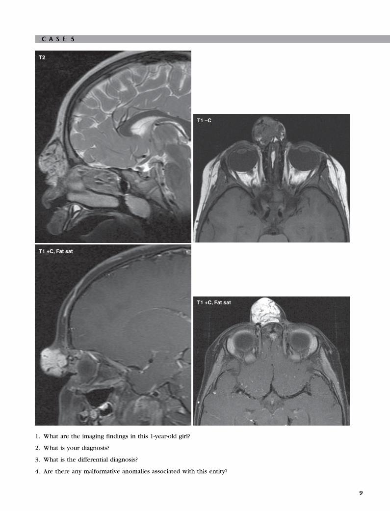
1. What are the imaging findings in this 1-year-old girl?
2. What is your diagnosis?
3. What is the differential diagnosis?
4. Are there any malformative anomalies associated with this entity?
9

A N S W E R S
C A S E 5
Diagnosis: Nasal Hemangioma
1. The image shows a well-circumscribed lobular mass,
with increased T2 signal and avid contrastenhancement noted in the subcutaneous fat of the
nasal bridge. There are vascular flow voids within the
mass seen best on T2-weighted image. No evidence
of intracranial extension exists.
2. The patient has nasal hemangioma.
3. In general, vascular malformations such as venous
malformations, lymphatic malformations, andarteriovenous malformation (AVM) can be
considered. Differential diagnosis would be affected
by the location as well. In this case encephalocele,
nasal dermoid, and nasal glioma could also be
considered, but the previously defined imaging
features lead to a correct diagnosis.
4. Yes, the malformative anomalies include PHACES
syndrome (posterior fossa malformations,
hemangiomas, arterial abnormalities, coarctation of
the aorta, and eye abnormalities); Dandy-Walker
malformation; spine anomalies if located in the
lumbar region; and Kasabach-Merritt syndrome.
ReferenceMulliken JB, Glowacki J: Hemangiomas and vascular
malformations in infants and children: a classification
based on endothelial characteristics, Plast Reconstr
Surg 69(3):412–422, 1982.
Cross-ReferenceBlickman JG, Parker BR, Barnes PD: Pediatric radiol-
ogy—the requisites, ed 3, Philadelphia, 2009, Mosby
Elsevier, pp 314–316.
CommentHemangiomas are the most common tumors in infancy
and childhood and account for 7% of benign soft tissue
tumors. Hemangiomas are tumors that express a loca-lized increase in angiogenic growth factors. Mulliken
and Glowacki first described the classification of vascu-
lar anomalies based on clinical, histologic, and cytologic
features. Vascular anomalies are divided into two major
groups: (1) vascular tumors (hemangiomas), and (2) vas-
cular malformations including venous malformations,
lymphatic malformations, AVMs, and arteriovenous fis-
tulas. The correct classification is important becausetreatment options differ significantly between the two
groups. Hemangiomas have cellular proliferation of
endothelial cells, and they are true neoplasms. Heman-
giomas have subgroups consisting of infantile, con-
genital, noninvoluting, intramuscular, and kaposiform
hemangioendothelioma types. The most common type
10
is infantile hemangioma. Hemangiomas test positive for
immunological markers such as glut1, FcrII, merosin,
and Lewis Y antigen. A 3:1 female-to-male ratio exists.
Hemangiomas usually appear in the first week of life
and can be located in the head and neck (60%), the
trunk (25%), and the extremities. The patient displaysa typical history of rapid neonatal growth (3 to 9
months) and slow involution characterized by hypercel-
lularity during the proliferating phase, as well as fibrosis
and diminished cellularity during the involuting phase
(18 months to 10 years). This typical clinical history is
oftentimes diagnostic; however, certain cases would
require imaging confirmation.
Imaging findings would obviously depend on thephase of the hemangioma. In the rapid-growth phase,
ultrasound would reveal a variable echogenicity mass
with increased flow on color-coded Doppler sonogra-
phy. Involuting hemangiomas are heterogeneous masses
with less intense color flow and fibrofatty changes.
A magnetic resonance image (MRI) is advised with T1,
T2, and postcontrast T1-weighted images. Fat suppres-
sion is of additional value in identifying this lesion. OnMRI, typically a parenchymal, well-circumscribed mass
with intermediate signal intensity on T1 and increased
signal intensity on T2-weighted images is visualized.
Avid contrast uptake is also noted, and the presence of
flow voids within and around the soft tissue mass is an
important feature.
In the majority of cases, no treatment is required
because of spontaneous involution. Indications for treat-ment in the remainder of the cases are primarily func-
tional, such as obscuration of vision or breathing or
persistent cutaneous ulceration, high cardiac output fail-
ure. Systemic or intralesional steroid agents can be used
for treatment. Promising results with systemic propran-
olol admission has been reported.

C A S E 6
1. What are the imaging findings on the ultrasound examination?
2. What is the most likely diagnosis in this 14-year-old girl?
3. What is the next test that should be performed?
4. Which long-term complication develops frequently?
11

A N S W E R S
C A S E 6
Diagnosis: Hashimoto Thyroiditis
1. Diffusely heterogeneous, hyperperfused thyroid gland
2. Hashimoto thyroiditis
3. Blood workup with determination of thyroid
hormones and thyroid antibodies
4. Hypothyroidism
ReferenceLorini R, Gastaldi R, Traggiai C, et al: Hashimoto’s thy-
roiditis, Pediatr Endocrinol Rev 1(Suppl 2):205,
2003.
Cross-ReferenceBlickman JG, Parker BR, Barnes PD: Pediatric radiol-
ogy—the requisites, ed 3, Philadelphia, 2009, Mosby,
p 324.
CommentHashimoto thyroiditis, also known as autoimmune thy-
roiditis or chronic lymphocytic thyroiditis, is the most
common acquired disorder of the thyroid gland in
children and the most common cause of hypothy-
roidism in the United States. Dr. Hakaru Hashimoto first
described this disorder in 1912. Hashimoto thyroiditis isan autoimmune disease of unknown cause and may
finally destroy the thyroid gland with resultant hypothy-
roidism. Incidence is estimated to be 1.3%. Blood tests
are mandatory to determine the thyroid function by
detecting the levels of thyroid hormones in general
and to identify antithyroid peroxidase antibodies or
antithyroglobulin antibodies in particular. Currently no
cure exists for Hashimoto thyroiditis; however, thehypothyroidism can be treated effectively by substitut-
ing the deficient thyroid hormones. Symptoms are usu-
ally related to the progressive drop in thyroid
hormones and not specific for Hashimoto thyroiditis.
Hashimoto thyroiditis is more frequent in females and
increases in frequency over age during childhood and
adolescence.
Ultrasound examination is the primary imagingmodality of choice. On ultrasound, the thyroid gland is
usually diffusely enlarged, hypoechoic, and reveals sig-
nificant hyperperfusion on color-coded Doppler sonog-
raphy and power Doppler sonography, especially in
the acute phase. Micronodulation characterized by mul-
tiple, small hypoechoic micronodules ranging between
1 and 6 mm in diameter are considered to be indicative
of Hashimoto thyroiditis. Occasionally, discrete focalnodules are identified and fine needle aspiration cytol-
ogy is necessary to confirm diagnosis. Hashimoto thy-
roiditis appears to occur more frequently in diabetic
12
patients. Ultrasonography is also helpful to rule out
additional lesions. Scintigraphy is rarely necessary to
confirm diagnosis.

C A S E 7
1. This patient had these three sequential films (first, second, and third figures) within a 24-hour period. What patient
group accounts for the largest proportion of these cases?
2. What plain film finding may precede the development of this abnormality?
3. What complications do you look for?
4. What lung pathologic conditions promote this abnormality?
13

A N S W E R S
C A S E 7
Diagnosis: Pulmonary InterstitialEmphysema
1. Premature infants (however, it can happen in any
patient in whom airway pressures exceed the
integrity of the airway epithelium).
2. Hyperinflation with endotracheal tube in place.
3. Pneumatoceles, pneumothorax,pneumomediastinum, pneumoperitoneum.
4. Surfactant deficiency, neonatal pneumonia,
meconium aspiration, persistent pulmonaryhypertension.
ReferenceKuhn JP, Slovis TL, Haller JO: Caffey’s pediatric diag-
nostic imaging, ed 10, Philadelphia, 2004, Mosby,
p 814.
Cross-ReferenceBlickman JG, Parker BR, Barnes PD: Pediatric radiol-
ogy—the requisites, ed 3, Philadelphia, 2009, Mosby,
pp 28–29.
CommentThe lungs of a premature infant are stiff and noncompli-
ant. This is due to lack of surfactant, normally produced
by type II alveolar cells, beginning in week 24 of gesta-tion and usually complete by week 32. Without this
protein, alveolar distension and adequate oxygenation
must be maintained by high-pressure ventilation with
oxygen-rich air. This combination injures the already
fragile alveolar walls and allows air to leak between
the lining cells into the interstitial spaces and lympha-
tics. The air is first visible lying parallel to the bronchi,
forming characteristic lucent lines and dots. Movementof the lungs with respiration causes the air to change
configuration. It can dissect centrally or peripherally,
causing pneumomediastinum and pneumothorax, or it
can become centrally confluent, creating a pneumatocele.
Air in the mediastinum can dissect downward through
the inferior pulmonary ligaments and result in sterile
pneumoperitoneum.
Aggressive treatment with exogenous surfactant inthe delivery room can decrease the severity of the lung
disease and avert this complication. Once present, it is
treated by decreasing ventilatory pressures. Jet ventila-
tion allows greater control of airway pressures. If unilat-
eral, decubitus positioning onto the affected lung can
effectively decrease pressure locally and hasten healing.
Pneumatoceles occasionally must be surgically removed
if they become intractably large.Chest radiographs, which are a valuable monitor for
this disease, can alert clinicians to dangerous levels of
14
hyperinflation that could precipitate air leaks, enabling
diagnosis of the emphysema and its complications and
determining efficacy of therapeutic measures.

C A S E 8
1. What are the findings in this neonate’s radiograph?
2. Before what gestational age do you expect to find this entity?
3. Under what conditions might you find it outside the usual age group?
4. What condition can mimic this entity and has very different treatment?
C A S E 9
1. What is the most common cause of this appearance in an infant or toddler?
2. What organism (or organisms) commonly causes this illness?
3. In older children, this appearance is more likely from what?
4. What is the normal lung volume in a quiet infant?
15

A N S W E R S
C A S E 8
Diagnosis: Surfactant Deficiency Disease
1. Moderate lung inflation better on the left, hazy
granularity more evident on the right, normal heartsize.
2. 32 weeks.
3. Seen more often in male patients (2:1). (The
presence of maternal diabetes, maternal or fetal
hemorrhage, sepsis, and multiple gestations further
increases the likelihood of developing this
condition.)
4. Group B streptococcal pneumonia.
ReferencesKuhn JP, Slovis TL, Haller JO: Caffey’s pediatric diag-
nostic imaging, ed 10, Philadelphia, 2004, Mosby,
pp 77–79.
Donoghue V: Radiologic imaging of the neonatal chest,
ed 2, Berlin-Heidelberg-New York, 2008, Springer,
pp 67–72.
Cross-ReferenceBlickman JG, Parker BR, Barnes PD: Pediatric radiol-
ogy—the requisites, ed 3, Philadelphia, 2009, Mosby,
pp 26–30.
CommentSurfactant is manufactured by type II alveolar cells,
beginning around week 24 of gestation and peaking at
about week 32. This compound lowers the surface ten-
sion of alveolar epithelium and prevents alveolar col-lapse. In its absence, the infant’s lungs fail to aerate
and appear uniformly white on radiography. The stan-
dard delivery room treatment is intubation and trache-
ally administered synthetic surfactant, which is then
distributed throughout the lungs by hand bagging.
If distribution is uniform, the lungs inflate and the radio-
graphic appearance changes to one of diffuse granular-
ity, which may look very fine, almost hazy in infantsless severely affected, or more dense and coarse. Non-
uniform distribution (e.g., if intubation was too deep
and bypassed a lobe or an entire lung) would give a
more uneven picture of hyperinflated areas and persis-
tently dense areas. Additional doses of surfactant may
be given over the first few days of life but with caution
(because this can promote pulmonary hemorrhage).
The surfactant is given in the hope that by the time itbreaks down (in 24 to 72 hours), the infant will be pro-
ducing sufficient endogenous surfactant to keep the
alveoli open.
16
C A S E 9
Diagnosis: Bronchiolitis
1. Bronchiolitis.
2. Respiratory syncytial virus and, much less
commonly, adenoviruses.
3. Reactive airway disease.
4. Tidal volume is the maximum volume during anormal inspiration. An infant, of course, cannot
cooperate for a film taken in deep inspiration.
Therefore the films of a normal infant breathing
quietly will often appear to be of small volume.
ReferenceSchuh S, Lalani A, Allen U, et al: Evaluation of the utility
of radiography in acute bronchiolitis, J Pediatr 150(4):429–433, 2007.
Cross-ReferenceBlickman JG, Parker BR, Barnes PD: Pediatric radiol-
ogy—the requisites, ed 3, Philadelphia, 2009, Mosby,
pp 32–36.
CommentsBronchiolitis is a viral respiratory illness that affects the
smaller airways of infants and toddlers. It results in
fever, congestion, wheezing, and diffuse lower respira-
tory tract signs. The disease is self-limited and usually
managed on an outpatient basis.
Chest radiographs, as in this example, usually show
hyperinflation and areas of atelectasis. During the ill-
ness, the location of atelectasis often changes rapidly.These findings are manifestations of the inflammatory
changes in the small airways. The airways are quite flex-
ible in this age group and can collapse partially or
completely during expiration, resulting in air trapping
and hyperinflation and/or atelectasis.
Similar radiographic findings are seen in reactive air-
way disease. In general, bronchiolitis is seen in the
infant, and reactive airway disease is observed in theolder child; however, an overlap occurs in their
incidences.

C A S E 1 0
1. This example is an Aunt Minnie. What is the disease process in this 11-year-old girl?
2. What are some nonpulmonary manifestations of this disease?
3. What laboratory test is used to make the diagnosis?
4. What is the prognosis of this disease?
17

A N S W E R S
C A S E 1 0
Diagnosis: Cystic Fibrosis
1. Cystic fibrosis (CF).
2. Pancreatic insufficiency, malabsorption, failure to
thrive, liver cirrhosis, infertility in males.
3. A sweat chloride test.
4. Mild examples of the disease exist; however, themedian age of survival is 36.8 years. Most patients die
from respiratory failure.
ReferenceRowe SM, Miller S, Sorscher EJ: Cystic fibrosis, N Engl J
Med 352(19):1992–2001, 2005.
Cross-ReferenceBlickman JG, Parker BR, Barnes PD: Pediatric radiol-
ogy—the requisites, ed 3, Philadelphia, 2009, Mosby,
pp 36–37.
CommentsCF is an inherited, autosomal recessive disorder of exo-
crine gland function. The most common manifestations
of CF are chronic respiratory infection and pancreaticenzyme insufficiency. CF is caused by defects in the
gene for cystic fibrosis transmembrane conductance
regulator (CFTR), which results in decreased secretions
of chloride, increased reabsorption of sodium and water
across epithelial cells, and ultimately to abnormal sticky
mucous production. This viscous mucous is less effec-
tive in clearing secretions and results in a lung environ-
ment conducive to infection, as well as the othermanifestations of this disease. CF is diagnosed by an
abnormal sweat chloride test. The sweat is analyzed
for its chloride content—the chloride will be high in
CF. Specific gene typing is useful in further characteriz-
ing the disease, because more than 1400 types of muta-
tions have been found in the CFTR gene.
The radiographs shown here reveal the typical find-
ings of hyperinflation, diffuse interstitial thickening,tram tracking of bronchiectasis, and mucous plugging.
Although diffuse disease is usual, focal infiltrates and/or
focal areas of more severe disease are common. As in
this case, hilar enlargement because of lymphadenopa-
thy (a manifestation of chronic infection) often is seen.
Computed tomography (CT) examination generally shows
more involvement than evident on chest radiographs. CT
findings include, but are not limited to, peribronchialthickening, centrilobular opacity, bronchiectasis, mucous
plugging, hilar adenopathy, bullae formation, and lung
abscess.
The radiographic findings are nearly pathognomic for
CF; hence the use of the term Aunt Minnie in this case.
Rarely, other causes of chronic inflammatory disease
18
may resemble CR; these include asthma and immunode-
ficiency syndromes.
Hemoptysis is common in advanced cases, and arteri-
ography and direct embolization of the bronchial
arteries may be necessary to treat it.

C A S E 1 1
1. What are the findings in this teenage boy?
2. What is the differential diagnosis?
3. What is the classification system for Hodgkin disease?
4. Is 18F-fluorodeoxyglucose (18F-FDG)-positron emission tomography (PET) computed tomography (CT) imaging
useful in lymphoma?
19

A N S W E R S
C A S E 1 1
Diagnosis: Hodgkin Lymphoma
1. Lymphadenopathy in the mediastinum and the right
hilum; splenomegaly with large hilar splenule;subsequent PET-CT showed uptake in multiple
lymph node areas and in the bone.
2. Lymphoma, tuberculosis or other granulomatous
infectious process, Langerhans histiocytosis, ormetastatic disease.
3. Stage I: One lymph node region involved (e.g., the
right neck or right axilla or mediastinum).Stage II: Involvement of two lymph nodes on same
side of diaphragm (e.g., both sides of neck).
Stage III: Lymph node involvement on both sides of
diaphragm (e.g., groin and armpit).
Stage IV: Involves the spread of cancer outside the
lymph nodes (e.g., to bone marrow, lungs, liver).
4. PET scans may show more disease than recognized
on CT alone and may allow distinction between
residual fibrotic mass versus mass with viable tumor
after treatment.
ReferenceOlson MR, Donaldson SS: Treatment of pediatric Hodg-
kin lymphoma, Curr Treat Options Oncol 9:81–94,
2008.
Cross-ReferenceBlickman JG, Parker BR, Barnes PD: Pediatric radiol-
ogy—the requisites, ed 3, Philadelphia, 2009, Mosby,
pp 41–43.
CommentsThis 16-year-old boy, whose images are shown in this
case, presented with a several-month history of fever,
night sweats, fatigue, bone pain, and weight loss. He
was found to have pancytopenia. Lymph node biopsy
showed classical Hodgkin lymphoma, and he was staged
as IV B because of apparent bone marrow involvementseen on PET scanning (bone scan and bone radiographs
were negative).
Hodgkin disease is a common hematological malig-
nancy that has two peaks of incidence: (1) between 15
and 35 years and (2) older than 55 years. The commonly
used staging system (cited previously) is the Ann Arbor
Staging System. In addition to the areas of involvement,
bulky mediastinal disease involving more than one thirdof the intrathoracic diameter is also taken into consider-
ation. Varying definitions of low, intermediate, and high-
risk groups include all of these factors, as well as the
histology.
The World Health Organization classification sepa-
rates Hodgkin lymphoma into two groups: (1) classical
20
(which includes lymphocyte-depleted, nodular-scleros-
ing, mixed cellularity, and classical lymphocyte-rich cell
types) and (2) the lymphocyte-predominant type.
Ninety percent of Hodgkin lymphoma is of the classical
type, with Reed-Sternberg cells, and positive for CD15
and CD30 (CD stands for cluster of differentiation andis a protocol used to identify and investigate the cell sur-
face molecules present on leukocytes). Immunohistolo-
gical differences are seen between the subgroups of
classical Hodgkin lymphoma, but the response to treat-
ment is similar. Lymphocyte-predominant Hodgkin lym-
phoma expresses markers not usually seen in the
classical type; those are B cell markers (CD20, CD79a,
CD75) and epithelial membrane antigen and lympho-cyte marker (CD45). This type of Hodgkin disease has
a more indolent course and good prognosis with treat-
ment, but the patient has a slightly higher risk of devel-
oping non-Hodgkin lymphoma.

C A S E 1 2
1. What are the findings in this teenage boy?
2. What is the differential?
3. Does the calcification narrow the differential?
4. Is local therapy effective in this process?
21

A N S W E R S
C A S E 1 2
Diagnosis: Pulmonary NodulesDue to Osteosarcoma
1. Multiple variable-sized discrete nodules appear in the
lungs. A large mass on the left appears to invade the
mediastinum, and it contains calcification.
2. Metastatic disease most likely; fungal infection, other
multifocal infection, septic emboli, Wegener
granulomatosis, and Langerhans histiocytosis much
less likely.
3. Yes, it makes calcium-producing neoplasm such as
osteosarcoma more likely.
4. After initial chemotherapy, resection of multiple lung
metastatic lesions in a patient with osteosarcoma is
effective and results in longer disease-free survival.
ReferenceAntunes M, Benardo J, Salete M, et al: Excision of pulmo-
nary metastases of osteogenic sarcoma of the limbs,Eur J Cardiothorac Surg 15(5):592–596, 1999.
Cross-ReferenceBlickman JG, Parker BR, Barnes PD: Pediatric radiol-
ogy—the requisites, ed 3, Philadelphia, 2009, Mosby,
pp 37–38.
CommentNodular lung disease consists of multiple round opacities
that can range from 1 mm to 1 cm or larger. The smaller
lesions are only visible on computed tomography exami-
nation and are referred to as miliary. Further characteriza-
tion of the nodules includes a description of their
margins (smooth or irregular), presence or absence of
cavitation, calcification, and their distribution.
Multiple lesions of different sizes, especially in sub-pleural or peripheral locations, suggest metastatic dis-
ease. Multiple small smooth or irregularly marginated
nodules in a perilymphatic distribution suggest sarcoido-
sis. Silicosis and coal workers’ pneumoconiosis may also
have this appearance. Upper lobe predominance is seen
in coal workers’ pneumoconiosis. Small nodules of
ground-glass opacity can be seen in extrinsic allergic
alveolitis or bronchiolitis. Miliary or larger nodules areseen in the hematogenous spread of tuberculosis, fungal
infection, or metastatic disease. If the lesions include
some with thin-walled cavities, Langerhans cell histiocy-
tosis should be considered. Other lesions that cavitate
include metastatic squamous cell carcinoma, Wegener
granulomatosis, rheumatoid lung disease, septic emboli,
and multifocal infection. Lymphoproliferative disorders,
lymphoma, leukemia, and Kaposi sarcoma may causeirregular nodules in a bronchovascular distribution. Cal-
cification can be seen in granulomatous disease,
22
hamartomas, metastatic tumor such as osteosarcoma,
rarely as a consequence of infection and in abnormal
calcium metabolism, with so-called metastatic pulmo-
nary calcifications.
The patient in this example had osteosarcoma of the
proximal femur with lung metastases at presentation 5years earlier. By the time of these images, the patient
had multiple tiny lesions (>100 total) resected from
each lung. Chemotherapy is the initial treatment of
nonmetastatic and metastatic osteosarcoma. Surgical
resection of the primary lesion generally follows chemo-
therapy. Experimental administration of the bone-
seeking radioisotope samarium (if the lesions are
calcified and avid on nuclear bone scan) has recentlybeen used to treat metastatic disease in osteosarcoma.
The outcome in patients with lung metastases from oste-
osarcoma is improved by the combination of chemother-
apy and surgery. A strong correlation exists between the
degree of necrosis of the aggregate metastatic disease
and the need for reoperation. In one study, those that
needed reoperation had less than 80% necrosis of
metastases.

C A S E 1 3
1. These three computerized tomography (CT) images represent the progression of a disease process in a teenagerbeing treated for leukemia. What do they show?
2. What is the differential diagnosis?
3. What is the name of the finding seen on the last CT image?
4. In what sort of patient is this likely to occur?
23

A N S W E R S
C A S E 1 3
Diagnosis: Fungal Disease Lungs
1. A small nodule progresses to a larger nodule with a
hazy border; then cavitation is seen with a centralsoft tissue component.
2. Fungal mycetoma, abscess, metastatic disease, septic
embolus, hematoma, and hydatid disease.
3. Air crescent sign.
4. This process is often seen in an
immunocompromised patient.
ReferenceDemirkazik FB, Akin A, Uzun O, et al: CT findings in
immunocompromised patients with pulmonary infec-
tions, Diagn Interv Radiol 14:75–82, 2008.
Cross-ReferenceBlickman JG, Parker BR, Barnes PD: Pediatric radiol-
ogy—the requisites, ed 3, Philadelphia, 2009, Mosby,
p 34.
CommentCT of the lung is used to detect infection in the immu-nocompromised patient. It is considerably more sensi-
tive to early infection than chest radiography. In
patients with febrile neutropenia, it is reported that a
CT will find 20% more pneumonias 5 days earlier than
the chest radiograph. In addition to increasing the
detection rate, a careful analysis of the CT features can
help to determine the likely cause of the infection.
The patient in this example had invasive aspergillosis.The typical findings include single or multiple nodules,
often with surrounding ground-glass opacity, the so-
called halo sign. A lesion may cavitate and contain a solid
nodule within it. This is called the air crescent sign.
These features were all present in the patient.
The halo sign is seen in about half of patients with
invasive aspergillosis and is caused by hemorrhage and
necrosis around the necrotic nodule, which containsthe fungal hyphae. This sign can also be seen in some
bronchopneumonia, as well as tumors such as adenocar-
cinoma, alveolar cell carcinoma, Kaposi sarcoma, and
metastasis. About 40% of nodules seen in fungal pneu-
monia are cavitary, and one half of them demonstrate
the air crescent sign. This finding is not specific for inva-
sive Aspergillus spp.; however, in the proper setting, it
is highly suggestive for fungal disease (clinicians believeit to be caused by retracted and infarcted lung tissue
within the cavity).
Although nodular disease is the most frequent mani-
festation of Aspergillus spp. infection on CT, ground-
glass opacity and consolidation can also be seen.
24
Other fungal infections are less common; increasingly
pulmonary candidiasis is the cause of fever in the immu-
nocompromised patient and its appearance is similar to
Aspergillus spp.

C A S E 1 4
1. What are the imaging findings in this neonate with respiratory distress?
2. What is the differential diagnosis?
3. What are the possible complications?
4. What are the predisposing factors?
25

A N S W E R S
C A S E 1 4
Diagnosis: Neonatal Pneumonia
1. Frontal radiograph of the chest demonstrates
bilateral patchy interstitial markings in minimallyhyperexpanded lungs. Of note, an umbilical artery
catheter is seen, with the tip projecting over the T3/4
interspace.
2. Differential diagnosis of these findings includessurfactant deficiency disease, neonatal pneumonia,
transient tachypnea of the newborn, and meconium
aspiration.
3. Complications of neonatal pneumonia include
pleural effusion, empyema, abscess, and
pneumatocele formation.
4. Factors that predispose to neonatal pneumoniainclude prolonged premature rupture of membranes,
maternal ascending infection, placental infection,
and perineal contamination.
ReferenceSwischuk LE: Imaging of the newborn, infant and
young child, ed 5, Philadelphia, 2004, Lippincott
Williams & Wilkins, pp 43–46.
Cross-ReferenceBlickman JG, Parker BR, Barnes PD: Pediatric radiol-
ogy—the requisites, ed 3, Philadelphia, 2009, Mosby,
pp 30–31.
CommentsNeonatal pneumonia is most often bacterial (Streptococ-cus, Staphylococcus aureus, and Escherichia coli) but
may be viral (adenovirus, herpes simplex, influenza,
and parainfluenza). Nonspecific findings are seen on
chest radiograph, with hyperexpansion and diffuse
increase in interstitial markings most common. Perihilar
streaky densities and diffuse haziness favors viral infec-
tion. Coarse, patchy parenchymal infiltrates favor bacte-
rial infection. Frank consolidation is rare in neonates.Pleural effusion may accompany bacterial pneumonia.
Infection with group B streptococci may mimic surfac-
tant deficiency disease with granularity, perhaps more
prominent in the lower lobes.
Most commonly, the infant is infected in utero or dur-
ing passage through the birth canal. Infants typically
present with respiratory distress in the first 48 hours
of life. Predisposing factors include prolonged prema-ture rupture of the membranes, ascending infection
from the vagina, placental infection, and contamination
from a poorly prepared perineum or maternal fecal
material. Long-term complications include chronic lung
disease.
26
Delayed onset of right diaphragmatic hernia has been
recognized as a complication, especially with group B
streptococci infections. Other complications include
empyema, abscess, and pneumatoceles.

C A S E 1 5
Immediately after birth 2 days after birth
1. What are the imaging findings in this newborn on the first plain radiograph?
2. What is your diagnosis?
3. What is the differential diagnosis?
4. What are the predictors of morbidity and mortality?
27

A N S W E R S
C A S E 1 5
Diagnosis: Congenital Diaphragmatic Hernia
1. The plain chest radiograph obtained immediately
after birth shows complete opacification of the lefthemithorax, mediastinal shift to the right, and
nonvisualization of the left hemidiaphragm. The
plain radiograph of the chest obtained on the second
day of life shows interval aeration of the fluid-filled
bowel loops in the left chest cavity. Bowel in the
chest, mediastinal shift, nonvisualized diaphragm,
position of nasogastric (NG) tube (in cases with
herniation of the stomach) are all helpful plainradiograph findings for diagnosis. T1-weighted
coronal image of the same patient obtained during
the third trimester shows herniation of the bowel
loops. T1 signal of the bowel lumen is from the
meconium content.
2. Congenital diaphragmatic hernia (CDH): Bochdalek
type.
3. Congenital cystic adenomatoid malformation,
congenital lobar emphysema, and pneumonia
complicated by cavitary necrosis.
4. Degree of pulmonary hypoplasia, pulmonaryhypertension, size of the hernia, presence of liver
and spleen in the chest.
ReferenceJohnson AM: Congenital anomalies of the fetal/neonatal
chest, Semin Roentgenol 39(2):197–214, 2004.
Cross-ReferenceBlickman JG, Parker BR, Barnes PD: Pediatric radiol-
ogy—the requisites, ed 3, Philadelphia, 2009, Mosby,
pp 21–22.
CommentCDH occurs in 1 of every 2000 to 3000 live births and
accounts for 8% of all major congenital anomalies.
CDH can be described as cases in which abdominal con-tents are herniated into the chest. The three basic types
of CDH include (1) the posterolateral Bochdalek hernia
(occurring at approximately 6 weeks gestation), (2)
the anterior Morgagni hernia, and (3) the hiatus hernia.
The left-sided Bochdalek hernia occurs in approximately
85% of cases. Bochdalek hernia is more common on the
left than on the right (5:1). The hernia may have variable
abdominal contents: stomach, small and/or large bowel,liver, and spleen. Most common signs and symptoms
include severe respiratory distress. This type of hernia
typically presents at or soon after birth and is often
detected at prenatal imaging. Less severe cases may
present later in life or incidentally on radiograph. Large
CDH results in pulmonary hypoplasia and pulmonary
28
hypertension. Prognosis is primarily related to the
degree of lung hypoplasia and pulmonary hypertension.
Fetal magnetic resonance image (MRI) and fetal ultra-
sound have provided valuable information about lung
volume, which is an important prognostic indicator in
predicting pulmonary capacity after birth. Overall thesurvival rate is about 50%. Prenatal diagnosis, supportive
care for pulmonary hypoplasia and respiratory failure,
and use of extracorporeal membrane oxygenation
(ECMO) resulted in improved survival rates. In utero
surgical repair is an option when in utero diagnosis is
made with ultrasound and MRI. Early diagnosis is crucial
in guiding the mode of delivery (ex utero intrapartum
treatment [EXIT] procedure) and immediate postnatalcare. Surgical outcome is still variable. Up to one third
of the cases have associated major malformations. Malro-
tation or stomach volvulus can be seen as a gastrointes-
tinal tract abnormality, and 50% of the cases may have
congenital heart disease.

C A S E 1 6
1. What are the imaging findings in this 3-year-old girl with persistent fever, cough, and malaise?
2. What is your diagnosis?
3. What is the differential diagnosis?
4. What is the most common microorganism?
29

A N S W E R S
C A S E 1 6
Diagnosis: Lung Abscess
1. Round large mass with air fluid level in the left lower
lobe, surrounded by normal lung parenchyma.
2. Lung abscess.
3. Primary versus secondary abscess: secondary to a
congenital lesion such as a bronchogenic cyst,
congenital cystic adenomatoid malformation, or
pulmonary sequestration (in this particular case, the
plain radiograph also includes diaphragmatic hernia
[e.g., stomach in the chest cavity]).
4. The main causative organisms are usually
streptococcal and anaerobic species, Staphylococcus
aureus and Klebsiella spp.
ReferencePuligandla P, Laberge JM: Respiratory infections: pneu-
monia, lung abscess and empyema, Semin Pediatr
Surg 17(1):42–52, 2008.
Cross-ReferenceBlickman JG, Parker BR, Barnes PD: Pediatric radiol-
ogy—the requisites, ed 3, Philadelphia, 2009, Mosby,pp 32–126.
CommentA lung abscess develops when a localized area of paren-
chymal infection becomes necrotic and then cavitates.
Primary lung abscesses occur in healthy children with-
out lung abnormalities, whereas secondary abscesses
occur in children with underlying lung disease thatmay be either congenital (e.g., cystic lung lesion) or
acquired (e.g., cystic fibrosis, immunodeficiency). Aspi-
ration may play a significant role, especially in children
with neurodevelopmental delay or immune deficiency.
Clinically, abscesses may develop indolently over a few
weeks with tachypnea, cough, and fever being the most
common symptoms.
Radiologic evaluation starts with plain films. Ultra-sound has the benefit of avoiding radiation exposure;
however, it may be less diagnostic in older children,
especially in deeper locations. Computed tomography
(CT) of the chest helps identifying underlying lung
lesions predisposing to the development of the abscess
such as a bronchogenic cyst, congenital cystic adenoma-
toid malformation, or pulmonary sequestration.
Up to 90% of patients with lung abscess may be ade-quately treated with intravenous antibiotic therapy. The
duration of parenteral treatment varies from as little as 5
days to as long as 3 weeks; oral therapy may be required
after intravenous medications have been stopped. Ulti-
mately, antibiotic therapy should be tailored to the
organisms present. Ultrasound- or CT-guided aspiration
30
may be indicated for diagnosis and treatment. The over-
all outcome of children with a lung abscess is quite
good, with mortality rates much lower than those of
adults and mostly occurring in children with secondary
lung abscesses or underlying medical problems.

C A S E 1 7
1. What are the imaging findings in this 3-year-old boy with cough and fever?
2. What is your diagnosis?
3. What is the differential diagnosis?
4. What is the cause?
31

A N S W E R S
C A S E 1 7
Diagnosis: Round Pneumonia
1. Round well-demarcated opacity in the left lower
lobe; patient had previous cardiac surgery andosteopenia.
2. Round pneumonia.
3. Bronchogenic cyst, neuroblastoma, congenital cystic
adenomatoid malformation (CCAM), pulmonary
sequestration.
4. Round pneumonia most commonly seen with
Streptococcus pneumoniae infection.
ReferenceKim YW, Donnelly LF: Round pneumonia: imaging find-
ings in a large series of children, Pediatr Radiol 37(12):1235–1240, 2007.
Cross-ReferenceBlickman JG, Parker BR, Barnes PD: Pediatric radiol-
ogy—the requisites, ed 3, Philadelphia, 2009, Mosby,
p 32.
CommentRound pneumonia is bacterial in origin, with a very
round, well-defined appearance on chest radiography,
simulating a mass. It respects the lobar anatomy without
crossing the fissures. Round pneumonia is seen in chil-
dren younger than 8 years of age and is commonly seen
in the lower lungs.
Clinical presentation is fever and cough, general mal-
aise, and may present with abdominal pain. If a child hassymptoms of pneumonia and round density is seen on
the chest radiograph, additional imaging is not required.
In children, collateral pathways or air circulation (chan-
nels of Lambert and pores of Kohn) are not well devel-
oped until 8 years old. This lack of well-developed
collateral circulation is thought to hinder spread of bac-
terial infection and predispose to the round appearance.
If computed tomography is performed, air broncho-grams can be seen. Magnetic resonance imaging is not
necessary in the diagnostic workup of round pneumonia.
Treatment of choice is with antibiotic agents. Round
pneumonia is one of the few indications in which a fol-
low-up chest radiograph is required after the child
becomes asymptomatic to exclude an underlying mass.
32

C A S E 1 8
1. What are the imaging findings in this newborn with a large lateral chest wall and abdominal wall mass?
2. What is your diagnosis?
3. What is the differential diagnosis?
4. Does this entity regress spontaneously?
33

A N S W E R S
C A S E 1 8
Diagnosis: Venolymphatic Malformation
1. Plain film reveals a large subcutaneous soft tissue
mass extending from upper chest down to the pelvis.Ultrasound shows a large complex cystic mass with
internal echoes. Magnetic resonance imaging (MRI)
reveals a large lobular, septated mass in the left
lateral chest wall, extending from the chest to the
left lateral abdomen. The caudal part of the mass is
precontrast T1 bright (either from high-protein
content or posthemorrhage), and the superior aspect
of the mass is T1 hypointense, revealing serpiginousareas on T2 that show avid contrast enhancement.
The caudal part represents the macrocystic
lymphatic malformation component, and the cranial
part represents the venous malformation
component.
2. Venolymphatic malformation
3. Venous malformation or lymphatic malformationalone
4. No, vascular malformations never regress or involute
spontaneously and may grow at a rate greater than
normal somatic growth.
ReferencesLegiehn GM, Heran MK: Classification, diagnosis, and
interventional radiologic management of vascular mal-
formations, Orthop Clin North Am 37(3):435–474,
vii–viii, 2006.
Mulliken JB, Glowacki J: Hemangiomas and vascular
malformations in infants and children: a classificationbased on endothelial characteristics, Plast Reconstr
Surg 69(3):412–422, 1982.
Cross-ReferenceBlickman JG, Parker BR, Barnes PD: Pediatric radiol-
ogy—the requisites, ed 3, Philadelphia, 2009, Mosby,
pp 314–315.
CommentFew areas within medical diagnosis are fraught with as
many persistent misconceptions and misnomers as
within the group of vascular anomalies. In 1982 Mulliken
and Glowacki published a landmark article proposing
characterization of vascular anomalies based on biologic
and pathologic differences. It is most important to make
the differentiation between vascular tumors (heman-giomas) and vascular malformations, including venous
malformations, lymphatic malformations, capillary mal-
formation, mixed type of venolymphatic malformations,
arteriovenous malformations (AVMs), arteriovenous fis-
tulas, because the prognosis and treatment options differ
significantly between the two groups.
34
Vascular malformations are described as lesions pres-
ent at birth growing commensurately or pari passu
with the child, composed of vascular channels lined
with flat “mature” endothelium exhibiting normal rates
of endothelial cell turnover. Vascular malformations
are further subdivided based on their flow rate observedin angiograms: low-flow venous malformations and high-
flow AVMs, with a separate group categorization for
lymphatic malformations and hemangiomas. Vascular
malformations are commonly seen in the head and neck
region followed by trunk and extremities.
Lymphatic malformations have been called lymphan-
giomas in the past, which is a misnomer. They are multi-
cystic (can be micro-macrocystic or mixed); vascularchannels and spaces are separated by fibrous septa.
The lumen may contain lymphatic fluid or proteina-
ceous material. Ultrasound is the first line of imaging
in evaluation of vascular anomalies. Lymphatic malfor-
mations appear as cystic cavities with layering debris.
They can have bleeding or inflammatory changes in
these cysts. No flow is detectable within a lymphatic
vascular malformation. MRI is very helpful in definingthe extent of disease and further characterization. It
may reveal increased T1 signal secondary to bleeding,
protein content and inflammatory changes, show
increased T2 signal, and may show peripheral contrast
enhancement of the fibrous septa.
Vascular malformations may present as a combina-
tion of venous and lymphatic malformation (as in the
presented case). It is important to clearly identify theparts of this lesion as lymphatic and venous because
the used sclerotic agents differ.
Successful management requires an experienced mul-
tidisciplinary team allowing effective communication
and integration of the most current clinical, pathologic,
and image-based diagnosis and intervention.

C A S E 1 9
Deep inspiration Expiration
1. What is the principal finding comparing inspiration with expiration?
2. What is the most likely diagnosis?
3. Is this finding acute or chronic?
4. What to do next?
35

A N S W E R S
C A S E 1 9
Diagnosis: Aspirated Carrot in the Left MainBronchus
1. Air trapping in the right lung.
2. Nonradioopaque foreign body in the right main
bronchus.
3. Acute. No signs of infection and/or fluid retention
and/or chronic atelectasis is seen in the right lung.
4. Bronchoscopy with removal of the foreign body.
Cross-ReferenceBlickman JG, Parker BR, Barnes PD: Pediatric radiol-
ogy—the requisites, ed 3, Philadelphia, 2009, Mosby,
p 15.
CommentChildren who suffer from acute, unexplained shortness
of breath with wheezing should be suspected of a for-
eign body aspiration until proven otherwise. Especiallysmall children and babies who explore their surround-
ings by putting many objects in their mouths are at risk
for aspiration. Older children more frequently choke on
food, especially hard candies and peanuts. It is impor-
tant to be aware of the fact that most aspirated foreign
bodies are not radioopaque and are consequently not
visible on plain films.
By acquiring chest films in different degrees of inspi-ration (inspiration versus expiration), foreign bodies
may indirectly be proven and located. Depending on
the degree of obstruction, the lung distal to the foreign
body may be partially or totally atelectatic. However, a
ball valve effect may also result in a progressive hyperin-
flation of the lung distal to the obstruction increasing
with every breath taken. The progressive air trapping
may lead to a life-threatening mediastinal shift to thecontralateral side compromising the ventilation of the
contralateral lung. Imaging shows a hyperinflation of
the air-trapped lung on inspiration, which persists on
expiration. On expiration, however, the contralateral
lung will deflate. The mediastinal structures will shift
to the side of the nonobstructed lung.
The trachea and bronchi should also be studied in
detail; air within the upper airways may serve as a natu-ral contrast to a foreign body. In this particular case, the
air column within the right bronchus was amputated
just distal to the carina.
Aspirated foreign bodies are more frequently found
in the right bronchus than in the left bronchus. The
right bronchus is slightly larger than the left bronchus
and in a more favorable orientation in relation to the tra-
chea for aspiration than the left main bronchus.If the patient’s history and the clinical findings are
highly suggestive for a foreign body aspiration, most
36
children will go immediately into the bronchoscopy
suite. In all other instances, plain chest films in inspira-
tion and expiration should be performed. If the patient’s
compliance to follow instructions is limited, lateral
decubitus should be considered. The air-trapped lung
will not deflate in the dependent position (lateraldecubitus).
Please do not forget that a foreign body may also
have been expelled by an adequate coughing reflex
but has been swallowed subsequently. Consequently,
if a foreign body is not visible on chest films and the
clinical history is highly suggestive of a foreign body
aspiration, additional abdominal films should be
acquired. The gastrointestinal tract should be examinedfrom the mouth until the anus.

C A S E 2 0
1. What are the imaging findings on plain radiography?
2. What are the imaging findings on chest computed tomography (CT)?
3. What is the most likely diagnosis?
4. Which patient population is more at risk for infection?
37

A N S W E R S
C A S E 2 0
Diagnosis: Pulmonary Tuberculosis
1. Enlarged mediastinal contour, possibly the result of
enlarged lymph nodes.
2. Necrotic paratracheal lymph nodes, deformed right
upper lobe, and high-grade compression of left
mainstem bronchus.
3. Pulmonary tuberculosis.
4. Immunocompromised patients and young children.
ReferenceSantos JF: Tuberculosis in children, Eur J Radiol
55:202–208, 2005.
Cross-ReferenceBlickman JG, Parker BR, Barnes PD: Pediatric radiol-
ogy—the requisites, ed 3, Philadelphia, 2009, Mosby,
pp 33–34.
CommentPulmonary tuberculosis (TBC) results from the inhala-
tion of mycobacterium tuberculosis bacilli. A primary
complex or Ghon complex results from the multiplica-tion of TBC bacilli in one bronchiole or alveolus and
the host’s local inflammatory acute reaction. From this
initial lesion, the bacilli may spread into the regional
lymph nodes. Most frequently hilar lymph nodes are
involved; depending on the location of the primary
infection, paratracheal or subcarinal nodes may be
affected. The combination of a primary focus and adja-
cent calcified hilar lymph nodes are known as Rankecomplex. This primary complex may go undetected on
plain radiography. In most cases the intrapulmonary
lesion heals with frequently subtle signs of focal pulmo-
nary scarring or fibrosis. Rarely, the primary focus may
persist and evolve into a larger focal pneumonitis. The
affected lymph nodes may develop fibrosis or calcifica-
tions during the healing phase. TBC bacilli may, how-
ever, survive within the lymph nodes for many years.In rare cases the lymph nodes may enlarge so signifi-
cantly that they encroach or obstruct adjacent bronchi
with resulting pulmonary atelectasis. Affected caseous
lymph nodes may eventually erode bronchial walls
with resultant endobronchial TBC and a fistulous tract.
Depending on the location, fistulous tracts may extend
into the pericardium or esophagus.
In addition, TBC bacilli may spread from the primarycomplex though the lymphatic system or bloodstream
into almost any part of the body. Most frequently, the
upper lung lobes are affected next to the liver, spleen,
meninges, pleura, and bones. Massive lymphohe-
matogenous dissemination may result in miliary or
38
disseminated TBC. Young children are more at risk for
miliary TBC than older children or adults.
On imaging, a primary complex may be difficult to
detect. CT usually reveals enlarged lymph nodes with
or without caseation and/or calcification. CT is
extremely helpful to identify the degree of bronchialobstruction or other complications. The number of
newly diagnosed TBC infections is unfortunately again
on the rise. TBC should be included in the differential
diagnosis when enlarged, possibly calcified lymph
nodes are encountered.

C A S E 2 1
1. This 3-month-old infant presented with difficulty feeding. What other symptoms might the caregiver report?
2. What are the findings on upper gastrointestinal examination (first and second images)?
3. What examination should the clinician order next?
4. Why is one needed?
39

A N S W E R S
C A S E 2 1
Diagnosis: Double Aortic Arch
1. Stridor; normal initially, then with progression of
symptoms; choking on solid feeds but able toswallow liquids.
2. Lateral view: posterior compression on the
esophagus, anterior compression on the trachea;
anteroposterior view: “hourglass” compression ofthe esophagus.
3. Magnetic resonance image or magnetic resonance
angiography has no radiation dose, but sedation maybe dangerous if stridor is a prominent symptom.
Computed tomographic angiography (third image)
usually does not require sedation, but the radiation
dose is large.
4. One arch is usually smaller than the other arch, and
the surgeon generally will choose that side to divide.
ReferenceKirks DR, Griscom NT, editors: Practical pediatric
imaging, ed 3, Philadelphia, 1998, Lippincott-Raven,
pp 14–15 and pp 77–80.
Cross-ReferenceBlickman JG, Parker BR, Barnes PD: Pediatric radiol-
ogy—the requisites, ed 3, Philadelphia, 2009, Mosby,
pp 14–15 and pp 78–79.
CommentAlthough this is not the most common aortic arch anom-
aly (that would be left aortic arch and aberrant right sub-clavian artery), it is the most common vascular ring.
Persistence of both fetal aortic arches occurs, and each
gives off its own subclavian and carotid artery. The sin-
gle ascending aorta divides around and encloses the tra-
chea and esophagus, coming together posteriorly in a
single descending aorta. If the ring is tight, the child
might present with stridor at birth. Otherwise, symp-
toms may slowly develop as the child grows. Dysphagiais often the presenting symptom when the diet changes
to include solid food. The right arch is usually larger,
rises higher, and extends more posterior than the left
arch.
40

C A S E 2 2
1. What are the radiographic findings in this 1-month-old cyanotic infant?
2. What aspect of the radiograph is potentially confusing? What causes this?
3. What proportion of these patients has a right-sided aortic arch?
41

A N S W E R S
C A S E 2 2
Diagnosis: Tetralogy of Fallot
1. Cardiomegaly with broad, upturned silhouette; right-
sided aortic arch, concave pulmonary arterialsegment.
2. The appearance of increased pulmonary blood flow;
large aorticopulmonary collaterals replace
hypoplastic pulmonary artery system.
3. Twenty-five percent of patients with tetralogy of
Fallot (TOF).
ReferencePark MK: Pediatric cardiology for practitioners, ed 4,
St Louis, 2002, Mosby, pp 189–196.
Cross-ReferenceBlickman JG, Parker BR, Barnes PD: Pediatric radiol-
ogy—the requisites, ed 3, Philadelphia, 2009, Mosby,
pp 55–56.
CommentClinicians postulate that a single intrauterine event in
fetal life starts the chain of defects that causes TOF,
the most common congenital cyanotic heart defect. This
insult causes hypoplasia of the right ventricular (RV)
outflow tract (1). This in turn causes malalignment of
the membranous and muscular septa, leaving an open-ing, the ventricular septal defect (VSD) (2). In addition,
the aorta is drawn medially by the lack of supporting tis-
sue and ends up straddling the septum (3). As the RV is
exposed to systemic pressures by the VSD and is forced
to pump against the semiobstructed RV outflow tract, it
hypertrophies (4). Ten percent of patients have an asso-
ciated atrioventricular canal abnormality, whereas 15%
to 20% have pulmonary atresia.Palliation with shunts to improve pulmonary blood
flow (the Blalock-Taussig shunt was the first of its kind)
has given way to early definitive surgery to enlarge the
outflow tract and close the VSD. This eliminates the
complications of systemic shunting: excessive or
uneven blood flow and the development of pulmonary
hypertension. Complications of definitive repair include
aneurysm of the outflow tract patch, restenosis withdiminishing or asymmetric pulmonary blood flow, and
stenosis of repaired valves.
42

C A S E 2 3
1. This 17-month old girl presented with a 5-day history of periorbital and pedal edema, not tolerating feeding. Two
images from the upper gastrointestinal (GI) tract show what finding?
2. What eponym is associated with this finding?
3. What did the biopsy show?
4. How does the significance of this finding in children differ from that in adults?
C A S E 2 4
GB supine SAG
1. What are the imaging findings and most likely diagnosis?
2. List some predisposing factors.
3. What imaging studies should be performed with suspected gallbladder disease?
4. What are the possible complications of this disease?
43

A N S W E R S
C A S E 2 3
Diagnosis: Menetrier Disease(Giant Hypertrophic Gastritis)
1. Prominent, thickened gastric rugal folds.
2. Menetrier disease—named for French pathologist
Pierre Eugene Menetrier (1859 to 1935).
3. Hyperplastic mucosa with eosinophilic and
neutrophilic infiltration, occasional cells withcytomegalovirus (CMV) inclusion bodies.
4. In adults, the condition is chronic and increases the
chance of gastric cancer, whereas in children it ismost often self-limited and secondary to infection
(CMV most often, Campylobacter pylori is also
reported).
ReferenceStringer DA, Babyn PS: Pediatric gastrointestinal imag-
ing and intervention, ed 2, Hamilton-London, 2000,
BC Decker Inc, pp 286–287.
Cross-ReferenceBlickman JG, Parker BR, Barnes PD: Pediatric radiol-
ogy—the requisites, ed 3, Philadelphia, 2009, Mosby,
p 85.
CommentThis condition causes protein-losing enteropathy thatcan result in hypoalbuminemia—helping to distinguish
this disease from allergic gastritis, which can also cause
enlarged rugal folds. The soft tissue edema secondary to
the hypoalbuminemia may be subtle but should provide
the alert practitioner with the differential diagnosis.
Moreover, a viral prodrome often exists in the history.
44
C A S E 2 4
Diagnosis: Cholelithiasis
1. Sonographic images of the gallbladder demonstrate
mobile, shadowing, echogenic foci within thegallbladder consistent with gallstones.
2. Parenteral nutrition, sickle cell disease and other
hemolytic anemias, malabsorption syndromes
including short-gut syndrome, and cystic fibrosis.
3. Ultrasound is the imaging modality of choice in cases
of possible gallbladder disease. With the question of
acalculous cholecystitis, nuclearmedicine scan shouldbe performed to evaluate gallbladder function.
Magnetic resonance cholangiopancreatography is
also useful to further evaluate gallbladder disease.
4. Obstruction and infection (cholecystitis, cholangitis,pancreatitis).
ReferenceSiegel MJ, Coley BD: Pediatric imaging, Philadelphia,
2006, Lippincott Williams & Wilkins, pp 172–173.
Cross-ReferenceBlickman JG, Parker BR, Barnes PD: Pediatric radiol-
ogy—the requisites, ed 3, Philadelphia, 2009, Mosby,
pp 107–108.
CommentsGallstones are relatively rare in the pediatric population.
Common causes include furosemide therapy, malab-
sorption, total parenteral nutrition, Crohn disease, cys-
tic fibrosis, bowel resection, and hemolytic anemiasuch as sickle cell disease. Patients present with colicky
right upper quadrant pain. Ultrasound is the imaging of
choice and will demonstrate intraluminal echogenic sha-
dowing structures within the gallbladder that may move
with change in patient positioning. Differential diagno-
sis would also include gallbladder polyp and cholesterol
granuloma. Complications of cholelithiasis include cho-
lecystitis, choledocholithiasis, cholangitis, and pancrea-titis. Gallbladder sludge may be a precursor to stone
formation and is probably the result of stasis.

C A S E 2 5
1. What are the imaging findings in this 12-year-old patient presenting with dysphagia? Frontal and lateral chestradiographs were obtained after the administration of oral contrast under fluoroscopy. What is the differential
diagnosis?
2. What would the findings be on a chest radiograph?
3. What would be the typical findings on a dynamic esophagram?
4. What are the possible complications?
45

A N S W E R S
C A S E 2 5
Diagnosis: Achalasia
1. Frontal and lateral views demonstrate dilated upper
and midesophagus with nonobstructive taperednarrowing of the distal esophagus in the region of
the lower esophageal sphincter. Differential
diagnosis includes achalasia, esophageal stricture,
gastroesophageal reflux, caustic ingestion, and
epidermolysis bullosa.
2. On plain chest radiograph a dilated air-filled
esophagus is classically seen in patients with
achalasia.
3. A dilated proximal esophagus with distal beaking,
failure of peristalsis to clear the esophagus in the
recumbent position, antegrade and retrograde
motion of barium in the esophagus, and pooling of
barium in the esophagus that occurs late in the
course of disease.
4. Esophagitis, increased risk for aspiration, and
increased risk for esophageal carcinoma.
ReferenceSchlesinger AE, Parker BR: Disorders of the esophago-
gastric junction. In Kuhn JP, Slovis TL, Haller JO, edi-
tors: Caffey’s pediatric diagnostic imaging, ed 10,
Philadelphia, 2004, Mosby, pp 1575–1579.
Cross-ReferenceBlickman JG, Parker BR, Barnes PD: Pediatric radiol-
ogy—the requisites, ed 3, Philadelphia, 2009, Mosby,
p 82.
CommentsAchalasia is an esophageal motility disorder with failure
of the lower esophageal sphincter to relax, resulting in
failure of the esophagus to advance food into the stom-
ach. Often a deficiency of cells is found in Auerbach
plexus. Although it is relatively rare in children, those
children with achalasia present with dysphagia, chestpain, vomiting of undigested food, and severe bad
breath. These children also may have prolonged cough-
ing spells after meals. Weight loss is common. Definitive
diagnosis is with manometry. On plain chest radiograph
one may see a dilated air-filled esophagus. Classic find-
ings on esophagram include dilated proximal esophagus
with distal beaking; failure of peristalsis to clear the
esophagus of barium with the patient in the recumbentposition; antegrade and retrograde motion of barium in
the esophagus secondary to uncoordinated, nonpro-
pulsive, tertiary contractions; and pooling or stasis of
barium in the esophagus that occurs late in the course
of disease. Differential diagnosis includes esophageal
stricture, gastroesophageal reflux, caustic ingestion,
46
and epidermolysis bullosa. Treatment options include
endoscopic balloon dilatation or surgical repair.
Complications include esophagitis as a result of stasis
and the retention of foodstuff and upper gastrointestinal
secretions in the esophagus, increased risk for aspira-
tion, and increased risk for esophageal carcinoma, com-pared with the general population.

C A S E 2 6
1. What are the imaging findings in this 4-year-old girl with injuries from a motor vehicle accident?
2. Would one expect to see associated injuries?
3. What is the differential diagnosis?
4. What is the cause?
47

A N S W E R S
C A S E 2 6
Diagnosis: Liver Trauma
1. Subcutaneous tissue laceration is present in the
anterior midabdomen. A focal intraparenchymalhypodensity extends from the left lobe of the liver
continuing inferiorly along the inferior aspect of the
right lobe of the liver, with subcapsular hypodense
fluid collection in the left lobe representing a deep
traumatic laceration and small subcapsular
hematoma. No injury exists to the biliary tree,
hepatic veins, or portal vein. In addition, a chest tube
is noted in the left lower chest wall; however, thecurrent image does not show the pneumothorax.
2. Forty-five percent of the cases have splenic injury,
33% have rib fractures, and some have duodenal
hematoma or pancreatic injury.
3. In the correct clinical setting, this image is
attributable to traumatic injury.
4. Blunt trauma is more common than penetrating
injury.
ReferenceYoon W, Jeong YY, Kim JK, et al: CT in liver trauma,
Radiographics 25(1):87–104, 2005.
Cross-ReferenceBlickman JG, Parker BR, Barnes PD: Pediatric radiol-
ogy—the requisites, ed 3, Philadelphia, 2009, Mosby,
p 118.
CommentLiver is the most commonly injured solid organ in
abdominal trauma. The right lobe of the liver is more
commonly affected than the left lobe. About two thirds
of patients have accompanying hemoperitoneum. Blunt
trauma is more frequent than penetrating trauma. Motor
vehicle injuries are the leading cause, followed by fall
and assault. Patients generally present with right upper
quadrant pain and tenderness and hypotension. Malepatients are more commonly affected than female
patients.
Trauma to the liver may present as parenchymal lac-
eration, subcapsular hematoma, parenchymal hema-
toma, active hemorrhage, infarction, periportal edema,
or biliary injury. The most widely used grading system
for blunt hepatic injury is the liver injury grading system
established by the American Association of Surgery ofTrauma (ASST). This system includes descriptions of
injuries to abdominal organs that are based on the most
accurate assessment at autopsy, laparotomy, or com-
puted tomography (CT).
Grade 1: subcapsular hematoma (<10% surface area),
laceration, less than 1 cm depth; grade 2: subcapsular
48
hematoma 10% to 50% of surface area, parenchymal
hematoma less than 10 cm in diameter or laceration 1
to 3 cm parenchymal depth; grade 3: subcapsular hema-
toma more than 50% surface area, intraparenchymal
hematoma more than 10 cm or expanding, laceration
more than 3 cm in depth; grade 4: parenchymal disrup-tion involving 75% of hepatic lobe or more than three-
segment involvement in a single lobe; grade 5: vascular
hepatic avulsion.
Ultrasound imaging is very helpful to screen for
abdominal injuries after trauma; however, contrast-
enhanced CT is more widely used because it enables
examination of the entire abdomen and pelvis in a very
short time, including the skeleton.Use of the ASST grading system is helpful when
describing the injury, as well as when deciding on acute
treatment options; however, the best predictor of the
need for surgical intervention in patients with blunt
liver trauma is hemodynamic instability, not the severity
of injury as determined with CT. More than 90% of initi-
ally hemodynamically stable children with solid organ
injuries can be successfully treated conservatively. Mul-tiorgan involvement carries a higher risk of mortality.
Complications include biloma, delayed hemorrhage,
hemobilia, hepatic infarcts, and pseudoaneurysms.

C A S E 2 7
1. What are the imaging findings in this 11-year-old boy with injuries from a motor vehicle accident?
2. What is your diagnosis?
3. What is the classic radiographic triad in this entity?
4. What is the number one differential?
49

A N S W E R S
C A S E 2 7
Diagnosis: Traumatic Splenic Injury:Grade 4-5
1. Hypodense nonenhancing spleen with small amount
of enhancing parenchyma in the inferior and middle
aspect of the organ. In addition, intraperitoneal fluid
surrounds the liver and is seen in the cul de sac. The
lungs show fluffy increased densities somewhat
sparing the periphery representing lung contusionor, less likely, aspiration; relatively small caliber of
the aorta and inferior vena cava compared with the
portal vein suggest hypovolemic shock.
2. Traumatic splenic injury grade 4-5.
3. Elevated left hemidiaphragm, pleural effusion, left
lower lobe atelectasis. In addition, rib fractures and
findings of retroperitoneal hematoma, such as anobscured left kidney, psoas shadow, and medial
displacement of the descending colon medially can
be seen.
4. Too early arterial phase artifact that results from
differences in enhancement of red and white pulp.
The spleen appears heterogeneous and corrugated.
Congenital cleft, splenic infarct, or abscess should
also be considered in the differential.
ReferencesCloutier DR, Baird TB, Gormley P, et al: Pediatric splenic
injuries with a contrast blush: successful nonopera-
tive management without angiography and emboliza-
tion, Pediatr Surg 39(6):969–971, 2004.
Upadhyaya P, Simpson JS: Splenic trauma in children,
Surg Gynecol Obstet 126(4):781–790, 1968.
Cross-ReferenceBlickman JG, Parker BR, Barnes PD: Pediatric radiol-
ogy—the requisites, ed 3, Philadelphia, 2009, Mosby,
pp 118–119.
CommentThe management of childhood solid organ injury has
evolved from standard explorative laparotomy for sus-
pected abdominal injury to routine nonoperative man-
agement. This approach has been validated as safe inhemodynamically stable children with splenic injury
and has led to a decrease in childhood splenectomy.
After a publication discussing the advantages of a con-
servative management of splenic injuries by Upadhyaya
et al. in 1968, this strategy has been successfully applied
to other solid organ injuries in children and also in the
adult patient population.
Splenic trauma has been defined as the parenchymalinjury to spleen with or without capsular disruption.
50
Blunt trauma is more frequently observed than
penetrating trauma. Motor vehicle injuries are the lead-
ing cause followed by handlebar or bicycle injuries.
Patients generally present with left upper quadrant pain
and tenderness, hypotension, rib pain, and ecchymosis.
Male patients are more commonly affected than femalepatients.
The American Association of Surgery of Trauma has
established the most widely used grading system for
splenic injury.
Grade 1: subcapsular hematoma (< 10% surface
area), laceration, less than 1 cm depth; grade 2: subcap-
sular hematoma 10% to 50% of surface area, parenchy-
mal hematoma less than 5 cm in diameter or laceration1 to 3 cm parenchymal depth without involvement of
trabecular vessels; grade 3: subcapsular hematoma more
than 50% surface area, intraparenchymal hematoma
more than 5 cm or expanding, laceration more than 3
cm in depth or involving the trabecular vessels; grade
4: laceration involving segmental or hilar vessels pro-
ducing major devascularization, parenchymal disruption
involving 25% of spleen; grade 5: completely shattered,hilar vascular injury that devascularizes spleen.
Ultrasound imaging is very helpful for initial screen-
ing after abdominal trauma; however, computed tomog-
raphy is more widely used because it enables
examination of the entire abdomen and pelvis in a very
short time. Unlike in the adult patient population, the
presence of arterial blush (active extravasation) does
not dictate the need for embolization in all cases. Non-operative management with monitoring of hemody-
namic status is still the method of choice. In addition,
unlike adults the grading system does not determine
the outcome in children. Additional organ injuries
should be actively searched for, such as pancreatic and
renal injuries ipsilateral to the spleen, as well as rib frac-
tures or bowel wall hematomas or perforations. Multior-
gan involvement carries a higher risk of mortality.Prognosis is very good with early diagnosis and appro-
priate intervention.

C A S E 2 8
1. Why could this child be at risk for an intestinal incarceration?
2. Which anatomic structure is not closed?
3. Is there a sex predeliction?
4. Which imaging modality should be used for confirmation of the diagnosis?
51

A N S W E R S
C A S E 2 8
Diagnosis: Bilateral Inguinal Hernias
1. Bilateral inguinal hernias.
2. Processus vaginalis.
3. Boys are more frequently affected than girls.
4. Ultrasound.
ReferenceSiegel MJ, Coley BD: Pediatric imaging, Philadelphia,
2006, Lippincott Williams & Wilkins, pp 226–227.
CommentInguinal hernias can be seen as incidental findings on
plain radiography of the abdomen. Most inguinal hernias
are asymptomatic. Occasionally, herniated bowel loopsmay incarcerate with subsequent development of an
acute abdomen. A less dramatic presentation may occur
in so called sliding inguinal hernias. These children may
present with intermittent episodes of mild intestinal
obstruction. Most inguinal hernias in children are indi-
rect (i.e., bowel loops herniate into the inguinal canal
through a patent processus vaginalis). In boys the bowel
loops may reach the scrotum; in girls they many reachthe labia majora. Depending on the size of the processus
vaginalis, a single bowel loop or multiple bowel loops
may be herniated, as well as different amounts of omen-
tum. Frequently a mild degree of free fluid is seen along
the herniated bowel loops. Direct inguinal hernias in
which the bowel loops herniate medially to the epigas-
tric vessels, or femoral hernias in which the bowel loops
herniated through the femoral sling, are uncommon inchildren.
Ultrasonography is the most sensitive imaging modal-
ity to identify the herniated bowel loops. Color Doppler
sonography will also give valuable information about
the bowel wall perfusion and will enhance differentia-
tion between inguinal hernia and a hydrocele or
varicocele.
52

C A S E 2 9
TRV ML
RT LT
1. If not found by prenatal ultrasound, how might this lesion present in childhood (first, second, and third images)?
2. Which tumors have increased incidence?
3. Are the ureters long and redundant (fourth image)?
53

A N S W E R S
C A S E 2 9
Diagnosis: Horseshoe Kidney
1. Urinary tract infection, urinary stone, trauma.
2. Wilms tumor in children; adenocarcinoma and
transitional cell carcinoma in adults.
3. No, they are the appropriate length for their distance
from the bladder.
ReferenceKuhn JP, Slovis TL, Haller JO: Caffey’s pediatric diag-
nostic imaging, ed 10, Philadelphia, 2004, Mosby,pp 1764–1768.
Cross-ReferenceBlickman JG, Parker BR, Barnes PD: Pediatric radiol-
ogy—the requisites, ed 3, Philadelphia, 2009, Mosby,
pp 125–129.
DiscussionThis malformation represents fusion of primitive renal
blastema resulting in lack of migration. It is quite fre-
quent, found in 1 in 500 to 1000 autopsies. The kidneys
are more medially oriented, with their fused lower poles
anterior to the aorta and inferior vena cava. Often sev-
eral renal arteries supply them from the aorta and a
range of abnormality is seen—from a thin fibrous bridge
to a single pelvic renal complex, from a symmetricV-shape to asymmetric fusion resembling crossed fused
renal ectopia. The more distorted forms generally come
more quickly to clinical attention. They are prone to ure-
teropelvic junction obstruction, vesicoureteral reflux,
stones, and ureteral duplication. (However, they also fre-
quently have an extrarenal pelvis, which should not be
confused with hydronephrosis.) Their anterior position
makes them more prone to traumatic injury. They havean association with trisomy 18, imperforate anus, Turner
syndrome, and infants of diabetic mothers. When they
are found clinically in the absence of those abnormal-
ities, often an increased incidence of congenital cardiac
anomalies is seen.
54

C A S E 3 0
1. This newborn female had a palpable abdominal mass on physical examination. A plain film was obtained
(first image). What do you see?
2. What is the differential diagnosis?
3. An ultrasound was ordered (second image). What are the findings?
4. What is the differential diagnosis now?
55

A N S W E R S
C A S E 3 0
Diagnosis: Neonatal Ovarian Cyst withSecond Follicle, Otherwise Uncomplicated
1. “Blank” area in the right lower quadrant with
rounded margin, bowel loops displaced, no
calcification.
2. Mesenteric cyst, ileal duplication cyst, ovarian cyst,
meconium cyst, multicystic dysplastic kidney,
unilateral hydronephrosis (e.g., ureteropelvic
junction obstruction), choledochal cyst.
3. Clear fluid-filled single large cyst with second
internal cyst.
4. Ovarian cyst, mesenteric cyst (other choices tend to
contain more complex fluid or be multichambered).
ReferenceFotter R, editor: Pediatric uroradiology, Berlin-Heidel-
berg-New York, 2001, Springer, pp 345–347.
Cross-ReferenceBlickman JG, Parker BR, Barnes PD: Pediatric radiol-
ogy—the requisites, ed 3, Philadelphia, 2009, Mosby,
pp 151–153.
CommentSmall follicular cysts can be seen on routine prenatal
ultrasound after the twenty-sixth gestational week. Clin-icians believe high levels of maternal or placental
human chorionic gonadotropin cause large cysts. Addi-
tional maternal causes have been implicated (maternal
diabetes, placental enlargement because of Rh sensitiza-
tion), as well as adrenogenital syndrome in the fetus.
The cysts can become very large and take up much of
the abdomen. This increases the risk of ovarian torsion
and amputation. The cyst fluid may show evidence ofhemorrhage, with clot or resolving debris, depending
on the timing of the event.
Most ovarian cysts will resolve as maternal hormones
ebb in the newborn infant. However, large cysts that
compromise bowel or respiratory function, have rup-
tured and caused ascites or are in danger of doing so,
or cause pain suggesting ovarian torsion are either
removed surgically (with ovarian salvage) or decom-pressed by needle aspiration.
56

C A S E 3 1
1. These figures show the upper and lower tract images from a voiding cystourethrogram (VCUG) in a 1-year-old male
toddler who also has a gastrostomy tube. What is the grade of this vesicoureteral reflux (VUR)?
2. What factors make it more likely that reflux will resolve spontaneously?
3. This patient has an asymptomatic younger sister. Should she get tested?
4. What is the role of radionuclide cystography (RNC) in the evaluation of VUR?
57

A N S W E R S
C A S E 3 1
Diagnosis: Vesicoureteral Reflux
1. Grade 3 on the right, grade 4 on the left
2. Young age at diagnosis, lack of associated renal
anomalies (duplication, ureteropelvic junction
obstruction), lack of associated ureteral anomalies
(duplication, ectopic insertion, ureterocele)
3. Yes. The sibling has a 45% chance of having reflux.
4. RNC has a much lower radiation dose than
fluoroscopic VCUG but less anatomic delineation. It
is best suited for follow-up studies to track resolution
of known VUR. Some centers use it for sibling
screening studies.
ReferenceChow JS, Lebowitz RL: Vesicoureteral reflux. In Reid J,
editor: Pediatric radiology curriculum, Cleveland,
2005, Cleveland Clinic Center for Online Medical Edu-
cation and Training. Available from: https://www.
cchs.net/pediatricradiology.
Cross-ReferenceBlickman JG, Parker BR, Barnes PD: Pediatric radiology—
the requisites, ed 3, Philadelphia, 2009, Mosby,
pp 146–148.
CommentVUR is more frequent in girls than in boys and less fre-
quent in African-American children. Reflux of infected
urine leads to scarring and stunted renal growth (reflux
nephropathy), as well as pyelonephritis. Long-termsequelae include hypertension and renal failure. Nor-
mally, the ureters take an angled submucosal track
through the muscular bladder wall to reach their ori-
fices in the trigone, making a one-way valve that resists
backflow of urine from the bladder. If this track is short,
is distorted by a ureterocele or bladder diverticulum, or
misses the trigone entirely, then reflux is more likely.
VCUG is the standard method for diagnosing reflux.The aim is to fill the bladder with contrast (retrograde,
through a small-diameter urethral catheter) until the
patient is able to void around the catheter, thereby
exposing the ureterovesical junctions (UVJ) to maxi-
mum pressure. However, if the patient voids at a low
volume (often the case with infants), maximum pres-
sure may not be achieved. Additional filling through
the same catheter usually results in greater volume onthe second or third cycle. Oblique views isolate each
UVJ in turn. Grading is on a 5-level scale, taking into
account height of contrast ascent, configuration of the
58
calyceal angles, and distension/ectasia of the ureter
and collecting system:
Grade 1: contrast in ureter only
Grade 2: contrast in ureter and renal collecting system,
with no distension
Grade 3: grade 2 þ blunting of the calyceal anglesGrade 4: grade 2 þ reversal of the calyceal angles þ dis-
tension and mild ureteral ectasia
Grade 5: distended collecting system with bulbous
calyces þ distended, ectatic ureter

C A S E 3 2
1. What are the findings in this teenage girl?
2. What is the diagnosis?
3. What are some complications of this malformation? Which is manifested in this patient?
4. How can this problem be treated?
59

A N S W E R S
C A S E 3 2
Diagnosis: Calyceal Diverticulum
1. Ultrasound shows a cystic structure in the kidney
with a small echogenic focus within it. Computedtomography (CT) shows the stone in the cystic space
that, on delayed scan, contains dense contrast.
2. Calyceal diverticulum.
3. The diverticulum is the source of urinary stasis and
may predispose to recurrent or refractory infection
or, as in this case, stone formation.
4. A percutaneous approach was used in this patient
with success. In some cases the diverticulum may
need to be surgically removed.
ReferenceGearhart JP, Rink RC, Mouriquand PDE: Pediatric urol-
ogy, Philadelphia, 2001, WB Saunders, p 134.
CommentThe calyceal diverticulum has also been called a congen-
ital calyceal cyst. By ultrasound, the lesion appears cys-
tic. In theory the location of the diverticulum may be
more central within the kidney than the typical corticalcyst, which tends to be more peripheral. Their size is
usually less than 1 cm, but they may be larger—
especially if dilatation of the remainder of the collecting
system exists for any reason. They usually freely con-
nect to the collecting system and will fill with contrast
on intravenous pyelogram or CT examination. It may
be necessary to obtain delayed films or scans to observe
the filling because the connection may be narrow.When the connection is actually stenotic, only faint con-
trast may be seen even on delayed imaging.
These lesions are usually asymptomatic, and no treat-
ment is needed. In the example shown, the patient had
recurrent urinary tract infection and flank pain.
60

C A S E 3 3
1. What are the findings in this teenage boy?
2. What is the likely cause?
3. What clinical test may be used to evaluate the patient?
4. What finding on the radiograph tells you the underlying cause of the bladder problem?
61

A N S W E R S
C A S E 3 3
Diagnosis: Neurogenic Bladder
1. Cystogram showing elongated trabeculated bladder
(Christmas tree shaped) with diverticulum formationand reflux into the prostatic ducts; sonographic
evidence of thick-walled trabeculated bladder.
2. Neurogenic bladder.
3. Urodynamic study.
4. Absent posterior elements of the lower lumbar and
sacral spine indicative of myelomeningocele (also
note dislocated right hip).
ReferenceGearhart JP, Rink RC, Mouriquand PDE: Pediatric urol-
ogy, Philadelphia, 2001, WB Saunders, pp 459–461.
CommentNeurogenic bladder in children is most often associated
with myelomeningocele and its variants, such as occult
dysraphism and sacral agenesis. Trauma is a less likely
cause; however, spinal cord, nerve root, or pelvic nerve
injury can also result in neurogenic bladder. Spinal con-
ditions such as neoplasm, infection, and transverse mye-litis are the rarest causes of the condition.
Several types of neurogenic bladder exist, depending
on the level of neurologic involvement (i.e., cerebral
cortex, spinal cord, sacral micturition centers, or distal
neural pathways). In myelomeningocele, a mixed type
usually exists; the findings may change with time as
the tethering of the cord may worsen and other factors,
such as bowel function and urinary tract infection,come into play. Evaluation of the neurogenic bladder
includes baseline and follow-up urodynamic studies,
periodic ultrasound examinations, and baseline and fol-
low-up voiding cystourethrograms as needed. The uro-
dynamic study is done by the urologist and allows the
measurement of the bladder capacities and detrusor
pressures simultaneously with the electromyographic
activity of the pelvic floor muscles indicative of sphinc-ter function. Detrusor contractility may be normal, over-
active, or underactive; the external sphincteric activity
may be synergic (normal), dyssynergic (overactive), or
inactive. The most common combination is overactive
detrusor and dyssynergic sphincter activity, resulting
in the elongated thick-walled bladder with trabecula-
tions and diverticula (so-called spastic bladder) as in this
example. Less commonly, normal detrusor and inactivesphincter activity results in the so-called flaccid bladder
(one which empties continuously).
Treatment of the neurogenic bladder is dependent on
the factors defined by the studies described previously.
In most patients, clean intermittent catheterization
(CIC) is the mainstay of management of the condition.
62
A child can accomplish CIC at a relatively young age if
he or she has the mental and physical capacity to do
so. The addition of pharmacologic agents and surgical
procedures are reserved for special situations.

C A S E 3 4
1. What is the diagnosis in this 9-year-old, previously healthy male who presented with scrotal pain and swelling?
2. What is the treatment?
3. What is the prognosis?
4. What are the predisposing or coexisting factors?
63

A N S W E R S
C A S E 3 4
Diagnosis: Testicular Microlithiasis
1. Multiple tiny shadowing echogenic foci throughout
both testes consistent with testicular microlithiasis.
2. Close follow-up is the only recommendation in cases
of testicular microlithiasis.
3. The risk of testicular neoplasm, particularly germ cell
tumors, increases 13 to 21 times in patients with
testicular microlithiasis.
4. Coexistence of many benign and malignant
pathologic conditions have been reported with
testicular microlithiasis including cryptorchidism,
Down syndrome, and infertility. The most important
accompanying pathologic condition is testicular
neoplasm, particularly germ cell tumors.
ReferencesCohen HL, Haller JO: Abnormalities of the male genital
tract. In Kuhn JP, Slovis TL, Haller JO, editors: Caf-
fey’s pediatric diagnostic imaging, ed 10, Philadel-
phia, 2004, Mosby, p 1935.
Cast JEI, Nelson WM, Early AS, et al: Testicular micro-
lithiasis: prevalence and tumor risk in a populationreferred for scrotal sonography, AJR Am J Roentgenol
175:1703–1706, 2000.
CommentsTesticular microlithiasis is the presence of calcifications
within the lumina of the seminiferous tubules. It is an
uncommon and usually incidental finding on testicular
ultrasound. Studies report 13 to 21 times increased riskof testicular neoplasm, particularly germ cell tumors;
therefore patients are monitored closely using ultra-
sound. However, imaging findings are nonspecific.
64

C A S E 3 5
RT LT
Transverse oblique scrotal wall
SAG LT TEST MID
SAG RT TEST MID
1. What are the imaging findings in this 6-year-old boy with acute scrotal swelling?
2. What is your diagnosis?
3. What is the clinical presentation?
4. What is the number one differential?
65

A N S W E R S
C A S E 3 5
Diagnosis: Idiopathic Acute Scrotal Edema
1. Bilateral, diffuse severe edema of the scrotal wall,
with normal testicles, no hydrocele; diffuse severeedema of scrotal wall is better depicted in transverse
oblique images.
2. Acute scrotal edema.
3. An acute onset of scrotal swelling and erythema
associated with scrotal discomfort.
4. It is most important to differentiate from entities
that present with scrotal swelling that require
immediate surgical attention (e.g., testicular torsion,
incarcerated hernia, or other causes of acute scrotum
that require immediate medical attention such as
orchiepididymitis).
ReferenceLee A, Park SJ, Lee HK: Acute idiopathic scrotal edema:
ultrasonographic findings at an emergency unit, Eur
Radiol 19(8):2075–2080, 2009.
Cross-ReferenceBlickman JG, Parker BR, Barnes PD: Pediatric radiology—
the requisites, ed 3, Philadelphia, 2009, Mosby,
pp 154–155.
CommentDiffuse scrotal swelling in acute onset has a wide differ-
ential diagnosis including testicular torsion, orchiepidi-
dymitis, scrotal trauma, incarcerated inguinal hernia,
testicular tumor with internal hemorrhage, and Fourniergangrene. The less commonly encountered entities are
idiopathic scrotal swelling and scrotal swelling second-
ary to a systemic disease. The latter two entities are
important to distinguish from the previously mentioned
entities, because they can be treated medically and do
not require surgical attention. These patients present
with an acute onset of diffuse scrotal swelling without
erythema or pain. In the idiopathic form, erythemaalways accompanies the swelling.
High-frequency ultrasonography performed with lin-
ear transducer with color Doppler analysis is the imag-
ing modality of choice when evaluating scrotal disease.
Diffuse edematous swelling in the subcutaneous fat in
the scrotal wall, normal bilateral testicles with normal
flow, and absence of hydrocele narrows the differentials
to diffuse scrotal edema either idiopathic or secondaryto a systemic disease. The idiopathic form can be unilat-
eral or bilateral (often it is bilateral).
The presented case was known to have lupus nephri-
tis and had laboratory evidence of nephrotic syndrome.
Presence of diffuse edema in the scrotum may be
explained by the inferior location of the scrotum and
66
loose connective tissue thus enabling gravity to loculate
edema. Treatment of underlying systemic disease will
help to resolve scrotal edema. The idiopathic form is
self-limiting and resolves spontaneously. Management
includes reassurance, activity restriction, scrotal sup-
port, cooling of the scrotal wall, and close observation.Pain medication is advised as necessary. Use of medi-
cations such as steroidal and antihistaminic agents is
controversial. Close follow-up with ultrasound is recom-
mended to exclude late complications.

C A S E 3 6
LT HIP COR FLEXα = 62°β = 53°
α
β
RT HIP COR FLEX
1. When is the best time to examine infants for this abnormality?
2. What clinical history raises or lowers your suspicion of a positive study?
3. What imaging procedures are used in evaluating this abnormality, and in what situations are they used?
67

A N S W E R S
C A S E 3 6
Diagnosis: Developmental Dysplasiaof the Hip
1. Because maternal hormones linger in the neonatal
period and contribute to joint laxity, the first
evaluation should be postponed until about 2 to 4
weeks of age.
2. Congenital developmental dysplasia of the hip
(DDH) is seen more frequently in girls (8F:1M),
multiple gestation, and breech presentation. Positive
family history should also raise suspicion. Certain
populations have higher frequency (some Asian andNative American tribal groups that swaddle infants
with leg adduction and extension) and lower
frequency (African-American infants).
3. Ultrasound in infants; plain film in older children;computed tomography (CT) for postoperative
studies; magnetic resonance imaging (MRI) to detect
complications.
ReferenceReid J: Developmental dysplasia of the hips. In Reid J,
editor: Pediatric radiology curriculum, Cleveland,
2005, Cleveland Clinic Center for Online Medical Edu-cation and Training. Available from: https://www.
cchs.net/pediatricradiology.
Cross-ReferenceBlickman JG, Parker BR, Barnes PD: Pediatric radiology—
the requisites, ed 3, Philadelphia, 2009, Mosby,
pp 202–203.
CommentThe hips in infants are ideally evaluated with ultra-
sound, which allows both static and dynamic imaging.
A linear, high-frequency transducer (7 to 10 MHz) is first
oriented along the iliac bone with the femur in neutral
position to obtain the coronal views. Acetabular angles
are measured, using the horizontal line of the iliac bone
for reference: the (alpha) angle of the iliac portion ofthe acetabulum should be close to 60 degrees and the
(beta) angle of the cartilaginous labrum should be less
than 70 degrees (first image). The femoral head should
be covered at least 50% by the iliac portion of the ace-
tabulum: this is illustrated by extending the horizontal
iliac line though the femoral head (the center should
lie at or below that line). The second image is a coronal
view through a dysplastic, subluxed hip. Imaging thehip while rotating the femur with adduction and abduc-
tion should see the head remain stable in the joint.
Transverse images through the posterior aspect of the
acetabulum are obtained with the femur flexed. As the
knee is pushed in a “piston” maneuver (Barlow test),
68
the head should remain stable and not appear to rise
over the acetabular margin.
Treatment initially consists of strategies to promote
normal articulation, to mold the developing acetabulum
and femoral head, through bracing the legs in flexion
and abduction. Ultrasound can be used to follow hipdevelopment while the infant is in the brace. Pulvinar
(a fibrofatty pad that normally cushions the hip joint)
in abnormal position or amount can interfere with hip
reduction. This is easily identified by ultrasound.
Progressive DDH can develop in older children who
have spastic adduction of the lower limbs because of
neurologic disease and do not go through the normal
stages of motor development that lead to upright pos-ture and walking. Anteroposterior plain film of the pel-
vis will reveal acetabular angles greater than 25
degrees, as demarcated by a horizontal line connecting
the medial corners of the right and left triradiate carti-
lages (Hilgenreiner’s line) and an angled line connecting
the superior acetabular margin with Hilgenreiner’s line
on that side (third image).
Hip dysplasia develops gradually, and the acceleratedpace of corrective bracing or operative reduction can
lead to the treatment-related side effect of avascular
necrosis of the femoral head. MRI scan may be neces-
sary if this complication is suspected. Arthrography is
usually used during open reduction to ensure anatomic
placement. Low-dose CT scan of the hips is often per-
formed to evaluate postoperative result when the child
is in a spica cast.

C A S E 3 7
1. This 5-year-old boy fell on his outstretched arm and has pain in his elbow. He arrives in a splint and views of the
elbow (first and second images) are given to you. What are the findings on the anteroposterior view of the elbow?
2. How does the lateral view of the elbow help you?
3. What do you ask for next? Why?
4. What eponym is associated with this injury?
69

A N S W E R S
C A S E 3 7
Diagnosis: Monteggia Fracture
1. The elbow looks normal.
2. The anterior dislocation of the radial head is evident
only on this view.
3. Ask for a view of the entire forearm (third image)
because radial head dislocation is associated with
ulna fracture.
4. Giovanni Battista Monteggia (Italian physician and
surgeon, 1762 to 1815) described this fracture
pattern in 1814.
ReferenceJohnson KJ, Bache E: Imaging in pediatric skeletal
trauma, Berlin-Heidelberg-New York, 2008, Springer,pp 270–272.
Cross-ReferenceBlickman JG, Parker BR, Barnes PD: Pediatric radiology—
the requisites, ed 3, Philadelphia, 2009, Mosby,
pp 194–196.
CommentThese fractures occur most frequently in children
between 5 and 7 years old. Seventy percent have this
pattern, with anterior radial head dislocation and ulna
fracture. Lateral dislocation and ulna fracture is the sec-
ond-most common pattern, with posterior dislocation
occurring rarely. It is also unusual to see fracture of both
radius and ulna associated with anterior radial head dis-
location. Because the forearm fracture is the more obvi-ous injury, it can divert attention from the dislocation
that, if untreated, can lead to loss of mobility.
70

C A S E 3 8
1. What kind of fracture is depicted in this teenage patient?
2. What are the other types of growth plate fractures?
3. What are the most significant complications of this type of fracture?
4. What imaging study is best in evaluating these complications?
71

A N S W E R S
C A S E 3 8
Diagnosis: Salter-Harris Fracture
1. Salter-Harris II fracture.
2. These fractures all involve the physis but also include
the metaphysis (as in this example), the epiphysis, or
both.
3. Complications of this type of fracture are related to
growth disturbance.
4. Plain films may be suggestive or diagnostic of
premature growth plate fusion; magnetic resonance
image (MRI) is more sensitive and informative.
ReferenceRoger LF, Poznanski AK: Imaging of epiphyseal injuries,
Radiology 191(2):297–308, 1994.
Cross-ReferenceBlickman JG, Parker BR, Barnes PD: Pediatric radiol-
ogy—the requisites, ed 3, Philadelphia, 2009, Mosby,
pp 193–197.
CommentsSalter-Harris fractures include the following types:
Salter-Harris I: fracture completely across the physis
with or without displacement
Salter-Harris II: fracture involving the physis and
metaphysis
Salter-Harris III: fracture involving the physis and the
epiphysis
Salter-Harris IV: fracture involving the physis,
metaphysis, and the epiphysis
Salter-Harris V: axial load injury resulting in crush injuryof the physis
The most common type is a Salter-Harris II fracture,
representing about 75% of the injuries. These fractures
are diagnosed with plain film radiography. Computedtomography is often useful to delineate multiplane com-
plicated fractures, particularly in Salter-Harris III and IV
types when the joint surface is involved.
Type II fractures infrequently result in growth distur-
bance. As the fractures become more severe, especially
in the rare type V, growth interruption becomes more
common. The prognosis is worse when the fracture
involves the lower extremity, irrespective of the Salter-Harris classification. In general, the metaphysis and the
epiphysis receive their arterial blood supply from sepa-
rate sources so that a fracture through the physis does
not result in arterial disruption. Two exceptions to this
rule are the proximal femoral and distal radial epiphy-
seal centers, both of which receive arterial supply that
72
crosses the physis and is disrupted in the fracture. The
fracture outcome depends on the degree of arterial dis-
ruption. Early fusion of a portion or all of the growth
plate is the outcome of arterial compromise. In partial
fusion (i.e., involving only a portion of the growth
plate), a tilted joint surface may result. In completefusion, extremity length discrepancy is the result. Plain
films are useful in monitoring healing, and growth
abnormalities are often evident. Growth recovery lines
not parallel to the epiphysis on follow-up films suggest
a growth disturbance. MRI is the ideal imaging study
to analyze the growth plate for this complication.

C A S E 3 9
1. What do you see in the images of this 4-month-old infant?
2. In addition to these films, what other studies should be ordered on this patient?
3. Are the findings highly specific for any entity?
4. Will follow-up films be helpful?
73

A N S W E R S
C A S E 3 9
Diagnosis: Child Abuse
1. Metaphyseal fractures.
2. Complete skeletal survey, head computed
tomography and magnetic resonance imaging,
ophthalmologic examination.
3. Nonaccidental trauma.
4. In situations that are highly suspicious clinically and/
or in which few findings exist on the initial skeletal
survey, a repeat study in 2 weeks may be useful.
ReferenceKleinman PK, Marks SC Jr: A regional approach to clas-
sic metaphyseal lesions in abused infants: the distal
tibia, AJR Am J Roentgenol 166(5):1207–1212, 1996.
Cross-ReferenceBlickman JG, Parker BR, Barnes PD: Pediatric radiol-
ogy—the requisites, ed 3, Philadelphia, 2009, Mosby,
pp 200–202.
CommentsThe bucket handle fracture of the distal tibia was foundfirst in this infant. The other fractures were clinically
silent, which is usually the case in metaphyseal frac-
tures. Any type of fracture may occur in child abuse;
however, metaphyseal fractures, also referred to as clas-
sic metaphyseal lesions, are highly specific for this
entity. These fractures are usually seen in the infant
(younger than 12 months) and rarely seen in older chil-
dren. They are frequently the result of shaking, whichalso may cause fatal or devastating neurologic injury.
This mechanism helps to explain the often-symmetrical
appearance of these fractures in the long bones. The
fracture line extends for a variable distance in the pri-
mary spongiosa portion of the metaphysis of long bones
and the metaphyseal equivalent elsewhere. The fracture
may appear to involve the “corner” of the bone; in more
extensive injury, a “bucket handle” fracture may beseen. These fractures may be more apparent in the heal-
ing phase, but dating of the injury is not usually possible
because they do not heal with obvious callus formation.
Over time, as the fractures heal, the metaphyseal frag-
ments are incorporated into the adjacent bone and
become less conspicuous; finally the bone becomes nor-
mal in appearance.
In addition to the metaphyseal fractures, fractures ofthe ribs posteriorly are also highly specific for child
abuse. The mechanism for these posterior rib fractures
is typically shaking with the chest gripped tightly during
the assault. The fractures occur because squeezing of
the thorax causes the posterior rib head to lever over
the transverse process of the vertebra. This type of
74
fracture is almost never seen in accidental trauma. Frac-
tures can occur in other portion of the ribs as well, but
these are less specific for abuse. Other mechanisms for
fractures include direct blows or, in the extremities,
twisting injuries.

C A S E 4 0
1. What is the imaging finding on the frontal and lateral views of the tibia and fibula in this 18-month-old child?
2. What is the most common presenting sign?
3. What is the mechanism of injury?
4. What other sites are common for this type of fracture?
75

A N S W E R S
C A S E 4 0
Diagnosis: Toddler’s Fracture
1. Nondisplaced spiral fracture of the midshaft tibia
(i.e., toddler’s fracture).
2. Refusal to bear weight or limping in a previously
normally ambulating child.
3. Twist of the foot with the knee stable.
4. Midshaft, proximal or distal metaphysis of the tibia,
cuboid (near calcaneal head), and calcaneus (near
apophysis or along base).
ReferenceSwischuk LE: Imaging of the newborn, infant and
young child, ed 5, Philadelphia, 2004, Lippincott Wil-
liams & Wilkins, pp 775–779.
Cross-ReferenceBlickman JG, Parker BR, Barnes PD: Pediatric radiol-
ogy—the requisites, ed 3, Philadelphia, 2009, Mosby,
p 197.
CommentsToddler’s fracture was originally described as a spiralfracture of the distal tibia resulting from torque or twist-
ing of the lower leg. It has since been expanded to
include other fractures that produce similar clinical find-
ings, namely, refusal to bear weight or limp. Typically,
there is a subtle, nondisplaced oblique fracture of the
distal tibia in a child 9 months to 3 years of age. In addi-
tion, included are buckle fractures of the distal tibia and
fibula. Fractures through the base of the first metatarsal,impaction cuboid fractures, and impaction fractures of
the calcaneus are also considered toddler’s fractures
and typically result from direct compression. Toddler’s
fractures are often very difficult to identify on plain
radiographs and may only be diagnosed on follow-up
radiographs after 10 to 14 days with periosteal reaction
and callus formation. They typically heal rapidly with no
deformity. If seen in a nonambulating infant, nonacci-dental trauma must be excluded.
76

C A S E 4 1
1. What is the diagnosis in this 12-year-old male adolescent? What type of Salter fracture is it?
2. How often is it bilateral?
3. Does a normal hip radiograph exclude the diagnosis?
4. What is the differential diagnosis?
77

A N S W E R S
C A S E 4 1
Diagnosis: Slipped Capital Femoral Epiphysis
1. Slipped capital femoral epiphysis (SCFE), Salter-
Harris I fracture caused by repetitive stress of weightbearing.
2. Up to 20% of SCFE injuries are bilateral at
presentation.
3. No, normal hip radiograph does not exclude the
diagnosis of SCFE.
4. Differential diagnosis includes Legg-Calve-Perthes
disease (younger) and inflammatory disease.
ReferenceHarcke HT, Mandell GA, Maxfield BA: Trauma to the
growing skeleton. In Kuhn JP, Slovis TL, Haller JO,editors: Caffey’s pediatric diagnostic imaging, ed 10,
Philadelphia, 2004, Mosby, pp 2277–2279.
Cross-ReferenceBlickman JG, Parker BR, Barnes PD: Pediatric radiol-
ogy—the requisites, ed 3, Philadelphia, 2009, Mosby,
p 196.
CommentsSCFE is a Salter-Harris type I fracture of the capital fem-
oral epiphysis, which slips posterior and medially rela-
tive to the metaphysis, toward or below Klein’s line (a
line drawn along the top of the femoral neck toward
the acetabulum that usually transects a small portion
of the capital femoral epiphysis). Findings are best seen
on frog-leg views. SCFE most commonly presents withhip pain in boys (10 to 17 years old) more frequently
than in girls (8 to 15 years old). African-Americans are
at increased risk. Most patients are overweight. SCFE is
the most common adolescent hip disorder. Radio-
graphic findings include widening of the physis, medial
and posterior displacement of the physis relative to the
metaphysis, and irregularity of the metaphysis. Treat-
ment is surgical and includes pinning to prevent furtherslippage and early closure of the physis. Complications
include avascular necrosis.
78

C A S E 4 2
1. What are the imaging findings in this 12-year-old male patient with leg pain after trauma?
2. What is the diagnosis?
3. What is the typical prognosis?
4. What is Jaffe-Campanacci syndrome?
79

A N S W E R S
C A S E 4 2
Diagnosis: Nonossifying Fibroma
1. Frontal and lateral views of the tibia and fibula
demonstrate an eccentric oval radiolucency with athin bulging cortex and multiple septations in the
distal metaphysic of the tibia. A pathologic spiral
fracture extends from the midshaft of the tibia
through the lesion.
2. Nonossifying fibroma (NOF) with pathologic
fracture.
3. Typically, NOFs undergo spontaneous resolution.
4. Multiple NOFs with extraskeletal congenital
anomalies including cafe-au-lait spots, mental
retardation, hypogonadism or cryptorchidism, ocular
abnormality, and cardiovascular malformations isknown as Jaffe-Campanacci syndrome.
ReferenceFletcher BD: Benign and malignant bone tumors. In
Kuhn JP, Slovis TL, Haller JO, editors: Caffey’s pediat-
ric diagnostic imaging, ed 10, Philadelphia, 2004,
Mosby, pp 2383–2385.
Cross-ReferenceBlickman JG, Parker BR, Barnes PD: Pediatric radiol-
ogy—the requisites, ed 3, Philadelphia, 2009, Mosby,
p 189.
CommentsNOF, also known as fibroxanthoma, and fibrous cortical
defect are nonaggressive developmental defects thattypically occur within the metaphysis of growing long
tubular bones in children, most commonly about the
knee and close to the physis.
NOF typically occurs in children and adolescents;
70% occur in teenagers. The age range is reported to
be 3 to 42 years.
On plain radiographs, NOF is usually an eccentric
round or oval radiolucency measuring 1 to 3 cm witha thin bulging cortex. They are most commonly located
within the posteromedial aspect of the distal femoral
metaphysis. No associated soft tissue mass is present.
These lesions are typically asymptomatic, but the
increased risk of fracture, especially in larger lesions,
leads to pain that is the most common presenting sign
in those cases that are not diagnosed incidentally. Spon-
taneous resolution with associated ossification is theusual outcome of these lesions.
Differential diagnosis includes unicameral bone cyst,
fibrous dysplasia, and chondromyxoid fibroma. The
presence of extraskeletal congenital anomalies (e.g.,
cafe-au-lait spots, mental retardation, hypogonadism
or cryptorchidism, ocular abnormality, cardiovascular
80
malformations) in association with multiple NOFs con-
stitutes the clinical and radiologic spectrum known as
Jaffe-Campanacci syndrome.

C A S E 4 3
1. What is the diagnosis?
2. What are the complications of hereditary multiple exostosis (HME)?
3. Which bones are involved?
4. What is the cause of the condition?
81

A N S W E R S
C A S E 4 3
Diagnosis: Hereditary Multiple Exostosis
1. HME, also known as diaphyseal aclasia
2. Complications of osteochondromas include
fractures, bony deformities, neurologic and vascular
injuries, bursa formation, and malignant
transformation.
3. An osteochondroma can affect any bone preformed
in cartilage—most frequently, the growing ends of
long bones.
4. They may arise spontaneously or as a result of
previous trauma.
ReferenceHelms CA: Miscellaneous bone lesions. In Brant WE,
Helms CA, editors: Fundamentals of diagnostic radi-
ology, ed 3, Philadelphia, 2007, Lippincott Williams &
Wilkins, pp 1186–1187.
Cross-ReferenceBlickman JG, Parker BR, Barnes PD: Pediatric radiol-
ogy—the requisites, ed 3, Philadelphia, 2009, Mosby,
pp 168–169.
CommentsHME, also known as diaphyseal aclasia, is an inherited
autosomal dominant disorder characterized by multiple
osteochondromas or exostoses in a cartilage cap that
arise from the metaphysis and point away from the adja-
cent joint. Osteochondromas are the most common
bone tumors in children; they may be solitary or multi-ple and may arise spontaneously or as a result of previ-
ous trauma. When multiple, they can be associated
with short stature and deformity as the result of asym-
metric growth at the knees and ankles. The risk of
malignant degeneration is between 1% and 20%.
In HME the osteochondromas are located close to the
metaphyses and may be sessile or pedunculated. The
cortex of the lesion is continuous with the cortex ofthe bone. Most are found at the rapidly growing ends
of long bones, but they also commonly involve the
medial borders of the scapulae, ribs, and iliac crests.
Findings on plain radiographs are usually diagnostic;
however, if a magnetic resonance image is taken, then
visualization of a cartilaginous cap is typical.
Complications of osteochondromas include fractures,
bony deformities, neurologic and vascular injuries,bursa formation, and malignant transformation. Axial
lesions are more likely to undergo malignant transforma-
tion than peripheral lesions. HME affects both genders
equally and usually appears in childhood between the
ages of 2 and 10 years, with most cases discovered by
the time the child is 4 years old.
82
HME should be differentiated from Ollier disease,
which is a nonhereditary disorder characterized by mul-
tiple, usually unilateral, enchondromas that can be quite
large and disfiguring. Enchondromas are benign cartilag-
inous neoplasms that are usually solitary lesions and
arise in intramedullary bone. Plain radiographs demon-strate expansile lytic lesions containing chondroid calci-
fications. Solitary enchondromas have a predilection for
the small bones of the hands and feet. In Ollier disease,
most lesions occur in the long bones.

C A S E 4 4
1. What are the imaging findings?
2. What is your diagnosis?
3. What is the classification system in approaching this anomaly?
4. How is the bone histology in this entity?
83

A N S W E R S
C A S E 4 4
Diagnosis: Failure of Formationand Segmentation of the Vertebra
1. Plain anteroposterior view of the spine shows focal
levoscoliosis centered in the proximal lumbar spine.
The L1 is a left hemivertebrae (formation anomaly)
resulting in levoscoliosis. Coronal reformat computed
tomography (CT) of the lumbar spine depicts
hemivertebra more clearly. Coronal T2-weighted viewshows the left L1 hemivertebra and lack of disk space
between T12 and L2 on the right (segmentation
anomaly). In addition, the magnetic resonance image
(MRI) shows butterfly vertebrae at T6 and T9, which
were not as clearly visualized on the plain radiography.
2. Segmented hemivertebrae and fused hemivertebra.
3. Formation failure can be partial or complete, andsegmentation anomaly can be fully segmented,
partially segmented, or unsegmented depending on
the relative development of the intervertebral disk.
4. The bone histology is completely normal unless a
concurrent metabolic abnormality is present.
ReferenceGrimme JD, Castillo M: Congenital anomalies of the
spine, Neuroimaging Clin N Am 17(1):1–16, 2007.
CommentFailure of the formation and segmentation of the verteb-
rae are generally studied under the same title as segmen-
tation anomalies; however, they refer to failure of
different embryologic processes. Vertebral formation
occurs in three steps: (1) membrane development, (2)
chondrification, and (3) ossification. Failure of vertebral
formation can be partial (wedge vertebrae) or complete
(vertebral aplasia, hemivertebra). Abnormal vertebraecan be supranumerary or replace the normal vertebral
body. Imaging findings of vertebral body formation fail-
ure is determined by deficient vertebral body failure: lat-
eral formation failure (common) results in classic
hemivertebrae; anterior formation failure (common)
results in sharply angulated kyphosis; and posterior for-
mation failure (rare) results in hyperlordotic curve. Seg-
mentation and fusion anomalies result from aberrantvertebral column formation.
The thoracolumbar region is the most common site.
Asymmetric height with relative hypoplasia of one of
the chondrification centers results in a wedge vertebrae
(hemivertebrae is the term used when one half of the
vertebrae fails to form). The process may involve single
or multiple levels, which may result in congenital scoli-
osis, kyphosis, and rotational abnormalities. Hemiverte-bra can be fully segmented, partially segmented, or
unsegmented depending on the relative amounts of
84
intervertebral disk space and fusion present at the levels
above and below it. Most cases are asymptomatic or
detected during scoliosis evaluation.
Congenital scoliosis is often associated with abnorm-
alities of other organ systems, such as those seen in
VACTERL (vertebral anomalies, anorectal atresia, cardiacdefects, tracheoesophageal fistula, renal anomalies, and
limb defects) syndrome. Spinal dysraphism may be asso-
ciated with congenital scoliosis. Abnormal formation
and segmentation of the spine can be associated with
genitourinary malformations such as cloacal exstrophy.
In addition, they can be encountered in various spinal
cord malformations such as diastematomyelia.
Plain radiography is often used to classify the scoli-otic deformity and to monitor its progression over time.
CT of the spine with two-dimensional coronal and sagit-
tal reformats allows for detailed evaluation of the anat-
omy. MRI provides benefits similar to CT, but it has
the advantage of a lack of ionizing radiation. In addition,
it provides detailed information about the spinal cord
and the nerve roots. Spinal cord tethering is a contrain-
dication to implantation of fixation devices.Differential diagnosis would include inherited spinal
dysplasias, such as mucopolysaccharidosis, and achon-
droplasia or vertebral fractures (pathologic or trau-
matic). Knowing the pedicle count per vertebral level
is helpful in differential diagnosis; in a case of dysplasia
or fracture, the count would be normal.

C A S E 4 5
1. What are the imaging findings?
2. What is your diagnosis?
3. Does treatment require surgery?
4. Are computed tomography and magnetic resonance imaging (MRI) part of the diagnosis?
85

A N S W E R S
C A S E 4 5
Diagnosis: Medial Elbow Dislocation andAvulsion Fracture of the Medial Epicondyle
1. Medial dislocation of the elbow with posteroinferior
dislocation of the medial epicondyle, representing an
avulsion fracture of the medial epicondyle.
2. Posteromedial elbow dislocation with avulsion
fracture of the medial epicondyle.
3. Most cases can be reduced without surgery unless
other associated fractures exist that may require
surgical intervention.
4. In a great majority of the cases, plain radiographs are
sufficient to evaluate the elbow before and after
reduction. MRI is only necessary in cases that present
with instability of the joint status postreduction to
document the integrity of the ligaments and repair
the ones that show impairment.
ReferenceKuhn MA, Ross G: Acute elbow dislocations, Orthop
Clin North Am 39(2):155–161, 2008.
Cross-ReferenceBlickman JG, Parker BR, Barnes PD: Pediatric radiol-
ogy—the requisites, ed 3, Philadelphia, 2009, Mosby,
pp 194–196.
CommentThe elbow is the most commonly dislocated major joint
in the pediatric population, with a peak between 5 and
10 years old. The elbow joint is highly stable, relying
more on the bony anatomy for stability rather than the
ligaments, which explains the relatively higher inci-
dence in the developing bony skeleton. Elbow disloca-
tions make up 10% to 25% of all elbow injuries, and amale predominance (M:F/2:1) is seen. Sports injuries
make up 40% of the cases. The mechanism of injury is
believed to be a hyperextension moment. A fall on the
outstretched hand is a common event.
Elbow dislocations have been traditionally described
by the resulting location of the involved radius and ulna
in relation to the distal humerus. This classification
divides elbow dislocations into posterior, anterior, anddivergent. Posterior elbow dislocations make up over
90% of all elbow injuries. Associated injuries with elbow
dislocation are common in children and seen in 50% of
the cases. With open physes, a medial epicondyle avul-
sion is the most common associated injury. Incarcera-
tion of the fragment into the elbow joint can occur.
Although prereduction and postreduction radiographs
reveal periarticular fractures in 12% to 60% of disloca-tions, operative findings have revealed unrecognized
osteochondral injuries in nearly 100% of acute elbow
86
dislocations. Anterior dislocations are usually the result
of a direct posterior force to a flexed elbow. Associated
fractures of the olecranon are commonly seen with
anterior dislocation. Before any reduction maneuvers,
assessment of neurovascular status is mandatory. Neuro-
vascular injuries are rare but can be potentially devastat-ing. Multiple case reports exist of brachial artery injuries
with posterior dislocation.
Plain films are the modality of choice, performed
in two orthogonal planes. Postreduction radiographs
should be obtained, and the affected limb’s neurovascu-
lar status should be documented before and after reduc-
tion. Recreation of a normal radiocapitellar line should
be evident on all radiographic views.Immediate atraumatic reduction is the goal before
muscle spasm and swelling begins. Otherwise, con-
scious sedation or general anesthesia may be needed
for muscle relaxation. If the patient complains of per-
sisting pain and limited elbow mobility, magnetic
resonance arthrography of the elbow should be consid-
ered. In the appropriate setting (and with a dedicated
musculoskeletal sonographer) high-resolution ultra-sound can be helpful to exclude effusion and/or osteo-
chondral lesions.

C A S E 4 6
T2 axial fat sat
1. What are the imaging findings in this 13-year-old boy complaining of pain?
2. What is your diagnosis?
3. What is the differential diagnosis?
4. What is the commonly accepted upper limit of lesion diameter?
87

A N S W E R S
C A S E 4 6
Diagnosis: Osteoid Osteoma
1. Plain radiography shows a radiolucent cortical lesion
less than 1.5 cm in size surrounded by a scleroticrim. Computed tomography (CT) shows the nidus
more clearly. On T1-weighted magnetic resonance
image (MRI), the nidus is seen as a hyperdense focus.
A T2-weighted image shows edema in the
surrounding bone marrow.
2. Osteoid osteoma.
3. Brodie abscess, stress fracture, osteoblastoma,osteoma.
4. The commonly accepted upper limit of lesion
diameter is 1.5 to 2 cm. This measurement helps
distinguish these lesions from osteoblastomas.
ReferenceGangi A, Alizadeh H, Wong L, et al: Osteoid osteoma:
percutaneous laser ablation and follow-up in 114
patients, Radiology 242(1):293–301, 2007.
Cross-ReferenceBlickman JG, Parker BR, Barnes PD: Pediatric radiol-
ogy—the requisites, ed 3, Philadelphia, 2009, Mosby,
p 189.
CommentOsteoid osteomas are benign bone-forming tumors
affecting the cortical bone that commonly present
between 10 and 20 years of age. Most patients display
classical symptoms of bone pain that flares nocturnallybut is promptly relieved with salicylate agents. Osteoid
osteoma has a male predominance and a male-to-female
ratio of at least 2:1. The most common location of oste-
oid osteoma is the long bones of the lower extremities;
the proximal femur is the most commonly affected, fol-
lowed by the tibia. However, virtually any bone can be
affected. Metaphysis and diaphysis are commonly
affected sites. Sometimes the tumor can be intraarticu-lar, where it presents with joint pain and effusion.
A plain radiograph is the initial modality of choice.
CT and MRI are found to work equally well in establish-
ing an accurate radiologic diagnosis. MRI is performed
with contrast enhancement that shows peak enhance-
ment in the early arterial phase followed by washout.
Edema can be seen surrounding the nidus and may
obscure visualization of the nidus. 99mTc-methylenediphosphonate (99mTc-MDP) bone scintigraphy shows
increased uptake and can be used to localize the lesion.
Osteoid osteomas do not reveal progressive growth
or malignant transformation. They may regress sponta-
neously. Therapy changes from conservative treatment
using analgesic agents to surgical excision of the nidus.
88
A successful alternative to open surgical excision is
image-guided thermal, radiofrequency, or laser ablation,
which is more commonly performed because of a
decreased risk of complications such as fractures.

C A S E 4 7
1. What is the most common stress that results in this type of fracture?
2. Name the commonly affected bones in elbow fractures in the decreasing order of frequency.
3. Describe the classification system for elbow fractures.
4. What does CRITOE stand for?
89

A N S W E R S
C A S E 4 7
Diagnosis: Supracondylar Elbow Fracture
1. Hyperextension-rotation injuries with valgus or varus
stress are the most common cause of supracondylarelbow fracture in children.
2. Supracondylar humerus (80%), lateral epicondyle of
the humerus, medial epicondyle of the humerus,
radius, and ulna.
3. The classification system is used for extension type
injuries, which are classified according to both the
direction and the degree of displacement. Gartlandtype I fracture, minimally displaced; Gartland type II
fracture, displaced with intact posterior cortex;
Gartland type III fracture, displaced with no cortical
contact.
4. Ossification centers of the elbow: capitellum, radius,
internal (medial) epicondyle, trochlea, olecranon,
and external lateral epicondyle. Knowing the age of
appearance is important for appropriate diagnosis:
Capitellum, 1 year; Radial head, 5 years; Internal
epicondyle (medial epicondyle), 7 years; Trochlea,
10 years; Olecranon, 10 years; External epicondyle
(lateral epicondyle), 11 years. Girls tend to have anearlier appearance of ossification centers by 1 to
2 years (compared with boys).
ReferenceOmid R, Choi PD, Skaggs DL: Supracondylar humeral
fractures in children, J Bone Joint Surg Am 90
(5):1121–1132, 2008.
Cross-ReferenceBlickman JG, Parker BR, Barnes PD: Pediatric radiol-
ogy—the requisites, ed 3, Philadelphia, 2009, Mosby,
pp 194–196.
CommentSupracondylar humerus fractures are the most common
elbow fractures in children, accounting for 60% to 80%of pediatric elbow fractures. These injuries are usually
seen in skeletally immature patients in the first decade
of life and are associated with a high rate of complica-
tions, with rates of nerve injury reported between 6%
to 16%, and a high incidence of malunion leading to
cubitus varus/valgus. The majority of the fractures are
of the extension type, resulting in posterior displace-
ment of the distal fracture fragment, as presented in thiscase. Flexion injuries are less common, result from a
direct blow to the posterior aspect of a flexed elbow,
and are often unstable.
Elbow radiographs are performed in two standard
planes: (1) the anteroposterior view with the elbow
fully extended and the forearm in supination, and
90
(2) the lateral view, with the elbow in 90-degree flexion
and the forearm in neutral position.
Many radiologic landmarks and lines are defined to
help describe and diagnose elbow fractures. The ante-
rior humeral line is defined in the lateral view as a longi-
tudinal line drawn along the anterior cortex of thehumerus and should traverse the middle third of the
capitulum. The radiocapitellar line is defined in the lat-
eral view as a line along the center of the radius and
should intersect the capitulum. The articular surfaces
of the elbow are contained within the joint capsule.
Three fat pads lie over the capsule: (1) the anterior over
the coronoid fossa, (2) the posterior over the olecranon
fossa, and (3) the third over the supinator as it wrapsaround the radius. Fracture, hematoma, and effusion
into an intact capsule may cause capsular distension,
improving the visibility of the fat pads and identifying
the occult fractures. In the setting of acute trauma, a vis-
ible posterior fat pad should be regarded as abnormal,
particularly to the radial head or to a nondisplaced
supracondylar fracture. The anterior fat pad can be seen
in normal conditions; if the pad displaced anteriorly,then it is regarded as abnormal.
It is important to distinguish between flexion and
extension fractures because flexion injuries often
require surgical stabilization and open reduction.

C A S E 4 8
1. Is the umbilical arterial catheter (UAC) line properly placed?
2. Is the umbilical venous catheter (UVC) line properly placed?
3. Would you recommend advancement of the peripherally inserted central catheter (PICC) line?
4. Does neck extension move the endotracheal (ET) tube cranially or caudally?
91

A N S W E R S
C A S E 4 8
Diagnosis: Supporting Devices in a Neonate
1. The UAC line is seen to the left of UVC line, making a
curve at the insertion site at the site of umbilicus,downward to the iliac artery and then looping up
and into the aorta. It terminates at T9, above the
expected major branches of abdominal aorta, in a
good location for high UAC line placement.
2. The UVC line is to the right of UAC line, terminating
close to the right atrium base, in a good location.
3. No. The left-sided PICC line terminates in the mid/distal superior vena cava, central location. In addition
to the placement of the catheters, the lungs are
hyperinflated in this premature infant. The interstitial
markings are coarse, and focal round lucencies
extending from the hilum to the periphery of the
lungs indicate developing interstitial emphysema.
4. Neck flexion moves the ET tube caudally, and
extension moves the ET tube cranially. When
evaluating proper positioning of the ET tube,
attention should be paid to the location of the chin.
If the chin goes down, the ET tube goes down.
ReferenceLobo L: The neonatal chest, Eur J Radiol 60(2):
152–158, 2006.
CommentVascular catheters, ET tubes, and enteric tubes are of
crucial importance in a sick neonate to support the
lungs, the circulatory system, and the gastrointestinalsystem. Plain films are essential in ensuring proper
placement of these devices.
UVCs are commonly used in the first week of life.
They are introduced through the umbilical stump, fol-
low the umbilical vein, left portal vein, ductus venosus,
middle or left hepatic vein, inferior vena cava, and right
atrium. UVCs provide general venous access, commonly
used for premature infants. They can be used for totalparenteral nutrition, exchange transfusions, and central
venous pressure monitoring. Ideally, the distal tip
should be just above the hemidiaphragm, which gener-
ally correlates with level of T8 through T9.
UACs are used for frequent blood sampling, continu-
ous monitoring of arterial blood pressure, and adminis-
tration of medications. Ideally, for high approach UAC
line placement the distal tip should be between T6and T10 to avoid major aortic branches; for low UAC
placement the distal tip should be below L3. The UAC
is introduced from the umbilical stump, follows the
umbilical artery, common iliac artery, and aorta. The
downward looping is characteristic.
92
Complications of UAC and UVC include malposition-
ing, thrombus, thrombus along the tip of the catheter or
portal venous thrombosis, pseudoaneurysm (usually
arterial), and abscess formation. High positioning of
the UVC in the right atrium can cause fibrillations. Plain
radiographs are very helpful in determining the appro-priate positioning. Ultrasound imaging with Doppler is
very helpful in evaluation of thrombus or abscess
formation.
ETs are used to provide respiratory support and sur-
factant administration in neonates. The ET tube is intro-
duced via the upper airways, and the distal tip should
reach no lower than midtrachea. This can be deter-
mined based on the visualization of the air-filled lucenttrachea and carina. In cases in which the airway cannot
be clearly visualized as a radiolucent column, placement
of the distal tip in relation to the thoracic vertebra
becomes helpful. The carina is generally at the T4 and
T5 disk spaces, and the distal tip should be above the
carina. Complications include migration of the ET tube
to mainstem bronchi, resulting in atelectasis of the con-
tralateral site and hyperinflation of the ipsilateral site.Misplacement of the ET tube in the esophagus may
result in perforation of the esophagus or stomach.
Enteric tubes can be aimed for the stomach or duode-
num as the distal locations, via nasal or oral approach.
Misplacement in airways may result in pneumothorax
and bronchopleural fistula, as well as hypoventilation
asphyxia and accidental infusion of fluids into the lungs.


Fair Game

C A S E 4 9
RT
RT
1. Patient A: This 2-year-old boy with posthemorrhagic hydrocephalus and treated with ventriculoperitoneal (VP)
shunt presented with vomiting. What does the abdomen and chest view from the shunt survey show (first image)?
What study is shown in the second image, and what are the findings?
2. Patient B: This 2-year-old boy with VP shunt also has gastroesophageal reflux and has been hospitalized on previous
occasions with feeding intolerance. The fourth figure is a skull view from a shunt survey from one of those
previous occasions. He now presents with vomiting again, and the fifth image is taken. What has changed? What is
seen in the third image?
3. Patient C: This female infant was shunted soon after birth for Dandy-Walker malformation. At 5 months, a head
computed tomographic (CT) scan showed what findings the last image? What complication can result from this?
95

A N S W E R S
C A S E 4 9
Diagnosis: Ventriculoperitoneal ShuntComplications
1. The first image shows the VP shunt coiled tightly in
the right upper quadrant, where before it had been
free in the peritoneum. The second image is an
indium 111 diethylenetriamine pentaacetic acid (In
111 DTPA) scan in which the isotope was injected in
the shunt bulb. Imaging after 30 minutes showscollection of the cerebrospinal fluid (CSF) around the
catheter tip, with no free drainage to the
peritoneum.
2. In the month between the fourth and fifth images,
the sutures have expanded. Ultrasound (third image)
found an abdominal fluid collection around the tip of
the shunt (arrow).
3. Findings include overshunting, resulting in
overlapping sutures. To prevent the complication of
craniosynostosis, drainage pressure was adjusted to
reexpand the brain slightly.
Cross-ReferencesKuhn JP, Slovis TL, Haller JO: Caffey’s pediatric diag-
nostic imaging, ed 10, Philadelphia, 2004, Mosby,pp 633–636.
Aldana PR, James HE, Postlethwait RA: Ventriculogall-
bladder shunts in pediatric patients, J Neurosurg
Pediatr 1(4):284–287, 2008.
CommentAs better medical care ensures greater survival of prema-
ture infants with posthemorrhagic hydrocephalus,children with closed-head trauma, and children with
brain tumors, the population of children with shunted
central nervous system ventricles is growing. About 40%
of shunts will eventually fail, either because of infection
or mechanical dysfunction. Shunting in an individual
patient generally begins with the VP approach. In a tiny,
premature infant, a temporizing reservoir might be
installed that drains to the subgaleal space; peritonealdrainage can be established when the infant gains suffi-
cient size and cutaneous thickness. As the child grows,
infection and fibrotic reactions can render the perito-
neal space unusable. The neurosurgeon can attempt to
establish drainage sites in the pleural spaces, the right
atrium and superior vena cava, and even in the
gallbladder.
Complications of overshunting include subdural hem-orrhage (occurring when the shrinking brain disrupts
bridging vessels) and craniosynostosis, necessitating
later surgical cranial expansion.
When the child with a shunt has a medical problem,
it is axiomatic that the shunt is responsible until proven
otherwise. Therefore the child generally receives a head
96
CT scan to assess the ventricular size and position of the
intracranial catheter. Next, plain films are taken that
cover the pathway of the extracalvarial shunt tubing. It
is essential to have comparison studies, both for the
head CT scan and for the plain-film survey. Large ventri-
cles on head CT scan may be the normal, balanced statefor that patient, just as small ventricles may represent
overshunting. Disconnection of the distal shunt tubing
can be difficult to see when the valve is radiolucent:
increasing space across the connection may be the only
clue. Breakage most often occurs at the base of the
neck—the point of maximum flexion stress. The tip
should move around in the abdomen between studies.
If it stays in one place, then it may be surrounded byfibrotic tissue and a high-pressure fluid collection might
form. One exception to this is the recently perfected
practice of inserting the shunt tip into the gallbladder;
the tubing will remain closely coiled in the right upper
quadrant, so clinical information is important to distin-
guish this from shunt fixation. The child can outgrow
the shunt: as the torso lengthens, distal tubing is
retracted from the peritoneum into the subcutaneoustunnel and can become obstructed. The chest and abdo-
men should also be assessed for signs of pneumonia,
enteritis, or other medical problems that could cause
the child’s symptoms.

C A S E 5 0
1. What are the computed tomographic (CT) findings in this 9-month-old girl?
2. Which structures are involved?
3. What is your diagnosis?
4. What is the most likely cause?
97

A N S W E R S
C A S E 5 0
Diagnosis: Labyrinthitis Ossificans
1. Increased density of the cochlea, vestibulum, and
semicircular canals.
2. Membranous labyrinth.
3. Labyrinthitis ossificans and bony obliteration of
membranous labyrinth.
4. Secondary ossification after acute meningitis with
Streptococcus pneumoniae or Haemophilus
influenzae.
ReferenceAferzon M, Reams CL: Labyrinthitis ossificans, Ear Nose
Throat J 80:700–701, 2001.
CommentMembranous labyrinthitis with secondary ossification of
the membranous labyrinth is the most common cause
of acquired, bilateral sensorineural hearing loss (SNHL)
in children. Ossification of the membranous labyrinth
usually results from an infectious or traumatic insult.
In children, typically SNHL occurs 2 to 18 months after
an acute meningitis episode. The most frequent agentsinclude S. pneumoniae and H. influenzae. The ossifica-
tion occurs within the lumen of the otic capsule in
response to the destructive, inflammatory process. The
most common involved inner ear structure is the scala
tympani of the basal turn. Clinically, a near complete
deafness and vestibular malfunction may develop. Clini-
cians believe that extension of bacterial meningitis into
the inner ear structures evolves along the cochlear aque-duct or internal auditory canal. Deafness after bacterial
meningitis is observed in 5% to 20% of cases. Unfortu-
nately, profound labyrinthine ossification is associated
with a very poor prognosis.
Diagnosis is likely based on the combination of find-
ings on high-resolution petrous bone CT and a matching
positive, previous history of bacterial meningitis. CT
may reveal an increased density of the cochlea and ves-tibulum. In early cases or in mild forms, the fibrosis and
osteoid deposition may go undetected by CT. Magnetic
resonance imaging (MRI) is especially helpful to rule
out other reasons for acute deafness. In rare cases, dur-
ing the acute phase, contrast-enhanced MRI may show
an increased enhancement of the labyrinthine mem-
brane. Prognosis has significantly improved because of
the advent of cochlear implants.
98

C A S E 5 1
1. What are the imaging findings on the T2-magnetic resonance image (MRI) in this 16-month-old girl with respiratory
infection and a prolonged febrile seizure?
2. Which additional functional information is seen on diffusion-weighted imaging?
3. What is the most likely diagnosis?
4. What condition is this child at risk to develop on follow-up?
99

A N S W E R S
C A S E 5 1
Diagnosis: Acute Hippocampal Injuryafter Febrile Seizure
1. T2-hyperintense swelling of the right amygdaloid
body and hippocampus.
2. Restricted diffusion and cytotoxic edema within the
right hippocampus.
3. Ischemic injury of the right hippocampus because ofprolonged febrile seizure.
4. Mesial temporal sclerosis with intractable seizures.
ReferenceProvenzale JM, Barboriak DP, VanLandingham K, et al:
Hippocampal MRI signal hyperintensity after febrile
status epilepticus is predictive of subsequent mesial
temporal sclerosis, AJR Am J Roentgenol 190:976–983,
2008.
CommentProlonged or complex febrile seizures in early child-
hood may result in ischemic injury of the hippocampus
with subsequent development of mesial temporal scle-
rosis. Mesial temporal sclerosis is associated with tem-
poral lobe seizures. Febrile status epilepticus is defined
as a seizure that lasts at least 30 minutes or a series of
seizures lasting more than 30 minutes without interictal
recovery in the setting of a febrile disease. Experimentalanimal studies showed that seizure-related injury is espe-
cially pronounced in the pyriform cortex and amygdale;
however, the hippocampus is also involved. Acute MRI
showed that the T2 hyperintensity of the hippocampal
structures is preceded by a reduction of the apparent
diffusion coefficient indicating cytotoxic edema. Histo-
logically, the edema is rapidly followed by neuronal
necrosis and tissue fragmentation. Provenzale and col-leagues showed a strong correlation between the
degree of hippocampal signal abnormality on acute
imaging and subsequent hippocampal damage (respec-
tively, development of mesial temporal sclerosis). Proven-
zale and colleagues also concluded that the identification
of a marked signal abnormality after a prolonged febrile
seizure might warrant the use of neuroprotective agents
in the future.Differential diagnosis may include a low-grade gang-
lioneuroma, astrocytoma, or a dysembryoplastic neuroe-
pithelial tumor. High-resolution imaging of the temporal
lobe usually allows the clinician to differentiate between
acute postictal hippocampal swelling and a neoplasm or
malformation.
100

C A S E 5 2
1. What are the imaging findings in this 14-month-old boy with left hip pain?
2. What is seen on contrast-enhanced magnetic resonance imaging (MRI)?
3. What is the final diagnosis?
4. What is the most frequent causative agent?
101

A N S W E R S
C A S E 5 2
Diagnosis: Spondylodiscitis
1. Levoscoliosis, narrowed disk space between L2 and L3,
and irregular endplates L2 and L3.
2. Narrowed disks L2 and L3, adjacent bone marrow
edema and contrast enhancement, and mild epidural
enhancement.
3. Spondylodiscitis.
4. Staphylococcus aureus.
ReferenceKayser R, Mahlfeld K, Greulich M, et al: Spondylodiscitis
in childhood: results of a long-term study, Spine
30:318–323, 2005.
Cross-ReferenceBlickman JG, Parker BR, Barnes PD: Pediatric radiology—
the requisites, ed 3, Philadelphia, 2009, Mosby,
pp 290–291.
CommentChildhood spondylodiscitis is a rare disease; the exact
incidence is unclear. In spondylodiscitis, both the diskand the adjacent vertebral bodies are infected. In adults,
rarely an isolated diskitis is seen. In children, because of
the differences of the blood supply of the developing
spine, an isolated diskitis may be observed more
frequently. The clinical presentation is frequently non-
specific and may include refusal to walk or sit, back
pain, inability to flex the lumbar spine, spinal scoliosis,
and a loss of the normal lumbar lordosis. In addition,children may present with hip pain. Laboratory tests
usually show nonspecific signs of infection, with a slight
to moderately increased white blood cell count. Blood
cultures are frequently negative. Initial radiographic
studies frequently include an ultrasound of the hip as a
first step, combined with radiography of the pelvis and
spine. The radiography of the spine is frequently nega-
tive in the acute phase, because the typical radiographicfindings usually do not appear until 2 to 3 weeks after
onset of symptoms. Consequently, the diagnosis of a
spondylodiscitis is usually delayed. Imaging findings on
radiography include narrowing of the intervertebral
disk, irregularity of the vertebral endplates, scoliosis,
and possibly a widened paraspinal soft tissue line or an
obscured psoas contour. Gas inclusions indicate abscess
formation. MRI is the imaging modality of choice. Thedisk is usually narrowed and may show a strong contrast
enhancement. In addition, T2-hyperintense bone mar-
row edema is seen in the adjacent vertebral bodies.
The adjacent soft tissue may enhance, and an extension
into the spinal canal with a possible epidural abscess
should be excluded. In addition, a typical complication
102
is an abscess formation in the adjacent paravertebral soft
tissues, including a psoas abscess.
The cause of childhood spondylodiscitis remains
unclear. Most frequently a generalized infection with
sepsis is considered. Trauma, injections, or punctures
may also be considered. S. aureus is the most frequentlyfound organism in nontuberculous spondylodiscitis.
Pneumococci, salmonella, and Escherichia coli infec-
tion are less common. Tuberculous spondylodiscitis is
again on the rise and should always be considered.
The therapeutic consequences are considerable. In non-
tuberculous spondylodiscitis, antibiotic agents and
immobilization are usually recommended. If an epidural
or paravertebral abscess develops, then abscess drain-age is recommended. After the acute infection has
resolved, frequently local fibrosis or bony ankylosis
develops in the affected spinal segment.
Back pain may also result from spondylolysis; spondy-
lolisthesis; traumatic injuries; disk degeneration and
herniation; Scheuermann disease; tumors (primary, sec-
ondary, and hematogenous); and miscellaneous condi-
tions (e.g., metabolic disorders, sickle cell disease,osteoporosis).
Refusal to walk, nocturnal waking with crying, and
back pain in general in a child should always alert the
clinician to the possibility of a spondylodiscitis.

C A S E 5 3
1. Summarize all visible imaging findings.
2. What is the diagnosis?
3. This condition is usually associated with what problem?
4. Which fruits are used to describe the findings on prenatal ultrasound?
103

A N S W E R S
C A S E 5 3
Diagnosis: Arnold-Chiari II Malformation
1. Small posterior fossa, embracement of the brainstem,
tectal beaking, tonsillar herniation, kinking of thecervical cord, compressed brainstem, colpocephaly,
hypoplastic falx cerebelli, corpus callosum
dysgenesis or agenesis, large massa intermedia, and
occipital stenogyria.
2. Arnold-Chiari II malformation.
3. Open (nonskin covered) myelomeningocele.
4. Lemon-shaped calvarium and banana-shaped
cerebellum.
ReferenceBarkovich AJ: Pediatric neuroradiology, diagnostic
imaging, Salt Lake City, 2007, Amirsys Inc, pp III
16–III 19.
Cross-ReferenceBlickman JG, Parker BR, Barnes PD: Pediatric radiology—
the requisites, ed 3, Philadelphia, 2009, Mosby,
pp 272–274.
CommentThe Arnold-Chiari II malformation is named after
Dr. Julius Arnold and Dr. Hans Chiari. This is a complex
malformation that primarily involves the posterior fossa,
which is too small. An extensive, complex combination
of supra- and infratentorial malformations (and therefore
findings) result from this condition. Arnold-Chiari II
malformation is present in all patients with an open,nonskin-covered myelomeningocele. Clinicians believe
that Arnold-Chiari II malformation results, at least in
part, from leakage of cerebrospinal fluid from the open
neural tube, which may prevent an adequate expansion
of the rhombencephalic vesicle (believed to produce
the small posterior fossa). Most patients are diagnosed
prenatally. Functional outcome depends on the degree
of hydrocephalus and the extent of the cerebral- andcerebellar-associated malformations. Magnetic resonance
imaging is the best imaging modality to summarize all
findings. Imaging findings include the small posterior
fossa, a squeezed cerebellum and brainstem (resulting
in a compression of the brainstem against the clivus), her-
niation of the cerebellar tonsils into the upper cervical
spinal canal with kinking of the spinal cord, embrace-
ment of the brainstem by the cerebellar hemispheres,deformity (beaking) of the tectal plate, ascending hernia-
tion of the superior cerebellum through the tentorium
cerebelli, supratentorial hydrocephalus with colpoce-
phaly, thinning and/or dysgenesis of the corpus callosum,
large massa intermedia, and a fenestrated hypoplastic
falx cerebri with interdigitation of the mesial gyri.
104
The occipital cortex may mimic polymicrogyria, which
Barkovich has described as stenogyria.
Arnold-Chiari II malformation may be associated with
syringohydromyelia in 50% to 70% of children. An
increased incidence of diastematomyelia also exists.
Diagnosis is frequently made during the prenatalscreening. Increased a-fetoprotein is highly suggestive
of an open myelomeningocele. On prenatal ultrasound
the downward displacement of the flattened and elon-
gated cerebellum mimics a banana, whereas the inward
depression of the frontal bones mimics a lemon.
Most children require a ventriculoperitoneal shunt to
treat the hydrocephalus. Prognosis is variable and
depends on the degree of hydrocephalus and associatedmalformations.

C A S E 5 4
1. What are the presented imaging findings?
2. What is your diagnosis?
3. What are the differential diagnoses of cystic posterior fossa lesions?
4. What additional lesions may occur with the presented syndrome?
105

A N S W E R S
C A S E 5 4
Diagnosis: Dandy-Walker Malformation
1. Cystic dilation of the fourth ventricle, hypoplastic
elevated and rotated upper vermis, enlargedposterior fossa, hydrocephalus, and keyhole
appearance of the fourth ventricle.
2. Dandy-Walker malformation (DWM).
3. Blake pouch, retrocerebellar arachnoid cyst, and
mega cisterna magna.
4. Hydrocephalus, corpus callosum dysgenesis,
migrational abnormalities (gray matter heterotopias,
polymicrogyria, schizencephaly), occipital
encephaloceles, and hydromyelia of the cervical
cord.
ReferencePatel S, Barkovich AJ: Analysis and classification of cere-
bellar malformations, AJNR Am J Neuroradiol
23:1074–1087, 2002.
Cross-ReferenceBlickman JG, Parker BR, Barnes PD: Pediatric radiology—
the requisites, ed 3, Philadelphia, 2009, Mosby,pp 212–214.
CommentDWM is named after the neurosurgeon Walter E. Dandy
and the neurologist Arthur E. Walker. A DWM is charac-
terized by a cystic dilation of the fourth ventricle
because of defective development of the anterior and
posterior velum medullare. The vermis is hypoplasticor may be completely absent. A spectrum of DWM is
known, and depending on the degree of malformation,
different names are used. The more extensive anomalies
are known as classic DWM or DW-variant; the less
severe forms are known as Blake pouch cyst or mega
cisterna magna. It should be kept in mind that not every
cystic lesion within the posterior fossa is a DWM. Differ-
ential diagnosis includes a retrocerebellar cyst. In addi-tion, DWM should be differentiated from a cerebellar
hypoplasia or brainstem malformation, as well as from
an infarcted cerebellum. The classic DWM is character-
ized by a cystic dilation of the fourth ventricle, usually
with an absent choroid plexus, an upward rotated hypo-
plastic vermis, a large posterior fossa with elevation of
the torcular herophili, and elevated straight sinus. The
tentorium cerebelli may be hypoplastic. Frequently asupratentorial hydrocephalus occurs. Hydrocephalus
does not have to be present at birth. In 60% of patients,
associated findings are seen that include hydrocephalus,
corpus callosum dysgenesis, migrational abnormalities
(gray matter heterotopias, polymicrogyria, schizence-
phaly), occipital encephaloceles, and hydromyelia of
106
the cervical cord. The neurocognitive development
depends on the associated malformations; a majority of
children will have a mental retardation. In addition,
DWM is reported to be associated with cardiac anoma-
lies and polydactyly. Magnetic resonance imaging
(MRI) easily makes the diagnosis. A multiplanar MRIdepicts all previously described findings. Differential
diagnosis from a Blake pouch cyst and a mega cisterna
magna may be challenging. A key feature is the identifi-
cation of an intact vermis. In addition, DWM should be
differentiated from acquired injuries of the cerebellum
and vermis. Prognosis may differ significantly. Fre-
quently, a DWM is diagnosed intrauterine by prenatal
ultrasound examination. The fourth ventricle may havea typical keyhole appearance.

C A S E 5 5
A B
C D
1. What are the four categories of the pathologic condition shown in these four different children?
2. What are the four diagnoses?
3. Place these children in order according to their prognosis, from good to poor.
4. Which child or children will most likely display additional lesions in the brain?
107

A N S W E R S
C A S E 5 5
Diagnosis: Various Brainstem Lesions
1. Ischemia, infection, tumor, and phakomatose.
2. Acute brainstem ischemia, acute disseminated
encephalomyelitis (ADEM), diffuse infiltrative
brainstem glioma, and unidentified bright objects
(UBOs) in neurofibromatosis.
3. D, B, C, and A.
4. The children in categories B (basal ganglia and
thalami) and D (related to the neurofibromatosis).
Cross-ReferenceBlickman JG, Parker BR, Barnes PD: Pediatric radiology—
the requisites, ed 3, Philadelphia, 2009, Mosby,
pp 264–265.
CommentIn children, many lesions may mimic primary brain
tumors. A diffuse infiltrative brainstem glioma may have
a long, benign course with only minimal clinical symp-
toms, significantly delaying diagnosis. In addition, symp-
toms may be attributed to various other causes. Diffuse
infiltrative brainstem gliomas have a very slow growthand respect functional neurological center for a long
time. Hydrocephalus occurs usually late during the
course of the disease. Acute brainstem infarction is rare
in children and presents with an acute onset of severe
neurologic deficits, including various cranial nerve
palsies and frequently dysregulation of respiration,
hypoventilation, and dysregulation of temperature con-
trol. Prognosis is poor; most children die in the acutephase or may progress into a locked in syndrome. Acute
brainstem infarction shows restricted diffusion on
diffusion-weighted imaging and is usually symmetric in
distribution. A thrombus within the basilary artery should
be excluded. A mild mass effect may be observed because
of the reactive edema. ADEM is an autoimmune reactive
inflammation in response to a previous infection—
frequently an upper respiratory tract infection. Lesionsare frequently multifocal and involve the brainstem,
basal ganglia, and thalami. Symptoms may be acute
and diffuse. On imaging, lesions are T2 hyperintense,
ill defined, and show a mild mass effect. On diffusion-
weighted imaging, lesions usually show an increased dif-
fusion compatible with vasogenic edema. If treated
early, symptoms and lesions may resolve completely.
UBOs are seen in neurofibromatosis and are not yetcompletely understood. These lesions are benign and
usually without any clinical symptoms related to their
location, extent, and size. Lesions are ill defined and
T2 hyperintense, may show mild mass effect, and infre-
quently show a contrast enhancement. Lesions may
change over time or disappear completely. Diagnosis is
108
made in the setting of a neurofibromatosis. Multiple
additional lesions may be seen throughout the brain.
It is essential to correlate imaging findings with the
clinical findings to identify or rule out additional lesions
and to use all current imaging modalities and functional
sequences available to differentiate and characterizetumors from the various tumorlike lesions.

C A S E 5 6
1. What are the imaging findings in this 12-year-old girl with dystonia?
2. What is the diagnosis?
3. What are the most likely histologic diagnosis and grade?
4. What are the most likely findings on proton magnetic resonance spectography (1H-MRS) measuredwithin the lesion?
109

A N S W E R S
C A S E 5 6
Diagnosis: Cerebral Astrocytoma
1. Ill defined, T2-hyperintense mass lesion within the
left thalamus, internal capsule and basal ganglia,minimal contrast enhancement, multiple small cysts,
minimal edema, mild obstruction of left lateral
ventricle.
2. Cerebral astrocytoma.
3. A low-grade fibrillary astrocytoma.
4. Increased cholin and reduced N-acetylaspartate
(NAA) concentration.
Cross-ReferenceBlickman JG, Parker BR, Barnes PD: Pediatric radiology—
the requisites, ed 3, Philadelphia, 2009, Mosby, p 252.
CommentCerebral astrocytomas are the most frequent supraten-
torial neoplasms in childhood. They represent more
than 30% of all childhood supratentorial tumors. The
majority of cerebral astrocytomas are fibrillary astrocyto-
mas and may be of various degrees of malignancy; most
of them are, however, low grade. Pilocytic astrocytomasare, with exception of a location within the hypotha-
lamic-chiasmatic region, rare in a supratentorial loca-
tion. The tumor may be solid, solid with necrosis, or
solid with multiple cysts. Tumors are frequently in a
deep location involving the thalamus or basal ganglia
and may extend into the mesencephalon. On magnetic
resonance image, low-grade variants are typically T2hyperintense and show minimal or no contrast enhance-ment. Because the lesion diffusely infiltrates the adja-
cent brain, tumor borders are usually ill defined. The
mass effect can be minimal or significant and compli-
cated by obstructive hydrocephalus because of a com-
pression of the adjacent ventricles. Higher-grade
malignancies, especially a grade IV astrocytoma or glio-
blastoma multiforme, typically show a more extensive
degree of contrast enhancement with big tumor cystsand extensive vasogenic edema. These lesions may be
mistaken for abscesses. Diffusion-weighed imaging
(DWI) is especially helpful in differentiating abscesses
from tumor necrosis, because an abscess will be charac-
terized by a restricted diffusion, whereas a necrotic
tumor cyst will show an increased diffusion rate. Symp-
toms depend on the location of the tumor and may
include seizures, focal neurologic deficits, or symptomsrelated to increased intracranial pressure, either by the
tumor or by a complicating obstructive hydrocephalus.
Currently, functional imaging, including DWI, diffusion
tensor imaging, perfusion-weighted imaging, and 1H-MRS
may help to characterize and determine the grade of
malignancy of the tumor; however, a final histologic
110
diagnosis is not yet possible. Tumor biopsy remains
the gold standard for diagnosis. Therapy is determined
by the location, age, and clinical symptoms. A deep,
central location usually prevents surgical resection; a
hemispheric location is more accessible for a surgical
resection or at least tumor debulking followed by eitherchemotherapy and/or radiotherapy.

C A S E 5 7
T2
T1
T1 +C
1. What are the imaging findings in this 15-month-old boy?
2. What is the most likely diagnosis?
3. What are the four most frequent tumors in the posterior fossa in children?
4. What is one of the most significant prognostic factors for long-term survival?
111

A N S W E R S
C A S E 5 7
Diagnosis: Medulloblastoma
1. A large, solid, and contrast-enhancing tumor dorsal to
the fourth ventricle; an ill-defined dorsal tumorborder with some edema within the vermis; and
T2 hypointense with high cellularity.
2. Medulloblastoma dorsal to the fourth ventricle
originating in the vermis.
3. Pilocytic astrocytoma, medulloblastoma, brainstem
glioma, and ependymoma.
4. Residual tumor volume after neurosurgical resection.
Cross-ReferenceBlickman JG, Parker BR, Barnes PD: Pediatric radiology—
the requisites, ed 3, Philadelphia, 2009, Mosby, p 243.
CommentMedulloblastoma is, depending on the age and gender of
the patient, one of the most frequent primary neo-
plasms of the posterior fossa. Medulloblastoma is espe-
cially frequent in boys in their first decade of life.
Overall, medulloblastoma (25%) is second to pilocytic
astrocytoma (35%). Brainstem gliomas (25%) and epen-dymomas (12%) are the third and fourth most frequent
tumors of the posterior fossa in children. In total, these
four variants represent 97% of all posterior fossa tumors.
Medulloblastomas most frequently arise dorsal to the
fourth ventricle either in the midline (vermis, 75% to
90%) or in a somewhat more lateral position (10% to
15%; also known as lateral medulloblastoma). Conse-
quently, the fourth ventricle is pushed anteriorly andserves as an anterior tumor border. The compression
of the fourth ventricle may result in an obstructive
hydrocephalus. Clinical symptoms are either related to
the obstructive hydrocephalus or to local tumor infiltra-
tion and may include ataxia, gait disturbance, nausea,
vomiting, and headaches. Medulloblastomas have a high
cellularity and are consequently dense on computed
tomography. On magnetic resonance imaging thelesions can be differentiated from ependymomas, which
are typically located within the fourth ventricle (in con-
trast to the medulloblastomas, which are primarily
located dorsal to the fourth ventricle). Medulloblasto-
mas usually display an ill-defined dorsal border because
of infiltration of the adjacent vermis or cerebellar hemi-
spheres; they are T1 hypointense to isointense and
T2 isointense or hyperintense. Contrast enhancementmay be strong but is occasionally absent. Cerebrospinal
fluid (CSF) metastases may occur when the tumor has
invaded the fourth ventricle. Tumor metastases may be
seen within the third ventricle and lateral ventricles or
along the spinal cord. A preoperative work-up should
include the entire spinal axis. Prognosis depends on
112
the residual tumor bulk after neurosurgical resection.
The smaller the residual component, the better the
prognosis. The residual tumor component is even of
higher prognostic significance than the initial tumor
size. In addition, the histology, immunohistochemistry,
and neurogenetic results will also help to determine prog-nosis. Adjuvant treatment is decided by the combined
information collected by imaging, immunohistochemis-
try, neurogenetic analysis, and residual tumor bulk after
neurosurgery. Prognosis significantly improved in the last
decade, with good long-term prognosis in most cases.

C A S E 5 8
T2
T2*
T2
T1
1. What are the findings on head ultrasonography for this preterm neonate?
2. What is the diagnosis and grading?
3. What are the findings on magnetic resonance imaging (MRI)?
4. What is the anatomic correlation to the T2 and T2 hypointense lines and lesions?
113

A N S W E R S
C A S E 5 8
Diagnosis: Germinal Matrix Hemorrhagewith Venous Infarction
1. Bilateral germinal matrix hemorrhage, mild
hydrocephalus, and focal fan-shaped hyperechoic
signal of the left periventricular white matter.
2. Bilateral germinal matrix hemorrhage (GMH).
Grading is: right, grade III; left, grade IV.
3. Extensive T2 and T2 hypointense lesions with
confluent areas within the left hemispheric white
matter and mild diffuse brain edema.
4. Thrombosed intramedullary veins because of
obstructed, subependymal veins.
Cross-ReferenceBlickman JG, Parker BR, Barnes PD: Pediatric radiology—
the requisites, ed 3, Philadelphia, 2009, Mosby, p 235.
CommentThe germinal matrix is a highly perfused cell layer
located along the ventricles from which the neurons
originate that migrate toward the cerebral cortex. The
germinal matrix is characterized by a high metabolic
activity and is vulnerable for focal hemorrhages, espe-
cially in the preterm child. During ongoing develop-
ment and maturation, the germinal matrix will
progressively involute and finally disappear completely.Consequently, the risk for developing a GMH decreases
with progressive gestational age.
GMHs are classified into four grades. Grade I is
defined as a focal hemorrhage confined to the germinal
matrix. In grade II the hemorrhage will rupture into the
ventricles that are not widened, whereas in grade III
GMH the ventricles will be widened. This hydrocepha-
lus is believed to result from focal adhesions along theventricles, especially at the sylvian aqueduct and the
outlets of the fourth ventricle, as well as by obliteration
of the pachionic granulations. GMH grade IV has previ-
ously been defined as a GMH with extension into the
adjacent hemispheric white matter (grade IV GMH is
believed to represent a venous ischemia of the periven-
tricular white matter because of compression and/or
thrombosis of the deep, subependymal venous systemby the hemorrhage). Secondary hemorrhagic conversion
may complicate the hemorrhage. Clinically, grades I and II
GMH may go undetected or may result in a focal seizure.
Grade III is evident because of an increasing head
circumference and neurologic instability. The develop-
ment of a hydrocephalus is the major complication of
GMH. Obstructive hydrocephalus may require frequent
cerebrospinal fluid (CSF) punctures or even placementof a ventriculoperitoneal shunt. Diagnosis is usually
made by transfontanellar ultrasound. GMH grade I is
114
characterized by a focal, hyperechoic subependymal
lesion along the ventricles (most frequently at the cau-
dothalamic groove). Grade II hemorrhage is seen as a
larger GMH in which blood products may cover the cho-
roid plexus, resulting in a CSF-blood sedimentation level
in the dependent parts of the ventricles or by a hypere-choic lining of the ventricles. Grade III is easily recog-
nized by the enlarging ventricles. In grade IV
hemorrhage, a hyperechoic signal is seen within the
periventricular white matter (frequently in a fan-shaped
pattern that follows the distribution of the venous drain-
age of the white matter into the deep venous system).
A focal hemorrhage is seen as a focal hyperechoic mass
lesion within the ischemic white matter. MRI is helpfulfor a better delineation and estimation of the degree of
injury to thewhitematter. Functional sequencesmay give
important information about functional outcome and
prognosis. Follow-up examinations of GMH are usually
done by serial bedside head ultrasound examinations.
Color-coded duplex sonography with spectral analysis
of the arterial flow profile with estimation of the resistive
index may give important, indirect information about theintracranial pressure and the amount of brain edema.
Serial, routine head ultrasound examinations are espe-
cially useful in children who are sedated and relaxed
because of, for example, extracorporeal membrane oxy-
genation in heart failure.

C A S E 5 9
T2 DW
T1 ADCI1. What are the imaging findings in this 3-year-old child?
2. What is the diagnosis and what could be the cause?
3. What is the added value of diffusion-weighted imaging (DWI)?
4. What is the prognosis?
115

A N S W E R S
C A S E 5 9
Diagnosis: Hypoxic-Ischemic Injury
1. T2-hyperintense swelling of the cerebral cortex;
extensive white matter edema with correspondingcytotoxic edema on DWI and apparent diffusion
coefficient; basal ganglia, thalami, and insular cortex
preserved.
2. Hypoxic-ischemic injury (HIE) of the brain, afterdrowning or cardiac arrest.
3. DWI allows for differentiation between cytotoxic
and vasogenic edema; cytotoxic edema linked toirreversible cell injury.
4. Poor because of extensive, probably irreversible
injury of the cerebral cortex and white matter.
Cross-ReferenceBlickman JG, Parker BR, Barnes PD: Pediatric radiology—
the requisites, ed 3, Philadelphia, 2009, Mosby,
pp 233–234.
CommentHIE is a very unfortunate combination of hypoxic injury
to the brain because of hypoventilation and simulta-neous ischemic injury to the brain (because of hypoper-
fusion resulting from, for example, cardiac arrest). HIE
is a devastating injury that can occur intrauterine, peri-
natally during birth, or postnatally. HIE may also occur
in near sudden infant death syndrome, after drowning,
and in children with congenital heart diseases compli-
cated by a sudden cardiac arrest. Depending on the
age of the child (preterm versus term), the degree ofhypoxia-ischemia, and the duration of hypoxia-ischemia,
the distribution and severity of brain injury will vary.
Preterm children with perinatal HIE will have injury to
the periventricular white matter, which can evolve into
periventricular leucomalacia (PVL), whereas term
babies typically infarct their basal ganglia and thalami.
Depending on the severity and duration of hypoxia-
ischemia, various overlapping combinations of injurymay be encountered. In addition, complicating germinal
matrix hemorrhages may be seen in preterm neonates.
Transfontanellar ultrasound is believed to be limited in
the early detection of HIE. High-end ultrasound exami-
nation with combined color-coded duplex sonography
and spectral analysis may, however, identify brain
edema by measuring the resistive index. In addition,
ultrasound may exclude other causes for the observedneurology. Neonates with severe HIE may be floppy,
lethargic, have a low Apgar score, or may present with
seizures. In older children with HIE, decreased con-
sciousness, hypoventilation, hypothermia, or focal neu-
rologic deficits may be observed. Magnetic resonance
imaging (MRI), including DWI and quantitative proton
116
magnetic resonance spectography (1H-MRS), give impor-
tant functional data about the degree of injury. DWI can
reveal ischemic injury to the brain before conventional
MRI (T1- and T2-weighted imaging) shows pathologic
condition. 1H-MRS may show increased concentrations
of lactate within the brain, indicating anaerobic metabo-lism, while a decreased concentration of N-acetylaspar-
tate (NAA) and creatine indicates neuronal cell injury
and energy failure.OnconventionalMRI a reduced cortico-
medullary differentiation with swelling and increased T2hyperintensity of the white matter are the most striking
findings. Depending on the mechanism of injury, the basal
ganglia and/or thalami may also be swollen. In neonates
the T1-hyperintense and T2-hypointense signal of the mye-linated white matter tracts in the posterior limb of the
internal capsula (PLIC) have proven to be of prognostic
value for outcome. If the PLIC signal is lacking, prognosis
is poorer. In addition, intracortical T1-hyperintense foci
within the central region indicate HIE with intracortical
petechial hemorrhages. The intramedullary veins may
also be prominent because of increased venous pressure
or thrombosis. Functional MRI is especially helpful inexamining children with HIE because it gives valuable,
early information before conventional MRI shows the
pathologic condition. Consequently, neuroprotective
treatments can be started earlier and more selectively.

C A S E 6 0
1. Patients A and B illustrate different aspects of the same condition. What are the findings in patient A (first and
second images)?
2. Patient B was involved in a motor vehicle accident and complained of abdominal and neck pain. Abdominal
computed tomography (CT) and cervical spine plain films were obtained (third and fourth images). What are the
condition-related findings that made the patient uniquely vulnerable to injury?
3. What other systems can be involved with this condition?
4. What is one embryologic explanation of some of the findings?
117

A N S W E R S
C A S E 6 0
Diagnosis: Goldenhar Syndrome(Goldenhar-Gorlin Syndrome,Facioauriculovertebral Sequence)
1. Left hemifacial microsomia (left auditory canal also
aplastic), and left pulmonary aplasia with fusion ofleft ribs 1 and 2.
2. Horseshoe kidney (a low, transverse position that
makes it more likely to be crushed against the lumbarspine); laceration and extravasation of contrast seen
here; the fusion of C4 and C5 (the cervical spine is
less flexible and more prone to injury at the levels
above and below the fusion).
3. Cardiovascular, central nervous system (CNS), or
limbs.
4. Unilateral insult at the time of formation of the firstand second branchial arch derivatives.
ReferenceTaybi H, Lachman RS: Radiology of syndromes,metabolic
disorders and skeletal dysplasias, ed 4, Baltimore,
1996, Mosby, pp 356–358.
Cross-ReferenceBlickman JG, Parker BR, Barnes PD: Pediatric radiology—
the requisites, ed 3, Philadelphia, 2009, Mosby, p 305.
CommentThe striking facial, ocular, and auricular anomalies in
this syndrome are what usually bring it to clinical atten-
tion and obtain a diagnosis for the child. However, the
associated anomalies in other systems may be subtlerand can be missed without rigorous search. These
abnormalities are highly varied and have no particular
pattern; screening echocardiogram, frontal radiograph
of the chest, renal ultrasound, head CT and magnetic
resonance imaging, and physical examination will pro-
vide a complete picture for the individual patient.
118

C A S E 6 1
1. A 9-year-old girl has a lump on her anterior neck in the midline that has recently enlarged. The first and second
images are taken from an ultrasound study. What does it show?
2. The plastic surgeon wants to remove it. What should be done before surgery?
3. The third and fourth images are from the magnetic resonance imaging (MRI) scan of a 1-month-old girl with feeding
difficulty. What are the findings?
4. What is the final diagnosis?
119

A N S W E R S
C A S E 6 1
Diagnosis: Thyroglossal Duct Cyst
1. Spheric cystic and solid subcutaneous mass about
1 cm in diameter, with no vascular flow; dermoidcyst, thyroglossal duct cyst (especially if it has been
previously infected), thyroid rest, branchial arch
remnant cyst.
2. A thyroid ultrasound to make sure the child has anormal thyroid.
3. Cystic mass in base of the tongue, cordlike structure
extending from the cyst to the level of thyroid.
4. Persistent thyroglossal duct with a cyst inside the
tongue, just below site of the foramen cecum.
ReferencesMoore KM: The developing human, ed 2, Philadelphia,
1977, Saunders, pp 160–180.
Barkovich AJ, Moore KR, Jones BV, et al, editors: Diag-
nostic imaging: neuroradiology, Salt Lake City,
2007, Amirsys, II 4:18–21.
Cross-ReferenceBlickman JG, Parker BR, Barnes PD: Pediatric radiology—
the requisites, ed 3, Philadelphia, 2009, Mosby,
pp 309–311.
CommentThe thyroid begins to form in the fourth week of fetal
life as a pit in the base of the tongue, at the junction
of the proximal one third (derived from the third bran-
chial arch) and the distal two thirds (derived from thefirst branchial arch). As the fetus enlarges, the pit deep-
ens, forming a duct in the midline—the thyroglossal
duct—with the developing thyroid at its distal tip. Ulti-
mately this duct passes inferiorly through the base of
the tongue; then it passes anteriorly to the surface of
the neck, passing superior and anterior to the hyoid
bone and anterior to the thyroid and cricoid cartilages.
The rapidly enlarging bilobed thyroid reaches its placein the lower neck by the end of the seventh week; usu-
ally by that time the duct above it has disappeared, per-
sisting only as the vestigial pit in the tongue—the
foramen cecum—and occasionally as the pyramidal lobe
of the thyroid, projecting upward from the isthmus.
However, remnants of the duct can persist as cysts any-
where along its pathway. Similarly, remnants of thyroid
can be left behind as solid masses.The complexity of the duct’s path complicates its
imaging workup. Cysts in the anterior neck are easily
studied using ultrasound. However, cysts or masses in
the base of the tongue may not be accessible, and com-
puted tomography or MRI might be necessary. In some
cases the thyroid is completely arrested in its descent,
120
and the mass may actually represent the patient’s entire
complement of thyroid tissue. Therefore ultrasound of
the thyroid to establish its normal configuration is man-
datory before surgery is contemplated. If thyroid tissue
is not in its normal place, then nuclear medicine thyroid
scan must be done to determine its presence andlocation.
Because the path of the thyroglossal duct crosses the
territories of the second, third, and fourth branchial
arches, the differential diagnosis of a mass in the base
of the tongue or anterior neck must also include cystic
remnants of these structures. Generally these structures
are off-midline and more lateral.

C A S E 6 2
1. What do you see on the magnetic resonance images (MRIs) of this 8-year-old boy?
2. What is the differential diagnosis?
3. What are the different cell types of rhabdomyosarcoma?
4. What is the prognosis?
121

A N S W E R S
C A S E 6 2
Diagnosis: Rhabdomyosarcoma
1. A large exophytic mass in the right neck and a
lymphadenopathy in the left neck.
2. A primary tumor such as rhabdomyosarcoma, Ewing
sarcoma, peripheral neuroectodermal tumor,
or other rare sarcomas (less likely lymphoma).
3. Embryonal, botryoid (embryonal variant), alveolar,
and undifferentiated.
4. The prognosis in rhabdomyosarcoma varies widely
based on the location, cell type, and stage. Those
with localized disease have a 5-year survival rate of
85% with combined treatment including
chemotherapy, surgery, and radiotherapy.
ReferenceArndt CAS, Crist WM: Common musculoskeletal tumor
of childhood and adolescence, N Engl J Med
341:342–352, 1999.
Cross-ReferenceBlickman JG, Parker BR, Barnes PD: Pediatric radiology—
the requisites, ed 3, Philadelphia, 2009, Moby,pp 325–338.
CommentRhabdomyosarcoma is the most common soft tissue sar-
coma in childhood. The name comes from the Greek
words rhabdo, meaning rod shaped, and myo, meaning
muscle. The tumor arises from primitive muscle cells;
tumor cells are usually positive for desmin, vimentin,myoglobin, actin, transcription factor myoD, and
elements of differentiated muscle cells. The incidence
is four to seven per 1 million children younger than
15 years old in the United States (or about 250 cases
annually). Two thirds of the patients are younger than
10 years old. The boy-to-girl ratio is 1.2:1.4 to 1. Most
cases occur sporadically and the cause is unknown.
However, a pattern of familiar cancer exists, includingosteosarcoma and rhabdomyosarcoma in the Li-Fraumeni
syndrome, which is seen in children with first-degree
relatives with adrenocortical carcinoma, breast cancer,
or other tumors that occur before the age of 45 years.
Tumor-suppressor gene p53 mutations are associated
with this syndrome.
Rhabdomyosarcoma can occur anywhere in the body
but does not arise primarily in bone. The most commonsites are the head and neck (28%), extremities (24%),
and genitourinary (GU) tract (18%). Other sites include
the trunk (11%), orbit (7%), retroperitoneum (6%), and
other sites in less than 3% of patients. The botryoid var-
iant of embryonal rhabdomyosarcoma arises in mucosal
cavities, such as the bladder, vagina, nasopharynx, and
122
middle ear. Lesions in the extremities are most likely
to have an alveolar type of histology. Metastases are
found predominantly in the lungs, bone marrow, bones,
lymph nodes, breasts, and brain.
The images shown in this case are of an unfortunate
boy who was noted to have an egg-sized mass 3 monthsbefore this MRI was obtained. Initial treatment with
antibiotic agents had had no effect on the mass, and
biopsy 1 month later showed embryonal rhabdomyosar-
coma. The mass grew rapidly. The family sought several
medical opinions, and he was first treated with herbal
supplements. He never received conventional therapy
and died at home 8 months after the MRI examination.

C A S E 6 3
1. What are the imaging findings in this 11-year-old boy with stridor and a previous history of long-term tracheostomy?
2. What is the diagnosis?
3. Why is airway obstruction more detrimental in children compared with adults?
4. What are the most common laryngeal anomalies?
123

A N S W E R S
C A S E 6 3
Diagnosis: Subglottic Stenosis
1. Three-dimensional (3D) virtual bronchoscopy image
reconstructed from axial thin-slice noncontrastcomputed tomography (CT) scan shows narrowing
of the subglottic airway with irregular contours.
Two-dimensional (2D) sagittal reformat at the same
level shows increased soft tissue density, likely from
granulation, causing stenosis of the subglottic
trachea.
2. The diagnosis is subglottic stenosis.
3. In children the larynx and trachea are significantly
smaller than in adults. This discrepancy is because of
the increased relative size of the child’s adenoids and
lingual and palatine tonsils, leaving little margin for
obstruction. One millimeter of glottic edema leads to
35% obstruction of the airway. In the subglottis,
1 mm of edema leads to a 44% narrowing. In
addition, the resistance to airflow is inverselyproportional to the fourth power of the radius of the
airway. Therefore 1 mm of concentric edema in a
newborn trachea (radius approximately 2 mm)
increases resistance 16 times, resulting in significant
airway compromise.
4. Laryngomalacia, vocal fold paralysis, and congenital
subglottic stenosis are the most common anomalies.
ReferenceTekes A, Flax-Goldenberg R: Diagnostic imaging of the
pediatric airway. Operative techniques in otolaryngol-
ogy, Head Neck Surg 18(2):115–120, 2007.
Cross-ReferenceBlickman JG, Parker BR, Barnes PD: Pediatric radiology—
the requisites, ed 3, Philadelphia, 2009, Mosby,
pp 13–15.
CommentSubglottic stenosis is one of the most common causes ofairway obstruction in infants and children. It is the sec-
ond most common cause of stridor in infants and the
most common laryngotracheal anomaly requiring tra-
cheostomy in children younger than 1 year old. It is
the most common serious long-term complication of
endotracheal (ET) intubation in neonates. Subglottic ste-
nosis may be categorized as congenital or acquired. The
diameter of the normal subglottic lumen is 4.5 to5.5 mm in a full-term neonate and approximately
3.5 mm in a preterm baby. A subglottic airway diameter
of 4 mm or less in a full-term infant or 3 mm or less in a
premature infant is considered narrow and consistent
with a diagnosis of subglottic stenosis. Subglottic steno-
sis is considered congenital when no other apparent
124
cause of the stenosis exists. Congenital subglottic steno-
sis implies that a child is born with a small laryngeal
lumen and that trauma of intubation or another cause
did not contribute to the stenosis. After intubation,
it is difficult to distinguish congenital from acquired ste-
nosis. A congenitally malformed larynx leads to respira-tory distress that may require intubation. Inflammation
and scarring may occur even though an age-appropriate
size of ET tube was used. Therefore the true incidence
of congenital subglottic stenosis is difficult to deter-
mine. The majority of cases of subglottic stenosis are
acquired and most commonly associated with ET tube
intubation and many other factors, including laryngo-
pharyngeal reflux, infection, and the associated inflam-matory response. ET tubes may cause pressure injury
to the glottis, whereas tracheotomy tubes may cause
severe stomal stenosis in the trachea or infraglottic
region.
Plain films may be the first modality of choice; how-
ever, thin-slice CT images provide better anatomic detail
and allow for 3D reconstructions of the larynx and air-
ways. Short scan time allows for scanning of the childwithout intubation or sedation, making it more prefera-
ble than magnetic resonance imaging.
Acquired subglottic stenosis may present years after
ET intubation. Surgical correction of subglottic stenosis
aims to provide an adequately enlarged lumen while
preserving vocal quality and airway protection. Treat-
ment success is predicted on a thorough preoperative
evaluation and tailoring the repair to address the sever-ity and location of the individual lesion.
Differential diagnosis is broad and includes—but is
not limited to—tracheomalacia, laryngomalacia, laryn-
geal cleft, vascular compression, teratoma, vascular
anomalies (e.g., hemangiomas, lymphatic malforma-
tion), and recurrent respiratory papillomatosis.

C A S E 6 4
1. What are the imaging findings?
2. What is the diagnosis?
3. In 1982, Mulliken and Glowacki introduced a classification system of vascular anomalies. When did theInternational Society for the Study of Vascular Anomalies (ISSVA) accept this system?
4. What are the differential diagnoses?
125

A N S W E R S
C A S E 6 4
Diagnosis: Venous Malformation
1. A large serpiginous, tubular, T2-bright, enhancing left
facial soft tissue mass exists, involving thesubcutaneous fat extending into the masticator space
and within the fossa for the left temporomandibular
joint.
2. Venous malformation.
3. Eleventh meeting of ISSVA in 1996 (Rome, Italy).
4. Hemangiomas and lymphatic malformations.
Hemangiomas are soft tissue mass lesions that show
arterial flow within their parenchyma; venous
malformations do not have any arterial flow. Venous
malformations typically present with tubular
serpiginous T2-bright avidly enhancing mass lesions,whereas lymphatic malformations are generally
cystic lesions that are again T2 bright but do not
enhance except for the wall of the cysts or
septations.
ReferenceMulliken JB, Fishman SJ, Burrows PE: Vascular anoma-
lies, Curr Probl Surg 37(8):517–584, 2000.
Cross-ReferenceBlickman JG, Parker BR, Barnes PD: Pediatric radiology—
the requisites, ed 3, Philadelphia, 2009, Mosby,
pp 314–315.
CommentVenous malformations represent a subcategory of vascu-lar malformations that are congenital malformations of
vessels that are not true neoplasms.
In 1982, Mulliken and Glowacki published a land-
mark article proposing characterization of vascular
anomalies based on biologic and pathologic differences.
This classification system clarified the misconceptions
and misnomers that had been developing within the
group of vascular anomalies during the past 200 years.Vascular anomalies are divided in two major groups:
(1) hemangiomas (true vascular tumors) and (2) vascu-
lar malformations (including venous malformations, lym-
phatic malformations, capillary malformations, mixed
type of venolymphatic malformations, arteriovenous
malformations, and arteriovenous fistulas).
Venous malformations represent dysplasia of small
and large venous channels. They generally present aspainful soft tissue masses, sometimes with a bluish
hue. They may bleed and result in cosmetic problems.
Venous malformations are commonly diagnosed at birth
but may become apparent at any age. They may sud-
denly enlarge secondary to hemorrhage or hormonal
influences.
126
Radiographs may show a soft tissue mass. Computed
tomography is typically not used in the diagnostic
workup. Ultrasound can show mixed echogenicities
within tangles of hypoechoic tubular structures. Color
Doppler sonography does not show any arterial flow.
Venous malformations reveal increased T2 signal andappear as channel-like serpiginous areas. They show
avid contrast enhancement and can have calcifications
that appear as signal voids (phlebolith). Venous malfor-
mations are infiltrative lesions crossing multiple soft
tissue planes involving subcutaneous fat, bone, neuro-
vascular bundles, or even viscera. Magnetic resonance
imaging is preferred for monitoring the disease, whereas
ultrasound is commonly used to rule out deep venousthrombosis after treatment.
Differential diagnosis includes hemangiomas and lym-
phatic malformations. Hemangiomas are soft tissue mass
lesions that show arterial flow within their parenchyma;
venous malformations do not have any arterial flow.
Venous malformations typically present with tubular
serpiginous T2-bright, avidly enhancing mass lesions,
whereas lymphatic malformations are generally cysticlesions that are again T2 bright but do not enhance
except for the wall of the cysts or septations.
Aspirin may be used to avoid thrombosis. Elastic
compression garments are recommended. The primary
modality of treatment is percutaneous sclerosis, such
as direct injection with ethanol or other sclerotic agents
under fluoroscopy or ultrasound guidance (performed
under general anesthesia). Potential complications ofpercutaneous treatment of vascular malformations
include skin necrosis, nerve damage, extremity
swelling, muscle atrophy, and deep vein thrombosis.
Often this is a life-long problem, with treatment aimed
at reducing symptoms rather than eliminating disease.
A multidisciplinary approach facilitates appropriate
diagnosis, management, and treatment of these cases.

C A S E 6 5
1. This 7-month-old infant arrived at the emergency department with intractable diarrhea. He received chest
and abdomen films. What do you see on the chest film (first image)?
2. What are the differential diagnoses for this age group?
3. What are the findings on the computed tomography scan (second and third images)?
4. What does the metaiodobenzylguanidine (MIBG) scan show (fourth image)?
127

A N S W E R S
C A S E 6 5
Diagnosis: Paraspinal, Posterior MediastinalNeuroblastoma
1. The left paraspinous line is elevated away from the
spine.
2. Infection; neuroenteric cyst; meningocele;
pulmonary sequestration; tumor originating in neural
elements (neurofibroma, neuroblastoma,
ganglioneuroma) or in the left adrenal gland
(adenoma, neuroblastoma). (Diarrhea can be caused
by high circulating catecholamines, making
neuroblastoma more likely in this patient.)
3. The mass is solid, paraspinal, contains calcium, lies
above the diaphragm, and does not involve the
adrenal gland.
4. High uptake in the left paraspinal area, where no
physiologic uptake is expected.
ReferenceKuhn JP, Slovis TL, Haller JO: Caffey’s pediatric diag-
nostic imaging, ed 10, Philadelphia, 2004, Mosby,
pp 1210–1215.
Cross-ReferenceBlickman JG, Parker BR, Barnes PD: Pediatric radiology—
the requisites, ed 3, Philadelphia, 2009, Mosby,
pp 43–45, 143–144.
CommentNeuroblastomas can form anywhere along the parasym-
pathetic neuronal chain. Sixteen percent of neuroblasto-
mas are found in the chest (almost all in the posterior
mediastinum). Forty percent show calcification. Up to
20% have extradural extension, even in the absence of
symptoms. The tumor may also extend along the spinesuperiorly and inferiorly, as well as through the retro-
crural space. However, because thoracic neuroblastoma
tends to present at an earlier age and at an earlier stage
than abdominal neuroblastoma, the prognosis is better.
128

C A S E 6 6
1. This 3½-year-old girl had a 1-week history of dyspnea on exertion. What are the findings in the first, second, andthird images?
2. After a chest tube was placed, the fourth image was obtained. What is seen now?
3. What are the differential diagnoses?
129

A N S W E R S
C A S E 6 6
Diagnosis: Pleuropulmonary Blastoma
1. A cystic and solid tumor displacing and compressing
the normal right lung medially.
2. Evacuation of pleural effusion; a large round tumor
mass extending from the right lung.
3. Cystic adenomatoid malformation, lung abcess,
hamartoma, malignant mesenchymal tumor, and
intraparenchymal bronchogenic cyst.
ReferencesKuhn JP, Slovis TL, Haller JO: Caffey’s pediatric diag-
nostic imaging, ed 10, Philadelphia, 2004, Mosby,
pp 1134–1137.
Priest JR, McDermott MB, Bhatia S, et al: Pleuropulmon-
ary blastoma: a clinicopathologic study of 50 cases,
Cancer 80(1):147–161, 1997.
Cross-ReferenceBlickman JG, Parker BR, Barnes PD: Pediatric radiology—
the requisites, ed 3, Philadelphia, 2009, Mosby, p 38.
CommentThe pluripotent mesodermal layer in the developing
embryo gives rise to cartilage, skeletal and smoothmuscle,
and fibrous tissue. Its persistence in the mature infant,
however, makes it a source of malignant growths. Thesetumors are named for the predominant tissue found on his-
tology, but their radiographic features are similar. Foci of
mesenchyme can be found in developmental cystic lesions
that are otherwise thought to be benign (e.g., broncho-
genic cyst, cystic adenomatoid malformation, congenital
pulmonary cyst, cystic hamartoma); clinicians theorize
that these foci account for the malignant transformation
seen in some lesions. Pleuropulmonary blastoma is themost frequently found representative of this group of
tumors. Unlike the adult tumor of the same name, the
epithelial elements in the pediatric tumor are benign; the
malignant elements have sarcomatous and blastematous
features.
Three subtypes are described. Type I is purely cystic
and found in young infants. Type II (cystic and solid)
and type III (purely solid) occur later, but this tumoris found almost exclusively in children younger than
5 years old. Type I has a slightly better prognosis.
This tumor can be regarded as a dysontogenetic analog
to Wilms tumor, neuroblastoma, and hepatoblastoma.
130

C A S E 6 7
From AO AO
Inverted
From AO
AO
1. If a lung mass was seen in utero, what are the differential diagnoses in light of this chest radiograph?
2. Does the location of the abnormality help in the differential diagnoses?
3. Name the two major types of this entity.
4. The venous drainage was likely to the inferior vena cava. Which type is this example?
131

A N S W E R S
C A S E 6 7
Diagnosis: Pulmonary Sequestration
1. Sequestration, congenital cystic adenomatoid
malformation (CCAM), or bronchogenic cyst.
2. Yes, sequestration usually occurs in the lower lobes.
3. Intralobar and extralobar.
4. Extralobar; the small cysts seen in the lesion byultrasound suggest a possible component of CCAM.
ReferencePatterson A: Imaging evaluation of congenital lung
abnormalities in infants and children, Radiol Clin
North Am 43(2):303–323, 2005.
Cross-ReferenceBlickman JG, Parker BR, Barnes PD: Pediatric radiology—
the requisites, ed 3, Philadelphia, 2009, Mosby,
pp 25–26.
CommentPulmonary sequestration usually presents as an asymp-
tomatic mass seen in an imaging study or is found
before birth by fetal ultrasound (as in this example).These masses are usually located in the lower portion
of the lung (more commonly on the left than on the
right). When located beneath the diaphragm in unusual
cases, it is called extrapulmonary sequestration. The
mass is usually solid in appearance and does not contain
air-filled alveoli. No bronchial connection exists.
Diagnosis can usually be made by a combination of
chest radiograph, sonography, and computed tomogra-phy or magnetic resonance imaging. The key to diagno-
sis is the identification of the blood supply.
Sequestration is characterized by its blood supply and
pleural investment. A large systemic artery (often from
the aorta) leads to the mass, and its venous drainage is
either to the pulmonary venous system (intralobar type,
which accounts for 75% of all sequestrations) or the sys-
temic venous system (extralobar type, which accountsfor 25% of the total). Some variability exists in the
venous drainage. The extralobar type has a separate
pleural investment. This type is more likely to be asso-
ciated with other congenital abnormalities such as dia-
phragmatic hernia, heart disease, and CCAM. The
intralobar sequestration is considered more likely to
become infected, because some connections may exist
to the adjacent lung via the pores of Kohn, adjacentbronchi, or foregut communication. One could make
an argument for observation in some cases; however,
in the United States most sequestrations are resected
because of the risk (or prior occurrence) of infection.
132
The mass in this patient was electively resected;
it had elements of CCAM, as well as the anomalous arte-
rial and venous blood supply. It is not unusual to see a
combination of lesions as in this case.

C A S E 6 8
1. What are the differential diagnoses of this neonatal chest image?
2. Is this patient likely to have surfactant deficiency? Why or why not?
3. What pathophysiologic result can you report regarding the initial findings in this disease?
4. What is the outcome of this condition?
C A S E 6 9
1. What are the findings in this term newborn with respiratory distress?
2. What are the differential diagnoses?
3. If a chest radiograph taken on day 2 of life is normal, what is the most likely diagnosis?
4. What is the natural history of this condition?
133

A N S W E R S
C A S E 6 8
Diagnosis: Meconium Aspiration Syndrome
1. Meconium aspiration syndrome and, less likely,
group B streptococcal pneumonia.
2. Yes; not for the same reason as in premature infants
but because of the depletion effect of meconium on
the surfactant normally present in term or postterm
infants.
3. Partial and/or complete obstruction of the airways
with the particulate matter (meconium).
4. Most patients do not have serious sequelae; however,
a higher incidence of reactive airway disease occurs
later in childhood when compared with controls.
ReferenceTaussig LM, Landau LI: Pediatric respiratory medicine,
ed 2, Philadelphia, 2008, Mosby.
Cross-ReferenceBlickman JG, Parker BR, Barnes PD: Pediatric radiology—
the requisites, ed 3, Philadelphia, 2009, Mosby,
pp 30–31.
CommentMeconium aspiration syndrome is the most common
cause of respiratory distress in the term or postterm
neonate. It is caused by the aspiration of meconium
before, during, or immediately after delivery. The diag-
nosis may be obvious to the neonatologist, but the radi-
ologist must consider other causes when the clinical
information is incomplete. The aspiration of meconiumresults initially in the paradoxic combination of atelecta-
sis and hyperinflation, both manifestations of varying
degrees of bronchial and bronchiolar obstruction. Sur-
factant is depleted by components of meconium, espe-
cially the free fatty acids, which also results in
atelectasis. This leads to the classic radiographic appear-
ance of large lungs with diffuse patchy opacities (atelec-
tasis). Later the radiographic findings reflect more of achemical pneumonitis or persistent pulmonary hyper-
tension. Air leaks into the mediastinum, pleural space,
or pericardium may occur as a result of barotrauma
and the inherent obstructive nature of the initial aspira-
tion of particulate meconium. General respiratory sup-
port and surfactant administration is the usual
treatment. Antibiotic agents are not routinely recom-
mended but may be needed if secondary bacterial pneu-monia develops. Extracorporeal membrane oxygenation
is sometimes necessary in the most severely affected
patients.
134
C A S E 6 9
Diagnosis: Transient Tachypneaof the Newborn
1. Mild hyperinflation, interstitial prominence, fluid in
the fissures, and possibly a small amount of right
pleural fluid.
2. Transient tachypnea of the newborn (TTN), mild
meconium aspiration, or neonatal pneumonia.
3. TTN.
4. The infant may require some supplemental oxygen
but will generally normalize within 1 to 2 days.
ReferenceKuhn JP, Slovis TL, Haller JO: Caffey’s pediatric diag-
nostic imaging, ed 10, Philadelphia, 2004, Mosby,
pp 72–73.
Cross-ReferenceBlickman JG, Parker BR, Barnes PD: Pediatric radiology—
the requisites, ed 3, Philadelphia, 2009, Mosby, p 30.
CommentTTN is caused by the retention of fetal lung fluid in the
lungs after birth. It is also known as the wet lung syn-
drome, and its clinical presentation is tachypnea with
minimal or no oxygen requirement. Often a history
exists of prolonged maternal labor, caesarian section(25%), maternal asthma, or diabetes. Lung fluid is nor-
mally removed by capillary resorption, lymphatic
resorption, and retrograde flow out of the trachea. The
reason for delayed resorption is obvious in some (but
not all) cases. A genetic predisposition may exist to
diminished b-adrenergic responsiveness in the infant
and mother. An increased incidence of asthma and
atopy is noted at 4- to 5-year follow-up of childrenwho had TTN.
Radiographic findings include mild hyperinflation, as
well as fluid in the alveolar space, in the interstitial
space, and/or in the pleural space.

C A S E 7 0
1. What is seen in this teenage patient?
2. What are the differential diagnoses?
3. Is this type of tumor ever malignant?
4. What is the treatment?
135

A N S W E R S
C A S E 7 0
Diagnosis: Mediastinal Teratoma
1. Computed tomography and magnetic resonance
imaging show a large anterior mediastinal mass withcalcification and fatty and cystic components.
2. The components of this mass make teratoma, a type
of germ cell tumor, the only diagnosis. Other anterior
mediastinal masses include lymphoma, thymoma,and ectopic thyroid gland.
3. Yes.
4. Resection alone, if benign.
ReferenceSellke FW, editor: Sabiston and Spencer surgery of the
chest, Philadelphia, 2005, Saunders.
Cross-ReferenceBlickman JG, Parker BR, Barnes PD: Pediatric radiology—
the requisites, ed 3, Philadelphia, 2009, Mosby, p 40.
CommentKnowing the site from which the tumor arises can help
the clinician to narrow down the differential diagnosisof masses in the mediastinum. The mediastinum is
divided into three parts: (1) anterior, (2) middle, and
(3) posterior. In the anterior mediastinum, masses are
most likely the result of teratoma, other germ cell
tumors, lymphoma, thymoma, and substernal thyroid.
The mediastinum is the most common site of extragona-
dal germ cell tumor. Germ cell tumors may be benign or
malignant. Benign tumors are called teratomas if theyhave tissue from at least two out of three primitive germ
cell layers. Theories for their development include deri-
vation from the cells in the region of the third brachial
cleft or pouch, origination from totipotent cells, and ini-
tiation from germinal nests of cells located along the
urogenital ridge that failed to migrate to the gonads in
embryologic development.
Teratomas in the mediastinum can becomeextremely large; in older children and adults they may
produce no symptoms. In younger patients, symptoms
because of mass effect (particularly on the trachea and
bronchial tree) are more common.
There are three types of teratomas: (1) mature,
(2) immature, and (3) teratoma with malignant compo-
nents. The mature type is benign, accounts for 85% of
these tumors, and is the diagnosis in this case. The imma-ture type contains mature epithelial and connective
tissue components, as well as immature elements with
neuroectodermal and mesenchymal elements. This type
may be cystic and contain calcification, as well as hair
and sebaceousmaterial. Clinicians classify teratomaswith
malignant components by the type of malignant tissue
136
seen histologically; these are nonseminomatous germ cell
types, adenocarcinoma or squamous carcinomas, mesen-
chymal or sarcomatous types (or a combination).
No imaging can absolutely distinguish between the
three types of teratoma. The malignant types are likely
to be poorly marginated and to invade adjacent struc-tures. Serum markers, such as a-fetoprotein, b-human
chorionic gonadotropin, and lactate dehydrogenase,
may be elevated. Surveillance of these markers can be
used to monitor tumor response to therapy. In the latter
two types of teratoma, therapy is often a combination of
surgery, chemotherapy, and radiation therapy.

C A S E 7 1
1. What are the imaging findings in this neonate? What is the most likely diagnosis?
2. How is this different than a pulmonary sequestration?
3. Which type is associated with other congenital anomalies?
4. Should intravenous (IV) contrast agents be used for computed tomography (CT)? Why or why not?
137

A N S W E R S
C A S E 7 1
Diagnosis: Congenital Cystic AdenomatoidMalformation
1. Frontal chest radiograph demonstrates large cystic
and solid mass in the right upper and midlung. Axial
and coronal reformatted CT scans further delineate
the multiple small and large cysts within the lesion.
2. Congenital cystic adenomatoid malformation
(CCAM) typically has no connection with systemic
arterial circulation, no lobar predominance, and
contains air. Pulmonary sequestration has a
connection with the arterial circulation, has apredilection for the left upper lobe, and typically
does not contain air.
3. Type II CCAM is associated with other congenital
anomalies in 50% of patients (skeletal, intestinal,renal, and cardiac).
4. Yes. When CT is performed with the possible
diagnosis of CCAM, IV contrast agents must be usedto prove that no systemic arterial supply exists (to
rule out pulmonary sequestration).
ReferenceSwischuk LE: Imaging of the newborn, infant and
young child, ed 5, Philadelphia, 2004, Lippincott
Williams & Wilkins, pp 86–88.
Cross-ReferenceBlickman JG, Parker BR, Barnes PD: Pediatric radiology—
the requisites, ed 3, Philadelphia, 2009, Mosby,
pp 24–25.
CommentCCAM is a relatively rare congenital lung abnormality
that is often identified on prenatal ultrasound. Somecases regress before birth. Imaging findings depend on
the size of cysts and the absence or presence of fluid
within the cysts. Classically, the cysts communicate
with the bronchial tree and are air filled early on; most
lesions are solitary. CCAM is usually unilateral with no
lobar predilection. Fifty percent of cases are classified
as type I and contain one or more large (2 to 10 cm)
cysts. Forty percent are type II, which contain numer-ous small cysts that are uniform in size. This type is asso-
ciated with other congenital anomalies in 50% of the
cases, including renal, skeletal, cardiac, and intestinal
anomalies. Type III is the most rare (< 10% of cases)
and appears solid but contains microcysts. Ultrasound
and CT are used to better delineate the size of cysts.
A risk of infection exists, and surgical removal is recom-
mended in these symptomatic cases. In addition, a smallrisk of malignant degeneration (rhabdomyosarcoma)
exists; therefore many clinicians advocate surgical
138
removal even if the CCAM is asymptomatic. Differential
diagnosis includes pulmonary sequestration (systemic
arterial supply, usually left lower lobe), congenital
diaphragmatic hernia, and necrotizing pneumonia.

C A S E 7 2
1. What are the imaging findings on the frontal chest radiograph and the contrast enhanced T1-weighted coronal
magnetic resonance image (MRI) in this child with respiratory distress? What is the most likely diagnosis?
2. What are the different types of foregut duplication cysts?
3. What are the differential diagnoses of a well-defined soft tissue density mass with smooth borders seen
on chest radiograph?
4. What are some of the complications of bronchogenic cysts that can be seen on a chest radiograph?
139

A N S W E R S
C A S E 7 2
Diagnosis: Bronchogenic Cyst
1. The frontal radiograph of the chest demonstrates a
well-defined soft tissue mass with smooth bordersoverlying the right upper lung zone. The contrast
enhanced T1-weighted MRI images demonstrate a
well-defined, thin-walled cystic structure in the right
paratracheal region, consistent with a bronchogenic
cyst.
2. Bronchogenic cyst, enteric cyst, and neurenteric
cyst.
3. Fluid-filled bronchogenic cyst, round pneumonia,
congenital cystic adenomatoid malformation
(CCAM), and neurogenic tumor.
4. Mass effect and airway compression.
ReferenceEffmann EL: Anomalies of the lung. In Kuhn JP,
Slovis TL, Haller JO, editors: Caffey’s pediatric diag-
nostic imaging, ed 10, Philadelphia, 2004, Mosby,
pp 904–905.
Cross-ReferenceBlickman JG, Parker BR, Barnes PD: Pediatric radiology—
the requisites, ed 3, Philadelphia, 2009, Mosby,
pp 23–24.
CommentA bronchogenic cyst is a type of foregut duplication cyst
and is a developmental lesion probably resulting from
abnormal airway branching. These cysts are most com-monly mediastinal (85%) but may be intrapulmonary or
even found in the neck or pericardium. Occasionally,
prenatal diagnosis of an intrapulmonary bronchogenic
cyst is made. They often communicate with the bronchial
tree, and about two thirds are aerated. Radiographi-
cally, mediastinal bronchogenic cysts present as a well-
defined mass in the paratracheal or subcarinal region.
Intrapulmonary bronchogenic cysts are usually in themedial third of the lung and more frequently in the
lower lobes. Infants present with respiratory distress
or feeding difficulties; the condition may cause airway
or esophageal compression. Differential diagnosis of
fluid-filled cysts includes round pneumonia, neoplasm
(neurogenic tumor), lymphadenopathy, and loculated
effusion. Differential diagnosis of air-filled cysts includes
CCAM, pneumatocele, abscess, and cavitating nodule.
140

C A S E 7 3
1. What is the diagnosis?
2. What is the most common site of involvement in this disease?
3. What are the most common extraosseous sites of involvement?
4. What are the most common findings in the lungs?
141

A N S W E R S
C A S E 7 3
Diagnosis: Langerhans Cell Histiocytosis
1. Langerhans cell histiocytosis (LCH).
2. The skeletal system.
3. The skin, central nervous system (CNS), liver, spleen,
lungs, lymph nodes, soft tissue, and bone marrow.
4. Initially diffuse interstitial disease with progression tohoneycomb pattern and large cysts.
ReferenceSchmidt S, Eich G, Geoffray A, et al: Extraosseous Lan-
gerhans cell histiocytosis in children, Radiographics
28:707–726, 2008.
Cross-ReferenceBlickman JG, Parker BR, Barnes PD: Pediatric radiology—
the requisites, ed 3, Philadelphia, 2009, Mosby,
pp 39–40.
CommentThe most common manifestations of LCH are bone
lesions, with extraosseous involvement less frequently
seen. Extraosseous sites of involvement include skin(55%), CNS (35%), hepatobiliary system and spleen
(32%), lungs (26%), lymph nodes (26%), soft tissues
(26%), bone marrow (19%), salivary glands (6%), and
digestive tract (6%). Approximately 10% of patients with
LCH present with lung manifestations that are typically
1 to 10 mm nodules, usually cavitated, with a slight pre-
dominance of upper lobe involvement. Spontaneous
pneumothorax occurs in approximately 10% of casesand may be the presenting feature. Lung lesions may
remain static or progress rapidly.
Initial chest radiographs may show diffuse, bilateral,
symmetric interstitial disease with a characteristic reti-
culonodular pattern resulting from the summation of
nodules and thin cystic walls. As the disease progresses,
the radiographic features gradually evolve into a honey-
comb-like pattern.Thin-section computed tomography is valuable for
the diagnosis and follow-up of pulmonary LCH. Multiple
bilateral small nodules with early signs of cavitation or
cysts with varied wall thickness (preferentially in the
upper and middle lobes with sparing of the costophre-
nic angles) are highly suggestive of LCH.
The peak age at initial diagnosis is from 1 to 3 years,
but the disease may manifest at any age. Boys are moreoften affected than girls.
With CNS disease, the pituitary stalk is typically
involved; patients present with diabetes insipidus.
Mastoid air cells may also be involved, with patients
presenting with hearing loss.
142

C A S E 7 4
R L
P
A
Left chest
1. What are the imaging findings in this 8-year-old girl?
2. What is the diagnosis?
3. Ultrasound is superior to computed tomography (CT) in characterizing the nature of fluid collection in the pleuralspace. True or false?
4. Describe the three stages of this entity.
143

A N S W E R S
C A S E 7 4
Diagnosis: Empyema as a Complicationof Pneumonia
1. Radiographic findings reveal a near complete
opacification of the left hemithorax with mediastinal
shift to the right and a right lateral displacement of
the trachea. (These findings confirm that the
opacification is from a process that results in
increased volume.) The ultrasound reveals aloculated septated fluid collection in the left pleural
space. The CT scan shows that pneumatoceles may
be noted in opacified left lower lobe with
enhancement of the left pleura.
2. Pneumonic infiltrate in the left lower lobe with
pneumatoceles and adjacent pleural fluid is the
diagnosis. (In the presence of the infiltrate with
pneumatoceles, the pleural fluid is considered to
represent empyema.)
3. True.
4. Stages are (1) exudative stage in which protein-rich
pleural fluid remains free flowing; (2) fibrinolytic
stage in which viscosity of the pleural fluid increases;
and (3) organizing stage in which loculations form.
ReferenceEastham KM, Freeman R, Kearns AM: Clinical features,
etiology and outcome of empyema in children in thenorth east of England, Thorax 59:522–525, 2004.
Cross-ReferenceBlickman JG, Parker BR, Barnes PD: Pediatric radiology—
the requisites, ed 3, Philadelphia, 2009, Mosby,
pp 32–35.
CommentPleural empyema is defined as pus in the pleural cavity
and has significant clinical morbidity. Empyema is an
exudative effusion with pH less than 7.2, and lactate
dehydrogenase greater than 1000 mg/dl. Most empyema
in childhood follows acute bacterial pneumonia (most
commonly streptococcus pneumonia). In rare instances
it can be associated with viral infections or tuberculosis.
More unusual causes of empyema include spread fromother sites of sepsis, such as from septic emboli, lung
abscess, subphrenic abscess, osteomyelitis of a rib, or
as a result of a missed inhaled foreign body. The most
common clinical presentation is persistent fever and
sepsis, respiratory distress, and persistent elevated
C-reactive protein despite antibiotic treatment for pneu-
monia. Radiologic workup starts with a plain radiograph
of the chest. The imaging findings on the plain radio-graph can range from presence of pneumonic infiltrate
along with pleural effusion (as evidenced by blunting
144
of the ipsilateral costophrenic sinus) to complete opaci-
fication of the entire hemothorax, depending on the
severity of the disease. Mediastinal shift can be observed
if the volume of the fluid is high. Ultrasound imaging is
very helpful to differentiate exudative from transudative
effusion. Transudate appears as anechoic fluid, whereastransudate contains echogenic debris, septations, and
loculations. In addition, ultrasound is helpful for image-
guided drainage. CT is helpful in cases with progressive
and persisting illness, despite adequate antibiotic treat-
ment and pleural drainage. The CT examination should
be performed with intravenous contrast agents where
additional findings such as lung abscess, cavitary necrosis
of pneumonic infiltrate, and pericarditis can be visualized.CT should not be used to differentiate free fluid from locu-
lated fluid or transudative effusion from exudative effu-
sion. Typically the pleura will enhance, and calcification
may occur. Plain radiographs obtained in anteroposterior
and decubitus positions and ultrasound are sufficient to
make that differentiation.
Management of pleural empyema depends on the size
of the effusion and symptoms; generally, conservativetreatment with antibiotic agents is the first line of choice.
In severe cases, drainage with thrombolytic therapy to
the pleural space, video-assisted thorascopic surgery, or
thoracotomy may be necessary. Pleural empyema is more
frequently seen in immune-compromised children (e.g.,
human immunodeficiency virus, posttransplant status,
and severe combined immunodeficiency).

C A S E 7 5
1. What are the findings on the chest radiography?
2. What are the imaging findings on the computed tomography (CT)?
3. What is the most likely diagnosis?
4. What is the anatomic correlation for the structure within the cyst?
145

A N S W E R S
C A S E 7 5
Diagnosis: Pulmonary Hydatid Cyst
1. A right-sided, air-filled intrapulmonary cystic lesion
with a floating membrane in the base and anadjacent pneumonic infiltrate and pleural effusion.
2. An air-filled intrapulmonary lesion with a serpent or
whirl sign.
3. Hydatid cyst of the lung.
4. A collapsed parasitic membrane after bronchial
rupture.
ReferenceRamos G, Orduna A, Garcıa-Yuste M: Hydatid cyst
of the lung: diagnosis and treatment, World J Surg
25:46–57, 2001.
CommentHydatid disease is a primary or secondary infestation of
the lung caused by Echinococcus granulosus. Hydatid
disease is an anthropozoonosis, which is endemic in
several regions of the world and is considered to be
a major health problem affecting both humans and
animals. Humans are intermediary hosts, becominginfested by eating food that is contaminated with the
feces of a primary host (e.g., dog) containing Taenia
spp. eggs. The ingested eggs reach the liver via the
portocaval system and the lungs or other organs via
the general circulation. The Taenia spp. eggs may also
directly reach the lungs via inhalation and/or lymphatic
distribution. The development of the parasitic larval
stage in the host’s organs manifests in the form of a cyst(hydatid). The liver is most frequently affected (50% to
60%), followed by the lung (10% to 30%). Hematoge-
nous spread may result in hydatid cysts in almost every
part of the body.
The hydatid cyst can develop anywhere in the lung;
however, it is more frequently noted in the lower lobes.
The intact hydatid cyst is liquid and may have remnants
of hooklets and scolices in the base of the cyst. Thecysts range from a few centimeters to large cysts that fill
out an entire hemithorax. On plain radiography a well-
defined, rounded, soft tissue lesion may be seen. If the
cyst ruptures into the bronchial system, the fluid within
the cyst will be replaced by air. Frequently, the scolices
and collapsed parasitic membrane are visible within the
dependant part of the cyst floating on residual fluid
within the cyst. On CT the collapsed parasitic mem-brane results in the serpent or snake sign and the spin
or whirl sign. The cyst may be surrounded by an allergic
or inflammatory pulmonary infiltration or atelectatic
lung. Rarely the cyst may rupture into the pleural cavity.
In the majority of cases only one cyst is encountered;
multiple cysts are much more rare but may be unilateral
146
and/or bilateral. Either ultrasound or CT should be used
to exclude additional lesions within the liver.
Next to imaging studies, serologic assays using immu-
noglobulin G enzyme-linked immunosorbent assay and
immunoelectrophoresis are helpful in confirming
diagnosis.

C A S E 7 6
1. This term newborn was discharged and then returned at 3 days of age in cardiopulmonary collapse,when this chest radiograph was obtained. What happened physiologically that led to readmission?
What are the findings?
2. What are the ultimate goals of repair?
3. At what age are the surgeries performed?
4. What is the only other alternative therapy at this time?
147

A N S W E R S
C A S E 7 6
Diagnosis: Hypoplastic Left Heart Syndrome
1. Pulmonary vascular resistance fell on schedule
toward the normal range, allowing the ductusarteriosus to close. Unfortunately, the ductus was
supplying all systemic flow because this patient has a
tiny aorta, atretic aortic valve, and diminutive left
ventricle and atrium. On the image, cardiomegaly
(reflecting dilated right ventricle and atrium) and
pulmonary arterial and venous overload are seen.
2. To allow the right ventricle to be the systemic
ventricle, pumping through a neoaorta; supply the
pulmonary arteries with low-pressure blood; stop the
mixing of oxygenated and deoxygenated blood in the
heart.
3. At presentation, between 3 and 6 months, and
between 18 months and 4 years.
4. Cardiac transplantation.
ReferenceSena L: Coarctation of the aorta and hypoplastic left
heart, 2005: In Reid J, editor: Pediatric radiology
curriculum, Cleveland, 2005, Cleveland Clinic Centerfor Online Medical Education and Training. Available
from: https://www.cchs.net/pediatricradiology.
Cross-ReferenceBlickman JG, Parker BR, Barnes PD: Pediatric radiology—
the requisites, ed 3, Philadelphia, 2009, Mosby,
pp 56–57.
CommentThis cardiac defect is entirely compatible with fetal life,
because it relies on the high pulmonary vascular resis-
tance, patent foramen ovale, and wide-open ductus that
are naturally present to bypass the inadequate systemic
cardiac chambers and outflow tract. The coronary
arteries connect to the tiny aorta and are perfused by
retrograde flow. Most of the time this defect is diag-nosed on prenatal ultrasound. At birth, prostaglandin
infusion is begun to prevent ductus closure, and the
atrial septum is assessed for adequacy of communica-
tion to allow oxygenated left atrial contents to flow into
the right atrium. Balloon septostomy and even extracor-
poreal membrane oxygenation (ECMO) may be needed
for stabilization. Chest radiograph findings reflect right
heart dilatation and small left ventricle. The pulmonaryarteries can be large if the atrial left-to-right shunt is
unrestricted. If the defect is small, then the pulmonary
veins obstruct and venous edema occurs.
Stage 1 repair takes place as soon as the patient is sta-
ble. The main pulmonary artery is separated from its
bifurcation and main branches. The right pulmonary
148
artery and right subclavian artery are connected side to
side (modified Blalock-Taussig shunt). Alternatively, the
right ventricle is connected directly to the right pulmo-
nary artery (Sano connection). The main pulmonary
artery and tiny ascending aorta (with its coronaries)
are incorporated, augmented with graft material, andconnected to the descending aorta. The ductus arterio-
sus is tied off, and the atrial septum is excised to maxi-
mize mixing of left atrial inflow. Thus the first of the
three goals (mentioned in question two) is satisfied
but the other two are not.
Stage 2 repair seeks to accomplish the second goal,
because prolonged exposure to systemic blood pressure
leads to irreversible changes in the pulmonary arteries.However, it can only take place when the patient’s pul-
monary vascular resistance finally falls below systemic
venous pressure. The surgeon replaces the Blalock-
Taussig shunt with a Glenn shunt, which connects the
superior vena cava to the right pulmonary artery. It is
termed a bidirectional Glenn because blood flows to
both the right and left pulmonary arteries. However,
systemic venous blood still reaches the right atriumthrough the inferior vena cava (IVC), allowing admixed
blood to flow out through the neoaorta.
Stage 3 is performed when the patient’s growth out-
strips the supply of available oxygenated blood and cya-
nosis deepens. The surgeon connects the IVC to the
right pulmonary artery. A baffle in the right atrium
directs blood through the connection.
Chest radiograph appearance after repair may reflectsome asymmetry in pulmonary blood flow (R > L), but
the difference should not be great unless an obstruction
exists. Aneurysmal dilation or leakage of the grafted
neoaorta can occur.

C A S E 7 7
1. What are the imaging findings in this tachypneic, acyanotic 2-month-old child?
2. What other anatomic regions should be closely inspected for signs of associated abnormality?
3. What cardiac defect is most likely to be uncovered on a teenage girl’s camp physical?
149

A N S W E R S
C A S E 7 7
Diagnosis: Left-to-Right Shunt (This Patient:Trisomy 21 with Complete AtrioventricularCanal)
1. Cardiomegaly, wormlike hilar shadows, pulmonary
arterial congestion, and mild pulmonary edema.
2. Ribs, shoulders, vertebrae, and sternum.
3. Atrial septal defect (ASD).
ReferencePennington DJ: Acyanotic congenital heart disease,
2005: In Reid J, editor: Pediatric radiology curricu-
lum, Cleveland, 2005, Cleveland Clinic Center forOnline Medical Education and Training. Available
from: https://www.cchs.net/pediatricradiology.
Cross-ReferenceBlickman JG, Parker BR, Barnes PD: Pediatric radiology—
the requisites, ed 3, Philadelphia, 2009, Mosby,
pp 47–51.
CommentIn fetal life the heart begins as a single tube that divides
both transversely (into atria, ventricles, and inflow-out-
flow tracts) and longitudinally (separating pulmonary
from systemic circulation). Abnormal connections
between the systemic and pulmonary circuits can per-
sist that, because of the lower resistance on the pulmo-
nary side, result in blood shunting across from thesystemic side. Because the shunted blood is already oxy-
genated, the patient is not cyanotic. Clinical and plain
film findings relate to the size and position of the shunt
and associated intra- and extracardiac malformations.
Plain film findings relate to volume overload: blood
recirculates on the pulmonary side, effectively increas-
ing blood volume. Both atria and the right ventricle
dilate and dilatation of pulmonary arteries occurs, even-tually leading to leakage and interstitial edema. These
shunts can occur as part of various syndromes that can
be suspected from chest imaging signs such as vertebral
anomaly (vertebrae, anus, cardiovascular tree, trachea,
esophagus, renal system, and limb buds syndrome),
right-sided aortic arch (tetralogy of Fallot, mirror image
branching), omphalocele (general association), Sprengel
deformity (Holt-Oram Syndrome), asplenia (generalassociation), heterotaxy (general association), 11 or
13 pairs of ribs � hypersegmented sternum (trisomy
21 [Down syndrome]), and hypoplastic or missing ribs
(trisomy 13 and 18).
Patent ductus arteriosus (PDA): The ductus arteriosus
is a normal fetal structure that connects the descending
aorta to the main pulmonary artery, allowing blood to
bypass the high-resistance pulmonary bed. After birth,intrinsic muscles should close the ductus. As breathing
150
opens the alveoli and resistance falls, flow can reverse
in a ductus that is still patent, overloading the pulmo-
nary circuit.
Ventriculoseptal defect (VSD): Incomplete division of
the ventricles may leave small defects that close on their
own or large ones that need surgical patching. The mostcommon location is in the membranous septum (70% to
80%), but spontaneous closure is more common in mus-
cular VSDs. They have equal incidence in males and
females. Presentation can be insidious, with feeding
fatigue and increased respiratory infections.
ASD: This is the most frequent shunt lesion of all and
predominates in female patients (2:1). It is also the
lesion most frequently found in teenagers. The atrialseptum forms first from the septum primum that, in its
late development, forms a defect—the ostium secun-
dum. If the septum secundum that forms later fails to
cover this hole, the most frequent type (75%) of defect
is formed—the ostium secundum ASD. These also most
frequently close spontaneously. Ostium primum ASDs
and sinus venosus ASDs form earlier in fetal cardiac
development and usually need surgical repair.Atrioventricular (AV) canal: Also known as endocar-
dial cushion defect, these fetal structures form the core
of the heart, their malformation resulting in various
combinations ofmitral and tricuspid valve leaflet deformi-
ties, ostium primum ASD, and membranous VSD. About
40% of patients with AV canal will have trisomy 21.
Aorticopulmonary window: This is a persistent
defect in the spiral septum that divides the ascendingaorta from the main pulmonary artery. It is a rare anom-
aly and may be associated with ASD, PDA, and aortic
coarctation.
Systemic shunts: Shunt vascularity and cardiomegaly
with negative echo raises the possibility of shunts out-
side the heart such as vein of Galen malformation, soft
tissue hemangioma, and hepatic hemangioendothe-
lioma. Ultrasound and magnetic resonance imaging(MRI) are appropriate diagnostic examinations for these
entities.

C A S E 7 8
1. What are the chest radiograph findings in this cyanotic term newborn (first image)? What does the angiogram
(second image, postmortem) show?
2. What helps differentiate this entity on plain film from other entities causing cyanosis?
3. What intracardiac connection is necessary for survival?
4. Why is such a patient more hypoxemic with feeding?
151

A N S W E R S
C A S E 7 8
Diagnosis: Total Anomalous PulmonaryVenous Connection
1. The chest shows hazy pulmonary edema in the
classic venous pattern and a normal heart size.
Injection of the pulmonary veins fills the portal
system and opacifies the hepatic sinusoids.
2. Term gestation excludes surfactant deficiency.
Venous edema implies left-sided cardiac obstruction.
Normal heart size suggests that the obstruction lies
before the left atrium.
3. Atrial septal defect (ASD) is necessary for survival.
4. With feeding, the full esophagus compresses the
aberrant pulmonary veins still further in the
esophageal hiatus.
ReferenceKirks DR, Griscom NT, editors: Practical pediatric
imaging, ed 3, Philadelphia, 1998, Lippincott-Raven,pp 562–566.
Cross-ReferenceBlickman JG, Parker BR, Barnes PD: Pediatric radiology—
the requisites, ed 3, Philadelphia, 2009, Mosby,
pp 54–55.
CommentNormally, oxygenated blood returns from the lungs to
the left atrium through the pulmonary veins so that it
can be pumped out to the rest of the body by the left
ventricle. However, persistence of fetal vessels can
result in abnormal connections of the pulmonary veins
to structures feeding back to the right side of the heart.
This can be partial (which can be difficult to detect and
can include Scimitar syndrome and pulmonary seques-tration in its spectrum) or complete (which is described
here). This is usually in conjunction with an ASD, but
the oxygenated blood is diluted with deoxygenated
blood. The end result is hypoxemia, which is immedi-
ately apparent at birth.
The abnormal connection can be above or below the
diaphragm. In the first instance, the connection can be
to the superior vena cava or the coronary sinus andresults in volume overload of the right atrium and pul-
monary arterial system. Plain film findings mimic those
of left-to-right shunts. The moderate hypoxemia would
be a differentiating point, however, from those other
shunts. When the pulmonary drainage passes below
the diaphragm through the esophageal hiatus (to the
ductus venosus or portal venous system), obstruction
occurs at the diaphragm. The pulmonary veins leak withthe increased pressure and pulmonary edema ensues,
worsening the hypoxemia.
152

C A S E 7 9
1. The magnetic resonance image (MRI) is from a 2-year-old boy with elevated blood pressure in the arms but not inthe legs. His chest radiograph was normal. What does the image show?
2. What is this condition called?
3. What vessels supply the distal aorta?
4. What is the treatment for this condition?
153

A N S W E R S
C A S E 7 9
Diagnosis: Coarctation of the Aorta
1. Focal narrowing of the aorta after the take off of the
left subclavian artery.
2. Coarctation of the aorta.
3. Collateral blood flows though the intercostal,
superior epigastric, and mediastinal vessels.
4. Surgical repair with either resection and direct
anastomosis or subclavian patch or balloon dilatation
of the narrowing.
ReferenceKuhn JP, Slovis TL, Haller JO:Caffey’s pediatric diagnostic
imaging, Philadelphia, 2004, Elsevier, pp 1292–1294.
Cross-ReferenceBlickman JG, Parker BR, Barnes PD: Pediatric radiology—
the requisites, ed 3, Philadelphia, 2009, Mosby,
pp 58–59.
CommentCoarctation of the aorta is classified into two types. One
is the juxtaductal type, which is usually seen in younginfants and children and is associated with transverse
arch and isthmus hypoplasia. Closure of the ductus
arteriosus may result in significant acute narrowing of
the aorta and acute heart failure. The second type, post-
ductal coarctation, tends to present later in life and may
be asymptomatic, discovered by the finding of abnormal
blood pressure or heart murmur.
Coarctation constitutes 5% of congenital heart dis-ease, with a male predominance (2:1). Associated anoma-
lies include patent ductus arteriosus (66%), ventricular
septal defect (33%), other heart lesions, Shone complex
(left heart obstructions), and Turner syndrome.
In the early presentation, the infant may have cardio-
megaly and pulmonary edema. In the older patient, mild
or no cardiomegaly may be present. The “Figure 3” sign
in the left upper mediastinum may be seen, and thenotch is the site of coarctation with poststenotic dilata-
tion of the aorta. Rib notching from the dilated intercos-
tal arteries is most common, involving the inferior
surface of the third to fifth ribs; however, it does not
usually occur until the patient is 5 years old.
Diagnosis in the young infant is usually confirmed by
echocardiography. In the older child, the distal arch
may be difficult to visualize by echocardiography. Inthese cases, either computed tomography (CT) or MRI
is useful. CT is a fast, easy study to perform but requires
iodinated contrast and radiation. MRI may require seda-
tion because it is a longer test, but no radiation is
involved and flow quantification is possible.
154
The prognosis is excellent for isolated coarctation,
with either a surgical or catheter approach to repair.
However, mortality may be higher if the patient has
more complex heart disease. Postoperative complica-
tions include aneurysm or pseudoaneurysm, recurrent
coarctation, and persistent hypertension.

C A S E 8 0
1. Images of two infants are shown here. One has had an anteroposterior (AP) chest radiograph; the other has had
multiple computed tomography (CT) images. Both have findings related to the same structure. What is thisstructure?
2. What are the findings on the chest radiograph? What is the likely cause?
3. What is the finding on the CT images?
4. How common is the CT finding in children?
155

A N S W E R S
C A S E 8 0
Diagnosis: Ductus Arteriosus
1. Ductus arteriosus.
2. On the chest radiograph, the heart and aorta are
enlarged; the pulmonary vessels are enlarged and
indistinct. The likely cause is left-to-right shunting
secondary to patent ductus arteriosus (PDA).
3. Linear calcification between the aorta and pulmonary
artery.
4. Calcification of the ligamentum arteriosum on
noncontrast CT is seen in 13% of children.
ReferenceBisceglia M, Donaldson JS: Calcification of the ligamen-
tum arteriosum in children: a normal finding on CT,AJR Am J Roentgenol 156:351–352, 1991.
Cross-ReferenceBlickman JG, Parker BR, Barnes PD: Pediatric radiology—
the requisites, ed 3, Philadelphia, 2009, Mosby, p 52.
CommentThe ductus arteriosus is derived from the sixth aorticarch. It connects the pulmonary artery to the aorta. After
week 6 of fetal life, most of the right ventricular outflow
passes through the ductus to the aorta rather than to the
lungs. Only about 5% to 10% of the right ventricular out-
flow passes through the lungs. The ductus arteriosus is
an important structure in fetal development; it diverts
blood from the fluid-filled, high-resistance lungs to the
aorta, which contributes to the development of the fetalorgans and other structures. Closure of the ductus before
birth may lead to right heart failure. After birth the ductus
normally undergoes closure by day 10 of life.
Persistence of the PDA represents 5% to 10% of all
congenital heart diseases, excluding PDA seen in prema-
ture infants. It occurs in approximately 8 of 1000 live
premature births. In term infants the incidence is about
1 in 2000 births. The female-to-male ratio is 2:1. Thislesion is diagnosed by the combination of its clinical
presentation and echocardiography.
When the defect is large, heart failure with tachypnea
and poor weight gain or failure to thrive are the main pre-
sentations. Patients who present with heart failure need
medical therapy followed by a definitive procedure to
close the PDA by either surgery or catheterization.
When the ductus closes normally, the ligamentumarteriosum is formed; it calcifies in some patients. This
finding is relatively common on chest radiograph
(although the calcification may be quite small and unap-
parent). The calcification is readily apparent on CT
imaging, especially when no contrast is used. Awareness
of this appearance is important for the clinician not to
156
mistake the calcification for evidence of a significant
condition manifesting as calcified lymphadenopathy or
mass.

C A S E 8 1
1. What are the imaging findings on the computed tomography (CT) for this 2½-year-old boy with failure to thrive?
2. What is the most frequently occurring symptomatic vascular ring?
3. Which symptoms are most common with vascular rings?
4. Which nerve may show an aberrant course?
157

A N S W E R S
C A S E 8 1
Diagnosis: Dysphagia Lusoria
1. Vascular sling with aberrant right subclavian artery
and left aortic arch.
2. Vascular sling with aberrant left subclavian artery
and right aortic arch.
3. Airway obstruction and dysphagia.
4. The recurrent nerve.
ReferenceHermans R, Dewandel P, Debruyne F, et al: Arteria
lusoria identified on preoperative CT and nonrecur-
rent inferior laryngeal nerve during thyroidectomy:
a retrospective study, Head Neck 25:113–117, 2003.
CommentVascular rings or slings become clinically symptomatic
because of tracheal compression in combination with
symptoms of dysphagia and/or recurrent episodes of
aspiration. Airway compression is usually only present
when a complete, tight vascular ring is present. Vascular
rings result from an abnormal development of the initi-
ally paired aortic arch system. The combination of aright aortic arch with an aberrant left subclavian artery
that runs dorsally to the esophagus is most common.
A left-sided aortic arch with an aberrant right subclavian
artery (originating from the descending aorta) is more
unusual (0.5 % of asymptomatic patients). Associated
cardiac anomalies are somewhat more frequent, espe-
cially when a right aortic arch is present. In addition, fre-
quently an aberrant course of the phrenic nerve exists,which can be of importance for surgical repair. Normally
the right recurrent inferior laryngeal nerve passes below
the right subclavian artery. If the normal right subclavian
artery is absent, the recurrent nerve will move cranially.
The aberrant right subclavian artery is also known as
the arteria lusoria. Consequently, symptomatic compres-
sion of the esophagus by an aberrant right subclavian
artery is also known as dysphagia lusoria.Conventional radiography frequently fails to identify
the aberrant right subclavian artery. Diagnosis is easily
made on contrast-enhanced CT of the chest. Three-
dimensional reconstruction may be helpful in the preop-
erative setting.
158

C A S E 8 2
1. What are the plain film findings in this 8-year-old patient (first image)?
2. What pros and cons should be weighed when choosing between computed tomography (CT) and ultrasound
for the follow-up cross-sectional examination?
3. What should be apparent in both modalities (as demonstrated in ultrasound images, (second and third images)?
159

A N S W E R S
C A S E 8 2
Diagnosis: Appendicitis
1. A soft tissue mass and paucity of gas in the right
lower quadrant, calcified fecalith, and localized ileus.
2. No operator dependence (pro: CT; con: ultrasound);
no radiation (pro: ultrasound; con: CT); finds
interloop abcesses (pro: CT; con: ultrasound); works
even for large body size (pro: CT; con: ultrasound);reliably visualizes the normal appendix (pro: CT;
con: ultrasound); easily excludes gynecologic disease
(pro: ultrasound; con: CT); localizes pain to
palpation (pro: ultrasound; con: CT); sensitive to free
air—an unusual complication (pro: CT; con:
ultrasound).
3. Appendix enlarged more than 6 mm in diameter,
fecalith, free fluid, and fluid collection.
ReferencePitt S, Reid J: Appendicitis, 2005: In Reid J, editor: Pedi-
atric radiology curriculum, Cleveland, 2005, Cleve-
land Clinic Center for Online Medical Education
and Training. Available from: https://www.cchs.net/
pediatricradiology.
Cross-ReferenceBlickman JG, Parker BR, Barnes PD: Pediatric radiology—
the requisites, ed 3, Philadelphia, 2009, Mosby,pp 99–101.
CommentImpaction of the appendix, with inspissation of secre-
tions and superinfection, can occur at any age. Because
the appendix and the cecal tip to which it is attached
are quite mobile in early life, the eventual location of
the appendix and the way it will express its inflamma-tion are extremely variable: locations like retrocecal,
subhepatic, alongside the bladder, or in the pouch of
Douglas might result in symptoms mimicking renal
stone, cholelithiasis, cystitis, or pelvic inflammatory
disease.
160

C A S E 8 3
10 min 15 min
3 hrs60 min
2 min 5 min
30 min
24 hrs6 hrs
45 min
1. When is neonatal jaundice considered a pathologic condition?
2. What entities are in the differential list?
3. How do these imaging findings help diagnose the problem? (Patient A, first image; patient B, second image
[longitudinal along portal vein] and third image [transverse through porta hepatis])
4. Why is it important to find this entity before 8 weeks of age?
161

A N S W E R S
C A S E 8 3
Diagnosis: Biliary Atresia
1. Persistence of jaundice past the first 2 weeks of life is
a pathologic condition, particularly if direct(conjugated) hyperbilirubinemia exists.
2. Idiopathic hepatitis, inherited metabolic syndromes
(galactosemia, a1-antitrypsin deficiency), cystic
fibrosis, sepsis, gallstones, bile plug syndrome(caused by dehydration and inanition), biliary atresia
(BA), and choledochal cyst.
3. Patient A has no excretion of isotope to the bowel inover 24 hours (only to the bladder), liver is
transverse, suggesting heterotaxy.
Findings shows that Patient B has liver parenchyma
that is coarsely echogenic, consistent with cirrhosis,
no choledochal cyst, and echogenic cord that takes
the place of the common duct.
4. In many cases of BA, an ongoing inflammatory aspect
exists that progressively eradicates the bile ducts
unless halted by surgery.
ReferencesCassady C: Newborn jaundice. In Reid J, editor: Pediat-
ric radiology curriculum, Cleveland, 2005, Cleveland
Clinic Center for Online Medical Education and
Training. Available from: https://www.cchs.net/
pediatricradiology.
Siegel MJ, editor: Pediatric sonography, ed 3, Philadelphia-
Baltimore, 2002, Lippincott Williams & Wilkins,
pp 290–291.
Cross-ReferenceBlickman JG, Parker BR, Barnes PD: Pediatric radiology—
the requisites, ed 3, Philadelphia, 2009, Mosby,
pp 103–105.
CommentThe cause of BA is unclear. In some infants it may repre-
sent a true atresia—nonformation of the biliary tree infetal life. In others it appears to be an autoimmune
inflammatory reaction to a virus contracted after birth.
In any case the blockage of bile flow leads to hepatocyte
injury, fibrosis, and cirrhosis. Early diagnosis is essential,
both in an attempt to remove the inflamed bile ducts,
if present, and to decompress the hepatocytes and save
them. Bile duct absence can occur at any level; surgi-
cally approachable disease is confined to abnormalityat the level of the right and left hepatic ducts and below.
Ultrasound can detect abnormalities highly asso-
ciated with BA, although no sign is absolutely specific.
At the organ level, heterotaxy (transverse liver, polysple-
nia or asplenia) should raise suspicion that BA is also
present. A gallbladder should be carefully searched for
162
(but only after the infant has received nothing by mouth
for 3 to 4 hours to maximize distension). The presence
of a gallbladder does not exclude BA; if it appears small
and irregular, or if a thick, echogenic cord is found
in the gallbladder bed, then BA may well be present.
A gallbladder that does not contract after a milk feedingsuggests obstruction by BA. Another echogenic cord
paralleling the portal vein (triangular in cross-section)
is thought to represent the fibrosed common duct. No
proximal duct dilatation occurs within the liver,
although irregular bile lakes adjacent to portal triads
may be seen.
Lack of isotope excretion into the bowel on nuclear
medicine (technetium-99m iminodiacetic acid [99mTc-IDA]) scan can be helpful, if sufficient hepatic parenchy-
mal functioning exists for adequate uptake. Biopsy looks
for bile duct proliferation, periportal fibrosis, and giant
cell proliferation, but this also overlaps with neonatal
hepatitis.
In the Kasai procedure, the surgeon cuts back
through the porta hepatis to expose patent bile ducts.
A small bowel loop is then brought up to this hepaticplate to funnel the bile into the gastrointestinal tract.
This procedure has its best chance of success if it is per-
formed before the infant is 8 weeks old.

C A S E 8 4
1. This 1-month-old infant with ambiguous genitalia (46, XY) has renal failure. The kidneys are imaged by ultrasound
(first and second images) and magnetic resonance imaging (MRI) (repetition time [TR] 4500; echo time [TE] 180,
postgadolinium) (third image). What is the most important thing to exclude?
2. Why were the kidneys removed surgically 1 month later?
3. This syndrome shares attributes with what two other, related syndromes?
4. What is the cystic structure in the pelvis on the fourth image?
163

A N S W E R S
C A S E 8 4
Diagnosis: Denys-Drash Syndrome
1. Wilms tumor.
2. The kidneys were failing, and the child needed to be
prepared for transplant. In addition, near-universal
occurrence of nephroblastomatosis with this
disorder makes the probability of Wilms tumor 90%.
3. WAGR (Wilms tumor, aniridia, genitourinary [GU]
anomalies, mental retardation) and Frasier syndrome
(chronic renal failure, XY gonadal dysgenesis,
increased risk of gonadoblastoma).
4. A utricular cyst—a mullerian duct remnant.
ReferencesTaybi H, Lachman RS: Radiology of syndromes,metabolic
disorders and skeletal dysplasias, ed 4, Baltimore,
1996, Mosby, pp 130–131.
Shapiro O, Welch TR, Sheridan M, et al: Mixed gonadal
dysgenesis and Denys-Drash syndrome: urologists
should screen for nephrotic syndrome, Can J Urol
14(6):3767–3769, 2007.
Cross-ReferenceBlickman JG, Parker BR, Barnes PD: Pediatric radiology—
the requisites, ed 3, Philadelphia, 2009, Mosby,
pp 140–142.
CommentMutation in the Wilms tumor suppressor gene results in
abnormalities in fetal development of the GU system.
Among the disorders mediated by this gene, Denys-Drash syndrome consists of a triad of medullary glomer-
ulosclerosis, Wilms tumor, and ambiguous genitalia. In
this infant, pathologic examination of the kidneys found
severe glomerulosclerosis but no definite nephroblasto-
mas. The testes were intraabdominal.
164

C A S E 8 5
Spine
1. Patient 1 is an 8-year-old girl who had abdominal pain and vomiting after a deceleration injury caused by a sledding
accident (first and second images). Patient 2 (6 years old, third and fourth images) began vomiting after
duodenoscopy and biopsy.
What do they have in common?
2. Under what other circumstances can this condition be found?
3. What is the treatment?
4. What complications can occur?
165

A N S W E R S
C A S E 8 5
Diagnosis: Duodenal Hematoma
1. Fluid collection in the region of the duodenum on
ultrasound (patient 1, second portion of duodenum;patient 2, third portion) and deformation of the
barium column on fluoroscopy consistent with
intramural collection.
2. Child abuse, lap belt injury, crush injury,anticoagulant agent overdose, thrombocytopenia,
and Henoch-Schonlein purpura.
3. Usually conservative, although computedtomography–guided and laparascopic drainage have
been reported if prolonged duodenal obstruction
exists.
4. Early infection; delayed stricture.
ReferenceStringer DA, Babyn PS: Pediatric gastrointestinal imag-
ing and intervention, ed 2, Hamilton-London, 2000,
BC Decker Inc, pp 400–408.
Cross-ReferenceBlickman JG, Parker BR, Barnes PD: Pediatric radiology—
the requisites, ed 3, Philadelphia, 2009, Mosby, p 119.
CommentThe duodenum is uniquely vulnerable to crush injury as
it passes across the spine to reach the ligament of Treitz.
Unlike the rest of the small bowel, which is mobile in
the peritoneum and can move away from the force,
the duodenum is retroperitoneal and fixed. If duodenalinjury is found in a trauma victim, then associated inju-
ries of the spine and gallbladder should be excluded.
166

C A S E 8 6
1. This child was having a routine follow-up echocardiogram for his known patent ductus arteriosus, when the
sonographer saw a fluid collection above the heart. A chest radiograph was obtained (first image). What are somedifferential possibilities?
2. Five months later, the child returned, now in respiratory distress. What are the chest radiographic findings (second
image)? How do they help you with your differential list?
3. A magnetic resonance imaging (MRI) scan was obtained (third image repetition time [TR] 310, echo time [TE] 2.5
postcontrast; fourth image TR 2900, TE 77, precontrast]). What does it show? How does this scan help you?
167

A N S W E R S
C A S E 8 6
Diagnosis: Foregut Duplication Cyst
1. Differential possibilities include bronchogenic cyst,
neuroenteric cyst, lateral meningocele, abscess,pericardial cyst, and cervical radicular cyst.
2. A large right-sided mass pushes the trachea toward
the left. The gradual clinical course excludes abscess.
No vertebral anomaly exists, making a neuroentericcyst less likely. The child has no clinical signs of
neurofibromatosis type 1 and no widened neural
foramina are visible; however, the lateral
meningocele is best imaged with MRI.
3. The study was able to show that the cyst did not
communicate with the spinal canal. The cyst fluid is
different from cerebrospinal fluid. This scan is
helpful because lateral meningocele is excluded.
ReferencesStringer DA, Babyn PS: Pediatric gastrointestinal imag-
ing and intervention, ed 2, Hamilton-London, 2000,
BC Decker Inc, pp 191–195.
Barkovich AJ, Moore KR, Jones BV, et al, editors: Diag-
nostic imaging: neuroradiology, Amirsys, III, 2007,
Salt Lake City, pp 6–49.
Cross-ReferenceBlickman JG, Parker BR, Barnes PD: Pediatric radiology—
the requisites, ed 3, Philadelphia, 2009, Mosby,
pp 43–77.
CommentThe pathologic examination revealed an undifferenti-
ated foregut duplication cyst. The primitive foregut
divides in the fourth gestational week with budding of
the trachea; the tracts then continue parallel develop-ment. Rests of pluripotent cells can persist anywhere
along both tracts and form cysts. They are lined with
secretory epithelium that may show differentiation
toward gastrointestinal tissue, respiratory tissue, or both
in the same cyst or in neither cyst. They may or may not
be associated with other foregut malformations, such as
esophageal atresia, or communicate with either tract.
Their slow growth and gentle deformation of surround-ing structures often allows them to achieve great size
before they come to clinical attention.
Of all the differential possibilities, excluding lateral
meningocele before surgery is the most important. The
lack of neurologically important signs or symptoms is
also reassuring.
168

C A S E 8 7
1. Patient A (first image) is a 10-year-old boy with symptoms of appendicitis. Patient B (second image) is a 13-year-old
girl who was involved in a motor vehicle accident. Each got a computed tomography (CT) scan. Patient 1’s
appendicitis was confirmed, but both he and patient B had unexpected findings in the upper abdomen. What
unexpected findings are present?
2. What are the differential diagnoses?
3. Patient A had a magnetic resonance imaging (MRI) follow-up after 18 months (third image repetition time 323,
echo time 80, postgadolinium). Patient B had a CT follow-up after 10 months (fourth image). How does that helpyou with your differential list?
169

A N S W E R S
C A S E 8 7
Diagnosis: Solid and Papillary EpithelialNeoplasm
1. Well-circumscribed cystic-appearing tumors in the
pancreas body (patient A) and tail (patient B); no
infiltration or fluid accumulation in surrounding
mesentery and no adenopathy.
2. Cystic tumor, congenital cyst, pseudocyst,
lymphangioma, and necrotic lymphoma.
3. Both tumors have increased in size (making
congenital cyst less likely), and patient B’s tumor
looks even more cystic. No inflammation is evident
(probably not pseudocyst), and no splenomegaly or
adenopathy is seen (lymphoma unlikely). Congenital
cysts are often seen in combination with renal
cysts and hepatic cysts (not present here) and can bepart of the von Hippel-Lindau complex that
includes renal and cerebellar lesions. Cystic tumor
versus lymphangioma seems the most likely
diagnosis.
ReferenceSiegel MJ: Pediatric body CT, Philadelphia, 1999,
Lippincott Williams & Wilkins, pp 273–274.
Cross-ReferenceBlickman JG, Parker BR, Barnes PD: Pediatric radiology—
the requisites, ed 3, Philadelphia, 2009, Mosby, p 115.
CommentThis rare tumor has many names (solid and papillary
epithelial carcinoma, solid and papillary epithelialneoplasm (SAPEN), epithelial carcinoma, papillary carci-
noma, papillary-cystic carcinoma, Hamoudi tumor,
Frantz tumor, solid pseudopapillary tumor), reflecting
the varying proportions of its tissue elements, but all
agree on its low grade of malignancy and good progno-
sis when surgically resected. The majority is seen in
young women in their teens and 20s, but can be seen
in younger patients of either gender. The most commonsite of origin is the pancreatic tail.
The reader may be puzzled as to why such a long
delay occurs before resection of these tumors. In the
case of patient A, the tumor was small and well-localized
to the pancreas, the patient was postoperative from
emergency surgery, and confidence existed in both the
diagnosis of SAPEN and the likelihood of the patient’s
coming in for follow-up. When the tumor began toenlarge, the decision was made to electively resect it.
In the case of patient B, the patient’s presentation after
trauma led to the erroneous diagnosis of hematoma.
When she presented again later with nonspecific
abdominal pain, the fresh look with no preconceived
ideas resulted in the correct diagnosis of SAPEN.
170

C A S E 8 8
1. This 8-year-old boy had symptoms indicating possible appendicitis and had this computed tomography scan (first
image). What is the finding?
2. This 2-day-old infant had feeding intolerance. This is an image from the upper gastrointestinal tract (second image).
What condition does it suggest?
3. This Doppler ultrasound image (third image) was obtained on a 13-year-old boy with several days of crampy
abdominal pain who finally came to the emergency department when he began to vomit. What could this be?
4. This 2-year-old boy had a 2-week history of painless melena with a single episode of bright-red blood in the diaper.Here are four images taken 15 minutes apart from a Tc-99m sodium pertechnetate scan (fourth image). What is
seen from the image?
171

A N S W E R S
C A S E 8 8
Diagnosis: Meckel Diverticulum/Omphalomesenteric Duct Remnant
1. A fluid collection exists in the pouch of Douglas that
surrounds a cystlike structure with an enhancing
wall. The differential diagnoses include duplication
cyst with torsion or infection, abscess, and
appendicitis (patient could be malrotated). The
pathologic condition can be identified as infectedMeckel diverticulum with acute perforation.
2. The image suggests localized dilatation of the middle-
to-distal small bowel with no contrast (the study alsoshowed no malrotation). The proximal bowel has
been decompressed, so apparently these loops are
not in continuity. The differential diagnosis would
include internal hernia with torsion. At operation:
small bowel torqued around a persistent
omphalomesenteric duct remnant.
3. An echogenic mass is seen with Doppler flow inside
a bowel loop shown in cross-section. This is the
usual presentation for an intussusception. An
uncomplicated intussusception, it must be
remembered, is uncommon in this age group.
However, a fluid-filled structure appears next to theintussusceptum that looks to be paired with it. At
operation: Meckel diverticulum acting as lead point
for ileocolic intussusception.
4. A focus of isotope uptake appears in the right lower
quadrant at the same time as uptake in the stomach.
The pathologic condition can be identified as Meckel
diverticulum with gastric mucosa.
ReferencesKuhn JP, Slovis TL, Haller JO: Caffey’s pediatric diag-
nostic imaging, ed 10, Philadelphia, 2004, Mosby,pp 140, 160, 1430–1432.
Moore KM: The developing human, ed 2, Philadelphia,
1977, Saunders, pp 206–207, 214–215.
Cross-ReferenceBlickman JG, Parker BR, Barnes PD: Pediatric radiology—
the requisites, ed 3, Philadelphia, 2009, Mosby,
pp 90–91.
CommentThe omphalomesenteric (or vitelline) duct connects the
yolk sac to the primitive midgut and nourishes the
developing fetus through week 8 of gestation. Normally
the duct closes and disappears after that time. It can per-
sist, however, as a true diverticulum from the antime-
senteric border of the distal ileum, containing all fourintestinal layers, which Johann Friedrich Meckel
described in 1809.
172
A memory aid is the rule of twos: 2% of the popula-
tion have it, and 2% of these patients are symptomatic.
It is usually found within 2 feet (from the ileocecal
valve); it is 2 inches (in length); two types of ectopic
tissue are most commonly present (gastric and pancre-
atic), the most common age at clinical presentation is2 years old, and male patients are two times as likely
to be affected. The secretions from the ectopic tissue
can ulcerate the diverticulum and cause bleeding,
which can be profuse in infants. A 99mTc scan will
detect the gastric mucosa in the diverticulum; because
this is present in only 15% of the diverticula, the anom-
aly can still be present despite a negative scan. The
diverticulum can also become impacted and inflamed;enteroliths can be seen on plain films. The diverticulum
can invert and become the lead point of an
intussusception.
The duct itself can persist, either in total as a fistulous
tract from the small bowel to the umbilicus (with thick
secretions, not the clear fluid seen with persistent ura-
chus) or segmentally as a cyst. The duct may close but
persist as a fibrous band. The connection to the baseof the umbilicus acts as an axis around which the small
bowel can volvulize.

C A S E 8 9
1. This infant failed to pass meconium for 24 hours and had progressive abdominal distension. What entities are in the
clinical differential diagnoses?
2. A water-soluble contrast enema has been performed. What does this image of the full colon show?
3. Based on this image and the fact that the infant passed some meconium when the enema tip was removed,
what are the differential diagnoses?
4. What other maternal history would be helpful?
173

A N S W E R S
C A S E 8 9
Diagnosis: Neonatal Small Left ColonSyndrome
1. Diagnoses include low ileal atresia and stricture,
meconium ileus, colonic atresia and stricture,
megacystis microcolon intestinal hypoperistalsis
syndrome, Hirschprung disease, small left colon
syndrome. (High intestinal obstruction usually results
in passage of a small amount of succus entericusfrom the patent distal bowel.)
2. This image shows an abrupt transition from
distended cecum, ascending and transverse colon toa much smaller descending and sigmoid colon; the
rectum appears close to normal diameter; the cecum
is in the right lower quadrant; the meconium is seen
proximal to the transition; and reflux into the
terminal ileum is seen.
3. Differential diagnoses are colonic stricture,
Hirschprung disease, or small left colon syndrome.
4. If the mother was diabetic (small left colon), drug
addicted (ischemic strictures), or if she was toxemic
and got magnesium sulfate therapy (ileus and
functional hypoperistalsis).
ReferenceStringer DA, Babyn PS: Pediatric gastrointestinal imag-
ing and intervention, ed 2, Hamilton-London, 2000,BC Decker Inc, pp 493–494.
Cross-ReferenceBlickman JG, Parker BR, Barnes PD: Pediatric radiology—
the requisites, ed 3, Philadelphia, 2009, Mosby,
pp 92–94.
CommentThe basic decision that must be made is between surgi-
cal and functional disease. In this case the enema was
able to exclude ileal pathologic condition. The passage
of meconium and the knowledge that the mother was
diabetic point the diagnosis more toward neonatal small
left colon syndrome, a self-limiting functional condition
in which the colon slowly acquires a normal peristaltic
pattern. However, even the reassuring appearance ofthe rectum cannot entirely exclude Hirschprung dis-
ease. The patient should be closely monitored for feed-
ing or stooling problems and reexamined if needed.
174

C A S E 9 0
1. What are the differential diagnoses of an abdominal mass in the newborn infant?
2. What are the radiographic (first image), sonographic (second image), and fluoroscopic (third image) findings in this
case?
3. How do these studies help narrow the differential diagnoses?
175

A N S W E R S
C A S E 9 0
Diagnosis: Small Bowel Duplication Cyst
1. Hydronephrosis, multicystic dysplastic kidney,
mesenteric cyst, duplication cyst, meconium cyst,neuroblastoma, and hemangioepithelioma.
2. Left-sided mass, no calcifications, bowel loops
draped around the mass, clear fluid with little debris,
and smooth wall with discernable layers.
3. These tests reveal that the mass is more likely to be
intraperitoneal than retroperitoneal and less likely a
meconium cyst or tumor.
ReferenceKirks DR, Griscom NT, editors: Practical pediatric imag-
ing, ed 3, Philadelphia, 1998, Lippincott-Raven, p 926.
Cross-ReferenceBlickman JG, Parker BR, Barnes PD: Pediatric radiology—
the requisites, ed 3, Philadelphia, 2009, Mosby, p 90.
CommentIn fetal life the bowel begins as a solid cord of endoder-
mal cells that hollows out to form a tube. In some places
the hollowing out process goes slightly awry and a sec-
ond lumen can form parallel to the first. This might
open onto the main channel at both ends or at one
end, or it might pinch off and form an enclosed space.The epithelial lining of mucous-producing cells forms,
no matter what the lumen size or configuration. If the
secondary lumen is pinched off, the mucus produced
by the lining accumulates and distends, forming a cyst.
The wall contains smooth muscle. These two layers
help in diagnosis by ultrasound: the epithelial layer is
echogenic, whereas the smooth muscle layer is
hypoechoic.The most common sites of formation are the terminal
ileum (one third of cases), distal esophagus, stomach,
and jejunum. Presentation depends on size: larger cysts
result in distension, pain, palpable mass, and obstruc-
tion, whereas smaller cysts can be the lead points of
intussusception. In a toddler or older child, Meckel
diverticulum (omphalomesenteric duct remnants) must
be included in the differential diagnoses.
176

C A S E 9 1
1. What are the two salient observations in this newborn’s image?
2. What associated anomalies must be excluded, and how is this done?
3. When do most repairs take place?
4. What complications can occur, and how are they diagnosed?
177

A N S W E R S
C A S E 9 1
Diagnosis: Esophageal Atresia andTracheoesophageal Fistula
1. The blind-ending pharyngeal pouch (nasogastric
tube) does not reach the stomach and air in the
bowel.
2. All of these infants must be screened for the skeletal,
renal, anal, and cardiac anomalies that can coexist
with esophageal atresia and tracheoesophageal
fistula (EA-TEF) in the VATER or VACTERL
associations in about half of the patients (VATER/
VACTERL ¼ vertebrae, anus, cardiac, trachea,esophagus, renal, limb) (in about half of the
patients). Plain film can be used to diagnose vertebral
and limb abnormalities, whereas ultrasound is the
most efficient screen for the brain, kidneys, and
heart. Anorectal malformations may be apparent
clinically.
3. Most repairs take place shortly after birth.
4. Postoperative follow-up must be alert for early
anastomotic leaks, late anastomotic strictures, and
recurrent fistulas. Pneumothorax,
pneumomediastinum, or pleural effusion might be a
sign on plain films of developing complications.
Leaks may be seen with water-soluble contrast
swallow.
ReferenceBenson JE: The pediatric esophagus. In Ekberg O, editor:
Radiology of the pharynx and the esophagus, Berlin-
Heidelberg, 2004, Springer-Verlag, pp 195–206.
Cross-ReferenceBlickman JG, Parker BR, Barnes PD: Pediatric radiology—
the requisites, ed 3, Philadelphia, 2009, Mosby,pp 76–78.
CommentIncomplete differentiation of the presumptive trachea
from the esophagus and/or failure of the esophagus to
recanalize during fetal weeks 4 to 8 results in a spec-
trum of abnormalities that usually comes to clinical
attention immediately, when the infant cannot swalloworal secretions, has profound respiratory distress, and a
tube cannot be passed to the stomach. The largest
group (82%) has esophageal atresia (EA) with a tra-
cheoesophageal fistula (TEF) connecting the trachea to
the distal esophageal remnant. This is visible on plain
film, with an air-filled sac projecting in the neck (per-
haps with a coiled tube) and air in the stomach and
gut. A few infants in this spectrum (6%) have an isolatedTEF. A few others not considered part of this group
178
have laryngeal cleft (a high defect at the level of the adi-
tus that can be confused clinically and fluoroscopically
with TEF).
The surgeon may wish the radiologist to more exten-
sively study a smaller group before surgery—the 10%
that have EA without TEF or with a TEF from the proxi-mal esophageal segment. These infants will have no air
distally in the gastrointestinal tract. The timing of the
surgery may depend on the growth of the proximal
and distal esophageal segments. The smaller the dis-
tance is between the ends, as demonstrated by the
approximation of radioopaque catheters under fluoros-
copy, the more successful is the repair.
Repair of the TEF may not relieve all respiratorysymptoms; tracheomalacia almost always occurs when
TEF exists, and the residual tracheal weakness may
demand its own surgical intervention. Embryonic rem-
nants in the esophageal wall can cause luminal narrow-
ing. These can take the form of respiratory tract traces,
such as cartilage, which will stiffen the wall and inter-
fere with peristalsis. This can occur in conjunction with
TEF and EA, and it may be overlooked because of themagnitude of these other abnormalities.

C A S E 9 2
1. How would the patient in these images present?
2. What do the films show?
3. What contrast media would you use for the enema?
4. What test should be ordered later for this patient?
179

A N S W E R S
C A S E 9 2
Diagnosis: Meconium Ileus
1. This newborn presents with abdominal distention,
bilious emesis, and failure to pass meconium.
2. The left lateral decubitus view shows dilated bowel
loops with bubbly-appearing material within the air-
filled loops. No air fluid levels exist. The enema
shows a microcolon and reflux of contrast into themeconium-filled ileum and more proximally into
dilated empty small bowel. These findings are typical
of meconium ileus.
3. Many prefer slightly diluted gastrograffin for its
hypertonic and slippery qualities as a contrast media.
4. A sweat test should be ordered later to detect cystic
fibrosis (CF).
ReferencesLeonidas JC, Berdon WE, Baker DH, et al: Meconium
ileus and its complication: a reappraisal of plain film
roentgen diagnostic criteria, AJR Am J Roentgenol
108(3):598–609, 1970.
Kao SCS, Franken EA: Nonoperative treatment of simple
meconium ileus: a survey of the Society for PediatricRadiology, Pediatr Radiol 25(2):97–100, 1995.
Cross-Reference:Blickman JG, Parker BR, Barnes PD: Pediatric radiology—
the requisites, ed 3, Philadelphia, 2009, Mosby,
pp 92–93.
CommentMeconium ileus is almost always a complication of CF
and is seen in the newborn. It is a distal ileal obstruction
caused by inspissated meconium. This occurs because
of abnormal composition of meconium because of the
enzyme deficiencies seen in CF. In its simple form, the
obstruction is functional rather than structural. Plain
film findings include dilated bowel loops with bubbly
appearance of the bowel contents and lack of air-fluidlevels. The absence of air-fluid levels is due to thick
meconium filling the distal small bowel.
Meconium ileus can be diagnosed and treated with a
contrast enema. In longstanding and distal fetal ileal
obstruction, the colon is small (microcolon) from lack
of antegrade meconium passage. In many cases of meco-
nium ileus, contrast does pass the level of obstruction
into the dilated proximal bowel (as in this example).Many advocate using gastrograffin as the contrast agent
when this diagnosis is suspected, because it is hyper-
tonic, causes increased small bowel secretion, and has
a slippery quality that will allow the tenacious meconium
to pass into the colon. Caution in the use of nondiluted
gastrograffin is advised because its hypertonicity may
180
cause fluid loss through the bowel and significant elec-
trolyte disturbances.
Stenosis, atresia (or atresias) of the ileum, volvulus of
the ileum, pseudocyst formation, and bowel perforation
may complicate meconium ileus. Meconium peritonitis
is the term used for in utero bowel perforation. Calcifi-cations may be seen in the abdomen. These calcifica-
tions occur in the meconium that enters the peritoneal
cavity after perforation. Complications of meconium
ileus almost always require surgical intervention.

C A S E 9 3
1. What are the findings in these three films of two patients (both neonates)?
2. In a premature infant, what is the diagnosis?
3. What is the presumed cause of this condition?
4. How would this patient be treated?
181

A N S W E R S
C A S E 9 3
Diagnosis: Necrotizing Enterocolitis
1. Dilated featureless bowel loops, extensive intestinal
pneumatosis, and air in the portal venous system andin the peritoneal cavity.
2. Necrotizing enterocolitis (NEC).
3. Ischemia and infection.
4. Bowel rest, antibiotic agents, and general support.
ReferenceBin-Nun A, Bromiker R, Wilschanski M, et al: Oral pro-
biotics prevent necrotizing enterocolitis in very low
birth weight neonates, J Pediatr 147(2):192–196,
2005.
Cross-ReferenceBlickman JG, Parker BR, Barnes PD: Pediatric radiology—
the requisites, ed 3, Philadelphia, 2009, Mosby,
pp 70–71.
CommentNEC is a common affliction of the neonate; the exact
cause is unknown. Risk factors include prematurity,enteral feeds, ischemic and/or reperfusion mucosal
injury, and the presence of bacteria. Normal gut micro-
flora is vital for bowel health, and recent studies (animal
models and clinical trials) have shown that probiotic
agents may decrease the incidence of NEC. The age of
onset of this disease is inversely proportional to the ges-
tational age. In other words, the extremely premature
infant is more likely to develop NEC several weeks afterbirth, whereas in a term infant the onset may be at 1 to
3 days.
The presentation of NEC is often nonspecific: feeding
intolerance, abdominal distention, increasing apnea and
bradycardia, lethargy, and temperature instability. How-
ever, rapid deterioration may occur. In addition to
abdominal distention, palpable bowel loops may be
present, and most patients experience abdominal ten-derness, abdominal wall erythema, and hematochezia.
Radiographs may show diffuse or localized dilated air-
filled bowel loops (a nonspecific finding). Intestinal
pneumatosis and portal venous air are diagnostic of
NEC. The air in the bowel wall may be linear or rounded
and is secondary to bacterial invasion of the bowel
wall. The portal venous air occurs when air in the
bowel wall enters the veins draining the bowel. Intra-peritoneal air is secondary to bowel perforation and
requires intervention—either open surgery or simple
drain placement when the patient cannot tolerate lapa-
rotomy. Sonography has been used to evaluate infants
with possible NEC; air in the bowel wall, air in the
portal venous system, peritoneal air or fluid, abscess,
182
or fixed loop can be seen. The arterial blood supply to
the small bowel may demonstrate extremely high peak
systolic velocity in early NEC; this finding may help dif-
ferentiate a more benign cause of feeding intolerance
from early NEC.
The medical treatment of NEC is bowel rest, antibi-otic administration, and general support. Bell has
described the stages of NEC; this categorization allows
therapy to be tailored to the individual patient and helps
determine prognosis.
NEC, whether medically managed or after surgery,
may result in bowel strictures. If symptoms suggest
such a complication, then appropriate contrast studies
are necessary, including enema and/or small bowelstudy.

C A S E 9 4
1. What are the vascular findings in this 5-year-old boy?
2. What are the causes of this abnormality?
3. In this example, what other finding is the likely cause?
4. What is the natural history of the vascular finding?
183

A N S W E R S
C A S E 9 4
Diagnosis: Portal Vein Thrombosisand Multifocal Hepatoblastoma
1. Vague lucency in the location of the main portal vein
and multiple serpiginous vessels around the area.
2. Portal vein thrombosis (PVT) and its cavernous
transformation.
3. Multiple low-density masses appear in the liver andin the porta hepatis area consistent with the tumor.
In this 5-year-old patient, the masses are due to
multifocal hepatoblastoma with metastasis to the
lymph nodes of the porta hepatis. Adjacent or
invasive tumor may cause PVT.
4. In some cases the PVT can resolve without specific
treatment.
ReferenceNovick SL, Fishman EK: Portal vein thrombosis: spec-
trum of helical CT and CT angiographic findings,Abdom Imaging 23:505–510, 1998.
Cross-ReferenceBlickman JG, Parker BR, Barnes PD: Pediatric radiology—
the requisites, ed 3, Philadelphia, 2009, Mosby,
pp 110–111.
CommentSepsis, hypercoagulable states, myeloproliferative disor-
ders, severe dehydration, umbilical vein catheterization,
ascending cholangitis, cirrhosis, portal hypertension, and
adjacent or invasive neoplasm may cause PVT. Besides
hepatoblastoma, other neoplasms associated with PVT
are hepatocarcinoma, cholangiocarcinoma, and pancre-
atic carcinoma. Inmost cases it is not possible to differenti-
ate a tumor within the portal vein from thrombosis alone.Sometimes, however, the tumor thrombus may enhance
with contrast. Cavernous transformation of the portal vein
is the development of periportal venous collaterals; this
may occur with slowly occurring or long-standing PVT.
PVT may extend into the superior mesenteric vein and
the splenic vein. Over time, the portal veinmay recanalize.
This is especially true in patients with sepsis.
In many patients, PVT is asymptomatic. Symptomsmay be present because of the underlying cause of the
PVT or in some cases, gastrointestinal bleeding from var-
ices may occur. When the mesenteric veins are also
involved, abdominal pain may be present.
Because of the widespread use of ultrasound, PVT is
recognized with increasing frequency. It is well charac-
terized by duplex sonography. Multidetector computed
tomography (CT) is a simple, fast, and exquisite modal-ity for analysis of the portal vein. Dual-phase CT arteri-
ography and three-dimensional reconstruction allow
184
complete vascular analysis and definitive evaluation of
the other abdominal organs. Magnetic resonance imag-
ing is a valuable alternative imaging modality.

C A S E 9 5
1. List the imaging findings on the upper gastrointestinal (GI) barium study in this 10-year-old girl who presented
with abdominal pain and weight loss. What is the likely diagnosis?
2. List three presenting features.
3. List three possible serious intraabdominal complications.
4. Is surgery a recommended form of treatment?
185

A N S W E R S
C A S E 9 5
Diagnosis: Crohn Disease
1. Spot views of the ileocecal region obtained during this
upper GI series with small bowel follow-throughdemonstrate luminal narrowing of the terminal ileum
with thickening of the mucosal folds, as well as linear
and transverse ulceration resulting in “cobblestoning.”
No evidence of obstruction is seen with oral contrast
opacifying the cecum and ascending colon. Findings
are most consistent with Crohn disease.
2. Diarrhea, colicky abdominal pain, weight loss, and
malabsorption.
3. Enteroenteric fistulas, sinus tracts, and abscesses.
4. No. Surgical resection of the affected bowel is not
typically recommended in Crohn disease because30% to 50% recur after resection.
ReferenceParker BR: Small intestine. In Kuhn JP, Slovis TL,
Haller JO, editors: Caffey’s pediatric diagnostic imag-
ing, ed 10, Philadelphia, 2004, Mosby, pp 1631–1633.
Cross-ReferenceBlickman JG, Parker BR, Barnes PD: Pediatric radiology—
the requisites, ed 3, Philadelphia, 2009, Mosby,
pp 96–98.
CommentCrohn disease, also known as regional enteritis, is charac-
terized by segmental transmural granulomatous inflam-
mation of the intestine. Peak incidence is in youngadulthood, but 25% present before teenage years. The ter-
minal ileum is the most frequently involved segment, but
it may occur anywhere in the GI tract from the mouth to
the anus. Clinical presentation may include diarrhea, col-
icky abdominal pain, weight loss, and malabsorption.
Upper GI and small bowel follow-through with barium
demonstrates thickening of the mucosal folds, linear and
transverse ulceration resulting in cobblestoning, “rosethorn” ulcers, pseudopolyps, luminal narrowing, and fis-
tulas. Computed tomography findings are similar but
more sensitive for mesenteric thickening and abscess for-
mation. Magnetic resonance imaging with contrast is
most helpful to distinguish active disease from fibrosis
and to evaluate anal fistulas. Major intraabdominal compli-
cations include enteroenteric fistulas, sinus tracts, and
abscesses. Other complications include toxic megacolon,gallstones, sclerosing cholangitis, ankylosing spondylitis,
and a 20-fold increase in adenocarcinoma. Differential
diagnosis includes ulcerative colitis (not transmural, con-
tinuous from anus advancing in a retrograde fashion and
not skip lesions), lymphoma, and infectious colitis (Yersi-
nia typically affects the terminal ileum, tuberculosis).
186

C A S E 9 6
1. What is the most likely diagnosis?
2. What are the differential diagnoses, and what can help differentiate each?
3. Are associated congenital anomalies common?
4. What is the prognosis?
C A S E 9 7
1. What are the imaging findings in this newborn, and what are the differential diagnoses?
2. How does the insertion of the umbilical cord aid in narrowing the differential diagnosis?
3. Do most cases have associated anomalies?
4. Does maternal a-fetoprotein help narrow the differential?
187

A N S W E R S
C A S E 9 6
Diagnosis: Omphalocele
1. Omphalocele.
2. Omphalocele, which is a midline defect in the
anterior abdominal wall that most commonly
contains liver and small bowel; gastroschisis is
off-midline, usually in the right paraumbilical region.
3. In newborns with omphalocele, 50% to 70% have
associated abnormalities (e.g., genitourinary [GU],
gastrointestinal [GI], cardiac, skeletal).
4. The prognosis for survival in patients with
omphalocele is high (near 90%) if the chromosomes
are normal and no life-threatening associated
anomalies exist.
ReferencesSchlesinger AE, Parker BR: Abdominal wall abnormal-
ities. In Kuhn JP, Slovis TL, Haller JO, editors: Caffey’s
pediatric diagnostic imaging, ed 10, Philadelphia,
2004, Mosby, p 1429.
Farmer DL: Abdominal wall defects. In Kuhn JP,
Slovis TL, Haller JO, editors: Caffey’s pediatric diag-
nostic imaging, ed 10, Philadelphia, 2004, Mosby,pp 112–113.
Cross-ReferenceBlickman JG, Parker BR, Barnes PD: Pediatric radiology—
the requisites, ed 3, Philadelphia, 2009, Mosby,
pp 88–89.
CommentOmphalocele is a herniation of abdominal contents, usu-
ally liver and small bowel, into the umbilical cord. The
umbilical cord inserts onto the sac, and the defect is at
midline; most are sporadic. Associated structural abnorm-
alities are seen in 50% to 70% of patients, including
GU (ureteropelvic junction obstruction, ectopia, cloacal
exstrophy), GI (tracheoesophageal fistula, imperforate
anus, malrotation, omphalocele), cardiac (septal defects,transposition, absent inferior vena cava), musculoskeletal
(dysplasia, clubfoot, vertebral anomalies), and central
nervous system (encephalocele, holoprosencephaly, cer-
ebellar hypoplasia). Most are diagnosed in utero with ele-
vated a-fetoprotein in the mother and midline abdominal
wall defect seen on fetal ultrasound. Differential diagnosis
includes gastroschisis (off-midline) and umbilical hernia.
Because the abdominal contents are covered by perito-neal membrane, surgical correction need not be per-
formed immediately. Nearly all cases of omphalocele
have an intestinal malrotation with resultant increased
risk of midgut volvulus.
188
C A S E 9 7
Diagnosis: Gastroschisis
1. An anterior abdominal wall defect exists that
contains bowel loops. Differential diagnoses includegastroschisis, omphalocele, and cloacal exstrophy.
2. Normal insertion of the umbilical cord exists with
gastroschisis; insertion is abnormal with
omphalocele and exstrophy variants.
3. Only about 15% of patients with gastroschisis have
associated anomalies, and they are usually much less
severe than with omphalocele.
4. Maternal a-fetoprotein is elevated in both
gastroschisis and omphalocele.
ReferencesSchlesinger AE, Parker BR: Abdominal wall abnormal-
ities. In Kuhn JP, Slovis TL, Haller JO, editors: Caffey’s
pediatric diagnostic imaging, ed 10, Philadelphia,
2004, Mosby, p 1429.
Farmer DL: Abdominal wall defects. In Kuhn JP,
Slovis TL, Haller JO, editors: Caffey’s pediatric diag-
nostic imaging, ed 10, Philadelphia, 2004, Mosby,
p 112.
Cross-ReferenceBlickman JG, Parker BR, Barnes PD: Pediatric radiology—
the requisites, ed 3, Philadelphia, 2009, Mosby,
pp 88–89.
CommentGastroschisis is a right paraumbilical abdominal walldefect that involves all three layers of the abdominal
wall. Small bowel is always involved and is, by defini-
tion, nonrotated. Gastroschisis is rarely associated with
other anomalies; the associated anomalies tend to be
less severe than those seen with omphalocele and
include hypoplastic gallbladder, hiatal hernia, and
Meckel diverticulum. The umbilical cord insertion is in
the normal location with gastroschisis (abnormal withomphalocele). Postoperative complications almost
always include motility disorders. In gastroschisis the
herniating bowel loops are exposed without membra-
nous coverage as in omphalocele. Intestine exposed to
amniotic fluid becomes edematous, matted, and
inflamed. Differential diagnosis includes omphalocele
(midline, associated anomalies, umbilical cord inserts
onto hernia sac) and exstrophy (umbilical cord insertsabove the defect, absent bladder).

C A S E 9 8
1. What are the imaging findings in this 2-month-old girl with a palpable liver edge on a routine well-child visit?
What are the differential diagnoses?
2. What is the most common clinical presentation with this disorder?
3. What are some possible clinical symptoms?
4. What should be the first imaging modality used in an infant with hepatomegaly or a palpable abdominal mass?
189

A N S W E R S
C A S E 9 8
Diagnosis: Hemangioendothelioma
1. Contrast-enhanced axial computed tomography (CT)
scans through the liver demonstrate multiple, well-defined enhancing masses within both liver lobes.
Differential diagnoses include
hemangioendothelioma, hepatoblastoma, and
neuroblastoma metastases.
2. The most common clinical presentation of infants
with hemangioendothelioma is incidentally noted
hepatomegaly at a well-child visit.
3. Possible clinical symptoms include abdominal
distention and congestive heart failure.
4. Ultrasound should be the first imaging done in an
infant with hepatomegaly. On ultrasound,hemangioendotheliomas appear as high-flow vascular
lesions that are well-defined and predominantly
hypoechoic.
ReferenceRoos JE, Pfiffner R, Stallmach, Stuckmann G, Marincek B,
Willi U: Infantile hemangioendothelioma, Radio-
graphics 23:1649–1655, 2003.
Cross-ReferenceBlickman JG, Parker BR, Barnes PD: Pediatric radiology—
the requisites, ed 3, Philadelphia, 2009, Mosby,
pp 108–109.
CommentHemangioendothelioma is the third most commonhepatic tumor in children (12% of all childhood hepatic
tumors), the most common benign vascular tumor of
the liver in infancy, and the most common symptomatic
liver tumor during the first 6 months of life. A 2:1 female
predilection exists, and 45% to 50% of patients also have
cutaneous hemangiomas. Hemangioendotheliomas are
usually benign. Most tumors continue to grow during
the first year of life and then spontaneously regress,probably as a result of thrombosis and scar formation.
The tumor may be asymptomatic and discovered inci-
dentally. Most patients with hemangioendothelioma
present with hepatomegaly, abdominal distention, or a
palpable upper abdominal mass. In addition, extensive
arteriovenous shunting exists within the lesion, which
may lead to high cardiac output and congestive heart
failure in 50% to 60% of patients.On ultrasound, single or multiple lesions are well
demarcated from the surrounding liver parenchyma.
Sometimes calcifications are seen.
On noncontrast-enhanced CT, they are usually well-
defined and hypodense to the normal liver parenchyma.
With intravenous contrast-enhanced CT, nodular
190
peripheral puddling of contrast material in the early
phase is characteristic, with subsequent peripheral
pooling and central enhancement with variable delay.
Small lesions tend to be multifocal and frequently
enhance completely.
Differential diagnosis includes hepatoblastoma,which is a malignant embryonic tumor and the most
common primary hepatic neoplasm in infants and chil-
dren younger than 5 years old. A 3:2 male predilection
exists, and the tumor is more commonly located in
the right hepatic lobe (> 60% of cases). The serum
a-fetoprotein level is elevated in up to 90% of patients
with hepatoblastoma but is typically normal in cases of
hemangioendothelioma. The lesions are usually largeand solitary but may also be multifocal. Differentiation
from hemangioma is challenging.

C A S E 9 9
1. What is the diagnosis in this newborn male infant with a failure to pass meconium in the first 48 hours of life?
2. What is the normal rectosigmoid index, and how is it measured?
3. What is the typical clinical presentation?
4. What are the pathologic findings?
191

A N S W E R S
C A S E 9 9
Diagnosis: Hirschsprung Disease
1. A lateral view of the rectosigmoid region on this
contrast enema demonstrates a sigmoid colon largerthan the rectum, which is consistent with the
diagnosis of Hirschsprung disease.
2. The normal rectosigmoid index should be greater
than 1. The transverse measurement of the rectumshould be greater than the transverse measurement
of the sigmoid.
3. A failure to pass meconium in the first 24 hours oflife accompanied by increasing bowel distention.
4. The pathologic findings on rectal biopsy in
Hirschsprung disease is an absence of ganglion cells,
hypertrophy and hyperplasia of nerve fibers, and anincrease in acetylcholinesterase-positive nerve fibers
in the lamina propria and muscularis mucosa of the
affected portion of the colon.
ReferenceSwischuk LE: Imaging of the newborn, infant and
young child, ed 5, Philadelphia, 2004, Lippincott
Williams & Wilkins, pp 445–453.
Cross-ReferenceBlickman JG, Parker BR, Barnes PD: Pediatric radiology—
the requisites, ed 3, Philadelphia, 2009, Mosby,
pp 94–95.
CommentHirschsprung disease, or colonic aganglionosis, is char-acterized by the absence of myenteric and submucosal
ganglion cells, which results in decreased motility of
the affected bowel segment, a lack of propagation of
peristaltic waves into the aganglionic colon, and an
abnormal or absent relaxation of this segment and of
the internal anal sphincter. It most typically occurs in
full-term male newborns and is diagnosed in the first
weeks of life. Failure to pass meconium in the first24 hours accompanied by increasing bowel distention
is the classic presentation. Aganglionosis most com-
monly involves the distal colon, with greater than 80%
involving the rectum or rectosigmoid regions. Plain-film
findings include increasing gaseous distention of the
bowel. A contrast enema typically identifies a transition
zone at the level of aganglionosis with dilation of the
colon proximally. The normal rectosigmoid index isgreater than 1. If the transverse measurement of the rec-
tum is less than the transverse measurement of the sig-
moid, Hirschsprung disease should be suggested.
Suction rectal biopsy is required for a definitive diagno-
sis. The classic pathologic findings include an absence
of ganglion cells, hypertrophy and hyperplasia of nerve
192
fibers, and an increase in acetylcholinesterase-positive
nerve fibers in the lamina propria and muscularis
mucosa.
Short segment distal rectal aganglionosis and total
colonic aganglionosis are two subtypes that are extremely
difficult to diagnosis with a contrast enema.In older children, Hirschprung disease presents as
chronic constipation and may be complicated by toxic
megacolon.

C A S E 1 0 0
1. A 14-year-old female adolescent presents with abdominal distention and difficulty breathing. What are the findings
on the frontal radiograph of the chest?
2. What is the differential diagnosis based only on the chest radiograph?
3. What are the findings on the contrast enhanced computed tomographic (CT) scan of the abdomen and pelvis?
4. List some predisposing factors observed in approximately 50% of these patients.
193

A N S W E R S
C A S E 1 0 0
Diagnosis: Hepatocellular Carcinoma
1. An elevated right hemidiaphragm.
2. A differential diagnosis includes both intrathoracic
and intraabdominal processes that may cause
diaphragm elevation such as phrenic nerve paralysis,
atelectasis (less likely because no mediastinal shift
has developed), right subphrenic effusion, and alarge right upper-quadrant mass.
3. Large heterogeneously enhancing mass in the left
lobe of the liver.
4. Approximately 50% of children with hepatocellular
carcinoma (HCC) have underlying liver disease, often
associated with hereditary tyrosinemia, biliary
atresia, infantile cholestasis, Alagille syndrome,glycogen storage diseases, and chronic hepatitis.
ReferenceSchlesinger AE, Parker BR: Tumors and tumor-like condi-
tions. In Kuhn JP, Slovis TL, Haller JO, editors: Caffey’s
pediatric diagnostic imaging, ed 10, Philadelphia,
2004, Mosby, pp 1502–1503.
Cross-ReferenceBlickman JG, Parker BR, Barnes PD: Pediatric radiology—
the requisites, ed 3, Philadelphia, 2009, Mosby,
pp 111–112.
CommentsHCC is uncommon in the pediatric population but is the
second most common hepatic malignancy in childrenafter hepatoblastoma. Two age peaks exist: (1) 2 to
4 years of age and (2) 12 to 14 years of age. Approxi-
mately 50% of children with HCC have underlying liver
disease, often associated with hereditary tyrosinemia,
biliary atresia, infantile cholestasis, Alagille syndrome,
glycogen storage diseases, and chronic hepatitis; how-
ever, many arise without any known underlying liver
abnormality. Most patients with HCC present with aslowly enlarging, right upper-quadrant mass that may
be found by the patient, by the patient’s parents, or dur-
ing a routine physical examination. Many children pres-
ent with right upper-quadrant pain, nausea, vomiting,
and weight loss. Nearly 25% of patients demonstrate
jaundice.
Laboratory evaluations reveal an elevated a-fetoprotein(AFP) level in 40% to 50% of patients and, to a lesser extent,abnormal levels of beta–human chorionic gonadotropin
(b-hCG).Sonography typically demonstrates a heterogeneous
liver mass with increased size of the portal vein and
increased velocity of blood flow. A CT scan will demon-
strate a tumor that is typically lower attenuation than
194
the liver on noncontrast-enhanced scans but may be iso-
dense to the liver. With intravenous contrast, enhance-
ment is typically heterogeneous, and invasion into the
portal vein, inferior vena cava, and hepatic veins and
arteries can be appreciated, if present.
If surgical resection is under consideration, magneticresonance imaging with magnetic resonance angiogra-
phy will best demonstrate tumor margins and
vasculature.

C A S E 1 0 1
1. What are the imaging findings in the infant with bilious emesis? What is the diagnosis?
2. What is the classic clinical presentation?
3. What is demonstrated on a plain radiograph of the abdomen?
4. What is the treatment?
195

A N S W E R S
C A S E 1 0 1
Diagnosis: Midgut Volvulus
1. Images from an upper gastrointestinal (GI) series
demonstrate a dilated proximal duodenum withthickened folds and the classic corkscrew pattern
(i.e., proximal jejunum spiraling downward in the
right- or middle-upper abdomen), which are
characteristic of midgut volvulus.
2. Bilious emesis is the classic presentation of midgut
volvulus, but most cases of bilious emesis are not the
result of midgut volvulus.
3. On plain abdominal radiographs, a high-grade
obstruction is classic, but the bowel gas pattern can
be normal because of intermittent twisting and
obstruction.
4. Midgut volvulus is a surgical emergency. Surgical
correction with the Ladd procedure is the treatment
of choice.
ReferenceOrtiz-Neira CL: The corkscrew sign: midgut volvulus,
Radiology 242:315–316, 2007.
Cross ReferenceBlickman JG, Parker BR, Barnes PD: Pediatric radiology—
the requisites, ed 3, Philadelphia, 2009, Mosby,
pp 70–72.
CommentsIn normal embryos at approximately 6 weeks gestation,
the midgut herniates through the umbilical stalk andultimately undergoes a 270-degree counterclockwise
rotation around the superior mesenteric artery before
returning to the abdomen at 10 to 12 weeks gestation.
At this time the normal small bowel mesentery should
have a broad attachment, stretching diagonally from
the duodenojejunal junction (DJJ) in the left upper
quadrant to the cecum in the right lower quadrant.
Incomplete rotation of the intestine results in malrota-tion. Peritoneal bands (Ladd bands) fix the malposi-
tioned high-riding cecum to the posterior abdominal
wall, and more proximal bands often surround the duo-
denum and jejunum. Because of the shortened mesen-
teric root, malrotation predisposes patients to two
problems: (1) midgut volvulus and (2) small bowel
obstruction. If the gut twists around the shortened
mesenteric root, the accompanying superior mesentericvascular compromise (first venous, then arterial) can
lead to life-threatening ischemia of the small bowel
and gangrenous necrosis. In neonatal patients, malrota-
tion with midgut volvulus classically presents with bil-
ious vomiting and radiographic findings consistent
with high intestinal obstruction. In suspected
196
malrotation with midgut volvulus, an upper GI series
should be performed. Barium is administered either
orally or through a nasogastric tube. The normal DJJ lies
to the left of the left-sided spinal pedicle at the level of
the duodenal bulb on a true frontal view. The duodenal
C-sweep courses posteriorly, inferiorly, anteriorly, andthen superiorly. In approximately 60% of patients with
malrotation, associated anomalies are demonstrated
and include congenital heart disease with heterotaxy,
diaphragmatic hernia, and anterior abdominal wall
defects. Other associated anomalies include imperforate
anus, duodenal atresia, duodenal web, annular pancreas,
and biliary atresia. On upper GI series in the patient
with malrotation, the downward displacement of theDJJ is typically demonstrated, which is also displaced
to the right on the frontal view with an abnormal course
on the lateral view. In malrotation with midgut volvulus,
the findings also include a dilated, fluid-filled duode-
num, proximal small bowel obstruction, the corkscrew
pattern, mural edema, and thickened duodenal folds.
Malrotation with midgut volvulus is a surgical
emergency.

C A S E 1 0 2
1. What are the imaging findings in this 6-year-old boy with abdominal pain?
2. What is the diagnosis?
3. What is the differential diagnosis?
4. What are the complications?
197

A N S W E R S
C A S E 1 0 2
Diagnosis: Acute Pancreatitis
1. Ultrasonography (US) shows increased thickness and
hypoechogenicity of the pancreatic body and tail. Inaddition to US, computed tomography (CT)
demonstrates peripancreatic fluid and dilation of the
common bile duct at the level of the pancreatic head.
The duodenal loop adjacent to the pancreatic head is
dilated.
2. Acute pancreatitis is the diagnosis.
3. Acute pancreatitis is essentially the only diagnosis,considering the imaging findings.
4. Complications include fluid collections, pseudocyst
formation, pancreatic necrosis, chronic pancreatitis,
fat necrosis, thrombosis of the splenic vein, abscessformation, and fistula.
ReferenceBalthazar EJ, Freeny PC, Van Sonnenbergh E: Imaging
and intervention in acute pancreatitis, Radiology
193:297–306, 1994.
Benifla M, Weizman Z: Acute pancreatitis in childhood:
analysis of literature data, J Clin Gastroenterol
37:169–172, 2003.
Cross-ReferenceBlickman JG, Parker BR, Barnes PD: Pediatric radiology—
the requisites, ed 3, Philadelphia, 2009, Mosby,
pp 113–114.
CommentAcute pancreatitis occurs less frequently in children
when compared with adults. Alcohol use, gallstones,
and drugs are the leading causes of acute pancreatitis
in those who are idiopathic (23%), experience trauma
(22%), have structural and anatomic abnormalities
(e.g., pancreatic divisum, annular pancreas) (15%), are
on drug regimens (e.g., chemotherapeutic agents)
(12%), have developed viral infections (10%), or havehereditary and metabolic disorders (e.g., hypercholester-
olemia) (2%). In addition, acute pancreatitis occurs
more commonly in children with human immunodefi-
ciency virus (HIV) and cystic fibrosis. The most com-
mon clinical symptoms and signs are abdominal pain
with vomiting and abdominal tenderness with abdomi-
nal distention.
Increased level of amylase, lipase, and trypsinogen,along with typical abdominal pain and vomiting, are
highly suggestive of this diagnosis. Imaging is helpful
in confirming and limiting differential diagnoses and to
exclude complications.
Plain films obtained in the upright position are help-
ful to exclude bowel perforation. US is the first cross-
198
sectional imaging modality to be performed, because
ionizing radiation is not involved. It can show increased
thickness of the organ, increased echogenicity second-
ary to inflammation, fluid collections and necrosis, if any;
however, increased bowel gas can easily obscure visual-
ization of the pancreas in the midline view. CT per-formed with oral (water) and intravenous contrast is
found to be the most reliable imaging modality. Baltha-
zar has classified the findings of CT imaging in acute
pancreatitis to define the severity and to predict the
outcome in terms of morbidity and mortality. Balthazar
constructed a CT severity index (CTSI) for acute pancre-
atitis that combines the grade of pancreatitis with the
extent of pancreatic necrosis. The CTSI assigns pointsto patients according to the grade of acute pancreatitis
and the degree of pancreatic necrosis. More points are
given for a higher grade of pancreatitis and for more
extensive necrosis. Patients with a CTSI of 0 to 3 had
a mortality of 3% and a complication rate of 8%. Patients
with a CTSI of 4 to 6 had a mortality rate of 6% and a
complication rate of 35%. Patients with a CTSI of 7 to
10 had a 17% mortality rate and a 92% complicationrate. Defining the presence of necrosis is most impor-
tant because it significantly affects morbidity and
mortality.
Pancreas divisum is the most common congenital
anomaly of the pancreatic ductal system. This anomaly
results when the ventral and dorsal pancreatic ducts fail
to fuse. The ventral duct (Wirsung duct) drains only the
ventral pancreatic anlage, whereas the majority of thegland empties into the minor papilla through the dorsal
duct (Santorini duct). Annular pancreas is another rare
congenital anomaly in which the incomplete rotation
of the ventral anlage leads to a segment of the pancreas
encircling the second part of the duodenum. A defini-
tive diagnosis of these anatomic abnormalities can be
made with endoscopic retrograde pancreatography.
Magnetic resonance (MR) pancreatography has beenshown to be highly sensitive and specific for pancreas
divisum. MR cholangiopancreatography with secretin
stimulation helps identify pancreas divisum.

C A S E 1 0 3
1. What is the most common location for this entity?
2. What are the common presenting clinical symptoms?
3. What is the most common age?
4. What is the most common cause?
199

A N S W E R S
C A S E 1 0 3
Diagnosis: Acute Ileocecal Intussusception
1. Terminal ileum and ileocecal valve.
2. Alternating lethargy and irritability, colic abdominal
pain, palpable right abdominal mass, jellylike stool,
ileus, and vomiting.
3. Between 3 months and 1 year of age. After 3 years of
age, a lead point should be investigated such as
Meckel diverticulitis, polyps, lymphoma, and bowel
wall hemorrhage.
4. Idiopathic. A seasonal occurrence (winter, spring)
has been found with a viral infection, particularly
with adenovirus but also with enterovirus, echovirus,
and human herpes virus.
ReferenceKimberly E: Applegate intussusception in children:
imaging choices, Semin Roentgenol 43(1):15–21,
2008.
Cross-ReferenceBlickman JG, Parker BR, Barnes PD: Pediatric radiology—
the requisites, ed 3, Philadelphia, 2009, Mosby,pp 73–75.
CommentAfter pyloric stenosis, intussusception is the most com-
mon cause of acute bowel obstruction in young chil-
dren. Most cases are idiopathic because the cause of
the intussusception is due to hypertrophied lymphoid
tissue in the terminal ileum, which results in ileocolicintussusception. The classic triad of colicky abdominal
pain, vomiting, and bloody stools is present in fewer
than 25% of children. Abdominal radiographs have a
sensitivity of 45% for the detection of intussusception;
however, they do play an important role in screening
for other causes such as constipation, masses, stones,
volvulus, and bowel perforation with free peritoneal
air. The presence of a curvilinear mass within thecourse of the colon (crescent sign), particularly in the
transverse colon just beyond the hepatic flexure, is a
nearly pathognomonic sign of intussusception. Ultraso-
nography is the first modality of choice in diagnosing
intussusception with high accuracy, approaching 100%
in experienced hands with a sensitivity of 98% to
100% and a specificity of 88% to 100%. Pseudokidney
or bullseye appearance is often diagnostic. Air or bariumenema is the most common approach in reducing the
intussusceptum.
Pneumatic reduction of intussusception is an effec-
tive and safe procedure in children. The patient is
placed in the lateral decubitus position, and a rectal
tube is placed. After securing the rectal tube to avoid
200
air leakage, air insufflation of the colon is performed
under manometric (less than 120 mm Hg) and fluoro-
scopic control.
The examination is complete when air reflux into the
cecum and terminal ileum is visualized. Absolute contra-
indications to an enema include intestinal perforation orperitonitis. Recurrent cases after three attempts of
unsuccessful reduction with air or patients with perito-
nitis and intestinal perforations are treated with surgery.
The duration of symptoms is the most important fac-
tor that decreases the reduction success of an enema.
Duration of symptoms beyond 24 to 48 hours will signif-
icantly reduce the success of reduction.

C A S E 1 0 4
Liver
T2
T2
1. What are the imaging findings with ultrasonography (US) in this 2-year-old girl?
2. In addition, what does magnetic resonance imaging (MRI) and magnetic resonance cholangiopancreatography
(MRCP) show?
3. What are the differential diagnoses?
4. What is the cause?
201

A N S W E R S
C A S E 1 0 4
Diagnosis: Choledochal cyst
1. Dilation of the common bile and the intrahepatic bile
ducts.
2. Axial T2-weighted MRI and MRCP performed in the
coronal plane reveal the length of the involved duct,
the pancreaticobiliary junction (PBJ), and the length
of the common channel and intrahepatic ducts.Background stomach and bowel loops are visualized
in the MRCP image.
3. The diagnosis is type I choledochal cyst. Thedifferential diagnoses include chronic cholangitis,
obstructing cholelithiasis, pancreatic pseudocyst,
and hydatid cyst of the liver.
4. Anomalous junction of the common biliary andpancreatic ducts, which provides a conduit, mixing
pancreatic secretions and bile.
ReferencesChavhan GB, Babyn PS, Manson D, et al: Pediatric MR
cholangiopancreatography: principles, technique, and
clinical applications, Radiographics 28(7):1951–1962,
2008.Todani T,Watanabe Y, NarusueM, Tabuchi K, Okajima K:
Congenital bile duct cysts: classification, operative
procedures, and review of thirty-seven cases including
cancer from choledochal cyst, Am J Surg 134:263–269,
1977.
Cross-ReferenceBlickman JG, Parker BR, Barnes PD: Pediatric radiology—
the requisites, ed 3, Philadelphia, 2009, Mosby,
pp 103–106.
CommentCholedochal cysts are rare and observed in 1 in 100,000
to 150,000 live births in the United States versus 1 in
1000 live births in Japan. Choledochal cysts are rare
anomalies that appear as cystic or fusiform dilations ofthe extrahepatic or intrahepatic biliary tree or both.
Approximately two thirds of the patients are diagnosed
before 10 years of age. Todani and associates have clas-
sified choledochal cysts based on the cholangiographic
morphologic location and the number of intrahepatic
and extrahepatic bile duct cysts. Todani describes five
types of choledochal cysts. Type I cysts consist of a cys-
tic dilation of the common bile duct (type IA), a focalsegmental dilation of the distal common bile duct
(type IB), or a fusiform dilation of both the common
hepatic duct and the common bile duct (type IC). Type II
cysts are true diverticula of the common bile duct.
Type III cysts, or choledochoceles, are cystic dilations
involving only the intraduodenal portion of the common
202
bile duct. Type IV cysts consist of multiple intrahepatic
and extrahepatic cysts (type IVA) or multiple extrahe-
patic cysts only (type IVB). Type V cysts comprise single
or multiple cystic dilation of the intrahepatic bile ducts.
The prevalence of choledochal cysts is higher among
female patients; in most series, 70% to 84% of thepatients are women. Abdominal pain, jaundice, and an
abdominal mass make up the classic triad of signs
and symptoms in one third of the patients. US is the best
initial screening modality. MRCP is a fast, accurate, non-
invasive alternative to endoscopic retrograde cholangi-
ography (ERCP) in the evaluation of biliary tract
disease. The diagnosis of choledochal cyst is usually
made with US. However, information about the type ofcyst, the length of the involved duct, the presence and
location of protein plugs or calculi, the PBJ, and the
length of the common channel is required for preopera-
tive planning. ERCP is traditionally used to obtain this
information. However, MRCP has been shown to be
100% accurate in the evaluation of choledochal cysts.
Hepatobiliary scintigraphy with technetium-99m disofe-
nin provides physiologic information on hepatic uptakeand the accumulation of radionuclide in the dilated bili-
ary tree.
Complications include cholelithiasis, choledocho-
lithiasis, cirrhosis and portal hypertension, malignant
transformation (usually adenocarcinoma), and pancreati-
tis. To reduce the risk complications, a complete cyst
excision without compromising the pancreatic duct
and common pancreaticobiliary channel should be per-formed as soon as the diagnosis is made.

C A S E 1 0 5
1. What are the imaging findings in this 12-year-old asymptomatic girl?
2. What is the most likely diagnosis, and is this entity common in children?
3. What is the prognosis?
4. What are the differential diagnoses?
203

A N S W E R S
C A S E 1 0 5
Diagnosis: Focal Nodular Hyperplasia
1. Ultrasonography (US) of the liver shows a
well-circumscribed, large mass in the liver with arelatively high echo signal, compared with normal
liver parenchyma. Contrast-enhanced computed
tomographic (CT) examination shows a large
homogeneous-enhancing hypervascular mass that is
partially involving the left and right lobes of the liver
without evidence of calcification, necrosis, or central
scarring. A T2-weighted axial image of the liver
shows that the mass reveals relatively similar signalintensity to the liver with different architecture of
the parenchyma. A postcontrast T1-weighted image
shows homogenous enhancement of the mass, again
similar to normal liver parenchyma.
2. Focal nodular hyperplasia is the most likely diagnosis
and is rare in children. This entity is most commonly
seen in women in the third and fourth decades of
life.
3. The prognosis is excellent. Surgery is performed in
patients who are symptomatic or whose imaging
findings are equivocal.
4. Giant hemangioma, hepatocellular carcinoma
(fibrolamellar type), hepatic adenoma, metastasis,
and hepatoblastoma in young children.
ReferenceHussain SM, et al: Focal nodular hyperplasia: findings at
state-of-the-art MR imaging, US, CT, and pathologic
analysis, Radiographics 24:3–17, 2004.
Cross-ReferenceBlickman JG, Parker BR, Barnes PD: Pediatric radiology—
the requisites, ed 3, Philadelphia, 2009, Mosby,
pp 109–110.
CommentFocal nodular hyperplasia (FNH) is one of the most com-mon benign liver tumors, second only to hemangioma.
FNH is most commonly found in women (80% to 95%
of patients) in their third and fourth decades of life;
however, FNH is rarely seen in children.
FNH is a hyperplastic process in which all the normal
constituents of the liver are present but in an abnor-
mally organized pattern. Liver function tests in these
patients are usually within the reference range.FNH is classified into two types: (1) classic (80% of
patients) and (2) nonclassic (20% of patients). The gross
appearance of classic FNH consists of lobulated con-
tours and parenchyma that is composed of nodules sur-
rounded by radiating fibrous septa originating from a
central scar. Distinction between FNH and other
204
hypervascular liver lesions, such as hepatocellular ade-
noma, hepatocellular carcinoma, and hypervascular
metastases, is critical to ensure proper treatment. The
asymptomatic patient with FNH does not require biopsy
or surgery.
US is often the initial imaging modality that identifiesa focal liver lesion. Only a subtle change in echogenicity
may be demonstrated, compared with the surrounding
normal liver parenchyma. The conspicuity of the lesions
with US may improve when a central scar is present.
The lesions may be slightly hypoechoic, isoechoic, or
slightly hyperechoic. Magnetic resonance imaging
(MRI) has a higher sensitivity and specificity for FNH
than do US or CT. Typically, FNH is isointense orhypointense on T1-weighted images, is slightly hyperin-
tense or isointense on T2-weighted images, and has a
hyperintense central scar on T2-weighted images. FNH
demonstrates intense homogeneous enhancement dur-
ing the arterial phase of contrast administration and
delayed enhancement of the central scar during later
phases. A central scar is present at imaging in most
patients with FNH. The amount of scar tissue withinFNH and the size of the central scar may vary. The
central scar is typically high in signal intensity on
T2-weighted images (unlike hepatocellular carcinoma)
and low in signal intensity on T1-weighted images.
Typical FNH can be diagnosed with confidence with
CT or MRI. Atypical FNH may appear as a large lesion,
which is sometimes multiple in location. The tumor
may show less intense enhancement, unusual appear-ance or nonenhancement of the central scar, and pseu-
docapsular enhancement on delayed images. With
these patients, differentiating atypical FNH from benign
and malignant lesions such as hepatic adenoma, hepato-
cellular carcinoma, fibrolamellar carcinoma, and hyper-
vascular hepatic metastases may be difficult.
The best imaging modalities for characterizing FNH
are those that can delineate the lesion’s central scar orcan show Kupffer cell activity. The best modalities for
identifying the central scar are CT and MRI, and Kupffer
cell activity is best demonstrated by radionuclide scans.
Hepatocellular neoplasms such as hepatic adenoma and
hepatocellular carcinoma can also reveal increased
uptake. In the future, however, MRI superparamagnetic
contrast agents or liver-specific contrast agents may
challenge radionuclide scanning.

C A S E 1 0 6
1. What are the imaging findings in these infants with a palpable right upper quadrant mass?
2. What is the diagnosis?
3. What are the differential diagnoses?
4. A prior infection such as hepatitis is a predisposing factor. Is this statement true or false?
205

A N S W E R S
C A S E 1 0 6
Diagnosis: Hepatoblastoma
1. Patient 1: An axial contrast-enhanced computed
tomographic (CT) image of the liver shows a large,well-circumscribed, densely calcified mass in the
right lobe of the liver. Patient 2: A large, well-defined,
slightly exophytic mass is observed arising from the
right lobe of the liver. The T2-weighted signal
characteristics and contrast enhancement are
heterogeneous.
2. Hepatoblastoma.
3. Hemangioendethelioma, neuroblastoma metastasis,
mesenchymal hamartoma, and hepatocellular
carcinoma.
4. The statement is false. Hepatoblastoma is acongenital hepatic malignancy. It can either be
familial, or short-arm chromosome 11 deletions may
be present.
ReferenceSallam A, Paes B, Bourgeois J: Neonatal hepatoblastoma:
two cases posing a diagnostic dilemma, with a review
of the literature, Am J Perinatol 22:413–419, 2005.
Cross-ReferenceBlickman JG, Parker BR, Barnes PD: Pediatric radiology—
the requisites, ed 3, Philadelphia, 2009, Mosby,
pp 110–111.
CommentHepatoblastoma is the most common hepatic tumor invery young children and, after Wilm tumor and neuro-
blastoma, one of the most common abdominal malig-
nancies in older children. Although most commonly
found in infants, hepatoblastoma can be seen in chil-
dren up to 15 years of age. A painless abdominal mass
and hepatomegaly describe the most common clinical
presentation. Incidences of hepatoblastoma increase in
Beckwith-Wiedemann syndrome, hemihypertrophy,and familial adenomatous polyposis. The five histologic
subtypes are (1) pure fetal, (2) embryonal, (3) mixed
epithelial, (4) mesenchymal or macrotrabecular, and
(5) small-cell undifferentiated. The fetal type carries
the most favorable prognosis, and the small-cell un-
differentiated type presents the worst. Serum alpha-
fetoprotein (AFP) levels are elevated in more than 90% of
the patients, and tumors that fail to express AFP at diagno-sis are biologically more aggressive. Metastatic disease is
common; lungs and periaortic lymph nodes are com-
monly affected, whereas brain metastasis is rare. The
right lobe of the liver is affected in 60% of the patients,
and approximately 50% of the patients show calcifica-
tion. Hepatoblastomas are usually well-demarcated
206
heterogeneous tumors and are likely secondary to
tumor necrosis or hemorrhage.
Ultrasonography (US) with Doppler imaging is the
first modality of choice in defining the presence of the
mass and the extent of disease. Further cross-sectional
imaging with CT or magnetic resonance imaging is indi-cated because of the anatomic extent of the disease,
which involves the hepatic veins, portal veins, and infe-
rior vena cava; it is also indicated because the relation-
ship to the hepatic lobar anatomy is crucial for
preoperative planning and for monitoring the response
to chemotherapy. Chemotherapy is helpful in down-
staging the tumor, allowing for either surgical resection
of the tumor or liver transplantation. Hepatoblastomascan metastasize to the lungs, periaortic lymph nodes,
and rarely to the brain.
US can show a solid mass with a heterogeneous sig-
nal from hemorrhage or necrosis or calcification.
Color-coded duplex sonography shows hypervascular-
ity. CT imaging may show heterogeneous mass, predom-
inantly hypoattenuating, compared with normal liver
parenchyma. Contrast enhancement is nonuniform andless than normal liver parenchyma. Hepatoblastomas
may appear as a low-signal mass on T1-weighted images.
On T2-weighted images, the signal is typically high but
may be variable, secondary to the degree of hemorrhage
and necrosis. Contrast enhancement is heterogenous.

C A S E 1 0 7
6 hours after birth
1. What are the imaging findings in this newborn with bilious vomiting?
2. What is the diagnosis?
3. What are the differential diagnoses?
4. What is the cause?
207

A N S W E R S
C A S E 1 0 7
Diagnosis: Jejunal Atresia
1. In a plain anteroposterior view of the abdomen, few
air-filled, dilated proximal bowel loops are visible,which suggests an upper intestinal obstruction. No
air-filled bowel loops are noted in the distal intestine.
An upper gastrointestinal (GI) barium study confirms
the near complete obstruction in the proximal
jejunum, as evidenced by an apparent dilation of the
duodenum and only a small trickle of barium passing
into the remainder of the small bowel.
2. Jejunal atresia.
3. In a neonate, the differential diagnoses would
include extrinsic versus intrinsic obstructions, which
include duodenal atresia, jejunoileal atresia,
meconium ileus, meconium plug syndrome,
Hirschprung disease, ileal duplication cyst,
malrotation, and volvulus.
4. The most commonly accepted theory is in utero
ischemia, resulting from volvulus, occlusion of
superior mesenteric artery, and intussusception.
ReferenceBerrocal T, et al: Congenital anomalies of the upper
gastrointestinal tract, Radiographics 19(4):855–872,
1999.
Cross-ReferenceBlickman JG, Parker BR, Barnes PD: Pediatric radiology—
the requisites, ed 3, Philadelphia, 2009, Mosby, p 90.
CommentPatients with jejunoileal atresia present with bilious
emesis, a failure to pass meconium, or abdominal disten-
sion, based on the level of atresia. Proximal jejunal
involvement would present with bilious emesis,
whereas distal jejunal or ileal location would present
with a failure to pass meconium. Patients with delayed
partial or incomplete jejunal atresia may present with afailure to thrive. The radiologic work-up starts with
plain radiographic films of the abdomen. Plain films
reveal dilated small bowel loops proximal to the level
of atresia. Jejunal or proximal ileal obstruction presents
with few severely dilated loops of bowel. Distal ileal
obstruction generally presents with diffuse similarly
dilated multiple loops of bowel proximal to the level
of obstruction. The presence of a soft tissue mass or cur-vilinear calcification suggests complicated obstruction
such as perforation with meconium peritonitis, pseudo-
cyst formation, and segmental volvulus.
In the presented case, an upper GI study was per-
formed because the radiologic findings and clinical
symptoms suggested a proximal obstruction. The
208
passage of contrast from the duodenum to the proximal
jejunum was delayed, and only a small amount of con-
trast passed through. Sometimes the atresias that allow
passage of contrast material are incomplete.
Water-soluble enema studies are performed when dis-
tal bowel obstruction is suspected. The size of the colonis helpful in determining a differential diagnosis. Micro-
colon can be seen in distal obstructions such as ileal
atresia, meconium ileus, or total colonic Hirschprung
disease. A normal- or near normal–sized colon can be
seen in jejunal or proximal ileal atresias or ileal duplica-
tions. The earlier the timing of the insult in utero, the
smaller the caliber of the colon. The reflux of the con-
trast material to a normal-sized terminal ileum indicatesa distal ileal atresia. Jejunal atresias can be multiple,
and additional distal atresias may be seen.
Enema studies should be avoided if any sign of perfo-
ration is revealed. Prognosis depends on the amount of
residual functional small bowel. Potential complications
include dysmotility, functional obstruction, and delayed
ileus as a result of bands.
Ileal atresia is associated with several syndromes andchromosomal aberrations such as trisomy 21.

C A S E 1 0 8
1. What are the imaging findings in this newborn infant with bilious vomiting?
2. What is the diagnosis?
3. Does ultrasonography (US) play a role in the diagnosis?
4. Name associated syndromes.
209

A N S W E R S
C A S E 1 0 8
Diagnosis: Malrotation
1. An upper gastrointestinal (GI) examination shows
that the duodenum is redundant and theduodenojejunal junction (DJJ) is to the right of the
spine. Follow-up plain radiographic films of the
abdomen 8 hours after the upper GI examination
show that the colon is in the left upper and lower
quadrants.
2. Malrotation without volvulus.
3. The accuracy of US has been found higher in patientswho are symptomatic and can be used as a screening
tool. Reversal of the normal anatomic relation of the
superior mesenteric artery (SMA) and the superior
mesenteric vein (SMV) (i.e., the SMA is to the right of
the SMV) can be seen in malrotation; however, this
abnormal relation can be seen in cases with normal
rotation of the midgut.
4. Apple-peel intestinal atresia, Cornelia de Lange
syndrome, Cantrell pentalogy, cat-eye syndrome,
chromosomal abnormalities (e.g., trisomies 13, 18,
21), Marfan syndrome, and prune-belly syndrome.
ReferenceApplegate KE, Anderson JM, Klatte EC: Intestinal malro-
tation in children: a problem-solving approach to the
upper gastrointestinal series, Radiographics 26
(5):1485–1500, 2006.
Cross-ReferenceBlickman JG, Parker BR, Barnes PD: Pediatric radiology—
the requisites, ed 3, Philadelphia, 2009, Mosby,
pp 87–88.
CommentIntestinal malrotation is defined as the failure of the
embryologic midgut to rotate 270 degrees counterclock-
wise on its return from the omphalocele into the
abdominal cavity. This results in abnormal mesentericattachments and a shortening of the base of the mesen-
tery, which may lead to midgut volvulus, a potentially
life-threatening condition. Malrotation occurs in approx-
imately 1 in 500 births and is usually diagnosed in
newborns and young infants with up to 75% occurring
in symptomatic newborns and up to 90% occurring in
symptomatic infants within the first year of life.
The classic clinical presentation is bilious vomitingwith or without abdominal distension associated with
either duodenal obstructive bands or midgut volvulus.
Malrotation of the bowel can be associated with a
syndrome and other anomalies. Malrotation can be seen
in the patient with omphalocele and gastroschisis or
congenital diaphragmatic hernia in whom the normal
210
embryologic development and resulting positioning of
the developing gut were disrupted. Malrotation is very
common in children with heterotaxy syndrome.
In malrotation the mesenteric attachment of the mid-
gut, particularly the portion from the DJJ to the cecum,
is abnormally short; therefore the gut is prone to twistcounterclockwise around the SMA and SMV.
The modality of choice in radiologic evaluation of
malrotation is an upper GI series. The initial passage of
barium through the duodenum should be observed
directly with fluoroscopy to confirm the course of the
duodenum and the position of the DJJ. The normal posi-
tion of the DJJ is to the left of the left-sided pedicles of
the vertebral body at the level of the duodenal bulb onfrontal views and posterior (retroperitoneal) on lateral
views. However, variations of the normal location that
mimic malrotation may appear, particularly on frontal
views in the upper GI series. Inferior displacement of
the normal DJJ can be seen as a variation, which may
be caused by the displacement of the relatively mobile
ligament of Treitz by the adjacent distended stomach
or bowel.True AP positioning of the child is important because
oblique views may result in a misinterpretation of the
location of the DJJ. Redundancy of the duodenum repre-
sents a challenge, making the interpretation of the DJJ
difficult. In cases with equivocal findings, delayed
images with visualization of the cecum are helpful.
The cecum is abnormally positioned in 80% of patients
with malrotation, and proximal jejunal loops are seenin the right hemiabdomen. In patients with persisting
uncertainty, the upper GI series should be repeated.
In acutely symptomatic patients, the upper GI tract
examination should be performed urgently because
the treatment for acute volvulus is immediate surgery
with the Ladd procedure, during which the bands are
cut to eliminate any obstruction and the small bowel is
placed and fixed in the right abdomen and the colonin the left abdomen.

C A S E 1 0 9
1. What are the imaging findings?
2. What is the clinical presentation?
3. What are the differential diagnoses?
4. What is the cause?
211

A N S W E R S
C A S E 1 0 9
Diagnosis: Typhilitis
1. Thickening of the colonic wall with increased
contrast enhancement that is limited to theascending colon and cecum, as well as free fluid in
the pelvis.
2. Distension, right lower quadrant pain, bloody stools,
fever, and vomiting.
3. Benign and malignant tumors, inflammatory
processes (e.g., appendicitis, diverticulitis, Crohn
disease), infectious diseases, and congenitalconditions (e.g., cecal volvulus, duplication cyst).
4. Typhilitis is a necrotizing enterocolitis that affects
the right colon and ileum in patients with
neutropenia that can be seen secondary to leukemia,lymphoma, anaplastic anemia, acquired
immunodeficiency syndrome, chemotherapy,
or transplantation.
ReferenceDavila ML: Neutropenic enterocolitis: current issues
in diagnosis and management, Curr Infect Dis Rep
9(2):116–120, 2007.
Cross-ReferenceBlickman JG, Parker BR, Barnes PD: Pediatric radiology—
the requisites, ed 3, Philadelphia, 2009, Mosby,
pp 99–101.
CommentNeutropenic enterocolitis or typhilitis (from the Greekword typhlon, which means cecum) is a clinical syn-
drome that occurs in the setting of disease or chemother-
apy-induced neutropenia. The disease is characterized by
an inflammatory process involving the ascending colon
or small bowel or both and can result in ischemia, necro-
sis, bacteremia, hemorrhage, and perforation.
At present, computed tomography (CT) is considered
a first-line imaging modality for the evaluation of theileocecal area. Massively thickened wall of the colon is
limited to the ascending segment of the right colon in
the right appropriate clinical setting and is pathogno-
monic. Ultrasonography (US) of the right lower quad-
rant will demonstrate a target sign that represents the
thickened bowel with an echogenic mucosa.
Radiographically, small bowel obstruction, ileus, pau-
city of the right lower quadrant air, thickened wall ofthe right colon, and intestinal pneumatosis can be seen.
The diagnosis is supported by the findings of bowel wall
thickening on US or CT imaging.
The management of neutropenic enterocolitis is con-
troversial. Neither prospective nor high-quality retro-
spective studies concerning medical or surgical
212
therapies are available. Most authors recommend initial
conservative management with bowel rest, intravenous
fluids, total parenteral nutrition, and broad-spectrum
antibiotics. Surgical intervention is recommended in
the setting of obstruction, perforation, persistent gastro-
intestinal bleeding, and clinical deterioration.

C A S E 1 1 0
1. What are the imaging findings on computed tomography (CT)?
2. What is the diagnosis?
3. What kind of additional malformations should be excluded?
4. What could be a reason for an acute abdomen?
213

A N S W E R S
C A S E 1 1 0
Diagnosis: Polysplenia in Left Isomerism(Heterotaxia)
1. Symmetrical, hypoarterial mainstem bronchi,
multiple spleens, and two left lungs.
2. Polysplenia in left isomerism.
3. Cardiac anomalies, azygous continuation, bilateral
superior vena cava (SVC), preduodenal portal vein,and intestinal malrotation.
4. Midgut volvulus.
ReferenceBorenstein SH, Langer JC: Heterotaxia syndromes and
their abdominal manifestations, Curr Opin Pediatr
18:294–297, 2006.
Cross-ReferenceBlickman JG, Parker BR, Barnes PD: Pediatric radiology—
the requisites, ed 3, Philadelphia, 2009, Mosby, p 116.
CommentThe two major heterotaxia variations are asplenia syn-
drome (right isomerism) and polysplenia (left isomerism).
The asplenia syndrome is characterized as bilateral right
sidedness with a centrally located liver, an absence of the
spleen, and two morphologic right lungs. The polysplenia
syndrome is characterized as bilateral left sidedness withmultiple spleens and two morphologic left lungs.
In addition to the multiple spleens, several other
anomalies can be observed in polysplenia syndrome. Azy-
gos continuation of the inferior vena cava is common.
The hepatic veins may drain separately into a common
atrium and bilateral SVC may connect to the coronary
sinus. Next to an increased incidence of congenital heart
diseases, the transposition of the great vessels, and pul-monary valvulary stenosis, children with heterotaxia syn-
drome often have gastrointestinal abnormalities that may
include preduodenal portal veins, biliary atresia, and gas-
tric volvulus. A potentially life-threatening acute abdo-
men caused by midgut volvulus can result from
abnormalities of intestinal rotation.
Polysplenia syndrome or left isomerism can be
demonstrated on chest radiography by symmetricalmainstem bronchi with a left-sided morphology that is
characterized by a wide carinal angle and a hyparterial
course of the mainstem bronchi. These findings can be
easily confirmed with CT. In addition, both lungs will
only show a major fissure and no minor fissure. The
multiple spleens can be located anywhere within the
upper abdomen.
Prognosis is variable, and treatment is frequentlysymptomatic.
214

C A S E 1 1 1
1. What are the magnetic resonance imaging (MRI) findings in this 13-year-old girl with abdominal pain?
2. Which sequences are used?
3. What is the most likely diagnosis?
4. What are characteristic imaging findings that may be seen in this disease?
215

A N S W E R S
C A S E 1 1 1
Diagnosis: Crohn Disease
1. Diffuse contrast enhancing bowel wall thickening
that affect a distal ileal loop.
2. T2-weighted fat saturated sequence (first image);
long-echo, heavily T2-weighted sequence (second
image); T1-weighted precontrast sequence (third
image); and T1-weighted postcontrast with fatsaturation and intravenous glucagon sequence
(fourth image).
3. Crohn disease (CD).
4. Bowel wall thickening (ileum >4 to 5 mm),
T2-weighted hyperintensity of the bowel
wall, contrast enhancement of affected loops, wall
stratification, ulcers, cobblestone appearance ofmucosa, single or multiple stenosis, prestenotic
dilation, skip lesions, transmural extension with
extraluminal abscesses, fistulae, comb sign, fibrofatty
proliferation of mesentery, and mesenteric
lymphadenopathy.
Cross-ReferenceBlickman JG, Parker BR, Barnes PD: Pediatric radiology—
the requisites, ed 3, Philadelphia, 2009, Mosby,
pp 96–98.
CommentCD is named after Dr. Burril Crohn who described this
autoimmune and likely chronic, recurrent inflammatory
disease of the gastrointestinal (GI) tract as early as 1932.
CD may affect the entire GI tract from the mouth to theanus. The small bowel is affected in 30% to 40% of
patients, ileocolonic disease is seen in 40% to 55% of
patients, and 90% of the patients with small bowel CD
have terminal ileum involvement. Clinically, patients
present with relapsing and remitting episodes of abdom-
inal cramps, fever, diarrhea, and weight loss. Fre-
quently, the patients are young and will suffer for
many years from CD. The disease typically involves allbowel wall layers and may extend extramurally. Locally,
cryptitis and crypt abscesses may be seen. In chronic
cases, the relapsing inflammation frequently results in
fibrosis with thickened, stenotic bowel loops. Fre-
quently, multiple segments are affected with nonaf-
fected segments in between (skip lesions). Because
these patients have chronic symptoms, MRI with the
use of biphasic hyperosmolar endoluminal contrast isthe imaging method of choice. The use of intravenous
contrast and glucagon increases the diagnostic sensitiv-
ity. Imaging findings include focal or segmental bowel
wall thickening with matching transmural contrast
enhancement. The affected segments are frequently ste-
notic with dilation of more proximal bowel loops. In
216
addition, because the disease may extend transmurally,
the demarcation of the outer bowel wall may be lost
with extraluminal fluid collections or abscess formation.
Progressive transmural disease should be suspected if
the comb sign is observed, which is the result of prolif-
erating mesenteric vessels surrounded by fat. Chronicinfection also results in a fibrofatty proliferation of the
adjacent mesentery with creeping fat that may displace
and separate adjacent bowel loops. Finally, multiple
mesenteric lymph nodes are frequently observed. Differ-
ential diagnosis includes ulcerative colitis, which is,
however, typically limited to the mucosa and rarely
extends transmurally. Moreover, ulcerative colitis (UC)
typically extends without interruption retrograde fromthe anus to the cecum.

C A S E 1 1 2
BL
M
SAG BL
SAG TRANS LABIAL
1. What are the findings in this female newborn infant demonstrated on plain-film radiography (first image)?
2. What possible differential diagnoses come to mind?
3. What is the best imaging modality for diagnosis? What are the differentiating features on transabdominal
(second and third images) and transperineal (fourth image) images?
4. When is this entity usually detected?
217

A N S W E R S
C A S E 1 1 2
Diagnosis: Hydrometrocolpos
1. A smooth mass rising from the pelvis, displacing
bowel gas upward.
2. Overdistended bladder, ovarian cyst, gastrointestinal
(GI) duplication cyst, mesenteric cyst, enlarged
uterus, and teratoma.
3. Ultrasonography (US) can characterize the mass and
show its cystic character and intrauterine and
intravaginal location. Internal debris suggests the
presence of old blood products. The third image,shows the indentation of the cervix into the vaginal
fluid. Transperineal sonography confirms the
location of the cystic collection in the vagina and
may visualize the curving contour of the bulging
hymen.
4. In the newborn period or at puberty.
ReferenceKuhn JP, Slovis TL, Haller JO, editors: Caffey’s pediatric
diagnostic imaging, ed 10, Philadelphia, 2004,
Mosby, pp 1946–1947.
Cross-ReferenceBlickman JG, Parker BR, Barnes PD: Pediatric radiology—
the requisites, ed 3, Philadelphia, 2009, Mosby, p 151.
CommentEven before menarche occurs, the uterus and cervix
produce secretions that drain through the vagina. Secre-
tion surges in the immediate newborn period and againat puberty under the influence of female hormones—
maternal in the first instance and endogenous in the
second. If the egress from the vagina is blocked, then
secretions build up, distending the vagina and uterus.
Amenorrhea may bring the patient to clinical attention
at puberty. In this infant, abdominal distension and
discomfort are demonstrated.
The main differential point is determining whetherthe obstruction is caused by an intact hymen or if seg-
mental vaginal or cervical atresia exists. The former situ-
ation is a benign condition, although it deserves
treatment to alleviate the obstruction to prevent hema-
tosalpinx and endometriosis from reflux of blood pro-
ducts and endometrial cells into the fallopian tubes
and peritoneum. Such a development may eventually
predispose the patient to ectopic pregnancy and infertil-ity. In patients with extreme vaginal distension, urethral
obstruction can develop.
The presence of a vaginal septum or segmental atre-
sia is associated with urogenital sinus, ambiguous genita-
lia, renal anomalies, GI atresias, and congenital heart
disease.
218

C A S E 1 1 3
SAG BL
1. The mother of this full-term infant complains that his umbilicus is always wet. An ultrasound image is provided
(first image). What are the findings?
2. A pediatric voiding cystourethrogram is also provided (second image). What does it show?
3. What is usually done about this?
219

A N S W E R S
C A S E 1 1 3
Diagnosis: Patent Urachus
1. The bladder extends cephalad in a point. A channel
goes to the base of the umbilicus (arrow).
2. Leakage of contrast from the umbilicus through a
channel from the bladder. The bladder and urethra
are otherwise normal.
3. Surgical excision.
ReferenceFotter R, editor: Pediatric uroradiology, ed 2, Berlin-
Heidelberg-New York, 2008, Springer, pp 133–330.
CommentThe urachus is a normal structure that connects the fetal
bladder with the placenta and helps the fetus rid itself of
wastes. The present case represents a persistence of
that connection in an unusually patent state. More
often, a diverticulum at the dome of the bladder is foundincidentally. The urachal channel can still be present,
sealed off at each end, but containing a cyst. These cysts
can become infected and form abcesses. Rare carcino-
matous degeneration of these cysts in adulthood has
been reported; consequently, excision is usually
warranted.
220

C A S E 1 1 4
1. What is the diagnosis?
2. How might the clinical presentation be described?
3. Do all patients have symptoms?
4. What is the treatment for this patient?
221

A N S W E R S
C A S E 1 1 4
Diagnosis: Everting Ureterocele withDuplication of the Renal Collecting System
1. Duplication of the renal collecting system with
obstruction of the upper pole by an everting
ureterocele is the diagnosis. The lower pole may also
be partially obstructed.
2. In utero diagnosis includes urinary tract infection or
incontinence in the female patient later in life.
3. No, especially the patient with duplication without
obstruction of the upper pole or reflux into the
lower pole. In this situation, the finding is considered
a normal variant.
4. Incision of the ureterocele initially, followed by
upper pole resection, as long as no significant
function of the upper pole exists.
ReferenceGrattan-Smith J, Jones R: MR urography in children,
Pediatr Radiol 22:1679–1688, 2006.
Cross-ReferenceBlickman JG, Parker BR, Barnes PD: Pediatric radiology—
the requisites, ed 3, Philadelphia, 2009, Mosby,
pp 128–129.
CommentComplete duplication of the renal collecting system is a
common anomaly seen in girls more than boys (2:1) and
is frequently complicated by the obstruction of the
upper pole moiety either by ureterocele or stenosis at
its ectopic insertion. The upper pole ureter inserts
ectopically, medial and inferior to the trigone (Weigert-
Meyer rule). The ectopic ureter inserts anywhere from
quite close to the trigone to as far away as the lowerbladder, urethra, vagina, or perineum in a girl. The
ectopic insertion in a boy is always above the external
urethral sphincter. This difference explains why inconti-
nence is not a sign of complicated duplication in the
male patient; the lower pole inserts normally.
To evaluate patients with this anomaly, multiple stud-
ies may be necessary. Careful attention to the bladder,
especially during early filling when performing a voidingcystourethrogram, should show a ureterocele if it is
within the bladder. It is effaced and may evert as the
bladder is filled or as the patient voids. Reflux, when
present, usually involves the lower pole moiety. Ultra-
sound examination is usually able to demonstrate the
amount of dilation of the upper pole and its ureter and
provide some idea of the amount of parenchyma in
the upper pole moiety. The ureterocele may be seenin the bladder. Functional nuclear medicine studies will
allow quantitative analysis of the overall function of the
222
upper pole. Occasionally in difficult cases, especially
where the upper pole is dysplastic without a dilated col-
lecting system, magnetic resonance (MR) urography or
computed tomography or both may be useful. MR uro-
graphy is gaining more widespread use as its utility is
demonstrated.

C A S E 1 1 5
1. What are the findings in this male newborn infant?
2. What causes these findings?
3. Describe the possible consequences of this condition.
4. What is the treatment?
223

A N S W E R S
C A S E 1 1 5
Diagnosis: Posterior Urethral Valves
1. Findings include a posterior urethra dilated above
the level of the verumontanum and a trabeculatedand small bladder with bilateral grade V
vesicoureteral reflux (VUR). Computed tomography
(CT) shows no renal cortex on the right.
2. Bladder outlet obstruction by posterior urethralvalves (PUVs).
3. This obstruction can lead to VUR, an abnormal thick-
walled noncompliant bladder from longstandingobstruction, upper tract obstruction, and renal
insufficiency and failure.
4. Ablation or resection of the valve tissue and close
follow-up of the condition, secondary to theobstruction.
ReferenceBloom DA: Dilation of the neonatal urinary tract. In
Kuhn JP, Slovis TL, Haller JO, editors: Caffey’s pediat-
ric diagnostic imaging, Philadelphia, 2004, Mosby,
pp 206–213.
Cross-ReferenceBlickman JG, Parker BR, Barnes PD: Pediatric radiology—
the requisites, ed 3, Philadelphia, 2009, Mosby,
pp 133–135.
CommentThe patient whose images are shown presented at
6 years of age after a motor vehicle accident. The urinarytract abnormalities were incidental findings on a CT
scan, and the diagnosis of PUVs was confirmed by the
voiding cystourethrogram shown. No significant right
renal function was found by nuclear scintigraphy. He
has early-stage chronic renal disease with a decreased
glomerular filtration rate and is being followed expec-
tantly after the transurethral resections of the valves.
PUVs are fused mucosal folds at the bottom of theverumontanum not normally found but probably related
to the normal folds called plicae colliculi. These valves
result in a varying amount of obstruction and its
sequela. The bladder may become thickened, trabecu-
lated, and small with diverticula. The noncompliant
bladder may cause obstruction of the distal ureters.
When dilation of the ureters is seen in utero or after-
ward by ultrasound, it may be the result of this obstruc-tion or VUR, as in this case. Renal dysplasia is often
present in severe cases and is most significant in kidneys
that are subject to VUR. This dysplasia is the cause of
renal insufficiency or frank renal failure and may prog-
ress, even after the valves have been resected.
224
PUVs are the most common cause of renal failure in
infants. When findings suggesting PUVs are found in
utero, maternal oligohydramnios and resulting lung
hypoplasia may be revealed. Potter sequence may
occur. If perinephric collections (urinomas) or urinary
ascites develop, this pop-off phenomenon may be pro-tective of renal function. Fetal surgery has been advo-
cated by some and usually consists of the placement of
a shunt between the bladder and the amniotic cavity.

C A S E 1 1 6
1. What are the findings in this school-age child?
2. How is this renal lesion graded?
3. Is intervention indicated?
4. What is the prognosis?
225

A N S W E R S
C A S E 1 1 6
Diagnosis: Renal Laceration
1. A fracture of the left kidney with perinephric and
retroperitoneal hematoma and a disruption of thecollecting system with a small amount of contrast
noted in the perinephric collection.
2. Severe lacerations with extensive hematoma and a
disruption of the collecting system is a grade IV renallesion.
3. If the patient is clinically stable and the hematoma or
urinoma (or both) are not growing, nonsurgicalconservative management is recommended.
4. Without vascular pedicle injury, the prognosis is
excellent for healing without significant sequela.
ReferenceCasale AJ: Urinary tract trauma. In Gearhart JP, Rink RC,
Mouriquand PDE, editors: Pediatric urology, Philadel-
phia, 2001, Mosby, pp 923–931.
Cross-ReferenceBlickman JG, Parker BR, Barnes PD: Pediatric radiology—
the requisites, ed 3, Philadelphia, 2009, Mosby,pp 149–150.
CommentChildren are more vulnerable to renal trauma in part
because of their body proportions. The kidney is rela-
tively larger than in the adult, and the rib cage is more
flexible and does not afford as much protection. Muscu-
lature is not as developed. Children are more likely to bestruck by a motor vehicle, which is the most common
cause of renal laceration (35%). Other causes include
being a passenger in a motor vehicle accident, falling,
and assaults. Boys are more likely to suffer blunt renal
trauma than girls (2:1). Abnormal kidneys are more vul-
nerable to blunt injury. Hydronephrosis (38%), tumors
(7%), and malposition (7%) are three of the most com-
mon abnormalities seen underlying trauma. Renal injuryis most often associated with other injuries such as
trauma to the liver or spleen (46%), head (35%), and
bone (21%). For this reason, when renal injury is sus-
pected, imaging the complete abdomen and pelvis and
other parts of the body for multisystem injury is reason-
able, and using computed tomographic (CT) scans is
widely accepted in this country. Ultrasound examina-
tion is more useful in following perinephric collectionsafter the extent and severity of the injury is defined by
CT examination.
The grading system is useful for categorizing the
injury; however, it does not correlate well with out-
come and the need for intervention. Grade I is a renal
parenchymal contusion; Grade II is a small parenchymal
226
laceration and perinephric hematoma; Grade III is a
larger parenchymal laceration and hematoma; Grade IV
may have collecting system disruption and minor vascu-
lar tears; and Grade V is extensive laceration and collect-
ing system and vascular pedicle disruption.

C A S E 1 1 7
1. This image shows a normal finding. What is it?
2. In what age group does the bladder have this appearance?
3. Is there a differential diagnosis for this finding?
4. Is there any associated abnormality in the patient?
227

A N S W E R S
C A S E 1 1 7
Diagnosis: Bladder Ears
1. This image is from a voiding cystourethrogram
(VCUG). The bladder is not distended, andsymmetrical protrusions of the contrast-filled bladder
are demonstrated inferiorly toward the inguinal
region. These protrusions are called bladder ears.
2. In infants and young children.
3. This appearance should be differentiated from a
diverticulum.
4. An inguinal hernia.
ReferenceFernbach SK, Feinstein KA: Normal findings and ana-
tomic variants. InKuhn JP, Slovis TL, Haller JO, editors:Caffey’s pediatric diagnostic imaging, Philadelphia,
2004, Mosby, p 1747.
Cross-ReferenceBlickman JG, Parker BR, Barnes PD: Pediatric radiology—
the requisites, ed 3, Philadelphia, 2009, Mosby,
pp 125–126.
CommentThe symmetrical appearance of these outpouchings and
their broad necks and location are typical of the bladder
ear—a normal variant. The bladder ear or ears are not
seen when the bladder is distended. The bladder ear
may be unilateral and generally occurs because the pro-
cessus vaginalis is patent in this age group, allowing for
the herniation of the bladder into the inguinal canal. Ofcourse this finding is associated with a true inguinal her-
nia, which can be detected by physical examination.
This normal variant is not seen in the older patient.
Although a diverticulum can be similar in appear-
ance, attention to the timing of its appearance on the
VCUG, the age of the patient, and the broad communi-
cation with the bladder lumen make the bladder ear
normal variation more likely.
228

C A S E 1 1 8
1. What examination is shown in this male infant? Ultrasound examination had shown bilateral hydronephrosis
greater on the right than the left.
2. What are the findings?
3. One process is demonstrated on the left and two processes are seen on the right. What are they?
4. Which process must be addressed first if surgery is necessary?
229

A N S W E R S
C A S E 1 1 8
Diagnosis: Ureteropelvic JunctionObstruction with Vesicoureteral Reflux
1. A voiding cystourethrogram.
2. Bilateral vesicoureteral reflux (VUR).
3. Simple grade II VUR is visualized on the left. The
VUR on the right is seen with delayed filling and
emptying of the upper tract and dilation greater thanthat of the ureter, which suggests coexisting
ureteropelvic junction (UPJ) obstruction.
4. If severe enough, the UPJ obstruction should becorrected first; the VUR may spontaneously resolve.
ReferenceChurchill BM, FengWC:Ureteropelvic junction anomalies:
congenital UPJ problems in children. In Gearhart JP,
Rink RC, Mouriquand PDE, editors: Pediatric urology,
Philadelphia, 2001, Elsevier, pp 328–333.
Cross-ReferenceBlickman JG, Parker BR, Barnes PD: Pediatric radiology—
the requisites, ed 3, Philadelphia, 2009, Mosby,
pp 130–131.
CommentThe hydronephrosis indicating UPJ obstruction is almost
always found on routine maternal ultrasound screening.Before 1978 when screening fetal ultrasound became
routine, UPJ obstruction presented in many ways includ-
ing febrile urinary tract infection, abdominal mass,
abdominal pain, and hematuria after minor trauma.
Realizing that UPJ obstruction in the young child is
not complete and the condition is not static is impor-
tant. Complete obstruction in utero is known to be
the cause of the multicystic dysplastic kidney. Theseverity of the UPJ obstruction varies; mild obstruction
may not be clinically significant. When diagnosed early
in life, the degree of obstruction remains the same in
approximately one third of patients; the obstruction
improves in one third and worsens in one third. After
determining the severity of the obstruction, devising a
plan of timely follow-up is important to determine if
and when to surgically intervene.Two main causes of childhood UPJ obstruction exist:
(1) The most common cause of intrinsic obstructions is
a short hypoplastic adynamic ureteral segment at the
UPJ, which results in a patent but narrowed ureter.
(2) The most common cause of extrinsic obstructions is
vessels crossing the UPJ, either the renal artery or the
vein. These vessels can best be shown with computed
tomographic or magnetic resonance (MR) imaging. Pre-operatively determining the cause in all cases is not nec-
essary because open rather than endoscopic surgery is
230
the most common approach to treating UPJ obstruction
in the infant and young child, and these vessels may be
directly visualized during the procedure. In the case
of extrinsic obstruction, a dismembered pyeloplasty is
performed.
UPJ obstruction is seen in less than 1% of patientswith VUR. This coexistence limits the grading of the
VUR because dilation of the intrarenal collecting system
has already occurred.
Ultrasound examination is a reasonable way to follow
the amount of dilation immediately after birth and there-
after. The quantitative degree of functional obstruction is
best determined by nuclear diuretic urography using tech-
netium-99m mercaptoacetyltriglycine (99mTc-MAG3).Alternatively, MR urography is gaining popularity.

C A S E 1 1 9
1. What are the findings in this newborn?
2. What study was performed?
3. Why was the study performed?
4. What is the sex of the patient?
231

A N S W E R S
C A S E 1 1 9
Diagnosis: Urogenital Sinus
1. The findings include a short urogenital (UG) sinus.
The vagina is normal in size, and a cervicalimpression is revealed.
2. A catheter has entered the bladder; consequently, in
effect, the study is a voiding cystourethrogram
(VCUG). In this case, the modality can also be calleda genitogram.
3. The study is usually performed in patients with
ambiguous genitalia.
4. The patient is female. The presence of a cervical
impression indicates a uterus is present.
ReferenceCohen HL, Haller JO: Anomalies of sex differentiation.
In Kuhn JP, Slovis TL, Haller JO, editors: Caffey’s pediat-
ric diagnostic imaging, Philadelphia, 2004, Mosby,
pp 1976–1978.
Cross-ReferenceBlickman JG, Parker BR, Barnes PD: Pediatric radiology—
the requisites, ed 3, Philadelphia, 2009, Mosby,pp 134–135.
CommentFor social reasons, infants born with ambiguous genitalia
require emergency evaluation. This evaluation includes
physical examination, hormonal assays, chromosomal
determination, and imaging to determine the internal
genital anatomy. Detailed radiography of the lowergenitourinary tract is useful not only for diagnostic pur-
poses but also for guiding the surgical approach to
reconstruction.
Most patients have a UG sinus, which is a common
channel for the anterior urethra and posterior vagina.
The sinus usually terminates at the base of the phallus.
This sinus should be suspected whenever a single peri-
neal opening or any degree of genital ambiguity exists.A VCUG, as in this case, may demonstrate all the anat-
omy. The catheter may preferentially enter the bladder
or the vagina; filling either with contrast may be helpful.
If the anatomy is not clearly delineated with these tech-
niques, then a catheter should be placed in the UG sinus
as close to the perineal opening as possible and contrast
gently instilled. This procedure may opacify the UG
sinus and the vagina. The longer the UG sinus, the moremasculinized the patient will be. The vagina may be
small or normal in size. The vaginal opening to the UG
sinus may be stenotic or completely obstructed, result-
ing in hydrocolpos at birth or hematocolpos at puberty.
The cervical impression on the vagina ensures that a
uterus is present. If the uterus is not seen, ultrasound
232
examination may be useful to examine the internal
gonads and uterus.
This patient had congenital adrenal hyperplasia,
which is the most common cause of ambiguous genita-
lia in female patients.

C A S E 1 2 0
1. What is the diagnosis in this 12-year-old adolescent with flank pain?
2. What are the common long-term complications?
3. What are the common short-term complications?
4. List three or more common imaging findings.
233

A N S W E R S
C A S E 1 2 0
Diagnosis: Autosomal Dominant PolycysticKidney Disease
1. Autosomal dominant polycystic kidney disease
(ADPKD).
2. Renal insufficiency or failure and hypertension.
3. Hemorrhage and infection.
4. Enlarged kidneys, innumerable macrocysts (>1 cm),
possible pancreatic cysts, heart valve disorders, and
berry aneurysms.
ReferencesZerin JM: Computed tomography and magnetic resonance
imaging of the kidneys in children. In Gearhart JG,
Rink RC, Mouriquand PDE, editors: Pediatric urology,
Philadelphia, 2001, Saunders, pp 135–136.
Fernbach SK: Congenital renal anomalies. In Kuhn JP,
Slovis TL, Haller JO, editors: Caffey’s pediatric diag-
nostic imaging, ed 10, Philadelphia, 2004, Mosby,p 1772.
Cross-ReferenceBlickman JG, Parker BR, Barnes PD: Pediatric radiology—
the requisites, ed 3, Philadelphia, 2009, Mosby,
pp 136–137.
CommentADPKD occasionally develops in childhood. The cysts
are typically small and few in number early in life. As
the child grows, however, the cysts get larger and
increase in number. The kidneys are affected in 100%
of patients with liver cysts and in 50% of patients or less
with pancreatic cysts. ADPKD is associated with heart
valve disorders, hernias, and berry aneurysms. In
infants, the kidneys are usually normal in size with fewor no cysts identified. In older children, the kidneys
are enlarged with innumerable cysts that usually mea-
sure >1 cm. Prognosis declines in adulthood with renal
insufficiency and hypertension. Ultrasonography and
computed tomography or magnetic resonance imaging
(MRI) are useful for follow-up imaging. Differential diag-
noses include autosomal recessive polycystic kidney dis-
ease, multiple simple cysts, and tuberous sclerosis (incombination with angiomyolipomas).
234

C A S E 1 2 1
1. What are the findings on ultrasound, and what are the differential diagnoses?
2. What are the findings on intravenous pyelogram (IVP), and what are the differential diagnoses?
3. Are both sides affected?
4. What is the most common cause of an abdominal mass in the newborn?
C A S E 1 2 2
1. What is the likely diagnosis in this male newborn infant?
2. What organ systems are affected in this condition?
3. What is Potter syndrome?
4. Can this disorder occur in female newborn infants?
235

A N S W E R S
C A S E 1 2 1
Diagnosis: Ureteropelvic JunctionObstruction
1. On ultrasound, dilation of the renal pelvis, which is
disproportionate to the caliectasis, is demonstrated.
Differential diagnoses include ureteropelvic junction
(UPJ) obstruction and multicystic dysplastic kidney.
2. On IVP, good renal function with symmetrical
excretion is seen. Dilation of the renal pelvis is
disproportionate to the calyces, and the ureter is
normal in caliber. Differential diagnoses include UPJ
obstruction, high-grade reflux, and megacalycosis.
3. UPJ obstruction is bilateral in 10% of patients. The
left kidney is more commonly affected. In this
patient, bilateral disease exists with the right side
worse than the left.
4. UPJ obstruction.
ReferenceFernbach SK: Congenital renal anomalies. In: Kuhn JP,
Slovis TL, Haller JO, editors: Caffey’s pediatric diag-
nostic imaging, ed 10, Philadelphia, 2004, Mosby,
pp 1778–1784.
Cross-ReferenceBlickman JG, Parker BR, Barnes PD: Pediatric radiology—
the requisites, ed 3, Philadelphia, 2009, Mosby, p 131.
CommentUPJ obstruction occurs more often in boys than in girls
and more often on the left side than the right. Between
10% and 20% of patients are bilateral and may be asso-
ciated with lower pole duplication. UPJ obstruction is
the most common cause of a palpable abdominal mass
in the neonate. Many theorize about the cause of thisdisorder. Some propose that a crossing vessel or fibrous
scar at the UPJ is the cause, whereas others propose that
an abnormal innervation of the proximal ureter occurs
and that the UPJ obstruction may be a Hirschsprung
equivalent in the genitourinary tract. Classic findings
on ultrasound include significant hydronephrosis that
ends abruptly at the UPJ and a disproportionately large
renal pelvis compared with the calyces. Nuclear scintig-raphy using technetium-99m mercaptoacetyltriglycine
(99mTc-MAG3) with diuretic challenge demonstrates
hydronephrosis with poor drainage even after adminis-
tration of diuretic agents. Differential diagnoses include
multicystic dysplastic kidney, hydronephrosis secondary
to vesicoureteral reflux, and ureterovesicular junction
obstruction. Treatment is pyeloplasty. Prognosis is
excellent if renal function is not compromised beforethe surgical repair.
236
C A S E 1 2 2
Diagnosis: Prune-Belly Syndrome
1. Prune-belly syndrome.
2. Abdominal wall musculature, genitourinary tract,
gastrointestinal (GI) tract, cardiovascular system,
respiratory system, and musculoskeletal system.
3. Potter syndrome includes oligohydramnios,
secondary to renal disease with associated
pulmonary hypoplasia.
4. Yes, though it is extremely rare.
ReferenceBloom DA: Dilatation of the neonatal urinary tract. In
Kuhn JP, Slovis TL, Haller JO, editors: Caffey’s pediat-
ric diagnostic imaging, ed 10, Philadelphia, 2004,Mosby, pp 217–218.
Cross-ReferenceBlickman JG, Parker BR, Barnes PD: Pediatric radiology—
the requisites, ed 3, Philadelphia, 2009, Mosby,
pp 133–134.
CommentPrune-belly syndrome, also called Eagle-Barrett syn-
drome, is a result of the failure of the anterior abdominal
wall musculature to develop. It occurs almost exclu-
sively in male infants and involves the abdominal wall,
bladder, ureters, urethra, and kidneys. The abdominal
wall is thin or lax. The prostatic urethra is long and
dilated as a result of prostatic hypoplasia. Some patients
have a utricle diverticulum from the urethra; a large, ver-tically oriented, thick-walled bladder; an urachal rem-
nant from the dome of the bladder; and tortuous and
dilated ureters. Varying amounts of hydronephrosis
and varying degrees of renal dysplasia are seen. All have
cryptorchism. Occasionally, the corpora of the phallus
also fail to develop. Associated abnormalities occur in
the GI tract (malrotation, imperforate anus, gastroschi-
sis, Hirschsprung disease), heart (atrial septal defect,ventriculoseptal defect), musculoskeletal system (club-
foot deformity, dislocated hips), and respiratory system
(pulmonary hypoplasia). The diagnosis is often made
in utero when an enlarged bladder, dilated ureters, and
an abnormal abdominal wall are seen on prenatal ultra-
sound. Cases with in utero urethral obstruction do not
survive. All survivors have multiple medical problems,
including renal insufficiency and eventual renal failure.Prune-belly syndrome can be confused with a case of
severe posterior urethral valves on prenatal ultrasound.

C A S E 1 2 3
1. What is the diagnosis in this teenage girl with right flank pain?
2. What are the two different mechanisms responsible for the spread of disease?
3. How would this lesion appear on an ultrasound (US) image?
4. How would this lesion appear on a magnetic resonance image (MRI)?
237

A N S W E R S
C A S E 1 2 3
Diagnosis: Renal Abscess
1. Renal abscess.
2. The hematogenous spread of infection and infection
ascending from the lower urinary tract.
3. A renal abscess typically appears on US as a round
hypoechoic lesion with good through transmission.
4. On an MRI a renal abscess typically demonstrates as
high-signal intensity on both T1- and T2-weighted
MRIs with restricted diffusion seen on a diffusion-
weighted MRI.
ReferenceFernbach SK: Special infections. In: Kuhn JP, Slovis TL,
Haller JO, editors: Caffey’s pediatric diagnostic imag-
ing, ed 10, Philadelphia, 2004, Mosby, pp 1807–1809.
Cross-ReferenceBlickman JG, Parker BR, Barnes PD: Pediatric radiology—
the requisites, ed 3, Philadelphia, 2009, Mosby,
pp 148–149.
CommentOverall, renal abscesses are rare in the pediatric popula-
tion. Patients with a renal abscess can present with
fever, chills, abdominal or flank pain, and occasional
weight loss and malaise.
In patients with a cortical abscess, Staphylococcus
aureus has been identified as the most common causa-
tive agent. It is usually the result of the hematogenous
spread of infection. Corticomedullary abscesses, how-ever, are usually a result of ascending infection from
the urinary bladder and are usually caused by gram-neg-
ative organisms.
In patients with a renal abscess, the laboratory study
findings will show leukocytosis and an elevated sedi-
mentation rate. Pyuria and bacteriuria will not be evi-
dent unless the abscess communicates with the
collecting system. Because gram-positive organisms aremost commonly blood borne, urine cultures typically
show no growth.
Risk factors for renal abscess include recurrent uri-
nary tract infections, renal calculi, genitourinary instru-
mentation, vesicoureteral reflux, immunosuppression,
diabetes mellitus, and sickle cell disease.
In cases of corticomedullary renal abscess, approxi-
mately 66% of patients have a history of recurrent uri-nary tract infections, approximately 30% of patients
have nephrolithiasis, and almost two thirds of patients
have a history of urinary instrumentation.
On US a round hypoechoic lesion with good through
transmission is typically seen. Wall thickness increases
as the abscess progresses. With contrast-enhanced
238
computed tomography (CT), a nonenhancing rounded
hypoattenuating lesion is characteristic. Typically, a
renal abscess has high-signal intensity on both T1- and
T2-weighted MRIs with restricted diffusion seen on a dif-
fusion-weighted MRI.
Abscesses less than 3 to 5 cm should be initially treat-ed with appropriate antibiotics for at least 4 weeks.
Abscesses greater than 5 cm or those that fail to respond
to antibiotic treatment should be considered for CT-
guided percutaneous drainage.
The differential diagnosis includes hematoma and
hemorrhage into an angiomyolipoma (see fat density
on US, CT, and MRI).

C A S E 1 2 4
1. What are the most common causes of this abnormality?
2. What is the typical presentation?
3. What is the imaging method of choice?
4. What is the treatment?
239

A N S W E R S
C A S E 1 2 4
Diagnosis: Urethral Fracture
1. Straddle injury and pelvic fractures.
2. Hematuria or difficulty voiding after the injury.
3. Retrograde urethrogram with a catheter placed in the
distal urethra is the imaging method of choice with
patients suspected of an acute urethral injury.
4. Treatment of a urethral fracture includes placing a
urethral catheter and waiting for healing. Only one
attempt to pass the site of the tear is advised. If
unsuccessful, the urethral catheter must be placed
under anesthesia with the aid of a urethroscopic
procedure to prevent further injury.
ReferenceCasale AJ: Urinary tract trauma. In: Gearhart JP, Rink RC,
Mouriquand PDE, editors: Pediatric urolog, Philadel-
phia, 2001, Saunders, pp 937–940.
Cross-ReferenceBlickman JG, Parker BR, Barnes PD: Pediatric radiology—
the requisites, ed 3, Philadelphia, 2009, Mosby, p 150.
CommentMost anterior urethral injuries are the result of blunt
trauma to the perineum (straddle injuries), and many
have delayed manifestation, appearing years later as a
stricture. A urethral injury should be suspected in the
setting of a pelvic fracture, traumatic catheterization,
straddle injuries, or any penetrating injury near the ure-
thra. Symptoms include hematuria or an inability tovoid. Physical examination may reveal blood at the mea-
tus, or a rectal examination may disclose a high-riding
prostate gland. Extravasation of blood along the fascial
planes of the perineum is another indication of injury
to the urethra. The diagnosis is made by retrograde ure-
throgram, which must be performed before inserting a
urethral catheter to avoid further injury to the urethra.
Extravasation of contrast demonstrates the location ofthe tear. Further management depends on the extent
of the injury and accompanying fractures or solid organ
injuries or both.
The retrograde urethrogram is performed using gen-
tle injection of 20 to 30 ml of contrast into the distal ure-
thra. The presence of extravasation pinpoints the
existence and location of the urethral tear. If a urethral
tear is identified, the pediatric urologist will make oneattempt to pass a urethral catheter past the injury into
the bladder. If unsuccessful, the patient is then taken
to the surgical department and examined under anesthe-
sia; a urethral catheter is placed during the urethro-
scopic procedure.
240

C A S E 1 2 5
241
1. What are the imaging findings in this infant with a urogenital anomaly?
2. What is the diagnosis?
3. Is the central nervous system (CNS) involved?
4. What is the cause?

A N S W E R S
C A S E 1 2 5
Diagnosis: Classic Bladder Exstrophy
1. Plain radiography of the pelvis reveals pubic
diathesis and increased iliac wing angles. Sagittal T1-weighted magnetic resonance imaging (MRI) shows a
decreased curvature of the levator ani. T2-weighted
images show the decreased curvature of the
puborectalis and iliococcygeus muscle groups of the
levator ani.
2. Classic bladder exstrophy.
3. No, the CNS is not involved.
4. Exstrophy of the bladder is the result of an abnormal
rupture of the cloacal membrane early in the
embryonic period. Mesenchymal ingrowth into the
abdominal wall is therefore inhibited. Because thepelvis is derived from sclerotomal components of the
mesenchyme, the development of the pelvis is also
effected.
ReferenceGargollo PC, et al: Magnetic resonance imaging of pelvic
musculoskeletal and genitourinary anatomy in
patients before and after complete primary repair ofbladder exstrophy, J Urol 174(4 Pt 2):1559–1566,
2005.
Cross-ReferenceBlickman JG, Parker BR, Barnes PD: Pediatric radiology—
the requisites, ed 3, Philadelphia, 2009, Mosby,
pp 134–136.
CommentClassic bladder exstrophy is a rare developmental defect
(1 in 200,000 to 1 in 4000 live births), and boys are
affected two to three times more often than girls. Clas-
sic bladder exstrophy combines epispadias with a lower
defect at the midline of the abdominal wall, exposing an
open and protruding bladder.
Classic bladder exstrophy and epispadias result in theabnormal caudad position of the primordial of genital
tubercules, combined with a failure of the primordia
to migrate ventrally, which prevents the urogenital por-
tion of the cloacal membrane from regressing caudally
and results in the anterior placement of the membrane
on the abdominal wall. The abnormal position of the
cloacal membrane prevents mesenchymal migration
into the lower abdominal wall, resulting in a deficiencyof mesenchymal-derived tissues in this area. When the
cloacal membrane ruptures to form the urogenital and
anal orifices in the normal sequence of events, the
entire urogenital tract is exposed, producing an
exstrophic bladder and associated epispadias.
242
The exstrophy-epispadias complex of genitourinary
malformations can be as simple as a glandular epispadias
or an overwhelming multisystem defect such as a cloa-
cal exstrophy. Although transition cases have been
reported, this entity can be studied in two major cate-
gories: (1) classic bladder exstrophy, and (2) cloacalexstrophy. Cloacal exstrophy is the most severe form
of the disease with multisystem involvement.
Classic exstrophic patients have involvement of the
genitourinary system and pelvic bones and pelvic floor
muscles. The posterior part of the pelvis is externally
rotated, the acetabula are retroverted, and pubic rami
are shorter than the normal lengths. This results in
pubic diastases, a widening of the iliac wings, and asquare-shaped pelvic floor. Anatomically, the bladder
neck lies more superficial and the anus is anteriorly
displaced.
Radiologic evaluation starts with plain-film radiogra-
phy of the pelvis. Pubic diastases are well visualized
on the plain radiography; however, cross-sectional imag-
ing is required to better delineate the anatomy of the
pelvic floor. MRI is the modality of choice to studythe pelvic floor. The iliac wing angles are increased,
and the levator ani muscle group has a decreased curva-
ture that gives a flat look to the pelvic floor. Computed
tomography has the disadvantage of radiation and does
not show the pelvic floor muscles in detail.
Surgery is the treatment of choice and is designed to
(1) secure the initial closure of the bladder, (2) recon-
struct a functioning and cosmetically acceptable penisin the male infant and external genitalia in the female
infant, and (3) result in urinary continence with the
preservation of renal function. The timing of surgery
depends on the bladder volume at birth. Single-staged
repair or modern-staged reconstruction of the exstrophy
(MRSE) is performed, based on the experience and pref-
erence of the surgical team. MRSE is designed to close
the bladder, posterior urethra, and abdominal wall withor without osteotomy just after birth, and the repair
of the epispadias is recommended at ages 6 months to
1 year. Single- or modern-staged surgical correction can
be accompanied with iliac wing osteotomies. Pelvic
bones are softer early during life, thus the patient with
sufficient bladder volume for closure can be surgically
treated without osteotomies with sufficient approxima-
tion of the pubic bones. However, in selected cases, per-formance of an osteotomy is expected to improve the
shape of the pelvic floor, thus improving functionality—
specifically urinary continence.

C A S E 1 2 6
Sagittal right kidney
1. What are the imaging findings in this newborn boy, with abnormal findings in the ultrasound (US) image of the
prenatal kidneys?
2. What is the diagnosis?
3. What is the most common anomaly associated with this entity?
4. What is the cause?
243

A N S W E R S
C A S E 1 2 6
Diagnosis: Ectopic Insertion of the BilateralUreters
1. Imaging findings include the ectopic insertion of the
bilateral ureters to the proximal urethra and superior
to the sphincter. A single kidney with reflux is
demonstrated. A radiographic image of the abdomen
and chest shows the formation abnormality in the T6
vertebra (butterfly vertebra) and sacrum. Airdistention of the bowel loops is apparent without the
presence of air in the rectum, secondary to anal
atresia. US reveals a single small kidney with
increased parenchymal echogenicity and dilation of
the right renal pelvis.
2. Ectopic insertion of the ureter to the proximal
urethra with dilated right renal pelvis with reflux.
3. Hypoplasia or dysplasia of the renal moiety.
4. Failure of differentiation of the bladder and urethra at
the sixth gestational week is the cause. Normally, bythe end of the sixth gestational week, the ureter and
remaining mesonephric duct have separate openings
into the urogenital sinus. This absorption of the
common excretory duct into the urogenital sinus
occurs in such a way that the original meatus of the
mesonephric duct migrates in a cephalic and lateral
direction. Progressive development of the bladder,
bladder neck, and urethra results in continued lateraland cephalic migration of the urethral orifice and
more medial and caudal migration of the opening of
the mesonephric duct.
ReferenceBerrocal T, Lopez-Pereira P, Arjonilla A, et al: Anomalies
of the distal ureter, bladder, and urethra in children:
embryologic, radiologic, and pathologic features,Radiographics 22:1139–1164, 2002.
Cross-ReferenceBlickman JG, Parker BR, Barnes PD: Pediatric radiology—
the requisites, ed 3, Philadelphia, 2009, Mosby,
pp 128–129.
CommentAnomalies of the urogenital tract are among the most
common organ system anomalies found in the fetus or
neonate. Ectopic insertion of the ureter stems from
abnormal ureteral bud migration and usually results in
caudal ectopia.
Ectopic insertion of the ureters occurs far more often
in girls than it does in boys. In girls, ectopic ureters
insert in the embryologic mesonephric duct remnants,which can be found in the vagina, urethra, uterus, vesti-
bule, broad ligaments, and Gartner ducts. In boys, the
244
mesonephric duct remnants include the seminal vesi-
cles, vas deferens, ejaculatory ducts, and prostatic ure-
thra, all of which are also potential sites for ectopic
ureteral insertion. As a result, locations for ectopic ure-
teral insertion are typically sex-specific (i.e., supra-
sphincteric in boys and often distal to the urethralsphincter in girls); consequently, girls are usually incon-
tinent. However, suprasphincteric urethral insertions
can also occur in girls. Because of these differences in
ectopic ureteral location, clinical manifestations in boys
and girls are usually different. The fundamental differ-
ence between ureteral ectopia in girls and boys is that
in girls, ectopic ureters can terminate at a level distal
to the continence mechanisms of the bladder neck andexternal sphincter and thus may be associated with
incontinence. If symptomatic, boys tend to present
signs of epididymitis or orchitis, whereas girls often
present signs of urinary incontinence or dribbling. Fre-
quently, the diagnosis is delayed until toilet training.
An ectopic ureter can drain a single kidney, but
approximately 70% of cases are associated with com-
plete ureteral duplication. The Weigert-Meyer ruleapplies in complete ureteral duplication with each seg-
ment having its own ureteral orifice in the bladder.
The classic radiologic work-up includes US, voiding
cystourethrogram (VCUG), and intravenous pyelogram
(IVP). Initial screening must be performed with renal
US. In ectopic insertion of the ureter of a single collect-
ing system, the involved kidney is usually small and dys-
plastic and may not be visible on IVP or US. Renalscintigraphy is required to assess renal function. Scintig-
raphy is indicated to localize ectopic renal tissue, which
is typically dysplastic but may be responsible for contin-
uous dribbling. VCUG is performed to demonstrate
reflux into the ectopic ureter and to identify the end-
point of the ureter. However, when an ectopic ureter
drains below the bladder sphincter, the ectopic inser-
tion may not be visible on a VCUG. Magnetic resonanceurography is a promising imaging modality in selected
patients. It has an advantage over US and IVP in that it
is capable of demonstrating ectopic extravesical ureteric
insertions, thereby providing a global view of the
malformation.

C A S E 1 2 7
1. What are the imaging findings in this 15-year-old girl?
2. What is the diagnosis?
3. Do secondary sex characteristics normally develop?
4. What is the cause?
245

A N S W E R S
C A S E 1 2 7
Diagnosis: Mayer-Rokitansky-Kuster-HauserSyndrome
1. The absence of a uterus, the partial aplasia of the
vagina, and the absence of a right kidney; the ovaries
are present.
2. Mayer-Rokitansky-Kuster-Hauser syndrome.
3. Yes, the ovaries are present.
4. The cause of this syndrome is unclear, and a
multifactorial mode of inheritance has been proposed,
involving genetic and environmental factors.
ReferenceMorcel K, et al: Mayer-Rokitansky-Kuster-Hauser syn-
drome, Orphanet J Rare Dis 14(2):13, 2007.
Cross-ReferenceBlickman JG, Parker BR, Barnes PD: Pediatric radiology—
the requisites, ed 3, Philadelphia, 2009, Mosby,pp 150–151.
CommentThe Mayer-Rokitansky-Kuster-Hauser syndrome is defined
as the agenesis of the uterus and vagina with normal
ovarian function and the development of secondary sex
characteristics. Congenital vaginal aplasia, an unusual
anomaly of the mullerian ducts, has been estimated tooccur in 1 in 4000 to 5000 women. More than 90% of
patients with vaginal aplasia are also characterized by
the absence of a uterus and amenorrhea. This syndrome
is caused by the early arrest in the development of the
mullerian ducts. The clinical characteristics of the syn-
drome may include (1) vaginal aplasia with normal exter-
nal genitalia; (2) the absence of a uterus or the presence of
an extremely rudimentary uterus; (3) normally developedfallopian tubes; (4) normal ovaries; (5) normally devel-
oped secondary sex characteristics; and (6) anomalies of
the urinary and skeletal systems, middle ear abnormalities
and hearing loss, Klippel-Feil syndrome, and a normal
set of sex chromosomes. Because patients with the
Mayer-Rokitansky-Kuster-Hauser syndrome maintain
ovarian steroidgenesis, the possibility of developing
genital neoplasms dependent on estrogenic actionsexists, but uterine leiomyomas of the rudimentary uterus
in this syndrome are rare. Patients generally present with
amenorrhea and normally developed secondary sex char-
acteristics during the teenage years or when the patient is
ready to start sexual activity.
Treatment involves the creation of a neovagina and
psychologic support. Reproductive capacity is limited
due to the absence of a uterus. Surrogate mothers canbe considered.
246

C A S E 1 2 8
Left flank Right flank
1. What are the imaging findings in this newborn boy?
2. What is the diagnosis?
3. What are the differential diagnoses?
4. What is the most likely function of the kidneys?
247

A N S W E R S
C A S E 1 2 8
Diagnosis: Multicystic Dysplastic Kidney
1. Numerous noncommunicating hypoechoic cysts of
varying sizes with echogenic thinned renalparenchyma. No recognizable corticomedullary
differentiation is evident.
2. Multicystic dysplastic kidney (MCDK).
3. Hydronephrosis, multiple cysts in autosomal
dominant cystic kidney disease, Wilms tumor,
tuberous sclerosis, end-stage renal disease, and
congenital mesonephric nephroma.
4. Kidney function is significantly reduced.
ReferenceSchreuder MF, Westland R, van Wijk JA: Unilateral multi-
cystic dysplastic kidney: a meta-analysis of observa-
tional studies on the incidence, associated urinary
tract malformations and the contralateral kidney,
Nephrol Dial Transplant 24(6):1810–1818, 2009.
Cross-ReferenceBlickman JG, Parker BR, Barnes PD: Pediatric radiology—
the requisites, ed 3, Philadelphia, 2009, Mosby, p 130.
CommentMCDK is the result of a utero urinary obstruction, sec-
ondary to a developmentally atretic or stenosed collect-
ing system. MCDK is the second most common
intraabdominal mass in the neonatal period after hydro-
nephrosis. Most of the cases are diagnosed prenatally by
ultrasonography (US). Some patients may have delayedpresentation and can be diagnosed as an incidental
finding when symptoms of contralateral ureteropelvic
junction obstruction, urinary tract infection, or trau-
matic injury is being evaluated. Most commonly, the
kidneys are found in the renal fossa; however, ectopic
locations such as pelvic and chest kidneys have also
been reported. This entity can involve the whole kidney
or be segmental in duplicated kidneys. Generally, twomajor types are identified: (1) pelvoinfundibular and
(2) hydronephrotic. The pelvoinfundibular type is more
common and is believed to be the result of atresia of
ureter or renal pelvis; the cysts represent dilated caly-
ces. The hydronephrotic type is less frequent and is
believed to result from atretic segment of ureter, and
cysts represent the entire pelvocaliceal system. The
work-up should start with US imaging, followed bynuclear medicine scanning. A nuclear medicine scan
can reveal a nonfunctioning kidney and help differenti-
ate this entity from hydronephrosis; it could also iden-
tify the function of the contralateral kidney.
Genetically, MCDK is considered sporadic. Associated
abnormalities include genitourinary abnormalities in
248
25% to 30% of patients including contralateral uretero-
pelvic or ureterovesical obstruction, megaureter, cystic
dysplasia of the testis, vesicoureteral reflux, cardiac
and musculoskeletal anomalies, and associated syn-
dromes such as Turner syndrome, trisomy 21, and chro-
mosome 22 deletions. Bilateral MCDK leads to absentfetal and neonatal renal function with associated pulmo-
nary hypoplasia and is therefore generally considered
incompatible with extrauterine life. However, unilateral
MCDK is a condition that does not lead to any com-
plaints except for potential mechanical problems as a
result of a large abdominal mass in rare cases.

C A S E 1 2 9
Transverseleft flank
1. What are the imaging findings in this 6-month-old boy with a palpable abdominal mass?
2. What is the diagnosis?
3. What are the differential diagnoses?
4. Is this a hereditary entity?
249

A N S W E R S
C A S E 1 2 9
Diagnosis: Multilocular Cystic Renal Tumor
1. A large multilocular cystic renal mass with
honeycombed appearance and mildly enhancingseptations.
2. Multilocular cystic renal tumor (MCRT), previously
called multilocular cystic nephroma.
3. Cortical simple cysts, malignant cystic renal tumors,
multicystic dysplastic kidney, and calyceal
diverticulum.
4. No.
ReferenceSilver IM, Boag AH, Soboleski DA: Best cases from
the AFIP: multilocular cystic renal tumor: cysticnephroma, Radiographics 28(4):1221–1226, 2008.
Cross-ReferenceBlickman JG, Parker BR, Barnes PD: Pediatric radiology—
the requisites, ed 3, Philadelphia, 2009, Mosby, p 140.
CommentIn the past, MCRTs have been considered to be lesionsof developmental origin, hamartomas, or hamartomas
with a malignant potential. These are now known to
be true neoplasia, characterized by a multicystic tumor
lacking blastemal or other embryonal elements.
Cystic nephroma is a rare, nonhereditary benign
renal neoplasm that is purely cystic and is lined by an
epithelium and fibrous septa that contain mature
tubules. Cystic nephroma represents one end of a spec-trum. At the other end of this spectrum is cystic partially
differentiated nephroblastoma (CPDN), in which the
septa contain foci of blastemal cells. Cystic nephroma
and CPDN are indistinguishable from one another,
based on their gross and radiographic appearances,
and can be lumped under the term multilocular cystic
renal tumor. MCRT has a bimodal age and sex distribu-
tion and tends to occur in children, mostly boys withCPDN between 3 months and 4 years of age, and in
adults, mostly women with cystic nephroma between
40 and 60 years of age.
MCRT is usually solitary, but bilateral tumors have
been described. MCRT is characterized as a solitary,
well-circumscribed, multiseptated mass of noncommu-
nicating fluid-filled loculi that are surrounded by a thick
fibrous capsule and compressed renal parenchyma. Itmost frequently manifests itself in children as a painless
abdominal mass. Hematuria and urinary tract infections
are less common in children.
Plain-film radiographs may reveal a large mass in the
kidney bed. Ultrasound (US) is the first modality of
choice in children with intraabdominal mass. US may
250
show a renal multicystic mass with no solid or nodular
elements and septations. On computed tomography,
cystic nephromas typically appear as well-circumscribed,
encapsulated multicystic masses with variable-enhancing
septa and no excretion of a contrast agent into the loculi.
Magnetic resonance imaging reveals hypointense septa inall pulse sequences, which indicates a fibrous nature of
the septa. Although renal scintigraphy has been described
in the work-up of MCRT, it recently has had no definitive
diagnostic value.
Clinical or imaging features of MCRT cannot predict
its histologic characteristics; therefore surgery, either
nephrectomy or nephron-sparing surgery, is indicated
both for diagnosis and treatment. Prognosis is usuallyexcellent with surgery.

C A S E 1 3 0
1. What are the imaging findings?
2. What is the diagnosis?
3. What are the differential diagnoses?
4. What is the cause?
251

A N S W E R S
C A S E 1 3 0
Diagnosis: Nephroblastomatosis
1. Homogeneous, multifocal, hypodense, well-
demarcated, and mildly enhancing lesions in therenal cortex.
2. Nephroblastomatosis.
3. Lymphoma or leukemia, pyelonephritis, and
multifocal Wilms tumor.
4. Persistent metanephric blastema (nephrogenic
rests), which normally disappears by 36 weeks
gestation.
ReferenceLonergan GJ: Nephrogenic rests, nephroblastomatosis,
and associated lesion in the kidney, Radiographics18(4):947–968, 1998.
Cross-ReferenceBlickman JG, Parker BR, Barnes PD: Pediatric radiology—
the requisites, ed 3, Philadelphia, 2009, Mosby,
pp 140–141.
CommentNephroblastomatosis consists of diffuse or multifocal
nephrogenic rests in the kidneys. The presenting ages
range from birth to 7 years of age. Nephroblastomatosis
is typically clinically occult and may incidentally be
found as a palpable mass or in the work-up of children
at risk for Wilms tumor. The imaging pearl is that
nephroblastomatosis is homogeneous on all imaging
modalities. Diffuse nephroblastomatosis is usually seenas renal enlargement with a thick peripheral rim of tis-
sue that may show striated enhancement. Magnetic res-
onance imaging reveals nodules of low-signal intensity
on both T1-weighted and T2-weighted images. Ultra-
sound may demonstrate hypoechoic nodules but is less
sensitive than MRI and computed tomography. Contra-
lateral involvement should be ruled out. The two patho-
logic subtypes are (1) perilobar rests (90%) and (2)intralobar rests (10%). Syndromes associated with these
histologic subtypes are perilobar rests, which may have
an association with Beckwith-Wiedemann syndrome
and trisomy 18. Intralobar rests may be associated with
Denys-Drash syndrome, sporadic aniridia, and WAGR
(Wilms tumor, aniridia, genitourinary anomalies, and
mental retardation) syndrome. Most cases regress spon-
taneously. Approximately 35% of diffuse hyperplasticperilobar nephrogenic rests will develop Wilms tumor.
No specific treatment method currently exists. Chil-
dren with syndromes associated with Wilms tumor are
typically screened regularly for the development of
nephroblastomatosis and Wilms tumor. Nephroblasto-
matosis presents with nodules that are homogeneous,
252
whereas Wilms tumor tends to be heterogeneous. How-
ever, differentiation between the two entities with imag-
ing alone is not possible.

C A S E 1 3 1
1. What are the imaging findings in this newborn infant with hypotonia and feeding intolerance?
2. What is the diagnosis?
3. Is this condition hereditary?
4. What are the synonyms for this entity?
253

A N S W E R S
C A S E 1 3 1
Diagnosis: Omphalocele-Exstrophy-Imperforate Anus-Spinal Defects
1. Plain-film radiography of the abdomen shows a
significant enlargement of the abdomen with air-
filled bowel loops extending below the level of the
pelvis. An apparent widening of the pubis and a
superolateral displacement of the femoral heads are
demonstrated. In addition, a segmentation anomalyof the thoracic vertebrae (T6 to T8) is revealed. An
axial T2-weighted image through the pelvis shows a
protrusion of significantly dilated bowel loops.
A sagittal T1-weighted image shows inferior
displacement of the conus medullaris that terminates
in an intradural lipoma. A normal bladder is not seen
on the axial or sagittal images.
2. Omphalocele-exstrophy-imperforate anus-spinal
defects (OEIS).
3. Yes. Sporadic recurrence has been reported in
siblings.
4. Cloacal exstrophy, vesicointestinal fissure,
splanchnica exstrophia, and exstrophy-epispadias
sequence.
ReferenceSoffer SZ, et al: Cloacal exstrophy: a unified manage-
ment plan, J Pediatr Surg 35(6):932–937, 2000.
Cross-ReferenceBlickman JG, Parker BR, Barnes PD: Pediatric radiology—
the requisites, ed 3, Philadelphia, 2009, Mosby,
pp 134–136.
CommentOEIS is a rare complex and represents the most severeform of epispadias-exstrophy sequence, ranging from
phallic separation with epispadias, pubic diastasis,
exstrophy of the bladder (isolated), and cloacal exstro-
phy to complete spectrum of OEIS. OEIS occurs with
an incidence of 1 in 250,000 live births. The occurrence
of exstrophy of the bladder appears to be more com-
mon (1 in 30,000 to 40,000) than exstrophy of cloaca
(1 in 200,000 to 250,000) or OEIS (1 in 200,000 to400,000). The true incidence of OEIS is probably higher
because many cases are incorrectly diagnosed as ompha-
locele, which is the most prominent component of this
malformation complex. In humans, the cloaca is a phy-
logenetic embryonic structure where the genital, uri-
nary, and digestive organs have a common outlet. The
normal development gives origin to the lower abdomi-
nal wall with the bladder, intestine, anus, genital organs,part of the pelvis bones, and lumbosacral spine. OEIS is
254
considered to be a defect in blastogenesis, beginning in
the first 4 weeks of human development.
Differential diagnoses include omphalocele or gastro-
schisis (isolated), bladder exstrophy, and limb-body wall
complex. Associated, cardiac, and renal anomalies can
also be seen.A multidisciplinary approach with collaboration
among pediatric surgeons, urologists, orthopedic sur-
geons, neurosurgeons, gynecologists, and neonatolo-
gists is paramount in the management of children with
cloacal exstrophy. Initial management first consists of
identifying associated anomalies including renal anat-
omy, hydronephrosis, hydroureter, tethered cord, mye-
lodysplasia, and a separated pubic symphysis. Duringthe initial surgical management of the newborn, the
omphalocele must be repaired, bladder exstrophy
closed (either primarily or in stages) with or without
osteotomies, and the fecal stream diverted.

C A S E 1 3 2
1. What are the imaging findings in this 12-year-old girl complaining of flank pain?
2. What is the diagnosis?
3. What is the differential diagnosis?
4. Does any association with a genetic disease exist?
255

A N S W E R S
C A S E 1 3 2
Diagnosis: Renal Cell Carcinoma
1. A large, well-circumscribed, solid tumor with little
enhancement extends into the inferior vena cava(IVC).
2. Renal cell carcinoma (RCC).
3. Wilms tumor.
4. Yes. RCC is associated with Hippel-Lindau disease.
ReferencesLowe LH, Isuani BH, Heller RM: Pediatric renal masses:
Wilms tumor and beyond, Radiographic 20(6):
1585–1603, 2000.
Robson CJ: Staging of renal cell carcinoma, Prog Clin
Biol Res 100:439–445, 1982.
Cross-ReferenceBlickman JG, Parker BR, Barnes PD: Pediatric radiology—
the requisites, ed 3, Philadelphia, 2009, Mosby, p 143.
CommentRCC is rare in children, representing less than 0.1% of
all malignant tumors and only 2% to 6% of all renaltumors. However, RCC shares an equal prevalence with
Wilms tumor in the second decade of life. RCC presents
a mean age of 9 to 11 years of age, with an equal male-
to-female ratio.
Recent data have suggested that pediatric RCC may
be a different entity from adult RCC, with different clin-
ical presentations and behavioral characteristics, unique
genetic abnormalities, and distinct pathologic character-istics. Children usually present with signs and symp-
toms related to their primary tumor (mass, pain,
hematuria), whereas adults often present with signs
and symptoms of metastatic disease or paraneoplastic
symptoms. Pathologically, RCC in children differs from
that in adults in that a great proportion of pediatric
RCC exhibits a papillary histology (30% in children ver-
sus 15% in adults). In adults the majority (75%) of RCChas a clear-cell nonpapillary histology. The tumor forms
an infiltrative solid mass with variable necrosis, hemor-
rhage, calcification, and cystic degeneration. The tumor
invades locally with spread to adjacent retroperitoneal
lymph nodes. Wilms tumor outnumbers RCC in
childhood by a ratio of 30:1. Because RCC is usually signifi-
cantly smaller than Wilms tumor at presentation, the mass
may be subtle at intravenous urography and ultrasoundand is most easily identified with computed tomography
ormagnetic resonance imaging as a nonspecific solid intrar-
enalmasswith variable enhancement. Heterogeneous areas
of hemorrhage and necrosis may be present. A higher fre-
quency of calcification is demonstrated in RCC (25%) than
in Wilms tumor (9%). Typically, RCC commonly invades
256
the renal vein and may invade the IVC and extend into the
right atrium. Identification of the tumor thrombus is
extremely important for surgical planning because a com-
bined abdominal and thoracic approach may be required.
In addition, the contralateral kidney should be screened
for additional RCC manifestations.The Robson modification of Flocks and Kadesky clas-
sification correlates the stage at presentation with a
prognosis. Tumor, nodes, metastasis (TNM) classifica-
tion has been endorsed by the American Joint Commit-
tee on Cancer. The major advantage of the TNM
system is that it clearly differentiates individuals with
tumor thrombi from those with local nodal disease.
The prognosis is influenced by the stage at presenta-tion, with an overall survival rate of approximately 64%.
The best outcomes have resulted from radical nephrec-
tomy and regional lymphadenectomy. The tumor is
extremely resistant to chemotherapy, rendering meta-
static disease difficult to treat.
RCC is a very rare pediatric disease and should be
treated as an entity of its own because of the differences
in symptoms, therapy, and prognosis from the adultform of the disease.

C A S E 1 3 3
1. What are the imaging findings on ultrasonography (US)?
2. What does a voiding cystourethrogram (VCUG) reveal?
3. What could the imaging findings have been if the child had been imaged prenatally?
4. What is the value of renal nuclear medicine studies?
257

A N S W E R S
C A S E 1 3 3
Diagnosis: Posterior Urethral Valves
1. Tortuous, elongated ureters.
2. A dilated posterior urethra, a thin valve distal to the
verumontanum, and dilated, tortuous ureters.
3. Megacystis, oligohydramnios, bilateral
hydroureteronephrosis, ascites, and pulmonary
hypoplasia.
4. The ability to assess the degree of impaired renal
function.
ReferenceBloom DA: Dilatation of the neonatal urinary tract. In
Kuhn JP, Slovis TL, Haller JO, editors: Caffey’s pediat-
ric diagnostic imaging, ed 10, Philadelphia, 2004,Mosby, pp 204–223.
Cross-ReferenceBlickman JG, Parker BR, Barnes PD: Pediatric radiology—
the requisites, ed 3, Philadelphia, 2009, Mosby,
pp 133–134.
CommentPosterior urethral valves (PUVs) belong to the most fre-
quent reasons for urethral obstruction in boys. PUVs
result from the fusion and adhesions of mucosal folds
in the urethra just below the verumontanum. The veru-
montanum is an elevation or crest in the wall of the ure-
thra where the seminal ducts enter the urethra. These
valves may result in a chronic outlet obstruction of the
bladder. Depending on the degree and duration of intra-uterine obstruction, significant morbidity may be seen
as a result of renal dysfunction or failure and pulmonary
hypoplasia. In the most severe cases, neonates die
shortly after birth because of a Potter sequence. In less
severe cases, the chronic obstruction results in a hyper-
trophic, thickened bladder wall with trabeculations,
diverticula, and frequently additional hypertrophia of
the interureteric ridge. Vesicoureteric reflux (VUR) anda ureterovesical junction obstruction are seen in 40%
to 60% of patients. Chronic obstruction and VUR are
believed to be the principal factors in the development
of renal dysplasia. Occasionally, ascites, which may
result from urine leaking into the peritoneal cavity after
forniceal rupture, is seen on a prenatal US.
PUV can be diagnosed prenatally by US. Depending
on the degree of obstruction, an enlarged urinary blad-der and hydroureteronephrosis are seen. In addition,
the more severe the obstruction and related renal fail-
ure, the more likely oligohydramnios with pulmonary
hypoplasia is observed. In the postnatal period, US
is the first-line imaging modality. US is highly sensitive
in identifying the bladder wall thickening and
258
hydroureteronephrosis. In addition, US allows the clini-
cian to determine the degree of renal injury or dysplasia.
Frequently, the kidneys are smaller in size, hyperechoic,
and display a reduced corticomedullary differentiation.
The gold standard for diagnosis is retrograde urethrogra-
phy or VCUG. VCUG typically demonstrates a signifi-cant dilation of the posterior urethra up to the level of
the posterior urethral valves. In addition, VCUG may
reveal extensive VUR into dilated, tortuous ureters.
Intrarenal reflux is occasionally noted. Catheterization
of the urinary bladder may be difficult because the cath-
eter may coil into the enlarged posterior urethra just
superior to the valves. The use of a Coude catheter
may be helpful. Early diagnosis and prompt treatmentare essential in limiting the degree of renal injury or
failure.

C A S E 1 3 4
T2
T1 with contrast
T1Follow up imaging after chemotherapy
1. What is the most likely diagnosis?
2. Which key anatomic structures can be invaded or involved?
3. Which syndromes and genetic abnormalities predispose a patient for this lesion?
4. What are the differential diagnoses?
259

A N S W E R S
C A S E 1 3 4
Diagnosis: Wilms Tumor
1. Wilms tumor of the right kidney.
2. Tumor thrombus into the renal vein and the inferior
vena cava possibly reaching the right atrium.
3. Nephroblastomatosis, Denys-Drash syndrome,
hemihypertrophy, Beckwith-Wiedemann syndrome,
and aniridia.
4. Clear-cell sarcoma, renal cell carcinoma, rhabdoid
tumor, and mesoblastic nephroma.
ReferenceFeinstein KA: Renal neoplasms. In Kuhn JP, Slovis TL,
Haller JO, editors: Caffey’s pediatric diagnostic imag-
ing, ed 10, Philadelphia, 2004, Mosby, pp 1787–1795.
Cross-ReferenceBlickman JG, Parker BR, Barnes PD: Pediatric radiology—
the requisites, ed 3, Philadelphia, 2009, Mosby,
pp 141–143.
CommentWilms tumor is the most common renal malignancy inchildren. The median age of presentation is 3½ years.
Wilms tumors arise from a persisting primitive meta-
nephric blastema. The presentation is most frequently
unicentric; however, in 7% of children, Wilms tumors
are multicentric, and in 5% of children, they are bilat-
eral. Wilms tumors usually spontaneously occur; how-
ever, 1% of patients have a positive family history. An
increased risk for developing Wilms tumors exist innephroblastomatosis (multiple foci of nephrogenic rests
within the kidney). In addition, Wilms tumors are more
frequent in overgrowth syndromes such as hemihyper-
trophy or in Beckwith-Wiedemann syndrome and may
occur in nonovergrowth syndromes like Denys-Drash
syndrome (30% to 40% at risk). Finally, children without
an iris (aniridia) have a 50% risk of developing a Wilms
tumor. In these children, several studies recommendultrasonographic (US) screening in 3-month intervals
up to the age of 7 years.
Wilms tumors are usually large, asymptomatic
abdominal masses. Occasionally, children report inter-
mittent episodes of abdominal pain, which is probably
related to traction to the renal capsula. Microscopic
hematuria is seen in 30% of children, and hypertonia
in found in 25% of children. Rarely a left varicocele isdiagnosed, which may result from compression of the
left renal vein by the tumor. Overall prognosis is favor-
able with survival in more than 90% of cases.
Plain-film radiographs of the abdomen may show
indirect mass effects with displaced and compressed
organs or bowel loops. Calcifications are rare. US is
260
usually the first cross-sectional imaging modality. On
US the tumor is easily identified. Because the tumor is
frequently large on initial presentation, the small field
of view of US may have difficulty identifying the exact
architecture of the tumor and the degree of distortion
of the involved kidney. Tumor staging requires addi-tional studies, either contrast-enhanced computed
tomography (CT) or, if available, multiplanar magnetic
resonance imaging. Wilms tumors usually show a het-
erogeneous contrast enhancement. The renal architec-
ture, especially the renal pelvis, may show a significant
distortion. Extrarenal extension, retroperitoneal lymph
node involvement and tumor thrombus extending into
the ipsilateral renal vein may occur. The intravasculartumor thrombus can reach the right atrium. Metastases
may involve the liver and lungs. CT of the lungs is
recommended for tumor staging. If one Wilms tumor
is diagnosed, the affected kidney and the contralateral
kidney should be carefully studied to rule out additional
tumor sites. Differential diagnoses include many other
significantly less frequent primary renal malignancies.
Because imaging does not always allow for reliable dif-ferentiation from Wilms tumor, tumor biopsy is fre-
quently necessary. Wilms tumor usually responds well
to chemotherapy with significant tumor size reduction
facilitating surgical resection.

C A S E 1 3 5
T2 with fat sat
and fat sat
T1
T1 with contrast
1. What are the imaging findings on plain-film radiography in this 16-year-old girl with abdominal pain?
2. What are the imaging findings on magnetic resonance imaging (MRI)?
3. What is the most likely diagnosis, and what should be included in the differential diagnoses?
4. Which complications may occur?
261
A N S W E R S
C A S E 1 3 5
Diagnosis: Bilateral Cystic OvarianTeratomas
1. A large soft-tissue pelvic mass with peripheral
calcification.
2. An MRI demonstrates bilateral, partially cystic,
partially solid ovarian lesions left > right. Fat
inclusions are confirmed on fat-saturated sequences.
The urinary bladder is displaced downward.
3. Bilateral cystic teratoma is the most likely diagnosis.
Differential diagnoses should include ovarian cyst,
ovarian cystadenoma, malignant ovarian germ-cell
tumor, and pregnancy.
4. Ovarian torsion with possible ovarian ischemia and
rupture of the teratoma.
ReferenceSiegel MJ, Coley BD: Pediatric imaging, Philadelphia,
2006, Lippincott Williams & Wilkins, pp 359–366.
Cross-ReferenceBlickman JG, Parker BR, Barnes PD: Pediatric radiology—
the requisites, ed 3, Philadelphia, 2009, Mosby, p 153.
CommentBenign ovarian teratomas are the most frequent benign
ovarian tumors in childhood (50% to 65%). The peakage of presentation is between 6 and 11 years of age.
Clinically, teratomas may present as large, painless pel-
vic or abdominopelvic masses. If the child is suffering
from an acute onset of pain, then ovarian torsion with
resultant ovarian ischemia should be suspected. Most
teratomas are predominantly cystic, usually with an
excentric solid component that contains hair, fat, calcifi-
cations, and sebaceous material (dermoid plug). Occa-sionally, a dysplastic tooth may be encountered. If the
solid component appears to be large or enlarging on
follow-up imaging, a malignant transformation should be
ruled out. In 25% of children, bilateral ovarian teratomas
may be seen. Depending on the size of the cystic tera-
toma, determining, for certain, the ovary from which
the lesion originates can be difficult. The affected ovary
can be significantly displaced from its usual location. Inaddition, depending on the size of the teratoma, locat-
ing the contralateral ovary may also be difficult. Cystic
ovarian teratomas can be suspected on plain-film radiog-
raphy if a large pelvic or abdominal mass is identified
with intralesional calcifications in a young girl. Ultra-
sound should be the second imaging modality to iden-
tify the lesion or to rule out pregnancy or both. If the
lesion is large, ultrasound may be limited. On computedtomography (CT), the cystic component is hypodense.
The calcium depositions, fat, and possibly teeth are easy
262
to identify with CT. MRI is especially helpful for con-
firming the diagnosis. The selective signal suppression
of the T1-hyperintense fatty components using selective
fat saturation pulses facilitates diagnosis.
The most feared complications include ovarian tor-
sion in which the teratoma acts as a fulcrum with ische-mic injury of the ovary, spontaneous or traumatic
rupture of the cystic teratoma, or malignant transforma-
tion. Cystic teratomas should be surgically resected
while preserving the involved ovary if possible.

C A S E 1 3 6
1. Describe the imaging findings.
2. What is the most likely diagnosis?
3. What should be included in the differential diagnoses?
4. Which ultrasound (US) protocol or technique is shown?
263

A N S W E R S
C A S E 1 3 6
Diagnosis: Epididymitis
1. An enlarged, hypoechoic, hyperemic right
epididymis, and mild scrotal wall thickening.
2. Right-sided epididymitis.
3. Epididymoorchitis, scrotal cellulitis, testicular
torsion, and scrotal hernia.
4. High-frequency linear transducer and gray-scale and
color-coded Doppler US using stand-off pad.
ReferenceDonnelly LF: Pediatrics, diagnostic imaging, Salt Lake
City, 2005, Amirsys Inc.
Cross-ReferenceBlickman JG, Parker BR, Barnes PD: Pediatric radiology—
the requisites, ed 3, Philadelphia, 2009, Mosby, p 154.
CommentEpididymitis is an inflammation of the epididymis that
can be acute or chronic. Children present with scrotal
pain, edema, swelling, and erythema. Initially, pain
may be centered to the flank and lower abdomen. Withthe progression of inflammation, the epicenter of the
pain may migrate into the scrotum. In addition, the adja-
cent testicle may become involved, which is a condition
known as epididymoorchitis. If untreated, epididymal
abscess, testicular abscess, or sepsis may result.
Epididymitis is most frequently seen in adolescents
who become sexually active; it may also, however,
occur in infants and children. Acute epididymitis is bilat-eral in 5% to 10% of children. If epididymitis occurs in
prepubertal children, a urologic evaluation should be
initiated to rule out genitourinary anomalies (e.g.,
ectopic ureter, ectopic vas deferens, urethral duplica-
tion, posterior urethral valves, urethrorectal fistula). In
genitourinary anomalies, epididymitis is most likely the
result of a reflux of bacteria into the vas deferens. Hema-
togenous spread should be postulated if no anomaly isidentified. Treatment includes antibiotic and antiinflam-
matory therapy to control inflammation, as well as sup-
portive care that includes analgesics for pain control,
bed rest, scrotal support with scrotal elevation, and ice
packing for cooling. Aggressive treatment is especially
indicated in bilateral inflammation because bilateral epi-
didymitis may result in sterility. If treated adequately,
the prognosis is excellent.Next to a urine and blood work-up, US is especially
helpful in diagnosing epididymitis. Gray-scale US will
show an enlarged, usually hypoechoic epididymis with
significant hyperemia on color-coded Doppler sonogra-
phy. In addition, a scrotal wall thickening is frequently
seen, as well as a mild degree of free fluid along the
264
epididymis (reactive hydrocele). US is especially valu-
able to exclude abscess formation or coexisting or
developing orchitis. In addition, US and color-coded
Doppler sonography are extremely helpful to exclude
testicular torsion that may present with a similar acute
symptomatology. Finally, scrotal wall cellulitis and scro-tal hernias can easily be differentiated from epididymitis
by US.
In summary, a US examination in children with an
acute scrotum is essential to (1) diagnose epididymitis,
(2) exclude bilateral inflammation, (3) exclude testicular
torsion, and (4) rule out complications similar to abscess
formations. In addition, in young children, a urologic
work-up should be considered to rule out genitourinaryanomalies.
Computed tomography, magnetic resonance imag-
ing, and vesicoureteral reflux examinations are rarely
necessary but may be considered in complex cases.
US examinations should be performed with high-
frequency linear transducers and should include gray-
scale and color-coded Doppler imaging. The use of a
stand-off pad may increase image quality and may alsomake the examination more comfortable for the child.

C A S E 1 3 7
Right Left
1. What are the imaging findings?
2. What is the diagnosis?
3. What should be excluded in the differential diagnoses?
4. Within how many hours should treatment be started after presentation?
265

A N S W E R S
C A S E 1 3 7
Diagnosis: Testicular Torsion
1. An enlarged hypoechoic right testicle, an enlarged
echogenic epididymis, no intratesticular blood flow,peripheral capsular blood flow, and a reactive
hydrocele.
2. Acute right testicular torsion.
3. Epididymoorchitis, torsion of the testicular
appendage, appendix epididymis, and testicular
trauma.
4. Within 6 hours of presentation.
ReferenceCohen HL, Haller JA: Abnormalities of the male genital
tract. In Kuhn JP, Slovis TL, Haller JO, editors: Caf-fey’s pediatric diagnostic imaging, ed 10, Philadel-
phia, 2004, Mosby, pp 1917–1938.
Cross-ReferenceBlickman JG, Parker BR, Barnes PD: Pediatric radiology—
the requisites, ed 3, Philadelphia, 2009, Mosby, p 154.
CommentTesticular torsion is a true urologic emergency. Testicu-
lar infarction is the result of venous and arterial occlu-
sion caused by the twisting of the testicle along the
spermatic cord–vascular pedicle. An early diagnosis is
mandatory. Manual or surgical correction with detorsion
within 6 hours of presentation saves a majority of testi-
cles. If detorsion is performed later than 24 hours, irre-
versible testicular injury or ischemia has occurred innearly 100% of cases.
Clinically, children present with a sudden onset of
severe unilateral scrotal or inguinal pain or both. Fre-
quently, patients report prior episodes of intermittent
scrotal pain that spontaneously resolves. It is believed
that these episodes represent intermittent torsions with
spontaneous detorsions. In the acute phase, a swollen,
tender, and erythematous hemiscrotum is observed.On physical examination, the affected testicle is swollen
and painful to palpation. The testicle may be elevated in
an oblique or horizontal position. The cremaster reflex
is usually absent, and the Prehn sign is negative. Nausea
and vomiting are common; fever occurs in only 10% to
15% of children.
Testicular torsion is seen in neonates and boys
around 14 years of age. During development, the testi-cles are covered by and progressively attached to the
tunica vaginalis and scrotal wall. Frequently in neonates
the testicles have not yet descended into the scrotum,
which prevents the appropriate attachment to the
tunica vaginalis and increases the chance for torsion.
In older children, the testicle can easily rotate along
266
the long, free segment of the spermatic cord within
the tunica vaginalis because of the high attachment of the
tunica vaginalis. This predisposes the patient for an intra-
vaginal testicular torsion. Testicular torsion is more fre-
quently encountered in the colder times of the year,
whichmay be related to the retraction of the testicle whenexposed to colder temperatures.
Differential diagnoses include epididymoorchitis or
orchitis, torsion of a testicular appendage, or appendix
of the epididymis. In addition, testicular trauma, tumor,
and incarcerated inguinal or scrotal hernias should be
excluded. Occasionally, an acute appendicitis may clini-
cally mimic acute testicular torsion.
Diagnosis is easily made with gray-scale ultrasound incombination with color-coded Doppler sonography.
The involved testicle is usually enlarged and hypoe-
choic, and the ipsilateral epididymis is enlarged and
either hypoechoic or hyperechoic. The scrotal wall is
frequently thickened. On color-coded Doppler sonogra-
phy, no flow is seen within the involved testicle. A rim
of peripheral, capsular blood flow is often encountered.
A reactive hydrocele may occur. Comparison with thecontralateral testicle is helpful. Acute, bilateral torsion
is very rare. Ultrasonography allows a differentiation
from epididymoorchitis, tumor, or an incarcerated ingui-
nal hernia, as well as appendicitis. Additional studies are
rarely necessary.

C A S E 1 3 8
1. What key descriptive words help determine the radiographic classification of this entity?
2. What is the basic defect that causes these findings, and how is it usually inherited?
3. What classic physiognomic features aid in classification?
4. Which is the worst type to have?
267

A N S W E R S
C A S E 1 3 8
Diagnosis: Osteogenesis Imperfecta
1. Fractures, osteopenia, bowing versus straight, and
thin versus thick.
2. Defects in genes encoding type I collagen causes
osteogenesis imperfecta (OI). Between 85% and 90%
of new mutations are autosomal dominant.
3. Scleral color (blue versus white) and teeth (normal
versus brittle).
4. Type II.
ReferencesNIH Fact Sheet: Osteogenesis Imperfecta, Available
at http://www.niams.nih.gov/Health_Info/Bone/
Osteogenesis_Imperfecta/default.asp.Sillence DO, Barlow KK, Garber AP, Hall JG, Rimoin DL:
Osteogenesis imperfecta type II: Delineation of the
phenotype with reference to genetic heterogeneity,
Am J Med Genet 17:407–423, 1984.
Spranger JW, Brill PW, Poznanski AK: Bone dysplasias:
an atlas of genetic disorders of skeletal develop-
ment, ed 2, New York, 2002, Oxford University Press,
pp 429–449.
Cross-ReferenceBlickman JG, Parker BR, Barnes PD: Pediatric radiology—
the requisites, ed 3, Philadelphia, 2009, Mosby,
pp 165–167.
CommentSimilar to many syndromes with long histories, OI classi-fication is in transition between the phenotype and the
genotype. The traditional Sillence et al (1984) classifica-
tion built on a still older, tripartite classification in a
more systematic fashion. It enabled a rough clinical
prognosis, using age at the first fracture, scleral color,
dental fragility, and appearance of tubular bones to sort
patients into four basic types with some inner
subdivisions:Type I: birth or later; blue sclerae; normal teeth
(group A), fragile teeth (group B); straight or mildly
bowed bones; has a good prognosis.
Type II (“OI congenita”): in utero or at birth; blue
sclerae; broad crumpled long bones and ribs (group A),
broad crumpled long bones and intact ribs (group B), thin
fractured long bones and ribs (group C—a recessive
type); is usually lethal.Type III (“OI deformans progressiva”): in utero or
later; white sclerae (though blue at birth); teeth vari-
able; bones thick at birth then progressively thinning
with bowing and “dumbbell” deformity; is a severe
handicap and usually shortens lifespan.
268
Type IV: birth or later; white sclerae (blue at birth);
normal teeth (group A), fragile teeth (group B); straight
bones, occasional bowing; has a good prognosis, results
in a short stature.
Note: Collagen names (e.g., type I, II, III, IV) are not
related to the designation of OI types.Genetic testing of patients with phenotypical type IV
OI has found two groups that do not have type I colla-
gen mutations:
Type V: Resembles type IV-group A, but with hyper-
plastic callus formation and calcification of interosseous
membranes in the forearms and lower legs.
Type VI: Resembles type IV but may have recessive
inheritance.Further research has uncovered other genetic muta-
tions that affect collagen formation and lead to OI type
II–like recessive syndromes:
Type VII: Defect in CRTAP (cartilage-associated
protein).
Type VIII: Is caused by a deficiency of Prolyl 3-
hydroxylase 1 (P3H1) as a result of a mutation to the
LEPRE1 gene.One important differential diagnosis is child abuse.
OI fractures are mainly diaphyseal, not the metaphyseal
corner fractures that are classic in child abuse. How-
ever, it must be remembered that type I OI can have a
tendency to easy bruising. A negative collagen-mutation
test is not conclusive, and OI and child abuse can
coexist.

C A S E 1 3 9
1. The mother of patient A tested positive for syphilis 1 month before delivery, did not get treated, and was rapid
plasma reagin–positive (RPRþ) 1:128 at delivery. The infant’s serum was RPR reactive at 1:16 and had
antitreponeme antibodies; cerebrospinal fluid (CSF) was Venereal Disease Research Laboratory (VDRL) negative(�). The first and second images were obtained. Should the infant be treated for congenital syphilis?
2. This premature infant (patient B, third image) was transferred to the neonatal intensive care unit (NICU) to confirm
the suspicion of skeletal dysplasia. What are the long bone findings? What might be included in the differential
diagnoses?
3. Patient C had seizures shortly after birth. What does a computed tomographic (CT) scan of the head (fourth image)
show?
4. What other item might be in patient C’s prenatal history?
269

A N S W E R S
C A S E 1 3 9
Diagnosis: Congenital Infection
1. This infant should be treated for congenital syphilis.
Even though the RPR reactivity was less than themother’s, the CSF was negative, and the source of
the antibodies was likely maternal. The strong
maternal history and the presence of characteristic
bone lesions (metaphyseal fraying, submetaphyseal
lucent bands) in all the infant’s long bones (legs were
not included here but were affected) were enough
for treatment to be given in this case.
2. Long bone findings include widened, frayed
metaphyses and poor mineralization. Rickets, scurvy,
and congenital infection could be included in the
differential diagnoses.
3. Diffuse cortical calcifications.
4. A pet cat in the house.
ReferencesBarkovich AJ, editor: Pediatric neuroradiology, Salt
Lake City, 2007, Amirsys Inc, pp I.1.198–202, 208–211.
Silverman FN, editor: Caffey’s pediatric x-ray diagnosis,
ed 8, Chicago, 1985, Yearbook–Medical Publishers,p 827, 835–840.
Cross-ReferenceBlickman JG, Parker BR, Barnes PD: Pediatric radiology—
the requisites, ed 3, Philadelphia, 2009, Mosby,
pp 227–228.
CommentViruses, protozoa, and spirochetes cross the placenta
and infect the fetus, interfering with developmental pro-
cesses in all systems. The acronym TORCHS encom-
passes toxoplasmosis, rubella (now very rare as a
result of immunization), cytomegalovirus (CMV), her-pes, and syphilis but has been expanded to include
human immunodeficiency virus. The earlier in gestation
the infection occurs, the greater the damage. Liver fail-ure, chorioretinitis, hydrops, and seizures may all be
present at birth.
In bones, the growing metaphysis is targeted, disrupt-
ing mineralization and destroying spongiosa. Metaphysi-
tis is common to all of the infecting agents; frayed
metaphyseal margins are seen acutely, then transverse
bands of lucency migrate away from the recovering phy-
sis if the infection is contained. Periostitis can also beseen. In an acutely sick neonate with multisystem pro-
blems occupying the clinical staff in the first hours of
life, the radiologist may be the first to raise the question
of TORCHS infection when metaphysitis is seen in the
proximal humerus on the chest radiograph. Prompt
270
treatment may be instituted with antiviral drugs or with
penicillin for syphilis.
Early transplacental infection corrupts the formation
of neural tissue at its earliest stages; hydranencephaly
can result. Later infection causes an inflammatory
response, with meningoencephalitis leading to calcifica-tion, gliosis, and hydrocephalus. CMV infection, in par-
ticular, can result in cortical dysplasia and cerebellar
hypoplasia; calcifications tend to be more periventricu-
lar. Herpes can be contracted acutely during passage
through an infected birth canal with resulting multifocal
encephalitis.

C A S E 1 4 0
1. What are the differential diagnoses in this irritable 4-month-old child with soft-tissue swelling?
2. Which bones are most commonly involved?
3. What is the typical course of the disease?
4. List some possible long-term sequelae.
271

A N S W E R S
C A S E 1 4 0
Diagnosis: Caffey Disease
1. Differential diagnoses of diffuse and significant
periosteal new bone formation in an infant includeinfantile cortical hyperostosis (Caffey disease),
scurvy, hypervitaminosis A, and trauma.
2. Multiple bones are typically involved in Caffey
disease, most commonly flat bones, especially themandible but also the clavicle, ribs, scapula, skull,
and ilium. The diaphyseal region of the long bones
may also be involved.
3. Typically, the signs and symptoms of Caffey disease
regress spontaneously after many remissions and
relapses.
4. Typically, Caffey disease is a self-limiting conditionbut rarely long-term sequelae such as facial
asymmetry, exophthalmos, diaphragm paralysis, and
bowing of the limbs result.
ReferenceAzouz EM: Infections in bone. In Kuhn JP, Slovis TL,
Haller JO, editors: Caffey’s pediatric diagnostic imag-
ing, ed 10, Philadelphia, 2004, Mosby, pp 2361–2368.
Cross-ReferenceBlickman JG, Parker BR, Barnes PD: Pediatric radiology—
the requisites, ed 3, Philadelphia, 2009, Mosby,
pp 171–172.
CommentsInfantile cortical hyperostosis occurs in children underthe age of 7 months and is characterized by hyper-
irritability, soft-tissue swelling, and diffuse cortical
thickening of the underlying bones. The mechanism is
unclear, and no racial or sexual predominance exists.
The disease is usually self-limited but typically has remis-
sions and relapses over a period of 2 weeks and as long
as 6 months. Plain-film radiographs may show soft-tissue
swelling or cortical hyperostosis or both with significantwidening of the bone. The periosteal reaction pro-
gresses to subperiosteal new bone formation. The bones
most commonly affected are the flat bones—mandible
(75% involvement), clavicle, rib (especially the lateral
arches), scapula, skull, and ilium. The tubular bones
most commonly affected are the ulnae, which usually
show asymmetric involvement. Especially with a pro-
tracted course, a significant delay in musculoskeletaldevelopment and crippling deformities can occur in
some cases, depending on the location of the lesion.
Examples include facial asymmetry (mandibular lesion),
exophthalmos (orbital lesion), ipsilateral diaphragmatic
paralysis (scapular lesion), and bowing of the limbs. Pos-
sible residual radiographic changes include diaphyseal
272
expansion or longitudinal overgrowth (leading to leg-
length discrepancy), cortical thinning, bowing deformi-
ties, and osseous bridging with contiguous bones (e.g.,
ribs, radius, ulna). Differential diagnoses include hyper-
vitaminosis A (typically older than 1 year of age with
no mandibular involvement), healing scurvy (significantosteopenia, metaphyseal irregularity, decreased alkaline
phosphatase), healing rickets (splaying, irregularity of
the metaphysic with slower resolution of clinical and
radiographic findings), trauma (fractures predominate),
osteomyelitis (usually only one bone affected), leukemia
(lytic bone lesions with metaphyseal bands), neuroblas-
toma, osteogenesis imperfecta, and neoplasm.

C A S E 1 4 1
1. What is the diagnosis in this 7-year-old girl?
2. Name four imaging features or characteristics typically found with this disorder.
3. Which joints are most commonly affected?
4. What laboratory tests support the diagnosis?
273

A N S W E R S
C A S E 1 4 1
Diagnosis: Juvenile Rheumatoid Arthritis
1. Juvenile rheumatoid arthritis (JRA).
2. Osteopenia, periarticular soft-tissue swelling,
marginal erosions, and joint space narrowing.
3. Large joints are most commonly affected in
monoarticular JRA; small joints of the hands and feet
are most commonly involved in the polyarticular
type.
4. An elevated level of C-reactive protein. In addition,
antinuclear antibodies are positive in 25% of patients.
ReferenceBabyn PS, Ranson MD: The joints. In Kuhn JP, Slovis TL,
Haller JO, editors: Caffey’s pediatric diagnostic imag-
ing, ed 10, Philadelphia, 2004, Mosby, pp 2460–2469.
Cross-Reference:Blickman JG, Parker BR, Barnes PD: Pediatric radiology—
the requisites, ed 3, Philadelphia, 2009, Mosby,
pp 182–183.
CommentJRA is the most common cause of chronic arthritis in
children and is classified as pauciarticular (up to four
joints), polyarticular (at least five joints), or systemic
(fever, rash, solid organ involvement). Onset is usually
before the age of 16 years and, by definition, at least
6 weeks of symptoms including periarticular swelling,
limited range of motion, stiffness, and tenderness and
warmth overlying the involved joints must haveoccurred. Imaging findings include joint effusion, soft-
tissue swelling, osteopenia, marginal erosions, narrow-
ing of the joint space, subluxation, periosteal reaction,
and growth disturbance. Differential diagnoses include
psoriatic arthritis (large and small joints, spine, sacroil-
iac joint, nail changes); ankylosing spondylitis; Reiter
syndrome; inflammatory bowel disease (sacroiliitis,
enthesis); pigmented vilonodular synovitis (monoarticu-lar with hemosiderin deposition); septic arthritis; and
transient synovitis.
274

C A S E 1 4 2
1. What is the diagnosis in this 4-month-old infant?
2. In what other disorders is the Erlenmeyer flask deformity seen?
3. Why do these patients present with anemia? With hepatomegaly?
4. Are cranial nerve palsies a feature? Why?
275

A N S W E R S
C A S E 1 4 2
Diagnosis: Osteopetrosis
1. Osteopetrosis, also known as Albers-Schonberg or
marble bone disease.
2. Chronic anemia (i.e., sickle cell disease), glycogen
storage diseases (Gaucher, Niemann-Pick), and
fibrous dysplasia.
3. Bone marrow failure causes anemia, and
extramedullary hematopoiesis results in
hepatomegaly.
4. Yes, because of the narrowed neural foramina as a
result of thickened bone.
ReferenceSiegel MJ, Coley BD: Pediatric imaging, Philadelphia,
2006, Lippincott Williams & Wilkins, p 421.
Cross-ReferenceBlickman JG, Parker BR, Barnes PD: Pediatric radiology—
the requisites, ed 3, Philadelphia, 2009, Mosby,
pp 170–171.
CommentOsteopetrosis is a heterogeneous group of disorders
characterized by dense, brittle bones caused by
impaired bone resorption by osteoclasts. Autosomal
dominant and recessive forms exist. Imaging characteris-
tics include a bone-in-bone appearance of the vertebrae
and pelvis, a metaphyseal widening or flaring (Erlen-
meyer flask deformity), macrocranium with a thick skull
base, absent or underdeveloped paranasal sinuses, awidening of the costochondral junctions, dense bones,
bowing bones, and pathologic fractures.
276

C A S E 1 4 3
1. What is the diagnosis in this 12-year-old male adolescent with knee pain when participating in sports?
2. What is the classical clinical presentation?
3. How is the diagnosis made?
4. What are the characteristic findings on the lateral knee radiograph?
277

A N S W E R S
C A S E 1 4 3
Diagnosis: Osgood-Schlatter Disease
1. Osgood-Schlatter (OS) disease.
2. Localized pain and soft-tissue swelling over the tibial
tuberosity in an adolescent.
3. The diagnosis of OS disease is made clinically.
Typically, a lateral radiograph of the symptomatic
knee is obtained to rule out other pathologic
conditions such as a neoplasm or infection.
4. Characteristic findings on the lateral radiograph of
the knee include greater than 4-mm soft-tissue
swelling over the anterior proximal surface of the
tibial tuberosity, superficial ossicle in the patellar
tendon (in 30% to 50% of patients), irregular
ossification of the proximal tibial tuberosity,calcification within the patellar tendon, and
thickening of the patellar tendon. Soft-tissue swelling
is the key to this diagnosis because the other findings
may be observed in normal patients.
ReferenceGholve PA, et al: Osgood-Schlatter syndrome, Curr Opin
Pediatr 19(1):44–50, 2007.
Cross-ReferenceBlickman JG, Parker BR, Barnes PD: Pediatric radiology—
the requisites, ed 3, Philadelphia, 2009, Mosby, p 199.
CommentOS disease, also known as tibial osteochondrosis or trac-
tion apophysitis of the patellar tendon insertion on thetibial tubercle, is one of the most common causes of
knee pain in the adolescent. Most cases of OS disease
are caused by chronic microtrauma in the deep fibers
of the patellar tendon at its insertion on the tibial tuber-
osity, secondary to overuse of the quadriceps muscle.
Typically, 8- to 13-year-old girls and 10- to 15-year-old
boys present with pain and swelling over the tibial
tubercle; the disease is bilateral in 20% to 30% ofpatients. Boys are affected more frequently than girls.
The disease is generally benign and self limited. Approx-
imately 50% of patients have a history of precipitating
trauma. The diagnosis is primarily clinical but plain-film
radiographs of the knee are often done to rule out other
causes such as a neoplasm and infection. The lateral
view visualizes the knee in slight internal rotation.
Visualizing the classic findings is most useful, andthese findings include greater than 4 mm soft-tissue
swelling over the anterior proximal surface of the tibial
tuberosity, superficial ossicle in the patellar tendon (in
30% to 50% of patients), irregular ossification of the
proximal tibial tuberosity, calcification within the patel-
lar tendon, and thickening of the patellar tendon.
278
Soft-tissue swelling is the key to the diagnosis because
the other findings may be found in normal patients.
Computed tomography (CT) is not typically performed,
but fragmentation of the tibial tuberosity, calcification
within the patellar tendon, and thickening of the patel-
lar tendon with adjacent soft-tissue edema would becharacteristically demonstrated with CT. Magnetic reso-
nance T2-weighted images would show increased signal
intensity (edema) in the tibial tubercle, in the inferior
patellar tendon, and within the surrounding soft tissues.
Differential diagnoses include infrapatellar bursitis,
patellar tendonitis, and Sinding-Larsen-Johansson dis-
ease. Analgesics and nonsteroidal antiinflammatory
drugs may be given for pain relief and to reduce localinflammation. Rest from the offending activity is also
recommended. Surgery is reserved for resistant cases.

C A S E 1 4 4
1. What are the imaging findings in this 17-year-old male adolescent, and what are the differential diagnoses?
2. What causes the skeletal changes in this disease?
3. What characteristic findings would be demonstrated on magnetic resonance imaging (MRI)?
4. What is the most common clinical presentation of bone involvement in this disease?
279

A N S W E R S
C A S E 1 4 4
Diagnosis: Sickle Cell Anemia—Bone
1. On the lateral radiograph of the skull, evidence of a
widening of the diploic space with the hair-on-end
appearance is demonstrated. The frontal and lateral
radiographs of the knee demonstrate mixed sclerosis
and lucency in the distal femoral dia/metaphysis,
which is consistent with a healed infarction. The
lateral chest radiograph demonstrates central
depression of multiple vertebral body endplates,
resulting in the characteristic H-shaped vertebrae.
The differential diagnosis includes sickle cell anemiaand thalassemia.
2. Bone infarction and bone marrow hyperplasia.
3. On a T1-weighted MRI, a diffuse low signal of the
marrow and hematopoietic marrow would be
demonstrated, instead of the fatty marrow. With
infarction, isointense or slightly hyperintense
marrow is seen. With the injection of gadolinium, alack of enhancement of areas of avascular necrosis
and acute infarction is typical.
4. Pain.
ReferenceKottamasu SR: Bone changes associated with systemic
disease. In Kuhn JP, Slovis TL, Haller JO, editors: Caf-
fey’s pediatric diagnostic imaging, ed 10, Philadel-
phia, 2004, Mosby, pp 2420–2422.
Cross-ReferenceBlickman JG, Parker BR, Barnes PD: Pediatric radiology—
the requisites, ed 3, Philadelphia, 2009, Mosby,
pp 175–176.
CommentSickle cell disease is characterized by anemia associated
with acute painful crises, usually beginning in the sec-
ond or third year of life. The bone manifestations of
sickle cell disease are the result of chronic tissue hyp-oxia, which causes changes in the bone marrow, as well
as in the bone. Infarction, compensatory bone marrow
hyperplasia, secondary osteomyelitis, and secondary
growth defects all lead to bone and joint destruction in
sickle cell disease. Bone changes seen on imaging reflect
infarction with focal destruction and sclerosis of medul-
lary and cortical bone and secondary periosteal new
bone formation. Infarction of bone and marrow can leadto osteolysis (in acute infarction), osteonecrosis (avascu-
lar necrosis or aseptic necrosis), articular degeneration,
vertebral endplate depression, dystrophic medullary cal-
cification, and a bone-within-bone appearance. The
shortened survival time of the erythrocytes in sickle cell
anemia (10 to 20 days) leads to a compensatory marrow
280
hyperplasia throughout the skeleton with resultant deos-
sification that can bring about the characteristic changes
in bone. These changes include decreased density of the
skull, decreased thickness of the outer table of the skull
caused by a widening of diploe, hair-on-end striations of
the calvaria, osteoporosis that sometimes leads to bicon-cave vertebrae, coarsening of trabeculae in long and flat
bones, and pathologic fractures.
Bone changes are very similar in both sickle cell ane-
mia and thalassemia. In thalassemia, bone marrow
expansion is typically more pronounced and paraverteb-
ral masses caused by extramedullary hematopoiesis are
often seen. Avascular necrosis is less common in thalas-
semia than it is in sickle cell disease.

C A S E 1 4 5
1. What are the imaging findings on the frontal radiograph of the right hip?
2. What are the imaging findings on the axial computed tomographic (CT) image of the pelvis and on the T1- and
T2-weighted axial magnetic resonance images (MRIs) of the pelvis?
3. What is the diagnosis in these two patients?
4. What are the differential diagnoses of a bone lesion with a fluid-fluid level?
281

A N S W E R S
C A S E 1 4 5
Diagnosis: Aneurysmal Bone Cyst
1. Imaging findings include an expansile lytic lesion
that involves the metaphysis of the proximal rightfemur. The margins are well-defined, and multiple
internal septations are demonstrated.
2. The CT demonstrates an expansile lytic lesion with
well-defined margins in the left sacral ala. The MRIsdemonstrate a well-defined, lobulated lesion with
internal septations and multiple fluid-fluid levels.
3. An aneurysmal bone cyst (ABC).
4. ABC, fibrous dysplasia, chondroblastoma, giant cell
tumors, myositis ossificans, nonossifying fibroma,
simple bone cyst, and telangiectatic osteosarcomas.
ReferenceFletcher BD: Benign and malignant bone tumors. In
Kuhn JP, Slovis TL, Haller JO, editors: Caffey’s pediat-
ric diagnostic imaging, ed 10, Philadelphia, 2004,
Mosby, p 2381.
Cross-ReferenceBlickman JG, Parker BR, Barnes PD: Pediatric radiology—
the requisites, ed 3, Philadelphia, 2009, Mosby, p 192.
CommentAn ABC is an expansile osteolytic lesion with a thin wall
containing blood-filled cystic cavities. It may be radio-
graphically and histiologically difficult to differentiate
from telangiectatic osteosarcoma. ABCs occur in
patients 10 to 30 years of age, with a peak incidenceof 16 years; 75% of patients are younger than 20 years
of age. Any bone may be affected, but the most common
location is the metaphyseal region of the knee. The fre-
quency in decreasing order is as follows: lower leg
(24%), upper extremity (21%), spine with a predilection
for the posterior elements (16%), femur (13%), pelvis
and sacrum (12%), clavicle and ribs (5%), skull and man-
dible (4%), and foot (3%).The common presentation includes the acute onset
of pain that rapidly increases in severity over 6 to 12
weeks.
Trauma is considered an initiating factor. In approxi-
mately one third of patients, an ABC arises within a pre-
existing bone tumor (chondroblastoma, chondromyxoid
fibroma, osteoblastoma, giant cell tumor, or fibrous
dysplasia).ABCs may be purely intraosseous, arising from the
bone marrow cavity. In this case, they are primarily cys-
tic and slowly expand into the cortex. They may be
extraosseous, arising from the bone surface, eroding
adjacent cortex, and extending into the marrow space.
Four phases of pathogenesis are recognized: (1) initial
282
osteolytic phase; (2) active growth phase, characterized
by rapid destruction of bone and a subperiosteal blow-
out pattern; (3) mature phase, also known as the stage
of stabilization in which the formation of a distinct
peripheral bony shell and internal bony septae and tra-
beculae develop, producing the classic soap-bubbleappearance; and (4) healing phase with progressive cal-
cification and ossification of the cyst. On plain radio-
graphs, ABCs are expansile lucencies with thin,
smooth walls and possibly adjacent periosteal reaction.
Radiographs are usually adequate for diagnosis. Cross-
sectional imaging may be needed for better characteriza-
tion of the lesion, especially in the axial skeleton.
CT demonstrates the intraosseous and extraosseousextents of the lesion. Fluid-fluid levels may be seen but
are also present in malignant and other benign lesions
such as fibrous dysplasia, chondroblastoma, giant cell
tumors, myositis ossificans, nonossifying fibroma, sim-
ple bone cyst, and telangiectatic osteosarcomas.
MRI characterizes the lesion in greater detail than CT.
On MRI, T1-weighted images show predominantly low-
to-intermediate signal intensity with or without fluidlevels. Acute hemorrhage into the cyst may have high-
signal intensity. T2-weighted images show areas of
low-to-intermediate signal intensity or some areas of het-
erogeneous high-signal intensity, depending on the con-
tent of the cyst. Septal enhancement is characteristic. A
rim of low-signal intensity with internal septa may pro-
duce a multicystic appearance.

C A S E 1 4 6
1. Explain the imaging findings on the frontal and frog-leg views of the hip in this 8-year-old boy.
2. What is the diagnosis?
3. What is visualized on early radiographic images?
4. What are the long-term complications?
283

A N S W E R S
C A S E 1 4 6
Diagnosis: Legg-Calve-Perthes Disease
1. Flattening, sclerosis, and irregularity of
mineralization of the right capital femoral epiphysis.
2. Legg-Calve-Perthes disease—the avascular necrosis of
the capital femoral epiphysis.
3. Early radiographic images may be normal in Legg-
Calve-Perthes disease. The earliest appreciable
change is mild, lateral displacement of the femoral
head.
4. Coxa vara deformity and painful osteoarthritis caused
by the progressive destruction of the articular
cartilage.
ReferenceHarcke HT, Mandell GA: Osteochondroses and miscella-
neous alignment disorders. In Kuhn JP, Slovis TL,
Haller JO, editors: Caffey’s pediatric diagnostic imag-
ing, ed 10, Philadelphia, 2004, Mosby, pp 2319–2321.
Cross-ReferenceBlickman JG, Parker BR, Barnes PD: Pediatric radiology—
the requisites, ed 3, Philadelphia, 2009, Mosby, p 198.
CommentLegg-Calve-Perthes disease, or idiopathic avascular
necrosis of the capital femoral epiphysis, affects male
children four to five times more frequently than female
children. It develops between 3 and 12 years of age
with a peak incidence at 6 to 8 years of age. Findings
are bilateral in approximately 10% of the cases, but typ-ically both hips are not affected simultaneously. Patients
typically present with hip pain, a limp, and limited
range of motion.
Radiographs may be normal with the earliest findings
being the mild, lateral displacement of the femoral head.
With progression, subchondral lucencies may be seen,
followed by flattening, sclerosis, and irregularity of min-
eralization. Further progression brings collapse and frag-mentation of the sclerotic femoral head and a
shortening and widening of the femoral neck with meta-
physeal cystic changes and, ultimately, a coxa vara
deformity. Painful osteoarthritis develops in the third
and forth decades because of the progressive destruc-
tion of the articular cartilage. Four stages are recog-
nized, according to the Catterall classification, which is
based on radiographic findings. In stage I, no radio-graphic changes are demonstrated, and the diagnosis is
based on clinical and histologic findings. In stage II, scle-
rosis of the femoral head is seen with the preservation
of contour, with or without cystic change. A loss of
structural integrity of the femoral head is seen in stage
284
III with additional changes occurring in the acetabulum
in stage IV.
Hip ultrasound is helpful by demonstrating hip joint
effusion, which causes capsular distention that is asso-
ciated with Legg-Calve-Perthes disease if it lasts longer
than 6 weeks. Scintigraphy demonstrates early changesthat are not appreciated on plain-film radiography. Initi-
ally, uptake is decreased in the femoral head as a result
of the interruption of blood supply. Later, uptake is
increased as a result of revascularization, bone repair,
and degenerative osteoarthritis. Computed tomographic
(CT) scans are helpful for early diagnosis of bone col-
lapse and curvilinear zones of sclerosis and can also
demonstrate subtle changes in the trabecular patternof the bone.
Early on, magnetic resonance imaging (MRI) demon-
strates appreciable thickening of the femoral head carti-
lage with a loss of the normally high-signal intensity
from the epiphyseal marrow, which is replaced by irreg-
ular foci of low-signal intensity on T1- and T2-weighted
images. Dynamic contrast-enhanced MRI demonstrates
hypoperfusion.Four phases of Legg-Calve-Perthes disease are recog-
nized. In the early destructive phase, blood supply is
absent to the femoral head and the hip joint becomes
inflamed, stiff, and painful. This phase may last from sev-
eral months to 1 year. In the second phase, which may
last 1 to several years, the femoral head begins to
remodel into a round shape again. The joint is still
irritated and painful. In the third stage, the femoral headcontinues to model itself back into a round shape with
new bone. This phase lasts for 1 to 3 years. The fourth
and last stage is the remineralization or healing phase,
which can last up to a few years.

C A S E 1 4 7
1. What are the imaging findings in the lateral skull and right shoulder radiographs of this 5-year-old boy? What is the
diagnosis?
2. What bones are most commonly affected in this disorder?
3. What is the most common lesion seen in the spine?
4. What would be apparent on a radionuclide bone scanning with technetium-99m polyphosphate?
285

A N S W E R S
C A S E 1 4 7
Diagnosis: Langerhans Cell Histiocytosis
1. Findings include well-defined lytic lesions with
scalloped sclerotic borders. Langerhans cellhistiocytosis (LCH) is the diagnosis.
2. In decreasing order, the skull, femur, mandible,
pelvis, ribs, and spine.
3. Vertebra plana.
4. Localized increased uptake, which is characteristic of
bone lesions in LCH.
ReferenceAzouz EM, Saigal G, Rodriguez MM, Podda A: Langer-
hans cell histiocytosis: pathology, imaging and treat-
ment of skeletal involvement, Pediatr Radiol 35(2):103–115, 2005.
Cross-ReferenceBlickman JG, Parker BR, Barnes PD: Pediatric radiology—
the requisites, ed 3, Philadelphia, 2009, Mosby, p 179.
CommentEosinophilic granuloma refers to Langerhans cell histio-cytosis localized to the skeleton. Boys in the first decade
of life are most commonly affected, and painful swelling
is the most common presenting sign. The skull is the
bone most often affected, followed in frequency by
the femur, mandible, pelvis, ribs, and spine. Adjacent
soft-tissue swelling may occur. Multiple lesions develop
in approximately 25% of cases. LCH localized to the
skeleton carries a favorable prognosis, compared withmultisystem involvement, which indicates a poorer
prognosis, especially if present at the time of diagnosis.
Radiographically, a well-defined lytic lesion with scal-
loped sclerotic borders is characteristic. Vertebra plana
is the most common spinal manifestation, but a lytic
lesion involving the posterior elements is also recog-
nized. Radionuclide bone scanning with technetium-
99m polyphosphate may reveal a localized increaseduptake. Magnetic resonance imaging is very sensitive
but not specific with active lesions demonstrating low-
signal intensity on T1-weighted images and high-signal
intensity on T2-weighted images; it is specific with old
lesions demonstrating low-signal intensity on both T1-
and T2-weighted sequences. Extensive soft tissue and
bone marrow edema are usually seen.
The cause of LCH is unknown. LCH may, in someinstances, regress on its own without treatment. In
other cases, very minimal treatment will result in the
resolution of symptoms and a regression of the disease.
For patients with more extensive disease, systemic che-
motherapy will be beneficial.
286
LCHwas formerly divided into three disease categories:
(1) eosinophilic granuloma, (2) Hand-Schuller-Christian
disease, and (3) Letterer-Siwe disease, depending on the
severity and extent of involvement. This classification
and its related risk groups are no longer used.

C A S E 1 4 8
1. What are the findings on the frontal and lateral radiographs of the femur in this 15-year-old adolescent who is
experiencing hand swelling and lower-extremity pain and swelling? No intravenous (IV) contrast was administered.
2. What typical abnormal laboratory values would radiographic studies be expected to find in this patient?
3. What are the characteristic clinical symptoms of this disorder?
4. What are the long-term complications?
287

A N S W E R S
C A S E 1 4 8
Diagnosis: Mixed Connective Tissue Disorder
1. Extensive soft-tissue swelling with dense
calcifications.
2. The diagnosis is based on clinical findings and
significantly elevated antinuclear antibodies (ANAs)
and antibodies to ribonucleoprotein (anti-RNP) in the
patient’s blood.
3. Raynaud phenomenon, swollen hands, arthritis or
arthralgia, myositis, and esophageal dysmotility.
4. Pulmonary hypertension and infections.
ReferenceOstendorf B, Cohnen M, Scherer A: Diagnostic imaging
for connective tissue diseases, Z Rheumatol 65:553–562, 2006.
CommentMixed connective tissue disorder (MCTD) was first
described in 1972 as an overlap of three diseases: (1)
systemic lupus erythematosus, (2) scleroderma, and (3)
polymyositis. Patients present with clinical features of
all three of these diseases and typically have very highquantities of ANAs and anti-RNP detectable in their
blood. MCTD affects people from ages 5 to 80 years of
age with the onset typically in those 15 to 25 years of
age. Its cause is unknown, but it seems to be an autoim-
mune disorder. The female-to-male ratio of MCTD is
approximately 10:1.
MCTD has been more completely characterized in
recent years and is now recognized to consist of the fol-lowing core clinical and laboratory features: Raynaud
phenomenon (96%), swollen hands (66%), arthritis or
arthralgia (96%), acrosclerosis or esophageal dysmotility
(66%), myositis (51%), pulmonary hypertension (23%),
high level of anti–U1-RNP antibodies, and antibodies
against U1-70 kd small nuclear ribonucleoprotein
(snRNP).
288
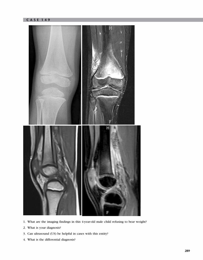
C A S E 1 4 9
1. What are the imaging findings in this 4-year-old male child refusing to bear weight?
2. What is your diagnosis?
3. Can ultrasound (US) be helpful in cases with this entity?
4. What is the differential diagnosis?
289

A N S W E R S
C A S E 1 4 9
Diagnosis: Acute Osteomyelitis
1. A plain-film reveals focal osteopenia in the medial
inferior aspect of femoral metaphysic and minimalloss of fat planes with no evidence of destruction in
the cortex or periosteal reaction. Magnetic resonance
imaging (MRI) reveals an increased T2-weighted
signal in the metaphysis of the femur with increased
T2-weighted signal in the adjacent soft tissue.
A T1-weighted sagittal image shows periosteal
elevation, and minimal subperiosteal fluid.
Postcontrast, fat-suppressed images show abnormalcontrast enhancement in the bone marrow and
surrounding soft tissue, indicating edema and
inflammation in these areas.
2. Acute osteomyelitis.
3. US changes may be demonstrated within 48 hours of
the onset of infection. The US appearance of
osteomyelitis includes loss of normal soft-tissuearchitecture and periosteal elevation, which may
appear as single or multiple echogenic lines
surrounding the cortical bone. US may also help
demonstrate some irregularity and interruption of
the cortical bone, although these findings would be
better demonstrated with plain-film radiographs or
computed tomography (CT). US helps not only in
making an accurate diagnosis but also aids in image-guided aspiration of the effusions.
4. Imaging findings may not be specific and may be
simulated by other diseases. Osteomyelitis is
primarily a disease of the infants and young childrenbut can be seen in any age group. A permeative bone
lesion in a child younger than 5 years old should raise
the suspicion for Langerhans cell histiocytosis (LCH)
and neuroblastoma metastasis. If the child is older
than 5 years of age, Ewing sarcoma, lymphoma,
leukemia, or LCH should be considered.
ReferenceSaigal G, Azouz EM, Abdenour G: Imaging of osteomyeli-
tis with special reference to children, Semin Muscu-
loskelet Radiol 8:255–265, 2004.
Cross-ReferenceBlickman JG, Parker BR, Barnes PD: Pediatric radiology—
the requisites, ed 3, Philadelphia, 2009, Mosby, p 181.
CommentOsteomyelitis implies bone and bone marrow inflamma-
tion, secondary to an infectious cause. Acute osteomye-
litis is primarily the disease of infants and younger
children, approximately 50% of the cases presenting
before 5 years of age. Osteomyelitis is most commonly
290
caused by bacterial organisms, most commonly by
Staphylococcus aureus, followed by b-hemolytic strep-
tococcus, although it can also be caused by fungi, para-
sites, and viruses. In cases with direct penetrating
trauma, Pseudomonas aeruginosa is the most common
organism. Salmonella is the most common cause inchildren with sickle cell disease.
The route of infection is hematogeneous in the great
majority of cases, followed by contiguous involvement
and direct inoculation.
Acute osteomyelitis commonly presents in the meta-
physis of the long bones (femur, tibia, humerus, fibula),
followed by short bones, pelvis, spine, and calvarium.
The metaphysis have rich blood supply and relativelyslow circulation, making it more susceptible for infec-
tious involvement.
Neonates are at higher risk for osteomyelitis, given
the weaker developing immune system. In addition, the
transphyseal sinusoids connect metaphyseal and epiphy-
seal blood vessels, allowing metaphyseal and epiphyseal
infection spread, which results in increased epiphyseal
damage.Radiographic changes in acute hematogenous osteo-
myelitis lag 7 to 10 days behind the evolution of infection.
The earliest finding is soft tissue swelling and displace-
ment or obliteration of fat planes. Bony destruction may
become visible after 7 to 14 days or even later. Periosteal
reaction can be seen at 7 to 10 days. In MRI, acute osteo-
myelitis shows loss of the high marrow signal on the T1-
weighted images, with increased corresponding signalintensity on the T2-weighted and short T1-weighted inver-
sion recovery (STIR) images. T1-weighted fat-suppressed
postcontrast images are better because suppression of
intrinsic fat results in better evaluation of the enhancing
soft tissues and fatty marrow. The high intramedullary
T2-weighted signal is secondary to edema, hyperemia,
and exudate. Cortical bone initially appears normal, which
explains the initially normal-appearing radiographs.The muscular and fascial planes adjacent to the infected
bone may demonstrate localized or diffuse increased
signal intensity on the T2-weighted and STIR images.
Well-established intraosseous, subperiosteal, and soft tis-
sue abscess collections are marked by peripheral enhance-
ment with central hypointensity on T1-weighted
postgadolinium images. Anatomical detail of MRI is better
than in Tc-bone scintigraphy, and the absence of ionizingradiation is an advantage, especially in the pediatric age
group. Three phasic bone scans, although highly sensitive
in the early detection (may be positive in 24 to 72 hours of
onset) of infection, can sometimes be ineffective in separ-
ating a bone marrow process from soft tissue disease,
given the lack of anatomical detail in bone scans. Tc-bone
scintigraphy, or whole body MRI, is superior to other
imaging modalities in detecting multifocal involvement.

C A S E 1 5 0
1. What are the imaging findings in this 14-year-old girl with asymmetry in the left leg and with an apparent limp?
2. What is the diagnosis, and what are the differential diagnoses?
3. What is McCune-Albright syndrome?
4. What is the cause?
291

A N S W E R S
C A S E 1 5 0
Diagnosis: Polyostotic Fibrous Dysplasia
1. The plain-film radiograph shows a loss of the normal
trabecular pattern with a ground-glass appearance ofthe bony matrix in the proximal left femur, distal left
femur, and proximal and middiaphyses of the left
tibia. The curvature of the femoral neck and
proximal shaft of the femur is significantly increased
because the proximal femoral lesion causes a severe
coxa vara abnormality (shepherd’s crook deformity),
which is a characteristic sign of the disease.
2. The diagnosis is polyostotic fibrous dysplasia.
Differential diagnoses include enchondroma,
eosinophilic granuloma, giant cell tumor, chronic
osteomyelitis, and postradiation changes.
3. McCune-Albright syndrome involves precocious
puberty in girls, with polyostotic fibrous dysplasia
and cutaneous hyperpigmentation.
4. Polyostotic fibrous dysplasia is a nonhereditary
disorder of unknown cause.
ReferenceSmith SE, Kransdorf MJ: Primary musculoskeletal tumors
of fibrous origin, Semin Musculoskelet Radiol 4
(1):73–88, 2000.
Cross-ReferenceBlickman JG, Parker BR, Barnes PD: Pediatric radiology—
the requisites, ed 3, Philadelphia, 2009, Mosby,
pp 178–179.
CommentFibrous dysplasia is a skeletal developmental abnormality
of the bone-forming mesenchyme that manifests as
a defect in osteoblastic differentiation and maturation.
Virtually any bone in the body can be affected. Histolog-
ically, the medullary bone is replaced by fibrous tissue,
which appears radiolucent on radiographs with the clas-
sically described ground-glass appearance. The initialmanifestations of fibrous dysplasia are most commonly
found in children 3 to 15 years of age. Approximately
70% to 80% of patients with fibrous dysplasias are
monostotic. This form most frequently occurs in the
rib (28%), femur (23%), tibia or craniofacial bones
(10% to 25%), humerus, and vertebrae in decreasing
order of frequency. Approximately 20% to 30% of
fibrous dysplasias are polyostotic. Polyostotic fibrousdysplasia more frequently involves the skull and facial
bones, pelvis, spine, and shoulder girdle. The sites of
involvement are the femur (91%), tibia (81%), pelvis
(78%), ribs, skull and facial bones (50%), upper extremi-
ties, lumbar spine, clavicle, and cervical spine, in
decreasing order of frequency.
292
Fibrous dysplasia may be associated with endocrino-
pathies in 2% to 3% of cases; these include precocious
puberty in girls, hyperthyroidism, hyperparathyroidism,
acromegaly, diabetes mellitus, and Cushing syndrome.
Radiologically, a lucent lesion is seen in the diaphysis
or metaphysis with endosteal scalloping and with orwithout bone expansion and the absence of periosteal
reaction. Usually, the matrix of the lucency is smooth
and relatively homogeneous; classically, this finding is
described as a ground-glass appearance. Irregular areas
of sclerosis may be present with or without calcifica-
tion. The lucent lesion has a thick sclerotic border and
is called the rind sign. Plain-film radiographs are highly
specific when characteristic features are present in alesion. However, the specificity decreases when the
lesion occurs at more complex sites such as the spine,
skull, and, sometimes, pelvis. Computed tomographic
findings complement plain-film radiographic findings.
Magnetic resonance imaging (MRI) shows decreased sig-
nal in both T1- and T2-weighted images. Typically, no
contrast enhancement is used. Cartilaginous islands
may reveal increased T2-weighted signal. MRI is helpfulafter surgery in demonstrating fibrocellular tissue prolif-
eration. Malignant degeneration is rare.
Two thirds of patients with polyostotic fibrous dys-
plasia are symptomatic before they are 10 years old.
The initial symptom is often pain in the involved limb,
associated with a limp, spontaneous fracture, or both.
In one series, a pathologic fracture was present in 85%
of patients with polyostotic fibrous dysplasias. Leg-length discrepancy of varying degrees occurs in approx-
imately 70% of patients with limb involvement. No cure
for fibrous dysplasia exists, and treatment focuses on
relieving the signs and symptoms.

C A S E 1 5 1
1. List the three most likely diagnoses.
2. List at least five plain-film imaging findings indicating malignancy.
3. Describe how to differentiate bone tumors based on location and the patient’s age.
4. What is the value of nuclear medicine studies?
293

A N S W E R S
C A S E 1 5 1
Diagnosis: Osteosarcoma
1. Osteosarcoma, Ewing sarcoma, and osteomyelitis.
2. Findings include (1) ill-defined destruction of
trabecular bone, (2) permeative or moth-eaten
appearance of the lesion, (3) wide zone of transition,
(4) pathologic fracture, (5) irregular cortical
destruction, (6) aggressive periosteal reaction,(7) Codman triangle, (8) sun burst appearance
with spiculae, (9) lamellated onion skin, and
(10) associated soft-tissue mass.
3. Osteosarcomas frequently involve the metaphysis of
long tubular bones, especially around the knee, with
a peak age of 10 to 15 years. Ewing sarcomas involve
flat bones and diaphysis of tubular bones and are
extremely rare in patients younger than 5 years of
age. The tumor may arise in bone or soft tissues.
4. Nuclear medicine studies facilitate differentiation
between malignant and benign bone lesions. These
studies help identify undetected tumor sites, bone
and bone marrow metastases, and may give
information concerning tumor viability.
ReferenceStoller DW: Magnetic resonance imaging in orthope-
dics and sports medicine, ed 3, Philadelphia, 2006,
Lippincott Williams & Wilkins, pp 2095–2104.
Cross-ReferenceBlickman JG, Parker BR, Barnes PD: Pediatric radiology—
the requisites, ed 3, Philadelphia, 2009, Mosby, p 186.
CommentOsteosarcomas are the most frequent primary malignant
bone tumors in children. The overall incidence of pri-
mary bone tumors is, however, low. Osteosarcomas
are diagnosed in 5.6 million children per year. Osteosar-
comas are most frequently diagnosed between 10 and
15 years of age during the adolescent growth spurt.Children most frequently present with pain, spontane-
ous pathologic fractures, or a focal swelling. An
increased risk for the development of osteosarcomas
has been reported in certain genetic disorders such as
hereditary retinoblastoma, or it may occur in the setting
of preexisting bone diseases such as Paget disease or
fibrous dysplasia. In addition, an increased risk for oste-
osarcoma exists in children who have been exposed toradiation. Osteosarcomas may metastasize into the
lungs, lymph nodes, liver, brain, and bones. Metastases
may show central calcifications. Plain films are usually
the first-line imaging modality, providing a good over-
view of the findings. Additional diagnostic work-up is,
however, essential and should provide information on
294
the exact location, extension, and biologic characteris-
tics of the primary tumor. For limb-saving surgical pro-
cedures, the orthopedic surgeons and oncologists are
especially interested to know which anatomic compart-
ments are involved, the possible extension into the adja-
cent growth plate or infiltration of the adjacent jointcapsula (or both), and the degree of subperiosteal tumor
extension. Magnetic resonance imaging (MRI) is the
best imaging modality for local tumor staging. Imaging
both adjacent joints is essential. Distant metastases can
be identified using nuclear medicine studies (bone scin-
tigraphy, positron emission tomography, computed
tomography [CT]). CT should be used to exclude pul-
monary, liver, and lymph node metastases. Brain MRIis used to rule out brain metastases. Differentiation from
osteomyelitis can be challenging, and nuclear medicine
studies may be helpful. Biopsies may be considered
but should be carefully planned together with the ortho-
pedic surgeon. If biopsy confirms a malignancy, the
orthopedic surgeon may have to resect the biopsy canal
because of possible tumor contamination.

C A S E 1 5 2
1. What is the striking finding on this abdominal radiograph of a 24-week pregnant woman?
2. What are the imaging findings on this fetal magnetic resonance image (MRI)?
3. What are the imaging findings on this postmortem radiograph?
4. What is the final diagnosis?
295

A N S W E R S
C A S E 1 5 2
Diagnosis: Osteogenesis Imperfecta Type II
1. The abdominal radiograph does not show the
osseous skeleton of the fetus (invisible fetus).
2. Short, deformed limbs, and an abnormally sized heart
(which appears large because the chest cage is small
with hypoplastic lungs).
3. Short, deformed, osteopenic bones, multiple
insufficiency fractures with excessive callus
formation, and a small chest cage.
4. Osteogenesis imperfecta (OI) type II.
ReferenceSolopova A, Wisser J, Huisman TA: Osteogenesis imper-
fecta type II: fetal magnetic resonance imaging find-ings, Fetal Diagn Ther 24(4):361–367, 2008.
Cross-ReferenceBlickman JG, Parker BR, Barnes PD: Pediatric radiology—
the requisites, ed 3, Philadelphia, 2009, Mosby,
pp 165–167.
CommentOI or brittle bone disease is a heterogeneous group of
inherited disorders characterized by increased bone fra-
gility. OI is the result of either a reduction of the normal
type I collagen production or from an abnormal colla-
gen synthesis. Typically, four different types of OI are
distinguished. Type I is the most frequent form. Clinical
symptoms are usually mild. Type II is the most severe
form. All children die within the perinatal period. Chil-dren with type III and IV survive the neonatal period
but suffer from significant complications. OI is classified
by clinical and molecular analysis.
Skeletal manifestations result from a generalized defi-
ciency of the membranous and endochondral bone
development and include significantly thinned calvar-
ium with a delayed closure of the fontanelles, sutures,
and excessive wormian bone formation. The classicalclinical stigmata include fragile bones, blue sclerae,
and ligamental laxity.
Bone changes may be identified as early as the
twelfth week gestation by prenatal ultrasound examina-
tion. Imaging findings include shortened, deformed
extremities. In addition, a reduced acoustic shadowing
of the long bones is observed, as well as an increased
nuchal translucency. Intrauterine growth retardation isoften present. Death usually occurs in the neonatal
period as a result of complications from infection, respi-
ratory distress (narrowed thorax as a result of multiple
rib fractures with pulmonary hypoplasia), cardiac insuf-
ficiency, or cerebral injury.
296
Newborns present with soft calvarian bones, a pecu-
liar triangular-shaped face with a beaked nose and blue
sclerae. Clinically, the extremities are usually short and
deformed as a result of the multiple fractures and are
typically in a froglike position. The radiologic hallmarks
are short, deformed, or angulated long bones with mul-tiple fractures in varied stages of healing with excess
callus formation, wormian bones, and poor skeletal
mineralization.
Typically, abdominal radiography of the mother will
not show the fetal skeleton (invisible fetus) because of
the poor mineralization of the fetal skeleton. Abdominal
radiography is rarely performed but may, however, be
considered in rare, potentially lethal skeletal malforma-tions of the fetus.
Fetal magnetic resonance imaging (MRI) is helpful
because an MRI simultaneously displays the skeletal mal-
formation and the hypoplastic chest and lungs. This
image may render valuable information about the imme-
diate postnatal time.

C A S E 1 5 3
eft
Right L1. What are the combined imaging findings?
2. Is this disorder an association or syndrome, and what is the name?
3. What are the additional possible findings that should be excluded?
4. This is a genetic disorder. Is this statement true or false?
297

A N S W E R S
C A S E 1 5 3
Diagnosis: VACTERL Association
1. Findings include a vertebral segmentation anomaly,
left renal agenesis, and radial aplasia.
2. The disorder is an association with the name VATER
or VACTERL.
3. Imperforate anus, cardiovascular anomalies,
tracheoesophageal malformation, esophageal atresia,
single umbilical artery, or limb anomalies.
4. False. This disorder is not genetic in origin.
Cross-ReferenceBlickman JG, Parker BR, Barnes PD: Pediatric radiology—
the requisites, ed 3, Philadelphia, 2009, Mosby,
pp 76–77.
CommentVATER or VACTERL is an acronym that refers to a con-
stellation of associated anomalies. The acronym relates
to the following findings: vertebral segmentation
anomalies, anal atresia, cardiovascular anomalies, tra-cheoesophageal fistula, esophageal atresia, renal or
radial anomalies or both, and limb anomalies. Duodenalatresia and a single umbilical artery have also been
described. This disorder is called an association rather
than a syndrome because the findings are linked, but
no specific genetic or chromosomal defect has been
identified. The VACTERL association has an estimated
incidence of 16 in 100,000 live births. The diagnosis is
made if at least three of the seven defects are present.
Children with a VACTERL association have normaldevelopment and intelligence. Vertebral anomalies, tra-
cheoesophageal fistula, and limb defects are seen in
approximately 70% of children. Renal or radial defects
(or both) or anal atresia are less frequent (50% to 55%
of patients). Cardiac defects are even less frequent.
Treatment is usually directly related to the present birth
defects.
298

C A S E 1 5 4
1. This 5-year-old developmentally delayed boy had crampy abdominal pain for several days. His pediatrician was
treating him for constipation. What can be seen on his abdominal radiograph (first image)?
2. The emergency department clinician sent the child for an ultrasound to rule out intussusception. What should be
the focus as he is examined?
3. What is important to note about the ultrasound images (second, third, and fourth images)?
4. As his abdomen is being observed during the scan, a purplish flat rash is seen in the groin. His mother explains that
the boy has complained about pain in his legs for the last few days. What additional question should be asked of the
emergency department clinician?
299

A N S W E R S
C A S E 1 5 4
Diagnosis: Henoch-Schonlein PurpuraSyndrome
1. Very little bowel gas, no sign of a large mass, and
stool impaction or obstruction.
2. The child is at the upper end of the age range for
uncomplicated, postinfectious intussusception. In
his age group, a lead point (polyp, hematoma,
lymphoma, lipoma, duplication cyst) becomes more
likely.
3. The ultrasound reveals a fluid-filled small bowel that
has thickened with clubbed, mucosal folds (second
image). One area of the small bowel in the
midabdomen has a very thick, hyperemic wall (third
image). No target sign of intussusception is
demonstrated. Low echogenicity is seen in thesubmucosal region, consistent with nonacute
hematoma (fourth image).
4. If hematuria is present.
ReferenceStringer DA, Babyn PS, editors: Pediatric gastrointesti-
nal imaging and intervention, ed 2, Hamilton,
Ontario, 2000, BC Decker, pp 406–407.
Cross-ReferenceBlickman JG, Parker BR, Barnes PD: Pediatric radiology—
the requisites, ed 3, Philadelphia, 2009, Mosby, p 149.
CommentHenoch-Schonlein purpura syndrome is an acute but
self-limiting diffuse vasculitis of uncertain causes that
affects the gut, skin, joints, kidneys, and (occasionally)
the central nervous system. The skin shows a purpuric
rash that spreads centrifugally. No signs of thrombocyto-penia are presented. The bowel wall, usually in the jeju-
num, can be affected at multiple points. The hematoma
can act as a lead point and cause intussusception that
may have to be surgically relieved.
300

C A S E 1 5 5
1. This 9-year-old patient had two computed tomographic (CT) scans 8 months apart. What is the difference between
them?
2. What portion of the total radiation burden in the U.S. population is now attributed to CT scanning?
3. Why is the concept of radiation dose especially important in pediatric radiology?
4. What four machine factors have the greatest effect on dose delivery?
C A S E 1 5 6
1. What are the imaging findings?
2. What is the most likely history?
3. How frequently does this complication occur?
4. Which anatomic area related to the primary lesion should be checked for additional lesions?
301

A N S W E R S
C A S E 1 5 5
Diagnosis: Radiation Exposure in Childrenfrom Computed Tomography
1. The second scan has less than one half of the
radiation dose of the first scan.
2. 25%.
3. Young tissues are more susceptible to radiation
damage. Children live longer, which allowsexpression of the damaged DNA as cancer. In
addition, a greater dose deposition is given to small
bodies because the same scan parameters are used
for adults as used for children.
4. Tube mA (direct, linear), kVp (direct; linear at lower
levels, nonlinear [greater effect] at higher levels),
gantry cycle time (direct, linear), and pitch (indirect,
linear).
ReferencesCoursey CA, Frush DP: CT and radiation: what radiolo-
gists should know, Applied Radiology 37(3):22–29,
2008.
Alliance for radiation safety in pediatric imaging: image
gently. (Web site) www.pedrad.org/.
CommentMultidetector CT is a powerful tool for diagnosing dis-
ease, but it comes at a price—an ever-increasing radia-tion dose. No visual penalty is paid for the higher
doses as it is with plain films; rather, the images become
incrementally more crisp and clear. The newer scanners
have wider effective beams for better overlap of slices,
which means more detailed three-dimensional recon-
structions. However, because the dose recorders are
built into the scanners, calculating the dose only by
overall volume scanned can lead to an underestimationof the actual dose by up to 35%.
Turning down the mA and kVp, rotating the gantry
faster, and passing the patient through the scanner fas-
ter will all decrease the deposited radiation. Add bis-
muth breast shields or eye patches for a head scan,
and the dose decreases further. However, the tradeoff
is increased noise in the image. Depending on the rea-
son for the scan, more or less noise might be tolerable.Persuading the clinician to select an ultrasound or mag-
netic resonance imaging is the surest form of dose
reduction. After all, the CT scan that is not used has a
dose of 0.
302
C A S E 1 5 6
Diagnosis: Posttreatment for HereditaryBilateral Retinoblastoma and SphenoidWing Osteosarcoma
1. Enucleation of the right eye ball and a partially
calcified tumor of the left sphenoid wing(osteosarcoma).
2. Posttreatment of bilateral retinoblastomas,
enucleation on the right, and radiotherapy on theleft.
3. Osteosarcomas may result from radiotherapy.
Osteosarcomas are also seen associated with
hereditary bilateral retinoblastoma.
4. The pineal gland.
Cross-ReferenceBlickman JG, Parker BR, Barnes PD: Pediatric radiology—
the requisites, ed 3, Philadelphia, 2009, Mosby, p 331.
CommentRetinoblastoma is the most common malignant ocular
tumor in childhood. The typical age of presentation for
unilateral cases is between 1 and 3 years. Bilateral cases
typically present during the first year. Trilateral retino-blastomas are rare and are usually hereditary. In 2% to
5% of children with hereditary bilateral retinoblastoma,
a third tumor may be found in the pineal gland. In
hereditary bilateral neuroblastoma, children are at risk
for secondary nonocular tumors later in life. The mean
latency of second tumors is 9 years. Osteosarcomas are
the most frequently encountered second malignancies.
Development of osteosarcoma may be accelerated byfocal radiotherapy. Osteosarcomas typically develop in
the field of radiation. Other tumors that may occur are
pinealomas, brain tumors, soft-tissue sarcomas, and mel-
anomas. The prognosis of children who develop a sec-
ond tumor is poor. In children with a unilateral
(sporadic) retinoblastoma, a 20% chance exists for
developing a retinoblastoma in the contralateral eye.
Close monitoring using magnetic resonance imaging ismandatory. If diagnosed early and treated adequately (a
combination of surgery, chemotherapy, and radiother-
apy), the prognosis today is favorable. The main factor
that determines the prognosis is direct tumor extension
via the optic nerve or adjacent sclera.
Secondary osteosarcomas are treated as primary
osteosarcomas.


Challenge

C A S E 1 5 7
1. What are the imaging findings on a prenatal magnetic resonance image (MRI)?
2. What are the imaging findings on a postnatal MRI at 5 months?
3. What is the most likely diagnosis?
4. To which group does this diagnosis belong?
305

A N S W E R S
C A S E 1 5 7
Diagnosis: Muscle-Eye-Brain Disease
1. Ventriculomegaly and a small, hypoplastic, kinked
brainstem.
2. Ventriculomegaly; diffuse cerebral polymicrogyria;
multiple subcortical cerebellar cysts; swollen, T2-
hyperintense cerebral white matter; hypoplastic
pons; malformed tectal plate; and elongatedmesencephalon.
3. Muscle-eye-brain disease (MEB).
4. Congenital muscular dystrophies (CMDs).
ReferenceVan der Knaap MS, Valk J: Congenital muscular dystro-
phies. In Magnetic resonance of myelination and
myelin disorders, ed 3, New York, 2005, Springer
Berlin Heidelberg, pp 451–468.
Cross-ReferenceBlickman JG, Parker BR, Barnes PD: Pediatric radiol-
ogy—the requisites, ed 3, Philadelphia, 2009, Mosby,
p 215.
CommentMEB belongs to the group of CMDs, which is a heteroge-
neous group of congenital, hereditary myopathies that
are often progressive. Frequently, the name of the diag-
nosis is based on the combined abnormalities of the
eyes and brain. MEB is inherited autosomal recessive.
Early clinical symptoms include muscular hypotonia
and poor visual contact. A hydrocephalus is seen inthe majority of patients. Motor development is generally
severely delayed. In addition, severe mental retardation
is usually present in combination with seizures. Ocular
signs include severe myopia, glaucoma, retinal degener-
ation, choroidal hypoplasia, and optic nerve hypoplasia.
MEB is caused by a defect in the O-mannosyl glycan syn-
thesis. No curative treatment is available.
MRI findings are extensive and prominent. Ventricu-lomegaly frequently ranges from mild-to-significantly
dilated. Additional striking features include a hypoplas-
tic pons and brainstem, multiple subcortical cerebellar
cysts, and variable degrees of cerebral polymicrogyria.
The cortex is characteristically thickened, and the inner
contour of the cortex reveals the polymicrogyria most
prominently. In addition, the cerebral white matter is
edematous and swollen with a significantly increasedT2-weighted signal. The mesencephalon is frequently
elongated with a malformation of the tectal plate. The
superior and inferior colliculi may be fused. In addition,
neuronal heterotopias may be seen within the cerebral
hemispheres, as well as a thinned, hypoplastic corpus cal-
losum and a partial absence of the septum pellucidum.
306
The characteristic features, especially the combination
of a ventriculomegaly and a hypoplastic, elongated, and
kinked brainstem may already be observed on prenatal,
fetal MRI. The cerebellar cysts may, however, be too small
to be detected intrauterine or may progress after birth.

C A S E 1 5 8
1. What are the imaging findings?
2. What is your diagnosis?
3. What is the underlying pathologic condition?
4. Which associated lesions may be observed?
307

A N S W E R S
C A S E 1 5 8
Diagnosis: Rhombencephalosynapsis
1. A single-lobed cerebellum, no vermis, a keyhole
fourth ventricle, and fused dentate nuclei.
2. Rhombencephalosynapsis.
3. A disruption of failed dorsal induction.
4. Hydrocephalus (the current patient is shunted),corpus callosum anomalies, and midline anomalies.
ReferencePatel S, Barkovich AJ: Analysis and classification of cere-
bellar malformations, AJNR Am J Neuroradiol 23:
1074–1087, 2002.
Cross-ReferenceBlickman JG, Parker BR, Barnes PD: Pediatric radiology—
the requisites, ed 3, Philadelphia, 2009, Mosby, p 212.
CommentRhombencephalosynapsis is a complex congenital mal-
formation of the posterior fossa, characterized by afusion of the cerebellar hemispheres, dentate nuclei,
and hypoplastic or fused superior cerebellar peduncles
(single-lobed cerebellum). In addition, the cerebellar
vermis is absent or hypoplastic. This malformation is
believed to be the result of a defective dorsal induction
or a differentiation of the cerebellar midline structures
or both. Rhombencephalosynapsis has been linked to
several gene defects, but it is also observed in condi-tions such as Gomez-Lopez-Hernandez syndrome.
Rhombencephalosynapsis is extremely rare; the clinical
presentation is usually nonspecific with ataxia, gait
abnormalities, and developmental delays. Associated
findings, such as corpus callosum agenesis or dysgene-
sis, septo-optic dysplasia, and hydrocephalus as a result
of aqueductal stenosis, are frequently observed. Migra-
tional abnormalities with cerebral polymicrogyria havealso been described. Rhombencephalosynapsis may be
diagnosed intrauterine by fetal magnetic resonance
imaging (MRI) or by postnatal MRI. Imaging findings
are usually obvious with the identification of a single-
lobed cerebellum with folia and cerebellar fissures
crossing the midline (transverse folia). In addition, the
dentate nuclei are fused and usually in a more midline
position dorsally to a narrowed or keyhole-shapedfourth ventricle. Midsagittal images may be misleading;
however, the pattern of the midline fissures do not
match the normal anatomy of the vermis (i.e., the pri-
mary fissure is missing). Coronal and axial images are
most useful. The prognosis is variable; most children
do not survive until adulthood. Additional lesions or
complications such as hydrocephalus or both shorten
survival.
308

C A S E 1 5 9
1. What are the imaging findings on magnetic resonance imaging (MRI)?
2. What is the diagnosis?
3. What characteristic is special on the corticospinal tracts in this syndrome?
4. Which other organs may show a pathologic condition?
309

A N S W E R S
C A S E 1 5 9
Diagnosis: Joubert Syndrome
1. Aplasia of the vermis, molar tooth appearance of the
brainstem, superior cerebellar peduncles that arethickened and run horizontally, and a bat wing– or
umbrella-shaped fourth ventricle.
2. Joubert syndrome.
3. The absence of the pyramidal decussation.
4. The kidneys.
ReferencePoretti A, Boltshauser E, Loenneker T, et al: Diffusion
tensor imaging in Joubert syndrome, AJNR Am J Neu-
roradiol 28:1929–1933, 2007.
Cross-ReferenceBlickman JG, Parker BR, Barnes PD: Pediatric radiol-
ogy—the requisites, ed 3, Philadelphia, 2009, Mosby,
p 212.
CommentJoubert syndrome was originally reported by Dr. Marie
Joubert who described several children with a similar clin-ical presentation that included episodic hyperpnea, abnor-
mal eye movement, rhythmic tongue protrusions, ataxia,
and mental retardation. Anatomically, the cerebellar
vermis is lacking. On MRI, the axial images show a
characteristic molar tooth appearance of the brainstem,
resulting from a horizontal course of the thickened supe-
rior cerebellar peduncles. The fourth ventricle is
deformed and resembles an umbrella or bat wing on axialplanes. Because the vermis is lacking, both cerebellar
hemispheres reach each other in the midline; conse-
quently, the midline slices reveal cerebellar fissures on
sagittal imaging instead of vermian sulci. In addition, den-
tate nuclei are lateralized. Tractography studies have
shown that the corticospinal tracts do not cross at the
pyramidal decussation, which may, in part, explain the
clinical presentation. Multiple gene loci that are linked toJoubert syndrome have been identified. The expression
may vary; children with Joubert syndrome may show dif-
ferent degrees of mental retardation and gait disturbance.
Currently, no prenatal test is available for early diagnosis.
Rarely associated supratentorial anomalies occur, and cor-
tical dysplasia and gray-matter heterotopia have been
reported. In children with Joubert syndrome, the kidneys
may be affected (cerebelloocculorenal syndrome). Renalultrasound should be considered during the diagnostic
work-up of these children. Experienced pediatric neurolo-
gists may suspect a diagnosis, based on the facial charac-
teristics that are not specific but indicative. With
ongoing research, many more malformations of the brain-
stem and cerebellum that share amolar tooth appearance
310
of the brainstem are described. Perhaps, Joubert syn-
drome is simply one of a spectrum of brainstem
malformations.

C A S E 1 6 0
1. What are the imaging findings on magnetic resonance imaging (MRI) and magnetic resonance angiography?
2. What is seen on the fractional anisotropy map?
3. What is the diagnosis?
4. To which group of anomalies does this malformation belong?
311

A N S W E R S
C A S E 1 6 0
Diagnosis: Semilobar Holoprosencephaly
1. Fused frontal lobes, small lateral ventricles, lack of
the anterior corpus callosum, unpaired anteriorcerebral artery (azygos artery), hypoplastic falx
cerebri, rudimentary temporal horns, and partial
separation of the thalami.
2. Continuous fiber tracts between the fused frontallobes and a horseshoe appearance.
3. Semilobar holoprosencephaly.
4. The ventral induction group of anomalies or the
diverticulation or cleavage group of disorders.
Cross-ReferenceBlickman JG, Parker BR, Barnes PD: Pediatric radiol-
ogy—the requisites, ed 3, Philadelphia, 2009, Mosby,
pp 212–213.
CommentA group of holoprosencephalies results from a disorder
of diverticulation or cleavage. Holoprosencephalies are
also known as disorders of ventral induction. Holopro-
sencephalies encompass an entire spectrum of malfor-mations; the mildest form is septo-optic dysplasia; the
most severe form is alobar holoprosencephaly. They
both result from a failure of cleavage of the prosence-
phalic vesicle during organogenesis. Frequently, asso-
ciated anomalies of the face are seen—“The face
predicts the brain.” Cyclopia has been described in
cases of alobar holoprosencephaly. Clinically, depending
on the severity of the malformation, different symptomsmay occur and include seizures, mental retardation, dys-
tonia, microcephaly, hypothalamic-pituitary dysfunction,
cyclopia, and fused metopic suture. Symptoms may be
mild in lobar holoprosencephaly and severe in alobar
holoprosencephaly. In septo-optic dysplasia, the anterior
horns of the ventricles have a boxlike configuration on
coronal MRI. In addition, an optic nerve pathologic
abnormality is seen on fundoscopy. Various degrees ofhypothalamic-pituitary dysfunction are observed. In the
lobar holoprosencephaly, a lobar brain is seen with hypo-
plastic frontal lobes, some frontal horn formation, and the
falx cerebri extends frontally. In semilobar holoprosence-
phaly, a partially formed fax cerebri and a partially formed
interhemispheric fissure are seen in the posterior part.
The anterior brain is fused, the thalami are partially
separated, and a small third ventricle and rudimentarytemporal horns are demonstrated. The septum pelluci-
dum is absent, the splenium of the corpus callosum is
present, whereas the truncus of the corpus callosum is
lacking. Hypoplastic olfactory bulbs and optic nerves
may exist. In alobar holoprosencephaly, a small holo-
sphere and monoventricle are seen, as well as fused
312
thalami. However, no third ventricle, falx cerebri or
corpus callosum, or interhemispheric fissure is seen. In
addition, no temporal horns are demonstrated, and mal-
formations of the Willis circle are observed with an azy-
gos or unpaired anterior cerebral artery. The prognosis
is frequently poor, especially in the most severe formsof holoprosencephalies. Diagnosis is usually made pre-
natally by either ultrasound or fetal MRI. In septo-optic
dysplasia, the prognosis will be determined by the asso-
ciated malformations. In up to 50% of cases, additional
lesions are identified by MRI. Postnatally, MRI is indicated
to identify all details of the malformation.

C A S E 1 6 1
1. What are the findings on magnetic resonance imaging (MRI)?
2. What is the diagnosis?
3. To which group of lesions does this malformation belong?
4. How many layers does the normal cortex have, and how many do you expect to find in this child?
313

A N S W E R S
C A S E 1 6 1
Diagnosis: Lissencephaly Type I
1. Symmetric T2-hypointense bands within the cerebral
white matter, lissencephalic cortex, and mildventriculomegaly.
2. Lissencephaly (type I) with extensive and arrested
neuronal migration.
3. Neuronal migration anomalies.
4. The normal cortex has six layers; this child has four
layers.
Cross-ReferenceBlickman JG, Parker BR, Barnes PD: Pediatric radiology—
the requisites, ed 3, Philadelphia, 2009, Mosby,
pp 219–222.
CommentNeuronal migration anomalies and malformations of cor-
tical development occur between the second and fourth
months gestation. In neuronal migration anomalies, cells
that should migrate from the periventricular germinal
matrix to the cortical surface of the brain are arrested
somewhere along their paths. These cells may rest atmultiple locations, either focal or as bands. Heterotopic
gray matter may be recognized on imaging focally as
subependymal heteroptopic cell masses or as bands
within the periventricular, central, or subcortical white
matter of the cerebral hemispheres. Disorders of corti-
cal organization may result in agyria, pachygyria, poly-
microgyria, or focal cortical dysplasia. Frequently,
combined malformations of neuronal migration and cor-tical organization are seen. In lissencephaly (type I), an
extensive arrest of the neuronal migration results in a
wide, T2-hypointense band of neurons within the white
matter of both hemispheres separated from the overly-
ing cortex by a thin band of T2-hyperintense white mat-
ter. The overlying cortex is typically lissencephalic and
has a four-layer architecture combined with a smooth
brain surface. The anomaly is usually symmetric. Ventri-culomegaly may be associated. The cerebellum is usu-
ally unremarkable. Children may present with
intractable seizures and a developmental delay. The
cause is not yet fully determined but is at least known
to be mediated genetically but is also believed to be
the result of intrauterine infections. MRI is the imaging
modality of choice to identify the exact anatomy of the
anomaly. High-resolution imaging will also display thatthe subcortical white matter has multiple linear bands
of T2-hypointensity, which most likely represents
streaks of neurons that almost reached the lissencepha-
lic cortex. Prenatal diagnosis on ultrasound may be diffi-
cult because the overlying fetal skull prevents a detailed
study of the cortex; the arrested neurons within the
314
white matter are also difficult to detect. Lissencephaly
(type I) should be differentiated from the neuronal
migration anomalies and malformations of cortical
development because differentiation will help deter-
mine prognosis.

C A S E 1 6 2
T2-FSE T1-SE +C
T1-SE +C
T2-FSE1. What are the imaging findings on an initial magnetic resonance image (MRI) in this 15-year-old girl?
2. Does the follow-up imaging, performed 13 months later, narrow the differential diagnoses?
3. What is the most likely diagnosis?
4. What is the prognosis?
315

A N S W E R S
C A S E 1 6 2
Diagnosis: Progressive MultifocalLeukoencephalopathy
1. Areas of T2-hyperintensity and nonenhancing T1-
hypointensity within the white matter of both
hemispheres (right is greater than left), minimal mass
effect, preserved superficial layers of the cortex, and
asymmetric distribution.
2. Rapid progression of the lesions, cortical thinning, a
strikingly asymmetric presentation, a metabolic
disease, or acute disseminated encephalomyelitis is
less likely.
3. Progressive multifocal leukoencephalopathy (PML)
in the immunocompromised child.
4. The prognosis of PML in a child with human
immunodeficiency virus (HIV) is poor, in general;
rapid increase makes the prognosis even worse.
Cross-ReferenceBlickman JG, Parker BR, Barnes PD: Pediatric radiology—
the requisites, ed 3, Philadelphia, 2009, Mosby, p 231.
DiscussionPML is a severe, demyelinating disease of the central
nervous system as a result of JC papovavirus infection
of the myelin-producing oligodendrocytes. The name
JC virus is derived from the initials of the index patient.PML typically occurs in immunocompromised indivi-
duals, such as children with congenital HIV infection
or other conditions associated with impaired T-cell func-
tion. In the course of the infection, extensive myelin
breakdown results in white matter destruction. Neuro-
logic symptoms are unspecific and include focal neuro-
logic deficits and dementia. Without treatment,
patients have a relentless downhill course. The diseaseis usually fatal within 1 year of diagnosis in 90% of
patients. The diagnosis can be established by the detec-
tion of the JC virus deoxyribonucleic acid (DNA) in
cerebrospinal fluid by polymerase chain reaction test-
ing. Many reports have described MRI findings in
patients with PML, but only few MRI abnormalities cor-
relate with patient survival. Conventional MRI reveals
patchy areas of T2-hyperintense and T1-hypointensedemyelination within the white matter. These lesions
do not usually show any contrast enhancement and
have minimal or no mass effect. Typically, these lesions
are asymmetric. On diffusion tensor imaging, the lesions
have a reduced fractional anisotropy, confirming the
injury to the white matter by the infection. On follow-
up imaging, lesions may show a rapid increase without
treatment. The overlying cortex may be thinned. Anychild with a rapidly expanding, focal white matter
lesion with a compromised immune system should be
316
suspected for PML until proved otherwise. The differen-
tial diagnoses may include acute disseminated encepha-
lomyelitis and radiation necrosis or metabolic white
matter diseases. The rapid progression and the clinical
history are usually helpful in differentiating the cause
of the observed lesions.

C A S E 1 6 3
1. What are the findings in a magnetic resonance image (MRI) of a 10-year-old girl with a history of progressive loss of
developmental milestones?
2. What does the 1H–magnetic resonance spectroscopy (1H-MRS) of the hemispheric white matter show?
3. What is the most likely diagnosis?
4. How can this diagnosis be confirmed?
317

A N S W E R S
C A S E 1 6 3
Diagnosis: Alexander Disease
1. Symmetric T2-hyperintense, T1-hypointense signal of
predominantly the frontal white matter and posteriorlimb of internal capsula; multifocal contrast-
enhancing brainstem lesions; periventricular T1-
hyperintensity; subcortical cysts; mild
ventriculomegaly; and cavum septi pellucid.
2. Increased levels of myoinositol and choline,
decreased N-acetyl aspartate, and mild lactate within
the white matter.
3. Alexander disease, a form of leukodystrophy.
4. Brain biopsy will confirm the diagnosis and reveal an
accumulation of Rosenthal fibers in hypertrophic,
fibrillary astrocytes and a mutation in the glialfibrillary acidic protein (GFAP) gene located on the
long arm (q) of chromosome 17 at position 21
(17q21).
ReferenceVan der Knaap MS, Valk J: Congenital muscular dystro-
phies. In Magnetic resonance of myelination and
myelin disorders, ed 3, New York, 2005, SpringerBerlin Heidelberg, pp 416–441.
Cross-ReferenceBlickman JG, Parker BR, Barnes PD: Pediatric radiology—
the requisites, ed 3, Philadelphia, 2009, Mosby,
pp 267–268.
CommentAlexander disease is a leukodystrophy first described by
W. Stewart Alexander in 1949 as progressive fibrinoid
degeneration of fibrillary astrocytes associated with
mental retardation in a hydrocephalic infant. Three sub-
groups have been identified: infantile, juvenile, and
adult. In the juvenile subgroup, the patients become
symptomatic between 4 and 14 years of age, typically
with a progressive loss of previously mastered develop-mental milestones. In addition, patients may present
with progressive bulbar and pseudobulbar symptoms,
an increase in swallowing problems and apneic attacks,
impaired speech or loss of speech, dysarthria, hoarse-
ness, spasticity, cerebellar ataxia, seizures, and progres-
sive behavioral and cognitive deterioration. Average
duration of the illness is 8 years. Most cases are spo-
radic, but familial cases have been described. Imagingfeatures are rather characteristic with predominant fron-
tal lobe involvement that may progress over the course
of the disease into the parietal regions. Five characteris-
tic MRI criteria have been defined, and four of the five
have to be fulfilled for diagnosis: (1) symmetric cerebral
white matter abnormalities with predominant frontal
318
involvement, (2) T2-hypointense, T1-hyperintense rim
of tissue along the ventricles, (3) focal lesions in the
basal ganglia and thalami, (4) brainstem abnormalities
(midbrain and medulla), and (5) contrast enhancement
of the described lesions including optic chiasm. In addi-
tion, 1H-MRS will show increased myoinositol and cho-line levels within the affected white matter, as well as
a reduced N-acetyl aspartate peak. Lactate may also be
observed. On microscopic examination, countless
Rosenthal fibers are seen throughout the brain within
hypertrophic fibrillary astrocytes. Macrocephaly results
from the combination of astrocytic hyperplasia and mas-
sive deposition of Rosenthal fibers.
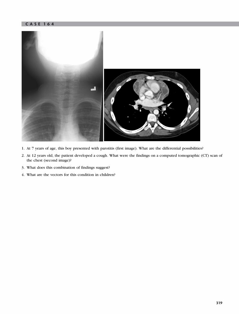
C A S E 1 6 4
1. At 7 years of age, this boy presented with parotitis (first image). What are the differential possibilities?
2. At 12 years old, the patient developed a cough. What were the findings on a computed tomographic (CT) scan of
the chest (second image)?
3. What does this combination of findings suggest?
4. What are the vectors for this condition in children?
319

A N S W E R S
C A S E 1 6 4
Diagnosis: Positive Human ImmunodeficiencyVirus: Parotitis and Thymic Cysts
1. All of the following differential possibilities are very
rare: acute bacterial parotitis, recurrent parotitis with
sialectasia, viral parotitis (mumps, Ebstein-Barr virus
[EBV], human immunodeficiency virus [HIV]), and
Sjogren’s syndrome, this patient’s first diagnosis.
2. CT findings included thymic cysts.
3. This combination of findings suggests HIV and
acquired immunodeficiency syndrome (AIDS).
4. The vectors for this condition in children are
congenital infection acquired through transfusion.
ReferenceKuhn JP, Slovis TL, Haller JO: Caffey’s pediatric diag-
nostic imaging, ed 10, Philadelphia, 2004, Mosby,
pp 1184–1189.
Cross-ReferenceBlickman JG, Parker BR, Barnes PD: Pediatric radiology—
the requisites, ed 3, Philadelphia, 2009, Mosby,
pp 324–325.
CommentAll parotitis is rare in children; consequently, all of the
unusual entities in the differential list have a goodchance of being correct. EBV, HIV, recurrent parotitis,
and Sjogren syndrome all have similar clinical presenta-
tions and relapsing and remitting courses. This patient
was not tested for HIV until the thymic cysts were
discovered.
320

C A S E 1 6 5
1. What is the finding in these transverse and midline sagittal plane images of the anterior upper neck of a 4-year-oldpatient?
2. What are the differential diagnoses?
3. The lesion is located in the midline of the anterior neck. If it were found in the lateral aspect, what would be a
likely diagnosis?
4. Why is localizing the thyroid gland important in this situation?
321

A N S W E R S
C A S E 1 6 5
Diagnosis: Thyroglossal Duct Cysts
1. A 1 to 2 cm round cystic structure with a high level
of echoes suggests a complicated cyst.
2. A thyroglossal duct cyst and lymphadenitis with
abscess are the most likely differential diagnoses.
Sebaceous cyst, lymphatic malformation, thymic
cyst, dermoid cyst, teratoma, lipomas, and sarcomaare less likely because of the size or ultrasound
characteristics.
3. Branchial cleft cyst.
4. If the thyroid tissue is not seen in the normal
location, then the gland may be ectopic and possibly
incorporated in the thyroglossal duct cyst.
ReferenceTunkel DE, Domenech EE: Radioisotope scanning of the
thyroid gland prior to thyroglossal duct cyst excision,
Arch Otolaryngol Head Neck Surg 124:597–599,
1998.
Cross-ReferenceBlickman JG, Parker BR, Barnes PD: Pediatric radiology—
the requisites, ed 3, Philadelphia, 2009, Mosby,
pp 309–311.
CommentThyroglossal duct cysts are the most common congeni-
tal cysts in the neck representing 75% of midline masses
and resulting from cystic dilation of epithelial remnants
of the thyroglossal duct tract, which is present duringthe embryologic inferior migration of the thyroid from
the base of the tongue to its normal final location. These
cysts present as midline neck masses at the level of the
thyrohyoid membrane and are closely associated with
the hyoid bone. Most patients present as children,
although presentation at any age is possible. Males and
females are equally affected, and the cysts are usually
asymptomatic, but they may become infected and formabscesses and draining fistulas.
In this example, the cyst is small; if larger, both an
elevation of the tongue and a swallowing dysfunction
would have developed. Ultrasound is the imaging
modality of choice because it is ideal for determination
of the nature of the mass and the localization of thyroid
tissue. In this case, the lesion appears as a complex cyst,
which is likely because of prior infection or hemor-rhage. Computed tomography (CT) and magnetic reso-
nance imaging (MRI) is also diagnostic but radiation
(CT), length of study (MRI), and expense make both
modalities impractical and unnecessary. Scintigraphy
may be helpful to localize ectopic location of the thy-
roid gland.
322
Surgical resection is considered the treatment of
choice. If the patient is euthyroid and thyroid tissue is
seen in the normal location, no further imaging of the
thyroid gland is necessary. In cases of abnormal thyroid
function, nuclear thyroid scans are indicated to search
for ectopic thyroid. Ectopic thyroid tissue may belocated in the sublingual space in 10% of patients and
at the base of the tongue in 90% of patients. In 75% of
patients, the ectopic tissue is the only functioning thy-
roid tissue. However, these patients are usually hypothy-
roid anyway because the ectopic tissue is dysplastic. A
careful histologic study of the specimen is important; a
small incidence of carcinoma arising in thyroglossal duct
remnants exists.

C A S E 1 6 6
1. What are the imaging findings in this 3-year-old African-American child that presented with right neck swelling?
What are the differential diagnoses?
2. What is Rosai-Dorfman disease?
3. What is Castleman disease?
4. What are the characteristic pathologic findings?
323

A N S W E R S
C A S E 1 6 6
Diagnosis: Rosai-Dorfman Disease
1. A frontal chest radiograph and intravenous contrast-
enhanced computed tomographic scans through theneck and upper chest demonstrate massive cervical
and mediastinal lymphadenopathy. The differential
diagnoses include lymphoma, infection, Castleman
disease, and Rosai-Dorfman disease.
2. Painless bilateral cervical lymph node enlargement
with the possible involvement of various extranodal
sites.
3. A benign process of angiofollicular hyperplasia of the
lymph nodes and typically does not affect children.
4. In Rosai-Dorfman disease, extensive reactive process
within the lymph node is typically seen, includingsignificant dilated sinuses containing sheets of
histiocytes with eosinophilic cytoplasm and small
nuclei with no evidence of malignancy.
ReferencesRosai J, Dorfman RF: Sinus histiocytosis with massive
lymphadenopathy: a pseudolymphomatous benign
disorder, Cancer 30:1174–1188, 1972.La Barge DV 3rd, Salzman KL, Harnsberger HR, et al:
Sinus histiocytosis with massive lymphadenopathy
(Rosai-Dorfman disease): imaging manifestations in
the head and neck, AJR Am J Roentgenol 191(6):
W299–W306, 2008.
CommentsRosai-Dorfman disease is a rare sinus histiocytosis and a
benign source of massive lymphadenopathy, which may
mimic a neoplastic process. This disease commonly pre-sents in children and young adults and in blacks more
commonly than in whites. It is characterized by painless
bilateral cervical lymph node enlargement and, less com-
monly, other groups of lymph nodes. Up to 43% of patients
have involvement of extranodal sites, especially the soft
tissues of the head and neck, paranasal sinuses, and nasal
cavity. Other sites that may be involved include the upper
respiratory tract, skin, salivary glands, orbit, bones, andtestis. These extranodal lesions may appear before lymph-
adenopathy is apparent.
Pathologic examination of the biopsied lymph nodes
demonstrates exaggerated reactive process with no evi-
dence of malignancy. Characteristic features include sig-
nificantly dilated sinuses containing sheets of histiocytes
with eosinophilic cytoplasm and small nuclei.
Differential diagnoses include Castleman disease, der-mopathic lymphadenitis, mucocutaneous lymph node
syndrome (Kawasaki disease), histiocytic necrotizing
lymphadenopathy (Kikuchi disease), vascular transfor-
mation of the lymph nodes, and inflammatory pseudotu-
mor of the lymph nodes.
324

C A S E 1 6 7
1. What are the imaging findings in this newborn with respiratory distress?
2. What is the diagnosis?
3. Does this condition have any association with other congenital anomalies?
4. What is the cause?
325

A N S W E R S
C A S E 1 6 7
Diagnosis: Choanal Atresia
1. A thickening and medial bowing of the lateral walls
of the nasal cavity, an enlargement of the vomer, andfusion of these elements. These findings are nicely
depicted in axial soft-tissue windows and
reformatted coronal and sagittal images.
2. Bilateral choanal atresia.
3. Choanal atresia is associated with Down, Treacher
Collins, and Apert syndromes, as well as Crouzon
disease. In addition, cardiac malformations; cleftpalate; malformations of the hands, fingers, and ears;
stunted growth; tracheoesophageal fistula; and
musculoskeletal abnormalities are all part of the
condition. In 30% of patients, choanal atresia is
associated with the CHARGE syndrome (the acronym
means Coloboma, Heart anomalies, choanal Atresia,Retardation of growth and development, and Genitaland Ear anomalies).
4. Causes include a lack of reabsorption of the
buccopharyngeal membrane, the permanence of the
Hochstetter nasobuccal membrane, and the theory in
which an alteration exists in the orientation of themesodermal cells that make up the nasal cavities.
ReferenceValencia MP, Castillo M: Congenital and acquired lesions
of the nasal septum: a practical guide for differential
diagnosis, Radiographics 28:205–223, 2008.
Cross-ReferenceBlickman JG, Parker BR, Barnes PD: Pediatric radiology—
the requisites, ed 3, Philadelphia, 2009, Mosby, p 302.
CommentNewborns are obligatory nasal breathers; therefore nasal
obstruction is an airway emergency in this population.
Choanal atresia is the result of an occluded nasal airway
and typically presents with respiratory distress duringfeeding, which may be alleviated by crying. Theories
proposed to explain the embryogenesis of choanal atre-
sia include persistence of the buccopharyngeal mem-
brane and the oronasal membrane, as well as the
mesodermal flow theory, in which a misdirection of
neural crest cell migration, secondary to local factors,
is implicated. Bilateral congenital choanal atresia should
be suspected when the infant presents signs of asphyxiaand cyanosis that improves with crying. Choanal atresia
will only present as an airway emergency if it is bilateral.
Choanal atresia occurs in 1 of 8000 live births and is
commonly (>75%) associated with syndromes and sys-
temic anomalies. Approximately two thirds of patients
are bilateral. In the most severe form of this condition,
326
the hard palate and vomer are fused with the ventral cli-
vus, and the nasopharynx is very small (nasopharyngeal
atresia). In another form, membranous choanal atresia is
a result of the failure of the buccopharyngeal membrane
to perforate. In more than 90% of patients with choanal
atresia, the abnormality is partly or completely osseous;pure membranous atresia is rare. Female infants are
affected twice as often as male newborns. Computed
tomography is the imaging modality of choice to diag-
nose and help determine whether the obstruction is
bony or membranous. Multiplanar reconstructions
should be performed to better evaluate the anatomy.
Normal choanal orifices measure more than 0.37 cm in
children younger than 2 years of age. The width of theposterior and inferior parts of the vomer is normally less
than 0.34 cm in patients younger than 8 years. Addi-
tional congenital anomalies are present in approxi-
mately 50% of the patients and should be actively
searched for in the presence of choanal atresia.

C A S E 1 6 8
1. This series of films shows a complication of the Nuss procedure to repair the pectus excavatum in a teenage boy.
The first film was on the first day after surgery, the next film was 2 days later, and the posteroanterior (AP) and
lateral films were taken 1 month after surgery. What is visualized?
2. What is the Haller index?
3. Why is pectus excavatum repaired?
4. At what age is the repair usually performed?
327

A N S W E R S
C A S E 1 6 8
Diagnosis: Pectus Excavatum RepairComplication
1. The Nuss bar became displaced as shown on the film
3 days and 1 month after surgery. It has rotated
superiorly 45 degrees. The pneumothorax and left
lower lobe atelectasis are also present on the early
postoperative films.
2. The ratio of the width of the chest (inside the rib
cage) to the narrowest AP diameter of the chest
(between the sternum and the anterior aspect of the
thoracic spine) as shown on a computedtomographic scan.
3. In addition to the cosmetic deformity, pectus
excavatum may result in cardiac compromise and
restrictive lung disease.
4. During the mid-to-late teenage years, although the
timing is controversial.
ReferenceVegunta RK, Pacheco PE, Wallace LJ, et al: Complica-
tions associated with the Nuss procedure: continued
evolution of the learning curve, Am J Surg 195
(3):313–316, 2008.
Cross-ReferenceBlickman JG, Parker BR, Barnes PD: Pediatric radiology—
the requisites, ed 3, Philadelphia, 2009, Mosby,
pp 15–17.
CommentPectus excavatum is the most common congenital
abnormality of the chest wall and occurs in 1 out of
1000 children and more often in boys than in girls.
The Nuss procedure is relatively new; it was firstdescribed in 1998 and uses a minimally invasive
approach. The Nuss bar is left in place for approxi-
mately 36 months and then removed. The repair of the
deformity is generally recommended when the Haller
index is greater than 3.25.
Minor complications of the Nuss procedure are com-
mon and include pneumothorax, pleural fluid, atelecta-
sis, breakage of wires used to secure the lateralstabilizer plate, intraoperative rupture of the intercostal
muscles, and pericardial tears without clinical signifi-
cance. Major complications are rare and include organ
perforation (heart and liver), laceration of the internal
mammary artery, pericardial effusion, ossification
around the Nuss bar, bar infections, and significant bar
displacement. Careful study of the postoperative films
will allow recognition of bar displacement. Althoughnot routinely obtained, a lateral film may be helpful as
it was in this example.
328

C A S E 1 6 9
1. What are the imaging findings in this 6-year-old boy who presented with stridor? What is the diagnosis?
2. What is the most frequent location of the lesions in this disease?
3. How do the lungs get involved?
4. Describe the typical presentation of these patients.
329

A N S W E R S
C A S E 1 6 9
Diagnosis: Respiratory Papillomatosis
1. A chest radiograph and intravenous contrast-
enhanced computerized axial tomographic scan ofthe chest demonstrate hyperinflation with multiple
lung nodules and cavitations. These findings are
consistent with the diagnosis of respiratory
papillomatosis.
2. In the larynx.
3. The lungs become involved due to endobronchial
spread of laryngeal lesions.
4. Most commonly with hoarseness, but may also have
a weak cry, stridor, and a failure to thrive in infancy.
ReferencesKuhn J: Larynx and cervical trachea. In Kuhn JP,
Slovis TL, Haller JO, editors: Caffey’s pediatric diag-
nostic imaging, ed 10, Philadelphia, 2004, Mosby,
pp 812–813.
Effman E: Pulmonary neoplasms, tumor-like conditions
and miscellaneous diseases. In Kuhn JP, Slovis TL,
Haller JO, editors: Caffey’s pediatric diagnostic imag-
ing, ed 10, Philadelphia, 2004, Mosby, pp 1129–1130.
Cross-ReferenceBlickman JG, Parker BR, Barnes PD: Pediatric radiology—
the requisites, ed 3, Philadelphia, 2009, Mosby,
pp 37–38.
CommentRecurrent respiratory papillomatosis (RRP) is relatively
rare in children. However, papillomas are the most com-
mon tumor found in the larynx in childhood.Benign tumors of the aerodigestive tract from the
human papilloma viral infection result in papillomatosis.
Lesions may be 1 mm to several centimeters in size. The
most frequent location is the larynx (95%) with the tra-
cheobronchial tree, lung parenchyma, nasopharynx,
and oropharynx and esophagus less common. Endo-
bronchial spread leads to lung nodules that may cavi-
tate. Complications include airway obstruction,hemorrhage, and rarely malignant degeneration.
Hoarseness is the most common clinical presenta-
tion, but it may also include a weak cry, stridor, and fail-
ure to thrive. Most patients present between the ages of
2 and 5 years. At least one half of affected children have
mothers with condyloma acuminata, but most infants of
mothers with genital papilloma viral infection do not
develop RRP.Diagnosis is made with laryngoscopy. Most cases can
be treated with laser therapy, but lesions frequently
recur.
Laryngeal tracheopapillomatosis spreads to the lungs
in less than 1% of patients and is associated with a poor
330
prognosis. Radiographic findings include thin-walled
cystic lesions, as well as solid pulmonary lesions that
tend to grow peripherally and lead to lung destruction.
Lesions are distributed predominantly in the posterior
lower lobes and are frequently associated with atelecta-
sis, bronchiectasis, and associated infections. Differen-tial diagnoses include granulomatous disease (Wegener
disease), metastatic disease, and septic emboli.

C A S E 1 7 0
1. What is the diagnosis in this teenager with acute onset shortness of breath 1 week after pharyngitis?
2. What is the most common causative agent?
3. What are the features of this syndrome?
4. What are the possible complications?
331

A N S W E R S
C A S E 1 7 0
Diagnosis: Lemierre Syndrome
1. Lemierre syndrome–pharyngitis with septic
thrombosis of the internal jugular veins.
2. Fusobacterium necrophorum.
3. Septic thrombosis of the internal jugular vein
develops after pharyngitis.
4. The direct spread from the internal jugular vein into
the mediastinum or cranial vault.
ReferencesDeSena S, Rosenfeld DL, Santos S, et al: Jugular thrombo-
phlebitis complicating bacterial pharyngitis (Lemierre’s
syndrome), Pediatr Radiol 26:141–144, 1996.
Lustig LR, Cusick BC, Cheung SW, et al: Lemierre’s syn-drome: two cases of postanginal sepsis, Otolaryngol
Head Neck Surg 112:767–772, 1995.
Cross-ReferenceBlickman JG, Parker BR, Barnes PD: Pediatric radiology—
the requisites, ed 3, Philadelphia, 2009, Mosby, pp 8–9.
CommentLemierre syndrome is pharyngitis with septic thrombo-
sis of the internal jugular veins and is usually caused
by the bacterium Fusobacterium necrophorum. It
typically affects young, previously healthy patients.
Lemierre syndrome develops most often after oropha-
ryngeal infection; the pathogens spread to the internal
jugular vein by several anatomic routes including local
draining veins, lymphatic tissue, or direct extensionthrough the soft tissue of the neck. The bacteria pene-
trate the jugular vein, causing an infected clot, from
which bacteria are seeded throughout the body. Pieces
of the infected clot break off and travel to the lungs as
emboli, blocking branches of the pulmonary artery. If
untreated, supportive infection can spread directly into
the ear, mediastinum, and cranial vault. Findings on clin-
ical examination of patients with Lemierre syndrome arequite variable. Signs of exudative tonsillitis, pharyngitis,
and thick gray pseudomembranes, as well as oral ulcers,
have been reported. However, oropharyngeal examina-
tion may be normal because sepsis typically occurs 1
week after primary infection, allowing time for physical
findings to resolve. Neck pain, swelling, and trismus
along the anterior border of the sternocleidomastoid
muscle and at the angle of the mandible are the clinicalmanifestations of a septic thrombophlebitis of the ipsi-
lateral internal jugular vein. Signs and symptoms of pul-
monary embolism may be the presenting clinical
finding, especially in patients in whom oropharyngeal
manifestations have resolved. Persistent fever and rigors
despite antibiotics, followed by the complaint of
332
pleuritic chest pain, hemoptysis, or dyspnea, may be
all that is required for a presumptive diagnosis.

C A S E 1 7 1
1. What are the imaging findings in this 6-month-old girl with chronic cough, wheezing, and noisy breathing?
2. What is the diagnosis?
3. What is the differential diagnosis?
4. Are any associated congenital anomalies identified?
333

A N S W E R S
C A S E 1 7 1
Diagnosis: Tracheobronchial Anomaly(Pig Bronchus)
1. Imaging findings include an extra bronchus
branching from the upper right lateral wall of the
trachea, nicely displayed as a supernumerary
bronchus; otherwise, the lung parenchyma and
interstitium are normal. The extra bronchus
branching was an incidental finding, and the plain-film radiography was negative.
2. The diagnosis is true right tracheal bronchus. This
type of tracheobronchial anomaly is called pig
bronchus because this morphologic condition is
normal in pigs.
3. In the light of computed tomographic findings, no
other differential diagnosis is evident.
4. Bronchiectasis, focal emphysema, and cystic lung
malformations may coexist. The prevalence of
tracheal bronchus in association with infantile lobaremphysema is unknown, but the association is more
frequent with a left tracheal bronchus.
ReferenceBerrocal T, Madrid C, Novo S, et al: Congenital anomalies
of the tracheobronchial tree, lung, and mediastinum:
embryology, radiology, and pathology, Radiographics
24(1):e17, 2004.
CommentBetween 1% and 12% of patients who undergo broncho-
graphy or bronchoscopy demonstrate some form of con-
genital tracheobronchial variant or anomaly. Contrary to
the numerous variations of lobar or segmental bronchial
subdivisions, abnormal bronchi originating from the tra-
chea or main bronchi are rare.Tracheal bronchus was first described in 1785 as an
airway malformation in which the right upper lobe
bronchus originates in the trachea. The term tracheal
bronchus includes a variety of bronchial anomalies that
affect the trachea or main bronchus and are directed
toward the upper lobe territory. This anomalous bron-
chus usually exits the right lateral wall of the trachea
less than 2 cm above the major carina and can supplythe entire upper lobe or its apical segment. The two
types are (1) displaced and (2) supernumerary bronchi.
If the anatomic upper lobe bronchus is missing a single
branch, then the tracheal bronchus is defined as dis-
placed; if the right upper lobe bronchus has a normal
trifurcation into apical, posterior, and anterior segmen-
tal bronchi, then the tracheal bronchus is defined as
supernumerary. The supernumerary bronchi may endblindly; in that case, they are also called tracheal diver-
ticula. If they end in aerated or bronchiectatic lung
334
tissue, they are termed apical accessory lungs or tra-
cheal lobes. Right tracheal bronchus has a prevalence
of 0.1% to 2% and left tracheal bronchus a prevalence
of 0.3% to 1% in bronchographic and bronchoscopic
studies. The displaced type of tracheal bronchus occurs
more frequent than the supernumerary type.Multidetector computed tomography (MDCT) is a
reliable, noninvasive imaging technique for the diagno-
sis of tracheal bronchus. Diagnostic sensitivity of MDCT
has been shown to be 100%. Three-dimensional recon-
struction of the airways makes the diagnosis easier.
The angle between the tracheal bronchus and trachea
varies from 22 to 108 degrees. During endotracheal intu-
bation, an endotracheal tube can occlude the lumen ofthe tracheal bronchus, resulting in atelectasis of the
involved lobe or segment. Recognition of the distance
between the tracheal bronchus and carina, the size of
tracheal bronchus and angle between the tracheal bron-
chus and trachea are important for the anesthesiologist
and helps avoid complications of intubation.
Patients are usually asymptomatic, but the diagnosis
of tracheal bronchus should be considered in cases ofpersistent or recurrent upper lobe pneumonia, atelecta-
sis or air trapping, and chronic bronchitis. Identification
of this anatomic variant would allow appropriate
changes in airway management.

C A S E 1 7 2
1. What are the imaging findings?
2. What is your diagnosis?
3. What are the differential diagnoses?
4. What is the cause?
335

A N S W E R S
C A S E 1 7 2
Diagnosis: Swyer-James Syndrome
1. A hyperlucent, hypoperfused small left lung with
bronchiectasis.
2. Swyer-James syndrome (SJS).
3. Hypoplastic pulmonary artery and primary or
secondary lung hypoplasia.
4. Severe bronchiolitis obliterans early during life
(<8 years of age).
ReferenceFraser RG, Pare PD, Fraser RS, Genereux GP: Diagnosis
of disease of the chest, ed 3, Philadelphia, 1996,
Saunders, pp 2177–2186.
Cross-ReferenceBlickman JG, Parker BR, Barnes PD: Pediatric radiology—
the requisites, ed 3, Philadelphia, 2009, Mosby,
pp 22–23.
CommentSJS refers to a small, hyperlucent lung with overdis-
tended alveoli and diminished pulmonary perfusion.SJS is the result of an obliterative bronchiolitis early in
life; that is, before pulmonary alveolarization is com-
pleted (<8 years of age). Early obliterative bronchiolitis
results in concentric fibrosis of the submucosal and peri-
bronchial tissues of the terminal bronchi. As a conse-
quence, progressive peripheral air trapping and
diminished pulmonary vascularization will result in pul-
monary hypoplasia. SJS is seen after Mycoplasmal,streptococcal, or staphylococcal infection. SJS has also
been described after severe respiratory syncytial virus
or influenza virus bronchiolitis.
The clinical findings are nonspecific. Most children
present with wheezing, episodes of hypoxemia, pneu-
monia, or atelectasis. Occasionally, SJS is clinically
asymptomatic and is discovered accidentally on chest
films. Pulmonary function tests show abnormal time-attenuation curves during inspiration and a prolonged
forced expiration. Air trapping is frequently confirmed.
On physical examination a unilateral small chest may
be observed. Typically, children with SJS have a history
of severe pneumonia, bronchitis, or bronchiolitis before
8 years of age. SJS may become apparent on imaging
within a few months of infection but may, however,
take several years to become apparent.Diagnosis of SJS is suggested on plain-film imaging,
especially if supported by a history of severe pneumonia
in early childhood. Plain-films of the chest will reveal a
hyperlucent small lung with small vessels. The mediasti-
nal structures may be shifted to the affected side as a
result of hyperinflation of the contralateral lung. An
336
expiration film shows little change in the lung volume,
compared with inspiration views. Follow-up chest films
may be helpful because the affected lung will not show
a significant growth.
Findings on high-resolution computer tomography
(CT) include a mosaic pattern of hyperlucency, a pau-city of pulmonary vessels, and irregularly dilated and
deformed segmental bronchi (cylindrical or saccular
bronchiectasis). The contralateral lung is usually hyper-
expanded. High-resolution CT should be performed in
inspiration and deep expiration if possible. Especially,
the forced expiration images will show focal air
trapping and the mosaic perfusion pattern.
Differential diagnoses include primary congenitallung hypoplasia, secondary lung hypoplasia after repair
of a congenital diaphragmatic hernia, or pulmonary
hypoplasia caused by a hypoplastic pulmonary artery.
The clinical history usually limits the differential diag-
noses. Biopsy is rarely necessary.

C A S E 1 7 3
1. What are the imaging findings in this 15-year-old patient with acute hemoptysis, dyspnea, and cough?
2. How would the imaging findings on chest computed tomography (CT) be described?
3. What is the differential diagnosis?
4. Which complication is visualized on CT and magnetic resonance imaging (MRI) (axial T2-weighted and diffusion
tensor images)?
337

A N S W E R S
C A S E 1 7 3
Diagnosis: Systemic Lupus Erythematosus
1. Bilateral, ill-defined airspace consolidation without
pleural effusion.
2. Patchy, bilateral, predominantly central, ground-glass
opacities.
3. Systemic lupus erythematosus (SLE) with acute lupus
pneumonitis versus Goodpasture syndrome.
4. Acute right-sided venous thrombosis with
temporooccipital venous ischemia.
ReferenceSwigris JJ, Fischer A, Gillis J, et al: Pulmonary and throm-
botic manifestations of systemic lupus erythematosus,
Chest 133:271–280, 2008.
CommentAcute lupus pneumonitis may present with an acute
onset of dyspnea, cough, fever, pleuritic chest pain,
and, occasionally, hemoptysis. Chest radiographic find-
ings are usually extensive. CT findings are nonspecific
with multiple areas of ground-glass opacities that may
mimic diffuse alveolar hemorrhage. The distribution issimilar to the “crazy paving” pattern as can be observed
in Goodpasture disease.
SLE is a systemic autoimmune disease with multisys-
tem involvement. The entire pulmonary system may be
affected including the airways, lung parenchyma, vascu-
lature, and pleura or respiratory musculature.
One feared complication of pulmonary involvement
is diffuse alveolar hemorrhage. The sudden onset of dys-pnea, acute ground-glass opacities, and declining hemat-
ocrit with or without hemoptysis should raise the
suspicion of diffuse alveolar hemorrhage. Patients with
SLE are at increased risk for developing pneumonia as
a result of the treatment with glucocorticoids or an
immunomodulatory drug. Respiratory distress syndrome
may occur in 5% to 15% of patients. Pulmonary arterial
hypertension results from the involvement of the pul-monary arteries.
Antiphospholipid antibodies may be present in a sig-
nificant number of patients and are associated with an
increased risk of thrombosis including dural venous
thrombosis, as seen in this patient.
Diagnosis can be difficult and relies on the combina-
tion of imaging findings and immunologic studies. SLE
may have its onset in childhood and is significantly morecommon in girls than in boys. Pleural and pericardial
effusions may be seen in addition to the pulmonary
manifestations. Imaging findings are nonspecific, and
the initial differential diagnoses include Goodpasture
syndrome, extrinsic allergic alveolitis, and various other
collagen-vascular diseases.
338

C A S E 1 7 4
1. This 13-year-old girl presented to the emergency department with hemoptysis. A chest x-ray film (first image) and
then a chest computed tomographic (CT) scan (second image) were obtained. What were the findings?
2. What were some differential possibilities for this finding?
3. Her urinalysis showed hyaline casts and red cells; consequently, a renal ultrasound (US) was obtained (third and
fourth images). What were the findings?
4. How does this change the differential diagnoses?
339
A N S W E R S
C A S E 1 7 4
Diagnosis: Microscopic Polyangiitis
1. Diffuse alveolar infiltrate consistent with pulmonary
hemorrhage.
2. Vasculitis, idiopathic pulmonary hemosiderosis,
capillary hemangiomatosis,
lymphangioleiomyomatosis, and tuberous sclerosis.
3. Enlarged kidneys (11.5 cm in length) with echogenic
parenchyma.
4. Narrowed to include abnormalities that affect both
the lungs and the kidneys and include Goodpasture
syndrome, Wegener granulomatosis, and
microscopic polyangiitis.
ReferencesBansal PJ, Tobin MC: Neonatal microscopic polyangiitis
secondary to transfer of maternal myeloperoxidase-
antineutrophil antibody resulting in neonatal pulmo-
nary hemorrhage and renal involvement, Ann Allergy
Asthma Immunol 93(4):398–401, 2004.
Fotter R, editor: Pediatric uroradiology, ed 2, Berlin
Heidelberg New York, 2008, Springer, pp 366–367.
Kuhn JP, Slovis TL, Haller JO, editors: Caffey’s pediatricdiagnostic imaging, ed 10, Philadelphia, 2004,
Mosby, pp 1057–1059.
CommentAutoimmune disease has different manifestations, depend-
ing on the tissue-specific antibodies involved. One of
the antibody markers in vasculitis is myeloperoxidase–
antineutrophil cytoplasmic antibody (MPO-ANCA), whichwas positive in this case. Capillary inflammation and fra-
gility leads to pulmonary hemorrhage in 12% of these
patients.
The renal enlargement and echogenicity on US
reflected edema and inflammation. A renal biopsy found
focal necrotizing and crescentic glomerulonephritis,
characteristic of MPO-ANCA positive vasculitis and
found in approximately 80% of patients. Goodpasturesyndrome also affects kidneys and lungs but through
antibasement membrane (ABM) antibodies; these
patients are ABM positive but MPO-ANCA negative.
Wegener granulomatosis is also MPO-ANCA positive
but has a preponderance of granulomatous inflamma-
tion, especially in the upper respiratory tract, which is
not a feature of microscopic polyangiitis.
MPO-ANCA is a very potent antibody and crosses theplacenta, where it can result in pneumonitis and neona-
tal pulmonary hemorrhage, as well as renal disease.
Exchange transfusion decreases the antibody burden,
and steroid administration blunts the effect of the anti-
bodies already in action. Symptoms will abate as long
as the infant is not making new antibodies.
340

C A S E 1 7 5
1. Is this newborn infant blue or pink?
2. Why must the ductus arteriosus remain open?
3. This patient had a successful correction but then suffered myocardial infarction. Why?
341

A N S W E R S
C A S E 1 7 5
Diagnosis: Pulmonary Atresia with IntactVentricular Septum
1. This infant is blue.
2. The ductus arteriosus is the largest supplier of blood
flow to the disconnected pulmonary arteries and
must be kept open. Prostaglandins must be given to
prevent closure of the ductus arteriosus.
3. The small, high-pressure right ventricle (RV)
decompresses through the sinusoids to the coronary
arteries, which can be otherwise obstructed. When
the outflow tract is opened and the RV is no longer
high pressure, the sinusoids close and the coronaries
and myocardium are without blood supply.
ReferencePark MK: Pediatric cardiology for practitioners, ed 4,
St Louis, 2002, Mosby, pp 214–218.
Cross-ReferenceBlickman JG, Parker BR, Barnes PD: Pediatric radiol-
ogy—the requisites, ed 3, Philadelphia, 2009, Mosby,
pp 47–56.
CommentPulmonary atresia with an intact ventricular septum is a
rare defect, accounting for less than 1% of congenital
heart disease. In some ways it represents a step beyond
the classic Fallot tetralogy and the tetralogy variant with
atresia of the pulmonic valve. In these two abnormal-
ities a ventriculoseptal defect (VSD) exists, and the RV
experiences at least a modicum of blood flow and is lessseverely hypoplastic than where the septum is intact.
With pulmonic atresia, the lack of a VSD completely iso-
lates the RV. Moreover, because much of the shaping of
the fetal vascular system depends on blood flow passing
through it, this defect also results in small, inadequate
central pulmonary arteries. Meanwhile, the fetal sys-
temic collateral vessels (including the ductus arteriosus)
that connect the aorta to the pulmonary arterial bed per-sist. An atrial septal defect must also exist to allow right-
to-left passage of deoxygenated blood.
The infant who is born with this defect is profoundly
hypoxemic. Surgery must first improve pulmonary
blood flow, which is usually performed with a Glenn
shunt (superior vena cava to the pulmonary artery), if
adequate central arteries exist. If not, but enough large
aortic collateral vessels exist, these can be unifocalized
to construct central, shuntable pulmonary arteries. If
the RV is adequate in size, it can then be connected to
the reconstructed arteries; if not, then it is bypassed
with a conduit that brings the inferior vena cava blood
back to the pulmonary circulation.
342

C A S E 1 7 6
1. What are the imaging findings on the chest radiograph in this young child who has difficulty breathing?
2. A computed tomographic (CT) scan was obtained 2 weeks after symptoms resolved, but the findings on the chestradiograph were essentially unchanged. What were the CT findings?
3. What were the differential diagnoses?
4. What is the most common congenital intrathoracic cyst?
343

A N S W E R S
C A S E 1 7 6
Diagnosis: Pericardial Cyst
1. Opacification of the middle and lower left
hemithorax with no significant mediastinal shift,making atelectasis unlikely. The differential diagnosis
includes infiltrate with large pleural effusion of space
occupying the lesion.
2. A large pericardial cyst, which was described as anondependent fluid density occupying much of the
left hemithorax and originating from the middle
mediastinum.
3. Pericardial cyst, bronchogenic cyst, and thymic cyst.
4. The most common intrathoracic cystic lesion is a
bronchogenic cyst. Most are found in the middle
mediastinum, but they can occur in the posteriormediastinum or, less commonly, between the
intrapulmonary arteries.
Cross-ReferenceBlickman JG, Parker BR, Barnes PD: Pediatric radiology—
the requisites, ed 3, Philadelphia, 2009, Mosby, p 59.
CommentPericardial cysts are an uncommon benign congenital
anomaly in the middle mediastinum. They represent
6% of mediastinal masses and 33% of mediastinal cysts.
Other cysts in the mediastinum are bronchogenic
(34%), enteric (12%), and thymic and others (21%). Of
the middle mediastinum masses, 61% are cysts. Pericar-
dial and bronchogenic cysts share the second most com-
mon cause after lymphomas.The incidence of a pericardial cyst is approximately 1
in 100,000. They are thought to result from a failure of
fusion of one of the mesenchymal lacunae that form
the pericardial sac; 75% have no associated symptoms
and are usually found incidentally during routine chest
x-ray or echocardiographic studies.
On a CT scan, pericardial cysts are thin-walled,
sharply defined, oval homogeneous masses. Their atten-uation is slightly higher than water density—30 to 40
Hounsfield units (HU). They fail to enhance with intra-
venous contrast.
The indications for resection of pericardial cysts
include size, symptoms, patient concern, and an uncer-
tainty of malignant potential, as well as to prevent life-
threatening emergencies. Before the development of
thoracoscopy, thoracotomy was the approach of choice.Currently, however, video-assisted thoracoscopic sur-
gery (VATS) is the approach most commonly used.
The VATS approach has many accepted advantages over
open procedures, including cosmetic, improved intrao-
perative visualization, shorter postoperative recovery,
reduced pain, and patient preference.
344

C A S E 1 7 7
1. What are the most significant findings on computed tomography (CT) and an upper gastrointestinal (UGI) study in
this teenager recovering from a stab wound to the right upper quadrant and chest?
2. What symptoms would the patient have?
3. What is the likely cause of this problem?
4. What other causes should be considered?
345

A N S W E R S
C A S E 1 7 7
Diagnosis: Duodenojejunal Intussusceptionaround the Feeding Tube
1. Nearly complete obstruction of the duodenum by
duodenojejunal intussusception. A metal-tipped
intestinal tube courses through the bowel involved
in the intussusception.
2. Bilious vomiting and pain.
3. The feeding tube is likely acting as a lead point for
the intussusception.
4. Other lead points should be considered. In thispatient, bowel wall hematoma or other bowel wall
injury may have been present. A preexisting bowel
wall mass may also act as a lead point.
ReferenceRedmond P, Ambos M, Berliner L, Pachter HL, et al: Iat-
rogenic intussusception: a complication of long intes-
tinal tubes, Am J of Gastroenterol 77:39–42, 1982.
Cross-ReferenceBlickman JG, Parker BR, Barnes PD: Pediatric radiol-
ogy—the requisites, ed 3, Philadelphia, 2009, Mosby,
pp 73–75.
CommentUpper small bowel intussusception is a well-known butuncommon complication of gastrostomy tube place-
ment and intubation of the upper stomach and small
bowel. It may be transient; however, it usually results
in partial or complete upper intestinal obstruction. A
gastrostomy tube may migrate into the duodenum or
into even the jejunum and, when pulled back, may
result in retrograde duodenogastric or jejunoduodeno-
gastric intussusception, or an antegrade intussusceptionmay occur around a long intestinal tube.
The intussusception may be diagnosed by UGI, CT,
or ultrasound examination. UGI examination in this
patient shows the nearly complete obstruction of the
duodenum with a luminal mass effect at the level of
obstruction. CT shows a rounded or sausage-shaped
mass that contains multiple layers representing the
intussuscepted bowel loops. In this patient, the enterictube is seen within the intussusception. Ultrasound find-
ings are characteristic with a targetlike mass seen when
the intussusception is visualized in a cross-sectional
image.
Careful withdrawal of the enteric or gastric tube usu-
ally results in the resolution of the intussusception.
However, in this patient, removal of the feeding tube
and two endoscopic procedures were unsuccessful.The intussusception was easily reduced with laparot-
omy; however, a small mass was palpated in the
346
proximal jejunum and a short segment was removed.
The pathologic examination revealed ulceration and
granuloma formation likely to be the result of irritation
from the indwelling feeding tube.
The ileocolic type of intussusception is most com-
mon in the distal small bowel extending into the colon.This type is most often seen in the young child from 6
months to 3 years of age. It is usually idiopathic,
although, perhaps caused by the lymphatic tissue in
the bowel wall, which becomes more prominent after
a viral illness. In ages below and above the idiopathic
range, lead points such as lymphoma, Meckel diverticu-
lum, duplication cyst, or bowel wall hematoma should
be considered.Enteroenteric intussusceptions fall into two general
categories: (1) tumor related and (2) postoperative.
Both are less likely idiopathic. Tumor- or mass-related
intussusceptions are the result of a lead point within
the bowel wall that promotes abnormal peristalsis. In
addition to those previously listed, other intussuscep-
tions are leiomyomas, polyps, lipomas, metastatic
lesions, and rarer entities. Postoperative intussuscep-tions may be the result of adhesions, abnormal motility,
or presence of intestinal tubes as in this patient. With
the now-widespread usage of CT imaging, intussuscep-
tion is more commonly encountered and, in many cases,
an insignificant finding.

C A S E 1 7 8
1. What are the findings?
2. What is the likely diagnosis in this 3-month-old infant? Are there any other considerations?
3. What causes the aortic findings?
4. Does the patient need treatment?
347

A N S W E R S
C A S E 1 7 8
Diagnosis: Infantile Hemangiomaof the Liver
1. Multiple discrete lesions are visualized in the liver—
hypoechoic on ultrasound and low-density on
computed tomographic (CT) studies (portovenous
phase). Magnetic resonance (MR) imaging shows that
the lesions are T2-hyperintense and discrete. As
shown on ultrasound and CT, the aorta is wide abovethe origin of the celiac trunk and smaller below.
2. The diagnosis is multifocal infantile hemangioma of
the liver. A significantly less likely diagnosis is amultifocal hepatoblastoma or metastatic
neuroblastoma. Imaging findings in these entities
would be atypical for hemangioma, and biopsy
would be necessary.
3. The significantly increased blood flow and shunting
in the liver results in the dramatic caliber change.
4. If the patient is symptomatic, treatment is indicated.With the significant shunting in the liver, this patient
presented with heart failure.
ReferenceKassarjian A, Zurakowski D, Dubois J, et al: Infantile
hepatic hemangiomas: clinical and imaging findings
and their correlation with therapy, AJR Am J Roent-
genol 182:785–795, 2004.
Cross-ReferenceBlickman JG, Parker BR, Barnes PD: Pediatric radiology—
the requisites, ed 3, Philadelphia, 2009, Mosby,
pp 108–109.
CommentInfantile hemangioma of the liver is a proliferative endo-thelial cell neoplasm that is characterized by initial rapid
growth in the first few months of life and spontaneous
involution. In this way, infantile hemangioma of the
liver is quite similar to hemangiomas affecting the skin
and other organs in infants. It must be differentiated,
however, from an epithelioid hemangioendothelioma,
which is a tumor that has malignant potential and does
not involute.The infantile hemangioma of the liver may be solitary
and large, multifocal or diffuse. Imaging findings are
nearly pathognomonic and, in combination with the
clinical presentation, may allow determination of
follow-up and treatment without biopsy. With ultra-
sound, the lesions are typically hypoechoic and discrete.
Considerable blood often flows in them, hepatic arteries
and veins are large, and portal and hepatic venous flowmay be more arterial in nature with Doppler interroga-
tion. The aorta tapers just after its take off of the celiac
348
trunk. CT shows low-density lesions with gradual
enhancement after intravenous contrast with the lesion
filling in from the periphery and eventually becoming
isodense with the adjacent normal liver. MR T1-
weighted images show slight hypointensity of the
lesions, which are bright on T2-weighted sequences.After contrast administration, the enhancement pattern
is similar to that seen in CT. The aortic finding is seen
in both CT and MR imaging. A calcification associated
with this lesion may exist.
Patients with infantile hemangioma of the liver may
present with high output heart failure. If the lesion is
large or the liver is diffusely involved, abdominal com-
partment syndrome may occur. Especially in the diffusetype, an association with hypothyroidism exists, proba-
bly because the tumor produces high levels of type 3
iodothyronine deiodinase. Often cutaneous hemangio-
mas are revealed in these patients. Infantile hemangi-
oma of the liver is more common in girls than it is in
boys (3-2:1) and greater than one half of patients with
these lesions are asymptomatic.
Treatment may be medical, embolization by noninva-sive techniques, or surgical. The mainstay of medical
management is steroid administration. The use of inter-
feron alfa-2a has been curtailed since the side effect of
spastic diplegia has been reported. Recent reports of
the addition of Propranolol to the steroid regimen in
the treatment of cutaneous lesions, large enough to
have associated heart failure, have been encouraging.
This treatment was successful in the patient discussedhere. Catheter-based intervention, surgical resection, or
liver transplant is reserved for patients who are refrac-
tory to medical management.

C A S E 1 7 9
1. What are the findings in these studies of three patients under 10 years of age?
2. All of these patients have had unexplained fevers. What category of disease is most likely?
3. If a biopsy of the lung lesions in two of these patients shows granulomas, what disease should be considered?
4. What causes the esophageal lesions?
349

A N S W E R S
C A S E 1 7 9
Diagnosis: Chronic Granulomatous Disease
1. Findings include nodularity and diffuse narrowing of
the distal two thirds of the esophagus; multiple small-lung nodules, one of which is cavitary; and a large,
rounded infiltrate or mass with subcarinal fullness.
2. Immunodeficiency is the most likely category.
3. Chronic granulomatous disease (CGD) should be
considered.
4. In this patient with CGD and dysphagia, the causes
may include granuloma formation, causing
obstructive symptoms or dysmotility or both.
ReferenceMarciano BE, Rosenzweig SD, Kleiner DE, et al: Gastro-
intestinal involvement in chronic granulomatous dis-
ease, Pediatrics 114(2):462–468, 2004.
Cross-ReferenceBlickman JG, Parker BR, Barnes PD: Pediatric radiology—
the requisites, ed 3, Philadelphia, 2009, Mosby,
pp 38–39.
CommentsCGD is a genetic immunodeficiency caused by the
inability of the phagocyte to kill certain bacteria and
fungi because of the reduced production of superoxide
anions. This results in recurrent infection and, in addi-
tion, the formation of granulomas as a result of an abnor-
mal response to inflammation. Infection is usually
caused by catalase-producing organisms. Granulomasare most frequently found in the gastrointestinal tract
but may be present in almost any organ of the body.
CGD is an inherited X-linked disorder in 65% of the
patients and autosomal recessive in the others. This dis-
ease was first described in the 1950s as a constellation
of recurrent infection—hypergammaglobulinema, hepa-
tosplenomegaly, and lymphadenopathy—mostly in boys
and usually fatal in the first 10 years of life. CGD is nowknown to be caused by deficiency of nicotinamide ade-
nine dinucleotide phosphate (NADPH) oxidase enzyme
of phagocytes. The most common molecular defect in
CGD is a mutation in the cytochrome b, b subunit (CYBB)
gene. It is located on the X chromosome and encodes for
gp91, which is the b subunit of cytochrome b558.
Differential diagnoses include other immunodefi-
ciency syndromes, some of which are leukocyte-adhe-sion deficiency, human immunodeficiency viral
infection, severe combined immunodeficiency, and Wis-
cott-Aldrich syndrome.
The standard assay for phagocytic oxidase activity is
the nitroblue tetrazolium (NBT) test. The colorless com-
pound NBT is reduced to blue formazan by the activity
350
of the NADPH or phagocyte oxidase (phox) enzyme
system.
Treatment for CGD includes infection prophylaxis,
improvement of neutrophil function, treatment of
actual infection, and eradication of the disease. Inter-
feron gamma has proved useful in the long term andreduces infection by improving phagocytosis. Hemato-
poietic stem cell transplant may be curative; however,
it has potentially high morbidity.

C A S E 1 8 0
1. What are the imaging findings in a 16-year-old adolescent with short stature and dry skin?
2. What is the diagnosis?
3. What are the differential diagnoses?
4. What is the cause of this condition?
351

A N S W E R S
C A S E 1 8 0
Diagnosis: Shwachman-Diamond Syndrome
1. Narrow thoracic cage, costochondral thickening,
short-flaring lower ribs, and fatty replacement of thepancreas.
2. Shwachman-Diamond syndrome (SDS).
3. Cystic fibrosis, Pearson syndrome, severe combined
immunodeficiency disease, and thrombocytopenia-
absent radius syndrome.
4. In most patients, SDS is associated with mutations in
the Shwachman-Bodian-Diamond syndrome gene
located on chromosome 7. SDS is inherited in an
autosomal recessive fashion.
ReferenceRobberecht E, Nachtegaele P, Van Rattinghe R, et al:
Pancreatic lipomatosis in the Shwachman-Diamond
syndrome. Identification by sonography and CT-scan,
Pediatr Radiol 15(5):348–349, 1985.
CommentSDS is a rare congenital disorder characterized by exo-
crine pancreatic insufficiency, bone marrow dysfunc-tion, and skeletal abnormalities. SDS is the second
most common cause of inherited pancreatic insuffi-
ciency after cystic fibrosis and the third most common
inherited bone marrow failure syndrome after Fanconi
anemia and Blackfan-Diamond anemia. More than 200
cases of SDS have been reported in the literature.
The patients present with diarrhea, short stature,
weight loss, and dry skin. Recurrent bacterial infectionsof the upper respiratory tract, otitis media, sinusitis,
pneumonia, osteomyelitis, bacteremia, skin infections,
aphthous stomatitis, fungal dermatitis, and paronychia
are common because of a neutropenia-neutrophil migra-
tion defect. These patients may appear emaciated with
abdominal distension accentuated by hypotonia and
hepatomegaly.
Ultrasonographic findings of the pancreas revealincreased echogenicity of the pancreas as a result of
fatty infiltration, whereas the same findings reveal as
decreased density on computed tomographic studies.
The size of the pancreas may be normal or atrophic.
Radiographic findings include delayed bone age and
thoracic dysostosis, consisting of costochondral
thickening, short-flaring lower ribs, and a narrow tho-
racic cage that is most obvious when the individual withSDS is younger than 2 years of age. Metaphyseal chon-
drodysplasia, shortening of the extremities, metaphyseal
widening, and “cup” deformity of the ribs (40% to 80%),
as well as valgus deformities of the elbows and knees,
can be seen.
352
Imperforate anus and Hirschsprung disease have
been associated with SDS. These associations may delay
the diagnosis of SDS because the presenting symptom is
constipation and not diarrhea. The sweat test helps dif-
ferentiate SDS from cystic fibrosis. In SDS, no increase
in chloride occurs. Pathologically, in contrast to cysticfibrosis, the pancreatic ductal architecture is spared;
thus an intact anion secretion and fluid flow occurs.
No therapy has been successful in completely revers-
ing neutropenia, anemia, or thrombocytopenia. Death
usually occurs from overwhelming sepsis or malig-
nancy. Median survival age is 35 years for all patients
with SDS. For patients whose course is complicated by
aplastic anemia, the median survival age is 24 years,whereas patients whose course is complicated by leuke-
mia have a median survival age of 10 years.

C A S E 1 8 1
T1
T2
5 days old neonate
5 days old neonate
Follow up at 10 months
1. What is the most likely diagnosis in this neonate?
2. If no additional lesions are present, in what stage would this child be categorized?
3. What finding appeared on follow-up imaging?
4. What is the prognosis?
353

A N S W E R S
C A S E 1 8 1
Diagnosis: Neuroblastoma—Stage 4S
1. Neuroblastoma of the left adrenal gland with
extensive liver metastases.
2. Stage 4S, using the International Neuroblastoma
Staging System (INSS).
3. Spontaneous regression.
4. The prognosis is favorable.
ReferenceDaneman A: Adrenal gland. In Kuhn JP, Slovis TL,
Haller JO, editors: Caffey’s pediatric diagnostic imag-
ing, ed 10, Philadelphia, 2004, Mosby, pp 1894–1908.
Cross-ReferenceBlickman JG, Parker BR, Barnes PD: Pediatric radiology—
the requisites, ed 3, Philadelphia, 2009, Mosby,
pp 143–145.
CommentNeuroblastoma is the most frequent solid neoplasm in
childhood (8% to 10%) and the most frequent malig-
nancy in the first year of life. Median age of diagnosisis 22 months. Neuroblastomas arise from neural crest
cells either within the adrenal medulla or along the
course of the sympathetic nerve chain, which explains
why neuroblastomas are found from the neck down into
the pelvis. In 90% to 95% of the children, catechola-
mines are identified in blood or urine samples.
Neuroblastomas may already be present prenatally;
the most frequent prenatal location is in the adrenalglands. Most children are symptomatic with local symp-
toms resulting from the compression of functional struc-
tures by the tumor. Horner syndrome can be observed
in cervical or mediastinal locations, and hypertonia
may result from an encasement of the renal arteries.
Neuroblastomas may occasionally be an incidental
finding on plain-film radiographs. The tumor may be
identified by the local mass effect or by intratumoral,disperse calcifications. Metastases are seen in up to
80% of children. The liver, bone marrow, skin, and
lymph nodes may be involved. Bone metastases to the
sphenoid bone and retrobulbar soft tissues with result-
ing orbital proptosis and ecchymosis may be characteris-
tically present and referred to as so-called raccoon eyes.
In addition, dural metastases may present with widened
cranial sutures. Clinically, neuroblastomas may presentwith a paraneoplastic syndrome known as opsoclonus-
myoclonus syndrome and is also described as dancing
eyes–dancing feet syndrome. These children present
with cerebellar ataxia and rapid eye movements. In up
to 50% of children with this syndrome, a neuroblas-
toma is found. Neonatal neuroblastomas may show a
354
spontaneous regression of both the primary tumor and
the metastases by a maturation of the neuroblastoma
cells into benign ganglioneuromas. Several biologic fac-
tors have been identified that may predict outcome.
An increased lactate dehydrogenase (LDH) and ferritin
levels, as well as an N-myc amplification, are related toa less-favorable prognosis. Final prognosis depends on
the tumor stage, patient age, site of the primary tumor,
and histologic factors. The cure rate for stages I and II
neuroblastomas is currently 85% to 90%. In addition,
neuroblastoma stage IVS (localized primary tumor as
defined for stage I and II with dissemination limited to
liver, skin, and/or bone) is also favorable.
Neuroblastomas are usually large on initial diagnosis.Imaging should determine the primary location (intra-
peritoneal versus retroperitoneal, adrenal versus extraa-
drenal) and distant disease. Findings suggestive of
neuroblastoma are a tumor in a paravertebral location,
spinal canal invasion, retroperitoneal vessel encase-
ment, and displacement of the great vessels away from
the spine. Midline extension, respectively, extension to
or beyond the contralateral pedicle, differentiates stageII from III neuroblastomas. If a tumor surrounds 75%
of the circumference of a major retroperitoneal vessel,
then surgical resection is usually contraindicated.
Intraspinal extension with acute cord compression
should be treated with emergent decompressive lami-
nectomy or radiotherapy.
Initial imaging usually includes a plain-film radio-
graph to search for calcifications and mass effect, fol-lowed by ultrasound examinations. Final staging
should be performed either by contrast-enhanced com-
puted tomography or preferably multiplanar magnetic
resonance imaging, possibly combined with magnetic
resonance angiography. Metaiodobenzylguanidine
(MIBG) nuclear medicine studies are helpful in identify-
ing the primary tumor and bone marrow involvement.
Differential diagnosis is wide. The most frequentadrenal mass in a neonate is a hemorrhage. These
lesions will, however, show a characteristic evolution
or regression on follow-up ultrasound. Adrenal carcino-
mas represent less than 1% of all adrenal neoplasms.
Adrenal adenomas, aldosteronomas, and pheochromo-
cytomas are very rare in children.
The presented case shows impressively how a stage
4S neuroblastoma can regress spontaneously withoutchemotherapy, radiotherapy, or surgery. The signal
intensity of the liver recovered nearly completely, and
the primary adrenal lesion disappeared.

C A S E 1 8 2
1. What are the imaging findings on ultrasonography (US), magnetic resonance imaging (MRI), magnetic resonance
angiography (MRA), and magnetic resonance venography (MRV) in this 6-year-old girl with cyanosis andtachycardia?
2. What are the findings on a brain MRI?
3. What is the diagnosis?
4. How can the cerebral complication be explained?
355

A N S W E R S
C A S E 1 8 2
Diagnosis: Congenital Absence of the PortalVein (Abernethy Type Ib)
1. US shows anomalous drainage of the splenic vein
into the inferior vena cava (IVC). MRI, MRA, and
MRV studies reveal an absent portal vein, a patent
hepatic artery, anomalous drainage of the splenic and
superior mesenteric veins into the IVC, a left-sided
pelvic kidney, and multiple focal hepatic nodules.
2. T1-hyperintensity of the lentiform nuclei, which
indicates hepatic encephalopathy and a peripheral-
enhancing septic embolus in the right temporal lobe.
3. Congenital absence of the portal vein (Abernethy
type Ib) with a portosystemic shunt.
4. By a septic embolus into the brain as a result of
multiple pulmonary arteriovenous shunts.
ReferenceNiwa T, Aida N, Tachibana K, et al: Congenital absence
of the portal vein: clinical and radiologic findings,
J Comput Assist Tomogr 26(5):681–686, 2002.
CommentThe congenital absence of the portal vein is a rare mal-
formation in which the mesenteric and splenic veins
drain directly into the IVC, renal vein, or iliac vein. This
congenital portosystemic shunt basically bypasses theliver. The liver has a normal arterial blood supply over
the hepatic artery and drains into the IVC through the
hepatic veins. The congenital absence of the portal vein
is the result of the abnormal development of the portal
venous system from the fourth through the tenth week
of gestation. Portosystemic anomalies are classified in
two groups: type I—the portal blood is completely
diverted into the IVC as a result of an absent portal vein;and type II—the portal vein is intact, but portal blood
is diverted into the IVC through a side-to-side extrahe-
patic communication. Type I is again subclassified into
type Ia, in which the superior mesenteric and splenic
veins do not join, and type Ib, in which the superior
mesenteric and splenic veins join before draining into
a systemic vein. Portosystemic shunting causes hyperga-
lactosemia as a result of bypassing the liver. Increasedblood galactose should raise the suggestion of a portal
vein anomaly.
The congenital absence of the portal vein may be
associated with other congenital anomalies including
cardiac defects, biliary atresia, and polysplenia.
The portosystemic shunt with hyperammonemia may
result in a portosystemic encephalopathy, frequently
identified by an increased T1-weighted signal of the len-tiform nuclei within the brain. In addition, the hyperam-
monemia may result in multiple arteriovenous shunts
356
within the lungs. The cause remains unclear. As in this
child, the arteriovenous shunts may present with cyano-
sis and tachycardia. In addition, this shunting is believed
to increase the risk for septic emboli in, for example,
the brain. After this child suffered from an episode with
a seizure, a brain MRI revealed a septic embolus at thecorticomedullary junction of the right temporal lobe.
Finally, the congenital absence of the portal vein may
result in focal nodular hyperplasia of the liver. In addi-
tion, liver masses such as adenoma, hepatoblastoma,
and hepatocellular carcinoma may exist.
US is usually the first-line imaging modality that
reveals the congenital absence of the portal vein and
anomalous drainage of the splenic and mesenteric veins.MRI is helpful in the exact delineation of the malforma-
tion and may rule out additional lesions. Chest CT or
lung perfusion-ventilation scintigraphy should be con-
sidered to rule out pulmonary arteriovenous fistula.
Treatment is usually symptomatic; if patients do not
respond to medical therapy, then liver transplantation
may be considered.

C A S E 1 8 3
aging
Follow up im1. What are the imaging findings on ultrasonography (US) and magnetic resonance imaging (MRI) in this boy with
bilateral lower limb edema?
2. What is the most likely diagnosis?
3. What are the differential diagnoses?
4. Which treatment was performed before the follow-up US was performed?
357

A N S W E R S
C A S E 1 8 3
Diagnosis: Primary IntestinalLymphangiectasia (Waldmann Disease)
1. Submucosal fluid collection along multiple small
bowel loops, fluid within the bowel loops, and
ascites.
2. Primary intestinal lymphangiectasia (PIL).
3. Secondary intestinal lymphangiectasia as a result ofintestinal lymphoma, lymphenteric fistula, Whipple
disease, Crohn disease, intestinal tuberculosis, or
sarcoidosis.
4. A low-fat diet with medium-chain triglyceride
supplementation.
ReferenceVignes S, Bellanger J: Primary intestinal lymphangiecta-
sia (Waldmann’s disease), Orphanet J Rare Dis 3:5,
2008.
CommentPIL is a rare disorder first described in 1961 by
Waldmann and colleagues (Waldmann disease) and is
characterized by dilated submucosal lymph vessels
along the small bowel. Consequently, lymph leaks into
the intestinal lumen and into the peritoneal cavity and
may result in a protein-losing enteropathy with hypoal-
buminemia, hypogammaglobulinemia, and lymphope-nia. The low-serum albumin and hypoproteinemia may
present with bilateral lower limb edema, anasarca, pleu-
ral effusion, pericardial effusion, and chylous ascites. In
addition, children may present with fatigue, abdominal
pain, nausea, inability to gain weight (or even weight
loss), growth retardation, and various symptoms result-
ing from malabsorption (e.g., vitamin deficiencies, hypo-
calcemia with resulting convulsions). PIL is generallydiagnosed before the patient is 3 years of age. The cause
remains unclear. High-resolution US examinations of the
bowel, which will show submucosal, linear fluid collec-
tions that indicate intestinal lymphangiectasia combined
with fluid within the bowel loops and ascites, can aid in
determining the diagnosis. Magnetic resonance imaging
(MRI) will reveal similar findings with T2-hyperintense
submucosal fluid collections, dilated lymphatics withinthe villi, and a “halo or target sign.” Diagnosis may be
confirmed by intestinal biopsy. Treatment includes a
low-fat diet with medium-chain triglyceride supplemen-
tation. The absence of fat in the diet limits the degree
of intestinal lymphangiectasia. Consequently, intestinal
MRI before and after maintaining a low-fat diet may
show the dynamics of PIL. As presented in this patient,
the degree of submucosal intestinal lymphangiectasiaand chylous ascites decreased after 1 week on a fat-free
diet (images not shown). PIL should be differentiated
358
from secondary intestinal lymphangiectasia, which may
result from intestinal lymphoma, lymphenteric fistula,
Whipple disease, Crohn disease, intestinal tuberculosis,
or sarcoidosis. The prognosis is favorable if the patients
comply with a life-long low-fat diet.

C A S E 1 8 4
1. What can be seen in the imaging findings of this teenage patient?
2. What are the differential diagnoses?
3. If the patient has sickle cell disease, what is the likely cell type?
4. What is the prognosis of this condition?
359

A N S W E R S
C A S E 1 8 4
Diagnosis: Renal Medullary Carcinoma
1. Multifocal mass in the right kidney, diffuse peritoneal
and retroperitoneal masses, and inhomogeneousmasses in the mediastinum.
2. Primary renal tumors such as renal cell carcinoma
(clear cell) and its rare subtypes, Wilms tumor, clear
cell sarcoma, rhabdoid tumor, and mesoblasticnephroma with widespread metastasis. Lymphoma
not likely because only one kidney is involved.
3. Renal medullary carcinoma.
4. Almost always metastatic at diagnosis and is
eventually fatal.
ReferencePrasad SR, Humphrey PA, Catena JR, et al: Common and
uncommon histologic subtypes of renal cell carci-
noma: imaging spectrum with pathologic correlation,
Radiographics 26(6):1795–1806, 2006.
Cross-ReferenceBlickman JG, Parker BR, Barnes PD: Pediatric radiology—
the requisites, ed 3, Philadelphia, 2009, Mosby,pp 140–145.
CommentsRenal medullary carcinoma, also referred to as the sev-
enth sickle cell nephropathy, is a very rare malignant
neoplasm seen almost exclusively in patients with sickle
cell hemoglobinopathy. It is believed to arise from the
medullary collecting ducts and can be confused histo-logically with another rare type of renal cell carcinoma
called collecting duct carcinoma. The association with
sickle cell disease is helpful in making the proper diag-
nosis. Medullary carcinoma occurs in a younger popula-
tion; age range is 10 to 40 years of age (the mean age is
22 years). Collecting duct carcinoma occurs between
the ages of 13 and 83 years (the mean age is 55 years).
Both tumors are aggressive and commonly metastatic atpresentation. The mean duration of survival for patients
diagnosed with medullary carcinoma is 15 weeks.
Grossly, the tumor appears as an infiltrative, hetero-
geneous kidney mass with a medullary epicenter when
small. It may be multifocal within the kidney. Histologi-
cal analysis shows poorly differentiated, mucin-produc-
ing eosinophilic cells in sheets with inflammatory,
fibrous or edematous stroma. Sickled erythrocytes arefound in most tumors.
The tumor is also heterogeneous in appearance by all
imaging modalities. This heterogeneity is due to the
presence of necrosis and hemorrhage in parts of the
tumor. The tumor is hypovascular by angiogram. Spread
is lymphatic and hematogenous; in addition to regional
360
and distant lymph nodes, the liver and lung are often
involved by metastasis.
The relationship between sickle cell disease and
renal medullary carcinoma is not known. One theory
is that this carcinoma arises from caliceal epithelium
and patients with sickle cell disease have been shownto have epithelial proliferation of the terminal collecting
ducts in the adjacent papillary mucosa. This prolifera-
tion of epithelial cells may predispose a patient to the
development of the neoplasm.

C A S E 1 8 5
1. What can be seen on these postvoid images of a school-age boy from a voiding cystourethrogram (VCUG) and
subsequent intravenous pyelogram (IVP)?
2. What is the diagnosis?
3. What is the cause of this abnormality?
4. What are some associated syndromes and conditions?
361

A N S W E R S
C A S E 1 8 5
Diagnosis: Bladder Diverticulum
1. The findings include two diverticula, one rightward
and the other leftward, both low in the bladder, andeach considered a so-called Hutch diverticulum. The
IVP shows evidence of a distal left ureteral
obstruction.
2. Bladder diverticulum, an obstruction of the leftureter at the ureterovesical junction (UVJ), secondary
to the Hutch diverticulum (paraureteral).
3. A weakness in the detrusor muscle with herniationof the bladder mucosa through the defect.
4. Menkes syndrome, Ehlers-Danlos syndrome, cutis
laxa, and Williams syndrome are associated with
congenital diverticulum.
ReferenceNguyen HT, Cilento BG Jr.: Bladder diverticula, urachal
anomolies, and other uncommon anomalies of the
bladder. In Gearhart JP, Rink RC, Mouriquand PD, edi-
tors: Pediatric urology, Philadelphia, 2001, Saunders,
pp 565–568.
Cross-ReferenceBlickman JG, Parker BR, Barnes PD: Pediatric radiology—
the requisites, ed 3, Philadelphia, 2009, Mosby,
pp 125–126.
CommentsBladder diverticulum may be acquired or congenital.
Acquired diverticula are usually associated with a trabe-culated bladder and are multiple. They are usually the
result of bladder outlet obstruction, infection, or iatro-
genic causes such as a complication of ureteral reim-
plantation. Congenital diverticula are more likely
solitary, occurring in an otherwise normal bladder, and
more frequently found in boys. The diverticulum may
be broad based or narrow necked, depending on the
size of the bladder musculature defect. The congenitaltype often occurs near the trigone and may affect the
nearby UVJ. They may contribute to the development
of vesicoureteral reflux (only inevitable if the ureter
inserts directly into the diverticulum). However, reflux
associated with a paraureteric diverticulum may resolve
spontaneously. In 5% of patients with paraureteric
diverticula, ureteral obstruction develops, as in this
patient. The diverticulum compresses the ureter, andinflammation or infection leads to fibrosis and scarring
and ultimately obstruction. Large diverticula can be a
source of urinary stasis and predispose the patient to
urinary tract infection.
Detecting diverticula is sometimes challenging
because of their dynamic quality and their elusive
362
nature. On VCUG, they may be easier to see at the end
of voiding. They are more difficult to visualize on blad-
der ultrasonography and are rarely shown by IVP.
Surgery is indicated if any of these complications
occur.

C A S E 1 8 6
1. What are the findings on these two images of this infant?
2. What may be the cause?
3. Can this condition occur spontaneously?
4. What other imaging can be used to follow or further evaluate the process?
363

A N S W E R S
C A S E 1 8 6
Diagnosis: Adrenal Hemorrhage
1. Low density in and around the enlarged right adrenal
gland is demonstrated, as well as some fluid aroundthe left adrenal gland.
2. Abdominal trauma with adrenal hemorrhage is a
common cause. Adrenal neoplastic masses are
usually not primarily hypodense on computedtomographic (CT) images; but neuroblastoma can be
complicated by hemorrhage or have cystic
components that may result in areas of hypodensity.
3. In a newborn, adrenal hemorrhage may be
spontaneous; that is, related to birth or the stress of
other perinatal illnesses.
4. If spontaneous hemorrhage is suggested,ultrasonography (US) is the best way to follow the
lesion. If a concern for neoplasm exists, magnetic
resonance imaging (MRI) allows for the detection of
any significant solid component and definitive
identification of blood and its by-products.
ReferenceWestra SJ, Zaninovic AC, Hall TR, et al: Imaging of the
adrenal gland in children, Radiographics 14(6):
1323–1340, 1994.
Cross-ReferenceBlickman JG, Parker BR, Barnes PD: Pediatric radiology—
the requisites, ed 3, Philadelphia, 2009, Mosby,
pp 143–144.
CommentsThe patient in this case was the victim of child abuse
and had a fatal brain injury. Adrenal hemorrhage is an
uncommon result of accidental and nonaccidental
abdominal trauma. Typically, no significant sequelae of
this injury are found, even in bilateral involvement.
Adrenal function is normal. Right lobe hepatic or renal
injury is frequently associated.Adrenal hemorrhage in the newborn is usually
asymptomatic but may result in a palpable mass, hypo-
volemic shock, anemia, and jaundice. Its cause is not
known, but birth trauma, especially in a large infant,
and serious illnesses, which include sepsis and hypoxia,
have been proposed as causes. Adrenal hemorrhage is
associated with renal vein thrombosis, especially on
the left side.The CT finding of ovoid hypodense material within
the center of the adrenal gland and the retention of a
roughly triangular-shaped gland are typical of traumatic
adrenal hemorrhage.
Adrenal hemorrhage in the newborn is usually found
with US and, in most cases, can be followed with US.
364
The amount of hemorrhage varies from small to so large
that the gland itself is no longer visible, and the kidney
is displaced inferiorly. Hemorrhage may be echogenic
acutely and then it resolves in stages, first mixed in
echogenicity and finally sonolucent as the blood clot
liquefies. Calcification can be seen at the site of adrenalhemorrhage. This is a late development, and the mass
has usually completely resolved by this time. If calcifica-
tions are seen in an adrenal mass in a neonate, neuro-
blastoma becomes more likely.
Although MRI may allow differentiation between a
neoplasm and hemorrhage when other studies are
inconclusive, it cannot reliably differentiate adrenal
hemorrhage from a hemorrhagic tumor in all patients.MRI is most useful to confirm the presence and chronic-
ity of hemorrhage associated with an adrenal mass.
Determination of the age of the hemorrhage is espe-
cially useful when child abuse is suspected. Acute adre-
nal hemorrhage less than 7 days old is isointense or
slightly low in signal on T1-weighted images and signifi-
cantly low in signal on T2-weighted images as a result of
intracellular deoxyhemoglobin. During the subacutephase (1 to 8 weeks), the clot begins to evolve. On
T1-weighted images, a hyperintense rim signal is
observed; if the hemorrhage is large, hyperintense T2-
weighted signal is seen in the liquefying clot. In the
chronic phase, both hemosiderin and calcification result
in low signal on T1- and T2-weighted images.

C A S E 1 8 7
1. What are the findings in these images of a premature infant?
2. What are the differential diagnoses?
3. What tests are helpful in differentiating the causes of the finding?
4. Why are the kidneys vulnerable to fungal disease?
365

A N S W E R S
C A S E 1 8 7
Diagnosis: Fungal Disease Kidneys
1. Several rounded echogenic nonshadowing masses in
the dilated renal collecting system.
2. Fungal balls, blood clots, other formed debris in the
infection, and nonshadowing stones.
3. Urinalysis and a urine culture.
4. In the premature infant, systemic candidiasis is a
well-known entity. Because blood flow to the
kidneys is up to 25% of the cardiac output, the organ
is commonly infected.
ReferenceBenjamin DK Jr, Fisher RG, McKinney RE Jr, et al: Candi-
dal mycetoma in the neonatal kidney, Pediatrics
104:1126–1129, 1999.
CommentsPrematurity, low–birth weight, central venous access,
the use of broad-spectrum antibiotics, intravenous
lipids, and total parenteral nutrition are all risk factors
for candidemia. A sustained and ongoing decline in the
platelet count over several days is another commonfinding in candidemia. In the cited study, 33% of
patients with candidemia and subsequent renal ultra-
sound examination had fungal balls or mycetomas. Not
all of these patients had positive urine cultures. Ultra-
sound findings of fungal disease are nonspecific and
include the presence of nonshadowing echogenic
debris within the collecting system, fungal balls within
a dilated collecting system, or echogenic focus in theregion of the nondilated collecting system. No urinalysis
abnormality is known to correlate with the presence of
fungus by culture; however, the urinalysis may suggest
other causes of the ultrasound findings. In the right clin-
ical setting, the presence of suggestive findings on ultra-
sound examination is sufficient to begin the patient on
antifungal therapy.
No long-term effect of kidney fungal disease on renalfunction is documented. Antifungal therapy may be
stopped before the ultrasound findings have cleared
because the imaging abnormalities may persist long
after viable organisms are present. In fact, seeing no
obvious improvement in the ultrasound long after the
urine culture is negative is not unusual. Continuing ther-
apy until the urine culture is negative, however, is
important.
366

C A S E 1 8 8
1. What are the imaging findings in this infant with skin lesions and thrombocytopenia? What is the likely diagnosis?
2. What is the most common type of tumor involved?
3. What is the typical clinical presentation?
4. What is the treatment?
367

A N S W E R S
C A S E 1 8 8
Diagnosis: Kasabach-Merritt Syndrome
1. A contrast-enhanced computerized axial
tomographic (CAT) scan and computed tomographicangiography (CTA) demonstrate a large vascular
tumor of the spleen. The most likely diagnosis is
Kasabach-Merritt syndrome (KMS), which is also
known as hemangioma-thrombocytopenia syndrome.
It consists of thrombocytopenia and coagulopathy
secondary to a vascular tumor. The vascular lesion
triggers intravascular coagulation with platelet
trapping and activation and consumption ofcoagulation factors.
2. Hemangioendothelioma is the most common
vascular tumor involved with KMS.
3. Patients with KMS typically present with a reddish-
brown skin lesion that evolves into a bulging mass
found in the trunk, upper and lower extremities,
retroperitoneum, and in the cervical and facial areas.
4. Treatment is typically surgical resection, if possible,
or embolization of the vascular lesion with
interventional radiology.
ReferenceBlei F, Karp N, Rofsky N, et al: Successful multimodal
therapy for kaposiform hemangioendothelioma com-
plicated by Kasabach-Merritt phenomenon: case
report and review of the literature, Pediatr Hematol
Oncol 15(4):295–305, 1998.
Cross-ReferenceBlickman JG, Parker BR, Barnes PD: Pediatric radiology—
the requisites, ed 3, Philadelphia, 2009, Mosby, p 109.
CommentKMS is a rare disease, usually seen in newborns or
young infants, in which a vascular tumor leads to throm-
bocytopenia and coagulopathy. It is also known as hem-
angioma-thrombocytopenia syndrome. The vascularlesion triggers intravascular coagulation with platelet
trapping and activation and consumption of coagulation
factors. Males are affected more frequently than females.
KMS is usually caused by a hemangioendothelioma,
especially the kaposiform type. Although these tumors
are relatively common, they rarely cause KMS. The natu-
ral history of kaposiform hemangioendothelioma is that
of slow regression, with the lesion leaving a reddish-brown discoloration. Large lesions do not become
completely involuted. When these tumors are large or
are growing rapidly, they can trap platelets, causing
severe thrombocytopenia.
KMS may be lethal; the estimated overall mortality
rate ranges from 10% to 37%. The typical patient initially
368
presents with a reddish-brown skin lesion that evolves
into a bulging mass. Tumors can be found in the trunk,
upper and lower extremities, retroperitoneum, and in
the cervical and facial areas. Generally, treatment of
the underlying vascular tumor results in a resolution of
KMS. If complete surgical resection is feasible, it pro-vides a good opportunity for cure. If surgery is not pos-
sible, various other techniques can be used to control
the tumor, especially embolization by interventional
radiology.

C A S E 1 8 9
T1 +C
T2
1. What are the imaging findings in this 1-year-old boy?
2. What is the diagnosis?
3. What are the differential diagnoses?
4. What is the cause?
369

A N S W E R S
C A S E 1 8 9
Diagnosis: Beckwith-Wiedemann Syndrome
1. Findings include bilateral apparent enlargement of
the kidneys, with multiple hypodense andhypointense, peripherally located, crescent-shaped
renal masses, representing nephroblastomatosis. In
addition, an enlargement of the pancreatic head is
demonstrated. Radiographs of the lower extremities
show an asymmetric overgrowth of the right lower
extremity.
2. Beckwith-Wiedemann syndrome (BWS).
3. WAGR (Wilms tumor, Aniridia, Genitourinaryanomalies, and mental Retardation) syndrome,
Perlman syndrome, Simpson-Golabi-Behmel
syndrome, Sotos syndrome, glycogen storage disease,
hyperpituitarism, omphalocele and gastroschisis, and
persistent hyperinsulinemic hypoglycemia of
infancy.
4. BWS is caused by alterations in the growth regulatory
genes on chromosome region 11, position 15.5.
ReferenceCohen MM: Beckwith-Wiedemann syndrome: historical,
clinicopathological, and etiopathogenetic perspec-
tives, Pediatr Dev Pathol 8(3):287–304, 2005.
Cross-ReferenceBlickman JG, Parker BR, Barnes PD: Pediatric radiology—
the requisites, ed 3, Philadelphia, 2009, Mosby, p 113.
CommentBWS is the most common and best-known congenital
overgrowth syndrome characterized by exomphalos
(anterior abdominal wall defect), macroglossia, gigan-
tism, hemihypertrophy, and visceromegaly. In 1964 in
Germany, Hans-Rudolf Wiedemann reported a familial
form of omphalocele with macroglossia. In 1969, J.
Bruce Beckwith of Loma Linda University in California
described a similar series of patients. Originally, Profes-sor Wiedemann coined the term EMG syndrome to
describe the combination of congenital exomphalos,
macroglossia, and gigantism. Over time, however, this
constellation was renamed BWS.
The incidence is estimated to be approximately 1 in
13,700 births. Of the known cases, 85% are sporadic,
and the remainder has autosomal dominant inheritance
with variable expressivity and incomplete penetrance.The clinical features are prenatal and postnatal over-
growth, macroglossia, anterior abdominal wall defects
(most commonly, exomphalos), and neonatal hypoglyce-
mia. Mental retardation and mild microcephaly are com-
mon. Strict maintenance of euglycemia reduces the risk
of nervous tissue damage. Mandibular progratism,
370
organomegaly (most commonly nephromegaly), and
hepatomegaly are seen. Skeletal age may be advanced,
and metaphyseal widening and cortical thickening may
be demonstrated. In addition, an increased risk of adrenal
hemorrhage or hemorrhagic cysts and adrenal calcifica-
tions exists with BWS.An increased risk of embryonal tumors is reported.
Wilms tumor is the most common cancer in children
with BWS, occurring in approximately 5% to 7% of all
children with BWS. The second most common cancer
occurring in patients with BWS is hepatoblastoma in
those up to 1 year of age. Most children develop Wilms
tumor before 4 years of age.
Tumor surveillance includes quarterly evaluationwith abdominal ultrasound to the age of 8 years, as well
as serum alpha-fetoprotein (AFP) to the age of 5 years.
Screening for cancer is recommended in children with
BWS, despite the observation that cancer does not
develop in most children with BWS. Cancer develops
in approximately 1 in 10 children with BWS; however,
this risk is high enough to warrant cancer screening.
The risk of cancer is dependent on age; the risk ishigher in patients younger than 4 years of age. Intervals
of 3 to 6 months are recommended for imaging. Longi-
tudinal abdominal ultrasonography is the modality of
choice, considering the lack of ionizing of radiation
and short-time intervals for screening. In patients when
the decision is difficult, contrast-enhanced magnetic res-
onance imaging or computed tomography can be per-
formed. AFP levels are also measured to monitor forhepatoblastoma. Neuroblastoma, rhabdomyosarcoma,
or adrenocortical carcinoma can also be seen. Fortu-
nately, these cancers are rare in children with BWS,
and screening for these has no proven benefit.
WAGR syndrome carries more than a 50% prevalence
of Wilms tumor (compared with 5% in patients with
BWS) and is differentiated from BWS because of its asso-
ciation with 11p13 deletion or mutation of the WT1tumor–suppressor gene, aniridia, and genitourinary
defects.
Prognosis depends on the expressivity of the gene
and malignant transformation.

C A S E 1 9 0
1. Based on the first, second, and third images, describe this image of skeletal dysplasia in a 3-year-old child.
2. Describe what the radiograph of the skull would show.
3. What two skeletal sites account for most of the neurologic morbidity in these patients (fourth and fifth images)?
371

A N S W E R S
C A S E 1 9 0
Diagnosis: Achondroplasia
1. A shortening of all tubular bones with a
disproportionate shortening of the proximalsegments (rhizomelic micromelia), a decrease in the
interpediculate distances from the thorax to the
sacrum, a narrowing of the foramen magnum, and
posterior vertebral scalloping.
2. A large calvarium with frontal bossing and a small
skull base.
3. The lumbar spinal canal and the foramen magnum.
ReferencesSpranger JW, Brill PW, Poznanski AK: Bone dysplasias:
an atlas of genetic disorders of skeletal develop-
ment, ed 2, New York, 2002, Oxford University Press,
pp 83–89.
Taybi H, Lachman RS: Radiology of syndromes, meta-
bolic disorders, and skeletal dysplasias, ed 4,St. Louis, 1996, Mosby, pp 749–755.
Cross-ReferenceBlickman JG, Parker BR, Barnes PD: Pediatric radiology—
the requisites, ed 3, Philadelphia, 2009, Mosby,
pp 164–168 and 281–283.
CommentAchondroplasia originates in a mutation of a gene that
controls the fibroblast growth factor, resulting in defi-
cient enchondral growth. It has autosomal dominant
inheritance, but most cases are new mutations, born
to unaffected parents. Because this anomaly is often
diagnosed prenatally (short femurs seen on ultrasound),
appropriate perinatal care can be provided and most
infants survive. Therefore achondroplasia is one of themore frequently encountered forms of dwarfism in daily
practice.
The result of the enchondral growth deficit is most
evident in the skull, where the calvarium is normal to
enlarged (membranous ossification) and the skull base
is small (enchondral ossification). Initial problems are
predominately respiratory as a result of the narrow nasal
passages and small thorax. Cervicomedullary compres-sion becomes apparent in childhood, whereas nerve
root compression from osteophyte formation and disk
herniation into the already-narrow spinal canal plague
middle-aged patients.
372

C A S E 1 9 1
R30
Standing
1. A 16-year-old male adolescent comes in for evaluation of scoliosis. What are the plain-film radiographic findings
(first through fourth images)?
2. This group of bony findings is known as what?
3. The urine will test positive for what class of compounds?
4. What eponymous syndromes share some of these same bony changes?
373

A N S W E R S
C A S E 1 9 1
Diagnosis: Dysostosis Multiplex—Mucopolysaccharidosis Type IV: MorquioSyndrome
1. Platyspondylia, thoracolumbar kyphosis, oarlike ribs,
small dysplastic tarsal bones in a wedge-shape and V-shaped distortion of the distal tibia and fibula, short-
thick elevated scapulae with poorly-formed glenoid
fossae, aseptic necrosis of the femoral heads, a
poorly formed acetabula, shortened limb bones with
widened metaphyses, odontoid hypoplasia, and
atlantoaxial instability (necessitating fusion).
2. Dysostosis multiplex.
3. The urine will test positive for mucopolysaccharides
(keratan sulfate) in this case.
4. Morquio, Hunter, Hurler, Maroteaux-Lamy, Scheie,Sanfilippo, and Sly.
ReferencesSpranger JW, Brill PW, Poznanski AK: Bone dysplasias:
an atlas of genetic disorders of skeletal develop-
ment, ed 2, New York, 2002, Oxford University Press,
pp 261–262 and 281–286.
Taybi H, Lachman RS: Radiology of syndromes,metabolic
disorders, and skeletal dysplasias, ed 4, Baltimore,
1996, Mosby, p 669 and 677–679.
Cross-ReferenceBlickman JG, Parker BR, Barnes PD: Pediatric radiology—
the requisites, ed 3, Philadelphia, 2009, Mosby, pp 167
and 282–283.
CommentThis spectrum of bony changes is also shared by other
complex carbohydrate storage diseases (e.g., mucolipi-
dosis, gangliosidosis, sialidosis, mannosidosis, galactosi-
dosis, fucosidosis) which, though individually rare,
combine in number to make dysostosis multiplex a
more frequent occurrence. Other components of the
pattern not illustrated here are macrocephaly with J-shaped sella, dysplastic carpal bones, and proximal
tapering of metacarpals 2 through 5, resulting in a V
shape with similar distortion of the distal radius and
ulna. A great variability in the degree of expression of
the bony changes among syndromes, within syndromes,
and even in the same individual is affected by age. How-
ever, their basic commonality is distinctive; even
describing it can set clinical testing in motion that canpinpoint a diagnosis.
374

C A S E 1 9 2
1. Five-year-old patient A had a magnetic resonance angiographic (MRA) scan obtained (first image). What were the
findings?
2. A year later, he had a computed tomographic angiographic (CTA) scan obtained (second image). What has
changed?
3. Patient B is 14 years old and carries the same diagnosis as Patient A. What additional features can be seen in the
third image?
4. How early can this syndrome present?
375

A N S W E R S
C A S E 1 9 2
Diagnosis: Loeys-Dietz Syndrome
1. MRA findings included a dilated aortic root
(measuring 2.2 cm) and a very tortuous thoracicaorta.
2. Increased dilation (now 3.5 cm) and tortuosity.
3. Scoliosis, dural ectasia, and carotid tortuosity.
4. At birth.
ReferencesJohnson PT, Chen JK, Loeys BL, et al: Loeys-Dietz syn-
drome: MDCT angiography findings, AJR Am J Roent-
genol 189:W29–W35, 2007.
Yetman AT, Beroukhim RS, Ivy DD, et al: Importance of
the clinical recognition of Loeys-Dietz syndrome inthe neonatal period, Pediatrics 119(5):1199–1202,
2007.
Cross-ReferenceBlickman JG, Parker BR, Barnes PD: Pediatric radiology—
the requisites, ed 3, Philadelphia, 2009, Mosby,
pp 281–283.
CommentLoeys-Dietz syndrome (LDS) shares many phenotypic
traits with other syndromes that affect the formation
of bones, joints, and fibrous tissue. In the newborn
period, these traits may take the form of arthrogryposis,
a descriptive term for contracted, distorted joints. Other
infants might have loose, hypermobile joints. Larsen,
Beals, Ehlers-Danlos, and Marfan syndromes can all pres-ent this way, as can LDS. Similar to patients with either
Marfan syndrome or the vascular subset of Ehlers-Danlos
syndrome, patients with LDS show abnormal changes in
the arterial walls, which lead to dilation. However,
those with LDS exhibit fast, aggressive disease that
encompasses the entire arterial tree, and the possibility
of dissection or rupture at a very early age is quite high.
Unlike the other two syndromes, however, patientswith LDS progress well with surgery, which can signifi-
cantly alter the course of their disease. A specific
genetic mutation appears to be responsible (TGFbR2[chromosome 3p22] or TGFbR1[chromosome 9q22]).
Testing for this mutation should be performed in all
patients in whom a vascular syndrome is suspected.
376

C A S E 1 9 3
1. This 2-year-old boy refuses to walk. What are the plain-film radiographic findings in the left leg (first image)?
2. What differential diagnoses are suggested by these findings?
3. Oncologic concerns prompted a magnetic resonance image (MRI) (second image—coronal short T1-weighted
inversion recovery [STIR] MRI; third image—T1-weighted MRI). What are the abnormal findings?
4. Medical history became available later that revealed, remarkably, a diet consisting solely of rice, milk, eggs,
and ground chicken. What is the diagnosis?
377

A N S W E R S
C A S E 1 9 3
Diagnosis: Scurvy
1. Osteopenia, prominent metaphyseal dense band
with osteopenic metaphyseal band along thediaphyseal margin, and a dense ring around the
margin of the epiphyses with central lucency.
2. Infiltrative process in the metaphysis
(neuroblastoma, lymphoma, leukemia) and metabolicprocess (scurvy, early rickets).
3. Abnormal edema-like signal in metaphyses (low on
T1-weighted MRI, high on STIR); cloaking periostealsignal over the distal femurs (high intensity on both
images), and compatible with blood products.
4. Vitamin C deficiency.
ReferenceSilverman FN, editor: Caffey’s pediatric x-ray diagnosis,
ed 8, Chicago, 1985, Yearbook Medical Publishers,
pp 674–679.
Cross-ReferenceBlickman JG, Parker BR, Barnes PD: Pediatric radiology—
the requisites, ed 3, Philadelphia, 2009, Mosby, p 174.
CommentThe lack of vitamin C interferes with cellular activity in
all parts of the body. In the bones, where mineralization
is the result of both cellular action (of the osteoblasts
and osteoclasts) and noncellular, diffusion-mediated cal-
cium deposition and resorption, this results in charac-
teristic radiographic findings. The zone of provisionalcalcification (ZPC) continues to become denser,
because calcium is laid down in this region without cel-
lular action and the resting cartilage no longer converts
to proliferating cartilage on the physeal side. The zone
thickens because it is not being converted to spongy
bone on the diaphyseal side. The ZPC is even more con-
spicuous because the diaphyseal bone resorption con-
tinues, particularly in the region immediately adjacenton the diaphyseal side. This atrophic band is called the
scurvy line. The ZPC widens horizontally because the
osteoclasts that normally remodel it are inactive and
form spurs. The spurs, the rest of the ZPC, and the
scurvy line are all brittle and prone to fissuring and frac-
turing; the corner sign, another distinctive finding, is
formed in this way. The epiphysis acts like a small meta-
diaphysis and undergoes the same changes. The ZPCforms a ring around the margin of the ossification center
while the center demineralizes.
Scurvy denatures the intercellular “cement” that
binds together the capillary endothelial cells, allowing
bleeding to occur even though clotting factors are
378
normal. This form of subperiosteal hemorrhage can
cloak the long bones.
Because the amounts of vitamin C needed for health
are so small, it takes 3 to 6 months of deprivation before
symptoms are noted. When vitamin C is restored, the
findings reverse: the bone remineralizes, the ZPC isremodeled, and growth resumes. Subperiosteal hema-
toma often calcifies dystrophically; only then is its true
extent appreciated. The central portion of the thick-
ened ZPC often remains intact and is visible as a trans-
verse line as the bone grows away from it.
The last U.S. outbreak of scurvy occurred in the
1950s, when boiled-milk formulas became popular.
Once it became evident that heating inactivated ascor-bic acid, it was added back to the diet in other ways
and the problem was eliminated. Currently, scurvy is
seen mainly in underdeveloped countries or in refugee
camps where the reliance on cereal crops or a monoto-
nous diet devoid of fruits and vegetables exists, but new
immigrants or temporary workers’ families can be a
source of new local cases.

C A S E 1 9 4
1. Describe the abnormalities seen in these studies of this newborn infant.
2. What type of bone dysplasia is this?
3. What type of inheritance is associated with this lesion?
4. Is this disease compatible with life?
379

A N S W E R S
C A S E 1 9 4
Diagnosis: Chondrodysplasia Punctata
1. The abnormalities include stippled epiphyses in the
proximal and distal humerus and in the proximalfemur. The proximal extremities are short. The
vertebral bodies are short in the anteroposterior
diameter, and coronal clefts are seen. The lungs are
small, and the stippled appearance of the sternum
cartilage is also noted. A lower abdominal mass
density is demonstrated.
2. Chondrodysplasia punctata is the type of dysplasia
demonstrated in this study. Symmetric involvement
is usually seen in the rhizomelic type.
3. Autosomal recessive.
4. Infants usually die within the first year of life.
ReferenceHertzberg BS, Kliewer MA, Decker M, et al: Antenatal
ultrasonographic diagnosis of rhizomelic chondrodys-
plasia punctata, J Ultrasound Med 18:715–718, 1999.
Cross-ReferenceBlickman JG, Parker BR, Barnes PD: Pediatric radiology—
the requisites, ed 3, Philadelphia, 2009, Mosby,
pp 165–166.
CommentThis patient has rhizomelic chondrodysplasia punctata
(RCDP), which was diagnosed by fetal ultrasound exam-
ination. He had lung hypoplasia probably related to a
long-standing urinary tract obstruction and oligohydram-nios. The soft-tissue mass in the pelvis was found to be
an enlarged bladder on ultrasound examination, possi-
bly secondary to posterior urethral valves. No further
imaging studies were obtained, and the patient died at
2 days of age. He was found to have typical molecular
changes associated with this disorder of peroxisome
biogenesis and the peroxin-7 gene.
RCDP is an autosomal recessive disease. Its manifesta-tions include the aforementioned skeletal changes, as
well as cerebellar atrophy, midface hypoplasia, catar-
acts, cervical canal stenosis, skin lesions, contractures,
and severe development delay. The prognosis is poor;
most of the affected children die before 10 years of
age; some die in the neonatal period as in this patient.
The finding of stippled epiphyses is not specific for
RCDP and can be seen in other entities such as theConradi-Hunermann syndrome and the brachytelepha-
langic types of chondrodysplasia.
380

C A S E 1 9 5
LT
RT1. Which knee is abnormal?
2. Describe the ultrasound abnormality demonstrated in these sagittal images.
3. What is the appearance of the knee during the physical examination?
4. What is the treatment of this entity?
381

A N S W E R S
C A S E 1 9 5
Diagnosis: Congenital Dislocationof the Knee
1. The left knee.
2. The tibia is anteriorly subluxated.
3. The knee is hyperextended. It may have limited
flexion.
4. Nonsurgical casting or splinting.
ReferenceKamata N, Takahashi T, Nakatani K, et al: Ultrasono-
graphic evaluation of congenital dislocation of the
knee, Skeletal Radiol 31:539–542, 2002.
Laurence M: Genu recurvatum congenitum, J Bone
Joint Surg (Br) 49:121–134, 1967.
CommentsCongenital dislocation of the knee (CDK) is uncommon
and occurs 80 times less than developmental dysplasiaof the hip. It is usually an isolated abnormality but may
be associated with multiple dislocations such as arthro-
gryposis or syndromes like Larsen or Ehlers-Danlos
syndrome. CDK was classified by Laurence in three clin-
ical categories: (1) hyperextension (the knee hyper-
extends more than 15 degrees but fully flexes);
(2) subluxation (the knee hyperextends more than
15 degrees, and some restriction of flexion is noted orit feels unstable); and (3) dislocation (the knee is unsta-
ble and extension/flexion is variable). When hyperex-
tension is present without subluxation or dislocation,
the abnormality is called genu recurvatum. Conserva-
tive management with traction and casting is the pre-
ferred treatment, but surgery may be necessary in the
more severe deformities such as dislocation.
The cause of CDK is not known. In isolated cases, the-ories of the causes include intrauterine position involv-
ing breech presentation and oligohydramnios. Fibrosis
and contracture of the quadriceps muscle is characteris-
tic of CDK. In addition, tight anterior joint capsule, hypo-
plasia of the suprapatellar bursa, and abnormal anterior
cruciate ligament have been described. Whether these
are the causes or the results of the dislocation is not clear.
Radiographic findings show fixed hyperextensionwhen the images are obtained without manipulation.
The presence of subluxation or dislocation can be seen
on the lateral view. Typically, the distal femoral epiphysis
is small for the age of the patient and may not be ossified,
even in the full-term newborn with this abnormality. In
addition to subluxation, the distal femoral epiphysis is
small in this patient. Ultrasound imaging shows the pres-
ence of subluxation or dislocation, and the quadricepstendon can be evaluated. It may be thin with increased
echogenicity, a sign thought to be secondary to fibrosis.
382
In this patient, not only is an obvious anterior subluxa-
tion of the tibia noted, but the quadriceps tendon is
slightly thinner and more echogenic on the affected side
(left) than on the normal side. This appearance will nor-
malize in time.

C A S E 1 9 6
1. What are the imaging findings in this teenage girl?
2. What is the differential diagnosis?
3. In light of the findings in this patient, can sclerosis of the bones also be demonstrated?
4. What hormones are responsible for this process in renal osteodystrophy?
C A S E 1 9 7
1. These two images show the same patient at age 7 and then at 20 years of age. What is the finding?
2. Do other types of this abnormality exist?
3. With what condition is this finding mostly associated?
4. Are any other associations found?
383

A N S W E R S
C A S E 1 9 6
Diagnosis: Secondary Hyperparathyroidism
1. Subperiosteal resorption of the radial aspects of the
middle phalanges of the second through fifth digits.
2. Primary, secondary, or tertiary hyperparathyroidism.
3. In some cases of secondary or tertiary
hyperthyroidism, diffuse bony sclerosis is also seen.
Metaphyseal sclerosis may be demonstrated in the
primary form.
4. A deficiency of 1,25 dihydroxyvitamin D (1,25-[OH]
2D), a metabolite of vitamin D formed in the kidney
and parathyroid hormone (PTH).
ReferenceKuhn JP, Slovis TL, Haller JO, editors: Caffey’s pediatric
diagnostic imaging, ed 10, Philadelphia, 2004,
Mosby, p 2245 and 2439–2441.
Cross-ReferenceBlickman JG, Parker BR, Barnes PD: Pediatric radiology—
the requisites, ed 3, Philadelphia, 2009, Mosby, p 173.
CommentThis patient with chronic renal insufficiency exhibits
changes of secondary hyperparathyroidism. The subper-
iosteal resorption is a classic finding in this entity.
The bony changes associated with renal disease are
called renal osteodystrophy. Other radiographic features
include osteopenia, patchy or generalized sclerosis, and
findings of rickets, osteomalacia, and secondary hyper-
parathyroidism. In the spine, osteosclerosis is often moresignificant along the vertebral body endplates, which
is referred to as a rugger jersey spine. Subperiosteal
resorption not only occurs on the radial side of the mid-
dle phalanges but also in the distal tufts of the terminal
phalanges and in the medial aspects of the proximal tibia,
humerus, and femur. Generalized bone resorption may
be present, and brown tumors may be seen. These
tumors are secondary to the accumulation of fibrous tis-sue and giant cells replacing or expanding the bone.
Brown tumors may lead to a pathologic fracture. The
finding of osteosclerosis is associated with higher levels
of PTH and alkaline phosphatase.
Primary hyperparathyroidism is caused by a parathy-
roid adenoma in most cases. Secondary hyperparathyroid-
ism is most commonly caused by renal insufficiency, but
intestinal malabsorption and vitamin D deficiency can alsobe causes. Tertiary hyperparathyroidism is the persistence
of findings after renal transportation for renal failure,
which is due to autonomic hyperfunction of the parathy-
roid glands.
384
C A S E 1 9 7
Diagnosis: Double-Layer Patella
1. The patella has two components, one in front of the
other.
2. Five types exist. This is a form of bipartite patella.
3. Multiple epiphyseal dysplasia (MED).
4. No other associations are found.
ReferenceRubenstein JD, Christakis MS: Case 95: fracture of dou-
ble-layered patella in multiple epiphyseal dysplasia,
Radiology 239:911–913, 2006.
Cross-ReferenceBlickman JG, Parker BR, Barnes PD: Pediatric radiology—
the requisites, ed 3, Philadelphia, 2009, Mosby, p 158.
CommentOnce thought to be specific for the diagnosis of autoso-
mal recessive MED, it is now known that some patients
with autosomal dominant disease also have this patellar
abnormality called a double-layer patella. MED is a het-
erogeneous skeletal dysplasia best classified by itsgenetic basis. Mutations are found in genes responsible
for coding cartilage oligomeric matrix protein, type IX
collagen, and matrilin-3. A disturbance of endochondral
ossification and hypoplasia of the epiphyseal cartilage
of tubular bones cause the resulting chondrodysplasia;
the vertebrae are usually normal. The epiphyseal
abnormalities are bilateral, symmetric, and more signifi-
cant in the lower extremities. Patients are usually of shortstature, normal intelligence, and may have early
osteoarthritis.
Patients with recessive MED may have a clubfoot
deformity, brachydactyly, scoliosis, cleft palate, joint
contracture and patellar subluxation, or dislocation
related to the double-layer patella. Surgical treatments
for the patella include resection of the posterior compo-
nent and surgical fusion of the two segments.

C A S E 1 9 8
T1 +C
T1 –C1. What are the imaging findings in this 10-year-old boy with back pain?
2. What is the classic radiographic presentation of this entity?
3. Is this entity closely related to primitive neuroectodermal tumor of the bone?
4. What are the differential diagnoses?
385

A N S W E R S
C A S E 1 9 8
Diagnosis: Extraosseous Ewing Sarcoma
1. The findings include a large, lobular T1-weighted
hypointense, T2-weighted heterogeneouslyhypointense, and heterogeneous-enhancing mass in
the lumbosacral spinal canal and paraspinal soft
tissues. It involves the right posterior spinal
musculature, extending into the spinal canal of the
spinal column, displacing the dura to the left, and
enlarging the neural foramina. In addition, signal
intensity changes, and expansion in the right lamina
indicates bone marrow involvement in this level.
2. The classic presentation includes a central,
diaphyseal, lytic, lamellated onion-skin or
permeative periosteal reaction. Any part of the bone
can be involved, and the onion-skin periostealreaction is not a pathognomonic sign.
3. These findings are related to primitive
neuroectodermal tumor.
4. Osteosarcoma, metastatic neuroblastoma,
Langerhans cell histiocytosis (LCH), or
rhabdomyosarcoma.
ReferenceWeber KL, Sim FH: Ewing’s sarcoma: presentation and
management, J Orthop Sci 6:366–371, 2001.
Cross-ReferenceBlickman JG, Parker BR, Barnes PD: Pediatric radiology—
the requisites, ed 3, Philadelphia, 2009, Mosby,
pp 186–187.
CommentEwing sarcoma is a small, blue, round-cell malignant
bone tumor. It is the fourth most common malignancy
of bone and the second most common primary malig-
nant bone tumor in children after osteosarcoma,
accounting for approximately 10% of primary malignant
bone tumors. The peak incidence of Ewing sarcomaoccurs during the second decade of life, with 80% of
the tumors occurring in patients younger than 20 years
of age. There is male preponderance, and this tumor
most frequently develops in the long bones and flat
bones such as in the pelvis and scapula. The most com-
mon presenting symptoms include pain, swelling, and a
mass. Approximately 20% of the patients have fever,
which may lead to the misdiagnosis of osteomyelitis.Upper and lower extremities are the most commonly
involved sites, followed by the pelvis, ribs, spine, and
any place in the soft tissues in a decreasing order of fre-
quency. Pelvis girdle or long tubular bone involvement
make up approximately 70% to 75% of patients. A
greater propensity exists for flat bones than for other
386
primary bone malignancies. Metastatic disease presents
with involvement of other bones or lung involvement
and lymph nodes.
Involvement of the spine is quite rare and occurs in
only 3.5% of patients with Ewing sarcoma. The most
common location is the lumbosacral spine. Thesepatients may exhibit neurologic deficits.
Radiographs are the initial modality of choice to
visualize aggressive, lucent, ill-defined intramedullary
lesion with cortical disruption. No ossified tumor matrix
is demonstrated. Magnetic resonance imaging (MRI) is
the best modality to visualize the local extent of disease.
An associated soft-tissue mass may be disproportionately
larger than the actual bone destruction. Computedtomography may be superior to MRI in showing the cor-
tical bone destruction. MRI is preferred in follow-up
imaging of local disease. Bone scintigraphy is used to
evaluate for metastatic disease, and positron emission
tomography is helpful in monitoring disease response.
Osteomyelitis is not a dominant differential diagnosis
in the central presentation of Ewing sarcoma; however,
in extremity involvement, osteomyelitis is an importantconsideration. Osteomyelitis more commonly presents
in younger children and has a rapid rate of progression
after the onset of symptoms. Osteosarcoma presents
with osteoid tumor matrix in 90% of the patients. LCH
also has an onset that can rapidly regress or disappear.
Chemotherapy and radiation therapy, followed by
surgery for local control, is the general management
plan in most patients. Poorer prognostic factors includelarger tumor volume and pelvic location and metastatic
disease.

C A S E 1 9 9
Follow up imaging after 1 year
1. What are the imaging findings?
2. What is the most likely diagnosis?
3. What appears to have occurred between both plain-film radiographs?
4. What may be the reason for the renal findings?
387

A N S W E R S
C A S E 1 9 9
Diagnosis: Rickets in Hypophosphatasia
1. A widened growth plate, cupping and irregularity of
all metaphyses, diffuse osteopenia, remineralizationon follow-up imaging, and medullary
nephrocalcinosis.
2. Rickets in hypophosphatasia.
3. This child has had successful phosphate replacement
therapy and vitamin D supplementation.
4. Nephrocalcinosis may be the result as a consequence
of vitamin D therapy.
ReferenceKottamasu SR: Metabolic bone diseases. In Kuhn JP,
Slovis TL, Haller JO, editors: Caffey’s pediatric diag-
nostic imaging, ed 10, Philadelphia, 2004, Elsevier,
pp 2242–2268.
Cross-ReferenceBlickman JG, Parker BR, Barnes PD: Pediatric radiology—
the requisites, ed 3, Philadelphia, 2009, Mosby,
pp 172–173.
CommentRickets is the result of an inadequate mineralization of
osteoid and newly formed bone in a growing skeleton.
Rickets may be the result of a variety of metabolic bone
diseases. It is classified as a disorder of vitamin D metab-
olism, phosphate metabolism, or calcium metabolism.
Vitamin D deficiency may have several causes including
nutritional and deprivational etiologic origins. A dietaryvitamin D deficiency can be the result of a strict vegetar-
ian diet or a lack of exposure to sunlight. In addition,
many diseases are associated with the development of
rickets; these include hepatobiliary and gastrointestinal
or renal disorders. Occasionally, rickets is the result of
anticonvulsant therapy that accelerates the catabolism
of active vitamin D levels. Chronic renal disease can
cause renal osteodystrophy, which is a combination ofrickets and secondary hyperparathyroidism.
Rickets as a result of hypophosphatasia is so-called
vitamin D–resistant rickets caused by an excess excre-
tion of phosphates in the proximal renal tubules. It is
characterized by a low serum phosphate level and a
low-to-normal serum calcium level. Treatment is primar-
ily with phosphate replacement and vitamin D supple-
mentation. Rickets as a result of hypophosphatasia isan X-linked dominant disorder.
Clinically, children present with bowleg deformities,
as well as swelling of the wrists, knees, and ankles.
The long bones have prominent or bulbous ends. The
costochondral junction is frequently expanded, which
388
may resemble a “rosary.” Scoliosis and thoracic kyphosis
are also seen. Most children have impaired growth.
Imaging findings are most prominent in areas of
active, rapid growth. Consequently, the best single
plain-film radiograph is of the knee that includes the
metaphyseal ends of the femurs and tibias. Alternatively,the wrist can be examined.
The characteristic imaging findings include a widen-
ing and irregularity of the growth plate and metaphyses.
The metaphyses typically show cupping. Diffuse osteo-
penia is seen, and the long bones are frequently bowed.
Further metabolic work-up is necessary to determine
the exact cause of the rickets. Occasionally, vitamin D
supplementation can result in a medullary nephrocalci-nosis, as noted in this patient. Follow-up imaging under
treatment may show a remineralization of the meta-
physes and a recovery of the osteopenia.
Differential diagnoses include leukemia and congeni-
tal syphilis.

C A S E 2 0 0
T1 with contrast and
fat satt
fat sat
T2 with fat sa
T1 with contrast and
1. What are the imaging findings in this 14-year-old boy with back pain?
2. What is the most likely diagnosis?
3. What other benign soft-tissue tumors should be considered possibilities?
4. What other malignant soft-tissue tumors should be considered?
389

A N S W E R S
C A S E 2 0 0
Diagnosis: Desmoid Tumor-AggressiveFibromatosis
1. Infiltrative, T2-hyperintense, strong-enhancing soft-
tissue tumor in the paraspinal musculature.
2. Desmoid tumor of the paraspinal musculature
(aggressive fibromatosis).
3. Hemangioma, hemangiopericytoma, and a peripheralnerve sheath tumor.
4. Rhabdomyosarcoma, fibrosarcoma, liposarcoma,
lymphoma, and neuroblastoma.
ReferenceMcCarville MB, Hoffer FA, Adelman CS, et al: MRI and
biologic behavior of desmoid tumors in children,
AJR Am J Roentgenol 189(3):633–640, 2007.
Cross-ReferenceBlickman JG, Parker BR, Barnes PD: Pediatric radiology—
the requisites, ed 3, Philadelphia, 2009, Mosby, p 330.
CommentAggressive fibromatosis or desmoid-type fibromatosis
is the most common type of fibromatosis in children.
Desmoid-type fibromatosis of childhood refers to a pro-
liferation of fibroblasts and myofibroblasts in musculoa-
poneurotic tissues (fascial sheaths and aponeuroses ofstriated muscles) that may act locally aggressive or
“malignant” as a result of diffuse infiltrative growth. Des-
moid-type fibromatosis is classified by the World Health
Organization as a tumor of intermediate grade. Although
a desmoid tumor does not metastasize, local infiltration
and compression of adjacent functional structures
including bone may result in significant complications.
The optimal treatment is controversial; a multidisciplin-ary approach includes surgery, chemotherapy, and radi-
ation therapy and is frequently mandatory. The goal of
treatment is to provide local control while preserving
function. Historically, surgery has been the primary
treatment. However, achieving the wide margin that
offers the best chance of avoiding recurrence is difficult
because of the infiltrative nature. Unfortunately, local
recurrence after surgical excision is frequent.On magnetic resonance imaging (MRI), desmoid-
type fibromatosis is usually T1-isointense or slightly
hyperintense to muscle but may show intermediate- or
high-signal intensity on T2-weighted imaging. Contrast
enhancement is usually intense. Desmoid fibromatosis
may infiltrate and displace adjacent structures. If located
in the paraspinal musculature, tumor extension may be
observed through the neuronal foramina into the spinalcanal with significant compression of the spinal cord.
Tumor margins may be sharp or ill-defined. Prediction
390
of a surgical cleavage plane may be difficult. MRI is espe-
cially helpful in the preoperative planning by identifying
tumor components that encircle neurovascular bundles.
A baseline MRI is essential for follow-up imaging during
and after treatment to differentiate between residual
and recurrent tumor, as well as postoperative changes.Computed tomography may be useful to identify bony
changes; MRI is, however, mandatory to determine the
exact extension of the lesion.


Note: Page numbers followed by f indicate figures.
INDEX OF TERMS
AAbernethy type Ib, 355f, 356Achalasia, 45f, 46Achondroplasia, 371f, 372Acute brainstem ischemia, 107f, 108Acute disseminated encephalomyelitis (ADEM), 107f, 108Acute hippocampal injuries
after febrile seizure, 99f, 100Acute ileocecal intussusception, 199f, 200Acute lupus pneumonitis, 338Acute osteomyelitis, 289f, 290Acute pancreatitis, 197f, 198Adrenal hemorrhages, 363f, 364Agenesis
corpus callosum, 3f, 4Aggressive fibromatosis, 389f, 390Albers-Schonberg disease, 275f, 276Alexander disease, 317f, 318Alobar holoprosencephaly, 312Ambiguous genitalia
and urogenital sinus, 231f, 232American Association of Surgery of Trauma, 48, 50Anal atresia
associated with acronym VACTERL, 298Aneurysmal bone cysts (ABCs), 281f, 282Aortic arch
double, 39f, 40ductus arteriosus derived from, 156and dysphagia lusoria, 157f, 158
Aorticopulmonary window, 150Apert syndrome
choanal atresia associated with, 326Aplasia
vagina and uterus, 245f, 246Appendicitis, 159f, 160Apple-peel intestinal atresia, 210Arnold-Chiari II malformations, 103f, 104Arthritis
juvenile rheumatoid arthritis (JRA), 273f, 274Arthrogryposis, 376Aspergillosis, 23f, 24Aspirations
in left main bronchus, 35f, 36and meconium aspiration syndrome, 133f, 134
Asplenia syndrome, 213f, 214Atresia
anal, 298apple-peel intestinal, 210biliary, 161f, 162choanal, 325f, 326esophageal, 177f, 178, 298jejunal, 207f, 208pulmonary, 341f, 342
Atrial septal defects (ASDs), 150, 152Atrioventricular (AV) canal, 149f, 150“Aunt Minnie,” 17f, 18Autoimmune disorders
autoimmune thyroiditis, 11f, 12Crohn disease (CD), 215f, 216human immunodeficiency virus (HIV), 198, 270, 320microscopic polyangiitis, 339f, 340mixed connective tissue disorders (MCTDs), 287f, 288systemic lupus erythematosus (SLE), 337f, 338
Autoimmune thyroiditis, 11f, 12Autosomal dominant polycystic kidney disease (ADPKD), 233f, 234Autosomal recessive disorders
chondrodysplasia punctata, 379f, 380cystic fibrosis (CF), 17f, 18muscle-eye-brain disease, 305f, 306Shwachman-Diamond syndrome, 351f, 352
Avulsion fracturesand medial elbow dislocation, 85f, 86
BBacterial infections
causing Lemierre syndrome, 332and neonatal pneumonia, 25f, 26causing osteomyelitis, 290pulmonary tuberculosis, 37f, 38causing renal abscesses, 238round pneumonia, 31f, 32causing spondylodiscitis, 101f, 102
“Barking cough,” 5f, 6Beckwith-Wiedemann syndrome, 369f, 370Bilateral cystic ovarian teratomas, 261f, 262Bilateral inguinal hernias, 51f, 52Biliary atresia, 161f, 162Bilious emesis
and jejunal atresia, 207f, 208and malrotation, 209f, 210
Bipartite patella, 384Bladder
classic bladder exstrophy, 241f, 242diverticulum, 361f, 362ears, 227f, 228neurogenic bladder, 61f, 62
Bladder diverticulum, 361f, 362Bladder ears, 227f, 228Bladder exstrophy, 241f, 242Bochdalek hernias, 27f, 28Bone tumors
Ewing sarcoma, 385f, 386hereditary multiple exostosis (HME), 81f, 82osteosarcoma, 293f, 294
Bonesaneurysmal bone cysts (ABCs), 281f, 282brittle bone disease, 295f, 296extraosseous Ewing sarcoma, 385f, 386Langerhans cell histiocytosis (LCH), 285f, 286manifestations of sickle cell anemia on, 279f, 280osteogenesis imperfecta type II, 295f, 296osteopetrosis, 275f, 276osteosarcoma, 293f, 294rickets in hypophosphatasia, 387f, 388
Bowelsmalrotation, 209f, 210
Brainand Alexander disease, 318lissencephaly type I, 313f, 314muscle-eye-brain disease (MEB), 305f, 306
Brainstemand Alexander disease, 318glioma, 112lesions, 107f, 108
Brittle bone disease, 295f, 296Bronchiolitis, 15f, 16
393

I N D E X O F T E R M S
Bronchogenic cysts, 139f, 140Bucket handle fractures, 73f, 74
CCaffey disease, 271f, 272Calyceal diverticulum, 59f, 60Candidemia, 366Cantrell pentalogy, 210Carcinoma
hepatocellular, 193f, 194renal cell with Hippel-Lindau disease, 255f, 256renal medullary, 359f, 360
Cardiac arresthypoxic-ischemic injuries, 115f, 116
Cardiac defectsatrial septal defect (ASD), 150, 152endocardial cushion defects, 150hypoplastic left heart syndrome, 147f, 148tetralogy of Fallot (TOF), 41f, 42
Cardiovascular anomaliesassociated with acronym VACTERL, 298
Cat-eye syndrome, 210Catheters
neonatal placement of, 91f, 92Cerebral astrocytoma, 109f, 110CHARGE syndrome, 326Child abuse
and adrenal hemorrhages, 363f, 364common injuries and fractures associated with, 73f, 74and duodenal hematomas, 165f, 166
Choanal atresia, 325f, 326Choledochal cysts, 201f, 202Cholelithiasis, 43f, 44Chondrodysplasia punctata, 379f, 380Chronic granulomatous disease (CGD), 349f, 350Chronic lymphocytic thyroiditis, 11f, 12Chronic renal insufficiency
secondary hyperparathyroidism, 383f, 384Classic bladder exstrophy, 241f, 242Cluster of differentiation (CD), 20Coarctation of aorta, 153f, 154Colonic aganglionosis, 191f, 192Computed tomography
radiation exposure from, 301f, 302Congenital abnormalities
absence of portal vein, 355f, 356achondroplasia, 371f, 372Beckwith-Wiedemann syndrome, 369f, 370bladder diverticulum, 362choanal atresia, 325f, 326chronic granulomatous disease (CGD), 349f, 350congenital absence of portal vein, 355f, 356congenital calyceal cysts, 59f, 60congenital cystic adenomatoid malformation (CCAM), 137f, 138congenital diaphragmatic hernia, 27f, 28developmental dysplasia of hip (DDH), 67f, 68gastroschisis, 187f, 188Hirschsprung disease, 191f, 192infections, 270Loeys-Dietz syndrome (LDS), 375f, 376Mayer-Rokitansky-Kuster-Hauser syndrome, 245f, 246midgut volvulus, 195f, 196muscle-eye-brain disease (MEB), 305f, 306omphalocele, 187f, 188osteogenesis imperfecta, 267f, 268pectus excavatum, 327f, 328pericardial cysts, 343f, 344prune-belly syndrome, 235f, 236pulmonary atresia, 341f, 342rhombencephalosynapsis, 307f, 308Shwachman-Diamond syndrome, 351f, 352subglottic stenosis, 123f, 124tetralogy of Fallot (TOF), 41f, 42thyroglossal duct cysts, 321f, 322
394
Congenital abnormalities (Continued)tracheobronchial anomaly, 333f, 334
Congenital absence of portal vein, 355f, 356Congenital calyceal cysts, 59f, 60Congenital cyanotic heart defects
tetralogy of Fallot (TOF), 41f, 42Congenital cystic adenomatoid malformation (CCAM), 137f, 138Congenital diaphragmatic hernia (CDH), 27f, 28Congenital dislocation of knee (CDK), 381f, 382Congenital infections
TORCHS, 270types of, 269f, 270
Congenital muscular dystrophy (CMD)muscle-eye-brain disease (MEB), 305f, 306
Cornelia de Lange syndrome, 210Corner signs
with scurvy, 377f, 378Corpus callosum agenesis, 3f, 4Cortex
lissencephaly type I, 313f, 314Crohn disease (CD), 185f, 186, 215f, 216Croup, 5f, 6Crouzon disease
choanal atresia associated with, 326Cystic fibrosis (CF)
and acute pancreatitis, 198diagnosis and image of, 17f, 18with meconium ileus, 179f, 180
Cystsaneurysmal bone cysts (ABCs), 281f, 282autosomal dominant polycystic kidney disease (ADPKD), 233f,234
bilateral cystic ovarian teratomas, 261f, 262bronchogenic, 139f, 140calyceal diverticulum, 59f, 60choledochal, 201f, 202foregut duplication, 167f, 168multicystic dysplastic kidneys, 247f, 248with multilocular cystic renal tumors (MCRTs), 249f, 250neonatal ovarian, 55f, 56pericardial, 343f, 344pulmonary hydatid, 145f, 146small bowel duplication, 175f, 176thymic with human immunodeficiency virus (HIV), 320thyroglossal duct, 119f, 120, 321f, 322
Cytomegalovirusas part of acronym TORCHS, 270
DDancing eyes–dancing feet syndrome, 354Dandy-Walker malformations, 95f, 96, 105f, 106Denys-Drash syndrome, 163f, 164Desmoid tumor-aggressive fibromatosis, 389f, 390Developmental dysplasia of hip (DDH), 67f, 68Diaphyseal aclasia, 81f, 82Diffuse infiltrative brainstem glioma, 107f, 108Double aortic arch, 39f, 40Double-layer patella, 383f, 384Down syndrome
choanal atresia associated with, 326and left-to-right shunt, 150and malrotation, 210
Drowninghypoxic-ischemic injuries (HIEs), 115f, 116
Ductus arteriosus, 155f, 156Duodenal hematomas, 165f, 166Duodenojejunal intussusception
around feeding tube, 345f, 346Duplication of renal collecting system
everting ureterocele with, 221f, 222Dwarfism
achondroplasia, 371f, 372Dysostosis multiplex
mucopolysaccharidosis type IV, 373f, 374

I N D E X O F T E R M S
Dysphagia lusoria, 157f, 158Dysplasia
chondrodysplasia punctata, 379f, 380developmental hip, 67f, 68fibrous, 291f, 292multicystic dysplastic kidneys, 247f, 248polyostotic fibrous, 291f, 292septo-optic, 312
EEagle-Barrett syndrome, 235f, 236Ectopic insertion of bilateral ureters, 243f, 244Edema
idiopathic acute scrotal, 65f, 66Elbows
medial dislocation and avulsion fracture, 85f, 86Monteggia fractures, 69f, 70supracondylar fractures, 89f, 90
Empyemaas pneumonia complication, 143f, 144
Enchondral growth deficitand achondroplasia, 371f, 372
Endocardial cushion defects, 150Eosinophilic granuloma, 285f, 286Ependymomas, 112Epididymitis, 263f, 264Epiglottis, 5f, 6Epiphysis
growth plate fractures, 72slipped capital femoral, 77f, 78
Erlenmeyer flask deformityand osteopetrosis, 275f, 276
Esophageal atresiaassociated with acronym VACTERL, 298and tracheoesophageal fistula (TEF), 177f, 178
Esophageal motility disordersachalasia, 45f, 46
Everting ureterocelewith duplication of renal collecting system, 221f, 222
Ewing sarcoma, 385f, 386Exocrine gland
cystic fibrosis (CF), 17f, 18Exocrine pancreatic insufficiency, 352Exstrophy
of bladder, 241f, 242Extraosseous Ewing sarcoma, 385f, 386Eyes
muscle-eye-brain disease (MEB), 305f, 306
FFailure of formation
and segmentation of vertebra, 83f, 84Febrile seizure
acute hippocampal injuries after, 99f, 100Feeding tube
duodenojejunal intussusception around, 345f, 346Fibromatosis
desmoid tumor-aggressive, 389f, 390Fibrous cortical defect, 79f, 80Fibrous dysplasia, 291f, 292Fibroxanthoma, 79f, 80Fibula
toddler’s fractures, 75f, 76Focal nodular hyperplasia (FNH), 203f, 204Foregut duplication cysts
bronchogenic, 139f, 140description and illustration of, 167f, 168
Foreign bodiesin left main bronchus, 35f, 36
Fracturesavulsion with medial elbow dislocation, 85f, 86bucket handle, 73f, 74and child abuse, 73f, 74
Fractures (Continued)Gartland, 89f, 90with hereditary multiple exostosis (HME), 81f, 82Monteggia, 69f, 70nonossifying fibroma (NOF), 79f, 80with osteogenesis imperfecta, 267f, 268with renal lacerations, 225f, 226Salter-Harris type I, 77f, 78supracondylar elbow, 89f, 90toddler’s, 75f, 76urethral, 239f, 240
Fungal diseasesof kidneys, 365f, 366of lungs, 23f, 24
Fusobacterium necrophorumcausing Lemierre syndrome, 331f, 332
GGallbladder
cholelithiasis, 43f, 44Gallstones, 43f, 44Gartland fractures, 89f, 90Gastric rugal folds
Menetrier disease, 43f, 44Gastroesophageal reflux, 95f, 96Gastroschisis, 187f, 188Gastrostomy tubes
duodenojejunal intussusception around, 345f, 346Genetic immunodeficiency diseases
chronic granulomatous disease (CGD), 349f, 350Genitograms, 231f, 232Genu recurvatum, 382Germinal matrix hemorrhages (GMHs), 113f, 114Giant hypertrophic gastritis, 43f, 44Goiter, 7f, 8Goldenhar syndrome, 117f, 118Growth plate
fractures, 71f, 72metaphysis fractures, 72and rickets in hypophosphatasia, 387f, 388
HHaemophilus influenzae type B (HIB), 6Haller index, 328Hashimoto thyroiditis, 11f, 12Hearing loss
and labyrinthitis ossificans, 97f, 98Hemangioendotheliomas
diagnosis and image of, 189f, 190with Kasabach-Merritt syndrome, 367f, 368
Hemangiomashemangioma-thrombocytopenia syndrome, 368infantile liver, 347f, 348nasal, 9f, 10
Hemangioma-thrombocytopenia syndrome, 368Hematomas
duodenal, 165f, 166Hemivertebrae, 84Hemorrhages
adrenal, 363f, 364germinal matrix hemorrhages (GMHs), 113f, 114
Henoch-Schonlein purpura syndrome, 299f, 300Hepatic tumors
focal nodular hyperplasia (FNH), 203f, 204hemangioendotheliomas, 189f, 190hepatoblastomas, 205f, 206hepatocellular carcinoma (HCC), 193f, 194infantile hemangioma of, 347f, 348
Hepatoblastomas, 205f, 206Hepatocellular carcinoma (HCC), 193f, 194Hereditary bilateral retinoblastoma
and sphenoid wing osteosarcoma, 301f, 302Hereditary multiple exostosis (HME), 81f, 82
395
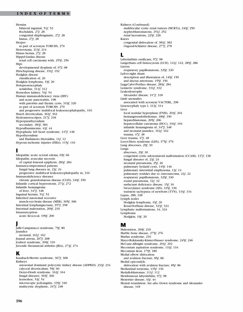
I N D E X O F T E R M S
Herniasbilateral inguinal, 51f, 52Bochdalek, 27f, 28congenital diaphragmatic, 27f, 28hiatus, 27f, 28
Herpesas part of acronym TORCHS, 270
Heterotaxia, 213f, 214Hiatus hernia, 27f, 28Hippel-Lindau disease
renal cell carcinoma with, 255f, 256Hips
developmental dysplasia of, 67f, 68Hirschsprung disease, 191f, 192Hodgkin disease
classification of, 20Hodgkin lymphoma, 19f, 20Holoprosencephaly
semilobar, 311f, 312Horseshoe kidney, 53f, 54Human immunodeficiency virus (HIV)
and acute pancreatitis, 198with parotitis and thymic cysts, 319f, 320as part of acronym TORCHS, 270and progressive multifocal leukoencephalopathy, 316
Hutch diverticulum, 361f, 362Hydrometrocolpos, 217f, 218Hyperparathyroidism
secondary, 383f, 384Hypoalbuminemia, 43f, 44Hypoplastic left heart syndrome, 147f, 148Hypothyroidism
and Hashimoto thyroiditis, 11f, 12Hypoxic-ischemic injuries (HIEs), 115f, 116
IIdiopathic acute scrotal edema, 65f, 66Idiopathic avascular necrosis
of capital femoral epiphysis, 283f, 284Immunocompromised patients
fungal lung diseases in, 23f, 24progressive multifocal leukoencephalopathy in, 316
Immunodeficiency diseaseschronic granulomatous disease (CGD), 349f, 350
Infantile cortical hyperostosis, 271f, 272Infantile hemangioma
of liver, 347f, 348Inguinal hernias, 51f, 52Inherited autosomal recessive
muscle-eye-brain disease (MEB), 305f, 306Intestinal lymphangiectasia, 357f, 358Intestinal malrotation, 209f, 210Intussusception
acute ileocecal, 199f, 200
JJaffe-Campanacci syndrome, 79f, 80Jaundice
neonatal, 161f, 162Jejunal atresia, 207f, 208Joubert syndrome, 309f, 310Juvenile rheumatoid arthritis (JRA), 273f, 274
KKasabach-Merritt syndrome, 367f, 368Kidneys
autosomal dominant polycystic kidney disease (ADPKD), 233f, 234calyceal diverticulum, 59f, 60Denys-Drash syndrome, 163f, 164fungal diseases, 365f, 366horseshoe, 53f, 54microscopic polyangiitis, 339f, 340multicystic dysplastic, 247f, 248
396
Kidneys (Continued)multilocular cystic renal tumors (MCRTs), 249f, 250nephroblastomatosis, 251f, 252renal lacerations, 225f, 226
Kneescongenital dislocation of, 381f, 382Osgood-Schlatter disease, 277f, 278
LLabyrinthitis ossificans, 97f, 98Langerhans cell histiocytosis (LCH), 141f, 142, 285f, 286Larynx
respiratory papillomatosis, 329f, 330Left-to-right shunt
description and illustration of, 149f, 150and ductus arteriosus, 155f, 156
Legg-Calve-Perthes disease, 283f, 284Lemierre syndrome, 331f, 332Leukodystrophy
Alexander disease, 317f, 318Limb anomalies
associated with acronym VACTERL, 298Lissencephaly type I, 313f, 314Liver
focal nodular hyperplasia (FNH), 203f, 204hemangioendotheliomas, 189f, 190hepatoblastomas, 205f, 206hepatocellular carcinoma (HCC), 193f, 194infantile hemangioma of, 347f, 348and neonatal jaundice, 161f, 162trauma, 47f, 48
Liver trauma, 47f, 48Loeys-Dietz syndrome (LDS), 375f, 376Lung abscesses, 29f, 30Lungs
abscesses, 29f, 30congenital cystic adenomatoid malformation (CCAM), 137f, 138fungal diseases of, 23f, 24neonatal pneumonia, 25f, 26pulmonary hydatid cysts, 145f, 146pulmonary interstitial emphysema, 13f, 14pulmonary nodules due to osteosarcoma, 21f, 22respiratory papillomatosis, 329f, 330round pneumonia, 31f, 32surfactant deficiency disease, 15f, 16Swyer-James syndrome (SJS), 335f, 336transient tachypnea of newborn (TTN), 133f, 134
Lupus, 288, 338Lymph nodes
Hodgkin lymphoma, 19f, 20Rosai-Dorfman disease, 323f, 324
Lymphatic malformations, 34, 324Lymphoma
Hodgkin, 19f, 20
MMalrotation, 209f, 210Marble bone disease, 275f, 276Marfan syndrome, 210Mayer-Rokitansky-Kuster-Hauser syndrome, 245f, 246McCune-Albright syndrome, 291f, 292Meconium aspiration syndrome, 133f, 134Meconium ileus, 179f, 180Medial elbow dislocation
and avulsion fracture, 85f, 86Medial epicondyle
dislocation with avulsion fracture, 85f, 86Mediastinal teratoma, 135f, 136Medulloblastomas, 111f, 112Membranous labyrinthitis, 97f, 98Menetrier disease, 43f, 44Mental retardation. See also Down syndrome and Alexander
disease, 318

I N D E X O F T E R M S
Merkel diverticulum/omphalomesenteric duct remnant, 171f, 172Metanephric blastema, 251f, 252Metaphysis
fractures with child abuse, 73f, 74growth plate fractures, 72
Microscopic polyangiitis, 339f, 340Midgut volvulus, 195f, 196Mixed connective tissue disorders (MCTDs), 287f, 288Monteggia fractures, 69f, 70Morgagni hernia, 27f, 28Morquio syndrome, 373f, 374Motor vehicle injuries. See also Fractures; Trauma
liver trauma, 47f, 48and posterior urethral valves, 223f, 224renal lacerations, 225f, 226
Mucopolysaccharidosis type IVdysostosis multiplex, 373f, 374
Multicystic dysplastic kidneys, 247f, 248Multifocal hepatoblastoma
and portal vein thrombosis (PVT), 183f, 184Multilocular cystic renal tumors (MCRTs), 249f, 250Muscle-eye-brain disease (MEB), 305f, 306Mycobacterium tuberculosis bacilli, 37f, 38Myelomeningocele
and Arnold-Chiari II malformations, 103f, 104and neurogenic bladder, 61f, 62
NNasal hemangiomas, 9f, 10Necrotizing enterocolitis (NEC), 181f, 182Neonatal jaundice, 161f, 162Neonatal ovarian cysts
with second follicle, 55f, 56Neonatal pneumonia
diagnosis and image of, 25f, 26and pulmonary interstitial emphysema, 13f, 14
Neonatal small left colon syndrome, 173f, 174Neoplasms
cerebral astrocytoma, 109f, 110focal nodular hyperplasia (FNH), 203f, 204hemangioendotheliomas, 189f, 190hepatoblastomas, 205f, 206hepatocellular carcinoma (HCC), 193f, 194infantile hemangioma of liver, 347f, 348medulloblastomas, 111f, 112multifocal hepatoblastoma, 183f, 184multilocular cystic renal tumors (MCRTs), 249f, 250nasal hemangiomas, 9f, 10neuroblastoma—stage 4S, 353f, 354osteosarcoma, 293f, 294renal cell carcinoma (RCC), 255f, 256solid and papillary epithelial neoplasms (SAPEN), 169f, 170Wilms tumor, 259f, 260
Nephroblastomatosis, 251f, 252Nephrogenic rest
in kidneys, 251f, 252Neuroblastomas
paraspinal or posterior mediastinal, 127f, 128Neuroblastoma—stage 4S, 353f, 354Neurogenic bladder, 61f, 62Neuronal migration anomalies
lissencephaly type I, 313f, 314Neutropenic enterocolitis, 211f, 212Nodular lung diseases, 21f, 22Nonossifying fibroma (NOF), 79f, 80Nuss procedure, 327f, 328
OOmphalocele, 187f, 188Omphalocele-exstrophy-imperforate anus-spinal defects
(OEIS), 253f, 254Omphalomesenteric duct, 171f, 172Opsoclonus-myoclonus syndrome, 354
Osgood-Schlatter disease, 277f, 278Osteochondromas
hereditary multiple exostosis (HME), 81f, 82Osteogenesis imperfecta, 267f, 268Osteogenesis imperfecta type II, 295f, 296Osteoid osteoma, 87f, 88Osteomyelitis, 289f, 290Osteopetrosis, 275f, 276Osteosarcoma
image and diagnosis of, 293f, 294pulmonary nodules due to, 21f, 22sphenoid wing, 301f, 302
Ovarian torsionand bilateral cystic ovarian teratomas, 261f, 262
Ovariesbilateral cystic ovarian teratomas, 261f, 262
PPancreas
pancreatitis, 197f, 198Shwachman-Diamond syndrome, 351f, 352
Pancreatitis, 197f, 198Paraspinal neuroblastomas, 127f, 128Parotitis
with human immunodeficiency virus (HIV), 320Patella
double-layer, 383f, 384Osgood-Schlatter disease, 277f, 278
Patent ductus arteriosus (PDA), 150, 156Patent urachus, 219f, 220Pectus excavatum
repair complications, 327f, 328Pericardial cysts, 343f, 344Peripherally inserted central catheters (PICCs), 91f, 92Pharyngitis
and Lemierre syndrome, 332Pig bronchus, 333f, 334Pilocytic astrocytoma, 112Pleural empyema, 143f, 144Pleuropulmonary blastomas, 129f, 130Pneumonia
empyema as complication of, 143f, 144neonatal, 25f, 26round, 31f, 32
Polymyositisas mixed connective tissue disorder, 287f, 288
Polyostotic fibrous dysplasia, 291f, 292Polysplenia in left isomerism, 213f, 214Portal vein
congenital absence of, 355f, 356Portal vein thrombosis (PVT)
and multifocal hepatoblastoma, 183f, 184Posterior mediastinal neuroblastomas, 127f, 128Posterior urethral valves (PUVs), 223f, 224, 257f, 258Posthemorrhagic hydrocephalus, 95f, 96Primary intestinal lymphangiectasia, 357f, 358Progressive multifocal leukoencephalopathy (PML), 315f, 316Prune-belly syndrome, 210, 235f, 236Pulmonary atresia
with intact ventricular septum, 341f, 342Pulmonary hydatid cysts, 145f, 146Pulmonary interstitial emphysema, 13f, 14Pulmonary nodules
due to osteosarcoma, 21f, 22Pulmonary sequestration, 131f, 132Pulmonary tuberculosis (TBC), 37f, 38
RRaccoon eyes, 354Radiation exposure
in children from computed tomography, 301f, 302Regional enteritis, 185f, 186Renal abscesses, 237f, 238
397

I N D E X O F T E R M S
Renal cell carcinoma (RCC), 255f, 256Renal collecting system
duplication with everting ureterocele, 221f, 222Renal insufficiency
secondary hyperparathyroidism, 383f, 384Renal lacerations, 225f, 226Renal medullary carcinoma, 359f, 360Renal (or radial) anomalies
associated with acronym VACTERL, 298Renal osteodystrophy, 384Respiratory distress
and meconium aspiration syndrome, 133f, 134Respiratory papillomatosis, 329f, 330Retinoblastoma
hereditary bilateral, 301f, 302Rhabdomyosarcoma, 121f, 122Rhizomelic chondrodysplasia punctata (RCDP), 379f, 380Rhombencephalosynapsis, 307f, 308Rickets in hypophosphatasia, 387f, 388Rosai-Dorfman disease, 323f, 324Round pneumonia, 31f, 32Rubella
as part of acronym TORCHS, 270Rugger jersey spine, 384
SSalter Harris fractures
description and illustration of, 71f, 72Type I slipped capital femoral epiphysis, 77f, 78
Sclerodermaas mixed connective tissue disorder, 287f, 288
Scrotumepididymitis, 263f, 264idiopathic acute scrotal edema, 65f, 66and testicular torsion, 265f, 266
Scurvy, 377f, 378Secondary hyperparathyroidism, 383f, 384Segmental anomalies
and failure of formation of vertebra, 83f, 84Semilobar holoprosencephaly, 311f, 312Sensorineural hearing loss (SNHL), 97f, 98Septo-optic dysplasia, 312Seventh sickle cell nephropathy, 359f, 360Shunts
complications, 95f, 96Shwachman-Diamond syndrome, 351f, 352Sickle cell anemia
diagnosis and illustration of, 279f, 280and renal medullary carcinoma, 359f, 360
Sinus histiocytosisRosai-Dorfman disease, 323f, 324
Slipped capital femoral epiphysis, 77f, 78Small bowel duplication cysts, 175f, 176Solid and papillary epithelial neoplasms (SAPEN), 169f, 170Sphenoid wing osteosarcoma
and hereditary bilateral retinoblastoma, 301f, 302Spiral fractures
of distal tibia, 76nonossifying fibroma (NOF), 79f, 80
SpleenKasabach-Merritt syndrome, 367f, 368traumatic splenic injuries: grade 4–5, 49f, 50
Spondylodiscitis, 101f, 102Staphylococcus aureus
causing osteomyelitis, 290causing renal abscesses, 238causing spondylodiscitis, 101f, 102
Straddle injuries, 240Stridor, 123f, 124Subglottic stenosis, 123f, 124Supracondylar elbow fractures, 89f, 90Surfactant deficiency
and pulmonary interstitial emphysema, 13f, 14
398
Surfactant deficiency disease, 15f, 16Swyer-James syndrome (SJS), 335f, 336Syphilis
as part of acronym TORCHS, 270Systemic lupus erythematosus (SLE)
image findings and diagnosis of, 337f, 338as mixed connective tissue disorder, 287f, 288
Systemic shunts, 150
TTeratomas
bilateral cystic ovarian teratomas, 261f, 262mediastinal, 135f, 136three types of, 136
Testestesticular microlithiasis, 63f, 64testicular torsion, 265f, 266
Testicular microlithiasis, 63f, 64Testicular torsion, 265f, 266Tetralogy of Fallot (TOF), 41f, 42Thymic cysts
with human immunodeficiency virus (HIV), 320Thyroglossal duct cysts, 119f, 120, 321f, 322Thyroid gland
goiter, 7f, 8Hashimoto thyroiditis, 11f, 12
Tibianonossifying fibroma (NOF), 79f, 80toddler’s fractures, 75f, 76
Tibial osteochondrosis, 278Toddler’s fractures, 75f, 76TORCHS infections, 270Total anomalous pulmonary venous
connection, 151f, 152Toxoplasmosis
as part of acronym TORCHS, 270Tracheal bronchus, 333f, 334Tracheobronchial anomaly, 333f, 334Tracheoesophageal fistula
associated with acronym VACTERL, 298and esophageal atresia (EA), 177f, 178
Transient tachypnea of newborn (TTN), 133f, 134Trauma
and adrenal hemorrhages, 363f, 364and child abuse, 73f, 74and duodenal hematomas, 165f, 166liver, 47f, 48renal lacerations, 225f, 226traumatic splenic injuries: grade 4–5, 49f, 50causing urethral fractures, 239f, 240
Traumatic splenic injuries: grade 4–5, 49f, 50Treacher Collins syndrome
choanal atresia associated with, 326Trisomy 21f, 150, 210Trisomy abnormalities
associated with malrotation, 210Tuberculosis, 37f, 38Tumors
cerebral astrocytoma, 109f, 110extraosseous Ewing sarcoma, 385f, 386focal nodular hyperplasia (FNH), 203f, 204hemangioendotheliomas, 189f, 190hepatocellular carcinoma (HCC), 193f, 194hereditary multiple exostosis (HME), 81f, 82Kasabach-Merritt syndrome, 367f, 368mediastinal teratoma, 135f, 136medulloblastomas, 111f, 112multilocular cystic renal tumors (MCRTs), 249f, 250nasal hemangiomas, 9f, 10neuroblastoma-stage 4S, 353f, 354osteoid osteoma, 87f, 88osteosarcoma, 293f, 294paraspinal neuroblastomas, 127f, 128

I N D E X O F T E R M S
Tumors (Continued)pilocytic astrocytoma, 112pleuropulmonary blastomas, 129f, 130posterior mediastinal neuroblastomas, 127f, 128renal cell carcinoma (RCC), 255f, 256renal medullary carcinoma, 359f, 360respiratory papillomatosis, 329f, 330retinoblastoma, 301f, 302rhabdomyosarcoma, 121f, 122solid and papillary epithelial neoplasms (SAPEN), 169f, 170sphenoid wing osteosarcoma, 301f, 302Wilms tumor, 259f, 260
Typhilitis, 211f, 212
UUmbilical arterial catheters (UACs), 91f, 92Umbilical venous catheters (UVCs), 91f, 92Unidentified bright objects
in neurofibromatosis, 107f, 108Upper respiratory infections
croup and epiglottis, 6Ureteropelvic junction (UPJ) obstruction
with vesicoureteral reflux, 229f, 230Ureters
bladder diverticulum, 361f, 362ectopic insertion of bilateral, 243f, 244
Urethraobstruction and posterior urethral valves (PUVs), 257f, 258posterior urethral valves (PUVs), 257f, 258urethral fractures, 239f, 240
Urethral fractures, 239f, 240Urethral obstruction
and posterior urethral valves (PUVs), 257f, 258Urogenital sinus, 231f, 232Uteropelvic junction (UPJ) obstruction, 235f, 236Uterus
Mayer-Rokitansky-Kuster-Hauser syndrome, 245f, 246
VVACTERL association, 297f, 298Vagina
Mayer-Rokitansky-Kuster-Hauser syndrome, 245f, 246Vaginal aplasia, 245f, 246Vascular anomalies
venolymphatic malformations, 33f, 34venous malformations, 125f, 126
Venolymphatic malformations, 33f, 34Venous malformations, 125f, 126Ventricular septum
intact with pulmonary atresia, 341f, 342Ventriculoperitoneal shunt complications, 95f, 96Ventriculoseptal defect (VSD), 150Vertebrae
failure of formation and segmentation, 83f, 84Vertebral segmentation
associated with acronym VACTERL, 298Vesicoureteral reflux (VUR)
diagnosis and illustration of, 57f, 58and posterior urethral valves, 223f, 224with ureteropelvic junction (UPJ) obstruction, 229f, 230
Viral infectionsand neonatal pneumonia, 25f, 26viral laryngotracheobronchitis, 5f, 6
Viral laryngotracheobronchitis, 5f, 6Vitamin C deficiency
and scurvy, 377f, 378Vitamin D deficiency
rickets in hypophosphatasia, 387f, 388Voiding cystourethrograms (VCUGs), 227f, 228, 229f, 230, 331f, 332,
333f, 334
WWaldmann disease, 357f, 358Wet lung syndrome, 133f, 134Wilms tumor, 259f, 260
399