Dynamic Excitations in Membranes Induced by Optical ... - NCBI
27
Dynamic Excitations in Membranes Induced by Optical Tweezers Roy Bar-Ziv,* Elisha Moses,* and Philip Nelson # *Physics of Complex Systems, Weizmann Institute of Science, Rehovot 76100, Israel, and # Department of Physics and Astronomy, University of Pennsylvania, Philadelphia, Pennsylvania 19104 USA ABSTRACT We present the phenomenology of transformations in lipid bilayers that are excited by laser tweezers. A variety of dynamic instabilities and shape transformations are observed, including the pearling instability, expulsion of vesicles, and more exotic ones, such as the formation of passages. Our physical picture of the laser-membrane interaction is based on the generation of tension in the bilayer and loss of surface area. Although tension is the origin of the pearling instability, it does not suffice to explain expulsion of vesicles, where we observe opening of giant pores and creeping motion of bilayers. We present a quantitative theoretical framework to understand most of the observed phenomenology. The main hypothesis is that lipid is pulled into the optical trap by the familiar dielectric effect, is disrupted, and finally is repackaged into an optically unresolvable suspension of colloidal particles. This suspension, in turn, can produce osmotic pressure and depletion forces, driving the observed transformations. 1. INTRODUCTION The function of biomembranes as tough, flexible partitions of cellular organelles involves a rich diversity of dynamic phenomena. Processes involving membrane shape transfor- mations such as exocytosis are controlled by the specific action of molecular machinery, which transduces readily available chemical energy and uses it to overcome viscous damping and elastic energy barriers (Alberts et al., 1989). On the micron scale, cell membranes typically inhabit an environment out of equilibrium. Their motion is governed by the interplay between thermal energy, strong dissipation due to the surrounding fluid, and chemical energy. The thermal part is not only responsible for the incessant fluc- tuations of bilayers, but also leads to nontrivial entropic forces (Helfrich and Servuss, 1984; Evans and Rawicz, 1990). From a physical point of view, it is of interest to come up with simplified artificial membrane systems that can function as “micromachines” in a thermal environment. Can a system with no biological components exhibit dy- namic processes similar to those occurring in the biological realm? We present here such a construction, composed only of lipid and water, with laser tweezers as the energy source. Artificial membrane vesicles made of lipids (“lipo- somes”) have been used extensively as model systems to study the physical properties of lipid bilayers. These include elasticity, equilibrium shapes and shape transitions, fluctu- ations, and adhesion (Deuling and Helfrich, 1976; Lip- owsky and Sackmann, 1995; Lipowsky, 1991; Seifert, 1997). It is now widely recognized that most of the equi- librium properties of bilayers, on length scales much larger than the bilayer thickness, can be explained within the framework of the curvature elasticity model (Canham, 1970; Helfrich, 1973; Evans, 1974). This includes studies that have focused on relaxation dynamics in thermal equi- librium (Brochard and Lennon, 1975; Schneider et al., 1984; Do ¨bereiner et al., 1995). Dynamic excitations of bilayers that take the membrane out of equilibrium have been less well studied, mainly because of the experimental difficulty in producing controlled perturbations in bilayers. Our approach follows the spirit of the micropipette aspi- ration technique, which allows one to apply small forces on the membrane and measure the elastic response (Evans and Needham, 1987; Evans and Rawicz, 1990; Elbaum et al., 1996). Laser tweezers have added a new tool for applying weak forces (Ashkin, 1970, 1980; Ashkin et al., 1987; Svoboda and Block, 1994; Simmons et al., 1996). To date the technique has mainly been used to manipulate micron- size particles and to measure forces acting in macromolec- ular function (Ashkin, 1997). The novelty of applying laser tweezers to lipid bilayers lies not in the ability to drag objects, but rather in the variety of remarkable qualitative transformations that they create in membrane structures. In addition, laser tweezers offer excellent spatial and temporal resolution not available with other techniques. When trapping macroscopic three-dimensional objects such as microbeads, one is concerned with the force applied by the tweezers. In contrast, when tweezers are applied to a two-dimensional surface we are interested in the energy transmitted per unit area, or in other words in the surface tension created by the laser. Laser-induced tension is one of the main ingredients of the theoretical picture to be elabo- rated below. Its existence is immediately clear when we tweeze a unilamellar giant floppy vesicle at a point along its contour. Within seconds of tweezing, the vesicle loses some of its area and becomes spherical with no visible fluctua- tions, a clear sign of tension (Bar-Ziv et al., 1995a). Laser-induced tension proved to be the key to under- standing our first new instability, the “pearling” transition Received for publication 15 September 1997 and in final form 20 March 1998. Address reprint requests to Dr. Roy Bar-Ziv, Center for Physics and Biology, Rockefeller University, 1230 York Ave., New York, NY 10021. Tel.: 212-327-8160; Fax: 212-327-7406; E-mail: barzivr@rockvax. rockefeller.edu. © 1998 by the Biophysical Society 0006-3495/98/07/294/27 $2.00 294 Biophysical Journal Volume 75 July 1998 294 –320
-
Upload
khangminh22 -
Category
Documents
-
view
0 -
download
0
Transcript of Dynamic Excitations in Membranes Induced by Optical ... - NCBI

Dynamic Excitations in Membranes Induced by Optical Tweezers
Roy Bar-Ziv,* Elisha Moses,* and Philip Nelson#
*Physics of Complex Systems, Weizmann Institute of Science, Rehovot 76100, Israel, and #Department of Physics and Astronomy,University of Pennsylvania, Philadelphia, Pennsylvania 19104 USA
ABSTRACT We present the phenomenology of transformations in lipid bilayers that are excited by laser tweezers. A varietyof dynamic instabilities and shape transformations are observed, including the pearling instability, expulsion of vesicles, andmore exotic ones, such as the formation of passages. Our physical picture of the laser-membrane interaction is based on thegeneration of tension in the bilayer and loss of surface area. Although tension is the origin of the pearling instability, it doesnot suffice to explain expulsion of vesicles, where we observe opening of giant pores and creeping motion of bilayers. Wepresent a quantitative theoretical framework to understand most of the observed phenomenology. The main hypothesis is thatlipid is pulled into the optical trap by the familiar dielectric effect, is disrupted, and finally is repackaged into an opticallyunresolvable suspension of colloidal particles. This suspension, in turn, can produce osmotic pressure and depletion forces,driving the observed transformations.
1. INTRODUCTION
The function of biomembranes as tough, flexible partitionsof cellular organelles involves a rich diversity of dynamicphenomena. Processes involving membrane shape transfor-mations such as exocytosis are controlled by the specificaction of molecular machinery, which transduces readilyavailable chemical energy and uses it to overcome viscousdamping and elastic energy barriers (Alberts et al., 1989).On the micron scale, cell membranes typically inhabit anenvironment out of equilibrium. Their motion is governedby the interplay between thermal energy, strong dissipationdue to the surrounding fluid, and chemical energy. Thethermal part is not only responsible for the incessant fluc-tuations of bilayers, but also leads to nontrivial entropicforces (Helfrich and Servuss, 1984; Evans and Rawicz,1990). From a physical point of view, it is of interest tocome up with simplified artificial membrane systems thatcan function as “micromachines” in a thermal environment.Can a system with no biological components exhibit dy-namic processes similar to those occurring in the biologicalrealm? We present here such a construction, composed onlyof lipid and water, with laser tweezers as the energy source.
Artificial membrane vesicles made of lipids (“lipo-somes”) have been used extensively as model systems tostudy the physical properties of lipid bilayers. These includeelasticity, equilibrium shapes and shape transitions, fluctu-ations, and adhesion (Deuling and Helfrich, 1976; Lip-owsky and Sackmann, 1995; Lipowsky, 1991; Seifert,1997). It is now widely recognized that most of the equi-librium properties of bilayers, on length scales much larger
than the bilayer thickness, can be explained within theframework of the curvature elasticity model (Canham,1970; Helfrich, 1973; Evans, 1974). This includes studiesthat have focused on relaxation dynamics in thermal equi-librium (Brochard and Lennon, 1975; Schneider et al.,1984; Dobereiner et al., 1995). Dynamic excitations ofbilayers that take the membraneout of equilibriumhavebeen less well studied, mainly because of the experimentaldifficulty in producing controlled perturbations in bilayers.
Our approach follows the spirit of the micropipette aspi-ration technique, which allows one to apply small forces onthe membrane and measure the elastic response (Evans andNeedham, 1987; Evans and Rawicz, 1990; Elbaum et al.,1996). Laser tweezers have added a new tool for applyingweak forces (Ashkin, 1970, 1980; Ashkin et al., 1987;Svoboda and Block, 1994; Simmons et al., 1996). To datethe technique has mainly been used to manipulate micron-size particles and to measure forces acting in macromolec-ular function (Ashkin, 1997). The novelty of applying lasertweezers to lipid bilayers lies not in the ability to dragobjects, but rather in the variety of remarkable qualitativetransformations that they create in membrane structures. Inaddition, laser tweezers offer excellent spatial and temporalresolution not available with other techniques.
When trapping macroscopic three-dimensional objectssuch as microbeads, one is concerned with the force appliedby the tweezers. In contrast, when tweezers are applied to atwo-dimensional surface we are interested in the energytransmitted per unitarea, or in other words in thesurfacetensioncreated by the laser. Laser-induced tension is one ofthe main ingredients of the theoretical picture to be elabo-rated below. Its existence is immediately clear when wetweeze a unilamellar giant floppy vesicle at a point along itscontour. Within seconds of tweezing, the vesicle loses someof its area and becomes spherical with no visible fluctua-tions, a clear sign of tension (Bar-Ziv et al., 1995a).
Laser-induced tension proved to be the key to under-standing our first new instability, the “pearling” transition
Received for publication 15 September 1997 and in final form 20 March1998.
Address reprint requests to Dr. Roy Bar-Ziv, Center for Physics andBiology, Rockefeller University, 1230 York Ave., New York, NY 10021.Tel.: 212-327-8160; Fax: 212-327-7406; E-mail: [email protected].
© 1998 by the Biophysical Society
0006-3495/98/07/294/27 $2.00
294 Biophysical Journal Volume 75 July 1998 294–320

(Bar-Ziv and Moses, 1994; Nelson et al., 1995). Longcylindrical vesicles subject to laser tweezing at a pointundergo a dramatic shape transformation into a modulatedstructure of a string of pearls completely delocalized fromthe tweezing point. The origin of this instability is a com-petition between the induced tension and the bending elas-ticity of the bilayer, as we have shown; we have quantifiedand characterized it, yielding a satisfactory agreement be-tween experiment and theory (Goldstein et al., 1996; Bar-Ziv et al., 1997b).
However, when examining the response of complex ve-sicular structures in which small vesicles are encapsulatedwithin larger ones, it becomes apparent that tension cannotbe the only ingredient in the physical picture of the laser-membrane interaction. When a parent vesicle has becomeround by laser tweezing, it can then expel an encapsulateddaughter vesicle, either spontaneously after the laser is shutoff, or by continuous tweezing. We will argue below thattwo new concepts are required to explain the observations:osmotic flow (Moroz et al., 1997) and a new “colloidalcreeping” mechanism to be introduced below. Both of thesemechanisms are rooted in the observation of irreversibleloss of membrane under the laser action. We will propose apicture in which the area detached from the bilayer by thelaser is repackaged in the form of a suspension of smallparticles (membrane fragments) that can produce osmoticeffects and depletion forces similar to those observed inother colloidal systems.
Several of the phenomena described in this paper wereannounced in earlier publications (Bar-Ziv and Moses,1994; Nelson et al., 1995; Bar-Ziv et al., 1995a,b, 1997b;Goldstein et al., 1996; Moroz et al., 1997). The purpose ofthis paper is to bring these results together, to describe newresults not previously described, and to create a unifiedphysical picture capable of explaining most of them. Theorganization of the paper is as follows. In Section 2 wepresent our physical picture for the interaction of the fo-cused laser beam that forms the optical tweezers with a fluidlipid bilayer. Section 3 is devoted to experimental proce-dures, protocol, and data analysis methods. In Section 4 wereview the pearling instability, which is currently our beststudied laser-induced dynamical shape transformation, andin Section 5 we present new results on expulsion in closedvesicles. In Section 6 we present excitations of bilayers inplanar structures, where the laser induces local unbindingand can create topological excitations. Finally, we presentnew results on exotic excitations of vesicles with a highsurface-to-volume ratio.
2. PHYSICAL PICTURE
In this section we will sketch a unified physical picture ofthe interaction of laser tweezers with a lipid bilayer, thenreview the qualitative experimental evidence for the specificelements of this picture. In the sequel we will then use thepicture to understand, in some cases quantitatively, severalof the most striking membrane transformations.
2.1. Laser-induced tension
The heart of the experiment is al 5 514 nm argon laser andan optical microscope with a strongly focusing objectivelens. Giant vesicles of various sizes and shapes are preparedby swelling pure lipids (typically dimyristoylphosphatidyl-choline (DMPC)) in water. The optical trap is produced byfocusing up toI 5 50 mW of laser intensity from a beam 6mm in diameter into a spot of sizew0 ' 0.3 mm (Svobodaand Block, 1994; Simmons et al., 1996). A strong gradientof light intensity is set at the focus of the objective lens, anda huge power density, 20 MW/cm2, passes through theD 54-nm-thick bilayers under study. Most of this energy justpasses through with minimal absorption and heating, be-cause the membrane is a thin transparent material at thelaser wavelength (see below). However, a tiny amount ofelectromagnetic energy,U, does interact with the lipid andgoes into polarizing it. This is
U 5 ~eL 2 eW! E ^uEu2&dV, (1)
whereeL andeW are the dielectric constants of the lipids andwater in the visible range, anduEu2& is the time-averagedelectric field intensity. The integral extends over the volumeof interaction of the electromagnetic field with the mem-brane. Because the thickness of the membraneD is muchsmaller than its lateral size, we can transform the integralinto a surface integral over the area of the membrane in thetrap. Using^uEu2& 5 I/w0
2, wherecW is the speed of light inwater, we get
U 5 SL ESt
dS, (2)
where
SL ;~eL 2 eW!ID
cWw02 < 4.5 z 1025erg cm22/mW z I. (3)
Because of the strong focusing of the Gaussian beam, theintegral extends over an area ofSt ' w0
2, which is muchsmaller than the total area of the vesicle.SL is anupper limiton the tension that could be induced in the membrane. Inpractice the actual tension will reflect only thenet energygain s after the membrane has been folded to fit into thetrap (see Section 2.4 below). For laser intensityI 5 10 mW,SL ' 5 3 1024 erg/cm2. As we will see, this is comparableto the tension needed to induce shape transformations inmicron-size membrane structures. The corresponding inter-action energy isU ' 10212 erg, comparable to the bendingrigidity of the bilayer and not much more than the thermalenergy kBT. It is important to note that because of thediffraction limit, w0 } l. Therefore, the laser-induced ten-sion drops significantly with wavelength,
SL } 1/l2, (4)
Bar-Ziv et al. Membrane Excitations 295

favoring in this case the use of an argon laser over near IRlasers, such as the commonly used 1064 nm YAG laser.
2.2. Membrane elasticity and entropic tension
What is the response of a lipid membrane to a local tensionof 1024–1023 erg/cm2? To answer this question we mustrecall some elements of membrane elasticity (for a recenttextbook see Safran, 1994). Lipid molecules self-assemblestrongly into bilayer membranes. Any deformation of themembrane that changes the packing of molecules from theiroptimum arrangement will incur an elastic free-energy cost,but some deformations are far less costly than others. Forexample, bending a bilayer deforms its individual monolay-ers, but by a small amount proportional toD/,, whereD isthe membrane thickness and, is the radius of curvature.Indeed, bends on the micron scale are so much easier thannet stretching of the membrane that we may neglect thelatter completely. This will be true as long as the appliedtensionS remains much less than the intrinsic stretch mod-ulus of the membrane, which is the scale of interest to us.The free energy cost of a membrane configuration is thengiven by
E 5k
2 ES 1
R11
1
R2D2
dS, (5)
wherek ' 10 2 15 kBT is the bending rigidity,R1 andR2
are the principal local radii of curvature, and the integral isover the surface of the membrane, the total area of which isfixed.
At zero temperature an ideal unconstrained membranewill adopt its preferred surface density of lipid moleculesand thus attain zero tension. At finite temperature thermalfluctuations soften the membrane and effectively render itstretchy: a weak external force acting on the membrane canunfold thermal wrinkles, increasing its apparent area with-out actually changing the microscopic surface density ofmolecules in the bilayer. Conversely, constraining the ther-mal fluctuations in a membrane lowers the entropy andgenerates a free energy cost per unit area or, in other words,an effective tension. For example, fluctuations can be con-strained by the combined constraints of fixed number oflipid molecules and fixed enclosed volume. Thus a closedvesicle will always have some nonzero surface tension. Thishas been established theoretically and beautifully demon-strated experimentally by Evans and co-workers (Helfrichand Servuss, 1984; Milner and Safran, 1987; Evans andRawicz, 1990; Seifert, 1995a). For 10-mm-size lipid vesi-cles, under no applied force, tension values are as low as1026 erg/cm2. In fact, this “entropic elasticity” persists overfive orders of magnitude, up to 0.1 erg/cm2, before thebilayer becomes a normal linearly elastic material (Evansand Rawicz, 1990). We dwell on this point because theoptical tweezers (somewhat fortuitously) operate in the mid-dle of this entropic regime.
Thus optical tweezers turned out to be a suitable tool forexciting dynamic shape transitions in bilayers, essentiallybecause of the clear separation of energy scales between thesoft, low-energy bending modes of the membranes and thehighly energetic stretching modes. Laser-induced tension of1023 erg/cm2 is a weak tension excitation compared withthe stretching modulus of;100 erg/cm2, but high whencompared with the equilibrium tension of;1026 erg/cm2.
2.3. Dynamics of tension andshape transformations
Because the membrane is a two-dimensional fluid, weshould think of its surface tension as a negative 2D pressure.What we have argued, then, is that the effect of the laser isto maintain this pressure at some valueS just outside thetrap. What happens next depends crucially on the initialmembrane configuration.
Tweezing an infinite planar membrane, or a very largeflaccid vesicle, will simply result in a slow inflow of ma-terial to the trap. The flow will be impeded by the viscousloss of the water it entrains (plus a negligible loss from thethin bilayer itself), so that the tension in the membranebecomes negligible outside a radius a few times the trapsize. Indeed, when tweezing in this configuration, we doobserve free Brownian fluctuation of the membrane awayfrom the trap.
More interesting phenomena can happen in other config-urations. As mentioned earlier, a spherical vesicle has min-imized its projected area for the given enclosed volume, andis under tension. Applying additional tension cannot furtherdecrease its surface area without releasing volume. In thiscase material flows into the trap until the tension is equal toS everywhere. If the laser is turned off at this point, thenthis equilibrium will be maintained. We will see below howthis tense state can store enough energy to drive membranereorganization. Perhaps most interesting, however, is thecase of an initiallycylindrical vesicle of radiusR0. Like thesphere, a cylinder cannot lose any surface area at fixedvolume without changing to some other shape. Plateau(1873) studied this problem in the previous century andnoted that a small periodic perturbation in the cylindricalshape could reduce area at fixed volume if its wavelengthexceeded 2pR0. Indeed, we have not seen an initial distur-bance of wavelength shorter than this.
Moreover, because of the volume constraint, the area loss(and hence the energy gain due to the work done by thelaser) is proportional to the square of the perturbation am-plitude. Because the cylinder was initially a metastableequilibrium shape, any shape perturbation costs some elas-tic bending energy, again proportional to the square of theamplitude. (In contrast, a flaccid vesicle reduces its bendingenergy by becoming more spherical.) Thus we get a com-petition: no shape change occurs unlessS exceeds a criticalvalue Spearl ; k/R0
2 (Bar-Ziv and Moses, 1994; Nelson etal., 1995; Goldstein et al., 1996). Before the tension reaches
296 Biophysical Journal Volume 75 July 1998
this value, the cylinder iseffectively rigid; tension musttherefore spread out through the incompressible membrane(as in the spherical case) instead of being lost to viscousdrag as the vesicle shape rearranges locally (as in the flaccidvesicle case).
Indeed, this situation persists even afterS rises past thepearling threshold (Goldstein et al., 1996). Here again thekey is the confined geometry of a thin cylinder. The fluidvolume which must rearrange for a shape change scaleslinearly with the amplitude (u)t of the modulation, and sothe rate of viscous energy loss is proportional to the viscos-ity h of water and tou2. On the other hand, the energy gainfrom the laser scales asu2 and so its rate is proportional toSuu. Thus initially the modulation grows at a slow ratefixed byS/h. Dimensional analysis then suggests that it alsomoves at a velocityvf ; S/h, and indeed we found that thisis an overestimate. Tension, on the other hand, spreadsdiffusively in a nearly incompressible membrane of nearlyfixed shape. Here dimensional analysis suggests a diffusionconstant ofKR0/h, where K ' 100 erg/cm2 is the bulkelastic modulus of the membrane.
Thus we find that tension initially outruns the advancingshape transformation. Although constant-velocity propaga-tion eventually catches up to diffusive spread, it turns outthat K is large enough that this does not occur in theobservable region of the initial pearling propagation (Gold-stein et al., 1996); we may thus take tension to be a constantequal toS throughout the pearling instability.
2.4. Detachment and escape of lipids
When a lipid molecule enters the laser spot, displacingwater, the system gains electric energy via Eq. 1. We foundthat continuous tweezing results in an irreversible loss ofmembrane surface area: for example, vesicles made roundand tense by the laser action remain that way indefinitely.Over 100 mm2 of membrane can disappear in this way.Thus, even though we cannot optically resolve the details ofthis process, we can nevertheless infer that lipids detach andescape from the membrane. Usually the remaining mem-brane does not become darker, and blobs of bulk lipid do notform, so some of the lost material must form membranefragments of a size well below our;200-nm resolution.Moreover, the trap volume of;3 3 10214 cm3 is too smallto contain all of the lost lipid, up to 100mm2 3 4 nm.
We gain further evidence that lipid is not simply foldingup inside the laser trap when we note that packing extramaterial into the spot would require bending the membrane,incurring a curvature energy penalty. If we assume that themembrane bends with a wavelength characterized by thetrap size, we get a lower bound for the packing cost per unitarea:Sc * 4k/w0
2 ' 3 3 1023 erg/cm2. Only for SL . Sc
would the membrane gain net energy proportional toS [SL 2 Sc by entering the trap. Experimentally we observe athreshold for tension effects at laser intensities ofIc ' 1–10mW, which corresponds toSc ' 0.5–5 3 1024 erg/cm2.
Either our estimate forSc is too high, or we must deducethat the huge energy flux through the bilayer creates addi-tional effects that supply the energy needed to repackage thelipids. (One could suppose that self-adhesion effects insidethe trap reduceSc somewhat, but the required near-cancel-lation of the folding energy seems unlikely to us.)
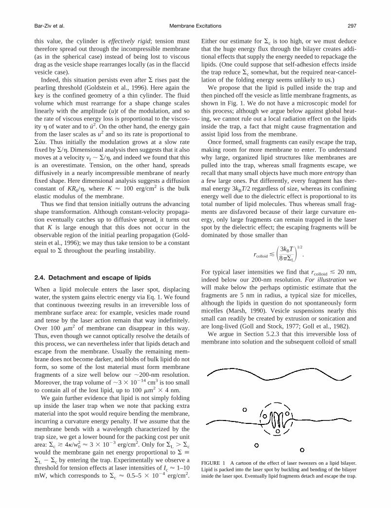
We propose that the lipid is pulled inside the trap andthen pinched off the vesicle as little membrane fragments, asshown in Fig. 1. We do not have a microscopic model forthis process; although we argue below against global heat-ing, we cannot rule out a local radiation effect on the lipidsinside the trap, a fact that might cause fragmentation andassist lipid loss from the membrane.
Once formed, small fragments can easily escape the trap,making room for more membrane to enter. To understandwhy large, organized lipid structures like membranes arepulled into the trap, whereas small fragments escape, werecall that many small objects have much moreentropythana few large ones. Put differently, every fragment has ther-mal energy 3kBT/2 regardless of size, whereas its confiningenergy well due to the dielectric effect is proportional to itstotal number of lipid molecules. Thus whereas small frag-ments are disfavored because of their large curvature en-ergy, only large fragments can remain trapped in the laserspot by the dielectric effect; the escaping fragments will bedominated by those smaller than
rcolloid & S3kBT
8pSLD1/2
.
For typical laser intensities we find thatrcolloid & 20 nm,indeed below our 200-nm resolution.For illustration wewill make below the perhaps optimistic estimate that thefragments are 5 nm in radius, a typical size for micelles,although the lipids in question do not spontaneously formmicelles (Marsh, 1990). Vesicle suspensions nearly thissmall can readily be created by extrusion or sonication andare long-lived (Goll and Stock, 1977; Goll et al., 1982).
We argue in Section 5.2.3 that this irreversible loss ofmembrane into solution and the subsequent colloid of small
FIGURE 1 A cartoon of the effect of laser tweezers on a lipid bilayer.Lipid is packed into the laser spot by buckling and bending of the bilayerinside the laser spot. Eventually lipid fragments detach and escape the trap.
Bar-Ziv et al. Membrane Excitations 297

particles thus formed in closed vesicles furnish the drivingforce for laser-induced spontaneous expulsion.
2.5. Colloidal effects
In the previous subsection we argued that the laser couldcreate a colloidal suspension of small membrane fragments.Any such particles created outside a confined region willsimply diffuse away, whereas those created inside a vesicleremain trapped and can lead to a number of surprisingeffects.
As long as the vesicle wall retains its integrity, anyosmotic activity of membrane fragments will be negligiblebecause of the far greater number of unavoidable smallsolute particles. The latter will clamp the vesicle volumebecause of their extremely slow diffusion through bilayermembranes (Alberts et al., 1989). Thus, for example, weexpect no colloidal effect in the case or pearling: here thereis very little area loss, the tweezing times can be quite small(under a second), and there is no membrane gap to permitosmotic flow.
Suppose, however, that a hole opens in the membrane, ofa size intermediate between small solutes (like sugar) andrcolloid. Then small solutes will exchange freely, leavingonly the osmotic effect of the membrane fragments. Be-cause the large fragments are concentrated inside, the neteffect is topull water into the vesicle, and thus create aninterior pressure. In Moroz et al. (1997) we argued that thiseffect supplies the driving force for vesicle expulsion.
Another well-known colloidal effect is the “depletioninteraction” (Israelachvili, 1991). Normally one thinks oftwo rigid surfaces in a colloidal suspension. Each surface ofareaA is surrounded by a small “depletion volume”Arcolloid:the center of each colloidal particle must stay outside thisvolume. The entropic cost of this forbidden volume can beeliminated by bringing the two surfaces into contact, elim-inating depletion volume and leading to a free energy gain.Equivalently, we can imagine the two surfaces being heldtogether by an effectivesuction, given by the osmotic for-mula peff 5 23kBTf/4p(rcolloid)
3, wheref is the volumefraction. We get an upper bound on this pressure by takingrcolloid ' 5 nm, the size of micelles, andf ' 0.3. Then2peff & 2 3 104 erg/cm3, a sizable pressure indeed.
We will see in Section 5.2.3 that inside a large vesicle, theexpected volume fraction is actually many thousands oftimes smaller than unity, because the laser destroys only alimited amount of membrane. However, in more confinedspaces, such as the thin layer between two bound bilayers,f can indeed approach unity, leading to large effects. Sup-pose, for example, that a bilayer edge is in contact with asecond bilayer, as occurs during vesicle expulsion. Thesliding contact keeps af ' 1 suspension on one sideseparated from pure water on the other. In this situation thesliding edge will feel an entropic force pushing it toward thecolloid side, because motion in this direction reduces thearea exposed to the colloid and hence the entropically costly
depletion volume, with no corresponding increase on thepure water side. We call this effect “colloidal creeping.”
The free-energy gain from colloidal creeping can also beconsiderable: multiplyingpeff by rcolloid in the above exam-ple gives an upper bound of 0.01 erg/cm2, again in the rangeof interest to us and, as we will see in Section 5.3.2,sufficient to enlarge a membrane pore once it forms.
2.6. Experimental evidence for thephysical picture
Before giving our detailed procedures and observations, wewill give some qualitative facts and simple estimates sup-porting the general physical picture sketched in the previoussubsections.
First we verify that the laser can apply forces of theexpected magnitude. Fig. 2 shows the result of pulling avery large flaccid vesicle. We show a section,;80 mmlong, of a much larger giant digalactosyldiglyceride(DGDG) vesicle. Tweezing at a point (arrow in Fig. 2,b–d)we were able to deform the membrane contour by slowly
FIGURE 2 Mechanical manipulation of a membrane. The line is a two-dimensional section from a DGDG giant vesicle that spanned the whole ofthe sample cell and filled more than our video frame. The vesicle wasobserved after hours of swelling in the sample cell. The dashed line inb–dindicates the undeformed contour ofa; the arrow defines the point oftweezing. The bar is 10mm.
298 Biophysical Journal Volume 75 July 1998

dragging it against the restoring elastic forces. Beyond acertain deformation (Fig. 2d), the restoring force overcamethe optical force and the membrane popped out of the trap,then slowly relaxed back to the thermal equilibrium contourof Fig. 2a. Throughout the process, the vesicle continued toexhibit visible thermal fluctuations, indicating that the in-duced tension is negligible, as expected from our discussionin Section 2.3 above.
The maximum applied optical force can be estimated bymeasuring the deformed contour at mechanical equilibrium(Fig. 2 d). The contour in Fig. 2d subtracted from that inFig. 2 a was approximated by a best fit to a Lorentzian,
h~x, y! 5h0
~x2 1 y2!/a2 1 1, (6)
with h0 5 16 mm anda 5 3.4mm. We computed the elasticbending energyE 5 1
2k*(¹2h)2dS, wherek 5 0.4 3 10212
erg is the bending modulus and dS is the surface element.Integrating over the surface deformation givesE 5 3 310211 erg. The optical force balancing the elastic restoringforce is roughlyE/h0 ' 4 3 1028 dyn. The tensionassociated with such a force is ultralow,S ' F/h0 ' 2 31025 dyn/cm, comparable to the minimum value for aflaccid vesicle mentioned in Section 2.2 above.
We can further verify the magnitude of the optical forcededuced from the mechanical pulling experiment by drag-ging a spherical vesicle with the tweezers. This is a standardprocedure for calibrating maximum trapping forces onspherical beads (Svoboda and Block, 1994; Simmons et al.,1996). For a given trapping intensity there is a maximumdragging velocityV, above which the trapped vesicle willpop out of the trap. At that velocity the optical trappingforce and the Stokes’ drag force coincide. Typically wemeasureF 5 6phRV' 1028 dyn, or 0.1 pN. This result isat least an order of magnitude less than the trapping forcemeasured on beads of similar size, because of the fact thatthe membrane is a two-dimensional object and hence theoptical force is weaker.
Turning to the specific phenomena of interest to us, wementioned in Section 2.3 that a cylindrical vesicle undersudden, uniform tension should have a thresholdSpearl 'k/R0
2, below which no shape transformation occurs. Indeed,we have observed that for a membrane tube of radiusR '1 mm made from DMPC lipid with a bending rigidityk '10212 erg, there is a threshold laser power for pearling thatcorresponds under Eq. 3 toSpearl ' 1024 erg/cm2, asexpected, a direct confirmation for the energy scale of thelaser-membrane interaction.
Tension alone cannot explain all of our phenomena, how-ever. In the expulsion experiment the laser puts a vesicle ofradiusRunder tension, but then is shut off. The vesicle thenspontaneously opens a pore to allow the expulsion of aninterior object of radiusr. One might naively suppose thatthe surface tension pulls open the pore. But anR 5 4.5 mmvesicle under a tension of 1023 dyn/cm has an area only 1%greater than the same membrane under zero external tension
(Seifert, 1995a). If the only energy storage mechanism werestretching of the membrane, all of the stored energy wouldbe spent by the time a pore of a size 1% the total vesicle areahad formed, or equivalently, by the time a volume 1.5% ofthe total vesicle volume had exited through this pore. Be-cause the daughter object can have a volume up to 50% ofthe parent vesicle (see Section 5.2 below), some othermechanism must be forcing water into the space betweenthe parent and daughter, pushing the daughter out. The onlymechanism that can force water against a pressure gradientis osmosis, and yet initially the fluids inside and out wereidentical. In Section 5.2.3 we will argue that the colloidproposed in the previous subsection provides the requiredosmotic activity.
3. EXPERIMENTAL PROCEDURES
3.1. Setup
The optical tweezers were set up using an inverted microscope, a highnumerical aperture lens, and an argon laser. The 488–514-nm Ar ion laserbeam (Coherent, Innova 70) creates a tighter beam waist than infraredlasers, and therefore a stronger electromagnetic field. Fig. 3 is a schematicdrawing of the setup. The inset is a close-up view of the vesicle suspensionchamber with a temperature control system with excellent long-term sta-bility of 5 mK.
The suspension of vesicles was kept in a closed sample cell made fromtwo thin coverslips sealed by wax or epoxy with a 50-mm mylar spacer.The cell was in contact with the objective lens on the bottom, and from thetop it was attached with immersion oil to a sapphire window that was thebottom plate of a temperature-controlled chamber. The temperature of thesapphire window and the objective lens was maintained by using waterflowing in a closed loop from a commercial heater-refrigerator bath(Lauda). The objective lens was also encased in a copper thermal sleeveencircling it, through which water from the temperature-regulated bathflowed. In principle, the bath can stabilize temperature to within60.01K,but because of heat losses we achieved only60.1K. Fine regulation of thetemperature within the vesicle cell was achieved by a thin foil heater(Minco) in the shape of an annulus placed between the sapphire and thevesicle cell. The temperature of the cell was monitored by a thermistor witha negative temperature coefficient and a sensitivity of;3000V/K, whichconstituted one leg of a Wheatstone bridge balanced by a precision (0.01%)resistance decade. The signal was fed back through a differential integratorand amplifier to a voltage-controlled power amplifier heating the foil. (Weare grateful to Y. Barad and V. Steinberg for providing us with theirhomemade low-noise instrumentation amplifier.) The temperature was setsuch that the heater always heated in steady state. Using an integration timeof ;10 s, the system averages out fluctuations and obtains long-termstability of 5 mK with ;1 W heating the foil.
3.2. Materials and preparation
Vesicles were prepared from commercially available lipids (Sigma),DMPC, stearoyloleoylphosphatidylcholine (SOPC), and DGDG, all ofthem uncharged, zwitterionic lipids, by standard protocols (Evans andNeedham, 1987), as described below. As far as we could tell, our resultswere not specific to one type of lipid, and all of the lipids we have usedconsistently displayed the same response to tweezing. In the following weshall not emphasize the chemical differences, because of the overall sim-ilarity in behavior. We preferred to work with lipids whose meltingtemperature was conveniently below our ambient working room tempera-ture. Lipid powder was added to a mixture of methanol and chloroform(2:1) at a concentration of 10 mg/ml. To produce giant vesicles, wedeposited;20 ml of lipid solution on a Teflon disk placed inside a beaker.
Bar-Ziv et al. Membrane Excitations 299

The Teflon was thoroughly cleaned before being used and was roughenedwith emery paper, as described by Evans and Needham (1987). Themethanol/chloroform solvent was allowed to evaporate and then was driedunder vacuum. This assists in obtaining a higher yield of unilamellarvesicles. Then a few milliliters of pure water or 0.1–0.5 molar sugarsolution, heated to 40°C, was added, and the beaker was left in the ovenovernight at a temperature of 40°C. Vesicle suspensions were readilyidentified as typical “clouds” and were gently aspirated into a syringe, thentransferred into the microscope cell. Glass components were cleaned bysonication in detergent and water, followed by multiple thorough rinses inultrapure water (Barnstead E-pure). This was followed by sonication in anorganic solvent, usually ethanol, for;10 min. The experiments wereperformed in the fluid state of the membranes in a closed cell, typically at15–20°C above the fluid-gel transition point.
A different preparation technique incorporating a simple kinetic proce-dure was used to produce long cylindrical structures. A few microliters oflipid solution was deposited on a clean coverslip and left for a few hoursunder vacuum in a desiccator, to allow the solvent to evaporate. Two slicesof mylar, 50mm thick, were placed on the glass as spacers, and an uppercoverslip was fixed to the bottom one with epoxy or melted wax, leaving
two opposite sides unglued. Warm water was then injected from the sideinto the cell with a syringe. This induced a flow capable of carrying alongglobules of lipids, which started to swell from the deposit on the bottomcoverslip. This injection procedure could be repeated a few times to createa number of strong flow pulses in the cell. Within a few hours of swelling,we observed tubes oriented in the direction of the flow extending overhundreds of microns, typically anchored at both ends to lipid globules thatwere still attached to the bottom coverslip. These structures remainedmetastable for hours to days.
3.3. Image analysis
Quantitative image analysis was carried out to follow dynamic shapetransformations of vesicles. We implemented a fast digitization algorithmof the two-dimensional contour extracted from the frame grabber, writtenas a user-defined interface within the NIH Image software. Do¨bereiner(1995) has presented excellent image analysis procedures to study vesicleshape transformations. A contour of a membrane obtained with phase-contrast microscopy is approximated by the contour perpendicular to thesteepest gradient of intensity along the typical halo profile. By measuringthe intensity distribution of the video pixels, one can locate the nominalline, to better than one-pixel resolution, using a smooth interpolatingscheme (Do¨bereiner, 1995). This kind of analysis is somewhat time con-suming, but is necessary for small shape changes.
In none of our shape changes did we need such an elaborate algorithm,and hence we developed a faster algorithm that finds the membranecontour to within pixel resolution, compromising for some digitizationnoise. The algorithm probes the vicinity of a contour point by averaging thepixel intensity over 13 pixels in eight directions and locating the contourpoint in the direction of maximum intensity, similar to the method of Duweet al. (1990). Once a set of points {xi, yi} is found, we smooth out thecontour and can proceed to analyze the shape changes according to need,as described in the specific sections below.
Once the contour was determined, we could measure a number ofquantities to follow the shape changes and the fluctuations. A simple wayto follow the decrease in fluctuations during tweezing (Section 5.2.1) andadhesion deformation (Section 5.2.2) of spherical vesicles was to measurethe fluctuations in the normalized mean square of the radius. These aredefined as
^R2& 2 ^R&2
^R&2 , (7)
where
^R& 5 ~1/N!Oi
N
uRW i~x, y! 2 RW CMu (8)
is the average radius of the contour relative to the center of mass, andsimilarly for ^R2&.
To quantify the shape fluctuations of open contours (Section 5.3.2), wetransform to the arclength parameterization:
si 5 si21 1 Î~xi 2 xi21!2 1 ~yi 2 yi21!
2 (9)
Ci 5 2arctanSyi11 2 yi21
xi11 2 xi21D, (10)
with proper care taken at the boundaries. From the starting point,s 5 0, tothe end point,s 5 s*, we computed the tangent angle to the contour,C(s).For each contour we numerically computed two parameters: the “curvatureintegral,”
KSdC
dsD2L ;
1
s*ESdC
dsD2
ds, (11)
FIGURE 3 Argon: argon laser (Coherent Innova 70) operating in the514.5 line. M1 and M2: mirrors. BE: beam expander composed of twopositive lenses. I: iris diaphragm. Inverted microscope (Zeiss Axiovert135). DM: dichroic mirror. CCD: CCD camera (Hamamatsu c2400 orCohu 6500). MON: Monitor. O: microscope objective (Zeiss plan-apo-chromat Phase363/1.4). Images are taken into the computer (Macintosh,Quadra 800) using a frame grabber (Data Translation) and Image software(NIH). S: sample. Inset: Obj: Objective Lens. CT: Copper tubing. Heater:thin foil 50 V (Minco). SW: sapphire window. W: optical window. T:temperature-regulated stage.
300 Biophysical Journal Volume 75 July 1998

and the “surface integral,”
^C2& ;1
s*EC2ds. (12)
These two quantities will help us evaluate and distinguish the geometriccontributions to the energy of the membrane, arising either from changingthe surface or from changes in the curvature.
Finally, to analyze the shape deformations in detail for axisymmetricalshapes (Section 5.3.3), we used the mode expansion method developed byDobereiner (1995). For each frame we rotated and translated the contour sothat it was aligned with the major axis of the inertia tensor. One can thenparameterize the vesicle contour by the tangent angle to the contourarclengthC(s) measured for each half-contour from the north pole,s 5 0,to the south pole ats 5 s*. Thus,
C~s! 5 ps
s*1 A1sinSp
s
s*D 1 A2sinS2ps
s*D1 A3sinS3p
s
s*D 1 · · ·
(13)
A2 measures the ellipsoidal deformation of the sphere (Do¨bereiner, 1995):
A2 5 1 1 Oi
SCisinS2psi
s*D 1 Ci11sinS2psi11
s* DDsi11 2 si
s*.
(14)
3.4. Heating and absorption
Working with focused laser and biological materials, one is always con-cerned with radiation heating effects. The most direct evidence that thereis no global heating of the water is that the membrane does not respondunless the laser is applied directly to it. To further confirm this, wemeasured the absorption directly and estimated the amount of heating dueto absorption at the laser wavelength by assuming that the trap is alocalized heat source (Block, 1990). The steady-state temperature rise ofthe surrounding liquid isDT 5 Iabs/lR (Block, 1990), whereIabs is theintensity absorbed at the laser wavelength,l is the thermal conductivity ofthe surrounding liquid, andR is the distance from the heat source. Theabsorption inside a trap of sizew0 is Iabs5 I0(1 2 e2w0/j), whereI0 is theincident intensity andj is the absorption length of the material at the laserwavelength. We measured the absorption of a highly concentrated lipidsolution (100 mg/ml DMPC in methanol-chloroform (1:1)). We found anupper bound of 0.024 on the optical density (absorbance), at 488–514 nm,from which we obtained an upper bound of 0.16 liter/mole-cm for themolar extinction coefficient,e# of lipids. We consider two extreme cases inwhich 1) the membrane bilayer is entirely crumpled inside the trap at aconcentration ofc# ' 0.45 mole/liter, and 2) the membrane is flat inside thetrap at a concentration ofc# ' 6 3 1023 mole/liter. The absorbance insidethe trap of sizew0 ' 0.3 mm is w0/j [ e#c#w0 ' 2 3 1026 for the first caseandw0/j ' 3 3 1028 for the latter case. Hence for an input intensity ofI0 5 50 mW (at the trap), the upper limit of the local temperature rise isDT ' (I0/lw0)(w0/j) ' 0.5 K and 53 1023 K, respectively. Both resultsfor DT should be taken as overestimates and upper bounds for the actualheating. For example, we have neglected local cooling due to convection.
4. PEARLING OF CYLINDRICAL VESICLES: ADYNAMICAL INSTABILITY
“Pearling” is the peristaltic transition that a membrane tubethat was stabilized by curvature elasticity undergoes when asudden tension is applied to it. Of the many excitations and
transitions that we have observed in membranes, this is thebest characterized by far and has played an important role inour understanding of nonlinear dynamics in membranestructures. For one thing, it is experimentally well con-trolled. Second, it can be formulated as a theoreticallytractable one-dimensional problem. Third, it clearly showsthe generation of tension in the membrane by the laser, atension that interacts and competes with the curvature elas-tic energy in a manner that lies at the heart of the action ofthe laser on membrane structures in general. Finally, ittypifies many of the problems and aspects that arise withdynamical problems: strong coupling to the surroundingflow, universal behavior and critical exponents at the tran-sition, dynamical selection of velocities and wavelength,strong fluctuations and their interplay with the linear growthregime, and a complex evolution of the fully developednonlinear regime. Clearly this is a rich system, exhibiting avariety of complex phenomena, yet it is simple enough to beunderstood in some depth.
Beyond establishing the existence of the instability, ourefforts have mainly concentrated on elucidation of the onsetand linear stage. Already this proved nontrivial, because thethermal fluctuations compete with the hydrodynamic modesat onset and mask the linear stage, which we were unable tostabilize. An experimental challenge would now be to slowdown or otherwise control the linear stage, to measure thelinear coefficients directly. Because we were unable to dothat, we measured them indirectly, through the existence ofpropagating front solutions. Although attempts to charac-terize the nonlinear stage (Bar-Ziv and Moses, 1994;Goveas et al., 1997) look promising, this is clearly a differ-ent level of difficulty.
Since we reported the instability, numerous observationshave been made on the excitation of pearls in a variety ofother situations, usually associated with mechanically in-duced tension. Specially interesting examples are the push-ing of the membrane from within by a microtubule (D.Kuchnir Fygenson, private communication) and the desta-bilization of a tether by pulling on a bead attached to themembrane (B. Pouligny, private communication).
Schematically, the mechanisms and stages in the progres-sion of the pearling instability can be described by thefollowing flow chart: Laser action3 Competition of ten-sion and curvature3 Instability3 Flow and Dynamics3Threshold and onset3 Propagation3 Marginal StabilityCriterion3 Quantitative comparison to Linear Theory3Nonlinear regime.
4.1. Phenomenology
Our picture of the instability is as follows. The laser grabsthe membrane at one point and begins pulling in lipid fromthe membrane that is outside of the trap. This causes a lossof area in the rest of the membrane, and an effective tensionin it. The instability begins at the point of tweezing andpropagates outward, at a rate that depends on the strength of
Bar-Ziv et al. Membrane Excitations 301

tweezing, or applied tension. Fig. 4 shows an example of apropagating, fully developed pearling instability. The linearsinusoidal perturbation rapidly increases in amplitude andcoarsens. This tendency continues into the highly nonlinearstage, where the amplitude has peaked, making the pearlsbig and almost spherical, while the diameter of the tube thatconnects the pearls becomes extremely small.
Interestingly, if the pearling has developed into the non-linear stage, then after the laser is shut off we observe thatpearls flow back and migrate toward the point of tweezing,where they collect until the tension relaxes. Measuring adecay of migration velocity shows that there is a monotonicdecrease in the tension after the laser is turned off.
A striking aspect of the instability, which differentiates itfrom other phenomena in membranes, is the nonlocal effectof the localized laser action. Nowhere else does a force,caused by the focusing of light down to a 0.3-mm spot,cause the instability of a membrane structure for hundredsof microns. This proves the global nature of the pearlinginstability, and dispels any worries about heating effects ofthe laser. This global instability is instead related to theclassic Rayleigh instability of a cylinder under surface ten-sion. An extreme example of this is given in Fig. 5a, wheretubes that formed junctions with each other are seen todestabilize along all of the different arms, and the pearling“jumps” across intersections (Fig. 5b). Further evidence forthe long range of the tension effect is seen in Fig. 6, wherea collage is made of many adjacent video frames that covera length of tube much larger than what we usually viewed.
It is possible to induce pearling by mechanical tensioninstead of the laser-induced tension, a fact that further
strengthens our picture of the laser action. This was done bysimple micromanipulation. We inserted a glass microneedleinto the sample and pulled on a lipid globule that had tubesattached to it. A rapid enough pull caused elongation of thetubes and produced uniform pearling modes with both lin-ear, small-amplitude modulations and nonlinear, isolatedpearl structures. This is shown in Fig. 7, where the glass
FIGURE 4 Propagation of a fully developed, large-amplitude pearlingfront outward from the illuminated laser spot. Time between frames inseconds: (a) 0, (b) 0.44, (c) 0.6, (d) 0.84, (e) 1.08, (f ) 1.14. The barrepresents 10mm.
FIGURE 5 (a) A network of connected intersecting tubes made fromDGDG lipids. The marks indicate different sections of the tube, and thecrosses indicate the position of the laser tweezers. (b) Upon tweezing of thetube at one place, the instability propagates to all sections. The tube herehas reached the nonlinear stage, and the pearls are stable over a long time.The bar represents 10mm.
302 Biophysical Journal Volume 75 July 1998

microneedle (not shown) was pulling at the end of the tube.The instability initiated with uniform, small-amplitude longmodes and then transformed into isolated spherical pearlsconnected by thin tubes, exactly as in the laser induced case.An additional example is presented in Fig. 8, where weinduced pearling by using the tweezers to drag a bead thatwas encapsulated in the vesicle wall.
Because a globule is an infinite reservoir of lipids, onecan, in principle, mechanically draw tubes of increasinglength without inducing tension (and hence pearling) bypulling slowly, so that new lipid can flow along with thepull from the lipid reservoir. However, if the pull is rapidand the tube long enough, lipid cannot flow from the glob-ule, and the surface area of the tube is effectively fixed. Forthe tweezers, no lipid is added near the trap. Thus mem-brane can only be delivered by a transformation to a newshape of equal volume but less area per unit length.
The production of tubes and their subsequent robustnessare in themselves an interesting issue. Our tubes are allmetastable, ending up after many hours (about a day) as a
large sphere on a very thin tube. The fact that these tubescome in a range of radii and lengths, and that they stillevolve as we observe them, complicates the picture suffi-ciently that at present there is no explanation of the exactdetails of their production and evolution.
4.2. Overview of theoretical approach
4.2.1. Basic considerations and assumptions
The origin of the pearling instability of a membrane cylin-der can be understood as a competition between the curva-
FIGURE 7 Pearling by a rapid mechanical pull with a glass microneedle.The needle (not shown, on right side of frame) was positioned perpendic-ular to the tube axis, and the direction of the pull was parallel to the axis.
FIGURE 8 Pearling by several rapid mechanical pulls performed con-secutively on a silica bead that was encapsulated in a vesicle and trappedby the tweezers. The typical time scale is seconds, and the tugging velocitywas typically 15mm/s. The bar represents 10mm.
FIGURE 6 A collage of videoframes showing the long range of theinstability. The tweezing point was justto the left of the picture. The bar scaleis 10 mm.
Bar-Ziv et al. Membrane Excitations 303

ture elasticity that stabilizes the tube and an externallyapplied surface tension that destabilizes the tube (Bar-Zivand Moses, 1994). As mentioned in Section 2.3 the desta-bilizing effect of tension arises from the geometrical factthat long-wavelength deformations on a cylinder can reducethe surface area while maintaining its enclosed volume, thussatisfying the tendency of a positive surface tension tominimize area. The most simplified approach is to assumethat the tension is constant in time and uniform along thetube. In Section 2.3 we recalled the argument of Goldsteinet al. (1996) that the laser applies a sudden tension thatspreads rapidly along the tube due to the incompressibilityof the membrane. Thus tension exists in the membranebefore any shape transformation can occur, and one cananalyze the initiation of the instability, assuming a constanttension. The same argument holds for the mechanicallyinduced instability where, by applying a rapid pull on aglobule with an attached tube, we induce a sudden force perunit length (tension) along the tube before a change of shapecan occur.
Granek and Olami (1995) have proposed an alternativetime dependence of the laser-induced tension resulting fromtwo competing effects: an assumed constant suction rate ofsurface area, rather than the constant tension assumed here,and the stabilizing tendency of the membrane to revert tozero tension. Their analysis predicted a coupling betweenthe time-dependent surface tension and the growth of theinstability. We have not been able to address the timedependence of the tension in our experiment and havetherefore assumed constant and uniform tension, at least inthe initial growth of the instability.
Olmsted and MacKintosh (1997) have argued that gradi-ents of tension are created in the laser experiment, becauseat the far ends of the cylinder the membrane is attached toa lipid reservoir at zero tension. However, our field of viewin the microscope limits our observation to a few wave-lengths on each side of the tweezing site, which is typicallychosen to be quite far from the ends of the tube, and hencewe are insensitive to gradients.
4.2.2. Linear stability analysis
Consider a membrane tube subject to uniform and constanttension. The energy of the membrane is then a sum of thecurvature elasticity and a surface energy,
F 5 kE2H2dS1 SEdS. (15)
To identify the instability we must calculate the energy ofsmall deformations of the shape and find those modes thatreduce the energy of the unperturbed tube (Bar-Ziv andMoses, 1994; Nelson et al., 1995; Granek and Olami, 1995;Gurin et al., 1996). We limit ourselves to shape deforma-tions that are axisymmetrical and in which the axis isunperturbed (peristaltic modes). For a surface given in termsof the local radius,R(z), in cylindrical coordinates the area
element is dS5 2pR(z)=1 1 Rz(z)2dz, where the subscriptdenotes the partial derivative, and the mean curvatureH is
H 5RRzz 2 1 2 Rz
2
2R~1 1 Rz2!3/2 . (16)
Linear stability analysis permits us to restrict our atten-tion to sinusoidal perturbations of the form
R~z! 5 r0 1 uqsin~qz!. (17)
The unperturbed tube radius,R0, is related to the amplituder0 via the volume conservation constraint:r0 5R0
=1 2 uq2/2R0
2. Integrating and keeping terms to quadraticorder inuq, we obtain the excess energy per unit length overthe unperturbed tube,
f 51
2
pk
R03 O
k
SS32 2 sD 1 ~s 2 1!k2 1 k4Duk2, (18)
where the nondimensional wavenumber isk 5 qR0, ands [SR0
2/k is the normalized ratio of surface tension to curva-ture. For S 5 0 all wavenumbers are stabilized by thecurvature. Fork 5 0 we have the Rayleigh instability of acylinder of fluid, where all modes withk , 1 are unstable,as they reduce the surface area at constant volume (Ray-leigh, 1892). For finiteS and k, long-wavelength modesbecome unstable only above a critical value,
Spearl53k
2R02. (19)
4.2.3. Dispersion relation
The linear stability analysis identifies the instability controlparameter and the unstable modes; it also seems to predictthat, for anyS, the most unstable mode isk 5 0, becausethat is where Eq. 18 is as negative as possible. Indeed,Rayleigh’s original analysis of the instability of a viscousthread gave this conclusion, leading to an irregular breakupof the thread (Rayleigh, 1892). But the pearling instabilityselects a well-defined, nonzero wavenumber (Fig. 4). Tounderstand this discrepancy, note that Eq. 18 says nothingabout therate of growth of a mode. In fact, modes withk '0 involve large-scale fluid motion, a process that incursstrong hydrodynamic dissipation. To obtain the growth ratesof the unstable modes and the fastest growing mode, one hasto solve the hydrodynamic equations of the fluid motion,which balance the tension against the viscous dissipationunder the appropriate boundary conditions.
We can easily carry out these steps using a rough approx-imation to the hydrodynamics. Neglecting the bending stiff-ness for the moment, Eq. 18 reduces to an energy for modek whose time derivative isPlaser5 (pS/R0)(21 1 k2)ukuk.This is minus the rate at which energy is given to the systemby the laser. To see where this energy goes, we note that intime dt a volumedV ' (2pR0/2k) z (2pR0) z (du/2) of fluidneeds to be moved from the narrowing parts of the tube to
304 Biophysical Journal Volume 75 July 1998

the thickening parts. To do this while maintaining no-slipboundary conditions at the wall of the tube requires a centralfluid velocity of v(0) 5 2pu/k, by the Poiseuille formula.The squared shear rate of this flow, times the viscosityh ofwater, is the rate of viscous energy loss per unit volume.Equating the total lossh(16p3/5k2)u2R0 to 2Plaser thengives the approximate growth rate,v(k) [ u/u 5 (5S/16p2R0
2)k2(1 2 k2). Thus we see that the growth rate indeedis not a maximum atk 5 0, but rather atk 5 1/=2 ' 0.7,roughly as observed.
Although the above derivation is transparent, quantitativecomparison to the experiment requires a more accuratetreatment (Goldstein et al., 1996; Bar-Ziv et al., 1997b). Inparticular, by neglecting bending stiffness we have missedthe threshold phenomenon mentioned in the previous sub-section. A useful approximate form for the complete dis-persion relation is (Bar-Ziv et al., 1997b)
v~k! 5 tk21bk2F32SS 2 Spearl
SpearlD~1 2 k2! 2 k2 2 k4G, (20)
where b ' 0.04 andtk 5 hR03/k. The k2 multiplier is
familiar from the simplified treatment in the precedingparagraph; it reflects the dissipation of long modes. Theterms in the brackets originate from the curvature andsurface energies of the mode. The dispersion relation pre-dicts a continuous transition with a selected wavenumber ofkmax ' 0 at onset andkmax3 0.68 for larger values of thecontrol parameter (Goldstein et al., 1996).
4.2.4. Marginal stability criterion and linear response
Most theoretical work to date has focused on the initial,linear instability (but see Goveas et al., 1997). This repre-sents a major obstacle to quantitative comparison of theoryand experiment, because the linear stage cannot be stabi-lized experimentally, nor can its growth rate be measureddirectly. In fact, the linear stage appears to be completelymasked by fluctuations of the membrane, with amplitudeand wavelength similar to those of the instability. We arethus driven to try to use linear theory to describe nonlinearphenomena, and this is made possible by the existence ofpropagation of the instability outward from the point oftweezing, in both directions along the tube. It is well knownthat for a variety of nonlinear systems in which a stable,patterned state (in our case the pearls) invades an unstable,uniform one (the straight, tense tube), the selected velocityand wavelength can be found from the linear dispersionrelation, using the “marginal stability criterion” (MSC)(Dee and Langer, 1983; Fineberg and Steinberg, 1987; vanSaarloos, 1988). This works because the front at its leadingedge has small amplitude and obeys the linearized equationof motion. Using the MSC, numerical evidence has beengiven for the existence of a front, along with precise analyticpredictions for the critical velocity and wavelength fortensionS well above the thresholdSpearl (Goldstein et al.,1996). One surprising feature of the analysis is that the
selected wavelength is slightly different from the mostunstable mode, leading to a deviation from the simplifiedmodel we sketched in Section 4.2.3; specifically, the veloc-ity of propagation was found to scale as
v# ; V/Vk 5 0.082S/Spearl, (21)
whereVk 5 k/hR02. Close to threshold, one must solve the
equations of Goldstein et al. (1996) numerically (T. R.Powers, unpublished calculation). A convenient interpolat-ing formula approximately reproducing the solution is
v# ; V/Vk 5 0.082~S/Spearl1 0.067! 2 3713e210.655S/Spearl.(22)
Equation 22 is approximately valid for anyS . Spearl.A different approach to analyzing the propagation is to
use a linear response to calculate the response of an unstablesystem to a localized, point-like perturbation (Gurin et al.,1996). Because of dispersion, the perturbation diffuses fromthe origin and is amplified by the unstable modes. Thisanalysis predicts an identical scaling of velocity and intro-duces an experimentally measurable delay time that is thetypical time needed for an appreciable amplitude to develop,
Dt 5 33CtkS Spearl
S 2 SpearlD, (23)
whereC is an experimental parameter measuring the am-plification that is needed to bring the amplitude to anexperimentally measurable value (Bar-Ziv et al., 1997b).
4.3. Onset and propagation
We have recently reported a quantitative study of the onsetof the instability and the propagation of unstable fronts(Bar-Ziv et al., 1997b). The experiment was carried out ontubes made from SOPC. Tubes were produced by firstdepositing lipid on the bottom plate, closing the cell with atop plate, and then filling the cell with water through smallfill holes in the side, which were then sealed with epoxy. Asthe lipid structures were self-assembling, a flow was in-duced by gentle rubbing along the top plate of the samplecell. Lipid globules detached as a result of the flow, and thetether they left behind usually evolved into a single-bilayertube. We determined that it was a single bilayer by opticalcontrast and by tweezing, which easily separated tubes thatwere composed of many bilayers into the separate constit-uent bilayers. In this specific sample the tube diameterswere relatively monodisperse, almost all of them in the1-mm range. This is probably related to the uniformity of thedrag force induced by a large-scale flow. The concept ofarea loss by application of the tweezers was vividly exem-plified on occasion by the formation of a small vesicle thatwas created inside the tubes upon prolonged tweezing. Thisvesicle typically had a radius comparable to that of the tubeand was visible, differing from the fragments that we hy-pothesize to form an osmotic gas. Such cases were dis-carded in the analysis of propagation.
Bar-Ziv et al. Membrane Excitations 305

Experimentally, the onset of pearling occurred when thelaser intensity exceeded a critical intensityIpearl, which wemeasured to beIpearl 5 (10 6 4) mW on tubes of radiusR0 5 (0.6 6 0.1) mm. Using these values, we obtainSL 54.5 3 1024 erg/cm2 for the laser-induced tension (Eq. 3)andSpearl 5 4.2 3 1024 erg/cm2 for the critical tension ofpearling, with errors of up to 40% in both. The roughagreement between the two estimates of the tension is aqualitative confirmation of our simple electrodynamicmodel for the laser-membrane interaction.
In Bar-Ziv et al. (1997b) we usedIpearlmeasured for onetube to evaluate the control parametere [ (I 2 Ipearl)/Ipearl5(S 2 Spearl)/Spearl for tubes with 14 values ofR0 in therangeR0 ' 0.4–1.0mm. (We also assumed that the packingenergySc was negligible. We return to this point below.)For each value ofe we measured the position of the frontX(t) 5 V z (t 2 Dt), with t 5 0, the time the laser was turnedon, and extracted the velocity of propagationV, the delaytime Dt, and the selected wavenumberqR0. We review theresults forDt and qR0 here for completeness (Figs. 9 and10). In particular, Fig. 10 qualitatively supports the predic-tion of a continuous bifurcation with long wavelength atthreshold (Bar-Ziv et al., 1997b).
The determination of the front velocity as obtained fromfigures such as Fig. 4 relied on our ability to distinguishbetween a pearled state and a “straight” fluctuating tube.The difficulty in determining the front position is visuallyapparent, and this is a “best case,” because the amplitude inFig. 4 is actually nonlinear, and higher than the fronts thatactually were used in the analysis. The arrowheads in thisfigure, as well those in Figure 1 of Bar-Ziv et al. (1997b),approximate a straight line and demonstrate how a propa-gation speed was obtained. We found out that this was donebest by eye and not by our computer algorithm. Because thetrap was positioned in the center of the video field of view,we typically obtained only three or four measurements ofXversust. As a consequence, the determination of the frontvelocity is noisy and limited, and, in particular, we are not
sensitive to slowing down of the front at large distancesfrom the trap.
For a more detailed comparison to the MSC, we nowanalyze the front velocity using the interpolation formula,Eq. 22. In particular, the behavior of pearling near thresholdnot only tests the MSC, but also lets us say something aboutthe possible folding energySc introduced in Section 2.4.Recall thatSc is a hypothetical packing cost per unit area forpulling material into the trap; if present, it reduces theeffective laser-generated tension.
Rather than measuringIpearlonce and using it to calibrateall of the other tubes, we will now simply fit the observedfront velocity V(I, R0) globally to Eq. 22 for a total of 30trials. We take a generic form for the laser-generated ten-sion, S 5 PI 2 Sc, whereP is a fitting parameter. Thediscussion of Section 2.1 gave a rough expectation thatP '5 3 1025 erg cm22/mW (Eq. 3), butSc is unknown.
We wish to test several predictions from our theory: 1)MSC predicts constant front velocity. This is observed inevery trial (data not shown). 2) MSC, combined with ourdielectric model of laser action, also predicts a linear rela-tion between velocity and laser power, as long as the latteris well above threshold. 3) Moreover, Eq. 22 claims that allof the curvesV(I) for various R0 collapse onto a singleuniversal curve when the dimensionless velocityv# is plottedagainstS/Spearl. 4) BecauseVk andSpearlboth scale as 1/R0
2,the slope ofV(I) is a universal number, independent oftubule radius. 5) As we just mentioned, we also have anestimate for the expected value of this slope.
Fig. 11 qualitatively confirms predictions 2) and 4). Wecarried out the fitting toP described above, again assumingSc 5 0. Evidently, five of the six tubule radii showncollapse onto one universal curve. We have not explainedthe dissident tubule; note, however, that itsR0 is not anextreme value. The data do not allow detailed assessment of3) beyond the universality of the slope, but we note thatpearling fronts do not propagate below threshold,s , 1,
FIGURE 9 Delay timeDt scaled byt 5 R3h/k as a function ofe. Thebest fit (solid line) yields Dt/t 5 (1.0 6 0.2) z 103 3 e21.06 0.15. Takenfrom Bar-Ziv et al. (1997b).
FIGURE 10 Selected wavenumberqR0 as a function ofe. The theoret-ical predictions for the fastest growing mode from the dispersion relationof GNPS (dashed curve) and their MSC wavenumber correction (solidcurve) are also plotted. Taken from Bar-Ziv et al. (1997b).
306 Biophysical Journal Volume 75 July 1998

consistent with 3). As for 4), the fit slopeP 5 2 3 1025 ergcm22/mW is about half our rough estimate (Eq. 3).
We were not able to extract a definite number forSc,because of the scatter in the data and random and systematicerrors in measuringR0. (Note thatR0 drops out of the slopedetermination above.) However, we can rule out the largevalue naı¨vely estimated in Section 2.4 above, in agreementwith the anecdotal evidence mentioned there. Thus the exactprocess taking place inside the laser spot remains a mystery.One possibility is that the dielectric forces estimated inSection 2.1 pull material into the spot, whereupon enoughenergy is extracted from the beam to pulverize whatevermaterial happens to be inside.
4.4. Nonlinear stage
Longer or stronger application of the tweezers in the exper-iment leads to dynamic structures of isolated spheres thatare interconnected by regions that have collapsed to verythin tubes (0.1–0.3-mm radius, depending onR0). Whereasthe initial instability propagates outward, these spherestravel back, flowing along the tube toward the point ofapplication of the tweezers, where they aggregate (Fig. 12).This motion typically continues for many seconds after thelaser has been turned off. The velocity of a single pearl wasconstant to a very good approximation over the stretch wecould measure (;30mm), ranging between 0.1 and 10mm/sfor DMPC. However, with time (measured in terms of thenumber of pearls,N, reaching the cluster), the velocitydecreased linearly, as shown in Fig. 13. This implies asimilar reduction in the force acting on the pearls. It can beshown that a linear decrease withN indicates an exponentialdecrease with arrival time.
The spheres are taut as they move, with all fluctuationsdamped by the surface tension. Eventually the pearls losethis tautness, but then they do not travel any more. Thevelocity of successive pearls as a function of time of arrivalat the cluster is a slow relaxation decay consistent with anexponential time dependence with a time constant on theorder of minutes. We did not measure the velocities as afunction of the laser intensity or tweezing time.
The motion of the pearls indicates a velocity scaleVp setby a gradient of pressure along the tube¹W P. For Poiseuilleflow of water in the very thin tubes that the motion of thepearls causes,Vp 5 R1
2¹W P/4h, whereR1 is the thin tuberadius. Taking as a rough estimate¹W P ' S/R1L, the Laplacepressure difference between pearl and tube over a typicaldistanceL between adjacent pearls, we obtainVp ' SR1/
FIGURE 13 Measured velocities of successive pearls as they arrive atthe cluster where the tweezers were applied. The velocities are normalizedto give the Stokes drag force that they encounter,F 5 6phRv, whereh isthe fluid viscosity,R is the pearl radius, andv is the velocity. The forcesare plotted as a function of both the time of arrival at the cluster and thepearl ordinal (inset). Full circles designate data of DGDG, and the otherpoints correspond to three different tubes of DMPC.
FIGURE 11 Dimensionless velocityV/Vk of pearl propagation versusdimensionless tensionS/Spearl for various tubule radiiR. The solid line isthe theoretical prediction (Eq. 17), withSc assumed equal to zero and onefitting parameterP (see text).
FIGURE 12 “Pearls on a string”: nonlinear, late stages of anR0 5 1 mmDMPC tube. The time between frames is 2 s, starting;20 s after the laserwas shut off. The bar represents 10mm. Taken from Bar-Ziv and Moses(1994).
Bar-Ziv et al. Membrane Excitations 307

4hL. ForR1 5 0.2mm, L 5 10 mm, andS 5 1023 erg/cm2,Vp is on the order of 5mm/s, in reasonable agreement withthe measured velocities.
At this point, our experimental precision and control forthe nonlinear regimen are far below what we can reach inthe linear stage. For this reason, we cannot compare ourresults quantitatively to the theoretical predictions ofGoveas et al. (1997). However, they did find qualitativeagreement with some of our results. Their predicted valuesfor the drift velocity of the pearls are on the same order ofmagnitude as the measured ones depicted in Fig. 13.
We conclude the discussion on cylindrical instabilities bypresenting a nonstandard case, in which the axial symmetryis not conserved. This instability was observed without anylaser action, but is a rare occurrence. The lowest such mode(Gurin et al., 1996) is an undulation of the cylinder. Fig. 14shows just such an excitation, probably produced by thesudden motion of one of the globules anchoring the tube.This would lead to excess area in the tube while conservingvolume.
To the best of our knowledge, there has been no previousreport of an undulation with a well-defined wavelength in afluid membrane tube. To see how this is possible, we sketcha crude model. Excess area gives rise to a negative surfacetension2S. Assuming, as in the pearling instability, thatthis tension is effectively uniform, we can write the energygained per unit length by the tube when its axis wanders asSdA/L. If the axis wanders sinusoidally with wavelength2pR0/q and amplitudea, then the time derivative d/dt SdA/L 5 (pq2S/2R0)aa. This energy gain, in turn, is spent onStokes drag. For smallq, we have roughly a force per unitlength of 4ph times the velocity. Equating the correspond-ing energy loss to the energy gain yields a growth rate ofa/a 5 q2S/2hR0, which vanishes asq3 0. Thus, perhapssurprisingly, the fastest growing mode is at a finite wave-length, as observed in Fig. 14. Because the only length scalein the problem isR0 itself, it is not surprising that the chosenwavelength is comparable toR0.
5. LASER-INDUCED EXPULSION IN VESICLES:AN ARTIFICIAL MACHINE ON THEMICRON SCALE
5.1. Summary
In this section we introduce the tweezers into structures withspherical topology. Two classes of transformations can be
observed, each raising its own problems and exposing dif-ferent physical mechanisms. Spontaneous expulsion of in-ner content will lead us to an understanding of tension in acompact topology, and to a conjecture regarding the effectof detached membrane fragments as an osmotic gas. Expul-sion under continuous action of the tweezers will lead us toinvoke the suspension of lipid particles as a mechanism forgenerating depletion forces, which can cause vesicles toslide and peel off each other. As a bonus, it led to theunexpected observation and measurement of the “buckling”instability of a spherical vesicle in an oscillatory processthat is as yet unexplained.
Our preparation procedure of swelling lipids in waterproduces an ensemble of giant closed vesicles, which oftenencapsulate many daughter vesicles. These complex struc-tures can be simplified by a continual process of tweezing inwhich inner vesicles are expelled, until one reaches inter-mediate forms of one mother–one daughter configurations.However, one often encounters such configurations withoutlaser manipulation. Spontaneously formed and manuallymanipulated configurations display identical expulsionprocesses.
For vesicles of closed spherical topology, the action oflaser tweezers induces the expulsion of interior objects froma vesicle without observable damage to either the “parent”or the “daughter” object. This phenomenon is thus quitedifferent from exocytosis or lysis, two processes in livingcells. In exocytosis the interior vesicle fuses to the outermembrane wall, opens, and empties its contents outside thecell. In lysis, the outer wall bursts, releasing the inner objectalong with the rest of the cell content. Remarkably, inexpulsion the interior object is completely unaffected andneed not even be a vesicle; we have observed qualitativelythe same phenomenon with glass beads. Equally remark-ably, the exit pore that opens to release the inner objecthealsimmediately without loss of the outer vesicle’s contents.
The conditions set up by laser tweezers can persist longafter the laser is shut off and can lead to spontaneousexpulsion of interior objects. We have reported this obser-vation and proposed a mechanism based on the laser-in-duced tension. Spontaneous expulsions have been observedonly after the expelling vesicles have been transformed intotense spheres, as signified by the absence of visible fluctu-ations (Bar-Ziv et al., 1995a; Moroz et al., 1997).
Expulsion by lysis can readily be induced in bilayervesicles by osmotic shock, by using a sugar solution thatproduces a sudden pressure change (D. Kuchnir Fygenson,unpublished observations). This pressure difference is hardto estimate, because the solute is released at a point distantfrom the vesicle, and the concentration grows in a diffusiveprofile that cannot be meaningfully characterized. The sur-prising fact about the laser expulsion is that it is slow andnondestructive; furthermore, this dramatic membrane reor-ganization happens for tensions estimated to be 1000 timessmaller than what is normally required for rupture. Ourmain hypothesis in explaining spontaneous expulsion wasthat the laser trap pulls in lipid and ejects it in the form of
FIGURE 14 Undulation mode of a tube. This phenomenon was rela-tively rare, and in this case was produced in a manner unrelated to the lasertweezers. It was observed after large-scale flow in the cell.
308 Biophysical Journal Volume 75 July 1998

submicron objects, the osmotic pressure of which thendrives the process (Moroz et al., 1997).
Expulsion can also be induced by continuous applicationof the tweezers: in this case one cannot decouple the me-chanical state of the vesicle from the action of the laser aswe did for spontaneous events. Surprisingly, in this case theouter vesicle is not tense, so osmotic pressure alone cannotbe driving the process. Whereas spontaneous expulsiontakes place typically within a fraction of a second, events inwhich the laser acts continuously can take up to a fewminutes, during which there is a constant feedback betweenthe state of the membranes and the constant influx of energycoming from the laser. We will describe in Section 5.2below a single experiment in which an initially complexvesicular structure was simplified in two steps, duringwhich the laser was acting continuously. Thus far we haveobserved this exact process of events only once, so wecannot present a detailed model. Instead we will propose aqualitative explanation below.
In the first part of this section we review our results onspontaneous expulsion in greater detail than in previousreports. We then present quantitative image analysis of themore complex expulsions induced by active tweezing, anddiscuss possible explanations.
5.2. Spontaneous expulsion
Fig. 15 shows 20 frames, 0.12 s apart, of a typical giantspontaneous expulsion event. Several questions arise con-cerning the energetics and dynamics of such a process:What nucleates the pore, what drives the inner vesicle out,and what maintains the process? We will argue that once theexit pore opens, the process is driven by the osmotic activity
of a colloid of submicron lipid fragments created by thelaser, and the corresponding Laplace surface tension. Wewill describe the stages leading to expulsion, elaborate onnew measurements, and review the model that has recentlybeen proposed.
5.2.1. Laser-induced rounding
Our experiments so far have indicated that spontaneousexpulsion occurs only after the parent vesicle has beenconverted into a spherical shape. The typical effect of thetweezers on an initially flaccid vesicle is shown in Fig. 16.Characteristic shape fluctuations of a floppy vesicle areshown in Fig. 16,a–c. The laser tweezers were then appliedto the vesicle (Fig. 16,d ande), and during a period of;2min or so, the fluctuations decreased until they were nolonger apparent (Fig. 16f ). At this point the vesicle hadbecome spherical and quite taut, indicating that tension waspresent in the membrane.
To follow the increase of tension and approach to sphe-ricity while tweezing, we extracted the contour of the ves-icle during tweezing and measured the fluctuations in thenormalized mean square of the radius of the vesicle (Sec-tion 3.3).
Fig. 17 is the temporal evolution of the mean squareradius for the same vesicle as in Fig. 16, showing strongfluctuations of the initially ellipsoidal floppy shape, super-imposed on a gradual decrease of fluctuations as the shapeapproached the sphere, indicating an increase in surfacetension. Vesicles pressurized in this way remain tense forhours. To obtain expulsion as in Fig. 15, we tweezed boththe inner and outer vesicles until they were round and taut,and then turned off the laser to let the expulsion proceed onits own. Note that after 100 s the signal is flat, an indicationof the reduced noise level obtained from the averaginginvolved in calculating R&.
FIGURE 15 Spontaneous expulsion after laser tweezing. Concentricvesicles (SOPC) were produced spontaneously, often with multiple daugh-ters. Both mother and daughter were initially in a floppy state, which hadto be tweezed before arriving at the state shown at top left. Frames are0.12 s apart. The bar represents 10mm. Time progresses from left to rightand from top to bottom.
FIGURE 16 Laser-induced rounding. (a–c) Snapshots of the vesiclebefore tweezing, exhibiting typical thermal fluctuations. Tweezing (d–e,arrow marks point of tweezing) brought the vesicle to a spherical shape (f ).The bar represents 10mm. Taken from Bar-Ziv et al. (1995a).
Bar-Ziv et al. Membrane Excitations 309

5.2.2. Tension-induced adhesion and expulsion
Often the tense inner vesicle wandered for some seconds inBrownian motion before encountering the outer wall. Upona close enough approach, adhesive forces snapped the ves-icles together rapidly, sometimes visibly deforming theouter vesicle toward the inner one before the two coulddraw together. Fig. 18 shows snapshots of two vesicles, afew seconds after they were tweezed. In this event we didnot observe expulsion. After some wandering (Fig. 18,aandb), the inner vesicle snapped into the outer one, deform-ing its shape (Fig. 18c) and then slowly relaxed back (Fig.18, d–f ) while they still adhered to each other.
Indeed, a measurement of the normalized mean square ofthe vesicle radius shows a rapid deformation of the outervesicle, followed by a slow relaxation of the deformed mode(Figs. 18 and 19). Going back to Fig. 15, we see that aftera short waiting period the inner vesicle indeed began toemerge by first snapping to adhesion. Tension-induced ad-hesion of vesicles is expected to occur very close to thespherical limit (Evans, 1985; Servuss and Helfrich, 1989;
Lipowsky and Seifert, 1991; Seifert, 1995b; Helfrich,1995).
In general, the point of initial tweezing was not related tothe point of the subsequent exit. Although the daughtervesicle’s volume was sometimes as large as 40% that of theparent, the parent always remained tense through the half-way point. In most events the daughter vesicle slowedconsiderably near the halfway point, and in some it stalledand retracted before this. Every vesicle that passed thehalfway point completed its exit rapidly, in one or two videoframes.
We analyzed another giant expulsion event, similar tothat in Fig. 15, and showed that the volume and area of thedaughter remained roughly constant, from which we inferthat at least one of its monolayer walls was intact through-out (Moroz et al., 1997). Remarkably, the area of theoutervesicle also remained constant through the halfway point.(Actually, we cannot measure the area or volume of aflaccid vesicle, because our method relies on knowing thatthe shape is spherical. What was observed is that the area isconstant at least through the halfway point, because in allevents the outer vesicle remained tense and spherical at leastthis far. For events remaining tense all the way to comple-tion, the area-conservation law held at all times.)
Thus not only was the final surface area equal to theoriginal, but at every intermediate step the outer vesicle area(excluding the absent cap where the daughter is emerging)remained constant. Correspondingly, the volumeDV of thespace between the vesicles was not constant, but rathergrew. In the final state the outer vesicle could end up flaccid(the case of a large daughter; Moroz et al., 1997) or couldremain tense (the case of smaller daughters; Fig. 15). Wewill present below a geometrical argument explaining whenan expelling vesicle ends up flaccid and when it remains
FIGURE 17 Time series of the normalized mean square of the radius ofthe vesicle while tweezing, reflecting the transformation of a floppy vesicleinto tense sphere. Contours are 0.12 s apart.
FIGURE 18 Tension-induced adhesion. Both vesicles have been lasertweezed. A strong deformation of the outer vesicle was observed (c) as itsnapped into contact with the inner one. Ind–f the deformation relaxedwhile the vesicles stayed stuck together. Frames are separated by 0.06 s.The bar scale represents 10mm.
FIGURE 19 Tension-induced adhesion: normalized mean square of theexternal vesicle’s radius during the snap to adhesion. The spike indicatesthe jump in the vesicle shape and is a rapid deformation, followed by aslow relaxation to the spherical shape. Data points are separated by 0.06 s.The noise level of our digitizing algorithm is reduced in this averagingprocess. The noise can be estimated from the fluctuations up tot 5 18 s,because the vesicle before buckling is perfectly spherical within ourresolution. We see that a perfect sphere is identified to within under apercent of the radius, or 53 1025 in these nondimensional units.
310 Biophysical Journal Volume 75 July 1998

taut. The daughter was fully detached and could be readilypulled away by the usual tweezer manipulation; there wasno tether connecting it to the outer vesicle.
Others have reported expulsion from vesicles tensed withthe pipette method or osmotic shock (D. Zhelev, privatecommunication; D. Kuchnir Fygenson, private communica-tion). Their events seem to require far greater tensions thanthe ones reported here. Furthermore, occasionally smallvesicles with umbilical attachments can turn themselvesinside out and thus appear to pass through a vesicle wall (E.Evans, private communication). We will return to this pos-sibility below. Vesicle expulsion can also be induced bychemical means (Menger and Gabrielson, 1994). The phe-nomena we found are triggered solely by laser action. Fi-nally, prompt laser-induced fusion of biological cell mem-branes has been reported by Schierenberg (1987); themechanism appears to be quite different from the sponta-neous rearrangement of pure lipid bilayers described here.
5.2.3. Model for spontaneous expulsion
For concreteness we will discuss the event shown in Morozet al. (1997), in which an outer vesicle of radiusR5 4.5mmexpels an inner one of radiusr 5 3.3 mm. The directobservation of membrane tensionS implies a correspondinghydrostatic pressurep 5 2S/R inside the large vesicle, andyet water is clearly seen to be entering, not leaving, theintermembrane space. This is direct evidence for the os-motic flow mechanism described in Section 2.5: an excessof some solute in the interior maintainsp while pulling morewater in to dilute the solute (Oster and Peskin, 1992). Withup to an atmosphere of osmotic pressure due to sugar onboth sides of the outer vesicle, significant volume changevia permeation through the bilayer is impossible. (More-over, the known permeation rate of 70mm/s for DMPC(Needham, 1995) is much too small to give the observedinflux.) If, however, a gap of widthw much larger than asugar molecule opens, then small solutes like sugar becomeirrelevant. (We will return in a moment to the origin of thegap.) Indeed, performing the experiment with a wide rangeof sugar concentrations had little effect on expulsion. As inSection 2.5, we propose that the relevant osmotically activespecies is rather a suspension of membrane fragments.
For concreteness we will again illustrate our mechanismas in Section 2.5, using the smallest reasonable value for thesize of these fragments,rcolloid 5 5 nm. To estimate thevolume fractionf, we note that roughly 10% of the original250 mm2 of surface disappeared permanently in the initialtweezing. Following Section 2.4, we suppose that it wasconverted toN spheres of volume (4p/3)rcolloid
3, about halfof which are created outside the vesicle and escape. Because10% of the original membrane occupied a volume of 0.1z250 mm2 z 4 nm, we getN/2 ' 105 trapped spheres, or avolume fractionf ' 2 3 1024. The ideal gas formula (van’tHoff’s law) then gives an osmotic pressureDp ' kBTN/2DV 5 17 dyn/cm2.
The pressureDp and the corresponding Laplace surfacetensionS 5 DpR/2 ' 4 3 1023 dyn/cm must overcome aline tensiong0 to enlarge the exit pore. We can estimateg0
directly by studying the rapid completion stage of expul-sion. Here the exit pore snaps shut, propelling the innervesicle a few microns in one or two video frames. From thepictures we estimate a speedv ' 3 3 1023 cm/s. Settingthis equal to the Stokes drag on a sphere of radiusr 5 3.3mm gives the order-of-magnitude estimate for the line en-ergy g0 ' 1.4 3 1027 dyn. Other events gave similarvalues. (Many authors have measured lipid bilayer lineenergies via electroporation and osmotic or hydraulic pres-sure in lipids different from ours (e.g., Taupin et al., 1975).Although most obtain values larger than our direct estimate,all are within a factor of 10. It is possible that a contami-nation or conversion of lysolipid, perhaps due to the laseritself, reduces our line tension.) As soon as the pore sizeexceedsg0/S ' 0.4mm, the Laplace surface tension will beable to overcome the edge energy and the pore will grow.
Having identified a suitable driving force, we now turn tothe actual osmotic flow. We argued earlier that a macro-scopic pore must open to defeat the volume-clamping ef-fects of dissolved small solutes, which far outnumber ourhypothetical membrane fragments. This pore must also bewide enough to admit water at the observed rateQ, which isas large as 100mm3/s (Moroz et al., 1997), and yet smallenough to trap the osmotically active species. Because thetensionS is far too small to create significant spontaneouspores, we instead focus attention on the rim of the parentvesicle. Whereas the line energyg0 tries to seal this rimtightly against the exiting daughter, thermal fluctuationsconstantly keep it open. We can estimate the average widthw of the gap by adapting Helfrich’s “steric repulsion” ar-gument (see Lipowsky, 1995). At the halfway point ofexpulsion, the rim of the exit pore is a tense fluctuating line,and so it feels an effective repulsive free energy of 1.89z(2pr(kBT)2/g0w
2), pushing it away from the daughter. Theline tension, however, creates a two-dimensional “disjoin-ing pressure,” or another contribution to the effective freeenergy of 2pg0w. Minimizing the total free energy gives anaverage gap width ofw 5 48 nm.
Thus the gap widthw proves to be roughly of the rightorder of magnitude to allow free and rapid diffusion ofsmall solutes like sugar, while still obstructing the colloidalparticles and hence giving rise to the required osmotic flow.w is somewhat larger than 2rcolloid, but there will still be anosmotic flow suppressed by 2rcolloid/w (Finkelstein, 1987;Moroz et al., 1997); recall also that 5 nm was just a lowerbound onrcolloid.
We still need to estimate the rateQ of osmotic flowthrough a slit of widthw and compare it to the experiment.For a slit of lengthL in a very thin membrane, dimensionalanalysis yields an estimate forQ of pw2L/h, whereh 5 0.78cP is the viscosity of water at 31°C, and we evaluated thegeometric factor for concreteness at a pore radius ofr/=2.Substituting the effective pressurep 5 Dp z (2rcolloid/w) and
Bar-Ziv et al. Membrane Excitations 311

L 5 2p(r/=2) gives Q ' 14 mm3/s, comparable to butsomewhat less than what is observed.
The conserved-area rule has an interesting geometricalconsequence. With it we can calculate the volumeDV1/2
between two spheres at the halfway point and compare it tothe volumeVR 5 (4p/3)R3 of a tense full sphere with theoriginal radiusR. One finds thatDV1/2 , VR for r/R greaterthan ;2/3. Because the final stage of expulsion is rapid,there is no time for the required extra volume to enter, andhence the final state of the outer vesicle cannot be full.Indeed, events like the one in Moroz et al. (1997) do end upflaccid. For smaller values ofr/R, the inequality is reversed:now there is no choice but to eject the excess water in a jet,and we expect the final vesicle to be tense, again as ob-served (see Fig. 15). There is even direct evidence for thejet: small expelled daughter vesicles sometimes end upsome distance away from the parent.
Finally, we turn to the intriguing question of pore initia-tion. Bilayer vesicles can coexist in contact indefinitely inthe absence of a specific fusogen such as Ca21 ions orpolyethylene glycol, or a pulse of high electric field (Chang,1987); membranes immobilized on mica plates must beforced together with pressures of thousands of atmospheresbefore they spontaneously fuse (Pashley and Israelachvili,1981). Indeed, in the absence of laser action, vesicles retainincluded objects indefinitely with no spontaneous expul-sion; vesicle expulsion without laser action (for example, bypipette aspiration) requires tensions hundreds or thousandsof times greater than those present here. What, then, createsthe initial defect that nucleates expulsion?
We can rule out the formation of a defect at the laser spot,because the exit pore has no preference to form there.Instead we speculate that the same submicron membranefragments responsible for the osmotic activity also becometrapped during the rapid jump to adhesion of the two largevesicles, and then provide an alternative pathway for expul-sion, circumventing the costly small-pore stage. We get ahint of how this could happen when we notice that theprimary barrier to membrane fusion is the hydration forcebetween two intact membranes. But a 5-nm fragment ishardly a smooth, intact membrane. Its enormous curvatureenergy of 8pk ' 1.5 3 10211 erg exceeds the line energycost of cutting it into a flat disk, and so on average it willpresent large exposed hydrophobic regions. Such membranefragments, in turn, could approach other membranes withless hydration repulsion than usual, and perhaps act asintermediaries catalyzing membrane fusion.
5.3. Expulsion by continuous tweezing:a single experiment
In this subsection we present an experiment in which newkinds of expulsions were observed. Unlike spontaneousexpulsion, these transformations required continuous driv-ing by the laser. Although this process, in its exact se-quence, was seen only once, quantitative analysis of itsstages reveals a surprising new phenomenon.
The initial state shown in Fig. 20a consists of a tensebilayer “daughter” vesicle trapped inside a tensedoublebilayer outer vesicle. This initial three-vesicle configurationformed spontaneously in a floppy state and was pressurizedby tweezing the inner and outer bilayers separately.
The experiment then consisted of two separate steps. Inthe first step (Fig. 21), the daughter vesicle exited from thesurrounding double vesicle. This induced the formation of anew kind of “fused hole” in which the outer two “parent”vesicles locally fused to form a curved fusion neck thatprevented exposure of the hydrophobic part of the bilayer.The final stage of this expulsion was characterized by aslow closure of the fused hole, allowing free volume ex-change between the interior and exterior of the parent ves-icles (Fig. 21p). This was apparent from the gradual ap-pearance of curvature fluctuations in the parent vesicles, incontrast to a simple pore, where rapid closure of the hole isdriven by the high line tension.
In the second stage, after the daughter vesicle was fullydetached and the fusion hole was closed, the two adheringvesicles were “peeled” from each other by continuous ac-tion of the laser (Fig. 22). This is a remarkable process,considering the fact that the two vesicles were of equal size!We analyzed both the inner vesicle and the outer engulfing
FIGURE 20 Starting points for the two steps. All three concentric ves-icles were initially floppy (not shown). The tweezers were gently appliedto pressurize all three and arrive at the state shown ina. The top rowdepicts the first step, starting with the daughter vesicle inside the parentvesicle (a). After the daughter was expelled (b,c), the two bilayers of theparent vesicle were subsequently separated (d). The bar represents 10mm.
312 Biophysical Journal Volume 75 July 1998

vesicle and found that here, as in the first step, the outervesicle did not remain tense during the entire process ofpeeling, and strong fluctuations appeared close to the finalstages of the expulsion. More surprisingly, the inner vesicle
exhibited a periodic sequence of buckling deformationsoccurring every 5 s, as the hole in the outer vesicle grewtoward final expulsion (Fig. 22p). The origin of the periodicshape oscillations is not entirely understood, but their ob-servation is unexpected and gives us an opportunity tomeasure the buckling deformation.
Fig. 20 shows the intermediate steps of the experiment.First, a floppy membrane and a smaller encapsulated vesiclewere brought into spherical shapes by the usual action of thelaser. This is the starting point of the experiment (Fig. 20a).Then the tweezers clamped together the outer and innervesicles, inducing a fusion neck between the two outervesicles, which led to the final expulsion of the inner vesiclein a slow process described below (Fig. 20b). In the secondstep the tweezers were focused on the two bilayers (Fig. 20c) and by continuous action peeled them off (Fig. 20d). Thereduction of actual area by tweezing in this step is clear,because twice the area of the initial vesicle was greater thanthe area of the two separated final vesicles.
5.3.1. First step: expulsion through a double bilayer
Expulsion through more than one bilayer produced a newkind of defect, a “fused hole.” Whereas pores in a singlebilayer are elastic because of the high line energy cost ofexposing the hydrophobic core of the bilayer, the fused holemust be softer, because it only involves the curvature en-ergy. In fact, the energy of the fused pore is much lowerthan that of a simple pore, but the mean curvature is stillgreater than zero; as seen in Fig. 21, the two principal radiiof curvature are;4 mm and;1 mm. Because it took thefused hole over a second to close, whereas an open poreclosed within a video frame (1/25 of a second), we infer thatthe effective line tensiongeff of the fused hole is at least anorder of magnitude smaller than the line tension of an openpore. This agrees with an order of magnitude estimate ofgeff 5 k/Rlip ' 1028 dyn, whereRlip is the radius of theconnection between the two concentric vesicles. This ob-servation in turn implies that the surface tension needed todrive expulsion can be very low, because the energy barrierfor pore formation isDE ' geff
2/S (Taupin et al., 1975).This equals;10kBT for S 5 1024 erg/cm2, compared to;150kBT for a true pore.
The expulsion of the daughter vesicle was assisted by thelaser as it pivoted around the laser spot while exiting. Asstated before, and as seen in the figure, the hole did notclose immediately after expulsion, but rather remained openfor ;2 s before retracting, implying a free exchange offluid. The presence of thermal fluctuations during expulsionshows that the tension is low.
5.3.2. Second step: peeling and expulsion without tension
At this point we were left with a double-bilayer vesicle,which we subjected to further tweezing, with the ultimateeffect of peeling the outer bilayer off, forming two separateunilamellar vesicles. This process was slow compared to
FIGURE 22 Second step: peeling a double bilayer vesicle. Video imageswere taken every 3 s. The bar represents 10mm, and the bright spot is thelaser trap. Time progresses from left to right and from top to bottom.
FIGURE 21 Time series of laser-assisted expulsion through two bilay-ers. The two engulfing vesicles fused to make a pore, marked by its curvedlips, which did not close immediately after exit of the inner vesicle.Fluctuations of the outer vesicles were observed during expulsion. Theframes are 1.2 s apart. The bar represents 10mm.
Bar-Ziv et al. Membrane Excitations 313

spontaneous expulsion; it lasted 60 s, during which the laseracted continuously, clamping together the vesicles at apoint. As separation proceeded, the volume between thevesicles increased from nearly zero (initially, we cannotresolve the two bilayers) to a final value of about half theinitial volume of the adhering vesicles. During this time theengulfing vesicle gradually slid over the inner sphericalvesicle by the constant opening of a giant pore. To conveythe dynamic nature of this remarkable process, we show atime series of video frames separated by 3 s (Fig. 22).
The laser point, marked by the bright spot on the left, isthe place at which the pore was initiated. As peeling pro-gressed, more area of the outer vesicle, which was initiallybound to the inner spherical vesicle by adhesion, was freedby the action of the laser. Especially close to expulsion weobserved the appearance of thermal fluctuations of the outervesicle, as seen toward the end of Fig. 22.
The observation of fluctuations in Figs. 21 and 22 is astriking feature that rules out the possibility that surfacetension, and hence Laplace pressure, drives the expulsion, incontrast to the situation in spontaneous expulsion (Section5.2.3). This posed no problem in Step One, because thefused hole had a small line tension. In Step Two, however,we have a true bilayer pore. What, then, drives Step Two?
We find a possible explanation again by using our col-loidal-effects framework (see Section 2.5), rememberingthat the laser is in operation all the while. As we noted there,the volume fraction of membrane fragments in the verysmall volume between bilayers can be high, leading to anenergy gain per unit area of up to 0.01 erg/cm2 when thepore creeps along the inner vesicle wall. Requiring that thisgain exceed the costg02pd(rp) of increasing the pore sizefrom rp to rp 1 drp and using the line energyg0 5 1.6 31027 dyn estimated above, we get 0.01 erg/cm2 z d(prp
2) $g0 z 2prp, which is indeed satisfied whenrp exceeds 0.2mm,a scale smaller than the laser spot size. Presumably the laseritself can disrupt the membranes to create an initial pore ofthis size.
Quantitative analysis of the peeling process is presentedin Figs. 23 and 24. The open contour of the outer vesiclewas extracted between the starting points 5 0, close to thelaser bright spot, and the end points 5 s*, which we chosemanually as the point where the two membranes separatedon the other side of the pore. We did not take into accountthe area of adhesion between the two membranes, which atall times conformed to the shape of the inner vesicle. Wethen computed the curvature and surface integrals of thecontour. The main source of high-frequency noise in thesemeasurements is the digitization algorithm, rather than in-trinsic fluctuations of the vesicle.
Looking at these two geometric quantities, we find a fewstriking features. The curvature integral defined in Section3.3, ^(dC/ds)2&, remained roughly constant for most of theprocess. Just before expulsion it increased rapidly, as mostof the membrane was no longer bound and therefore wasfree to fluctuate. In contrast, the surface integral^C2& didnot change by much close to expulsion. Instead, it gradually
increased from the beginning until approximately halfwaythrough the peeling process. This measurement shows thatat first, most of the energy pumped into the system by thelaser was invested not only in increasing the pore size butalso in unbinding the two membranes, which were held byadhesive forces. Once unbound, the area measured by ourtechnique remains constant, and the vesicle is peeled bysliding the pore along the inner vesicle. The curvaturefluctuations toward the end of the process indicate that notenough water is entering to keep the inner membrane full asits edge creeps along the inner vesicle.
5.3.3. Buckling oscillations in the second step
Another, more subtle feature observed in this peeling pro-cess is that the shape of the inner vesicle was not alwaysspherical, but underwent sudden ellipsoidal deformations,as demonstrated in Fig. 25. As we show, these deformationsdisplayed a periodicity of;5 s and reflected recurrentbuckling instabilities (Ou-Yang and Helfrich, 1988; Peter-son, 1987). The origin of the periodicity remains obscure,
FIGURE 23 Time series of the curvature integral for the outer vesicleduring Step Two (see text for detailed discussion). The time betweenmeasurements is 0.06 s.
FIGURE 24 Time series of the surface integral for the outer vesicleduring Step Two. The time between measurements is 0.06 s.
314 Biophysical Journal Volume 75 July 1998

and the opportunity to observe the dynamics of bucklingcleanly and in a repetitive fashion is rare.
The buckling is most evident when we look at the ellip-soidal modeA2 in the expansion for the vesicle contourdefined by Eq. 14 in Section 3.3. Fig. 26 shows the timeseries of the magnitude ofA2 over the entire process ofpeeling for one of the half-contours of the inner vesicle.
The periodic peaks in the graph correspond to strongdeformations when the vesicle buckled, and hence the shapeacquired a largeA2 value. The periodicity of the oscillationsis even more evident in the autocorrelation function ofA2 asdefined by
^~A2~0! 2 A2~t!!2&, (24)
which is shown in Fig. 27. The oscillations are also presentin the higher modeA3, but the signal is less pronounced andthe signal-to-noise ratio is worse.
A reliable measurement of the lineshape of the bucklinginstability is obtained by superimposing all of the cycles ofdeformation, as shown in Fig. 28. This clearly shows a timeasymmetry in the shape of the peak, because the vesiclebuckled in ;0.1 s and then relaxed back over about asecond. This is because buckling of a spherical vesicleoccurs as an instability above a threshold value of external
pressure,Pc 5 212k/r03, wherer0 is the radius of the sphere
(Ou-Yang and Helfrich, 1988; Peterson, 1987). This pro-vides an estimate of the forces involved in buckling.
The origin of these buckling oscillations is not clear to usand awaits further investigation. We divide possible expla-nations into three general categories: 1) a local effect, suchas periodic escape of the membrane from the trap; 2) aglobal feedback mechanism of energy storage and release,in which the vesicle buckles when the forces on it exceed acertain threshold; and 3) noise phenomena, such as mechan-ical vibrations.
The mechanical noise in our apparatus (Fig. 29) wasmonitored by looking at the flickering of the laser lightreflected from the bottom plate of the cell. We selected afew square regions in the fringes produced by reflection ofthe optical trap from the glass bottom plate, typically five byfive pixels, and measured the average intensity reflectedinto that region. The scattering signal has a characteristicpeak around 5 Hz, typical for mechanical vibrations, but nosign of a peak at the 0.2-Hz frequency of the shape defor-
FIGURE 28 The average dynamic lineshape of the buckling instabilityas obtained by a superposition of all of the periodic deformations. Timet 50 is the frame at whichA2 acquired a maximum value. The solid lines areeach a sum of two exponential functions and guide the eye to indicate thedifferent time scales of the excitation and the relaxation.
FIGURE 25 Two selected frames from the process of peeling, 0.12 sapart. In the first the vesicle is spherical, whereas in the second it hasundergone a buckling instability. The outer membrane fluctuates, indicat-ing a low surface tension.
FIGURE 26 Time series of theA2 mode of the inner vesicle during thesecond half of the process. The peaks correspond to buckling deformations.Data points were sampled every 0.06 s.
FIGURE 27 Time autocorrelation function of the ellipsoidal mode,A2,showing the 5-s periodicity.
Bar-Ziv et al. Membrane Excitations 315

mations. However, we cannot completely rule out otherpossible noise effects.
6. EXCITATIONS OF NONSPHERICAL VESICLES
6.1. Unbinding of planar membranes
Giant lamellar structures of bilayers are commonly ob-served after several hours of swelling, typically extendingfrom the bottom to the top plates of the sample over hun-dreds of microns. These structures are mainly multilamellarand appear to be bound, or loosely bound within our opticalresolution, because of kinetic constraints during formation,actual adhesion interactions, or mechanical pressure. Suchbound structures have been reported in the literature (Mutzand Helfrich, 1989).
When pinching loosely bound membranes together withlaser tweezers, we observed an unusual elastic response(Fig. 30). The membranes locally unbound and separated toa large intermembrane distance. One could maintain suchprofiles in steady state with no observable tension effectsaway from the trap. This is because the lateral extensions ofthe membranes were practically infinite.
Menes and Safran introduced a theoretical model thatincorporates bending elasticity, fluctuations, and intermem-brane interactions to calculate the membrane profiles sub-ject to a local pinch, similar to a “sticker” that forces themembranes to be in close contact (Bar-Ziv et al., 1995b).This model showed that the overall binding of two mem-branes with localized “stickers” is strongly influenced bythe elastic response and fluctuations of the embeddingmembrane, and is generally related to recent studies onmembranes with inclusions (Dan et al., 1994). The theoret-ical model produced unbinding membrane profiles, whichwere in good quantitative agreement with the experiment,using the pinch strength as a parameter. The theory went onto predict new scaling laws for the unbinding as a functionof pinch strength and size. The experiment, however, could
not check these predictions because of the narrow rangeover which the unbinding profiles were observed by videomicroscopy.
A natural extension of this work could be to study theinteraction of two pinches as a model for interaction be-tween adhesion sites embedded in membranes. Althoughthis approach has indeed been pursued theoretically (Menesand Safran, 1997), “optical pinches,” produced with lasertweezers, are not the best experimental model system for thestudy of the response of membranes to stickers. The majordrawbacks of this approach are 1) optical tweezers actcontinuously on the membranes, and possibly produce flowin the lipid itself and in the fluid around the pinch; 2) thepacking of lipids inside the pinch is below our resolution,and we do not know the correct boundary conditions that areset at the interface of the pinch; 3) the area lost from the trapinto solution could be trapped between the membranes,producing osmotic effects, as in the expulsion experiments.Thus, to model local membrane stickers one should seek amore controlled system of mechanical adhesion sites, forexample, by using coated beads.
6.2. Topological passages
Membrane “passages” are cylindrical necks that connecttwo membranes continuously, leaving no free edges. Shapeswith passages in equilibrium have been a subject of intenseexperimental and theoretical research, in the context of theconformal invariance of the curvature energy (Ou-Yang,1990; Mutz and Bensimon, 1991; Julicher et al., 1993;Michalet et al., 1994; Michalet, 1994; Charitat andFourcade, 1997). In membranous cell organelles, such as theGolgi apparatus and the endoplasmic reticulum, passagesare the basic topological building blocks that partition fluid
FIGURE 29 Power spectrum of mechanical noise obtained from theflickering of reflected laser tweezer light. A pronounced peak at 5 Hzis indicative of vibrations, with no indication of the 0.2-Hz bucklingfrequency.
FIGURE 30 Local unbinding of pinched membranes. (Top) Two boundmembranes. (Bottom) Pinching by laser tweezers (arrow) causes unbindingof the pair. The bar represents 10mm. Taken from Bar-Ziv et al. (1995b),where additional details are presented.
316 Biophysical Journal Volume 75 July 1998

volume by highly convoluted surfaces (Alberts et al., 1989;B. Fourcade, private communication). A passage betweentwo concentric spherical vesicles creates a stomatocyteshape, whereas two such passages form a torus.
We show here, for the first time, how to go beyondpassive observation to the active creation of such passages;instead of micromechanical means, we used laser tweezers.
Fig. 31 shows a dynamic process in which a complexpassage was formed. The initial configuration was a set offour floppy planar membrane contours obtained after a fewhours of swelling, each corresponding to two bound bilay-ers. The laser, marked by the bright spot, was focused to aspot on a pair of bilayers, pinching them together (Fig. 31a). The pinch locally separated the bilayers, creating atypical unbinding profile (Fig. 31,b and c). As the over-shoot grew, it reached the upper pair and fused to form apassage (Fig. 31,e and f). The laser was kept on, therebywidening the passage (Fig. 31g) and forming the remark-able structure in Fig. 31h. At this point the laser was movedand then shut off. The structure was not stable and decayedin two consecutive steps (Fig. 31,i–m).
Although it is hard to obtain three-dimensional informa-tion from these two-dimensional figures, the unstable struc-ture of Fig. 31h looks like a spherical vesicle inside a widepassage. However, it is unlikely that this is indeed a sphere,because it decayed spontaneously (Fig. 31i). Upon closeinspection we see that the structure of Fig. 31h could be anew kind of topological excitation, which in the language oftopology is a genus-2 defect. It can be envisioned as the
fusion of two passages into one complex hole, with twoconnections from one bilayer to the other. A view from theperpendicular side would have confirmed this conjecture.
6.3. Shapes with high surface-to-volume ratio
Among the variety of vesicle shapes that form spontane-ously in water are those that have a high surface-to-volumeratio, such as the stomatocytes (Deuling and Helfrich,1976). Theoretically, the shapes of vesicles are obtained asminima of the bending energy under the appropriate con-servation laws and are parameterized by two physical vari-ables, the dimensionless “reduced volume”v, and the areadifference between the two monolayers comprising the bi-layer, m0 (Miao et al., 1994; Seifert, 1997). Transitionsbetween equilibrium shapes have previously been inducedby changes in temperature (Berndl et al., 1990; Ka¨s andSackmann, 1991). Using laser tweezers, one can excite suchvesicles by rapidly “quenching” the variablesv andm0. Ifthe laser draws an equal amount of area from both mono-layers, thenv must change, whereas removal of an unequalamount of lipid changesm0. In the latter case even a smallamount would suffice to excite large shape transitions.Tension is released by shape changes, because there isample area in the vesicle to compensate for the loss andrestore fluctuations.
As a result of the quench, the original stomatocyte shapeis no longer the appropriate equilibrium configuration. Theshape then has to dynamically relax, and we follow itstrajectory in configuration space toward the equilibriumshape corresponding to the new values of the variables.Below we describe two particular examples of trajectoriesin configuration space during the relaxation of stomatocytes.
Fig. 32 shows an initially floppy stomatocyte vesicle.Applying the tweezers to the vesicle caused it to transforminto a rimmed pancake-like shape with restored up-downsymmetry. After the laser was shut off, the shape slowlyoscillated between the two symmetry states of the stomato-cyte configurations until it relaxed. Typically we observedone or two oscillations over a time scale of a few minutes.
The whole process of tweezing and relaxation was re-peated twice for this vesicle, and on the third try the shapediverged into a completely different trajectory with no ap-parent symmetry. We have found no thermodynamic argu-ment for the existence of these damped oscillations, or anydynamic mechanism that would account for them or for thelong time scale.
In the second case, shown in Fig. 33, the dynamic trajec-tory was completely different. Another stomatocyte vesiclewas chosen and tweezed. As the vesicle approached thesymmetrical shape it underwent a striking bifurcation into amultifinger starfish (Wintz et al., 1996), breaking the axialsymmetry. The laser was then shut off. We then observed agradual coarsening of the shape, as the starfish lost its armsone by one.
A possible explanation (T. Tlusty, private communica-tion) for the appearance of the starfish is the occurrence of
FIGURE 31 Excitation of a membrane passage with laser tweezers. (a)A set of four contours, each corresponding to two bilayers bound at aninterbilayer distance below our separating resolution. Time of frames inseconds: (a) 0, (b) 13, (c) 21.5, (d) 22.1, (e) 22.6, (f) 22.3, (g) 23.8, (h) 26.9,(i) 28.4, (j) 29.1, (k) 30.6, (l) 45, (m) 71.8. The bar represents 10mm.
Bar-Ziv et al. Membrane Excitations 317

a pearling instability in the rim of the stomatocyte. Assum-ing that the vesicle has no time to adjust its shape, tensionmay be built up for a short time. If at this time we regard therim alone, it is a toroidal vesicle that can undergo pearling.One can then obtain the number of arms in the starfish as anatural consequence of the area reduction. The volume andarea of the cylindrical rim areV 5 2p2r2R, S 5 4p2rR,wherer is the small radius of the torus andR its big one.Ignoring the central part, a final starfish withz sphericalarms and the same volumeV has areaS# 5 (144p5)1/3z1/3
(Rr2)2/3. To reduce area, we must comply withS# /S # 1,from which we obtain an upper limit on the number of armsz # (4p/9)(R/r). InsertingR/r ' 4 from Fig. 33c, we getz ( 6, in agreement with the observed shapes.
7. SUMMARY AND CONCLUSIONS
This paper combines previously unreported experimentalobservations with some new theoretical ideas. We havemeasured the optical force needed to bend a membrane,confirming the energy scale of the laser-membrane interac-tion. We then summarized the pearling instability and im-proved the analysis of the velocity scaling. We presentedevidence of pearling induced by mechanical means (attach-ing to a bead or a micropipette and pulling on it, or byinducing a flow). A measurement of pearl migration in thenonlinear regime was presented. A first observation of aspontaneous transition to the first asymmetrical mode (un-dulation at finite wavelength) of a tube was presented.
The discussion of expulsion in vesicles divides into a partdealing with spontaneous expulsion after tweezing, and a
part dealing with expulsion under continuous tweezing. Wehave quantified the steps leading to spontaneous expulsion:a measurement of the gradual decrease of fluctuations whiletweezing, and a measurement of the snap to adhesion as aprecursor to expulsion. In the second part we analyzed indetail the consecutive expulsion of three concentric vesi-cles. This uncovered phenomena such as the fused holeformed between adhering bilayers, and “buckling oscilla-tions” as one vesicle is peeled off another. The bucklinginstability was quantified and its full dynamic trace measured.
We reported the first observations of mechanical excita-tion of topological defects, in the form of fused necksbetween parallel bilayers. Excitation of stomatocytes leadsto relaxation via novel oscillatory behavior and to a cascadeof exotic starfish configurations.
The theoretical picture we presented includes a compre-hensive treatment of the folding of a membrane into theelectromagnetic trap formed by the tweezers. As lipids arepacked into the submicron spot they are repackaged andexpelled into the solution. There they can act as an osmot-ically active suspension that produces and maintains a pres-sure as expulsion proceeds.
FIGURE 32 Damped oscillations of a stomatocyte. The stomatocyte ofawas tweezed (b,c), bringing the vesicle to the symmetrical shape. The pointof trapping is marked by the arrow. The tweezers were then released. Thevesicle first evolved to one symmetry state of the stomatocyte (d ), but thenwent back through the symmetrical state (f ) to the opposite state (i). Axialsymmetry is a good approximation for all of the shapes in the figure.
FIGURE 33 Starfish excitations. The original stomatocyte was tweezedin a–c. The bifurcation occurred betweend and e. Time of frames inseconds: (a) 0, (b) 3, (c) 4, (d) 5, (e) 5, (f) 32, (g) 49, (h) 57, (i) 59, (j) 165,(k) 176, (l) 330. The laser was turned off inc. The vesicle continuedevolving beyond the threefold starfish ofl into a twofold shape (notshown).
318 Biophysical Journal Volume 75 July 1998

We have introduced the idea of colloidal creeping andhave shown that it may explain some of the motion of onemembrane along another, as the laser is applied.
A main hypothesis, the repackaging of lipid into subreso-lution membrane fragments, has no direct proof but muchcircumstantial evidence. This calls for possible experimen-tal verification. The best candidate at the moment is local-ized dynamic light scattering (LDLS) (Bar-Ziv et al.,1997a), a technique specially developed to probe dynamicson the micron scale in a specific single region. Attempts atusing fluorescence failed because of the high laser intensities.
At present we do not have a microscopic understandingof what really goes on inside the trap, but observe theresulting effect of tension outside. Still, we may havemissed essential ingredients associated with chemical pro-cesses involved in the laser-membrane interaction.
The use of the laser tweezers makes possible a newapproach for dynamic excitations in lipid bilayers. Ideally,this brings us closer to understanding nonequilibrium phe-nomena in cells and in biological membranes. The laserapplies a weak tension, pulling in material and then detach-ing surface area. This is just enough to excite shape trans-formations without catastrophically disrupting the membrane.
We thank R. Bruinsma, N. Dan, J.-P. Eckmann, B. Fourcade, D. Fygenson,M. Goulian, R. Granek, S. Gruner, J. Israelachvili, R. Kamien, M. Kraus,N. Levit, A. Libchaber, R. Lipowsky, T. Lubensky, A. Meller, X. Michalet,S. Milner, D. Nelson, Z. Olami, C. Peskin, P. Sens, R. Zeitak, and D.Zhelev for innumerable discussions. It is a special pleasure to acknowledgeour debt to T. Frisch, R. Goldstein, R. Menes, J. D. Moroz, T. Powers, S.Safran, U. Seifert, and T. Tlusty for collaboration on our earlier papers, toT. Tlusty for the argument relating starfish and pearling, and to T. Powersfor the unpublished calculation leading to Eq. 22.
This work was supported in part by US/Israeli Binational Foundation grant94-00190, National Science Foundation grant DMR95-07366, and theMinerva Foundation, Munich, Germany. We also thank the Aspen Centerfor Physics, where some of the work was done.
REFERENCES
Alberts, B., D. Bray, J. Lewis, M. Raff, K. Roberts, and J. Watson. 1989.Molecular Biology of the Cell. Garland, New York.
Ashkin, A. 1970. Acceleration and trapping of particles by radiationpressure.Phys. Rev. Lett.24:156–159.
Ashkin, A. 1980. Applications of laser radiation pressure.Science.210:1081–1088.
Ashkin, A. 1997. Optical trapping and manipulation of neutral particlesusing lasers.Proc. Natl. Acad. Sci. USA.94:4853–4860.
Ashkin, A., J. M. Dziedzic, and T. Yamane. 1987. Optical trapping andmanipulation of single cells using infrared laser beams.Nature. 330:769–771.
Bar-Ziv, R., T. Frisch, and E. Moses. 1995a. Entropic expulsion in vesicles.Phys. Rev. Lett.75:3481–3484.
Bar-Ziv, R., A. Meller, T. Tlusty, E. Moses, J. Stavans, and S. A. Safran.1997a. Localized dynamic light scattering: probing dynamics at thenanoscale.Phys. Rev. Lett.78:154–156.
Bar-Ziv, R., R. Menes, E. Moses, and S. Safran. 1995b. Local unbinding ofpinched membranes.Phys. Rev. Lett.75:3356–3359.
Bar-Ziv, R., and E. Moses. 1994. Instability and “pearling” states producedin tubular membranes by competition of curvature and tension.Phys.Rev. Lett.73:1392–1395.
Bar-Ziv, R., T. Tlusty, and E. Moses. 1997b. Critical dynamics in thepearling instability of membranes.Phys. Rev. Lett.79:1158–1161.
Berndl, K., J. Kas, R. Lipowsky, E. Sackmann, and U. Seifert. 1990. Shapetransformations of giant vesicles: extreme sensitivity to bilayer asym-metry.Europhys. Lett.13:659–664.
Block, S. M. 1990. Optical tweezers: a new tool for biophysics.In Non-invasive Techniques in Cell Biology, Modern Review of Cell Biology,Vol. 9. J. K. Foskett and S. Grinstein, editors. Wiley-Liss, New York.375–402.
Brochard, F., and J. F. Lennon. 1975. Frequency spectrum of the flickerphenomenon in erythrocytes.J. Phys. (France).36:1035–1047.
Canham, P. B. 1970. The minimum energy of bending as a possibleexplanation of biconcave shape of human red blood cell.J. Theor. Biol.26:61–81.
Chang, E. 1987. Pressure as a probe of vesicle fusion.In Cell Fusion. A.Sowers, editor. Plenum, New York. 353–364.
Charitat, T., and B. Fourcade. 1997. Lattice of passages connecting pas-sages.J. Phys. II (France).7:15–35.
Dan, N., A. Berman, P. Pincus, and S. A. Safran. 1994. Membrane-inducedinteractions between inclusions.J. Phys. II (France).4:11713–11725.
Dee, G., and J. S. Langer. 1983. Propagating pattern selection.Phys. Rev.Lett. 50:383–386.
Deuling, H. J., and W. Helfrich. 1976. The curvature elasticity of fluidmembranes: a catalog of vesicle shapes.J. Phys. (France).37:1335–1345.
Dobereiner, H.-G. 1995. The budding transition of phospholipid vesicles:a quantitative study via phase contrast microscopy. Ph.D. thesis. SimonFraser University, Burnaby, BC, Canada.
Dobereiner, H.-G., E. Evans, U. Seifert, and M. Wortis. 1995. Spinodalfluctuations of budding vesicles.Phys. Rev. Lett.75:3360–3363.
Duwe, H. P., J. Ka¨s, and E. Sackmann. 1990. Bending elastic moduli oflipid bilayers: modulation by solutes.J. Phys. (France).51:945–962.
Elbaum, M., D. Kuchnir Fygenson, and A. Libchaber. 1996. Bucklingmicrotubules in vesicles.Phys. Rev. Lett.76:4078–4081.
Evans, E. 1974. Bending resistance and chemically induced moments inmembrane bilayers.Biophys. J.14:923–931.
Evans, E. 1985. Detailed mechanics of membrane-membrane adhesion andseparation.Biophys. J.48:175–183.
Evans, E., and D. Needham. 1987. Physical properties of surfactant bilayermembranes: thermal transitions, elasticity, rigidity, cohesion, and col-loidal interactions.J. Chem. Phys.91:4219–4228.
Evans, E., and W. Rawicz. 1990. Entropy-driven tension and bendingelasticity in condensed-fluid membranes.Phys. Rev. Lett.64:2094–2097.
Fineberg, J., and V. Steinberg. 1987. Vortex-front propagation in Rayleigh-Benard flow.Phys. Rev. Lett.58:1332–1335.
Finkelstein, A. 1987. Water Movement through Lipid Bilayers, Pores, andPlasma Membranes: Theory and Reality. Wiley, New York.
Goldstein, R. E., P. Nelson, T. Powers, and U. Seifert. 1996. Frontpropagation in the pearling instability of tubular vesicles.J. Phys. II(France).6:767–796.
Goll, J., F. Carlson, Y. Barenholz, B. Litman, and T. Thompson. 1982.Photon correlation spectroscopic study of the size distribution of phos-pholipid vesicles.Biophys. J.38:7–13.
Goll, J., and G. Stock. 1977. Determination by photon correlation spec-troscopy of particle size distributions in lipid vesicle suspensions.Bio-phys. J.19:265–273.
Goveas, J. L., S. T. Milner, and W. B. Russel. 1997. Late stages of the“pearling” instability in lipid bilayers. J. Phys. II (France).9:1185–1204.
Granek, R., and Z. Olami. 1995. Dynamics of Rayleigh-like instabilityinduced by laser tweezers in tubular vesicles of self-assembled mem-branes.J. Phys. II (France).5:1348–1370.
Gurin, K. L., V. V. Lebedev, and A. R. Muratov. 1996. Dynamic instabilityof a membrane tube.J. Exp. Theor. Phys.83:321–326.
Helfrich, W. 1973. Elastic properties of lipid bilayers: theory and possibleexperiments.Z. Naturforsch.C28:693–703.
Bar-Ziv et al. Membrane Excitations 319

Helfrich, W. 1995. Tension-induced mutual adhesion and a conjecturedsuperstructure of lipid membranes.In Handbook of Biological Physics.R. Lipowsky and E. Sackmann, editors. Elsevier, Amsterdam. 691–721.
Helfrich, W., and R. Servuss. 1984. Undulations, steric interaction andcohesion of fluid membranes.Nuovo Cimento.3D:137–151.
Israelachvili, J. N. 1991. Intermolecular and Surface Forces. AcademicPress, San Diego.
Julicher, F., U. Seifert, and R. Lipowsky. 1993. Conformal degeneracy andconformal diffusion of vesicles.Phys. Rev. Lett.71:452–455.
Kas, J., and E. Sackmann. 1991. Shape transitions and shape stability ofgiant phospholipid vesicles in pure induced by area-to-volume changes.Biophys. J.60:825–844.
Lipowsky, R. 1991. The conformation of membranes.Nature. 349:475–481.
Lipowsky, R. 1995. Generic interactions of flexible membranes.In Hand-book of Biological Physics. R. Lipowsky and E. Sackmann, editors.Elsevier, Amsterdam. 548, 554.
Lipowsky, R., and E. Sackmann. 1995. Structure and Dynamics of Mem-branes. Elsevier, Amsterdam.
Lipowsky, R., and U. Seifert. 1991. Adhesion of membranes: a theoreticalperspective.Langmuir.7:1867–1873.
Marsh, D. 1990. CRC Handbook of Lipid Bilayers. CRC Press, BocaRaton, FL.
Menes, R., and S. A. Safran. 1997. Non-linear response of membranes topinning sites.Phys. Rev. E.56:1891–1899.
Menger, F., and K. Gabrielson. 1994. Chemically-induced birthing andforaging in vesicle systems.J. Am. Chem. Soc.116:1567–1568.
Miao, L., U. Seifert, M. Wortis, and H.-G. Do¨bereiner. 1994. Buddingtransitions of fluid-bilayer vesicles.Phys. Rev. E.49:5389–5407.
Michalet, X., D. Bensimon, and B. Fourcade. 1994. Fluctuating vesicles ofnonspherical topology.Phys. Rev. Lett.72:168–171.
Michalet, X. 1994. The`se de doctorat de l’Universite´ Paris VII.Milner, S. T., and S. A. Safran. 1987. Dynamical fluctuations of droplet
microemulsions and vesicles.Phys. Rev. A.36:4371–4379.Moroz, J. D., P. Nelson, R. Bar-Ziv, and E. Moses. 1997. Spontaneous
expulsion of giant lipid vesicles induced by laser tweezers.Phys. Rev.Lett. 78:386–389.
Mutz, M., and D. Bensimon. 1991. Observation of toroidal vesicles.Phys.Rev. A.43:4525–4527.
Mutz, M., and W. Helfrich. 1989. Unbinding transition of a biologicalmodel membrane.Phys. Rev. Lett.62:2881–2884.
Needham, D. 1995. Cohesion and permeability of lipid bilayer membranes.In Permeability and Stability of Lipid Bilayers. E. Disalvo and S. Simon,editors. CRC Press, Boca Raton, FL. 69.
Nelson, P., T. Powers, and U. Seifert. 1995. Dynamical theory of thepearling instability in cylindrical vesicles.Phys. Rev. Lett.74:3384–3387.
Olmsted, P. D., and F. C. MacKintosh. 1997. Instability and front propa-gation in laser-tweezed lipid bilayer tubules.J. Phys. II (France).7:139–156.
Oster, G., and C. Peskin. 1992. Dynamics of osmotic fluid flow.InMechanics of Swelling: From Clays to Living Cells and Tissues. T. K.Karalis, editor. Springer-Verlag, Berlin. 731–742.
Ou-Yang, Z. C. 1990. Anchor ring-vesicle membranes.Phys. Rev. A.41:4517–4520.
Ou-Yang, Z. C., and W. Helfrich. 1988. Instability and deformation of aspherical vesicle pressure.Phys. Rev. Lett.61:1325.
Pashley, R., and J. Israelachvili. 1981. Hydration forces between micasurfaces in aqueous electrolyte solutions.J. Colloid. Interface Sci.80:153–162.
Peterson, M. 1987. Comments on: instability and deformation of a spher-ical vesicle pressure.Phys. Rev. Lett.59:2486–2488.
Plateau, J. 1873. Statique Experimentale et Theorique des Liquides Soumisaux Seules Forces Moleculaires. Gautier-Villars, Paris.
Rayleigh. 1892. On the instability of a cylinder of viscous liquid undercapillary force.Philos. Mag.34:145–154.
Safran, S. 1994. Statistical Thermodynamics of Surfaces, Interfaces, andMembranes. Addison-Wesley, Reading, PA.
Schierenberg, E. 1987. Laser-induced cell fusion.In Cell Fusion. A.Sowers, editor. Plenum, New York. 409–418.
Schneider, M. B., J. R. Jenkins, and W. W. Webb. 1984. Thermal fluctu-ations of large quasispherical biomolecular phospholipid vesicles.J. Phys. (France).45:1457–1472.
Seifert, U. 1995a. The concept of effective tension for fluctuating vesicles.Z. Physik.B97:299–309.
Seifert, U. 1995b. Self-consistent theory of bound vesicles.Phys. Rev. Lett.74:5060–5063.
Seifert, U. 1997. Configurations of fluid membranes and vesicles.Adv.Phys.46:12–137.
Servuss, R. M., and W. Helfrich. 1989. Mutual adhesion of lecithinmembranes at ultralow tensions.J. Phys. (France).50:809–827.
Simmons, R. M., J. T. Finer, S. Chu, and J. A. Spudich. 1996. Quantitativemeasurements of force and displacement using an optical trap.Biophys.J. 70:1813–1822.
Svoboda, K., and S. M. Block. 1994. Biological applications of opticalforces.Annu. Rev. Biophys. Biomol. Struct.23:247–285.
Taupin, C., M. Dvolaitzky, and C. Sauterey. 1975. Osmotic pressureinduced pores in phospholipid vesicles.Biochemistry.14:4771–4775.
van Saarloos, W. 1988. Front propagation into unstable states: marginalstability as a dynamical mechanism for velocity selection.Phys. Rev. A.37:211–229.
Wintz, W., H.-G. Dobereiner, and U. Seifert. 1996. Starfish vesicles.Europhys. Lett.33:403–408.
320 Biophysical Journal Volume 75 July 1998