DR. MANISHA VICTOR - Dissertation submitted to
101
i “AUTOLOGOUS PLATELET RICH PLASMA IN TREATMENT OF CHRONIC CUTANEOUS ULCERS” By DR. MANISHA VICTOR Dissertation submitted to B.L.D.E (Deemed to be) University VIJAYAPURA, KARNATAKA In partial fulfillment of the requirements for the degree of MASTER OF SURGERY IN GENERAL SURGERY Under the guidance of DR. RAMAKANTH BALOORKAR MS ASSOCIATE PROFESSOR DEPARTMENT OF GENERAL SURGERY B.L.D.E. (Deemed to be) UNIVERSITY’S SHRI B.M.PATIL MEDICAL COLLEGE, HOSPITAL & RESEARCH CENTER,VIJAYAPUR- 586103 KARNATAKA 2018
-
Upload
khangminh22 -
Category
Documents
-
view
0 -
download
0
Transcript of DR. MANISHA VICTOR - Dissertation submitted to
i
“AUTOLOGOUS PLATELET RICH PLASMA IN
TREATMENT OF CHRONIC CUTANEOUS ULCERS”
By
DR. MANISHA VICTOR
Dissertation submitted to
B.L.D.E (Deemed to be) University
VIJAYAPURA, KARNATAKA
In partial fulfillment of the requirements for the degree of
MASTER OF SURGERY
IN
GENERAL SURGERY
Under the guidance of
DR. RAMAKANTH BALOORKAR MS
ASSOCIATE PROFESSOR
DEPARTMENT OF GENERAL SURGERY
B.L.D.E. (Deemed to be) UNIVERSITY’S
SHRI B.M.PATIL MEDICAL COLLEGE, HOSPITAL &
RESEARCH CENTER,VIJAYAPUR- 586103
KARNATAKA
2018

ii
B.L.D.E (deemed to be) UNIVERSITY’S
SHRI B.M.PATIL MEDICAL COLLEGE,
HOSPITAL & RESEARCH CENTER, VIJAYAPUR- 586103
DECLARATION BY THE CANDIDATE
I, Dr. MANISHA VICTOR, hereby declare that this dissertation/thesis
entitled “AUTOLOGOUS PLATELET RICH PLASMA IN TREATMENT
OF CHRONIC CUTANEOUS ULCERS” is a bonafide and genuine research
work carried out by me under the guidance of Dr. RAMAKANTH
BALOORKAR M.S (GENERAL SURGERY) Associate Professor, Department
of General Surgery, Shri B. M. Patil Medical College, Vijayapur, Karnataka.
Dr. MANISHA. VICTOR Date: Post Graduate Student,
Department of General Surgery,
Place: B.L.D.E.(deemed to be) UNIVERSITY‟S
Shri B.M.Patil Medical College, Hospital
& Research Centre, Vijayapur,
Karnataka

iii
B.L.D.E.(Deemed to be) UNIVERSITY’S
SHRI B. M. PATIL MEDICAL COLLEGE,
HOSPITAL AND RESEARCH CENTRE, VIJAYAPUR.
CERTIFICATE BY THE GUIDE
This to certify that the dissertation entitled “AUTOLOGOUS PLATELET
RICH PLASMA IN TREATMENT OF CHRONIC CUTANEOUS ULCERS” is
a bonafide research work done by Dr. MANISHA VICTOR, under my overall
supervision and guidance, in partial fulfillment of the requirements for the degree of
M.S. in General Surgery.
Date: Dr. RAMAKANTH BALOORKAR
Place: Vijayapur Associate Professor,
Department of general Surgery,
BLDE ( Deemed to be) University‟s
Shri B.M.Patil Medical College,
Research Centre and Hospital,
Vijayapur.
iv
B.L.D.E. (Deemed to be) UNIVERSITY’S
SHRI B. M. PATIL MEDICAL COLLEGE, HOSPITAL AND
RESEARCH CENTRE, VIJAYAPUR.
ENDORSEMENT BY THE HEAD OF DEPARTMENT
This to certify that the dissertation entitled “AUTOLOGOUS PLATELET
RICH PLASMA IN TREATMENT OF CHRONIC CUTANEOUS ULCERS” is
a bonafide research work done by Dr. MANISHA VICTOR, under the
guidance of Dr. RAMAKANTH BALOORKAR M.S Associate Professor,
Department of General Surgery at B.L.D.E (Deemed to be) University‟s Shri. B. M.
Patil Medical College Hospital and Research Centre, Vijayapur
Date: Dr. TEJASWINI VALLABHA
Place: Vijayapur Professor and HOD, BLDE (Deemed to be) University‟s
Shri B.M.Patil Medical College,
Research Centre and Hospital,
Vijayapur.
.

v
B.L.D.E. (Deemed to be) UNIVERSITY’S
SHRI B. M. PATIL MEDICAL COLLEGE, HOSPITAL AND
RESEARCH CENTRE, VIJAYAPUR.
ENDORSEMENT BY THE PRINCIPAL
This to certify that the dissertation entitled “AUTOLOGOUS PLATELET
RICH PLASMA IN TREATMENT OF CHRONIC CUTANEOUS ULCERS” is
a bonafide research work done by Dr. MANISHA VICTOR, under the guidance
of Dr. RAMAKANTH BALOORKAR M.S Associate Professor, Department of
General Surgery at B.L.D.E. BLDE (Deemed to be) University‟s Shri. B M Patil
Medical College Hospital and Research Centre, Vijayapur
Date: Dr. S. P. GUGGARIGOUDAR
Place: Vijayapur Principal
BLDE (Deemed to be) University‟s
Shri B.M.Patil Medical College, Hospital
& Research Centre, Vijayapur.

vi
B.L.D.E.(Deemed to be)UNIVERSITY’S
SHRI B. M. PATIL MEDICAL COLLEGE, HOSPITAL AND
RESEARCH CENTRE, VIJAYAPUR.
COPYRIGHT
DECLARATION BY THE CANDIDATE
I hereby declare that the BIJAPUR LIBERAL DISTRICT EDUCATION
UNIVERSITY (BLDE Deemed to be University), VIJAYAPUR, KARNATAKA,
shall have the rights to preserve, use and disseminate this dissertation / thesis in print
or electronic format for academic / research purposes.
Date: Dr. Manisha Victor
Place: Vijayapur Post Graduate Student,
BLDE (Deemed to be) University‟s
Shri B.M.Patil Medical College, Hospital
& Research Centre, Vijayapur.
© BLDE (Deemed to be) UNIVERSITY VIJAYAPUR, KARNATAKA

vii
ACKNOWLEDGEMENT
It‟s a great pleasure to express my gratitude to all the following people whose
support and advice helped me to complete this dissertation.
I owe a lot to Dr.Ramakanth Baloorkar, my guide and associate professor
whose patience and supervision helped me to complete this dissertation in time.
Under his guidance I have learnt many finer aspects of surgery.
I am thankful to Dr.Tejashwini Vallabha, Professor and Head of the
Department of General Surgery who encouraged and supported me during these three
years of my post graduate studies.
It‟s my privilege to thank Dr.Arvind.Patil, Professor whose advice and
discipline continues to inspire and push me during my postgraduate days.
I am thankful to my unit chief Dr. M. B. Patil for giving me opportunities to
learn and practice surgery. Dr.Vijaya Patil, Dr. M. S. Kotennavar, Dr.Basavaraj
Narasanagi for their valuable advice during my study.
I am grateful to Dr.Vikram. S, Dr.Hemanthkumar, Dr.Girish. Kulloli,
Dr.Deepak. Chavan, Dr.Dayanad.B, Dr.Surekha.R, Dr.Sanjeev.R for their help
during my study.
I am thankful to Dr.S.P.Guggarigoudar, Principal of Shri B M patil medical
college, hospital and research centre, Vijayapur for permitting me to utilize resources
in completion of my work.
I am thankful to our Medical Superintendent Dr.Vijayakumar
Kalyanappagol for his support and inspiration.

viii
I thank my seniors Dr Balakrishna.G, Dr Manoj.V, Dr Vijaykumar,
Dr.Suryaprakash, Dr.Santosh, Dr.Ritesh, Dr.Harsh for their suggestions and
advice.
I am immensely thankful to my dearest friends Dr.Nagaraj B, Dr.Charan,
Dr.Roshni, Dr.Pradeep, Dr.Dheeraj, Dr.Mithlesh without their support and right
guidance I would not be able to nurture and progress in these three years of my post-
graduate life.
I extend my thanks to my juniors Dr.Hanumanth, Dr.Aprajitha,
Dr.Ningappa, Dr.Pradyumna, Dr.Shruti, Dr.Radha, Dr.Sindhura for helping me
out with the work and support.
I thank my late grandfather Mr. Ishaiah, my Father Mr. A. Victor, my
Mother Mrs. Mary Victor for believing in me and restoring faith in every decision
and step I chose in life and career. I owe them this life.
I thank my best friends Dr. Nagaraj, Mr.Appasaheb, Dr. Pooja, Dr. Polash,
Dr. Fatima for their support, help, patience, love and belief in me.
Dr.Manisha.Victor.

ix
LIST OF ABREVATIONS
PRP : Platelet Rich Plasma
PPP : Platelet poor plasma
DFU : Diabetic foot ulcers
WBC : White blood cell
RBC : Red blood cell
ECM : Extracellular matrix
TcO2 : Transcutaneous oxygen tension
TNF-α : Tumor necrosis factor alpha
IL-1 : Interleukin 1
HBOT : Hyperbaric oxygen therapy
TPN : Topical negative pressure
PDGF : Platelet Derived Growth Factor
EGF : Epidermal growth factor
IGF : Insulin growth factor
KGF : Keratinocyte growth factor
TGF : Transforming growth factor
VEGF : Vascular endothelial growth factor
PDAF : Platelet derived angiogenesis factor
EDTA : Ethylenediaminetetraacetate
GM-CSF : Granulocyte macrophage colony stimulating factor
FDA : Food and drug administration
EMEA : Europe Middle East and Africa
SD : Standard deviation
HbA1c : Glycated hemoglobin

x
ABSTRACT
BACKGROUND: Chronic ulcers are most common problem affecting the population
worldwide. Chronic wounds fail to process through the expected healing process in
timely manner. Plasma rich plasma is considered to be advanced therapy for treatment
of chronic wounds.1
OBJECTIVES OF THE STUDY: To compare the clinical efficacy and local
injection of platelet rich plasma with conventional dressing in treatment of chronic
cutaneous ulcers.
MATERIALS AND METHOD: All patients presenting to B.L.D.E (Deemed to be)
University‟s Shri B.M.Patil Medical College Hospital and Research Centre Vijayapur
and admitted patients in whom the diagnosis of CHRONIC CUTANEOUS ULCER
from OCTOBER 2016 to JUNE 2018 were included in the study. A prospective
interventional study was conducted with 90 patients alternatively assigned to each
group i.e., 45 patients to local injections of autologous platelet rich plasma and 45
patients to conventional dressing with 10% povidone iodine solution. All patients
were examined, necessary investigations were done and appropriate treatment was
given. All cases were followed up till discharge of the patient from the hospital or till
closure of wound. „Primary efficacy end point‟ was complete ulcer closure.
„Secondary efficacy end points‟ include reduction in ulcer surface area over time, time
to achieve ulcer closure by either skin grafting or secondary suturing. All the data was
analyzed using the Z-test, student‟s T-test and the results were tabulated. A “p value”
of <0.05 was considered statistically significant.
RESULTS: Most of the patients included in the study were males and majority of
them presented with ulcer. The efficacy of the dressing was compared as the

xi
percentage of reduction in the surface area of the ulcer, percent of ulcer surface area
covered by granulation tissue and mean duration to outcome in the form of skin
grafting, secondary suturing or healing by secondary intention. Granulation tissue fill
up of the ulcers and wound contraction was better in local injection of autologous
platelet rich plasma group as compared to conventional dressing group.
CONCLUSION: Local injection of autologous platelet rich plasma can be considered
as a superior option in the management of chronic ulcers. But we advocate further
studies with larger sample size to substantiate the findings we made
KEY WORDS: Chronic ulcers, PRP.
xii
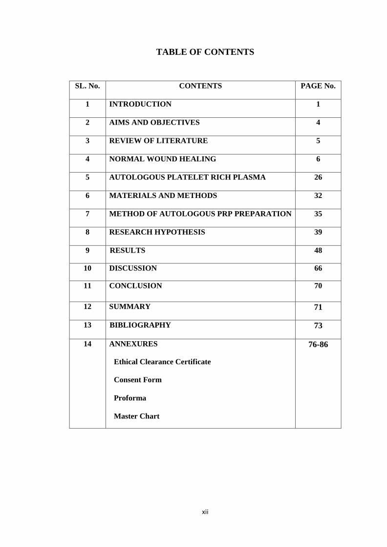
TABLE OF CONTENTS
SL. No. CONTENTS PAGE No.
1 INTRODUCTION 1
2 AIMS AND OBJECTIVES 4
3 REVIEW OF LITERATURE 5
4 NORMAL WOUND HEALING
6
5 AUTOLOGOUS PLATELET RICH PLASMA 26
6 MATERIALS AND METHODS 32
7 METHOD OF AUTOLOGOUS PRP PREPARATION 35
8 RESEARCH HYPOTHESIS
39
9 RESULTS 48
10 DISCUSSION
66
11 CONCLUSION
70
12 SUMMARY 71
13 BIBLIOGRAPHY 73
14 ANNEXURES
Ethical Clearance Certificate
Consent Form
Proforma
Master Chart
76-86
xiii
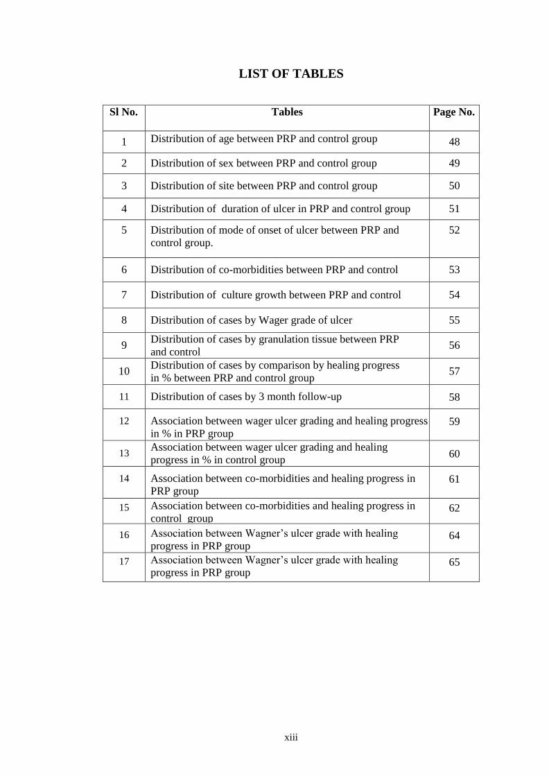
LIST OF TABLES
Sl No. Tables Page No.
1 Distribution of age between PRP and control group 48
2 Distribution of sex between PRP and control group
49
3 Distribution of site between PRP and control group 50
4 Distribution of duration of ulcer in PRP and control group 51
5 Distribution of mode of onset of ulcer between PRP and
control group.
52
6 Distribution of co-morbidities between PRP and control 53
7 Distribution of culture growth between PRP and control 54
8 Distribution of cases by Wager grade of ulcer 55
9 Distribution of cases by granulation tissue between PRP
and control 56
10 Distribution of cases by comparison by healing progress
in % between PRP and control group 57
11 Distribution of cases by 3 month follow-up 58
12 Association between wager ulcer grading and healing progress
in % in PRP group 59
13 Association between wager ulcer grading and healing
progress in % in control group 60
14 Association between co-morbidities and healing progress in
PRP group 61
15 Association between co-morbidities and healing progress in
control group 62
16 Association between Wagner‟s ulcer grade with healing
progress in PRP group 64
17 Association between Wagner‟s ulcer grade with healing
progress in PRP group 65

xiv
LIST OF CHARTS
Sl No. CHARTS Page No.
1 Distribution of age between PRP and control group 48
2 Distribution of sex between PRP and control group
49
3 Distribution of site between PRP and control group 50
4 Distribution of duration of ulcer in PRP and control group 51
5 Distribution of mode of onset of ulcer between PRP and control
group.
52
6 Distribution of co-morbidities between PRP and control 53
7 Distribution of culture growth between PRP and control 54
8 Distribution of cases by Wager grade of ulcer 55
9 Distribution of cases by granulation tissue between PRP
and control 56
10 Distribution of cases by comparison by healing progress
in % between PRP and control group 57
11 Distribution of cases by 3 month follow-up 58
12 Association between wager ulcer grading and healing progress
in % in PRP group 59
13 Association between wager ulcer grading and healing
progress in % in control group 60
14 Association between co-morbidities and healing progress in
PRP group 61
15 Association between co-morbidities and healing progress in
control group 62
16 Association between Wagner‟s ulcer grade with healing
progress in PRP group 63
17 Association between Wagner‟s ulcer grade with healing
progress in PRP group 65
xv
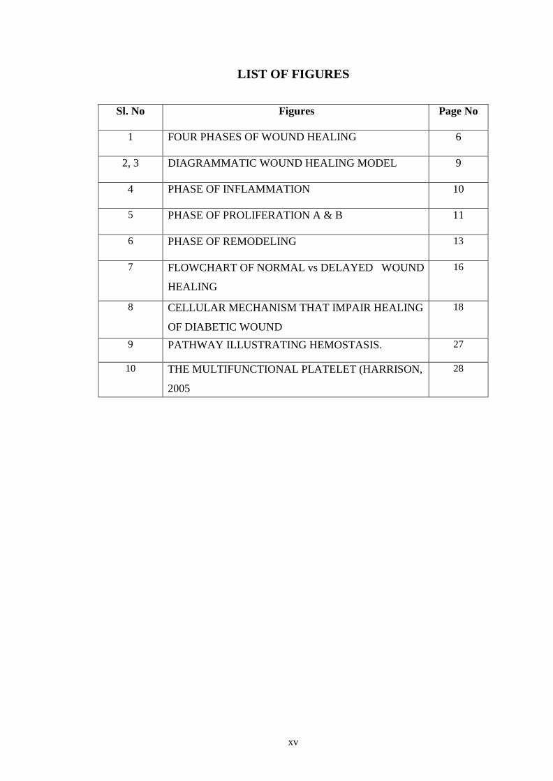
LIST OF FIGURES
Sl. No Figures Page No
1 FOUR PHASES OF WOUND HEALING 6
2, 3 DIAGRAMMATIC WOUND HEALING MODEL 9
4 PHASE OF INFLAMMATION 10
5 PHASE OF PROLIFERATION A & B 11
6 PHASE OF REMODELING 13
7 FLOWCHART OF NORMAL vs DELAYED WOUND
HEALING
16
8 CELLULAR MECHANISM THAT IMPAIR HEALING
OF DIABETIC WOUND
18
9 PATHWAY ILLUSTRATING HEMOSTASIS. 27
10 THE MULTIFUNCTIONAL PLATELET (HARRISON,
2005
28
xvi
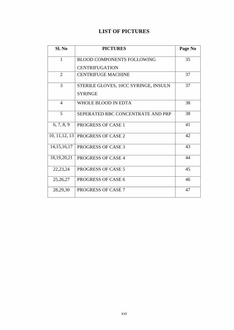
LIST OF PICTURES
Sl. No PICTURES Page No
1 BLOOD COMPONENTS FOLLOWING
CENTRIFUGATION
35
2 CENTRIFUGE MACHINE 37
3 STERILE GLOVES, 10CC SYRINGE, INSULN
SYRINGE
37
4 WHOLE BLOOD IN EDTA 38
5 SEPERATED RBC CONCENTRATE AND PRP 38
6, 7, 8, 9 PROGRESS OF CASE 1 41
10, 11,12, 13 PROGRESS OF CASE 2 42
14,15,16,17 PROGRESS OF CASE 3 43
18,19,20,21 PROGRESS OF CASE 4 44
22,23,24 PROGRESS OF CASE 5 45
25,26,27 PROGRESS OF CASE 6 46
28,29,30 PROGRESS OF CASE 7 47

1
INTRODUCTION
Chronic wounds are catastrophic health problem worldwide. These stubborn
wounds fail to heal in an expected manner of healing process.
The overall increase in the incidence of chronic wounds makes them a
tremendous socioeconomic burden globally.
Wound healing is a complex and dynamic process.(1)
Once a wound begins healing, normally the process resolves with complete
wound closure. However, healing of acute or chronic wounds can become impaired
by patient factors (i.e., co-morbidities) and/or wound factors (i.e., infection)(1)
. Wound
with impaired healing is difficult to treat because good standard wound care does not
always provide an improved healing outcome and often more advanced therapies are
employed. (1)
The standard management includes advance therapeutics with drugs
(antibiotics), intense local dressings (such as negative pressure/antimicrobial) and
multiples surgical interventions/reconstructions. Such intervention and modalities
requires experts and large resources. Still outcomes are unpredictable and associated
with morbidities. (2)
Platelet rich plasma is considered to be advanced therapy for chronic and acute
wounds.(1)
The curative properties of platelet rich plasma rely on the fact that platelets are
a physiological reservoir of growth factors, which have an active role in tissue
regeneration.(3)

2
Platelets are anucleated cell fragments that originate from megakaryocytes in
the bone marrow. (6)
It is well known that platelets contain a great variety of growth factors, with
healing functions. (3)
PRP is a volume of autologous plasma that has a platelet concentration above
baseline i.e., five times more than the normal platelet counts. (4)
When concentrating platelets, 7 fundamental protein growth factors are
concentrated.(5)
Continuous release of these growth factors has been proposed to promote
angiogenesis both in vitro and in vivo. (6)
Release of these angiogenic factors in platelet derived fraction preparations
could be useful in tissue regeneration and wound healing. (6)
Platelets also regulates hemostasis through vascular obliteration and fibrin clot
formation (6)
Among the three organelles described in platelets, namely lysosomes, alpha
granules and dense granules. The biggest compartments for protein storage are alpha
granules.(6)
High leukocyte concentration of PRP has an added antimicrobial effect. (7)
Since PRP is an autologous blood product, it carries no risk of transmitting
infection disease.(7)
Hence, such a physiological mixture of growth factors may be advantageous
clinically to achieve wound healing. (10)

3
This study is undertaken to evaluate the safety and clinical efficacy of local
injection of autologous platelet rich plasma with the conventional type of dressing in
management of chronic cutaneous ulcers.

4
AIM & OBJECTIVES OF THE STUDY:
To evaluate the safety and efficacy of local injections of Autologous Platelet
Rich Plasma in treatment of chronic cutaneous ulcers.

5
REVIEW OF LITERATRE
Chronic wounds are significant burden on healthcare facilities globally. The
management and treatment of chronic wounds are demanding to health care providers.
The subset of chronic wounds and the complications associated with them continue to
progress rapidly despite enormous progress in science of wound healing.
Clinically the process of wound healing is important to understand from several
prospective. These include:
Development of precise, least traumatic surgical technique; the clear
understanding of newer developments and anti-infective therapies affect wound
healing.(11)
Optimal outcome of wounds healing depends on complete evaluation of the
patient and of the wound and application of best practices and techniques. (12)
As noted by John Hunter (1728-1793), a keen observer of biological phenomena,
“… the injury alone has in all cases a tendency to produce the disposition and means
of a cure.”(12)
6
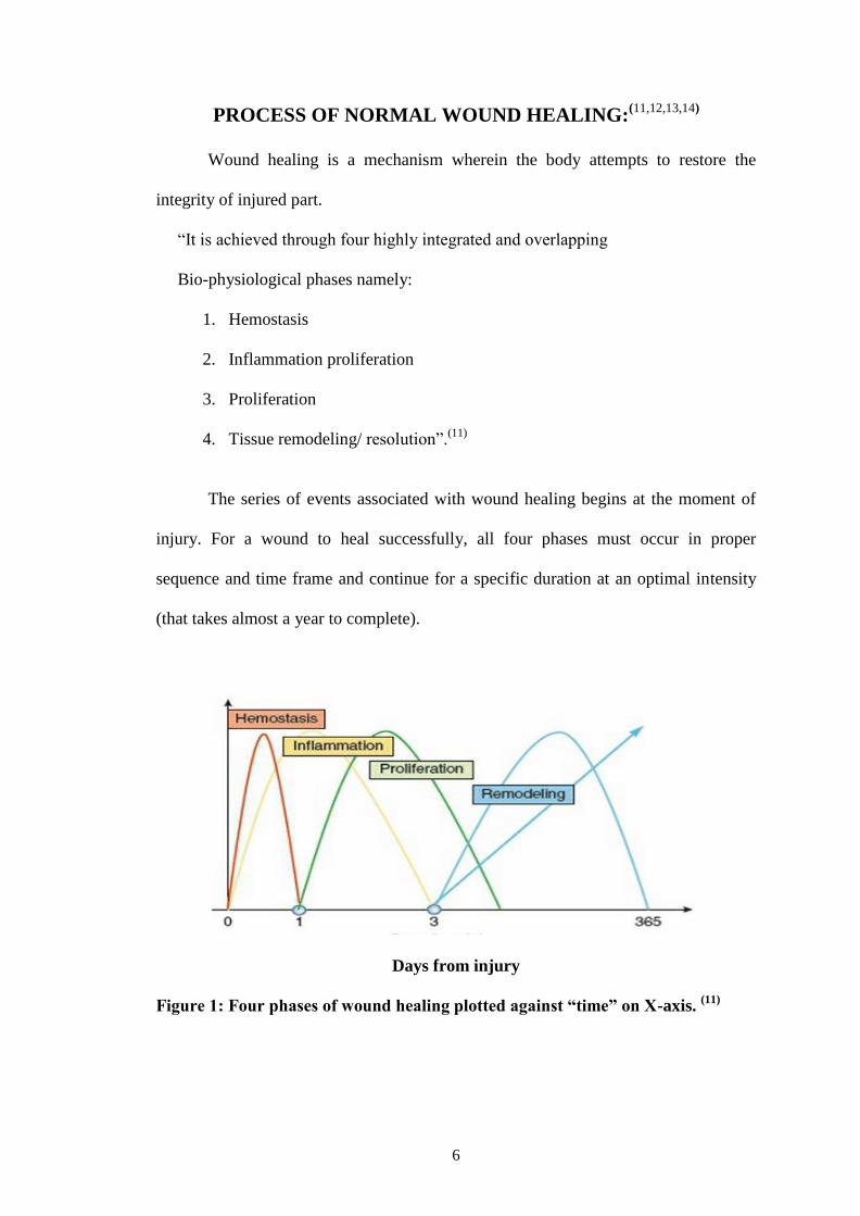
PROCESS OF NORMAL WOUND HEALING:(11,12,13,14)
Wound healing is a mechanism wherein the body attempts to restore the
integrity of injured part.
“It is achieved through four highly integrated and overlapping
Bio-physiological phases namely:
1. Hemostasis
2. Inflammation proliferation
3. Proliferation
4. Tissue remodeling/ resolution”.(11)
The series of events associated with wound healing begins at the moment of
injury. For a wound to heal successfully, all four phases must occur in proper
sequence and time frame and continue for a specific duration at an optimal intensity
(that takes almost a year to complete).
Days from injury
Figure 1: Four phases of wound healing plotted against “time” on X-axis. (11)
7
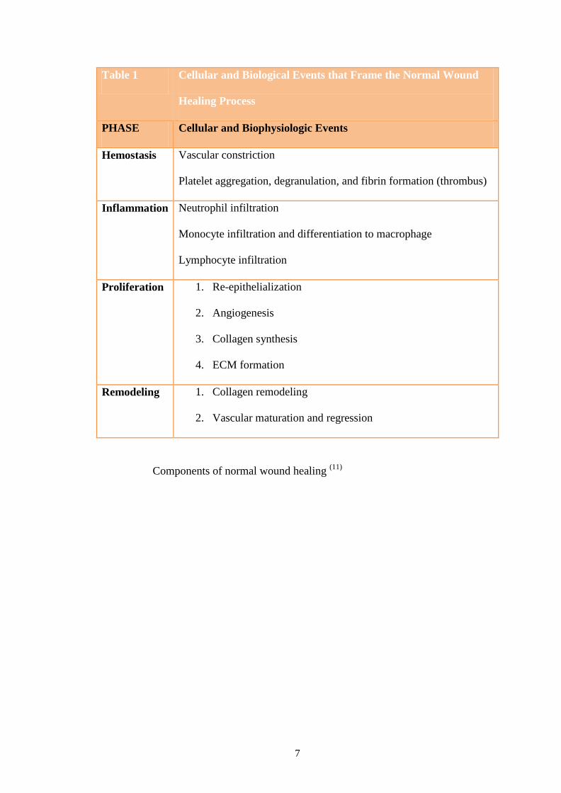
Table 1 Cellular and Biological Events that Frame the Normal Wound
Healing Process
PHASE Cellular and Biophysiologic Events
Hemostasis Vascular constriction
Platelet aggregation, degranulation, and fibrin formation (thrombus)
Inflammation Neutrophil infiltration
Monocyte infiltration and differentiation to macrophage
Lymphocyte infiltration
Proliferation 1. Re-epithelialization
2. Angiogenesis
3. Collagen synthesis
4. ECM formation
Remodeling 1. Collagen remodeling
2. Vascular maturation and regression
Components of normal wound healing (11)

8
HEMOSTATIC PHASE:
This phase immediately begins following wounding of the tissue.
Circulating factors that initiate the inflammatory phase of healing are platelets,
plasma proteins and leukocytes.
Platelets are anucleated cells produced in bone marrow by megakaryocytes.
The plasma membrane of each platelet contains specific receptors known as
glycoprotein Ia/Iia.(16)
The platelets contain granules with important factors for hemostasis and
inflammation. Platelets bind and anchor to vascular endothelium (Type IV). Together
platelet activation occurs and platelets change from round shape to flattened
configuration and discharge contents of cytoplasm which is known as “platelet release
reaction”(11)
.
These bioactive factors serve a dual purpose in hemostasis and wound healing.
This phase is characterized by increased vascular permeability, chemotaxis,
secretion of cytokines and growth factors into the wound.
It represents an attempt to arrest bleeding and maintaining milieu interior.

9
2
3
Figure 2 and 3: Diagrammatic wound healing model. Platelets are the first cells
to arrive at the wound and are critical to create a clot and hemostasis.(11)

10
INFLAMMATION:
Activated WBC migrate to wound site to engulf debris and injured tissue.
Chemical mediator Leukotaxine attract WBCs.
First polymorpho-nuclear neutrophil dominate. By 5th
day, granulocytes die
and monocytes predominate to continue scavenging activity.(14)
Monocytes must be present at the wound site to create a normal fibroblast
production. Depression of monocytes will delay wound healing.(14)
These cells secrete cytokines and growth factors which function in rapidly
amplifying process that affect all aspects of healing.
Figure 4: Phase of inflammation(11)
PROLIFERATION:
Primarily consists of epithelization mainly occur by proliferation and
migration of marginal basal cells.(14)
Marginal basal cell loose there attachment to underlying dermis, Enlarge and
migrate. Basal cells in wound zone undergo rapid mitotic division.(14)
Epidermis adjacent to wound begins to proliferate on day 1.
No regeneration of hair follicles, sweat and sebaceous glands in new epidermis.(14)

11
Figure 5
A. Phase of proliferation. Vast array of cells are recruited into the
wound bed and carry out diverse functions including proliferation
and deposition of ECM.
B. Fibroblast function.
GRANNULATION TISSUE FORMATION:
Proliferation and migration of surrounding connective tissue.
Fibroblast start synthesizing extra-cellular matrix.
STAGE OF VASCULARIZATION:
Formation of living granulation tissue is called “organization”. Wound clot is
invaded by macrophages which lead to capillary loop formation.
Capillary loops undergo canalization to form vascular arcade.
Which with time mature and acquire muscle coat to form arterioles.

12
COLLAGEN SYNTHESIS:
It is brought about by fibroblasts. Collagen is nothing but extracellular
secretion of fibroblasts.
Collagen is made up of glycine, lysine, hydroxyprolene and proline.
Initially tropocollagen is formed which later condenses to form fibril. Fibril
condenses to from collagen.
Collagen is not inert and undergoes constant turnover under the influence of
enzyme collagenase.
TYPES OF COLLAGEN: (14)
Collagen I: Tendon, bone, skin
Collagen II: Cartilage
Collagen III: Fetal dermis, aorta, esophagus, uterus
Collagen IV: Immature scar
REMODELING(14)
Also called as phase of maturation.
It begins by six weeks and lasts for two years.
Maturation of collagen is brought about by cross linking which is responsible
for tensile strength of scar.
Normal dermis contain 80% of collagen type I and 20% of type III, while scar
contains 50% of collagen type I and 50% of collagen type III.
Hydroxylation of lysine and proline will produce collagen type I which
requires Vitamin C Iron and α-ketogluteric acid.
STRENGTH OF SCAR: 3% by 1 week
20% by 3 weeks
80% by 12 weeks.(14)
13
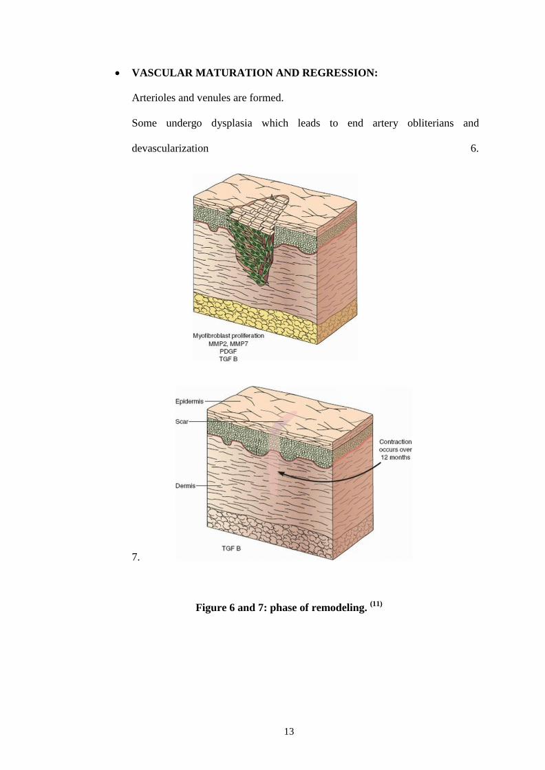
VASCULAR MATURATION AND REGRESSION:
Arterioles and venules are formed.
Some undergo dysplasia which leads to end artery obliterians and
devascularization 6.
7.
Figure 6 and 7: phase of remodeling. (11)

14
FACTORS AFFECTING NORMAL WOUND HEALING:(14)
GENERAL FACTORS:
1. Age : young age – better wound healing
2. Nutrition : a. Protein deficiency cause impairment of granulocyte action
and collagen formation.
b. Sulfur containing amino-acid determine tensile strength of
the scar
c. Vitamin C important for hydroxyltion of amino acids for
synthesis of collagen
d. Vitamin A is essential for re-epithelization.
c. Zinc is an important co-emzyme for DNA, RNA synthesis.
d. Metallo-enzymes.
3 Hematological: Anemia, granulocytopenia, immunodeficiency.
4. Diabetes : Microangiopathy
Hyperglycemia
Atherosclerosis
Decreased chemotaxis
Decreased phagocytosis
5. Corticosteroids: Have anti-inflammatory effect
Decrease protein synthesis
Inhibit fibroblast activity
Decrease capillary budding,
Decrease epithelization.
6. Cytotoxic drugs: inhibit cell division
7. Radiation

15
8. Uremia
9. Malignancy
LOCAL FACTORS:
1. Position of wound: parallel to Langer lines heal faster
2. Blood supply: directly proportional to wound healing
3. Tension: decreases wound healing
4. Infection: delay fibroblast action( fibroblast require alkaline medium for
collagen synthesis)
5. Wound over joints have poor wound healing
6. Foreign body at wound site cause inflammation
7. Necrosis
8. Local radiation: decreases vascularization and fibroblast activity
9. UV light: Promote wound healing
CHRONIC WOUND:(11,12,13,14,15)
“Chronic wounds are defined as wounds that have failed to proceed through orderly
process of healing that produce satisfactory anatomic and functional integrity”.(12)
Wounds heal within the time of 4-6 weeks, those that do not are termed at chronic
wounds.(11)
Non healing ulcers and wounds represent failure to achieve complete epithilization in
appropriate temporal sequence and tissue repair.(15)
Unresponsiveness to normal regulatory signals have implicated in predictive factor for
chronic wounds.

16
It may be due to-
- Failure of synthesis of normal growth factors
- Excessive breakdown of growth factors within the wound due to increased
proteolytic enzymes.
- Failure of function of normal anti-protease inhibitor enzymes.(12)
Often chronic wounds stall in inflammatory phase of healing.(15)
It is due to persistent proinflammatory state and paradoxically leads to increased
degradation of matrix.(11)
They have high pro-oxidant environment along with bacterial colonization, necrotic
tissue, foreign body, localized tissue hypoxia.(13)
Figure 7: Flowchart representing normal and delayed wound healing.(11)
Chronic wounds have been classified as:
- Vascular ulcers (venous and arterial)
- Diabetic ulcers
- Pressure ulcers(15)
17
MOST COMMON CAUSE OF CHRONIC WOUNDS:
DIABETES:
Diabetic foot ulcer (DFU) is a major complication of diabetes mellitus.
This medical condition affects more than 15% of population worldwide.
Recent study showed that up to 88% of all lower limb amputation are related to
diabetic foot ulcer (Alvarsson et al., 2012).(17)
Diabetic associated large vessel occlusion and end-organ microangiopathy each lead
to tissue ischemia and infection.(13)
Diabetic sensory neuropathy lead to repeated unnoticed trauma and constant pressure
on the wound.(13)
Tissue hypoxia is well demonstrated by reduced dorsal foot transcutaneous oxygen
tension (TcO2)(13)
Also the thickened basement membrane decrease perfusion of tissues.
VEGF up regulation in patients with diabetes impaired.(13)
Hyperglycemia further increases pro-inflammatory mediators i.e.,
TNF-α, IL-1.(13)
There is also loss of balance between metalloprotinases and MMP inhibitors (muller
et al., 2008) accelerating ischemia.(17)
These changes in structure and functions of cells at wound site lead to delayed healing
in DFUs.
Hence treatment of DFUs remain a challenge.(17)
18
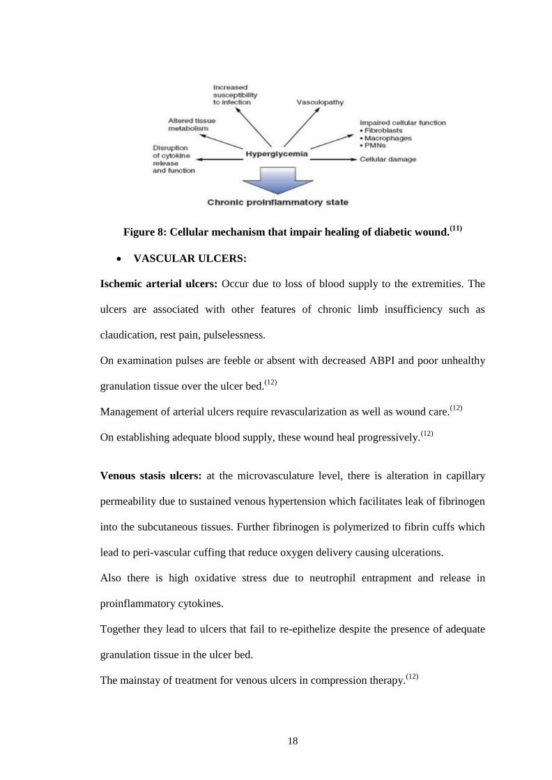
Figure 8: Cellular mechanism that impair healing of diabetic wound.(11)
VASCULAR ULCERS:
Ischemic arterial ulcers: Occur due to loss of blood supply to the extremities. The
ulcers are associated with other features of chronic limb insufficiency such as
claudication, rest pain, pulselessness.
On examination pulses are feeble or absent with decreased ABPI and poor unhealthy
granulation tissue over the ulcer bed.(12)
Management of arterial ulcers require revascularization as well as wound care.(12)
On establishing adequate blood supply, these wound heal progressively.(12)
Venous stasis ulcers: at the microvasculature level, there is alteration in capillary
permeability due to sustained venous hypertension which facilitates leak of fibrinogen
into the subcutaneous tissues. Further fibrinogen is polymerized to fibrin cuffs which
lead to peri-vascular cuffing that reduce oxygen delivery causing ulcerations.
Also there is high oxidative stress due to neutrophil entrapment and release in
proinflammatory cytokines.
Together they lead to ulcers that fail to re-epithelize despite the presence of adequate
granulation tissue in the ulcer bed.
The mainstay of treatment for venous ulcers in compression therapy.(12)

19
PRESSURE ULCERS:
Also called decubitus ulcers. “It is defined as a localized area of tissue
necrosis that develops when soft tissue is compressed between a bony prominence and
an external surface”(12)
Excessive pressure causes collapse of capillaries and impairs delivery of
nutrients and oxygen to the tissue.
Ulcer formation is further favored by moisture, friction and shear forces and
co-morbidities associated such as immobility, obesity.
There are four stages of pressure ulcer formation:
Stage I – non-blanching erythema of intact skin
Stage II – partial thickness skin loss involving epidermis or dermis
Stage III – Full thickness skin loss but not through the fascia.
Stage IV – Full thickness skin loss with extensive involvement of muscle and bone.
Treatment of pressure ulcers is multidisciplinary involving wound care teams
with therapist, nutritionists.
Recurrence rates are very high.(12)
INFECTION:
Infection is the common cause of delayed wound healing. Particularly beta-
hemolytic streptococci prevents wound from healing by any means.
Bacterial infection prolongs the inflammatory phase and interferes with
epithilization, wound contraction and collagen deposition.
Bacterial endotoxins continue the process of phagocytosis and collagenase
release, which degrade the collagen in the wound and surrounding structure.(13)

20
WAGNER CLASSIFICATION SYSTEM FOR ULCERS
Grade 0 Foot symptoms like pain only
Grade 1 Superficial ulcers
Grade 2 Deep ulcers
Grade 3 Ulcer with bone involvement
Grade 4 Forefoot gangrene
Grade 5 Full foot gangrene
DRESSING IN CHRONIC ULCERS:
Wound dressings represent an integral part of the management of chronic
ulcers.
Dressings should alleviate symptoms, provide wound protection and
encourage healing.
In choosing a dressing for a chronic ulcer, several factors have to be taken into
account. Infection need to be controlled.
A dressing must be comfortable and acceptable for the patient. Ideally, the
dressing should also aid in the management of the infection itself and promote healing
of the ulcer.
Desirable characteristics for wound dressings must incorporate the principles
of wound healing.
These dressings must also accommodate practical issues such as allowing
providing mechanical protection at the same time they must also be cost effective ,
should be hypoallergenic.
Various types of non-adherent or saline-soaked gauze dressings are often
regarded as standard treatment for chronic ulcers.
21
These dressings are designed to be atraumatic and to provide a moist wound
environment. These simple, relatively inexpensive dressings are not designed
specifically for managing infection but can be safely used in conjunction with
antibiotic treatments.
HYDROCOLLOIDS
Hydrocolloid dressings are semipermeable to vapour, occlusive to wound
exudate and absorbent. They are usually presented as an absorbent layer on a film or
foam. Examples of commercially available products include Duoderm (Convatec),
Granuflex (Convatec), and Comfeel (Coloplast). They are found to be the second
most popular choice of dressing (behind non-adherent) for chronic ulcers, espicially
diabetic ulcer. Despite their popularity, their use on infected wounds is controversial.
Hydrocolloid dressing creates a hypoxic and moist environment that may also
facilitate autolysis of necrotic material. Their use for highly exudative wounds can
lead to maceration of the surrounding skin.
Most authorities, however, have expressed concern that hydrocolloids may
increase the risk of infection developing within a wound .
Hydrocolloid dressings are designed to be left on the wound for prolonged periods
(1week), hence they are useful in clean ulcers but their role in infected ulcers is
controversial as infected wounds require repeated inspection of wound.
HYDROGELS:
Hydrogels are designed to facilitate autolysis of necrotic tissue and they
donate moisture to extensively dry wounds. They can lead to maceration when
applied to wounds that show moderate to severe exudate. Examples include
Aquaform (Maersk Medical)and Intrasite.
22
IODINE PREPRATION:
Antiseptics such as iodine-based preparations are commonly used on wounds,
although there is no evidence to support a beneficial effect. Typically they are applied
to locally infected wounds, usually in combination with systemic antibiotics.
Iodine comes in 2 main preparations: cadexomer-iodine and povidone iodine.
Iodine is bactericidal in vitro, with maximal activity at 0.1%–1% povidone iodine has
long been used as a skin antiseptic, but its antimicrobial effect on wounds is
debatable. Furthermore, some data have shown iodine solutions to be toxic to
fibroblasts and keratinocytes.
A randomized controlled trial of cadexomer-iodine versus saline-soaked gauze
on clean foot ulcers showed no significant difference in healing between the groups.
Certain iodine dressings are highly absorbent and therefore useful in preventing skin
excoriation in moderately exudating ulcers. In our own clinical practice, Cadexomer-
iodine pastes are used for wounds and povidone-iodine gauze for superficial ulcers.
Despite the lack of evidence, many consider iodine preparations to be appropriate
dressings for infected diabetic foot ulcers.
Povidone iodine preparation was used in control group in this study.
SILVER-IMPREGNATED DRESSING
The use of silver as a topical antimicrobial for acute and chronic wounds is
well established. It has been traditionally delivered as silver nitrate or as silver
sulfadiazine. Silver nitrate has cytotoxic effects on host cells, a property often
exploited in the treatment of hypergranulating tissue, but its application can be
uncomfortable. Silver sulfadiazine, which has the antimicrobial actions of both silver
and sulfadiazine, is used on burns and chronic wounds and is generally well tolerated.
23
The antimicrobial effects of silver are complex, including direct inhibition of
bacterial cell respiration, inactivation of intracellular enzymes and alterations to the
cell membrane. Silver-coated dressings that use elemental silver may be more
efficacious at killing bacteria than is silver sulfadiazine or silver nitrate. New silver-
impregnated dressings may be suitable for use for infected diabetic foot ulcers.
Examples include Megaheal and Hydroheal.
However, reports suggest of accelerated wound reepithelialization.
BIOLOGICAL DRESSING:
Biological Therapy (e.g., bilayered keratinocytes and fibroblasts and platelet
derived growth factor) are used when patients fail to improve after the approaches
described above have been applied for 3 weeks. Biological therapy should be
implemented only if wound size cannot be decreased by more than 10 percent within
a 3-week time period.
Chronic ulcers exhibit a decreased production of growth factors within the
wound. Cell therapy, also known as biological therapy, presents an appropriate
treatment option in some cases. Biological therapy is an ideal treatment for chronic
ulcers because it adds cells that release growth factors to a growth factor dependent
environment, increases cytokines and matrix proteins and promotes angiogenesis.
Thus accelerating healing time decreases the risk of wound infection.
The biological therapy consists of “The bilayer biologically active skin
construct”, composed of a surface layer of allergenic human keratinocytes over a layer
of allogeneic human fibroblasts suspended within a collagen matrix. The “Bilayer cell
therapy” has been shown to increase the healing rate of diabetic foot ulcers not
complicated by osteomyelitis or ischemia. Fibroblasts synthesize collagen and secrete

24
a matrix of growth factors and matrix proteins in physiological concentrations
essential for wound healing and epithilization.
HYPERBARIC OXYGEN THERAPY:
Hyperbaric oxygen therapy is based on the premise that the delivery of
supraphysiological concentrations of oxygen to diseased tissues will result in
beneficial physiological changes.
The therapy is based on achieving an atmospheric pressure of 2–3
atmospheres pressure which is administered using a sealed polyethylene bag over the
affected area and administering 100 percent oxygen to a pressure between 20 and
30mmHg. Treatment lasts for 2 to 2 ½ hours.
HBOT can be offered to patients who have diabetic foot ulcers for whom
atleast 30 days of standard wound care has failed and who have a Wagner grade III
lesion or higher. (meaning the ulcer must penetrate to tendon, bone or joint and may
be associated with deep abscess, osteomyelitis, gangrene, or septic arthritis) In
treatment ulcer, it is believed both that the function of phagocytic cells is improved,
assisting in the fight against any infection and that wound healing is independently
aided through effects on cellular processes. Thus, it has been suggested that HBOT is
useful for the treatment of infection and for the healing of chronic wounds.
TOPICAL NEGATIVE PRESSURE:
The practice of exposing a wound to sub-atmospheric pressure for an extended
period to promote debridement and healing was first described by Fleischmann et al
in 1993 following the successful use of this technique in 15 patients with open
fractures. The science behind topical negative pressure dressings is to apply a sub-
atmospheric pressure over the wound bed and maintain the negative pressure
environment by means of a semi permeable occlusive coverage. Since the wound is
25
occluded from the surrounding environment it is also called “ Limited access
dressing”.
Usage of a subatmospheric pressure causes,
Fourfold increase in blood flow in the local wound environment. (As
measured by a laser Doppler technique.)
Induces mechanical stress which causes an increase in cellular activity.
Increase in the rate of granulation tissue formation and reduction in the
bacterial load in the wound.
Clinically TNP removes large amounts of fluid from wounds especially acute
wounds. The resulting reduction in oedema is thought to aid in the
enhancement of blood and nutrient flow into the wound.
The mechanism behind the ability of the TNP to decrease bacterial count may
be attributable to three properties increased blood flow, decreased interstitial
edema and removal of harmful enzymes in wound.

26
AUTOLOGOUS PLATELET RICH PLASMA
Also called as PRP, maybe defined as “A component of plasma fraction of
autologous venous blood with platelet counts in the range between 4 to 6 above the
baselines considered to be therapeutic benefit (I million platelets/L)”.(6)
A sample of normal blood will contain 93% of red blood cells, 6% of platelets
and 1% of white blood cells. In PRP the ratio of RBC to platelets is reversed, thereby
increasing the factors that would be more useful in healing.(22)
“It is an endogenous therapeutic technology that has been gaining popularity
in regenerative medicine due to its potential to stimulate and accelerate tissue
healing”.(19)
Platelets were discovered in 1882 by Giulio Bizzozero. Though for many
years the dynamic and multifunctional nature of platelets remained unknown.(16)
Platelets are anucleated, discoid shaped cells that are derived from
megakaryocytes in the bone marrow through controlled fragmentation.(18)
Diameter of mature platelet is 2-5µm and half-life of platelets is 5-9 days. The
normal platelet count in blood is 150-400x103
per cubic mm of blood. An average
healthy adult can produce1011
platelets per day.(16)
Platelets are peculiar in their structure. Although they are anucleated yet have
well defined mitochondria. The plasma membrane of platelet is bilayered and contain
phospholipid which is a site for various surface receptor expression which help in
signaling and intracellular trafficing.(16)
They are specialized blood cells that release contents of their intracellular
granules in response to activation.(18)
The named biological markers on platelet plasma membrane are CD36, CD41,
CD42a, CD42b, CD61, CD63 IIbIIIa and GLUT-3.
27
Majorly platelet activity is associated with initiation of coagulation cascade to
achieve hemostasis. But they also play many important roles in pathophysiological
states.(16)
The surface receptors of platelets also contain α granules which participate in
extensive functions such as coagulation, inflammation, atherosclerosis, angiogenesis,
wound repair and tumorogenesis.(16)
Platelets have two major storage granules, which store biologically active
molecules that involve in initiation of coagulation and recruiting other cells during
inflammation.(16)
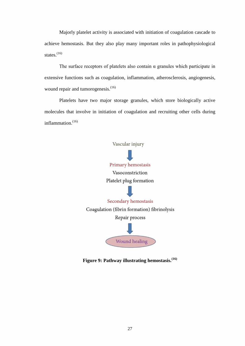
Figure 9: Pathway illustrating hemostasis.(16)

28
Figure 10: The multifunctional platelet (Harrison, 2005 [44](16)
PLATELET GROWTH FACTORS:
“Growth factors are solube and diffusible polypeptide substances that regulate
growth, defferentiation, proliferation, and cellular metabolism of numerous cell
types.”(19)
There are not less than sixty different biologically active substances in
platelets that alleviate in tissue repair and healing like chemotaxis, cell proliferation,
angiogenesis, immune modulation, cell differentiation, anti-bacterial activity,
intracellular matrix deposition and remodeling.(18)
Few of the named important growth factors that are released by activated platelets are-
PDGF (platelet-derived growth factor)
PDEGF (platelet derived epidermal growth factor)

29
TGF α and β (transforming growth factor)
EGF (epidermal growth factor)
IGF (insulin growth factor)
KGF (keratinocyte growth factor)
IL-8 (interleukin)
TNF-α (tumor necrosis factor)
GM-CSF (granulocyte macrophage colony stimulating factor).
“So far only PDGF has been approved by the United States Food and Drug
Administration (FDA) and by European Authorities (EMEA) for clinical application
in patients.”(19)
List of growth factors in Platelet Rich Plasma
Growth Factor
Effect
PDGF Macrophage activation and angiogenesis
Fibroblast chemotaxis and proliferative activity
Enhances collagen synthesis
Enhances the proliferation of bone cells
TGF-Beta Enhances the proliferative activity of fibroblasts
Stimulates biosynthesis of type I collagen and fibronectin
Induces osteoclast formation and bone resorption
IGF-I Chemotactic for fibroblasts and stimulates protein synthesis
Enhances bone formation by proliferation and differentiation of osteoblasts
PDEGF Promotes wound healing by stimulating the proliferation of keratinocytes
and dermal fibroblasts
PDAF Induces vascularization by stimulating vascular endothelial cells
PF-4 Stimulates the initial reflux of neutrophils into wounds
A chemoattractant for fibroblasts
A potent antiheparin agent
EGF Cellular proliferation
Differentiation of epithelial cells
VEGF Angiogenesis
Migration and mitosis of endothelial cells
Creation of blood vessel lumen
Creates fenestrations
Chemotactic for macrophages and granulocytes
Vasodilation (indirectly by release of nitrous oxide)
PDGF = Platelet-Derived Growth Factor; TGF = Transforming Growth Factor; IGF = Insulin
Growth Factor; PDEGF = Platelet-derived endothelial growth factor; PDAF = Platelet-
derived angiogenesis factor; PF-4 = Platelet Factor 4; EGF = Endothelial Growth Factor;
VEGF = Vascular Endothelial Growth Factor;

30
During the process of wound healing, the growth factors in platelet granules
act as messenger to regulate well-organised and complex series of events which
involve cell-cell, cell-matrix interactions which play an important role during various
phases of wound healing.(18)
“Autologous platelet derived wound healing factors were proposed to regulate
wound healing of chronic cutaneous ulcers by promoting the formation of granulation
tissue in the early healing phase. This conclusion was based on randomized,
prospective, double-blind, placebo-controlled studies, who showed improved healing
compared to usual treatments.”(20)
Platelet rich plasma functions as growth factor delivery system in high
concentrates and also function as tissue sealant system.
The wound repair is brought about by degranulation of α-granules which
releases locally active growth factors.
These active growth factors accelerate wound healing by attracting
undifferentiated immune cells into newly formed collagen matrix which further
undergo cell division and de-differntiation.
Platelets also suppress cytokine release and halt inflammation by interacting
with macrophages and promote healing and regeneration.(19)
PRP further promote neoangiogenesis and re-epithilization.
“Platelets in PRP also play a role in host defense mechanism at the wound site
by producing signaling proteins that attract macrophages.
PRP contain small number of leukocytes that synthesize interleukins as a part
of a non-specific immune response. Previous studies of PRP have demonstrated
antimicrobial activity against Escherichia coli, Staphylococcus aureus, including
methicillin-resistant Staphylococcus aureus, Candidia albicans, Cryptococcus
neoformans”.(20)

31
Pain reduction following platelet growth factor application was mentioned in
study by Crovetti et al.(18)
“The curative properties of PRP explained by the fact that platelets are
physiological reservoir of growth factors, that have an active role in tissue
regeneration”(21)
32
MATERIALS AND METHODS
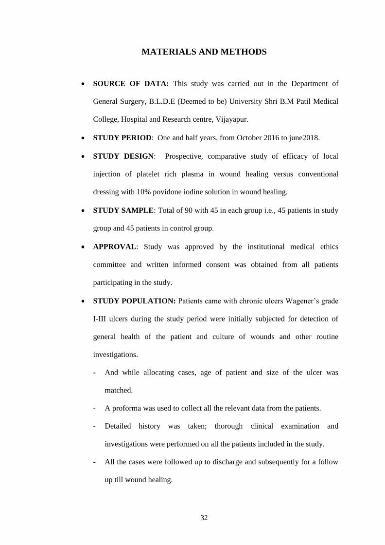
SOURCE OF DATA: This study was carried out in the Department of
General Surgery, B.L.D.E (Deemed to be) University Shri B.M Patil Medical
College, Hospital and Research centre, Vijayapur.
STUDY PERIOD: One and half years, from October 2016 to june2018.
STUDY DESIGN: Prospective, comparative study of efficacy of local
injection of platelet rich plasma in wound healing versus conventional
dressing with 10% povidone iodine solution in wound healing.
STUDY SAMPLE: Total of 90 with 45 in each group i.e., 45 patients in study
group and 45 patients in control group.
APPROVAL: Study was approved by the institutional medical ethics
committee and written informed consent was obtained from all patients
participating in the study.
STUDY POPULATION: Patients came with chronic ulcers Wagener‟s grade
I-III ulcers during the study period were initially subjected for detection of
general health of the patient and culture of wounds and other routine
investigations.
- And while allocating cases, age of patient and size of the ulcer was
matched.
- A proforma was used to collect all the relevant data from the patients.
- Detailed history was taken; thorough clinical examination and
investigations were performed on all the patients included in the study.
- All the cases were followed up to discharge and subsequently for a follow
up till wound healing.

33
- “Primary efficacy end point” was complete ulcer closure and “Secondary
efficacy end point” was time taken to achieve ulcer closure by either
secondary suturing or skin grafting.
INCLUSION CRITERIA:
Highest dimensions within 5cm breadth, 5cm Width, 0.5cm Depth
Wagner’s Classification Of Diabetic Ulcer Upto Grade-III
Traumatic Non-Healing Ulcers
Bedsores
Venous Ulcers
Other non-specific ulcers.
EXCLUSION CRITERIA:
Anemia – Hb <10gm% in adults, <12gm% in children <14 years
Uncontrolled Diabetes
Gross Nutritional Deficiency BMI <18 , Albumin <2gm/Dl of blood
Dyslipidemia
Wagner’s Classification Of Ulcer Grade-IV or More
Patient on immunosuppressive drugs
Known malignancies
Patient with bleeding disorders.
Radiotherapy to local area of ulcer.
METHOD OF COLLECTION OF DATA
• All eligible patients admitted in the Department of General Surgery in Shri B.
M Patil medical college with ulcer during the study period from October 2016
to June 2018 were initially evaluated for the presence of chronic ulcers and

34
co-morbidities associated with the patient. Routine investigations were done
and swab culture were sent from the ulcer for organisms. Ulcer dimensions
were measured.
• The study subjects will be randomly divided into two groups, Local injection
of platelet rich plasma (A group) and Povidone iodine dressing group (B
group).
• Study group were treated with local injection of PRP
• Control group was treated with mechanical debridement and dressed with 10%
Povidone Iodine.
• PRP was injected every 4th
day for 4 times and every alternate day normal
saline dressing was done while Povidone iodine dressing was done in control
group on daily basis.
• Measurements from the ulcer were taken on every 4th
day till 16 days.
• Statistical analysis was done by using Fisher exact test, Chi square test and p
value <0.05 was considered significant.
35
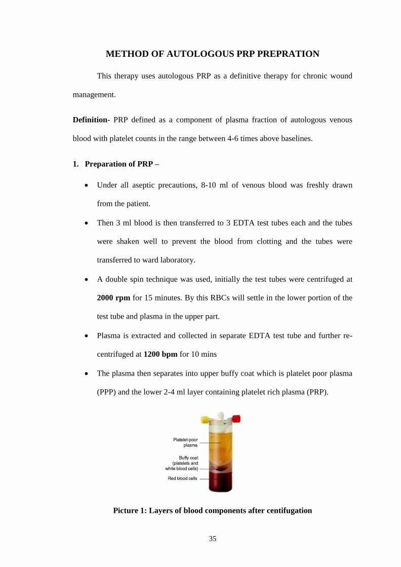
METHOD OF AUTOLOGOUS PRP PREPRATION
This therapy uses autologous PRP as a definitive therapy for chronic wound
management.
Definition- PRP defined as a component of plasma fraction of autologous venous
blood with platelet counts in the range between 4-6 times above baselines.
1. Preparation of PRP –
Under all aseptic precautions, 8-10 ml of venous blood was freshly drawn
from the patient.
Then 3 ml blood is then transferred to 3 EDTA test tubes each and the tubes
were shaken well to prevent the blood from clotting and the tubes were
transferred to ward laboratory.
A double spin technique was used, initially the test tubes were centrifuged at
2000 rpm for 15 minutes. By this RBCs will settle in the lower portion of the
test tube and plasma in the upper part.
Plasma is extracted and collected in separate EDTA test tube and further re-
centrifuged at 1200 bpm for 10 mins
The plasma then separates into upper buffy coat which is platelet poor plasma
(PPP) and the lower 2-4 ml layer containing platelet rich plasma (PRP).
Picture 1: Layers of blood components after centifugation

36
The centrifuge machine used is simple non-cooled one.
The process is performed at a room temp of 22-24ºC
This autologous PRP is then transferred to a insulin syringe and locally
infiltrated in the wound margins, just like a local infiltration of local
anesthesia. At a distance of approximately 0.2 ml/cm with equal quantity.
The process was repeated every 4th
day for minimum of 4 cycles.
Local dressing was performed on alternate days, with moist saline only.
From infected wounds swabs were taken for culture and sensitivity every
week, to monitor infection status of the wound.
Patient was thoroughly examined for general condition and local wound
condition and ulcer size (length, breath, depth) was measured.
Every 4th
day, the ulcer area and volume was calculated and photographed.
Wound area was calculated using the formula length in greatest dimensions
multiplied with breadth in greatest dimension.
For an ellipse wound the formula used was Length x breadth x 0.7854
(An ellipse is closure to a wound shape then square or rectangle)
The use of an ellipse for calculating wound measurement has been used in
RCTs in wound healing literature. (8, 9)
Treatment outcome will be defined as a percentage of change in the area,
which will be calculated as initial measurement minus assessment day
measurement divided by initial measurement.
HEALING IN % = Initial measurement – Final measurement X 100
Initial measurement

37
Picture 2: Centrifuge machine
Picture 3: Sterile gloves, 10cc syringe, insulin syring

38
Picture 4 : Whole blood collected in EDTA Tube
Picture 4: Separated RBC concentrate and PRP

39
RESEARCH HYPOTHESIS
Autologous Platelet rich plasma is a safe, simple, biocompatible technique
as a definitive management of chronic cutaneous ulcers without any adverse
events.
SAMPLING:
Study period from OCTOBER 2016 to JUNE 2018. All the patients admitted
during this period, who fulfill the inclusion criteria, will be included in this study.
ESTIMATION OF SAMPLE SIZE:
It is a interventional comparative study.
With anticipated mean difference of average duration of wound healing
between the two study groups as 5.1 weeks and Anticipated SD as 5.3 weeks.
With this the minimum sample size for both the groups is 90 and per each
group is 45.
With 95% Power and 99% Confidence level obtained from reference study. (4)
Formula Used
n = (Zα + Zβ)22SD
2 n = 90
_______________________
MD
2
WHERE
Z = Z statistic at a level of significance
MD = Anticipated Mean difference = 5.1
SD = Anticipated Standard deviation = 5.3
Student‟s t – test used to compare the subcutaneous injection of PRP and
standard wound management techniques based on the healing results
under study.
Hence the samples size of two groups is 90.

40
Statistical Analysis:
Data will be analyzed using-
- Mean ± SD
- Percentages
- Z proportion test
- T-test for comparison of mean
- Graphical presentation

41
PHOTOGRAPHS OF RESULTS WITH PRP INJECTIONS
6 7
8 9
Picture 6, 7, 8, 9, showing progressing healing of chronic ulcer over left hand
following 4 cycles of PRP. End result: complete wound closure in 20 days.

42
10 11
12 13
Picture 10, 11, 12, 13 showing wound contraction of 45% in a chronic ulcer over
left knee following 4 cycles of PRP. Duration of ulcer-3 mont

43
14 15
16 17
Picture 14, 15,16,17, show serial progression of chroinc ulcer of 6 months
duration with PRP injection brought healthy granulation tissue.

44
18 19
20 21
Picture 18, 19, 20, 21 : Show contraction of wound by >50% with 4 cycles of PRP
in a chronic ulcer over dorsum of right foot.

45
22 23
24
Picture 22, 23, 24: Show contraction of wound by >40% with early granulation
tissue following 4 cycles of PRP injections in chronic ulcer over right arm in
diabetic patient.

46
25
26
27
Picture 25, 26, 27: show appearance of healthy grannulation tissue following
local injections of PRP in a bedsore over gluteal region of 6 months of duration.
47
28
29
30
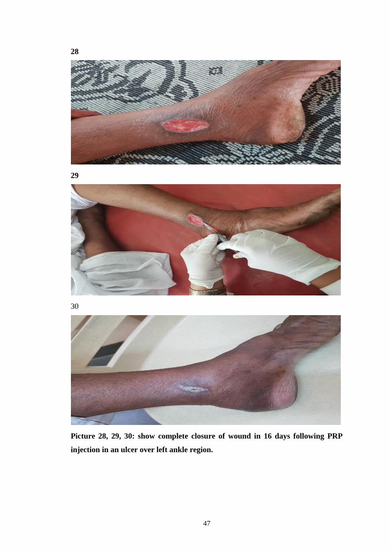
Picture 28, 29, 30: show complete closure of wound in 16 days following PRP
injection in an ulcer over left ankle region.

48
RESULTS
TABLE 1 : DISTRIBUTION OF AGE BETWEEN PRP GROUP (A) AND
CONTROL GROUP (B)
Age
(Years)
PRP
GROUP %
CONTROL
GROUP % Total %
Chi
square
test
< 40 11 24.4 7 15.6 18 20
P=0.2981
NS
40 – 49 8 17.8 5 11.1 13 14.44
50 – 59 10 22.2 9 20.0 19 21.11
60 – 69 11 24.4 14 31.1 25 27.78
70 – 79 5 11.1 6 13.3 11 12.22
80+ 0 0 4 8.9 4 4.44
Total 45 100.0 45 100.0 90 100
CHART 1: DISTRIBUTION OF AGE BETWEEN PRP GROUP AND
CONTROL GROUP
Table 1 and chart 1 show age distributiom between PRP group and control group with
MD with percentage distribution of age is maximum in between 60-69years (24.4%)
in PRP group and (31.1%) in control group.

49
TABLE 2: DISTRIBUTION OF SEX BETWEEN PRP AND CONTROL
GROUPS.
Gender Study
group
% Control
group
% Total % Chi square test
Male 37 82.2 31 68.9 68 76 P=0.1411 NS
Female 8 17.8 14 31.1 22 24
Total 45 100 45 100 90 100
CHART 2: DISTRIBUTION OF SEX BETWEEN PRP AND CONTROL
GROUPS.
Table 2 and chart 2 show sex distribution among two groups. In this study
both the groups were male predominant i.e., 76%

50
TABLE 3: DISTRIBUTION OF SITE BETWEEN PRP AND CONTROL
GROUPS.
Site of Ulcer Study
group
% Control
group
% Total % Chi
square
test
Lower
Extremity
38 84.4 35 78 73 81 P=0.4191
NS
Upper
Extremity
7 15.6 10 22 17 19
Total 45 100 45 100 90 100
CHART 3: DISTRIBUTION OF SITE OF ULCER BETWEEN PRP AND
CONTROL GROUPS.
Table 3 and chart 3 shows site distribution of ulcers in both groups. In this study
lower extrimity ulcers were predominant in both groups i.e., 81%.

51
TABLE 4: DISTRIBUTION BETWEEN DURATION OF ULCER BETWEEN
PRP AND CONTROL GROUP:
Duration Study
group
% Control
group
% Total % Pooled chi
square test
<1 MONTH 0 0 3 6.7 3 3.33
P=0.1508
NS
1-2 MONTHS 37 82.2 37 82.2 74 82.22
>2 MONTHS 8 17.8 5 11.1 13
14.45
Total 45 100.0 45 100.0 90 100
CHART 4: DISTRIBUTION BETWEEN DURATION OF ULCER BETWEEN
PRP AND CONTROL GROUP:
Table 4 and chart 4 show distribution of duration of ulcer between both
groups, maximum patients had ulcers of duration between 1-2months i.e., 82.2%

52
TABLE 5: DISTRIBUTION OF MODE OF ONSET BETWEEN PRP GROUP
AND CONTROL GROUP.
Mode of Onset Study
group %
Control
group % Total %
Chi
square
test
Trauma 20 44.4 12 26.7 32 35.55
P=0.1081
NS
Abscess
drainage
12 26.7 10 22.2 22 24.44
Debridement 3 6.7 11 24.4 14 15.5
Insidious 3 6.7 4 8.9 7 7.77
Varicose veins 2 4.4 0 0 2 2.22
Amputation 1 2.2 0 0 1 1.11
Bedsore 1 2.2 3 6.7 4 4.44
Burns 1 2.2 4 8.9 5 5.55
Fistulectomy 1 2.2 0 0 1 1.11
Insect bite 1 2.2 0 0 1 1.11
Cellulitis 0 0 1 2.2 1 1.111
Total 45 100.0 45 100 90
CHART 5: DISTRIBUTION OF MODE OF ONSET BETWEEN PRP GROUP
AND CONTROL GROUP.
Trauma was most common mode of onset in both the groups i.e., 35% average.

53
TABLE 6: DISTRIBUTION OF CO-MORBIDITIES BETWEEN PRP AND
CONTROL GROUP.
Co-Morbidities Study
group
% Control
group
% Total % Pooled
chi
square
test
Diabetic 17 37.8 20 44.4 37 41.11
P=0.4082
NS
Hypertension 2 4.4 5 11.1 7 7.78
Varicose veins 6 13.3 2 4.4 8 8.89
Diabetic &
HTn 2 4.4 1 2.2 3
3.33
Diabetic &
Hypertension 0 0 1 2.2 1
1.11
Foot Drop 0 0 1 2.2 1 1.11
Nil 18 40 15 33.3 33 36.66
Total 45 100.0 45 100 90 100
CHART 6: DISTRIBUTION OF CO-MORBIDITIES BETWEEN PRP AND
CONTROL GROUP:
Table 6 and chart 6 showing distribution of co-morbidities between both the
groups. In this study 37.8% and 44.1% were diabetics were diabetics with mean of
41.4%.

54
TABLE 7: DISTRIBUTION OF CULTURE GROWTH BETWEEN PRP AND
CONTROL GROUP.
Culture from
wound
Study
group
% Control
group
% Total % Chi
square
test
Sterile 28 62.2 25 55.6 53 58.89
P=0.5737
NS
S. aureus 10 22.2 9 20 19 21.11
Acinobacter 3 6.7 3 6.7 6 6.67
Citrobacter 3 6.7 4 8.9 7 7.78
Klebsiella 1 2.2 4 8.9 5 5.56
Total 45 100.0 45 100 90 100
CHART 7: DISTRIBUTION OF CULTURE GROWTH BETWEEN PRP AND
CONTROL GROUP.
Table 7 and chart 7 show the organisms isolated between both the groups. In
this study most of the ulcers were consided once the culture was sterile with mean of
58.89%.
28
10
3 3 1
25
9
3 4 4
0
5
10
15
20
25
30
Sterile S aureus Acinobacter Citrobacter Klebsiella
No
. of
pat
ien
ts
Culture from wound
Study group Control group

55
TABLE 8: DITRIBUTION OF WAGNER GRADE OF ULCER BETWEEN
PRP AND CONTROL GROUP:
WAGNER'S
ULCER GRADING
Study
group
% Control
group
% Total %
I 15 33.3 3 6.7 18 20
II 17 37.8 28 62.2 45 50
III 13 28.9 14 31.1 27 30
Total 45 100.0 45 100.0 90 100
CHART 8: DITRIBUTION OF WAGNER GRADE OF ULCER BETWEEN
PRP AND CONTROL GROUP:
0
10
20
30
40
50
60
70
I II III
33.3 37.8
28.9
6.7
62.2
31.1
No
. ofp
atie
nts
(%)o
WAGNER'S Ulcer Grading
Study group Control group

56
TABLE 9: DISTRIBUTION OF GRANULATION TISSUE BETWEEN PRP
AND CONTROL GROUP.
GRANNULATION
TISSUE
Study
group
% Control
group
% Total % Chi
square
test
Satisfactory 31 68.9 17 37.8 48 53.33
P=0.0031* Unsatisfactory 14 31.1 28 62.2 42 46.67
Total 45 100.0 45 100.0 90 100
CHART 9: DISTRIBUTION OF GRANULATION TISSUE BETWEEN PRP
AND CONTROL GROUP.
Table 8 and chart 8 show distribution of granulation tissue among two groups. A
significant satisfactory granulation tissue was observed in PRP group with P value of
0.0031 when compared to conventional dressing.
68.9
37.8
31.1
62.2
0
10
20
30
40
50
60
70
80
Study group Control group
No
. of
pat
ien
ts(%
)
Granulation Tissue
Satisfactory Unsatisfactory

57
TABLE 10: COMPARISION OF HEALING PROGRESS IN % BETWEEN
PRP AND CONTROL GROUP.
Healing
Progress in %
Study
group
% Control
group
% Total Chi square
test
20-30 5 11.1 10 22.2 15
P=0.00105*
30-40 18 40 27 60 45
40-50 14 31.1 7 15.6 21
50+ 8 17.8 1 2.2 9
Total 45 100.0 45 100 90
CHART 10: COMPARISION OF HEALING PROGRESS IN % BETWEEN
PRP AND CONTROL GROUP.
Table 10 and chart 10 showing comparison between healing progress in % among
both the groups after 20 days of observation. In this study healing of the ulcers were
maximum in PRP group with 31.1% patients having >50% healing in PRP group with
significant P value of 0.0015%
0
10
20
30
40
50
60
20-30 30-40 40-50 50+
11.1
40
31.1
17.8
22.2
60
15.6
2.2
No
. of
Pat
ien
ts(%
)
Healing Progress
Study group Control group

58
TABLE 11: DISTRIBUTION OF 3 MONTHS FOLLOW-UP BETWEEN PRP
AND CONTROL GROUP:
3 MONTH
FOLLOW-UP
Study
group %
Control
group % Total %
Contraction Of
Wound
19 42.2 8 17.8 27 30
Skin Grafting 8 17.8 14 31.1 22 24.44
Suturing 12 26.7 17 37.8 29 32.22
Did not follow up 6 13.3 6 13.3 12 13.34
Total 45 100.0 45 100 90 100
CHART 11: DISTRIBUTION OF 3 MONTH FOLLOW-UP BETWEEN PRP
AND CONTROL GROUP:
Table 11 and chart 11 showing 3 months follow up in both the groups with 42.2%
ulcers healing completely by secondary intension in PRP group when compared to
control group i.e., 17.8%.
0
5
10
15
20
25
30
35
40
45
Contraction Skin grafting Suturing Did not follow up
42.2
17.8
26.7
13.3
17.8
31.1
37.8
13.3
No
of
pat
ien
ts(%
)
3 Month FOllow up
Study group Control group

59
TABLE 12: ASSOCIATION BETWEEN WAGNER’S ULCER GRADING AND
HEALING PROGRESS IN % IN PRP GROUP:
WAGNER'S
Ulcer
Grading
Healing Progress Chi square
test 20.00 –
30
N(%)
30.00 –
40
N(%)
40.00 –
50
N(%)
50.00+
N(%)
Total
N(%)
I 0 0 7(50) 8(100) 15(33.3) P=0.001*
II 0 10(55.6) 7(50) 0 17(37.8)
III 5(100) 8(44.4) 0 0 13(28.9)
Total 5(100) 18(100) 14(100) 8(100) 45(100)
CHART 12: ASSOCIATION BETWEEN WAGNER’S ULCER GRADING
AND HEALING PROGRESS IN % IN PRP GROUP:
0
10
20
30
40
50
60
70
80
90
100
I II III
0 0
100
0
55.6
44.4 50 50
0
100
0 0
No
. of
pat
ien
ts(%
)
Healing Progress
20.00 – 30 30.00 – 40 40.00 – 50 50.00+
60
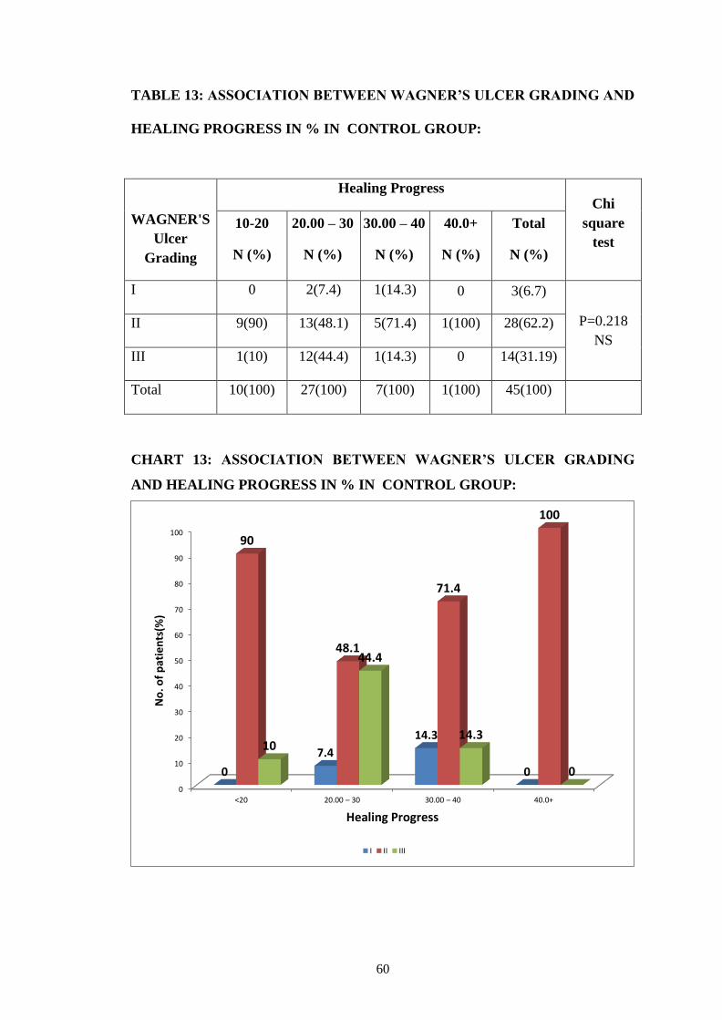
TABLE 13: ASSOCIATION BETWEEN WAGNER’S ULCER GRADING AND
HEALING PROGRESS IN % IN CONTROL GROUP:
WAGNER'S
Ulcer
Grading
Healing Progress Chi
square
test
10-20
N (%)
20.00 – 30
N (%)
30.00 – 40
N (%)
40.0+
N (%)
Total
N (%)
I 0 2(7.4) 1(14.3) 0 3(6.7)
P=0.218
NS
II 9(90) 13(48.1) 5(71.4) 1(100) 28(62.2)
III 1(10) 12(44.4) 1(14.3) 0 14(31.19)
Total 10(100) 27(100) 7(100) 1(100) 45(100)
CHART 13: ASSOCIATION BETWEEN WAGNER’S ULCER GRADING
AND HEALING PROGRESS IN % IN CONTROL GROUP:
0
10
20
30
40
50
60
70
80
90
100
<20 20.00 – 30 30.00 – 40 40.0+
0
7.4
14.3
0
90
48.1
71.4
100
10
44.4
14.3
0
No
. of
pat
ien
ts(%
)
Healing Progress
I II III

61
TABLE 14: ASSOCIATION BETWEEN CO-MORBIDITIES AND HEALING
PROGRESS IN PRP GROUP
Co-
Morbidities
Healing Progress Chi
square
test 20.00– 30
N(5)
30.00 – 40
N(%)
40.00 – 50
N(%)
50.00+
N(%)
Total
N(%)
Diabetic 4(80) 6(33.4) 4(28.6) 3(37.5) 17(37.8)
P=0.662
NS
Hypertension 0 0 1(7.1) 1(12.5) 2(4.4)
Diabetic and
Hypertension
0 2(11.1)
0 0 2(4.4)
Nil 1(20) 7(38.9) 8(57.1) 2(25) 18(40)
Varican
Veins
0 3(16.7) 1(7.1) 2(25) 6(13.3)
Total 5(100) 18(100) 14(100) 8(100) 45(100)
CHART 14: ASSOCIATION BETWEEN CO-MORBIDITIES AND HEALING
PROGRESS IN PRP GROUP
0
10
20
30
40
50
60
70
80
No
. of
pat
ien
ts(%
)
Healing Progress
20 – 30 30 – 40 40 – 50 50.00+

62
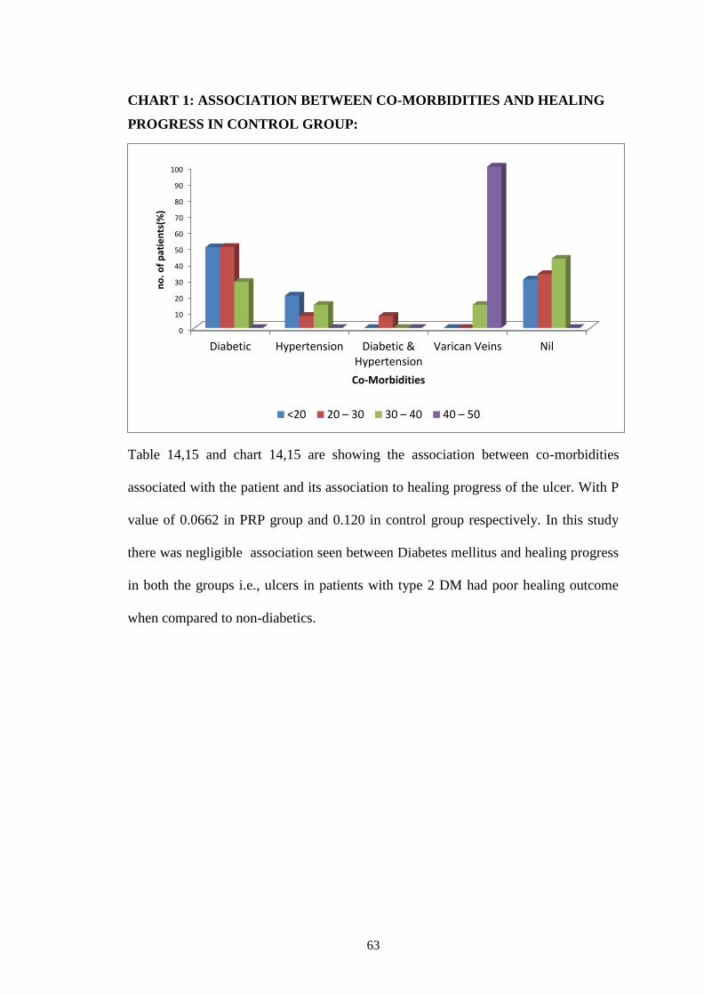
TABLE 15: ASSOCIATION BETWEEN CO-MORBIDITIES AND HEALING
PROGRESS IN CONTROL GROUP:
Comorbidities Healing Progress Chi
square
test
10-20
N(%)
20.00 – 30
N(5)
30.00 – 40
N (%)
40+
N(%)
Total
N(%)
Diabetic 5(50) 14(51.8) 2(28.6) 0 21(46.6)
P=0.120
NS
Hypertension 2(20) 2(7.4) 1(14.3) 0 5(11.1)
Diabetic and
Hypertension 0 2(7.4) 0 0 2(4.4)
Nil 3(30) 9(33.3) 3(42.9) 0 15(33.3)
Varicose
Veins 0 0 1(14.3) 1(100) 2(4.4)
Total 10(100) 27(100) 7(100) 1(100) 45(100)
63
CHART 1: ASSOCIATION BETWEEN CO-MORBIDITIES AND HEALING
PROGRESS IN CONTROL GROUP:
Table 14,15 and chart 14,15 are showing the association between co-morbidities
associated with the patient and its association to healing progress of the ulcer. With P
value of 0.0662 in PRP group and 0.120 in control group respectively. In this study
there was negligible association seen between Diabetes mellitus and healing progress
in both the groups i.e., ulcers in patients with type 2 DM had poor healing outcome
when compared to non-diabetics.
0
10
20
30
40
50
60
70
80
90
100
Diabetic Hypertension Diabetic &Hypertension
Varican Veins Nil
no
. o
f p
atie
nts
(%)
Co-Morbidities
<20 20 – 30 30 – 40 40 – 50

64
TABLE 16: ASSOCIATION BETWEEN WAGNER’S ULCER GRADE WITH
HEALING PROGRESS IN % IN PRP GROUP.
WAGNER'S
Ulcer Grade
Healing Progress Chi square
test 20.00 –
30
N(%)
30.00 –
40
N(%)
40.00 –
50
N(%)
50.00+
N(%)
Total
N(%)
I 0 0 7(50) 8(100) 15(33.3) P=0.001*
II 0 10(55.6) 7(50) 0 17(37.8)
III 5(100) 8(44.4) 0 0 13(28.9)
Total 5(100) 18(100) 14(100) 8(100) 45(100)
CHART 16:ASSOCIATION BETWEEN WAGNER’S ULCER GRADE WITH
HEALING PROGRESS IN % IN PRP GROUP.
0.00
10.00
20.00
30.00
40.00
50.00
60.00
70.00
80.00
90.00
100.00
I II III
0 0
100
0
55.6
44.4 50 50
0
100
0 0
No
. of
pat
ien
ts(%
)
Healing Progress
20.00 – 30 30.00 – 40 40.00 – 50 50.00+

65
TABLE 17: ASSOCIATION BETWEEN WAGNER’S ULCER GRADE WITH
HEALING PROGRESS IN % IN CONTROL GROUP.
WAGNER'S
Ulcer Grade
Healing Progress Chi
square
test
10-20
N (%)
20.00 – 30
N (%)
30.00 – 40
N (%)
40.0+
N (%)
Total
N (%)
I 0 2(7.4) 1(14.3) 0 3(6.7)
P=0.218
NS
II 9(90) 13(48.1) 5(71.4) 1(100) 28(62.2)
III 1(10) 12(44.4) 1(14.3) 0 14(31.19)
Total 10(100) 27(100) 7(100) 1(100) 45(100)
CHART 17:ASSOCIATION BETWEEN WAGNER’S ULCER GRADE WITH
HEALING PROGRESS IN % IN CONTROL GROUP.
Table 16,17 and chart 16,17 are showing association between Wagner ulcer grade
with healing progress in % in both PRP and control groups. There is sigificant
association between ulcer grade and healing % in PRP group with P valuve of 0.001
and 0.218 in control group. >50% healing was seen in 8 cases of grade I ulcer, 40-
50% healing results were seen in Grade II ulcers.
0
10
20
30
40
50
60
70
80
90
100
<20 20.00 – 30 30.00 – 40 40.0+
0
7.4 14.3
0
90
48.1
71.4
100
10
44.4
14.3
0
No
. o
f p
ati
en
ts(%
)
Healing Progress
I II III

66
DISCUSSION
Worldwide accepted standard therapy for chronic ulcers are debridement of
wound surgically/chemically, local wound dressings with topical bacteriostatic and
bactericidal agents such as povidone iodine, silver preparations etc., which are
thought to accelerate granulation tissue and promote wound healing. The prevalence
and incidence of chronic ulcers and the complications associated with them continue
to escalate even with proper and timely interventions. Platelet-rich plasma (PRP) is an
autologous product, extracted from the patient‟s plasma, which includes a high
platelet concentration in a fibrin clot that can be easily applied to the ulcer area. The
fibrin clot is absorbed during wound healing within days to weeks following its
application. Most of the studies on PRP reporting studies closure time and higher
healing percentage in patients using PRP and platelet derived products. This study
compares the efficacy of local injections of autologous platelet rich plasma with
conventional dressing in treatment of chronic ulcers.
A total of 90 patients with chronic cutaneous ulcers of Wagner‟s grade
between I-III were randomly divided into PRP group and conventional dressing group
with 45 patients each.
In this study most of the patients were in the age group of 60-69 years i.e., 11
out of 45 (24.4%) in PRP group and 14 out of 45 (31.1%) in control group (table 1
and chart 1).
In this study both the groups were male predominant i.e., 37 (82.2%) in PRP
group and 31 (68.9%) in control group accounting to 76%. (Table 2)
Site of ulcer (Table 3) was more on lower extremity in both the groups. i.e., 38
patients (84.4%) in PRP group and 35 patients (78%) in control group. The ulcers

67
were invariably seen oven the dorsal and plantar aspects of foot and at the ankle
region.
Most common mode of onset in the study was trauma in both the groups. 20
patients (44.4%) in PRP group and 12 patients (26.7%) in control group with average
of 35.55%. ulcers following abscess drainage was seen in 12 (26.7%) in PRP group
and 10 (22.2%) in control group. Other mode of onset were insidious more commonly
due to scratching, long standing varicose veins. Immobilization leading to bedsores
were seen in 1 patient in PRP group and 3 patients in control group.(Table 4)
Driver et al. (2006) carried out the first reported prospective, randomized,
controlled multicenter trial in the United States regarding the use of PRP for treatment
of diabetic ulcers. It included 32 patients. The authors found that 68.4% in PRP group
and 42.9% in control groups had complete closure of wounds with P value of 0.036
which was significant.
Sarvajnamurthy S et al. In 2013 studied on 12 patients with 17 chronic venous
ulcers were treated with PRP application and treatment outcome was measured by
percentage of improvement in area and volume of the ulcer. The mean duration of
healing of the ulcers was 5.1 weeks and 100% improvement in the area of the ulcers
was seen in 13 (76%).
In this study Wager‟s grade II ulcers were most commonly considered in both
PRP group and control group i.e., 17 (37.8%) in PRP group and 28 (62.2%) in control
group with mean average of 50%.
Most of the ulcers in the study were included once the culture from the wound
swab was sterile i.e., 28 (62.2%) in PRP group and 25 (55.6%) in control group and
the others comprised of S.aureus, Acinobacter and Citrobacter.
Coming to the healing progression in percentage, clearly healing was better

68
and faster in PRP group when compared to conventional dressing with 14 patients 40-
50% healing in PRP group when compared to conventional group i.e., 7 patients
(15.6%) and >50% healing seen in 8 (17.8%) of PRP group with high significant P
value of 0.00105.
In a study by Li L et al. (wound repair regen 2015) which was a prospective,
randomized controlled trial showed that standard treatment plus autologous platelet
rich plasma was statistically more effective than standard treatment. The subjects
defined as healing grade 1 were 85.4% while 67.3% in control group with
significance p value.
In this study, appearance of healthy granulation tissue was distinctively high
and early in PRP group i.e., 31 (68.9%) patients had satisfactory granulation tissue
when compared to the study group which had healthy granulation tissue in 17 patients
(37.8%) after treatment with povidone iodine dressing.
In a meta-analysis performed by CARTER et al. 2011 evaluated time to heal
in PRP subjects versus saline dressings. Two RCTs showed statistically significant
difference in wound area reduction compared to saline gauze dressings.
Similarly, a non RCT, comparative study showed significant area and depth
reductions with 2.5-3.5 fold decrease in time to reach 50% compared to pre-treatment
moist wound care controls.
Table16,17 and chart 16,17 are showing association between Wagner ulcer
grade with healing progress in % in both PRP and control groups. There is significant
association between ulcer grade and healing % in PRP group with P value of 0.001
and 0.218 in control group. >50% healing was seen in 8 cases of grade I ulcer, 40-
50% healing results were seen in Grade II ulcers.
In a study by JM de Leon et al.(2011) near physiological concentration of
69
PRP-gel in treatment of chronic ulcers which showed positive response occurred in
96.5% of wounds within 2.2weeks. in 47.5% area reduction occurred and 90.5% of
wounds had 63.6% volume reduction. Rapid treatment response was observed in 275
of 285 patients. The magnitude of response was consistently high, with statistically
significant outcomes reported for various groups.
In relation to associations between the study groups, there was significant
association with Wagner‟s ulcer grade and healing progress in % with high P value of
0.001 in PRP group and significance of P being 0.218 in control group.
There was no significant association seen between the mode of onset with and
healing progress between both the groups.
There was no significant association seen between the culture from the wound
and healing progress in both the groups.
There was no association seen between co-morbidities of the patient with the
healing progress in both the groups.

70
CONCLUSION
All the patients with chronic cutaneous ulcers of size less than 5cmx5cm in
greatest dimensions and ulcer grade between I-III of Wagener‟s grading were
managed with local injections of autologous PRP and were compared to the
ulcer management with conventional dressing i.e 10% povidone iodine
solution. Serial examination of ulcers have shown significant reduction in the
ulcer size with appearance of healthy granulation tissue, improvement in mean
duration of wound healing and contraction of wound in patients treated with
local injections of autologous PRP.
Local injections of autologous PRP in chronic cutaneous ulcers is more
effective when compared to conventional dressing with povidone iodine
dressing in achieving faster and complete wound healing and promoting
growth of healthy granulation tissue.
Since the PRP is derived from patients own blood, the risk of complications
were nil.
However, additional successful clinical evidence is required with validated
laboratory findings to establish local injection of autologous PRP as one of the
most effective alternative topical agents in treatment of chronic ulcers.
71
SUMMARY
This study was performed in Shri B.M Patil Medical College in the
department of general surgery during the study period of October 2016 to June
2018, to study the efficacy of local injection of PRP compared to conventional
dressing with 10% povidone iodine solution in treatment of chronic cutaneous
ulcers. Total of 90 patients with chronic ulcers were enrolled into the study
and randomly divided into PRP group and control group. With 45 patients in
each group. PRP was injected on every 4th
day for 4 cycles and regular
dressing was done with 10% povidone iodine in control group.
In our study we found that-
- 11 patients (24.4%) and 14 patients( 31.1%) were in the age group of 60-
69years.
- Both the groups were male predominant i.e., 37 patients (82.2%) in PRP group
and 31 patients (68.9%) in control group which accounted to 76%.
- Most common mode of onset in the study was trauma in both the groups. 20
patients (44.4%) in PRP group and 12 patients (26.7%) in control group with
average of 35.55%. ulcers following abscess drainage was seen in 12 (26.7%)
in PRP group and 10 (22.2%) in control group.
- Wager‟s grade II ulcers were most commonly considered in both PRP group
and control group i.e., 17 (37.8%) in PRP group and 28 (62.2%) in control
group with mean average of 50%.
- Culture from the wound swab was sterile i.e., 28 (62.2%) in PRP group and 25
(55.6%) in control group and the others comprised of S. aureus, Acinobacter
and Citrobacter.
72
- Healing was better and faster in PRP group when compared to conventional
dressing with 14 patients 40-50% healing in PRP group when compared to
conventional group i.e., 7 patients (15.6%) and >50% healing seen in 8
(17.8%) of PRP group with high significant P value of 0.00105.
- Appearance of healthy granulation tissue was distinctively high and early in
PRP group i.e., 31 (68.9%) patients had satisfactory granulation tissue when
compared to the study group which had healthy granulation tissue in 17
patients (37.8%) after treatment with povidone iodine dressing.
- There is significant association between ulcer grade and healing % in PRP
group with P value of 0.001 and 0.218 in control group. >50% healing was
seen in 8 cases of grade I ulcer, 40-50% healing results were seen in Grade II
ulcers.

73
BIBLOGRAPHY
1. Carter MJ, Fylling CP, Parnell LK. Use Of Platelet Rich Plasma Gel on
Wound Healing:
2. A Systematic Review and Meta-Analysis. Eplasty. 2011;11:((e38).Epub 2011
Sept 15.
3. Shrisvastava S, Singh PK, Taywade S. STARS therapy:”Sandeep‟s technique
for assisted regeneration of skin”. J Orthop Allied Sci.2016;4:5-7.
4. San Sebastian KM, Loboto I, Hernandez I, Burgos-Alonso N, Gomez-
Fernandez MC, et al. BMC Fam Pract. 2014;15:211.
5. Sarvajnamurthy S, Suryanaryan S, Budamakuntala L, Suresh DH. Autologous
Platelet Rich Plasma in Chronic Venous Ulcers: Study of 17 cases. J Cutan
Aesthet Surg. 2013 Apr-Jun: 6(2): 97-99.
6. Li L, Chen D, Wang C, Yuan N, Wang Y, He L, et al. Autologous platelet-rich
gel for treatement of diabetic chronic refractory cutaneous ulcers: A
prospective, randomized clinical trial. Wound Repair Regen. 2015 Jul-
Aug;23(4):495-505.
7. Martinez CE, Smith PC, Palma Alvarado VA, The influence of platelet-
derived products on angiogenesis and tissue repair: a concise update. Front
Physiol. 2015 Oct 20;(6):290.
8. Smith RG, Gassmann CJ, Campbell MS. Platelet Rich Plasma: Properties And
Clinical Applications, J Lancaster Gen Hosp. 2007(2):2.
9. Margolis DJ, Kantor J, Berlin JA, Healing of diabetic neuropathic foot ulcers
receiving standard treatment. A meta-analysis. Diabetic Care. 1999;22:692-5
10. Blume PA, Walters J, Payne W, Ayala J, Lantis J. Comparison of negative

74
pressure wound therapy using vacuum-assisted closure with advanced moist
wound therapy in the treatment of diabetic foot ulcers: A multicenter
randomized controlled trial. Diabetes Care. 2008;31:631-6.
11. Tzeng YS, Deng SC, Wang CH, Tsai JC, Chen TM, Burnout T. Treatment of
Nonhealing Diabetic Lower Extremity Ulcers with Skin Graft and Autologous
Platelet Gel: A Case Series.hindwai publish crop 2013, Article ID 837620-9
12. Miller MJ, Chandawarkar R. Wound Healing: Greenfield‟s Surgery Scientific
Principles and Practice: 6th
edn. China: Wolters Kluwer: 2017.p54-68.
13. Brunicardi FC, Anderson DK, Billar TR Dunn DL, Hunter JG, Matthews JB,
Pollock RE. Wound Healing: Schwartz‟s Principles of Surgery: 10th
edn: Mc
Graw Hill Education: 2015.p241-271.
14. Townsend CM, Evers BM, Beauchamp RD, Mattox KL. Wound Healing:
Sabiston Textbook of Surgery: 20th
edn: Elsevier 2017.p130-162.
15. Das S, Wound Healing: A Concise Textbook Of Surgery: 10th
edition: p 1-10.
16. Frykberg RG, Banks j: Challenges in the Treatment of Chronic Wounds.
Advances in wound care:2015 vol 4;9.
17. Ghoshal K, Bhattacharyya M: Overview of Platelet Physiology: Its Hemostatic
and Nonhemostatic Role in Disease Pathology: Hindwai publication: vol 2014,
Article ID 781857.
18. Tran TDX, Le P, Pham P: Diabetic foot ulcer treatment by activated platelet
rich plasma: a clinical study. Biomedical research and therapy 2014, 1(2):37-
42.

75
19. Rozam P, Bolta Z: Use of platelet growth factors in treating wounds and soft-
tissue injuries. Acta Dermatoven APA 2007, 16:156-165.
20. Alcantara DC, Zaragoza M, Gimenez E, et al: platelet rich plasma: New
Insights for Ccutaneius Wound Healing Management. J Funct. Biomater.
2008, 9, 10;doi10.3390/jfb9010010.
21. Lacci KM, Dardil A: Platelet-Rich Plasma: Support for Its Use in Wound
Healing. Yale J. of bio and med 2010 (8) 1-9.
22. Sebastian KMS, Labato I, Hernandez I, et at,: Efficacy and Safety of
autologous platelet rich plasma for the treatment of vascular ulcers in primary
care: Phase III study. BMC Family Practice 2014, 15:211.

76
ANNEXURES
ETHICAL CLEARANCE CERTIFICATE
77
INFORMED CONSENT FORM
B.L.D.E (DEEMED TO BE) UNIVERSITY’s
SHRI B.M. PATIL MEDICAL COLLEGE, HOSPITAL AND RESEARCH
CENTRE, VIJAYAPUR – 586103, KARNATAKA.
TITLE OF THE PROJECT: AUTOLOGOUS PLATELET RICH
PLASMA IN TREATEMENT OF
CHRONIC CUTANEOUS ULCERS.
PRINCIPAL INVESTEGATOR: DR MANISHA.V
Department of General Surgery
PG GUIDE: DR. RAMAKANTH BALOORKAR MS
Associate Professor of Surgery
Shri. B. M. Patil Medical College &
Hospital Vijayapur
PURPOSE OF RESEARCH:
I have been informed that this study will analyze the usefulness of local
injection of Platelet rich plasma in healing of chronic cutaneous ulcers.
I have been explained about the reason for doing this study and selecting
me/my ward as a subject for this study. I have also been given free choice for either
being included or not in the study
PROCEDURE:
I understand that relevant history will be taken. I will undergo detailed clinical
examination after which necessary investigations will be done whenever required,
which would help the investigator for appropriate management.

78
RISKS AND DISCOMFORTS:
I understand that I/my ward may experience some pain and discomfort during
the examination or during my treatment. This is mainly the result of my condition and
the procedure of this study is not expected to exaggerate these feelings which are
associated with the usual course of treatment.
BENEFITS:
I understand that I/my wards participation in this study will help to analyse the
effectiveness of PRP in healing of chronic cutaneous ulcers.
CONFIDENTIALITY:
I understand that medical information produced by this study will become a
part of this Hospital records and will be subjected to the confidentiality and privacy
regulation of this hospital. Information of a sensitive, personal nature will not be a
part of the medical records, but will be stored in the investigator‟s research file and
identified only by a code number. The code key connecting name to numbers will be
kept in a separate secure location.
If the data are used for publication in the medical literature or for teaching
purpose, no names will be used and other identifiers such as photographs and audio or
video tapes will be used only with my special written permission. I understand that I
may see the photograph and videotapes and hear audiotapes before giving this
permission.
79
REQUEST FOR MORE INFORMATION:
I understand that I may ask more questions about the study at any time.
DR.MANISHA V is available to answer my questions or concerns. I understand that I
will be informed of any significant new findings discovered during the course of this
study, which might influence my continued participation.
If during this study, or later, I wish to discuss my participation in or concerns
regarding this study with a person not directly involved, I am aware that the social
worker of the hospital is available to talk with me and that a copy of this consent form
will be given to me for careful reading.
REFUSAL OR WITHDRAWL OF PARTICIPATION:
I understand that my participation is voluntary and I may refuse to participate
or may withdraw consent and discontinue participation in the study at any time
without prejudice to my present or future care at this hospital.
I also understand that DR MANISHA V. will terminate my participation in
this study at any time after he has explained the reasons for doing so and has helped
arrange for my continued care by my own physician or therapist, if this is appropriate
80
.INJURY STATEMENT:
I understand that in the unlikely event of injury to me/my ward, resulting
directly to my participation in this study, if such injury were reported promptly, then
medical treatment would be available to me, but no further compensation will be
provided.
I understand that by my agreement to participate in this study, I am not
waiving any of my legal rights.
I have explained to _________________________________________ the
purpose of this research, the procedures required and the possible risks and benefits,
to the best of my ability in patient‟s own language.
Dr. RAMAKANTH BALOORKAR Dr .MANISHA
(Guide) (Investigator)

81
PROFORMA
CASE NO:
Name : IP No :
Age/sex: DOA:
Occupation:
Address:
CHIEF COMPLAINTS:
WOUND:
Mode Of Onset
Duration
Number
Site
Size and Extent
Associated Pain
Discharge
Others
Any Associated Disease
Past History Of Similar Wound
Personal History
PAST HISTORY:
Diabetes Mellitus
Hypertension
Peripheral vascular disease

82
PERSONAL HISTORY:
Diet
Sleep
Appetite
Bowel & bladder
7. GENERAL PHYSICAL EXAMINATION:
Mental Status Weight:
Built Height:
Nourishment BMI:
Hydration status
Pallor
Pulse
Blood Pressure
Any Obvious Deformity
A. LOCAL EXAMINATION :
INSPECTION-
NUMBER
SITE
SIZE
SHAPE
EXTENT
MARGIN
EDGE OF THE ULCER
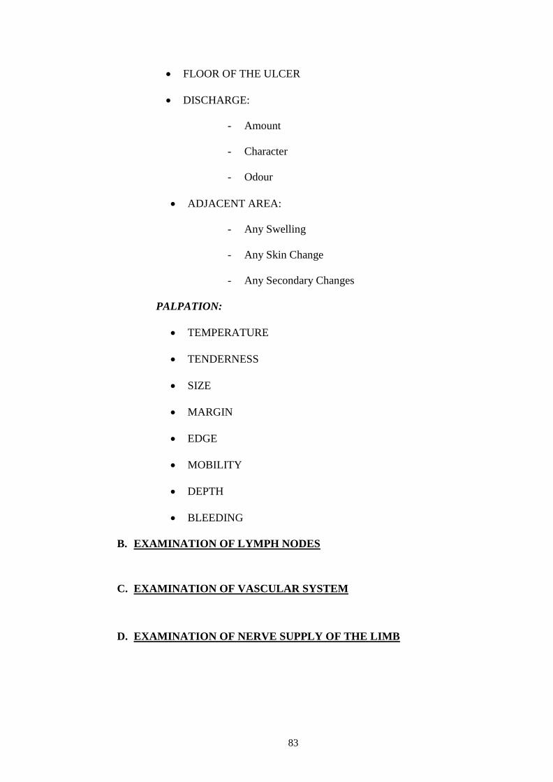
83
FLOOR OF THE ULCER
DISCHARGE:
- Amount
- Character
- Odour
ADJACENT AREA:
- Any Swelling
- Any Skin Change
- Any Secondary Changes
PALPATION:
TEMPERATURE
TENDERNESS
SIZE
MARGIN
EDGE
MOBILITY
DEPTH
BLEEDING
B. EXAMINATION OF LYMPH NODES
C. EXAMINATION OF VASCULAR SYSTEM
D. EXAMINATION OF NERVE SUPPLY OF THE LIMB

84
SYSTEMIC EXAMINATION
PER ABDOMEN:
Inspection:
Palpation:
Percussion:
Auscultation:
RERSPRATORY SYSTEM:
Inspection
Palpation
Percussion
Auscultation
CARDIO-VASCULAR SYSTEM:
Inspection
Palpation
Percussion
Auscultation
CENTRAL NERVOUS SYSTEM::
Higher Mental functions
Diagnosis
INVESTIGATIONS:
Hemoglobin
Total Count
Differential Count
Blood Urea

85
Serum Creatinine
BLOOD SUGAR RBS, FBS, PPBS (if diabetic)
Urine For Ketone Bodies (If Diabetic)
HbA1C ( If Diabetic)
Urine Routine
Culture Sensitivity Of Discharge
X-Ray of bone Or Joint Involved
COMMENTS:
PRE-TREATEMENT MEASUREMENTS:
SERIAL MEASUREMENTS DURING THE TREATEMENT:
FINAL MEASUREMENTS:
PROGRESSION OF HEALING: (In terms of %)
INFERENCE: