Journals Removed from UGC-Approved List of Journals - Govt ...
Upload
khangminh22Category
view
0download
0

Romanian Neurosurgery

EDITORIAL AND ADVISORY BOARD
EDITOR-IN-CHIEF Dr St. M. Iencean, MD, PhD
EXECUTIVE EDITOR
Al. Chiriac, PhD
ASSISTANT EDITORS
B. Costachescu
A. Iordache
ADVISORY BOARD - ROMANIA
Professor D. Adam, Romania
Dr. Fl. Exergian, Romania
Professor St.I. Florian, Romania
Professor R.M. Gorgan, Romania
Professor G.B.I. Iacob, Romania
Dr. Al. Lupsa, Romania
Professor I. Poeata, Romania
Dr. Al. Tascu, Romania
Dr. Marius Dabija (Assist. Prof.), Romania
ADVISORY BOARD - INTERNATIONAL Professor M.A. Arraez, Spain
Professor V. Astarastoae, Romania
Professor H. Bertalanffy, Germany
Professor J. Brotchi, Belgium
Professor P. Courtheoux, France
Professor J.P. Houtteville, France
Professor Y. Kato, Japan
Professor U. Kehler, Germany
Professor Christopher M. Loftus, USA
Dr. M.R. Mahmud, Nigeria
Professor J.Cl. Marchal, France
Professor P. Mertens, France
Professor B.K. Misra, India
Professor D.F. Muresanu, Romania
Professor L. Pendefunda, Romania
Professor S.C. Robertson, USA
Professor M. Samii, Germany
Professor J. Schramm, Germany
Professor M. Sindou, France
Professor B. Sutter, Austria
Professor F. Umansky, Israel
Professor T.T. Wong, Taiwan
EMERITUS EDITORIAL BOARD FOUNDING EDITOR Professor A.V. Ciurea, Romania
Assoc. Prof. Habil. H. Ples, Romania, Former Editor
_Professor Al. Constantinovici, Former Editor_
_Professor Constantin Arseni_

ROMANIAN
NEUROSURGERY
Vol. XXXV | No. 2 June 2021

Copyright © 2021 Romanian Society of Neurosurgery &
London Academic Publishing
All rights reserved. This book or any portion thereof may not be
reproduced or used in any manner whatsoever without the express
written permission of the Romanian Society of Neurosurgery or the
publisher except for the use of brief quotations in a book review or
scholarly journal.
ISSN 1220-8841 (Print)
ISSN 2344-4959 (Online)
First Printing: June 2021
London Academic Publishing Ltd.
27, Old Gloucester Street
WC1N 3AX
London, United Kingdom
Email: [email protected]
london-ap.uk
lapub.co.uk
journals.lapub.co.uk
journals.lapub.co.uk/index.php/roneurosurgery
Company Reg. No. 10941794
Registered in England and Wales
The opinions expressed in the published articles are the sole
responsibility of the authors and do not reflect the opinion of the editors
or members of the editorial board.

CONTENTS
129 Subarachnoid haemorrhage. A critical neurosurgical emergency
Alexandra Bibiriță, Daniel Teleanu, Alexandru Vlad Ciurea
135 Meningioma in shape. Can the appearance of tumour margins be considered as a prognostic factor?
A.I. Cucu, Mihaela Cosman, B. Dobrovat, Cristina Dascalu, Ioana Jitaru, R.B. Sandu, A. Tudor, Claudia Costea, Mihaela Turliuc, Gabriela Dumitrescu, Anca Sava, I. Poeata
143 The role of MRI-guided focused ultrasound in neurosurgery. A narrative review
Marius Gabriel Dabija, Ioan Sebastian Nechifor, Vlad Andrei Dabija, Bogdan Costachescu, Lucian Eva
148 The stent-assisted coil-jailing technique for very small intracranial aneurysm treatment
A. Chiriac, N. Dobrin, Georgiana Ion, Z. Faiyad, I. Poeata
152 Endovascular treatment options for carotid-cavernous fistulae
Roxana Codreanu, Rares Cristian Filep, Lucian Marginean
159 Deep cerebral vein thrombosis due to anaemia in a child
Rajneesh Misra, Sushil Kumar, Sandeep Sharma
162 Long term clinical outcome following decompressive surgery for Cauda Equina Syndrome. A single centre experience from India
Lohar Vishnu Kumar, Jaiswal Gaurav, Gupta Tarun Kumar, Jain Sachin Kumar, Lodha Krishna Govind, Yadav Kaushal

174 Visual outcome analysis in patients with posterior fossa tumours undergoing surgical treatment
Deepak Kumar Singh, Vrihaspati Kumar Agrahari, Mohd. Kaif, Rakesh Kumar, Kuldeep Yadav
180 Single versus double burr holes evacuation in the treatment of
chronic subdural hematoma. A tertiary centre experience
Shrish Nalin, Anurag Sahu, Kanika Gupta, Kulwant Singh
189 Upper cervical spine tuberculosis. A case report
A. Khelifa, L. Berchiche, W. Bennabi, M. Al-Zekri, A. Morsli
192 The use of corticosteroids in autoimmune encephalitis. Basic and clinical considerations
Bryan Lester Nahar-González, Ivan David Lozada-Martinez, Yandris Arevalo-Martínez, Loraine Quintana-Pajaro, Silvia Prada-Soto, Teresa Pacheco-Hernandez, William Florez-Perdomo, Yelson Alejandro Picón-Jaimes, Luis Rafael Moscote-Salazar
199 Paediatric brain abscesses in tribal region of India. A single
centre experience
Sachin Kumar Jain, Gaurav Jaiswal, Tarun Kumar Gupta, Krishna Govind Lodha, Vishnu Kumar Lohar, Kaushal Yadav
204 Lateral orbitotomy in the management of an intra-orbital
lipoma. A case report
Mandour Cherkaoui, Ait Lhaj El Houssaine, Khouya Ali Adil, Gazzaz Miloudi, E.L. Mostarchid Brahim
207 Primary calvarial cavernous haemangioma in a child. A case
report
Adel Khelifa, Walid Bennabi, Lakhder Berchiche, Abdelhalim Morsli
210 A solitary case of gliosarcoma an indication for TP53 mutation
analysis: a non-concordant finding. Case report
Ebtesam Abdulla, Nabeel Hameed, Roopa Arora

215 C7 contribution to the ulnar nerve. Literature review
Lívio Pereira de Macêdo, Arlindo Ugulino Netto, Kauê Franke, Pierre Vansant Oliveira Eugenio, Lucas Ribeiro de Moraes Freitas, Fábio Antônio Serra de Lima Júnior, João Vitor Romeiro de Paula, Silvya Nery Bernardino, Fernando Henrique Morais de Souza, Nivaldo S. Almeida, Hildo Rocha Cirne Azevedo-Filho
219 Burr-hole craniostomy versus mini-craniotomy in the treatment of chronic subdural hematomas. Analysis of clinical results
André Tokpa, Moussa Diallo, Louis Kéabléon Derou, Yves Soress Dongo, Bernard Fionko, Adéréhime Haïdara
225 Shunt in scrotum: unusual shunt complication in an operated case of post TBM hydrocephalus
Manoj Agarwal, Sharad Pandey, Pankaj Kumar, L.N. Gupta
228 Spectrum of non-traumatic craniovertebral junction disorders. Diagnosis and demonstration with magnetic resonance imaging and multidetector computed tomography
Neha Singh, Deepak Kumar Singh
234 Possibilities of endoscopic endonasal transsphenoidal surgery in treatment of growth-hormone pituitary adenomas
Ruslan V. Aksyonov, Orest I. Palamar, Andriy P. Huk, Dmytro S. Teslenko, Dmytro I. Okonskyi
239 Letter to the editor. Intracranial aneurysm: research in preclinical outcome models and human effectiveness of intraluminal devices
Tariq Janjua, Luis Rafael Moscote-Salazar, Amit Agrawal
241 Guidelines for authors


Romanian Neurosurgery (2021) XXXV (2): pp. 129-134 DOI: 10.33962/roneuro-2021-020 www.journals.lapub.co.uk/index.php/roneurosurgery
Subarachnoid haemorrhage. A critical neurosurgical emergency
Alexandra Bibiriță1, Daniel Teleanu2,3,
Alexandru Vlad Ciurea4,5 1 Resident of Neurosurgery at Bucharest University Emergency
Hospital, ROMANIA 2 Chief of Neurosurgery Department at Bucharest University
Emergency Hospital, ROMANIA 3 Assoc. Professor of Neurosurgery at “Carol Davila” University of
Medicine and Pharmacy, Bucharest, ROMANIA 4 Chief of Neurosurgery Department and Scientific Director of
Sanador Clinical Hospital, Bucharest, ROMANIA 5 Professor of Neurosurgery at “Carol Davila” University of Medicine
and Pharmacy, Bucharest, ROMANIA
ABSTRACT Subarachnoid haemorrhage (SAH) accounts for 3% of all strokes and is the cause of
5% of stroke mortality. SAH by rupture of cerebral aneurysm or arterial-venous
malformation (AVM) remains the most devastating cerebrovascular disease. During
admission for SAH, about 30-70% of patients suffer a rebleed, and from all rebleeds,
about 90% lead to death no matter the treatment. Available current scales help
predict the prognosis and guide the therapy. Considering that the lifestyle risk factors
for SAH are of increasing prevalence, it is expected that it will affect even more people
in the future. SAH should not be regarded as a disease but rather a set of events with
devastating complications requiring adequate management from debut extending
long after patient discharge.
EPIDEMIOLOGY
Subarachnoid haemorrhage stands for 3% of all cerebral vascular
strokes and is responsible for the 5% mortality coming from CVAs.
Incidence peak for SAH is 55-60 age group. During hospital admission
for this pathology 30-70% of patients suffer a rebleed and
approximately 90% of all rebleeds will end in death whatever the
treatment.
PHYSIOPATHOLOGY AND AETIOLOGY
Subarachnoid haemorrhage occurs as a consequence of a blood vessel
rupture in the subarachnoid space or near it. Following this rupture,
blood will invade the space between the pia mater and the arachnoid,
Keywords risk factors,
neurosurgical emergency, CT,
angiography, subarachnoid haemorrhage
scales, cerebral vasospasm
Corresponding author: Alexandra Bibirita
Bucharest University Emergency
Hospital, Romania
Scan to access the online version

130 Alexandra Bibiriță, Daniel Teleanu, Alexandru Vlad Ciurea
the subarachnoid space, which normally contains
just cerebrospinal fluid.
Etiologically, which is considering the reason that
led to the event, following types of SAH stand out –
traumatic SAH or spontaneous.
The spontaneous form of SAH occurs in cases of
high arterial blood pressure (hypertensive SAH) or is
the direct consequence of an aneurysmal rupture, a
cerebral or spinal arterial-venous malformation
rupture or secondary to cervical-cerebral arteries
dissection. [3,12] Actually about 85% of all SAH forms
are due to the rupture of a cerebral aneurysm. Brain
aneurysm typically occurs during spring and autumn
and is considered directly linked to temperature
changes, similar to CVAs. [2,12]
A rare form of spontaneous SAH is the idiopathic
subarachnoid hemorrhage, in the presence of risk
factors considered non-traditional and modifiable –
alcoholism, smoking, cocaine or amphetamine
abuse. Besides these forms, literature provides a
series of traditional risk factors, which are not subject
to medical or lifestyle intervention and thus called
non-modifiable, frequently linked to SAH occurrence:
family history of SAH, soft tissue disease (polycystic
kidney disease, neurofibromatosis type I, Ehlers-
Danlos syndrome mainly type IV and other collagen
abnormalities), female sex (1.5 times the risk),
African descent (2 times the risk), Japanese or Finnish
descent, vasculitis, even more rare factors –
parasitosis, Moya-Moya disease, eclampsia, blood
disorders, coagulation disorders. [1,2,11,12] It is
important to point out that as prevalence of the non-
modifiable risk factors gets higher, it is expected that
SAH incidence will increase in the following decades.
CLINIC OF SUBARACHNOID HAEMORRHAGE
Clinically the first symptom and nonetheless the
most constant is severe headache with sudden onset
– approximately 97% of cases, describes as a
thunderclap headache and acknowledged by the
patient as “the most intense headache I ever had”.
[12]
A more particular situation is the sentinel
headache – a sudden and severe pain that goes away,
allowing for a symptom-free period with neglection
of said episode, followed shortly by severe mental
status alteration, paroxysmal phenomena known by
neurosurgeons as “walk, talk and die patients.” [1,3]
Photophobia as an accompanying symptom is
frequent, together with ocular symptoms such as
subhialoid hemorrhage, retinal or vitreous
hemorrhage. In medical literature vitreous
hemorrhage associated with SAH is known as
Terson’s syndrome. [8]
Eye fundus examination may reveal papillary
edema secondary to increased intracranial
hypertension secondary to bleeding into the
subarachnoid space. Vegetative phenomena such as
emesis or syncope are common.
Sometimes, although not rarely the patient seeks
medical attention after a loss of consciousness
episode or for altered mental status, as neurological
status on admission might vary from slight confusion
to deep coma. Epileptic seizures occur frequently as
about 20% of patients develop such symptoms in the
first 24 hours since debut. They are considered to be
a straight effect of increased intracranial pressure,
associated hyponatremia or aneurysm site,
especially when it involves sylvian arteries territory.
[3,9]
Neurological examination reveals early
meningeal signs: neck stiffness, headache,
photophobia, ocular pain, emesis. Neck stiffness
usually develops within 6 to 24 h from the debut of
SAH. Within a few hours from debut signs such as
Kernig, Brudzinki or bilateral Lassegue can appear or
focal neurological signs. Psychiatric acute onset is
more common and sometimes even specific among
elderly patients. Some other accompanying signs or
symptoms are acute urinary retention, diminished or
abolished osteo-tendinous reflexes (usually after 4 to
6 h from the debut of the hemorrhage) and
eventually focal neurological signs. Elderly patients,
most commonly after 70 years old very often present
with a far worse neurological status on admission.
[9,12]
DIAGNOSIS
The elective diagnostic tool in SAH is the plain
cerebral CT examination on arrival in the emergency
room, which shows blood in the subarachnoid space.
The Fisher grade is thus immediately established.
This diagnostic imaging tool is nonetheless the actual
gold standard in diagnosing the subarachnoid
hemorrhage, its limitation being that when not
enough time since the debut of the hemorrhage has
passed, the first examination might turn out negative

131 Subarachnoid haemorrhage. A critical neurosurgical emergency
and will become positive a few tens of minutes later.
The Fisher grading system is based upon the results
of the plain cerebral CT examination and it
appreciates the risk of developing cerebral
vasospasm. It is summarized in the table below. [14]
Fischer
Grade
Presence of
blood in the SA
space
Other aspects Vasospasm
risk
Grade 1 No blood in the
cisterns
No clots 21%
Grade 2 Blood <1 mm
thickness
No clots 25%
Grade 3 Blood >1 mm
thickness
+/- clots 37%
Grade 4 Rare blood,
diffusely in the
basal cisterns
Intraventricular
hemorrhage or
parenchymal
21-37%
Table 1. Fisher Scale (1982).
Figure 1. Cranio-cerebral CT axial sequences showing the 4
Fisher grades (1 – grade 1, 2 – grade 2, 3 – grade 3, 4 – grade 4).
Source Future Neurol @ 2013 Future Medicine Ltd.
If the CT examination is negative but the clinical
suspicion of SAH is still high, a lumbar puncture may
be helpful for diagnostic purpose, but it is to be
performed with every precaution and only after eye
fundus examination excludes intracranial
hypertension, in order to avoid cerebral herniation.
This procedure can induce a rebleed if intracranial
pressure is too abruptly decreased, which is why a
minimal amount of a CSF sample is to be drawn. [3,5]
Lumbar puncture is according to available date the
most sensitive diagnostic tool for SAH. A positive
result consists of increased CSF pressure,
xanthochromic CSF, more than 100 000 thousand
erythrocytes per mm3, increased proteinorrhachia
(>50 mg/dl) and normal or slightly decreased
glycorrhachia (<50-70 mg/dl) [3,4,5].
Cerebral MRI imaging with FLAIR sequencing, CT
angiography or MRI angiography do not add very
much relevance to the SAH diagnosis in the first 24-
48 hours following debut, but they prove to be
excellent between day 3 and 7, with good premises
for identifying a cerebral aneurysm and its
anatomical features. [7]
Figure 2. Axial enhancement cerebral CT sequence – red arrow
points to an anterior communicating artery aneurysm –
Collection of Neurosurgery I Clinic, Emergency University
Hospital of Bucharest.
Digital subtraction angiography (DSA) - now a tool of
capital importance in the management of
subarachnoid hemorrhage – consists of selectively
injecting contrast material using a catheter inserted
into a large artery under radiologic screen. It is an
extremely useful resource in the evaluation of
cerebral aneurysms and it can also come in as a both
diagnostic and therapeutic tool, providing the
possibility of simultaneously embolizing the
incriminated aneurysm or AVM.

132 Alexandra Bibiriță, Daniel Teleanu, Alexandru Vlad Ciurea
Figure 3. Right sylvian artery giant dissecting aneurysm near
M1 and M2 segments junction. Right internal carotid
angiography clichee. Collection of Neurosurgery I Clinic,
Emergency University Hospital of Bucharest.
Aneurysm with sacs smaller than 5 mm are ideal for
endovascular coiling, while for the rest of them the
risk of recanalization or incomplete occlusion are
considered too high, thus needing stent or balloon
assisted coiling.
Figure 4. Digital subtraction angiography sequence – black
arrows points to a anterior communication artery aneurysm –
Open Source image.
MANAGEMENT
Once the diagnosis has been confirmed therapeutic
measures are immediately started based on
patient’s clinical and neurological status and in
closed connections with SAH etiology. Diagnosis is
further completed with other needed tests. The
patient should be admitted into a neurosurgical
service with dedicated intensive care unit in case of
need of advanced vital function support.
Drug treatment consists of maintaining an
optimal blood pressure of systolic BP < 160 mmHg,
considering that high blood pressure values are
associated with worsening SAH and rebleed risk.
Vasospasm prevention is to be addressed by
administering Nimodipine. Cerebral edema
prevention, maintaining normal volemia and
preventing hyponatremia are the next medical
considerations, as even with maximal therapy they
are extremely difficult to correct if they occur. Anti-
seizure prophylaxis is mandatory because of the
irritation of cerebral cortex by blood in the
subarachnoid space. [6,9]
The neurological status of the patient suffering
from SAH is to be appreciated with the help of Hunt
& Hess scale and WNFS scale (World Neurosurgical
Federation Society scale - 1988) and it is extremely
important in the surgical indication. Classification
into a Hunt & Hess (1968) grade is determined after
clinical and neurological assessment as follows: [15]
Grade I: absent or mild headache, absent or minimal
neck stiffness
Grade II: severe headache, franc neck stiffness,
possible cranial nerves paresis
Grade III: confusion or lethargy, mild focal deficits
Grade IV: stuporous, hemiparesis forte, decerebrate
Grade V: comatose, decerebrate.
WFNS Scale is based upon Glasgow Comma Scale
and the presence or absence of neurological deficits
as such:
Grade I: GCS 15 points, no neurological deficits
Grade II: GCS 13-14 points, no neurological deficits
Grade III: GCS 13-14 points, present neurological
deficits
Grade IV: GCS 7-12 points, whatever the neurological
deficits
Grade V: GCS 3-6 points, whatever the neurological
deficits.
Another scale of great clinical and therapeutical
significance is the Fisher scale previously presented,
with four degrees of severity, based upon computed-
tomography aspects such as the presence of blood

133 Subarachnoid haemorrhage. A critical neurosurgical emergency
in the ventricular system or parenchymal hematoma
and thickness of subarachnoid blood.
The right moment to perform surgery in
aneurysmal SAH is nowadays considered to be at 24-
72 hours from debut in patients with Hunt & Hess
grades 1 or 2 and consists of aneurysm securement
through classical surgical clipping or endovascular
coiling. Patients with a higher Hunt & Hess grade
(altered mental status or neurological deficits) are to
be admitted and monitored in the intensive care unit
for vital functions support with the goal of obtaining
a better neurological status in order for the
aneurysm securement to be achievable with a more
favorable risk/benefit balance.
Exception to this rule occurs when SAH of any
etiology is accompanied by large parenchymal
hematoma (Fisher grade IV) that come with vital risk
to the patient and it requires emergency intervention
whatever the Hunt & Hess grade. A similar situation
occurs when patient’s life is endangered on the sort-
term due to intracranial hypertension (secondary to
acute obstructive hydrocephalus following SAH) - a
external ventricular drainage is placed together with
an intracranial pressure monitoring device – in order to
obtain clinical and neurological amelioration until
definitive treatment of the cause that led to SAH is
possible. [9, 10]
Following surgery, after securing the aneurysm
whose rupture produced the subarachnoid
hemorrhage, together with general medical
measures and complications prevention, a
transcranial Doppler ultrasound is needed in order
to appreciate the blood flow through the main
cerebral arteries and possible vasospasm, as well as
control cerebral CT examination for the verifying of
occlusion devices used and for the visualizing the
aspect of the ongoing SAH. In cases of drug resistant
vasospasm intraarterial endovascular vasodilator
therapy may be used – vasodilator agents are
selectively injected directly into the cerebral arteries
under angiographical control.
COMPLICATIONS
In the postoperative period medical treatment
measures must be continued. Considering the risk of
rebleed, which stand at 30-70% despite adequate
treatment and knowing that 90% of rebleed lead to
death, postsurgical patients must be monitored in
the intensive care unit.
Despite these therapeutic resources, the major
risk of devastating complications particularly difficult
to treat persists – cerebral vasospasm and late
cerebral ischemia, vegetative phenomena of central
origin, parenchymal hematoma, rebleed, electrolytes
imbalance, normal pressure hydrocephalus. [10]
The risk of developing cerebral vasospasm stands
present since the debut of SAH but is considered to
be at its highest between days 3-7 and extending up
to 3 weeks. It is very accurately appreciated with the
help of the Fisher grading system, risk correlation
being proportionate with a higher grade.
Figure 5. Résumé of SAH complications and their mechanism.
Personal illustration.
NEUROLOGICAL REHABILITATION
The treatment of subarachnoid hemorrhage is a
multidisciplinary one, step-by-step and one which
address simultaneously the etiological factor, the
worsening factor and at the same time the
prevention of complications. [2,12] For a favorable
outcome and prognosis, and also for survivors’
rehabilitation a quick and adequate diagnosis is
required. It should happen in a primary
neurosurgery center with radiology and intensive
care optimal facilities available at all times. The
prognosis of these patients and their neurological
rehabilitation require the right cooperation between
the medical team consisting of emergency room
physician – radiologist – interventionist –
neurosurgeon – intensivist – neurologist and
rehabilitation physician.
CONCLUSIONS
A curative treatment of subarachnoid hemorrhage is
out of the question at the moment, with it being not
actually a disease but rather a course of pathological
events that develop in a chain like manner and that

134 Alexandra Bibiriță, Daniel Teleanu, Alexandru Vlad Ciurea
imply a series of serious consequences over cerebral
structures, both short term and long term.
Methods of treatment of SAH complications are
currently under development, and even though they
are based on hypotheses decades old, some of them
are highly debatable according to some authors,
such as subarachnoid space washout-out with drugs
designed to prevent vasospasm. [13]
Considering that about 15-20% of patients that
suffer from SAH decease before getting to medical
attention of any kind according to WHO data, and
that 40% of survivors from SAH of any kind will have
permanent neurological deficits, two things stand
out: early identification of high suspicion SAH cases
is critical and referral to an emergency neurosurgical
service is of paramount need.
ABBREVIATIONS AVM – arterial-venous malformation;
BP – blood pressure;
CSF – cerebrospinal fluid;
CVA – cerebrovascular accident;
ICP – increased intracranial pressure;
WHO – World Health Organization.
CONFLICTS OF INTEREST The authors have no conflicts of interest to declare.
REFERENCES
1. Kameda-Smith M, Aref M, Jung Y, Ghayur H, Farrokhyar F,
Determining the diagnostic utility of lumbar punctures in
CT negative suspected subarachnoid hemorrhage: A
Systematic Review and Meta-analysis, World Neurosurg.
2020; 148: e27-e34.
2. Kellermann I, Kleindienst A, Hore N, Buchfelder M,
Brandner S., Early CSF and Serum S100B, Concentrations
for Outcome Prediction in Traumatic Brain Injury and
Subarachnoid Hemorrhage, Clin Neurol Neurosurg.
2016; 145:79-83.
3. Nadkarni NA, Maas MB, Batra A, Kim M, Manno EM,
Sorond FA, Prabhakaran S, Naidech AM, Liotta EM,
Elevated Cerebrospinal Fluid Protein Is Associated with
Unfavorable Functional Outcome in Spontaneous
Subarachnoid Hemorrhage, J Stroke Cerebrovasc Dis.
2020 29(4):10460.
4. Lenhart M, Bretschneider T, Gmeinwieser J, Ullrich OW,
Schlaier J, Feuerbach S, Cerebral CT angiography in the
diagnosis of acute subarachnoid hemorrhage, Acta
Radiol., 2013, 38(5):791-6.
5. Raevis J, Elmalem VI, Pseudotumor cerebri syndrome
causing a terson like syndrome, Am J Ophthalmol Case
Rep. 2020; 20: 100993.
6. Vivancos J, Gilo F, Clinical management guidelines for
subarachnoid haemorrhage. Diagnosis and treatment,
Neurologia, 2014;29(6):353-70.
7. Wu L, Chen G, Signaling Pathway in Cerebral Vasospasm
After Subarachnoid Hemorrhage: News Update, Acta
Neurochir Suppl. 2016; 121:161-5.
8. Veldeman M, Albanna W, Weiss M, Conzen C, Schmidt TP,
Schulze - Steinen H, Wiesmann M, Clusmann H, Schubert
GA, Invasive neuromonitoring with an extended
definition of delayed cerebral ischemia is associated with
improved outcome after poor-grade subarachnoid
hemorrhage, J Neurosurg. 2020; 15:1-8.
9. Veldeman M, Albanna W, Weiss M, Conzen C, Schmidt TP,
Clusmann H, Schulze-Steinen H, Nikoubashman O, Temel
Y, Schubert GA, Treatment of Delayed Cerebral Ischemia
in Good-Grade Subarachnoid Hemorrhage: Any Role for
Invasive Neuromonitoring?, Neurocrit Care. 2020.
10. Probst MA, Hoffman JR, MD, Computed Tomography
Angiography of the Head is a reasonable next test
following a Negative Non-contrast Head Computed
Tomography in the Emergency Department work up of
Subarachnoid Hemorrhage, Ann Emerg Med. 2016 June;
67(6): 773–774.
11. Hughes JD, Bond KM, Mekary RA, Dewan MC, Rattani A,
Baticulon R, Kato Y, Azevedo-Filho H, Morcos JJ, Park KB,
Estimating the Global Incidence of Aneurysmal
Subarachnoid Hemorrhage: A Systematic Review for
Central Nervous System Vascular Lesions and Meta-
Analysis of Ruptured Aneurysms, World Neurosurg.
2018;115:430-447.
12. Flemming KD, Lanzino G, Management of Unruptured
Intracranial Aneurysms and Cerebrovascular
Malformations, Continuum (Minneap Minn). 2017
Feb;23(1, Cerebrovascular Disease):181-210.
13. Kodama, Namio & Sasaki, Tatsuya & Kawakami, Masahisa
& Sato, Masahiro & Asari, Jun. (2000). Cisternal irrigation
therapy with urokinase and ascorbic acid for prevention
of vasospasm after aneurysmal subarachnoid
hemorrhage. Outcome in 217 patients. Surgical
neurology. 53. 110-7; 53(2):110-7.
14. Lindvall P, Runnerstam M, Birgander R, Koskinen LO. The
Fisher grading correlated to outcome in patients with
subarachnoid haemorrhage. Br J Neurosurg. 2009
Apr;23(2):188-92.
15. Rosen DS, Macdonald RL. Subarachnoid hemorrhage
grading scales: a systematic review. Neurocrit Care.
2005;2(2):110-8.

Romanian Neurosurgery (2021) XXXV (2): pp. 135-142 DOI: 10.33962/roneuro-2021-021 www.journals.lapub.co.uk/index.php/roneurosurgery
Meningioma in shape. Can the appearance of tumour margins be considered as a prognostic factor?
A.I. Cucu1, Mihaela Cosman2, B. Dobrovat3,4, Cristina Dascalu4,
Ioana Jitaru1, R.B. Sandu1, A. Tudor1,
Claudia Costea4, Mihaela Turliuc1,4,
Gabriela Dumitrescu5, Anca Sava4,5, I. Poeata4 1 Department of Neurosurgery, "Prof. N. Oblu" Emergency Clinical
Hospital, Iasi, ROMANIA 2 Department of Neurosurgery, Emergency County Hospital, Braila,
ROMANIA 3 Department of Radiology, "Prof. N. Oblu" Emergency Clinical
Hospital, Iasi, ROMANIA 4 "Grigore T. Popa" University of Medicine and Pharmacy, Iasi,
ROMANIA 5 Department of Pathology, "Prof. N. Oblu" Emergency Clinical
Hospital, Iasi, ROMANIA
ABSTRACT Objective: The aim of this study was to evaluate the possible relationship between
the appearance of tumour margins of atypical meningiomas and the risk of tumour
recurrence, as well as progression-free survival. We also evaluated the correlations
between the tumour margins and the neuroimaging characteristics (e.g. brain
oedema and contrast enhancement) along with pathological features (e.g. brain
invasion and mean value of Ki-67 LI).
Material and methods: In our study, we included 81 patients diagnosed with atypical
meningioma (grade II meningioma), who have undergone surgery at the "Prof. Dr N.
Oblu" Emergency Clinical Hospital Iasi, between January 1, 2010, and December 31,
2019. We followed the MRI imaging characteristics (e.g. tumour margins patterns,
contrast enhancement, oedema grading and tumour volume), but also the
pathological characteristics such as brain invasion and the mean value of the Ki-67
labelling index. The assessment of tumour recurrence was made using MRI imaging
(T1+ contrast), over a follow-up period of 5 years after the surgery.
Results: In our study, we observed that 59.3% (n=48) of meningiomas had an
irregular appearance. The irregular margins predominated in the male population
(65.1%) and were statistically significantly correlated with brain oedema (p<0.001),
contrast enhancement (p<0.01), anatomical location (p<0.014) and the mean value of
the Ki-67 labelling index (p<0.01). The tumour margins were not correlated with brain
invasion or volume of meningiomas.
Conclusion: In our series of patients we found that the irregular margin was not a
prognostic factor for tumour recurrence over a period of 5 years or for progression-
free survival.
Keywords atypical meningioma,
tumour shape, tumour margins,
recurrence, prognostic factors
Corresponding author: Mihaela Cosman
Department of Neurosurgery, Emergency County Hospital,
Braila, Romania
Scan to access the online version

136 A.I. Cucu, Mihaela Cosman, B. Dobrovat et al.
INTRODUCTION Meningiomas are the most frequent primary tumors
of the central nervous system and represent about
one third of all primary brain tumors (1). A
preliminary study of ours has shown that in our
region, the incidence of these tumors has increased
in recent years (2, 3).
The World Health Organization (WHO)
classification of central nervous system tumors,
divides meningiomas into three major groups: WHO
grade I meningiomas are typical or benign (88-94%),
grade II meningiomas are atypical (5-7%), and grade
III meningiomas that are anaplastic or malignant (1-
2%) (4, 5). Although the vast majority of meningiomas
are benign, these tumors are still a challenge for
neurosurgeons and radiologists, especially in terms
of neuroimaging features and the orientation of
preoperative diagnosis. Regading this, various
authors have reported certain "malignant"
neuroimaging characteristics, such as: bony
destruction, hyperostosis of the adjacent skull,
extracranial tumor extension through the skull base,
marked peritumoral brain edema, arterial
encasement, absence of calcifications, presence of
irregular margins or "mushrooming" (6–8). However,
many of these studies have limitations, and no one
can specify a clear predictive value for these
characteristics. Moreover, there is no MRI or CT
feature that can clearly distinguish between a benign
and a malignant meningioma (9). Concerning the
prognostic value of the appearance of tumor
margins, previous studies have reported that the
irregular margins of meningiomas and their
lobulated appearance are associated with brain
invasion and denote malignant behavior of the
tumor (10–14).
The development of imaging techniques in recent
years, especially MRI, allowed to perform a predictive
analysis of malignancy degree. This was posible
corroborated with clinical data, tumor morphology
and also with imaging characteristics (study of
perfusion, diffusion coefficient, spectroscopy - in MRI
imaging) (15). It seems that these preoperative
imaging studies can improve both the management
strategy and the prognosis of these patients (16,17).
The aim of our article is to identify in a group of
atypical meningiomas (AMs), the prognostic value of
margins for recurrence and progression-free survival
(PFS), but also to analyze the correlations between
the tumor margins and other neuroimaging features
(brain edema and contrast enhancement) and
pathological characteristics such as brain invasion
and the mean value of Ki-67 labeling index (LI).
MATERIALS AND METHODS
In this study we evaluated the imaging characteristics
in 81 patients with AMs, who had undergone surgery
in the Department of Neurosurgery, from Prof. Dr. N.
Oblu Emergency Clinical Hospital, and the patients
were followed between January 1, 2010 - December
31, 2019. The preoperative MRI factors assesed
were: (1) tumor margins (regular vs. irregular), (2) the
grade of peritumoral brain edema (absent, mild,
moderate or severe), (3) contrast enhancement
(homogeneous vs. heterogeneous), (4) anatomical
location, (5) tumor volume and (6) recurrence. We
also evaluated pathological characteristics such as
brain invasion and the mean value of the Ki-67 LI. The
tumor margins were delineated on preoperative MRI
(contrast-enhanced T1WI) and were classified as
regular or irregular. Brain edema was evaluated in
the T2WI sequence, as hyperintense extension
adjacent to the tumor and for grading, we used Hale
scale: (0) no cerebral edema - absence of high T2WI
signal around the meningioma, (1) mild edema - ring
of high T2WI signal surrounding the meningioma,
but without mass effect, (2) moderate edema - more
extensive edema, but without mass effect, (3) severe
edema - mass effect on neighboring structures or
deep digitiform edema in the white matter (16,18).
Regarding the anatomical classification,
meningiomas were classified as follows: (1) skull
base meningiomas, (2) convexity meningiomas, (3)
parasagittal-falcine meningiomas, (4) posterior fossa
meningiomas and (5) intraventricular meningiomas.
Tumor volume was calculated according to the
formula = π/6 x length x width x height (18–21), and
mean tumor volume was 26.4 cm3. Patients
underwent MRI imaging annually, for a period of 5
years, and tumor recurrence/progression was
defined as any contrast-enhancement at the level of
the remaining tumor bed, or the increase in volume
of the remnant tumor (18).
The statistical data processing was made in SPSS
24.0 (SPSS Inc., Chicago, IL). The data were
characterized through descriptive statistics and
frequency distributions. We used the following tests:
Kolmogorov-Smironov test, t-Student, ANOVA tests,
Mann-Whitney and Kruskal-Wallis tests and Chi-
squared test. A p value of 0.05 was considered
137 Meningioma in shape
significant. The actuarial data were represented with
Kaplan-Meier plots, and the cumulative incidence
curves were compared using the log-rank test. The
study was approved by the Reasearch Ethics
Committee of the "Grigore T. Popa" University of
Medicine and Pharmacy.
RESULTS
The study group included 81 patients of which 53.1%
(n=43) were male. The mean age was 61 years (range
37-87 years). In the study group we observed that
59.3% (n=48) of meningiomas had irregular margins.
This appearance of irregular margins was mainly in
the male population, in a percentage of 65.1% (28
patients out of a total of 43 men male).
Results concerning preoperative imaging features
The appearance of the tumor margins were found to
have a significant statistical influence on brain
edema (p<0.001). In the study group, 73.3% (n=22) of
patients with severe brain edema had irregular
margins and 72.2% (n=13) of patients with moderate
edema had irregular margins. Beside this, 86.7% of
patients without brain edema had regular margins
(Table 1). Between the appearance of tumor margins
and contrast enhancement we identified a
statistically significant correlation (p<0.01). Also,
72.7% of tumors with regular margins were
homogeneous.
Between the tumor margins and the anatomical
location of meningiomas, we observed a statistically
significant difference (p<0.014). Thus, in the cases of
parasagical-falcine meningiomas, 90.5% (n=9) had
irregular margins, as well as 52.9% (n=9) of the skull
base meningiomas. 50% (n=7) of all convexy
meningiomas had irregular margins. There were no
statistically significant differences between the
tumor margins and tumor volume (p<0.221). We
noticed instead that 65.9% (n=27) of patients with
tumor volume ˃ 26.4 cm3 had irregular margins
(Table 1).
Results concerning pathological aspects
Of the 12 patients with brain invasion, 9 of them had
irregular margins. However, we did not identify a
statistically significant correlation between the
tumor margins and brain invasion. Between the
mean value of Ki-67 LI and the appearance of tumor
margins we found a statistically significant
correlation (p<0.01). 78.9% of patients with regular
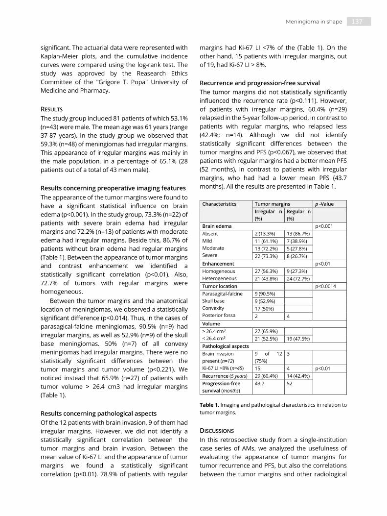
margins had Ki-67 LI <7% of the (Table 1). On the
other hand, 15 patients with irregular marginis, out
of 19, had Ki-67 LI > 8%.
Recurrence and progression-free survival
The tumor margins did not statistically significantly
influenced the recurrence rate (p<0.111). However,
of patients with irregular margins, 60.4% (n=29)
relapsed in the 5-year follow-up period, in contrast to
patients with regular margins, who relapsed less
(42.4%; n=14). Although we did not identify
statistically significant differences between the
tumor margins and PFS (p<0.067), we observed that
patients with regular margins had a better mean PFS
(52 months), in contrast to patients with irregular
margins, who had had a lower mean PFS (43.7
months). All the results are presented in Table 1.
Characteristics Tumor margins p -Value
Irregular n
(%)
Regular n
(%)
Brain edema p<0.001
Absent
Mild
Moderate
Severe
2 (13.3%) 13 (86.7%)
11 (61.1%) 7 (38.9%)
13 (72.2%) 5 (27.8%)
22 (73.3%) 8 (26.7%)
Enhancement p<0.01
Homogeneous
Heterogeneous
27 (56.3%) 9 (27.3%)
21 (43.8%) 24 (72.7%)
Tumor location p<0.0014
Parasagital-falcine
Skull base
Convexity
Posterior fossa
9 (90.5%)
9 (52.9%)
17 (50%)
2 4
Volume
˃ 26.4 cm3
< 26.4 cm3
27 (65.9%)
21 (52.5%) 19 (47.5%)
Pathological aspects
Brain invasion
present (n=12)
Ki-67 LI >8% (n=45)
9 of 12
(75%)
3
15 4 p<0.01
Recurrence (5 years) 29 (60.4%) 14 (42.4%)
Progression-free
survival (months)
43.7 52
Table 1. Imaging and pathological characteristics in relation to
tumor margins.
DISCUSSIONS
In this retrospective study from a single-institution
case series of AMs, we analyzed the usefulness of
evaluating the appearance of tumor margins for
tumor recurrence and PFS, but also the correlations
between the tumor margins and other radiological

138 A.I. Cucu, Mihaela Cosman, B. Dobrovat et al.
features (brain edema and contrast enhancement)
and pathological findings (brain invasion and the
mean value of Ki-67 LI).
Correlations between the tumor margins and
brain edema
Analyzing the correlation between tumor margins
and the degree of peritumoral cerebral edema, we
observed statistically significant differences
(p<0.001). Thus, almost half of the patients with
irregular margins (45.8%) had severe brain edema,
while 39.4% of patients with regular margins have no
cerebral edema, or it was minimal (21.2%). Thus, we
can consider that the irregular margins of a
meningioma may be a predictor of the occurrence of
brain edema (Figure 1).
Figure 1. Illustrative examples of the analysed MRI variables: (a) axial T1-weighted MRI shows an irregular tumor shape with
mushroom-like growth and peritumoral edema (white asterisk). (b) T2-weighted MRI shows the same tumor, with severe
peritumoral edema which compresses the lateral ventricles (Hale 3 grade) (black asterisk).
Consistent with our results, Lobato et al. reported a
statistically significant difference between the tumor
margins and brain edema. He found that patients
who had meningiomas with an irregular margins,
had a 2.9 times higher risk for brain edema (10).
Although cerebral edema can also occur as a result
of brain invasion, there are additional biological
mechanisms involved in the occurrence of this
phenomenon, such as ischaemia from tumor mass
effect or venous congestion (5, 22). Also, Nakano
observed with his collaborators a statistically
significant correlation between brain edema and
meningioma margins, both in univariate analysis and
in multivariate analysis (23). The conclusion of his
study was that the incidence of brain edema was
significantly higher in the group of tumors with
irregular margins (23).
Correlations between the tumor margins and
contrast enhancement
In our study, we identified a statistically significant
correlation (p<0.010) between the tumor margins
and contrast enhancement. Thus, 72.7% of tumors
with regular margins was homogeneous (Figure 2).
The heterogeneous aspect of meningiomas can also
be given by the presence of intratumoral necrosis
(24), and this can also indicate a malignant histology
of the tumor (10, 25). The most common mechanism
of necrosis in meningiomas is due to hypoxia (26),
and in turn, this occurs due to high metabolic
demands, and may be related to a more aggressive
tumor progression (27).

139 Meningioma in shape
Figure 2. Illustrative examples of the analyzed MRI variables: (a) Axial and coronal contrast enhanced T1- weighted MRI showing a
sphenoid wing meningioma with irregular margins and heterogeneous enhancement. (b) Axial and coronal contrast enhanced T1-
weighted MRI showing another sphenoid wing meningioma with regular margins and homogeneous enhancement.
Correlations between the tumor margins and
brain invasion
Analyzing the correlation between the margins and
brain invasion, although there was no statistically
significant difference between the two variables, we
found that of the 12 patients with brain invasion, 9 of
them had irregular margins (Figure 3, Figure 4.b.).
Figure 3. (a) Illustrative examples of the analyzed MRI variables: axial T1-weighted MRI shows an irregular tumor shape with
mushroom-like growth. (b) Brain invasion revealed as small ”islands” of tumor cells (black arrow) into adjacent nervous tissue, which
also shows astrocytic gliosis (HE, x200). (c) Brain invasion as irregular projections of the tumor into adjacent nervous tissue without
an intervening layer of leptomeninges at the tumor to brain interphase (black arrows). The adjacent brain parenchyma also exhibited
astrocytic gliosis (HE, x100).
Some authors consider that the presence of irregular
margins, as well as their lobulated appearance with
fringed extensions to the brain parenchyma is
associated with brain infiltration and denotes
malignant tumor behavior (10–14,28). Among these
authors, Adeli et al. observed a statistically significant
correlation between irregular margins and brain
invasion in 4% of 617 grade I, II and III meningiomas
(28). In relation to brain invasion, in the evaluation of
imaging factors, some authors observed that the
absence of a cerebrospinal fluid rim separating the
meningioma from brain, would suggest a higher
histological degree of the tumor, due to the
possibility of invasion of the adjacent brain tissue.
Correlations between the tumor margins and the
mean value of the Ki-67 LI
Correlating the tumor margins with the value of the
marker Ki-67 LI, we identified a statistically significant
difference (p<0.01). Thus, 78.9% of patients with
regular margins had Ki-67 LI <7% On the other hand,
out of 19 patients with Ki 67 LI> 8%, 15 of them had
meningiomas with irregular margins. Statistical
significant differences were also reported by Beculic
et al. between the two variables, noting that irregular
margins were associated with higher Ki-67 LI values
(30). Moreover, immunohistochemical detection of
Ki-67 LI remins an important tool in addition to
routine histopathological evaluation that can be
used to predict tumor behavior of AM (31,32) (Figure
4).
Figure 4. (a) Meningothelial tumor with sheet-like growth, increased cellularity and two mitotic figures/ high power field (black

140 A.I. Cucu, Mihaela Cosman, B. Dobrovat et al.
arrows) (HE, x400). (b) Leptomeningeal tumoral invasion with a thin intervening layer of pia mater at the tumor (black arrow) to brain
interphase (white arrow) (IHC, anti-Ki67 antibody, x200). (c) Nuclear immunoreactivity (8%) for Ki-67 LI in atypical meningioma, grade
II of malignancy (IHC, anti Ki-67- antibody, x200)
The influence of tumormarginson recurrence
Until now, several authors have shown that grade of
lobulation of a meningioma increases with the
malignancy (33,34), so implicitly the irregular
appearance of the tumor margins.
Irregular margins of meningiomas have been
attributed to different growth rates in certain regions
of the tumor (35) and are associated with increased
cell proliferation (33) and an increased risk of tumor
recurrence (7). In this regard, Zhang et al.
demonstrated in a study of 33 AMs that tumors with
irregular margins are more prone to malignant
progression compared with AMs with regular
margins (36). Also, Gobran et al. observed in its series
of 138 intracranial meningiomas (all grades), that
lobed-looking meningiomas had a recurrence rate of
23.5%, and mushroom meningiomas had a
recurrence rate of 62.5% over a five-year period,
observing a statistically significant correlation (37).
When we evaluated the relationship between the
tumor margins and the recurrence, although we did
not find any statistical corellation between tumor
margins and recurrence, according to other studies
(6, 38). However, we observed that the majority of
meningiomas that recurred had irregular margins
(60.4%). Moreover, 57.6% of regular-margin tumors
did not recur over a 5-year follow-up period.
In the literature, the existence of irregular
margins or "mushrooming" is considered to be an
important prognostic factors for recurrence, as it
could reflect the high proliferative potential of the
tumor (7). Also, Nakasu et al. noted that tumors
"mushrooming" tumors had the highest recurrence
rate, followed by lobulated tumors and then tumors
with smooth and irregular margins (7). Also Ildan et
al. observed in 137 patients with grade I-III
meningiomas, that mushroom - shaped
meningiomas was associated with a significantly
higher risk of recurrence than those with smooth
margins (39). However, this atypical imaging feature
of irregular margins cannot be considered as an
indicator of a higher-grade behavior, and cannot be
reliable and specific in differentiating benign and
malignant meningiomas (40).
The influence of tumor margins on the
progression-free survival
In our study, we observed that patients who had
meningiomas with irregular margins had a lower rate
of survival than those with regular margins (43.75
months vs. 52 months) (Figure 5). As we did not
identify a statistically significant difference between
the two variables in agreement with other authors
(6), we can consider that the irregular appearance of
meningiomas did not influence the PFS. However,
other authors such as Nakasu et al. observed that
irregular margins correlates with PFS (7, 41).
Figure 5. Progression-free survival depending on the
appearance of meningioma margins. It is notable that patients
with regular margins (blue line) had a better progression-free
survival (52 months vs. 43.75 months).
CONCLUSIONS
We found that in our series of patients, that irregular
margins has no prognostic value for tumor
recurrence over a period of 5 years follow up and
also for PFS, but instead correlates statistically with
grade of brain edema, contrast enhancement and
the mean value of Ki-67 LI. Even so, the appearance
of tumor margins must be a factor to consider when
evaluating neuroimaging of meningiomas and such
studies should be extended to other types of
intracranial tumors.

141 Meningioma in shape
REFERENCES
1. Budohoski KP, Clerkin J, Millward CP, et al. Predictors of
early progression of surgically treated atypical
meningiomas. Acta Neurochir (Wien) 2018; 160:1813-22.
2. Cucu AI, Costea CF, Carauleanu A, Dumitrescu GF, Sava A,
Sadiye Scripcariu I, et al. Meningiomas Related to the
Chernobyl Irradiation Disaster in North-Eastern Romania
Between 1990 and 2015. Rev Chim 2018; 69(6):1562-65.
3. Cucu AI, Costea CF, Turliuc MD, Ghiciuc CM, Costachescu
B, Popescu R, et al. Anatomical localization of intracranial
grade II meningiomas in North-Eastern Romania: our 25-
years experience. Romanian Neurosurg 2019; 33:232-38.
4. Louis DN, Perry A, Reifenberger G, et al. The 2016 world
Health organization classification of tumours of the
central nervous system: a summary. Acta Neuropathol
2016; 131(6):803e20.
5. Salah F, Tabbarah A, ALArab y N, Asmar K, Tamim H,
Makki M, et al. Can CT and MRI features differentiate
benign from malignant meningiomas? Clin Radiol 2019;
74(11): 898.e15-898.e23.
6. Nowak A, Dziedzic T, Krych P et al. Benign versus atypical
meningiomas: risk factors predicting recurrence. Neurol
Neurochir Pol 2015; 49(1):1-10.
7. Nakasu S, Nakasu Y, Nakajima M et al. Preoperative
identification of meningiomas that are highly likely to
recur. J Neurosurg 1999; 90(3):455-62.
8. Hanft S, Canoll P, Bruce JN. A review of malignant
meningiomas: diagnosis, characteristics, and treatment. J
Neurooncol 2010; 99(3):433-43.
9. Champeaux C, Dunn L. World Health Organization grade
II meningioma: a 10-year retrospective study for
recurrence and prognostic factor assessment. World
Neurosurg 2016; 89: 180-86.
10. Lobato RD, Alday R, Gómez PA et al. Brain oedema in
patients with intracranial meningioma. Correlation
between clinical, radiological, and histological factors and
the presence and intensity of oedema. Acta Neurochir
(Wien) 1996; 138(5):485-93; discussion 493-94.
11. Vassilouthis J, Ambrose J. Computerized tomography
scanning appearances of intracranial meningiomas. J
Neurosurg 1979; 50:320-27.
12. Dietemann JL, Heldt N, Burguet JL et al. CT findings in
malignant meningiomas. Neuroradiology 1982; 23:207-
209.
13. New PFJ, Hesselink JR, O’Carroll CP, Kleinman GM.
Malignant meningioma: CT and histological criteria
including a new CT sign. AJNR 1982;3:267-76.
14. New PFJ, Aronow S, Hesselink JR. National Cancer
Institute Study: evaluation of computed tomography in
the diagnosis of intracranial neoplasms. IV.
Meningiomas. Radiology 1980; 136:665-75.
15. Ros-Sanjuan A, Iglesias-Moroño S, Carrasco-Brenes A et
al. Atypical meningiomas: histologic and clinical factors
associated with recurrence. World Neurosurg 2019;
125:e248-e256.
16. Hale AT, Wang L, Strother MK, Chambless LB.
Differentiating meningioma grade by imaging features
on magnetic resonance imaging. J Clin Neurosci 2018;
8:71-5.
17. Lu Y, Liu L, Luan S, Xiong J, Geng D, Yin B. The diagnostic
value of texture analysis in predicting WHO grades of
meningiomas based on ADC maps: an attempt using
decision tree and decision forest. Eur Radiol 2019;
29(3):1318‐28.
18. Cucu AI, Costea CF, Turliuc MD, Dascalu CG, Jitaru I, Dinu
R, et al. The tumour volume influence on tumour
recurrence and progression-free survival in the case of
atypical meningiomas: Our experience on a series of 81
cases. Romanian Neurosurg 2020;52-7.
19. Char DH, Kroll S, Phillips TL. Uveal melanoma. Growth
rate and prognosis. Arch Ophthalmol 1997; 115:1014-18.
20. Gass JD. Comparison of uveal melanoma growth rates
with mitotic index and mortality. Arch Ophthalmol 1985;
103:924-31.
21. Richtig E, Langmann G, Müllner K, Richtig G, Smolle J.
Calculated tumour volume as a prognostic parameter for
survival in choroidal melanomas. Eye (Lond) 2004;
18(6):619-23.
22. Cucu AI, Turliuc MD, Carauleanu A, Poeata I, Costea CF,
Dumitrescu GF, et al. Chemical Aspects of Peritumoral
Cerebral Edema in Atypical Meningiomas. Rev Chim
2018; 69(10):2804-07.
23. Nakano T, Asano K, Miura H, Itoh S, Suzuki S.
Meningiomas with brain edema: radiological
characteristics on MRI and review of the literature. Clin
Imaging 2002; 26:243-49.
24. Osawa T, Tosaka M, Nagaishi M, Yoshimoto Y. Factors
affecting peritumoral brain edema in meningioma:
special histological subtypes with prominently extensive
edema. J Neurooncol 2013; 111(1): 49-57.
25. Elster AD, Challa VR, Gilbert TH et al. Meningiomas: MR
and histopathologic features. Radiology 1989; 170: 857-
62.
26. Kano T, Kobayashi M, Yoshida K, et al. Central tumour
necrosis of a large meningioma following acute anemia
caused by hysterectomy. Neurol Med Chir (Tokyo) 2009
;49(9):424e6.
27. Perry A, Stafford SL, Scheithauer BW, et al. Meningioma
grading: an analysis of histologic parameters. Am J Surg
Pathol 1997; 21(12):1455e65.
28. Adeli A, Hess K, Mawrin C, Streckert EMS, Stummer W,
Paulus W, et al. Prediction of brain invasion in patients
with meningiomas using preoperative magnetic
resonance imaging. Oncotarget 2018; 9(89):35974-82.
29. Sheporaitis LA, Osborn AG, Smirniotopoulos JG, et al.
Intracranial meningioma. AJNR Am J Neuroradiol 1992;
13(1):29e37.
30. Bečulić H. Correlation of Peritumoral Brain Edema with
Morphological Characteristics and Ki67 Proliferative
Index in Resected Intracranial Meningiomas. Acta Clin
Croat 2019; 58:42-9.
31. Cucu AI, Costea CF, Turliuc MD, FG, Dumitrescu A, Sava A,
et al. Are there any correlations between demographic
characteristics, tumor location, and Ki-67 labeling index

142 A.I. Cucu, Mihaela Cosman, B. Dobrovat et al.
in intracranial atypical meningiomas (WHO grade II)?
Rom J Morphol Embryol 2019; 60:567-572.
32. Cucu AI, Costea CF, Poeată I, Turliuc DM. Prognostic
factors in atypical meningioma. Romanian Neurosurg
2017; 31(2):165-71.
33. Hashiba T, Hashimoto N, Maruno M, et al. Scoring
radiologic characteristics to predict proliferative potential
in meningiomas. Brain Tumor Pathol 2006; 23:49-54.
34. Liu H, Zhou J, Li W, Liu G. Comparative analysis of the
magnetic resonance imaging features between
anaplastic meningioma and atypical meningioma. J
Craniofac Surg 2016; 27:e229-e233.
35. Zhou JL, Liu JL, Zhang J, Zhang M. Thirty-nine cases of
intracranial hemangiopericytoma and anaplastic
hemangiopericytoma: a retrospective review of MRI
features and pathological findings. Eur J Radiol 2012;
81:3504-10.
36. Zhang Q, Jia GJ, Zhang GB et al. A logistic regression
model for detecting the presence of malignant
progression in atypical meningiomas. World Neurosurg
2019; 126:e392-e401.
37. Gobran A, Li FC, Xu XK, Zhang SY. Factors associated with
recurrence of postoperative meningioma: a clinical study
of 138 patients. Romanian Neurosurg 2013; 20(4):379-87.
38. Streckert EMS, Hess K, Sporns PB et al. Clinical,
radiological, and histopathological predictors for long-
term prognosis after surgery for atypical meningiomas.
Acta Neurochir (Wien) 2019; 161(8):1647-56.
39. Ildan F, Erman T, Göçer AI et al. Predicting the probability
of meningioma recurrence in the preoperative and early
postoperative period: a multivariate analysis in the
midterm follow-up. Skull Base 2007; 17(3):157-71.
40. Bozdağ M, Er A, Ekmekçi S. Association of apparent
diffusion coefficient with Ki-67 proliferation index,
progesterone-receptor status and various
histopathological parameters, and its utility in predicting
the high grade in meningiomas. Acta Radiol
2021;62(3):401-13.
41. Spille DC, Heß K, Sauerland C et al. Brain Invasion in
Meningiomas: Incidence and Correlations with Clinical
Variables and Prognosis. World Neurosurg 2016; 93:346-
54.

Romanian Neurosurgery (2021) XXXV (2): pp. 143-147 DOI: 10.33962/roneuro-2021-022 www.journals.lapub.co.uk/index.php/roneurosurgery
The role of MRI-guided focused ultrasound in neurosurgery. A narrative review
Marius Gabriel Dabija1,2, Ioan Sebastian Nechifor1,
Vlad Andrei Dabija3, Bogdan Costachescu1,2,
Lucian Eva1 1 Department of Neurosurgery. “Nicolae Oblu” Emergency Clinical
Hospital, Iasi. ROMANIA 2 Department of Neurosurgery. “Gr. T. Popa” University of Medicine
and Pharmacy, Iasi. ROMANIA 3 “Gr. T. Popa” University of Medicine and Pharmacy, Iasi, ROMANIA
ABSTRACT Introduction. MRgFUS is a novel technology, which can have profound implications
in the current treatment of neurological disorders. Its applications range widely, from
the alteration of the blood-brain barrier, ablation of tumours to the treatment of
movement disorders.
Objective. To review, following thorough research of the literature, the principles of
its use in the treatment of neurological diseases and the main reported evidence of
its clinical implementation.
Material and method. Interrogation of the MEDLINE database, using the PubMed
search engine, for the following MESH words: “MRgFUS”, “FUS” “BRAIN”, from 2000 to
the current year.
Conclusion. MRgFUS can be safely used today for the treatment of Essential Tremor.
New research is warranted for the evaluation of its safety and effectiveness in other
neurological disorders.
INTRODUCTION MRI guided focused ultrasound (MRgFUS) of the brain is a novel
technology which has the potential to be implemented successfuly in
our modern neurosurgical practice. Because of its capability to alter the
blood brain barrier, ablate tumors and proven to be effective as a
treatment of different movement disorders, it is worthy for us to gain
familiarity with this technique. Unfortunately, novelty and the specific
technicality of this instrument confuses the clinician. Therefore, we aim
in this paper, by the means of a narrative review, to provide an insight
on the working mechanism and the uses of the technology and to
address the benefits and limitations of its applicability in the clinical
practice.
Keywords MRgFUS,
movement disorders, blood-brain barrier,
high focused ultrasound
Corresponding author: Bogdan Costachescu
Department of Neurosurgery.
“Nicolae Oblu” Emergency Clinical Hospital, Iasi, Romania
Scan to access the online version

144 Marius Gabriel Dabija, Ioan Sebastian Nechifor, Vlad Andrei Dabija et al.
METHOD
Research of the MEDLINE database, using the
PubMed search engine, for the following MESH
words: “MRgFUS”, “FUS” “BRAIN”, from 2000 to the
current year.
Background.
First studied by Lynn (1) at the start of the 20th
century, the ability to focus ultrasound waves in
order to produce a biological effect on the
intracranial content was a tempting idea. Aided by
the recent advances in high resolution MRI imaging,
neuronavigation and the development of
hemispheric piezoelectric transducer systems, we
are now able to accurately focus the ultrasound
waves intracranially, as demonstrated by the
commercially available system ExAblate 4000
(InSightec LTD). The result of their application on the
tissue can be divided into thermal and mechanical
effects. As ultrasounds propagates through the
tissue, they raise the local temperature and interact
with the gas molecules, producing bubbling,
oscillation and finally cavitation with the stretching of
the cellular membrane as a result (2) . Based on these
mechanical and thermal effects they can reversibly
or irreversibly alter the brain tissue, depending on
their strength and frequency.
The role of MRgFUS in tumor ablation
Ultrasound can aid in the treatment of deep brain
neoplastic lesions, considered unresectable, or in
patients that are unfit for surgery. Unfortunately, the
data is scarce due to the relative novelty of the
technology and the absence of clinical trials (3), (4).
In 2014, Daniel Coluccia published the results
regarding the thermoablation of a left thalamic
recurrent glioma, using ultrasound. He noticed a 10%
reduction in tumoral volume at 5 days post
procedure, with improvement in the neurologic
status of the patient. At the 21th day check-up no
increase in the volume of the tumor was observed
(5).
Preliminary results, published by Ernst Martin,
creates an insight on the thermoablative tumoral
effect of the HIFUS system. Although the author
reported clinical improvement in only one patient, it
opens the pathway to the development of new
clinical trials (6).
The role of MRgFUS on the permeability of the
blood-brain barrier
The BBB is a complex structure serving the role of an
interface between the blood and the cerebral tissue.
Its cytoarchitecture is mainly represented by a layer
of endothelial cells, binded one to another by tight
junction proteins, interconnected with neurons,
perycites and astrocytes (7). This renders it virtually
impermeable to exogeneous molecules and thus
limiting the effectiveness of different
pharmaceuticeuticals. MRgFUS can be used to
transiently open the BBB. Hynynen et.al, in his in vivo
study, demonstrated that the BBB can safely be
opened in rabbits, by applying low strength
ultrasounds combined with the intravenous
administration of preformed microbubbles. After
applying the ultrasound field, these microbubbles
oscillate, modify their form, thus stimulating the
cerebral blood capillaries and opening the BBB. The
effects are transient without significant side-effects
(8) (fig 1.). Combining the microbubbles with contrast
agent, enables us to accurately pinpoint the target
anatomic structure, with the aid of imaging (9).
Figure 1. Vascular cells being stimulated (red cubes) by the
ultrasound induced cavitation of the microbubbles (green
spheres).
Agessandro Abrahao et al, in their human trial of 4
participants suffering from ALS, demonstrated the
successful opening of the BBB, after sonification,
without side effects (10). MRgFUS can be used in
conjunction with systemic chemotherapy in the
treatment of intracranial neoplasms. Liu HL et al, in
their rat glioma model study, demonstrates a
significant survival improvement in the combined

145 The role of MRI-guided focused ultrasound in neurosurgery
treatment group versus the single treatment group
(53 days versus 29 days) (11). It appears that the
opening of the barrier can significantly enhance the
effect of immunotherapy on brain metastases, as
demonstrated by Thiele Kobus et al, in their study
(12). Finally, it has been shown that MRgFUS cand aid
gene therapy pharmaceuticals in their permeation of
the BBB as shown by Liu et al in their study (13).
The role of MRgFUS in the treatment of movement
disorders and chronic neuropathic pain
Lesioning of the brain by stereotactic techniques
along with DBS represent the mainstay of
neurosurgical treatment in movement disorders.
However, despite their effectiveness, they are not
without potential side effects and contraindications.
MRgFUS can be a valuable alternative tool in the
treatment of these pathologies. Based on the
principle of cavitation and thermoablation they can
produce discrete lesioning of target anatomical
areas, similar with stereotactic radiosurgery, but
without harming the surrounding healthy tissue (14).
Clearly, a positive ratio of reward versus risk
associated with the technique has led different
authors to implement it into clinical practice.
Zaaroor et al, in their study investigating the
treatment of symptomatic Parkinson Disease and
Essential tremor by HIFU VIM thalamotomy, reported
a significant improvement in quality of life and
reduction of symptomatology by approximatively
50% in the treated subjects. The effects persisted in
all but 6 patients, at the 24th month follow-up.
Common side effects were transient and
represented by gait ataxia, hand paresthesia and
asthenia (15).
Martínez-Fernández et al, in their randomised
trial investigating the role of HIFU sub-thalamotomy
for the treatment of Parkinson Disease numbering
40 subjects, 27 assigned to the procedure group
versus 13 to the sham procedure, reported
improvement of symptoms at 4 months post
procedure by approximatively 50% in the treated
subjects vs. the placebo. Common reported side
effects are represented by slurred speech,
dyskinesia and gait disturbance, with long term
persistence in 6 treated patients (16).
Elias et al, in their trial with 76 enrolled
participants, investigating the role of HIFU VIM
thalamotomy in the treatment of patients suffering
from essential tremor refractory to conservative
therapy, reported a 41% improvement of the tremor
at 3 months post procedure, respectively a 35%
percent improvement in the treated group versus
the placebo group. Common adverse effects
included gait ataxia and paresthesias ,these being
present in approximative 36% of the treated subjects
(17).
Jin Woo Chang et al, which investigated the use
of HIFU VIM thalamotomy in the treatment of
refractory essential tremor in 76 patients, reported
an overall of 53% improvement of the tremor at one
year post procedure. Gait disturbances and
paresthesias are reported as the most common
adverse effects (18).
Another study investigating the role of HIFU
thalamotomy in the treatment of Essential Tremor,
with 26 participants, published similar results (19).
The evidence offered by the literature, which
proves that HIFU thalamotomy is safe and efficient
has led the FDA to approve it, in 2016, for the
treatment of Essential Tremor (20), marking a
cornerstone in it’s pathway for clinical
implementation.
It has been proven that MRgFUS can be used in
the treatment of chronic neuropathic pain. First
described in 2009, FUS medial thalamotomy can be
a viable therapeutic approach of chronic neuropathic
pain (21). Marc N Gallay et al, reports improvement
of trigeminal neuralgia after central lateral FUS
thalamotomy (22). Further clinical trials are needed
in order to adequatly asses the possibility of these
technique to be safely and successfully used.
DISCUSSION
As previously shown focused ultrasound therapy has
the capability to significantly alter the current
paradigm of treatment used in neurological
diseases. Regarding it’s use on opening the BBB, it is
possible that the translation of fundamental science
into clinical practice, aided by the future realisation
of clinical trials, will usher a new era in Neuro-
Oncology (23), (24) and in the treatment of
neurodegenerative disorders.
Several studies are currently investigating its role
in the treatment of brain ischemia (25), (26). It
remains to be seen if it will be effective.
In the field of movement disorders, unfortunately
it is now limited only to the treatment of Essential
tremor, but probably in the near future, will be
extended for Parkinson Disease (27).

146 Marius Gabriel Dabija, Ioan Sebastian Nechifor, Vlad Andrei Dabija et al.
New progress in our understanding of the
neurological pathology combined with technical
improvements will probably advance this technology
as a treatment for chronic pain (28) and other
neuropsychiatric disorders, such as obsessive-
compulsive disorder and depression (29), (30).
CONCLUSION
MRgFUS is a promising technology in the field of
neuroscience. Although currently used in the
treatment of movement disorders, further studies
are needed for clear identification of its role in the
treatment of different neurological diseases.
REFERENCES
1. Lynn JG, Zwemer RL, Chick AJ, Miller AE. A new method
for the generation and use of focused ultrasound in
experimental biology. J Gen Physiol. 1942 Nov
20;26(2):179–93.
2. Al-Bataineh O, Jenne J, Huber P. Clinical and future
applications of high intensity focused ultrasound in
cancer. Cancer Treat Rev. 2012 Aug;38(5):346–53.
3. Ram Z, Cohen ZR, Harnof S, Tal S, Faibel M, Nass D, et al.
Magnetic resonance imaging-guided, high-intensity
focused ultrasound for brain tumor therapy.
Neurosurgery. 2006 Nov;59(5):949–55; discussion 955-
956.
4. Park J-W, Jung S, Jung T-Y, Lee M-C. Focused Ultrasound
Surgery for the Treatment of Recurrent Anaplastic
Astrocytoma: A Preliminary Report. 2006 May 1;829:238–
40.
5. Coluccia D, Fandino J, Schwyzer L, O’Gorman R, Remonda
L, Anon J, et al. First noninvasive thermal ablation of a
brain tumor with MR-guided focused ultrasound. J Ther
Ultrasound. 2014 Oct 16;2:17.
6. Martin E, Werner B, Bauer R, Leyen K van, Coluccia D,
Fandino J. Clinical neurological HIFU applications: the
Zurich experience. Transl Cancer Res [Internet]. 2014 Oct
[cited 2021 Apr 26];3(5). Available from:
https://tcr.amegroups.com/article/view/3156
7. Daneman R, Prat A. The Blood–Brain Barrier. Cold Spring
Harb Perspect Biol [Internet]. 2015 Jan [cited 2021 Apr
25];7(1). Available from: https://www.ncbi.nlm.nih.gov
/pmc/articles/PMC4292164/.
8. Hynynen K, McDannold N, Vykhodtseva N, Jolesz FA.
Noninvasive MR imaging-guided focal opening of the
blood-brain barrier in rabbits. Radiology. 2001
Sep;220(3):640–6.
9. Treat LH, McDannold N, Vykhodtseva N, Zhang Y, Tam K,
Hynynen K. Targeted delivery of doxorubicin to the rat
brain at therapeutic levels using MRI-guided focused
ultrasound. Int J Cancer. 2007 Aug 15;121(4):901–7.
10. Abrahao A, Meng Y, Llinas M, Huang Y, Hamani C,
Mainprize T, et al. First-in-human trial of blood–brain
barrier opening in amyotrophic lateral sclerosis using
MR-guided focused ultrasound. Nat Commun. 2019
Dec;10(1):4373.
11. Liu H-L, Hua M-Y, Chen P-Y, Chu P-C, Pan C-H, Yang H-W,
et al. Blood-brain barrier disruption with focused
ultrasound enhances delivery of chemotherapeutic
drugs for glioblastoma treatment. Radiology. 2010
May;255(2):415–25.
12. Kobus T, Zervantonakis IK, Zhang Y, McDannold NJ.
Growth inhibition in a brain metastasis model by
antibody delivery using focused ultrasound-mediated
blood-brain barrier disruption. J Controlled Release. 2016
Sep 28;238:281–8.
13. Lin C-Y, Hsieh H-Y, Pitt WG, Huang C-Y, Tseng I-C, Yeh C-K,
et al. Focused ultrasound-induced blood-brain barrier
opening for non-viral, non-invasive, and targeted gene
delivery. J Controlled Release. 2015 Aug 28;212:1–9.
14. Fishman PS, Frenkel V. Treatment of Movement
Disorders With Focused Ultrasound. J Cent Nerv Syst Dis
[Internet]. 2017 Jun 6 [cited 2021 Apr 26];9. Available
from: https://www.ncbi.nlm.nih.gov/pmc/articles/PMC5
462491/.
15. Zaaroor M, Sinai A, Goldsher D, Eran A, Nassar M,
Schlesinger I. Magnetic resonance–guided focused
ultrasound thalamotomy for tremor: a report of 30
Parkinson’s disease and essential tremor cases. J
Neurosurg. 2017 Feb 24;128(1):202–10.
16. Martínez-Fernández R, Máñez-Miró JU, Rodríguez-Rojas
R, Álamo M del, Shah BB, Hernández-Fernández F, et al.
Randomized Trial of Focused Ultrasound
Subthalamotomy for Parkinson’s Disease. N Engl J Med
[Internet]. 2020 Dec 23 [cited 2021 Apr 20]; Available
from: https://www.nejm.org/doi/10.1056/NEJMoa2016
311.
17. Elias WJ, Lipsman N, Ondo WG, Ghanouni P, Kim YG, Lee
W, et al. A Randomized Trial of Focused Ultrasound
Thalamotomy for Essential Tremor. N Engl J Med. 2016
Aug 25;375(8):730–9.
18. Chang JW, Park CK, Lipsman N, Schwartz ML, Ghanouni P,
Henderson JM, et al. A prospective trial of magnetic
resonance–guided focused ultrasound thalamotomy for
essential tremor: Results at the 2-year follow-up. Ann
Neurol. 2018;83(1):107–14.
19. Iacopino DG, Gagliardo C, Giugno A, Giammalva GR,
Napoli A, Maugeri R, et al. Preliminary experience with a
transcranial magnetic resonance–guided focused
ultrasound surgery system integrated with a 1.5-T MRI
unit in a series of patients with essential tremor and
Parkinson’s disease. Neurosurg Focus. 2018 Feb
1;44(2):E7.
20. Fishman PS. Thalamotomy for essential tremor: FDA
approval brings brain treatment with FUS to the clinic. J
Ther Ultrasound. 2017 Jul 13;5(1):19.
21. Martin E, Jeanmonod D, Morel A, Zadicario E, Werner B.
High-intensity focused ultrasound for noninvasive
functional neurosurgery. Ann Neurol. 2009 Dec
1;66(6):858–61.

147 The role of MRI-guided focused ultrasound in neurosurgery
22. Gallay MN, Moser D, Jeanmonod D. MR-Guided Focused
Ultrasound Central Lateral Thalamotomy for Trigeminal
Neuralgia. Single Center Experience. Front Neurol.
2020;11:271.
23. Dauba A, Delalande A, Kamimura HAS, Conti A, Larrat B,
Tsapis N, et al. Recent Advances on Ultrasound Contrast
Agents for Blood-Brain Barrier Opening with Focused
Ultrasound. Pharmaceutics [Internet]. 2020 Nov 21 [cited
2021 Apr 26];12(11). Available from: https://www.ncbi.
nlm.nih.gov/pmc/articles/PMC7700476/
24. Zhang DY, Dmello C, Chen L, Arrieta VA, Gonzalez-
Buendia E, Kane JR, et al. Ultrasound-mediated delivery
of paclitaxel for glioma: a comparative study of
distribution, toxicity and efficacy of albumin-bound
versus cremophor formulations. Clin Cancer Res Off J Am
Assoc Cancer Res. 2020 Jan 15;26(2):477–86.
25. Daffertshofer M, Gass A, Ringleb P, Sitzer M, Sliwka U, Els
T, et al. Transcranial low-frequency ultrasound-mediated
thrombolysis in brain ischemia: increased risk of
hemorrhage with combined ultrasound and tissue
plasminogen activator: results of a phase II clinical trial.
Stroke. 2005 Jul;36(7):1441–6.
26. Brown AT, Flores R, Hamilton E, Roberson PK, Borrelli MJ,
Culp WC. Microbubbles improve sonothrombolysis in
vitro and decrease hemorrhage in vivo in a rabbit stroke
model. Invest Radiol. 2011 Mar;46(3):202–7.
27. A randomized, sham-controlled trial of transcranial
magnetic resonance-guided focused ultrasound
thalamotomy trial for the treatment of tremor-dominant,
idiopathic parkinson disease | Cochrane Library
[Internet]. [cited 2021 Apr 26]. Available from:
https://www.cochranelibrary.com/central/doi/10.1002/c
entral/CN-01407058/full
28. Goellner E, Rocha CE. Anatomy of Trigeminal
Neuromodulation Targets: From Periphery to the Brain.
Neuromodulation Facial Pain. 2020;35:18–34.
29. Jung HH, Kim SJ, Roh D, Chang JG, Chang WS, Kweon EJ, et
al. Bilateral thermal capsulotomy with MR-guided
focused ultrasound for patients with treatment-
refractory obsessive-compulsive disorder: a proof-of-
concept study. Mol Psychiatry. 2015 Oct;20(10):1205–11.
30. Kim M, Kim C-H, Jung HH, Kim SJ, Chang JW. Treatment of
Major Depressive Disorder via Magnetic Resonance-
Guided Focused Ultrasound Surgery. Biol Psychiatry.
2018 Jan 1;83(1):e17–8.

Romanian Neurosurgery (2021) XXXV (2): pp. 148-151 DOI: 10.33962/roneuro-2021-023 www.journals.lapub.co.uk/index.php/roneurosurgery
The stent-assisted coil-jailing technique for very small intracranial aneurysm treatment
A. Chiriac1,2, N. Dobrin2, Georgiana Ion2, Z. Faiyad2, I. Poeata1 1 "Grigore T. Popa" University of Medicine and Pharmacy, Iasi,
ROMANIA 2 “Prof. Dr N. Oblu” Clinic Emergency Hospital, Iasi, ROMANIA
ABSTRACT The stent-assisted coil-jailing technique was initially introduced as an effective and
secure method in the treatment of large wide-necked intracranial aneurysms. Later
on, this technique has proved its efficiency in the safety and optimal treatment of very
small aneurysms. In this article, we will present the successful treatment of a very
small middle cerebral artery aneurysm using the stent-assisted coil-jailing technique
and review the current trends in this type of treatment.
INTRODUCTION With the advances in endovascular technologies, endovascular coil
embolization of ruptured cerebral aneurysms has become the first
efficient treatment option. Nevertheless, endovascular coiling of very
small aneurysms remain controversial. Most of these particular
vascular lesions are conformational characterized by a small dome and
large-neck. The technical difficulties are represented by the high risk of
procedure-related rupture and coil migration due to a very limited
aneurysm volumes and structural restriction of embolization coils.
Stent-assisted coil-jailing technique is a very popular endovascular
method that could be feasible and effective for such structural
complicated aneurysms. However, the technique should be used
carefully and above all, its usage must be safe and lead to good clinical
results with regard to aneurysm occlusion. In this report, we describe a
successfully treated very small middle cerebral artery aneurysm using
the stent-assisted coil-jailing technique in order to provide a more
comprehensive view of this challenging endovascular treatment and
eventually to offer helpful clinical information for the management of
such cases [8].
CASE PRESENTATION
A 50-year-old woman presented with a sudden-onset headache for
which she addressed the emergency department at a local hospital. A
Keywords very small aneurysm,
stent-assisted coil-jailing technique
Corresponding author: A. Chiriac
“Prof. Dr N. Oblu” Clinic Emergency
Hospital, Iasi, Romania
Scan to access the online version

149 The stent-assisted coil-jailing technique for very small intracranial aneurysm treatment
head CT-scan investigation was performed
immediately and showed a lenticular hematoma and
mild subarachnoid haemorrhage in the left Sylvian
fissure. Due to the suspicion of an aneurysmal
cerebral hemorrhage, the patient was immediately
transferred to our hospital for further diagnosis and
treatment. At admission she was confused and with
right hemiparesis. Her past medical history included
hypertension with no regular treatment. The brain
CT angiography raised the suspicion of an
aneurysmal dilatation on M1 segment of left middle
cerebral artery. A subsequent subtraction catheter
cerebral angiography confirmed a very small
aneurysm at the origin of left Charcot artery. Due to
the very small dimension of the aneurysm (2.3 mm ×
2.0 mm) and relatively large neck a stent-assisted
coil-jailing technique was proposed for the occlusion
of the lesion.
A (left)
B (right)
C D
E
F
G
H
I
J
K
Figure 1. A, B – Diagnostic cerebral CT and Angio-CT images; C,
D, E, F, G, H, I – Intraprocedural DSA images; Postprocedural
Cerebral CT Images.
The intervention performed the next day on a
biplane angiography system (INFINIX, Toshiba,
Canon Medical System) under general anesthesia by
our neurosurgical team with many years of
experience in neuroendovascular interventions.
Written informed consent was obtained prior to the
treatment. Commercially available 6F introducer
sheath from Merit Medical was placed into the right
femoral artery. A 6F Chaperon guiding catheters
(Microvention) was carefully advanced over 0.035
glidewire up to the proximal segment of the left
internal carotid artery. Using the 3D-CT images
analysis a favourable working angle roadmap for the
target aneurysm was obtained. A Prowler 10
microcatheter (Codman J&J) was then very carefully
advanced over a 0.014 Transed microwire (Boston

150 A. Chiriac, N. Dobrin, Georgiana Ion et al.
Scientific) and positioned into the aneurysm sac.
Keeping the microcatheter in this fixed position a
second Prowler Select Plus microcatheter (Codman
J&J) was inserted up to the M1 / M2 bifurcation level
of the left middle cerebral artery. With its help a 4/16
mm Enterprise 2 intracranial stent (Codman J&J) is
deployed over the aneurysm neck and the first
microcatheter in a jailed configuration. After the
Prowler Select Plus microcatheter withdraws, two
Galaxy G3 MINI microcoils (Cerenovus
Johnson&Johnson) were then inserted and detached
into aneurysm. The coils placement was done very
slowly, and gentle handling of the microcatheter was
used to avoid tension build-up in the aneurysm and
to allow the coil loop to form inside the
aneurysm. After detaching the last coil, the
microcatheter is carefully retracted so as not to
cause any movement of the stent. A DSA acquisition
is performed to check the permeability of all vessels
and complete occlusion of the aneurysm. Finally, the
entire guiding system is retracted with a compressive
dressing of the femoral puncture site. The patient
was placed into intensive care and received 75mg
clopidogrel and 100mg aspirin daily. A cerebral CT
scan was performed 7 days postoperatively and
showed the lenticular hematoma resorption. The
patient was discharged home after 3 days in good
neurological condition.
DISCUSSIONS
The literature reports that very small intracranial
aneurysms (≤3 mm) represent 13.2%–15.1% of all
intracranial aneurysms. On the other hand,
according to ISUIA study only 0.1% (per year) of small
aneurysms (<5 mm) of the anterior circulation will
manifested by rupture. There have been also several
particular studies that have reported incidences of
small aneurysms of up to 7% of all ruptured
aneurysms[s]. In this clinical situation an emergency
treatment must be initiated. Endovascular coil
embolization or microsurgical clipping of ruptured
very small cerebral aneurysms are still challenging
methods due to distinctive technical difficulties
[t,x,s]. If in the case of endovascular treatment the
very small size of the aneurysmal dome frequently
associated with a wide neck make it difficult to place
a coil safely, in the case of microsurgical treatment
the thin-walled and too small neck make it difficult to
accept a clip without narrowing or tearing the parent
artery [3,6,9].
The stent-assisted coiling technique represents
an efficacious adjuvant technique for treating wide-
necked aneurysms. The advent of these adjuvant
techniques has made possible the endovascular
occlusion of an increasing number of very small
aneurysms. The stent-assisted coil-jailing technique
has become often considered as the most efficient
option for the embolization of very small aneurysms.
This technique allowed, not only to provided the
necessary support to achieve a stable coil
arrangement, but also solved the problem of
overestimating the coil length in small aneurysms. By
this technique, if the coil is too long for a small
aneurysm, the remaining coil segment (tail) could be
purposely left and jailed between the stent and the
parent artery wall at the end of the embolization
procedure.
The main complication reported in very small
aneurysms was intraprocedural rupture. If the first
studies reported significantly higher rates of
procedural rupture, with the continuing
development of endovascular devices and greater
operator experience, the last ones did not show a
significant association between very small
aneurysms and intraprocedural rupture. It seems
that the preferred use of stent-assisted coil-jailing
technique might explain the evolution of the
previously mentioned results. Obtaining a stable
position of the microcatheter by fixing it between the
arterial wall and the stent, as well as the possibility of
coils insertion into aneurysm with the tip of the
microcatheter in its neck area, without the need of
navigation in the aneurysmal sac caused a significant
reduction in the risk of intraprocedural rupture in
very small aneurysms [1,3,6,9]
Another complication described in very small
aneurysms was that of thrombemolism. Because of
a restricted intra-aneurysmal space and an improper
dome/neck ratio, coil migration and coil loop
protrusion may frequently occur in very small
aneurysms embolization. The thromboembolic
complications rate for such events has been
reported in the literature as varying between 2.1%–
12%. The new designs of the intracranial stents and
development of the assisted coil techniques have
allowed the significant reduction of these
complications by their ability to prevent the
herniation of the coils and at the same time to fix
outside the blood flow the possible oversized coils.
These have also simplified the procedure and

151 The stent-assisted coil-jailing technique for very small intracranial aneurysm treatment
prevent possible complications caused by the
replacement of the coils [2,4,6].
The recurrence rates of previously coiled vary
small intracranial aneurysms in early published
reports were variably, in a range approximately
similar to those of larger intracranial aneurysms
(range from 21% - 15%) [s,t,x].With the increasing use
of the jail technique in cases of embolization of very
small aneurysms, new studies have reported much
lower recurrence rates in these vascular lesions
(range from 7.5 % - 1.5%.) [3.5.6.9].This technique
has proven useful for the dense packing of the very
small aneurysm and avoiding the recoiling of an
inappropriate aneurysm occlusion.
CONCLUSIONS
The stent-assisted coil-jailing technique has been
proven as an efficient approach for the treatment of
very intracranial aneurysms with excellent and safety
profiles. This technique allowed us both to support
the coil loops in the aneurysmal sac by stabilizing the
microcatheter and to solve the problem related to
the possible oversizing of the length of a coil.
REFERENCES
1. Beuing, O., Lenz, A., Donitza, A., Becker, M., Serowy, S., &
Skalej, M. (2020). Stent-assisted coiling of broad-necked
intracranial aneurysms with a new braided microstent
(Accero): procedural results and long-term follow-up.
Scientific reports, 10(1), 1-8.
2. Jindal, G., Miller, T., Iyohe, M., Shivashankar, R., Prasad, V.,
& Gandhi, D. (2016). Small Intracranial Aneurysm
Treatment Using Target (®) Ultrasoft (™) Coils. Journal of
vascular and interventional neurology, 9(1), 46–51.
3. Kim, S. M., Lee, H. G., Choi, B. S., Kim, J. S., Lee, S. J., Kim,
H. Y., ... & Jin, S. C. (2018). Recurrence of Small Cerebral
Aneurysms (< 4 mm) Treated Endovascularly Using
Target® Nano™ Coils. Journal of cerebrovascular and
endovascular neurosurgery, 20(2), 106.
4. van Rooij WJ, Keeren GJ, Peluso JP, Sluzewski M. Clinical
and angiographic results of coiling of 196 very small (< or
= 3 mm) intracranial aneurysms. (2009) AJNR Am J
Neuroradiol. Apr;30(4):835-9..
5. Rong-Bo Q, Hua J, Kai W, Ze-Lin S. Stent-jail technique in
endovascular treatment of wide-necked aneurysm. Turk
Neurosurg. 2013;23(2):179-82..
6. Li, C. H., Su, X. H., Zhang, B., Han, Y. F., Zhang, E. W., Yang,
L., ... & Gao, B. L. (2014). The stent-assisted coil-jailing
technique facilitates efficient embolization of tiny
cerebral aneurysms. Korean journal of radiology, 15(6),
850.
7. Zhang Y, Yang M, Zhang H, Zhang X, Li Y, Jiang C, Liu J,
Yang X. Stent-Assisted Coiling May Prevent the
Recurrence of Very Small Ruptured Intracranial
Aneurysms: A Multicenter Study. World Neurosurg. 2017
Apr;100:22-29.
8. Zhou Y, Peng Q, Wu X, Zhang Y, Liu J, Yang X, Mu S.
Endovascular Treatment of Tiny Aneurysms With Low-
Profile Visualized Intraluminal Support Devices Using a
"Compressed" Stent Technique. Front Neurol. 2020 Dec
18;11:610126.
9. Wang, J. W., Li, C. H., Tian, Y. Y., Li, X. Y., Liu, J. F., Li, H., &
Gao, B. L. (2020). Safety and efficacy of endovascular
treatment of ruptured tiny cerebral aneurysms
compared with ruptured larger aneurysms.
Interventional Neuroradiology, 26(3), 283-290.
10. Xue, G., Zuo, Q., Tang, H., Zhang, X., Duan, G., Feng, Z. &
Liu, J. (2020). Comparison of Low-Profiled Visualized
Intraluminal Support Stent-Assisted Coiling and Coiling
Only for Acutely Ruptured Intracranial Aneurysms: Safety
and Efficacy Based on a Propensity Score-Matched
Cohort Study. Neurosurgery, 87(3), 584-591.

Romanian Neurosurgery (2021) XXXV (2): pp. 152-158 DOI: 10.33962/roneuro-2021-024 www.journals.lapub.co.uk/index.php/roneurosurgery
Endovascular treatment options for carotid-cavernous fistulae
Roxana Codreanu1, Rares Cristian Filep2, Lucian Marginean2 1 Emergency Clinical County Hospital, Targu Mures, ROMANIA 2 University of Medicine, Pharmacy, Sciences and Technology
“George Emil Palade”, Ph.D. School of Medicine, Targu Mures,
ROMANIA
ABSTRACT A carotid-cavernous fistula is a pathological shunt between the internal or external
carotid arteries and the cavernous sinus (CS). The arteriovenous shunt can be direct,
between the internal carotid artery and the CS, or indirect, between meningeal
branches from the carotid arteries and the dural coverings of the CS. Direct fistulas
occur most commonly after craniofacial trauma, while indirect shunts result from
various chronic diseases. Signs and symptoms depend on the venous drainage
routes. Exophthalmos, chemosis, and diplopia are caused by venous drainage
through the superior and inferior ophthalmic veins. If venous egress is mainly
through the inferior petrosal sinus, patients complain of pulsatile tinnitus. Cortical
venous drainage is the most dangerous route because it can lead to focal
neurological deficits and intracerebral haemorrhage. Treatment of carotid-cavernous
fistulae can be achieved through different endovascular techniques, using
detachable balloons, coils, liquid embolic agents, covered stents, and flow-diverters.
This paper aims to report three cases with carotid-cavernous fistulae, which were
successfully cured using different transarterial and transvenous modalities.
INTRODUCTION Carotid cavernous fistula (CCF) refers to an aberrant arteriovenous
communication between the carotid arterial system and the venous
compartments of the cavernous sinus (CS). Based on the arterial
feeding source, CCF’s can be direct or indirect.1 According to the Barrow
classification of CCF’s, a direct, high-flow connection between the
internal carotid artery (ICA) and the CS is regarded as a “type A” fistula.
Indirect CCFs, which typically have a low flow, are classified as type B if
they are supplied only by the dural branches of the ICA, type C by dural
branches of the external carotid artery (ECA), and type D by both ICA
and ECA dural branches. The majority of type A CCF’s are more
commonly a result of blunt or penetrating trauma, accounting for 75%
of all CCF cases.1 Clinical manifestations of type A fistulas depend on
the venous outflow from the CS and include ocular pain, exophthalmos,
chemosis, ocular foreign body sensation, visual disturbances, and
headaches for an anterior superior ophthalmic venous route. Drainage
Keywords carotid-cavernous fistula, endovascular treatment,
transarterial, transvenous
Corresponding author: Rares Cristian Filep
University of Medicine, Pharmacy, Sciences and Technology “George
Emil Palade”, Ph.D. School of Medicine, Targu Mures, Romania
Scan to access the online version

153 Endovascular treatment options for carotid-cavernous fistulae
to the posterior venous outflow channels can lead to
neurologic symptoms, such as aphasia and
confusion.2 Digital subtraction angiography (DSA) is
the gold standard in the diagnosis of CCF. It must be
performed before any potential intervention.
Endovascular treatment is now the first therapeutic
option for CCFs because it is associated with high
occlusion and low complication rates.3 Depending on
the angioarchitecture of the fistula, endovascular
obliteration can be achieved by a combination of
coils, detachable balloons, liquid embolic agents, and
stents.4 This paper aims to present a case series of
three patients with CCF’s occluded using different
transarterial and transvenous treatment strategies.
CASE DESCRIPTIONS
Case 1
A 17-year-old female patient was admitted in July
2020 to the emergency department of an outside
hospital for craniofacial trauma with multifocal,
displaced mandibular fractures, active bleeding in
the oral cavity, and marked facial edema after a
motor vehicle accident. Urgent maxillofacial surgery
was performed to stop the bleeding and treat the
mandibular fractures.
After several days the patient complained of left
ocular pain. Periorbital edema, chemosis, and
exophthalmia developed gradually afterward. CT
angiography showed an enlarged left cavernous
sinus compared to the contralateral side (Fig. 1, a)
and a dilated, engorged superior ophthalmic vein.
(Fig. 1, b, c). Five days later, she was transferred to
our hospital for further investigations.
Catheter angiography confirmed a high-flow, type A,
left carotid-cavernous fistula with a 4.2 mm direct
“communication” between the ICA and the CS,
located on the horizontal part of the cavernous
segment (Fig. 1, d, e, f). Venous drainage was mainly
through the superior ophthalmic vein, markedly
enlarged, explaining her main signs and symptoms
(Fig. 1, e). Additionally, drainage occurred in the
pterygoid venous plexus, inferior petrosal sinus, and
jugular vein, and through the coronary sinus in the
contralateral cavernous sinus (Fig. 1, d). Based on the
angioarchitecture of the fistula, a decision was made
to occlude the fistula with coils through a
transarterial approach using the balloon-remodeling
technique.
Figure 1. Axial CT angiogram images in a. enlarged left CS (white arrows), b. engorged left SOV (white arrow), c., and left
exophthalmia. DSA images in d. frontal projection, the drainage through the coronary sinuses (arrowheads) in the contralateral CS,
IPS, and internal jugular vein, e. lateral projection, engorged SOV (white arrow), f. lateral projection, the “venous pouch” connected
to the ICA (white circle). CS=cavernous sinus; SOV=superior ophthalmic vein; DSA=digital subtraction angiogram; IPS=inferior
petrosal sinus; ICA=internal carotid artery.

154 Roxana Codreanu, Rares Cristian Filep, Lucian Marginean
On the following day, under general anesthesia,
through a right femoral approach, a 6F guiding
catheter was placed in the distal cervical ICA,
followed by navigation of a microcatheter in the CS
and a hypercompliant balloon in the carotid siphon
(Fig. 2, a). While the balloon was inflated over the
arterial “tear,” six bare platinum, detachable coils
were implanted through the microcatheter into the
left cavernous sinus (Fig. 2, b). No antiplatelet
therapy was used during the procedure. After the
first coil was deployed, a bolus of 5000 IU of
unfractionated heparin was administered
intravenously. After the 6th coil was introduced,
complete obliteration of the fistula was obtained (Fig.
2, c, d). There were no periprocedural complications.
Immediately after the procedure, the patient was
extubated. Her symptoms regressed completely
over the following days, and she was discharged four
days later. A control angiogram was performed three
months later, confirming the total exclusion of the
fistula without additional sequelae (Fig. 2, e, f).
Figure 2. a. blank roadmap image shows the inflated balloon in the ICA, overlying the coil mass; b. lateral projection fluoroscopy
image: the coil mass is better seen; c, d. frontal and lateral projection DSA images immediately after the procedure showing
complete obliteration of the fistula; e, f. control angiogram after three months demonstrating cure of the fistula. ICA=internal
carotid artery, DSA=digital subtraction angiogram.
Case 2
An 8-year-old boy was admitted to our hospital with
exophthalmos and chemosis of the left eye, which
developed gradually for the last week before
admission. Upon neurological examination, the
patient was conscious, cooperative, with motor
aphasia and right hemiparesis. His neurological
deficits resulted from a left hemispherectomy, which
he received after significant head trauma during a
motor vehicle accident seven months prior. The
following day, under general anesthesia, a catheter
angiography revealed a left high-flow Barrow type A
carotid-cavernous fistula (Fig. 3 a, b). The fistula was
also opacified retrogradely from the vertebral artery
(Fig. 3 c, d). In addition, due to the high flow,
aspiration from the external carotid artery via artery
of foramen rotundum was evident (Fig. 3, g).
A decision was made to cure the fistula by
sacrificing the left ICA with coils. A 6F guiding catheter
was placed in the left ICA, followed by navigation of a
microcatheter in the supraclinoid segment. Two
Penumbra Occlusion Devices were deployed distal
and proximal to the fistula (Fig. 3, e), achieving
complete obliteration of the ICA and fistula. The

155 Endovascular treatment options for carotid-cavernous fistulae
enlarged artery of the foramen rotundum was
occluded with a 1mm Target Nano coil (Fig 3. g, h). A
cure of the fistula was immediately seen upon
contrast injection in the vertebral artery (Fig. 3, i).
Chemosis and exophthalmia gradually improved
following the procedure, and the patient was
discharged three days later.
Figure 3. DSA images showing a, b. frontal and lateral projections, high-flow fistula with a large venous pouch, draining in the SOV;
c, d. the fistula is opacified retrogradely through the PCom, during contrast injection in the right VA; e, f. Two Penumbra Occlusion
Devices (white arrows) were deployed in the ICA distal and proximal to the fistula, with immediate occlusion of the ICA; g, h.
aspiration through an enlarged artery of the foramen rotundum (dashed arrow) was interrupted by occluding the artery with a
1mm Target Nano coil; i. final contrast injection in the VA shows no residual filling of the fistula.
DSA=digital subtraction angiogram; SOV=superior ophthalmic vein; PCom=posterior communicating artery; VA=vertebral artery;
ICA=internal carotid artery.
Case 3
A 33 years-old male patient was admitted to our
hospital with diplopia, ptosis of the left eyelid,
exophthalmia, and chemosis of the left eye. The
symptoms had a gradual onset in the last two weeks
before admission. MRI performed at another
institution revealed enlarged left CS and SOV (not
shown). Upon admission, his blood pressure and
other laboratory findings were within normal limits.
Neurological examination was also normal. The only
significant information that he recalls was an abrupt
deceleration from 100km/h, without head trauma,
while driving a truck, two weeks before symptom
onset. Catheter angiogram performed the following
day revealed a left, indirect, Barrow type D dural CCF,
opacified through meningeal branches from the ICA
and ECA (Fig. 4 a, b, c, d). Venous drainage occurred
through the SOV and IOV.

156 Roxana Codreanu, Rares Cristian Filep, Lucian Marginean
Figure 4. a, b. frontal and lateral projection DSA images showing a left dural, indirect CCF, and venous drainage through the SOV
and IOV; c, d. the fistula is opacified through meningeal branches from the ECA; e, f. complete cure of the fistula obtained with
transvenous Onyx injection.
DSA=digital subtraction angiogram; CCF=carotid-cavernous fistula; SOV=superior ophthalmic vein; IOV=inferior ophthalmic vein;
ECA=external carotid artery
Under general anesthesia, super-selective
catheterization of the ECA and its meningeal
branches was performed with a 1.3F Headway Duo
microcatheter (Microvention, California, USA) with
the goal of occluding the fistula trans arterially (not
shown). However, a sufficiently distal position could
not be obtained, raising the concern of an
incomplete obliteration. Therefore, a combined
transarterial and transvenous route was chosen to
occlude the fistula by injecting a liquid embolic in the
CS while preventing its reflux with a compliant
balloon inflated in the ICA.
The left internal jugular vein was punctured
under roadmap guidance, followed by the placement
of a 6F sheath (Fig. 5, a). A Scepter C compliant
balloon (Microvention, California, USA) was
navigated in the ICA (Fig. 5, b) and inflated to prevent
reflux of the liquid embolic. A 4F diagnostic catheter
was placed at the level of the left IPS, which was not
visible. After multiple failed attempts to recanalize
the IPS with a Terumo 35 guidewire, we managed to
cross the IPS and reach the CS with a coronary
angioplasty wire, Pilot 14 (Abbott, Chicago, USA) (Fig.
5, c, d) and a Headway Duo microcatheter.
Superselective contrast injection confirmed the
position inside the CS and the enlarged SOV (Fig. 5, e,
f). With the balloon inflated in the cavernous ICA, two
vials of Onyx (Medtronic, Dublin, Ireland) were slowly
injected through the Headway Duo microcatheter
until the fistula was no longer visible (Fig. 4, e, f). No
complications occurred during or after the
procedure. Symptoms improved almost immediately
on the angiographic table. The patient was
discharged five days later with residual diplopia.
Figure 5. a. A 6F sheath was placed in the IJV under roadmap guidance; b. the compliant balloon inflated had the role in preventing
reflux of the liquid embolic in the cavernous ICA; c, d. frontal and lateral projection roadmap images: a coronary wire was
successfully navigated from the IJV through the IPS in the CS; e, f. frontal and lateral projection DSA images confirming the
microcatheter position in the CS; g, h. frontal and lateral fluoroscopy images showing the final Onyx cast.
IJV=internal jugular vein; ICA=internal carotid artery; IPS=inferior petrosal sinus; CS=cavernous sinus; DSA=digital subtraction
angiogram.

157 Endovascular treatment options for carotid-cavernous fistulae
DISCUSSION
Endovascular treatment of carotid-cavernous
fistulae can be achieved by transarterial or
transvenous approaches. Furthermore, multiple
devices and occlusion strategies are available. The
selection of a treatment strategy depends mainly on
the angioarchitecture of the fistula. In the first case
presented, transarterial occlusion with coils was, in
our opinion, the safest plan due to a good
visualization of the shunt and the easy access to the
enlarged venous pouch. The treatment strategy in
the second case was dictated by the absence of the
left hemisphere, such that sacrifice of the ICA was
deemed the safest and fastest cure possible.
Transarterial treatment of direct CCF’s using
detachable coils was first reported by Halbach et al.
in 1991.5 Since then, this technique has been
assessed in multiple retrospective cohort studies.
The reported immediate complete obliteration rate
was around 70%, and up to 90% after later
reinterventions, with no periprocedural morbidity or
mortality.5–7 The main reasons behind incomplete
obliteration were a large arterial defect or a large
aneurysmal dilation of the cavernous sinus.6 To
preserve the parent artery and prevent coil
herniation in the ICA, non-detachable balloons or
intracranial stents can be used as reconstructive
treatment options.6,8 In addition, incomplete closure
of the fistula can lead to a change in hemodynamics,
leading to a redirection of blood drainage, e.g., from
the SOV to cortical cerebral veins, which can rupture
and cause an intracerebral hemorrhage.
Detachable coils have potential disadvantages in
the treatment of CCF’s. Due to the dense packing
needed to occlude the fistula, the coil mass can cause
mechanical compression on the nerves that travel in
the CS, with subsequent cranial nerve palsy (CNP).
Worsening or developing new CNP after fistula
closure seems to be associated with the total volume
of coils used. Almost 80% of patients who needed a
coil volume larger than 0.2 cm3 developed new or
worsening previous CNP. Only 70% of these patients
recovered completely, requiring a more extended
period than patients with lower coil volumes.9 To
achieve such a dense packing necessary to cure the
fistula, a mean number of 14-22 coils are needed per
patient.6,7 This large number of coils increases the
cost of the procedure and the irradiation to the
patient.
If obliteration of the fistula with coils cannot be
obtained transarterial, transvenous access is a viable
alternative. It can be achieved by a posterior route to
the CS through the internal jugular vein and inferior
petrosal sinus or an anterior approach through the
facial or superior ophthalmic vein.4,9 Considering the
internal trabecular structure of the CS, using only
detachable coils might be partially responsible for
the relatively low immediate occlusion rate.
Consequently, coils can be combined with liquid
embolic agents, either as a first-line approach or as a
bail-out strategy to achieve complete obliteration.4
This technique requires a combined transarterial
and transvenous access. Usually, embolic agents are
injected into the coil mass. At the same time, a non-
compliant balloon is inflated in the ICA, covering the
laceration to prevent emboli from occluding ICA
branches. Immediate obliteration rates reported for
this technique are higher than those for coiling
alone, reaching 90%.10 However, care should be
taken when injecting liquid embolic in the CS, not to
occlude normal draining veins and to cause a
hemorrhagic infarct. In the third case reported, the
structure of the fistula was so diffuse that we
considered coils impossible to position properly.
Therefore, only liquid embolic was slowly injected
into the CS. Concomitantly, a compliant balloon was
inflated in the cavernous segment of the ICA to
prevent liquid from refluxing into the artery, causing
an ischemic stroke.
Covered and flow-diverting stents can also
occlude the tear in the cavernous segment of the ICA
while preserving its patency.4 However, covered
stents are rigid, not designed for intracranial arteries,
making them difficult to navigate and deploy. The
first report of a direct CCF, caused by a ruptured
aneurysm in a 72 years-old female patient,
successfully obliterated with a Flow-Redirection
Endoluminal Device (FRED) flow-diverter, was
presented in 2012 at the ABC-WIN, Val d’Isere
meeting by Stefanita Dima and Lucian Marginean.
Since then, other reports have shown that
transvenous or transarterial coiling of the cavernous
sinus, with simultaneous flow-diverter implantation
in the ICA, achieves a 100% obliteration rate.11,12
CONCLUSIONS
Carotid-cavernous fistulae represent a
heterogeneous disease. This heterogeneity is caused
by the different angioarchitecture of each fistula.
These architectural particularities dictate the best

158 Roxana Codreanu, Rares Cristian Filep, Lucian Marginean
endovascular treatment modalities. Transarterial
balloon-assisted coiling is associated with relatively
high immediate obliteration rates and a low number
of complications. However, due to the large volumes
of coils needed to occlude the fistula and their
subsequent mass effect, cranial nerve palsies can
persist for more extended periods. Transvenous
occlusion with coils and liquid embolic has a higher
cure rate than coiling alone; however, inadvertent
reflux of liquid embolic into normal arteries can lead
to devastating ischemic complications.
As technology evolves, endovascular choices will
continue to expand, allowing higher immediate cure
rates and shorter recovery intervals. Flow-diversion
devices are promising adjuncts to the arsenal of
devices aimed at carotid-cavernous fistulas. They can
promote a faster thrombosis of the fistula, and more
importantly, offer a scaffolding surface over the
arterial laceration that can serve as a platform for
endothelial growth and complete reconstruction of
the internal carotid artery.
ETHICAL REQUIREMENTS
No personal data of the patient is available for identification.
CONFLICTS OF INTEREST The authors declare that there is no conflict of interest.
REFERENCES
1. Barrow, D. L. et al. Classification and treatment of
spontaneous carotid-cavernous sinus fistulas. J
Neurosurg 62, 248–256 (1985).
2. Thomas, A. J. et al. Proposal of Venous Drainage–Based
Classification System for Carotid Cavernous Fistulae With
Validity Assessment in a Multicenter Cohort.
Neurosurgery 77, 380–385 (2015).
3. Gemmete, J. J., Ansari, S. A. & Gandhi, D. M. Endovascular
Techniques for Treatment of Carotid-Cavernous Fistula. J
Neuro-ophthalmol 29, 62–71 (2009).
4. Zaidat, O. O. et al. Multimodal endovascular therapy of
traumatic and spontaneous carotid cavernous fistula
using coils, n-BCA, Onyx and stent graft. J Neurointerv
Surg 3, 255 (2011).
5. Halbach, V. V., Higashida, R. T., Barnwell, S. L., Dowd, C. F.
& Hieshima, G. B. Transarterial platinum coil
embolization of carotid-cavernous fistulas. Ajnr Am J
Neuroradiol 12, 429–33 (1991).
6. Bink, A. et al. Coil embolization of cavernous sinus in
patients with direct and dural arteriovenous fistula. Eur
Radiol 19, 1443 (2009).
7. Luo, C.-B. et al. Transarterial detachable coil embolization
of direct carotid-cavernous fistula: Immediate and long-
term outcomes. J Chin Med Assoc 76, 31–36 (2013).
8. Morón, F. E., Klucznik, R. P., Mawad, M. E. & Strother, C.
M. Endovascular treatment of high-flow carotid
cavernous fistulas by stent-assisted coil placement. Ajnr
Am J Neuroradiol 26, 1399–404 (2005).
9. Nishino, K. et al. Cranial nerve palsy following
transvenous embolization for a cavernous sinus dural
arteriovenous fistula: association with the volume and
location of detachable coils. J Neurosurg 109, 208–214
(2008).
10. Yu, Y. et al. Embolization of Direct Carotid Cavernous
Fistula With Onyx and Coils Under Transarterial Balloon
Protection. Cardiovasc Inter Rad 37, 679–685 (2014).
11. Nossek, E. et al. Use of Pipeline Embolization Devices for
treatment of a direct carotid-cavernous fistula. Acta
Neurochir 157, 1125–1130 (2015).
12. Baranoski, J. F. et al. Flow diverters as a scaffold for
treating direct carotid cavernous fistulas. J Neurointerv
Surg 11, 1129 (2019).

Romanian Neurosurgery (2021) XXXV (2): pp. 159-161 DOI: 10.33962/roneuro-2021-025 www.journals.lapub.co.uk/index.php/roneurosurgery
Deep cerebral vein thrombosis due to anaemia in a child
Rajneesh Misra, Sushil Kumar, Sandeep Sharma
St. Stephens Hospital, Tis Hazari, Delhi, INDIA
ABSTRACT An eighteen-month-old child presented with vomiting, fever and altered sensorium
of two days duration. He had anaemia and computed tomography of head revealed
hyper-dense internal cerebral veins, vein of Galen and inferior sagittal sinus and
bilateral thalamic hypo-density. The child improved with anti-coagulants and packed
cell transfusion.
INTRODUCTION Deep cerebral veins include the internal cerebral vein, vein of Galen,
basal vein of Rosenthal and the straight sinus. Approximately 10% of
the cases of cerebral venous thrombosis are contributed by deep
cerebral venous system, the remainder being that due to superficial
system (1). Symptoms of Deep Cerebral Venous Thrombosis (DCVT) are
confusing and non-specific. Hence the diagnosis is often delayed. A high
index of suspicion can save valuable lives if proper treatment is initiated
at an early stage. Recently we came across a case of DCVT with anemia
which is unusual and hence is being reported.
CASE REPORT
An 18-month-old child presented with 3-4 episodes of vomiting since 2
days, fever and tonic posturing of all four limbs since one day. On
examination, the child had fever, tachycardia of 150/min, respiratory
rate of 44/min. He was in altered sensorium. Fundus was normal and
the deep tendon reflexes were brisk. The plantar were extensor.
Investigations revealed Hb of 6.34 g/dL, RBC Count of 4.43* 106/mm3,
WBC 17.6*103/mm3, with a differential count of NE 64.4%, L28.2%, Mo.
7.1%, EO 0.3%; Hct 23.7%, MCV 53.4fL, MCH 14.2pg, MCHC 26.6g/dL,
Platelet count 1236*103/microlitre, MPV 6.4, Procalictonin 0.797.
Peripheral smear showed anisocytosis, hypochromia, microcytosis,
poikilocytosis, and target cells. Serum electrolytes, LFTs, KFTs were
normal. CSF was colorless, clear and on microscopy, contained 22
cells/microlitre with 98% polymorphs, 2% lymphocytes with RBC+ve,
Gram staining was negative and culture was sterile after 72 hours.
Computed tomography revealed hyperdensity in the internal
cerebral vein of Galen and the straight sinus (Fig. 1). There were bilateral
Keywords anaemia,
computed tomography, deep cerebral vein
thrombosis.
Corresponding author: Rajneesh Misra
St. Stephens Hospital, Tis Hazari, Delhi, India
.
Scan to access the online version

160 Rajneesh Misra, Sushil Kumar, Sandeep Sharma
thalamic hypodensities and hydrocephalus.
Radiologists were sure of thrombosis of deep
cerebral venous system and advised a follow up MRI
in the follow up period.
Figure 1. (Top row) Non-contrast (left) and contrast (right) axial
computed tomography scan showing bilateral thalamic hypo-
density and hyper-dense deep cerebral venous system (on
plain scan).
(Bottom row) Non contrast (left) and contrast(right) sagittal CT
showing hyper dense deep cerebral venous system on non-
contrast scan which remains as such with contrast.
Figure 2. (Top row) Follow up MR showing very small area of
infarct (DWI on left) surrounded by a ring of edema and
patency of deep venous system demonstrated in MRV (right).
(Bottom row) Follow up MRI demonstrating small hypo-intense
areas on T(left) and FLAIR (right) in bilateral thalami.
The patient was started on Acylovir and
anticoagulants (enoxaparin). External ventricular
drainage was instituted. He had recurrent seizures
which were controlled with anti-epileptics.
Sensorium improved over a period of time and the
EVD was removed. He was discharged after 20 days
of hospitalization in conscious state with power of
4/5 in all limbs with mild hypotonia.
Investigations for possible thrombophilia were
normal. These included cardiolipin antibodies (6.03-
Normal <12 MPL),Homocysteine(11.62micromol/L-
Normal 5.46-16.2), Protein C (67-Normal 70-140
IU/dL),MHFTR mutation(+ve),Phospholipid IgG(1.52-
Normal <10), Phospholipid IgM(2.42, Normal <10).On
follow up, he was ambulant with mild dystonia and
MRI done at 3 and 9 months revealed recanalization
of the deep cerebral venous system (Fig. 2).
DISCUSSION
Deep cerebral venous thrombosis is a rare cause of
stroke in children and may be associated with poor
prognosis (2). The diagnosis is often delayed as the
symptoms of venous occlusion are varied and non-
specific. Children may present with encephalopathy
as seen in our case or may have headache, seizures
or raised intracranial pressure.
In our patient, all the biochemical parameters
were normal. Hematological investigations revealed
microcytic, hypochromic anemia and the diagnosis
cerebral DVT was established with a non- contrast
computed tomography (CT).CT may be normal in
30% of the cases (2, 3). The thalami only or the basal
ganglia may show edema or infarction. At times, the
thalamic involvement may be unilateral even with
bilateral DCVT (4). Sidek et al in 2015 could collect 24
cases of isolated deep cerebral vein thrombosis and
added one of their own. Out of 25 cases, 15 were
bilateral infarctions (5).
Hyper-density in the deep venous system on CT is
suggestive of the diagnosis. MRI can serve as a
contributory tool by demonstrating hypo-intense
lesion on T2WI and hyper-intense image on T1WI(6, 7).
Though MRI appears to be sensitive for detection of
cerebral deep vein thrombosis, it could provide
additional diagnostic benefit in only two cases of
CDVT,where thrombosis could not be suspected on
CT(8).
Hydrocephalus was due of thalami causing
occlusion of the third ventricle. Recanalization can
occur (9) and was seen in our patient as well. Our
patient was treated with low molecular weight
heparin,Vitamin K and packed cell transfusion.
161 Deep cerebral vein thrombosis due to anaemia in a child
Clinical outcome was favorable. Thrombosis
resolved at 5 months.
Onset may be acute (less than 48 hours),
subacute (48hrs to 1 month) and more chronic
disease. Mortality was reduced from 48% to 13%
with the use of heparin, increasing the importance of
early diagnosis(10).The predisposing factors include
dehydration, hypercoagulable state, states of
infection and malignancy(2).
Acute thrombosis decreases the level of anti-
thrombin, protein C, and protein S. Therefore the
tests for thrombophilic states should be performed
at least 6 weeks after an acute thrombotic event and
INR should be maintained between 2-3.
Investigations for hypercoagulability panel were
negative in our patient except anemia.
Various mechanisms have been postulated to
explain iron deficiency leading to CSVT.Serum iron
prevents thrombopoiesis (11) and acts as an inhibitor
of thrombocytosis.Therefore, iron deficiency leads to
increased platelets and results in a hypercoagulable
state .Increased erythropoietin activity during iron
deficiency anemia stimulates megakaryocytosis.
Microcytosis decreases cell deformability and
increases viscosity leading to deranged flow pattern (12). In the presence of infection, increased metabolic
demand can create anemic hypoxia with
predisposition to venous thrombosis (13).
Ourpatient, had anemia with occult infection as
indicated by high TLC.This, in our opinion was
responsible for the DCVT. He did not have any
evidence of dehydration or thrombophilic state
(Cardiolopin antibody was negative and
homocysteine, Protein C, Phospolipid IgG and IgM
levels werereported to be normal). Early diagnosis
coupled with institution of low molecular weight
heparin and blood transfusion resulted in good
outcome.
REFERENCES
1. Ferro JM, Canhao P, Stam J, Bousser MG, Baringarr-
ementaria F. ISCVT investigations: prognosis of cerebral
deep vein thrombosis: results of international study on
cerebral vein and dural sinus thrombosis(ISCVT). Stroke
2004;34:664-670.
2. Spearman MP ,Jungreis CA, Wehner JJ, Gerszten PC,
Welch WC. Endovascular thrombolysis in deep cerebral
venous thrombosis. Am J Neuroradiol 1997;18:502-506.
3. Bousser MG, Ferro JM. Cerebral venous thrombosis: an
update. Lancet Neurol 2007;6:167-170.
4. Chang k, Tariq U, Khan RM, Nickles TP and Lock Jr JH.
Unilateral thalamic venous infarction in an infarct: A rare
presentation of bilateral deep cerebral venous
thrombosis. Case Rep Radiol 2018Oct 24;2018:3618619.
5. Sidek S, RahmatK,Ramli N. Imaging findings. Neurology
Asia 2015;20:191-195.
6. Kamal AK, Itrat A, Shoukat S, Khealani A, Kamal K.J Pak
Med Assoc 2006;56:557-559.
7. Hermann KA, SporerB,Yousry TA. Thrombosis of the
internal cerebral vein associated with transient unilateral
thalamic edema:a case report and review of literature.
AJNR Am J Neuroradiol2004;25(8):1351-5.
8. Crombe D, haven Fr, Gille M. Isolated deep venous
thrombosis diagnosed on CT and MR imaging. A case
study and literature review.JBR-BTR 2003;86:257-261.
9. Okamoto K, Ito J.quiz case of the month. Eur
Radiol1998;8:319-320.
10. Smith AG, Cornblath WT, Deveikis P. Local thrombolytic
therapy in deep cerebral venous thrombosis. Neurology
1997:48:1613-1619.
11. Hartfield DS, Lowry NJ,Keene DL, Yager JY. Iron deficiency:
A cause of stroke in infants and children. PediatrNeurol
1997;16:50-3.
12. Gold DW, Gulati SC. Myeloproliferativediseases.In:
Isselbacher KJ, Braunwald E, Wilson JD, Martin JD,Fauci
AS, Kasper DL, editors. Harrison’s Internal
Medicine.13thed. New York: McGraw Hill; 1994. p.1757-
64.
13. Keane S, Gallagher A, Ackroyd S, McShane MA, Edge JA.
Cerebral venous thrombosis during diabetic
ketoacidosis. Arch dis Child 2002;86:204-5.

Romanian Neurosurgery (2021) XXXV (2): pp. 162-173 DOI: 10.33962/roneuro-2021-026 www.journals.lapub.co.uk/index.php/roneurosurgery
Long term clinical outcome following decompressive surgery for Cauda Equina Syndrome. A single centre experience from India
Lohar Vishnu Kumar, Jaiswal Gaurav, Gupta
Tarun Kumar, Jain Sachin Kumar, Lodha Krishna
Govind, Yadav Kaushal
Department of Neurosurgery, Maharana Bhupal Government
Hospital & Ravindra Nath Tagore (RNT) Medical College, Udaipur,
Rajasthan University of Health Sciences, Jaipur, INDIA
ABSTRACT Study design: Retrospective study with prospective long-term clinical follow-up.
Background: Cauda equina syndrome (CES) is a neurosurgical emergency, mostly
caused by herniated lumbar disc for which urgent surgical decompression is
indicated. Data on long-term clinical outcome of bladder, bowel & sexual function
following decompression are elusive.
Aim: To evaluate the effectiveness of surgical decompression on recovery of CES
symptoms at long-term follow-up & role of timing of surgery on the outcome.
Methods: Records of 35 CES patients who underwent surgical decompression for
herniated lumbar disc during a five years period retrospectively analysed & patients
called for follow-up visits. Outcome measures comprised of history & clinical
examination pertinent to bladder, bowel & sexual function.
Results: Twenty-four patients (68.5%) were included for final evaluation who
attended follow-up visits, with a minimum follow-up period of one year. Most
common complaint at presentation was bladder dysfunction (100%) with urinary
retention in 16 patients (66.6%), faecal incontinence in 11 (45.8%), saddle anaesthesia
in 22 (91.6%) & erectile dysfunction in 6 patients (out of 15 males). Only four patients
underwent surgery within 48 hours of CES symptom onset, rest cases after 48 hours
duration. At follow-up, bladder dysfunction present in 33.3% with urinary retention
in 16.6%. Faecal incontinence persisted in 4 patients (16.6%) & saddle anaesthesia in
7 (29.1%). Sexual dysfunction was the most persistent complaint.
Conclusion: Long-term follow-up shows significant recovery of sphincteric function
in CES patients after surgical decompression. Urinary & bowel dysfunction improve
significantly. Timing to surgery didn’t affect the long-term outcome.
INTRODUCTION Cauda equina syndrome (CES) is clinically characterized by varying
degree of loss of bladder, bowel & sexual function & is often associated
with one or more of features like low back pain, unilateral or bilateral
Keywords Cauda Equina Syndrome,
long term follow-up, surgical decompression,
time to surgery
Corresponding author: Jaiswal Gaurav
Department of Neurosurgery, RNT
Medical College, Udaipur, India
Scan to access the online version

163 Long term outcomes following CES surgery
sciatica, leg weakness & is caused by compression of
lumbar and sacral nerve roots in cauda equina [1, 2,
25, 33, 35].
It is considered a neurosurgical emergency &
prompt surgical decompression represents the
therapeutic gold standard [1, 18, 28]. The most
common cause of CES is herniated lumbar disc (HLD)
& CES is considered the only primary absolute
indication for surgery in HLD patients [1, 8, 18, 28].
There are differing reports in the literature regarding
the optimum time to surgery but because of the rare
occurrence of this syndrome, there is a lack of unified
criteria for deciding the best timing for surgery and
determining factors that positively influence the
clinical outcome.
Hence, the influence of timing of surgery on
outcome has been a topic of debate in literature and
most studies show that decompression within 48
hours yield better outcome [1, 3, 18]. But many
others disagree & indicate that timing of surgery
does not appear to significantly improve the
outcome [4, 14, 25, 26, 28]. Regardless of the disease
onset, it is logical to conclude that operating sooner
rather than later is better.
Long term follow-up of CES patients is required
for evaluation of bladder, bowel & sexual function
recovery following surgical decompression and
several studies do include individual population with
follow-up of several years, however, those patient
numbers are small and evaluation of outcome
seldom includes defecation and sexual function [20].
Aim of this study was to present our experience
at this institute and assess the effectiveness of
surgical decompression on functional recovery of
CES symptoms after long term follow-up with
regards to bladder, bowel as well as sexual
dysfunction & evaluate the factors affecting the long-
term outcome after surgery following development
of CES symptoms, together with a thorough review
of literature.
MATERIALS & METHODS
Study design
Medical records & clinical data of consecutive
patients operated for lumbar herniated disc at this
tertiary care regional Government hospital during a
five years period from July 2014 to June 2019 with
coded admission diagnosis & clinical presentation of
CES were retrospectively reviewed for their baseline
characteristics, presenting complaints, pre-operative
neurological status, surgical complications, clinical
parameters at the time of discharge from the
hospital. Then selected patients included in the study
were called upon telephonically for follow-up visits
arranged in outpatient care clinic. All patients had
pre-operative MRI of lumbosacral spine for definitive
diagnosis of herniated disc causing clinical cauda
equina syndrome. The minimum follow-up period
was set to one year with mean follow-up of 28
months and maximum of 5 years. Patients were
evaluated for clinical improvement & outcome of
micturition, defecation, and sexual function and
possible predictors of outcome. The study was
approved by Institutional Review Board.
Inclusion criteria
- Patients with newly diagnosed CES operated within
the study period.
- Pre-operative MRI suggestive of herniated lumbar
disc causing cauda equina nerve root compression
(Figure 3 & 4).
- Follow-up of at least one year.
Exclusion criteria
- Cause of CES other than HLD, like tumors or
degenerative lumbar stenosis.
- Patients with re-operation for persistent symptoms
with post-operative MRI suggestive of
residual/recurrent disc herniation.
- Didn’t attend the follow up clinic.
A total of 35 patients were identified by screening the
records of all patients operated by the two
experienced senior neurosurgeons for lumbar
herniated disc at this institution & out of these, 24
patients were included for the final evaluation. Rest
eleven patients didn’t meet the inclusion criteria due
to various reasons & were excluded.
Operative procedure
All patients underwent standard laminectomy &
removal of herniated lumbar disc causing
compression of lumbosacral nerve roots. No patient
in this study had spinal fusion or any
instrumentation for instability.
Demographic & clinical data
Baseline parameters like age, sex, level of disc,
associated comorbidities, smoking status,
presenting symptoms, duration of symptoms of CES

164 Lohar Vishnu Kumar, Jaiswal Gaurav, Gupta Tarun Kumar et al.
& time to surgery were analyzed (table-1). Limb
weakness was graded according to MRC (Medical
research council) scale. Symptoms of bladder, bowel
& sexual dysfunction were separately measured.
Patients who were catheterized at the time of
presentation were considered having urinary
retention. Incontinence of faeces due to sphincteric
dysfunction & saddle anesthesia were included as
symptoms of bowel dysfunction relevant to CES &
were evaluated. Sexual dysfunction was present in
male patients as erectile dysfunction. Post-operative
complications were also noted & patient-related
clinical data at the time of discharge from the
hospital were analyzed.
Characteristics (n=24)
Median Age (in years) 40.0 years (Range 30-65)
Sex – Male
Female
15
09
Level of HLD L2-3 1 (4.1%)
L3-4 5 (20.8%)
L4-5 11 (45.8%)
L5-S1 9 (37.5%)
Two level HLD 2 (8.3%)
Comorbidities DM-2 3
HTN 4
Smoking status Smoker 8
Non-smoker 16
(HLD Herniated lumbar disc; DM-2 Diabetes mellitus type-2;
HTN Hypertension)
Table 1. Pre-operative patient related parameters.
Outcome Measures
A follow-up visit was arranged during a period
between July to August 2020 for all screened patients
of CES as per their convenience to attend the
outpatient care & relevant clinical history was taken
regarding present status of pain, limb weakness &
improvement in bladder, bowel & sexual function.
Thorough neurological examination was performed
in all patients including presence of motor deficit,
anal tone & perineal sensation. Improvement in limb
weakness was considered significant if it upgraded at
least two MRC (Medical research council scale)
grades from the pre-operative record.
Outcome of individual parameters was assessed
& compared between two intergroups of
chronological order 1) pre-operative v/s at discharge
& 2) at discharge v/s long term follow-up. This
comparison was done to evaluate the importance of
a long-term follow-up & long recovery time required
to see the results of surgical decompression.
Statistical analysis
The recorded data were analyzed using IBM-SPSS
software version 24.0 for windows OS 10 (SPSS Inc.
Chicago). Comparing independent groups with
categorical variables was done with Chi Square test.
For comparisons between paired groups of
categorical variables, McNemar's test was done with
Yates correction for continuity and validated with a
binomial exact test. Binary logistic regression models
were used to evaluate predictors for bladder, bowel
and sexual dysfunction at long term follow-up, with
inclusion of the following variables: age, sex, level of
herniated disc at presentation, duration of
complaints of CES at presentation & time to
decompression. Statistical significance determined
by a p value < 0.05.
RESULTS
A total of 594 patients underwent laminectomy with
discectomy surgery for herniated lumbar disc during
the five years study period at this institution, out of
which thirty-five (35) patients were diagnosed having
CES. So, the incidence of CES among patients
operated for lumbar herniated disc was 5.9% at our
center. Out of these 35 patients, eight didn’t attend
the follow-up visit due to various reasons (three of
them couldn’t be contacted, five didn’t came for
follow-up even after several requests made
telephonically & so couldn’t be examined at follow-
up). Three patients demonstrated significant
residual or recurrent disc herniation in the follow-up
MRI scans and were re-operated, so excluded from
the final evaluation. Hence, twenty-four patients
(68.5%) with CES were included in our study for
evaluation.
Demographic parameters of the patients who
were included are summarized in table-1. The
median age was 40 years with 15 males & 9 females.
The most common level of disc herniation was L4-5
(in 45.8%) followed by L5-S1 (37.5%) & two-level disc
herniation in two cases (8.3%), one with L2-3 & L3-4
levels & other one with L3-4 & L4-5 levels, & these

165 Long term outcomes following CES surgery
two patients underwent laminectomy & discectomy
at both levels simultaneously.
Pre-operative clinical parameters
The common presenting clinical features are listed in
Table-2. The most common presenting symptoms
were radicular leg pain (87.5%), urinary dysfunction
(100%) & saddle anesthesia (91.6%). Other common
complaints were weakness of either or both legs
(54.1%) & bowel dysfunction in form of faecal
incontinence of varying severity (45.8%). Two
patients had partial foot drop at presentation which
improved at long term follow-up. The least common
complaint reported was sexual dysfunction (25% in
whole sample) as 6 male patients having erectile
dysfunction (40%). None of the females reported any
sexual dysfunction. Sixteen patients (66.6%) were
catheterized either at the time of admission or
before surgery for urinary retention. Per-rectal
examination revealed perineal hypoesthesia in
thirteen (54.1%) & decreased anal tone in nineteen
(79.2%) patients. Three patients who reported
absence of radicular pain, also had bilateral lower
limb weakness, complete saddle anesthesia with
urinary retention.
Symptoms/signs Overall (n=24)
Low back pain 22 (91.6%)
Radicular leg pain
- Unilateral
- Bilateral
21 (87.5%)
16 (66.6%)
5 (20.8%)
Motor deficit
- U/l leg weakness
- B/l leg weakness
- Foot drop (U/l & partial)
13 (54.1%)
8 (33.3%)
3 (12.5%)
2 (8.3%)
Bladder dysfunction
- Urgency/dysuria
- Overflow incontinence
- Retention
24 (100%)
3 (12.5%)
5 (20.8%)
16 (66.6%)
Bowel dysfunction
- fecal incontinence
11 (45.8%)
Sexual dysfunction (Male)
- Erectile dysfunction
6/15 (40.0%)
Perineal hypoesthesia 13 (54.1%)
Saddle anesthesia
- Complete
- Partial
22 (91.6%)
15 (62.5%)
7 (29.1%)
Anal tone
- Normal
- Decreased
5 (20.8%)
19 (79.2%)
Table 2. Pre-operative clinical parameters.
Disease duration and time to surgery
The duration of complaints related to herniated
lumbar disc disease with low backache &/or radicular
leg pain are summarized in Table-3. The most
important parameter to be measured was the mean
duration of onset of cauda equina syndrome
symptoms (bladder &/or bowel dysfunction with or
without leg weakness) to surgical decompression.
For this, the duration of symptoms at admission was
added to the duration from admission to surgery.
The mean duration was 117 hours (approximately 5
days) with only four patients (16.6%) undergoing
surgery within 48 hours from symptom onset. We do
not have emergency spine surgery unit at this
institution; hence all cases were posted for surgery
on urgent priority basis in the elective theatre on the
same day or within 48 hours of admission. The time
to surgical decompression from onset of CES
symptoms of all included patients is shown in Figure-
1 & the time to surgery from admission to the
hospital in Figure-2. The average duration of hospital
stay was 6.5 days. Mean follow-up period was 28
months with range from 12-60 months. The duration
of symptoms relevant to herniated disc like low
backache and radicular pain with or without limb
weakness were also noted from patient’s history
sheets. Many of these were on conservative
approach of management with analgesics alone.
Those cases first presented to us with acute
involvement of bladder & bowel function. Some
cases developed CES without prior symptoms of
HLD. The longest duration of CES symptoms
recorded was 15 days in two cases (Figure-1).
Duration (Range)
Mean duration from onset of
symptoms of HLD to presentation
(in days)
152.3 (10-730)
Mean duration of onset of CES
complaints to surgery (in hours)
117.0 (36-360)
Mean duration from admission to
surgery (in hours)
33.5 (12-48)
Average duration of hospital stays
(in days)
6.5 (3-13)
Table 3. Duration of symptoms & time to surgical
decompression.
Surgical complications
All patients underwent standard laminectomy &
discectomy surgery to decompress the cauda equina

166 Lohar Vishnu Kumar, Jaiswal Gaurav, Gupta Tarun Kumar et al.
nerve roots, intraoperatively dural tear was
encountered in two cases but they were recognized
& managed intraoperatively without closure of dura
required & they had no post-operative CSF leak.
Post-operatively no major complications were
reported before discharge except one patient having
diabetes developed superficial wound infection on
post-operative day-4 which was managed
conservatively with antibiotics alone. No worsening
of neurological status reported in the post-operative
period & at discharge from hospital. All patients were
shifted to elective ICU care at least for 24 hours post-
operatively, no patient required ventilatory support
or prolonged ICU stay. Injectable antibiotics were
given routinely to all patients for a minimum of three
days post-operatively, no steroid use documented
for any patient.
Figure 1. Duration from CES symptom onset to surgery of all
patients.
Clinical outcome at long-term follow up
At a minimum follow-up period of one year, most of
the patients had significant improvement in their
pre-operative deficits (Table-4). Age at onset of CES,
sex, comorbidities, smoking status & level of disc
were not found to influence the outcome (not
statistically significant).
The assessment of pain relief was subjective &
patient reported & this was not quantified on pain
scale. Our study shows that a significant number of
patients (37.5%) still complain of radicular leg pain at
follow up. Patients who had limb weakness show
significant improvement when compared to pre-
operative & discharge records, & only 4 patients
(16.6%) had reported residual deficits.
Figure 2. Time to surgery after admission of all patients.
Pre-operative At discharge
(average 6.5 days)
At long-term follow
up
p-value**
Radicular leg pain
- Unilateral
- Bilateral
21 (87.5%)
16 (66.6%)
5 (20.8%)
15 (62.5%)
11 (20.8%)
4 (8.3%)
9 (37.5%)
6 (25.0%)
3 (12.5%)
MNY 0.003
BE <0.001
Motor deficit*
-U/l leg weakness
-B/l leg weakness
-Foot drop
13 (54.1%)
8 (33.3%)
3 (12.5%)
2 (8.3%)
12 (50.0%)
8 (33.3%)
3 (12.5%)
1 (4.1%)
4 (16.6%)
3 (12.5%)
1 (4.1%)
0 (0.0%)
MNY 0.016
BE <0.001
* if no improvement in motor power by at least 2 MRC grades; **outcome at long-term; MNY – McNemar’s test with Yates
correction; BE – Binomial Exact test
Table 4. Long-term outcome of leg pain & motor deficits.

167 Long term outcomes following CES surgery
Pre-operative At discharge
(average 6.5 days)
At long-term follow up p-value#
Urgency/dysuria 3 (12.5%) 3 (12.5%) 2 (8.3%) -
Overflow
incontinence
5 (20.8%)
4 (16.6%) 2 (8.3%) -
Retention 16 (66.6%) 14 (58.3%) 4 (16.6%) -
Total 24 (100%) 21 (87.5%) 8 (33.3%) MNY <0.001
BE <0.001
# outcome at long-term; MNY – McNemar’s test with Yates correction; BE – Binomial Exact test
Table 5. Long-term outcome of bladder dysfunction.
Long-term outcome of bladder dysfunction
At follow-up, 33.3% of patients complained of
bladder involvement at follow-up & this
improvement, when compared with either pre-
operative or at discharge data using McNemar’s test
with Yates correction for continuity, was statistically
significant (Table-5). Binomial exact test was used to
check the validity of this calculation. Out of 16
patients with retention initially, 12 were free from
catheter at long-term & only 16.6% were still
dependent on it despite proper bladder training in
post-operative period.
Two of the cases who had retention before surgery,
however, reported complaints of dysuria or urgency
at follow-up but were not catheter-dependent. Out
of the four patients operated within 48 hours after
CES symptom onset, three had retention & one had
incontinence pre-operatively, out of which one
patient still had retention at the follow-up visit. When
compared with the group of rest of the 20 patients
operated >48 hours after CES onset, no statistically
significant difference was observed in outcome of
bladder function between the two groups.
Table 6. Long-term outcome of bowel & sexual dysfunction.
Bowel & sexual dysfunction
Bowel dysfunction was measured with four different
parameters (Table-6). Two subjective parameters
including saddle anaesthesia & faecal incontinence
as reported by the patients. Saddle anesthesia
present pre-operatively in 22 cases (91.6%) including
15 complete & 7 partial loss of sensation in saddle
region. It improved at follow-up visit to seven (29.1%),
four (16.6%) & three (12.5%) respectively. Faecal
incontinence with partial or complete loss of control
over defecation including either intermittent
incontinence or soiling was reported by 11 patients
(45.8%) before surgery which improved significantly
at follow-up & prevalent in only 4 cases (16.6%) at
follow-up.
Pre-operative At discharge (average 6.5 days) At long-term follow up p-value##
Saddle anaesthesia
- Complete
- Partial
22 (91.6%)
15 (62.5%)
7 (29.1%)
19 (79.1%)
13 (54.1%)
6 (25.0%)
7 (29.1%)
4 (16.6%)
3 (12.5%)
MNY <0.001
BE <0.001
Faecal incontinence
11 (45.8%) 11 (45.8%) 4 (16.6%) MNY 0.046
BE 0.004
Perineal
hypoaesthesia
13 (54.1%) 11 (45.8%) 8 (33.3%) MNY 0.131
BE 0.016
Anal tone
-decreased
19 (79.2%)
15 (62.5%)
7 (29.1%)
MNY 0.005
BE <0.001
Sexual dysfunction
in males (ED), n=15
6 (40.0%) 6 (40.0%) 4 (26.6%) MNY 0.479
BE 0.50
## outcome at long-term; MNY – McNemar’s test with Yates correction; BE – Binomial Exact test

168 Lohar Vishnu Kumar, Jaiswal Gaurav, Gupta Tarun Kumar et al.
Objective parameters of bowel dysfunction
included presence or absence of perineal
hypoaesthesia & demonstration of anal tone. At
presentation, hypoaesthesia was present in 13
patients & decreased anal tone i.e. lax anal sphincter
in 19 patients, which improved at follow-up. The
association of saddle anaesthesia & decreased anal
tone with presence of urinary retention at
presentation & follow-up was statistically significant
(p-value < 0.05) & all 4 patients with retention at
follow-up had saddle anaesthesia, perineal
hypoaesthesia & decreased anal tone. Hence,
presence of saddle anaesthesia with perineal
hypoaesthesia and a lax anal tone was a predictor of
urinary dysfunction.
Sexual dysfunction which was prevalent in 25% of
the study cohort & reported by 6 male patients (40%)
having erectile dysfunction at presentation was still
present in 4 of them at follow-up. The lower initial
incidence of this complaint as compared to other
sphincteric disturbances may be due to response
bias, as none of the females complained any form of
sexual dysfunction, possibly reflecting some form of
barrier on both patient’s side as well as on doctor’s
side regarding the discussion of sexual health.
DISCUSSION
This study is one rare of its kind to prospectively
assess the long-term outcome following surgery for
CES secondary to herniated lumbar disc by directly
obtaining patient history & performing clinical
examination in the outpatient care clinic after a
minimum of one year.
The primary aim of this study was to evaluate
clinical outcome after a long period following
surgery, specifically the outcome of sphincteric
function. This long term follow-up is required for
neurological recovery which tends to occur many
years after decompression [5, 25]. Some previous
studies by Korse et al. & Hazelwood et al. have
evaluated outcomes at relatively longer duration [13,
20]. Together with highlighting the importance of a
long term follow up, we also reviewed the literature
to describe other features relevant to the context of
cauda equina syndrome and their possible
association with the outcome.
The sample size (n=24) in our study was relatively
small but considering the rare occurrence of this
syndrome [9, 15, 23], number of patients in this study
were considerable. We achieved a response rate of
68.5% due to unwillingness of many patients (31.5%)
to participate for the follow-up evaluation for various
reasons, personal or social. This shows a significant
number of patients were lost to follow-up & indicates
towards an attrition bias, but it could well be because
of dissatisfaction from the delivered care also & must
have been a major factor affecting the overall
outcome assessment.
The incidence of CES in patients undergoing
surgery for lumbar disc herniation in this study was
5.9%, which is slightly higher than previous literature
that quotes approximate incidence of 1-3% [8, 9, 14,
16, 19, 23, 25]. Recently Korse et al. quoted a higher
incidence of 10.8% in their study on 75 patients [19].
The most common cause of CES is lumbar disc
herniation [18, 34]. Others include tumors, spinal
stenosis, hematomas, trauma, and iatrogenic causes
[21]. The most common level of cauda equina nerve
root compression is L4-5 or L5-S1 level [6, 8, 16] and
our study population also reflects the same with
almost 83.3% (20/24) of cases demonstrating disc at
these two levels. We couldn’t demonstrate a
correlation between the level of disc and long-term
outcome.
The exact definition of cauda equina syndrome is
not available. It is described by various authors in
different terms and there is no agreed definition [32].
There have been attempt to quantify the clinical
aspects of Cauda equina syndrome by N. V. Todd
based on bladder and bowel dysfunction but has not
been validated for clinical practice [2, 35]. The five
characteristic features of CES are bilateral
neurogenic sciatica, reduced perineal sensation,
altered bladder function ultimately to painless
urinary retention, loss of anal tone and sexual
dysfunction. Not all will be present in any individual
patient [8, 32]. Results demonstrated that bladder
dysfunction, radicular leg pain, saddle anaesthesia
with decreased or loss of anal tone are the most
prevalent complaints within this population, while
leg weakness & bowel dysfunction being
comparatively less common and sexual dysfunction
reported least frequently. Bilateral sciatica also, was
a less common feature (in 20.8%). Unilateral foot
drop is also described as a feature of CES & two
patients in this study had partial unilateral foot drop
along with sphincter dysfunction with evidence of
disc herniation at L3-4 & L4-5 levels respectively.
These two patients recovered at follow-up. Bilateral
foot drop can also occur but is an extremely rare

169 Long term outcomes following CES surgery
condition as described by Mahapatra et al. in a case
of CES due to L3-4 prolapsed disc [24]. Low-backache
being non-specific, was not assessed as an
independent outcome measure, though many cases
still had this complaint in post-operative and follow-
up period.
There has also been an attempt to classify CES by
‘The British Association of Spinal Surgery’ into three
subgroups as CESS (suspected CES with absence of
sphincter dysfunction), CESI (CESS plus dysuria,
urgency or altered urinary sensation) and CESR
(painless retention with faecal or urinary overflow
incontinence) [10, 14]. However, classification into
two broad groups of incomplete (CESI) and complete
(CESR) cauda equina syndrome seems more realistic
for the true evaluation of the clinical outcome, as
these two groups represent the definite involvement
of sphincter function, specifically the bladder
function. We followed the same criteria to describe
CES clinically. All the 24 patients in our study cohort
had some form of bladder dysfunction and 21 of
them fall into CESR subgroup (16 with retention & 5
with overflow incontinence). Thereby, a comparison
couldn’t be made with the rest of the three patients
falling into CESI subgroup due to such skewed
distribution of variables.
The long-term outcomes related to bladder,
bowel & sexual dysfunction in this study broadly
agree with previous literature. However, similar to
previous studies, the proportions of patients with
residual symptoms differ in individual categories.
Our study noted a lower rate of bladder dysfunction
(33.3%) at long term follow up than previous studies,
with Hazelwood et al. finding 76%, Korse et al finding
36.1% & McCarthy et al finding 43% of patients to
have bladder dysfunction at long term follow-up [13,
20, 25]. The higher incidence of bladder dysfunction
by Hazelwood et al. may be due to use of the
objective USP score (Urinary symptoms profile score)
which have a high sensitivity to a range of urological
symptoms and patients would often report a
symptom-free bladder, only to show dysfunction on
the USP [5, 36]. This is likely due to the neural
damage sustained in CES that would preferentially
affect detrusor innervation and function over pelvic
floor strength or urethral patency [10]. Olivero et al.
have described an improvement of bladder function
in more than 90% of their cases with a sample size of
31 patients [26]. Kaiser et al. noted good outcomes
in 70% of their patients with CESR [16]. Similar to
these, patients in the retention group also had good
recovery patterns in our study with a prevalence of
66.6% reducing to 16.6% at follow-up. This
improvement is significant considering the generally
described poor outcome in previous studies [18, 20].
Konig et al. found no improvement in bladder
dysfunction in any case of a grade-4 CES (based on
Shi classification) regardless of the timing of surgery
[18, 31]. We did not classify our patients into these
grades as described by Shi et al. but the classification
seems promising and further prospective studies on
its application in prognostication of CES patients are
recommended [31].
Two important aspects of bladder dysfunction
not sufficiently mentioned in the previous literature
are the pre & post-operative urodynamic study (UDS)
and electromyography of bladder musculature.
Uroflowmetry and post-void residual urine (PVR) are
simple objective tests to establish the neurovesical
involvement in CES [17]. These can be further
confirmed with invasive multichannel UDS to
exclude other possible causes responsible for
persistent bladder dysfunction in the post-operative
period. Yamanishi et al. in their study on urodynamic
evaluation of surgical outcome of CES stated that
most of the patients could empty their bladder only
by straining or changing their voiding postures
postoperatively [37]. This adaptation by the CES
patients in post-operative period could well be a
reason for the subjective improvement of bladder
dysfunction in the present study and their catheter-
free status at follow-up, despite the presence of a
residual bladder dysfunction on objective
assessment. Lee et al. have evaluated the role of
electromyography as a tool for evaluating the
integrity of sacral spinal segments 2 to 4 to predict
the outcome of bladder dysfunction in CES & found
that results of electrically induced bulbocavernosus
reflex (E-BCR) examination seemed to give excellent
positive and negative predictive values for the
recovery of bladder function in patients with CES
[22]. These two factors should be considered
relevant predictors of bladder dysfunction and
should be evaluated in the follow-up.
Bowel dysfunction is a complex domain of Cauda
equina syndrome and includes two subjective and
two objective parameters as previously mentioned.
We describe a high prevalence of 91.6% saddle
anaesthesia and 45.8% faecal incontinence. This
incontinence of varying degree was strongly related

170 Lohar Vishnu Kumar, Jaiswal Gaurav, Gupta Tarun Kumar et al.
to the presence of two objective parameters of
perineal sensory loss and decreased anal tone. All
patients having some form of faecal incontinence
also had both these findings present. Most of the
previous studies report poor outcome of bowel
disturbances with Korse et al. finding a prevalence of
43% at long term follow-up from 47% at initial follow-
up and McCarthy et al. finding 60% bowel
disturbance at follow-up & one-third of these having
intermittent faecal incontinence or soiling [20, 25].
Our findings correlate with the previous studies but
since there is no unified criteria of defining bowel
dysfunction or disturbance in different studies [4, 13,
20] we could not compare individual parameters,
however, the overall recovery is significant for all
these 4 parameters. The role of rectal examination
testing the anal tone is described in previous studies.
Sphincter disturbance and saddle anaesthesia
appear to be the most reliable predictors of outcome
after surgery [7, 12, 21, 25]. There are few studies
suggesting that early surgery increases the likelihood
of an improvement in bowel dysfunction [8, 18, 25].
However, we could not verify this association.
Sexual dysfunction was reported less commonly
and complained only by the male population with
erectile dysfunction. There was no patient showing
any functional recovery at the time of discharge but
33.3% (2 of 6) patients had reported improvement in
erection at long term follow-up though it was not
statistically significant (p value 0.479). None of the
females reported any form of sexual dysfunction,
however this doesn’t rule out its prevalence in this
population considering the under-reported
complaints related to sexual dysfunction. McCarthy
et al. reported 50% of patients had some degree of
sexual dysfunction, and Korse et al. reported
dysfunction prevalence of 56% at 2 months,
marginally improving to 53% at 13 years [20, 25].
In our study, patients operated before 48 hours
fared no better than those operated after 48 hours,
specifically the sphincter function. However, since
only four cases were operated before 48 hours, this
conclusion didn’t reach a statistical significance. But,
considering the significantly good recovery in those
operated after 48 hours, it can be concluded that a
more important factor predicting the outcome is
‘time factor’ post-surgery, thus emphasizing the role
of a long-term follow-up of these cases. Four cases
presented more than 10 days after the onset of their
CES symptoms (figure-1), but had no statistically
significant difference between the outcome when
compared with those operated early upon. What are
the factors then, governing the neurological recovery
in such patients remains unanswered.
Figure 3. Pre-operative T1-weighted sagittal MRI image of a 36
years old male patient showing a large herniated disc at L5-S1
level (yellow-coloured arrow mark).
Recently, Bydon et al. in their study on 45 patients of
CES have stated that determinants of outcome in CES
are elusive, similar to the state of the literature on
surgical management of acute spinal cord injury, in
which the timing of surgery has not been
conclusively correlated with outcome [4]. Studies in
the porcine model have shown that cauda equina
compression can cause injury after as little as 2 hours
[4, 27, 29]. They have further postulated that time-
dependent cellular and physiologic effects of cauda
equina impingement are irreversible well before
patients reach surgery, leaving outcome dependent
on other factors. This together with the delayed
presentation of patients to seek medical help,
unavailability of adequate medical facilities in the
developing countries and delayed diagnosis by
primary care physician leads to a significant delay in
time to surgery. Heyes et al. in their recent series of
136 patients, demonstrated that regardless of type
of CES and independent of timing of surgical
intervention, most patients see a significant
improvement in bowel and bladder function
following surgical decompression [14]. The majority
of patients in their series also presented with
symptom duration of greater than 48 hours. We
consider this related to the long time required for
natural recovery of damaged nerve fibers after

171 Long term outcomes following CES surgery
decompression. Nerve injury is not necessarily a
direct result of injury to the nerve at the level of the
Cauda equina but more a mechanical injury to the
bladder wall musculature. Schoenfeld AJ & Bono CM
stated in their systematic review of timing on post-
operative recovery in Lumbar discectomy, functional
outcome was only adversely affected if
decompression was carried out 6 months post onset
of symptoms [30]. Considering all these facts,
outcome in our study were fairly good irrespective of
duration of CES symptoms & time to surgery.
Figure 4. Pre-operative axial MRI image of the same patient as
in figure-3, showing large postero-central disc herniation at L5-
S1 level causing compression over ventral thecal sac (yellow-
coloured arrow mark).
We also reviewed the pre & post-operative MRI scans
wherever available to evaluate the possible role of
MRI findings in clinical outcome (figure-3 to 6). Three
of the cases not included in our study cohort but
were first operated during the study period, had
evidence of significant residual or recurrent disc
herniation on post-operative MRI, were re-operated
for their persistent sphincteric dysfunction. This role,
of ineffective decompression or recurrence of disc
herniation, has not been considered in most of the
previous studies. Residual disc or recurrent disc
herniation can be a cause of long term sphincteric
dysfunction in these population of patients and
should not be overlooked. Another factor which can
influence the outcome is disc herniation size and
degree of compression of cauda equina nerve roots.
A previous study by Kaiser et al. however, found no
significant correlation between the size of disc
herniation relative to size of spinal canal and post-
operative urinary function in patients with CES [16].
Further research is recommended to evaluate this
probable association.
Figure 5. Post-operative sagittal MRI image of same patient as
in Figure-3.
Figure 6. Post-operative axial MRI image of same patient as in
Figure-4.
We also reviewed the literature for comparison of
different surgical techniques and found that most of
the previous studies recommend a wider
decompression [11]. There is no significant
difference with either bilateral laminectomy or
hemilaminectomy or interlaminar fenestration with
discectomy, with fusion or without fusion. Dave et al.
found patients operated with trans-foraminal
lumbar interbody fusion (TLIF) have decreased

172 Lohar Vishnu Kumar, Jaiswal Gaurav, Gupta Tarun Kumar et al.
incidence of surgical complications but similar
neurological outcome compared with non-fusion
group [6]. All the patients included in current study
underwent standard unilateral or bilateral
laminectomy with discectomy and without fusion.
Overall, the outcome predictors of CES surgery
are difficult to prognosticate. Timing of surgery is the
most extensively studied parameter in CES patients
but till now no consensus exists for the optimum
time window of surgery. However, we still
recommend urgent surgical decompression once
the patient presents with CES symptoms. We also
recommend a strict follow-up schedule for all
operated patients of CES till recovery of sphincteric
dysfunction achieved so as to further evaluate the
other causes responsible for delayed long-term
outcomes.
LIMITATIONS
All pre-operative data were obtained retrospectively
from the history files of the patients from hospital’s
medical record section. This implicates an observer’s
bias & limitation of the data quality. Most of the
recorded parameters were patient reported &
subject to individual variation.
CONCLUSION
This study assesses most of the issues relevant to
CES & demonstrated that regardless of timing of
surgical intervention, most of the patients see a
significant improvement in pain, limb weakness,
bowel and bladder function following surgical
decompression & this requires a long-term follow-
up. Recovery is not complete in all patients but
urinary & bowel dysfunction improve significantly.
Sexual dysfunction, however, have poor recovery.
This long-term outcome after surgery has
highlighted the importance of a strict schedule of
follow-up of all CES patients. Timing of surgery was
not shown to influence the clinical outcome in this
study. This doesn’t mean surgery should be deferred,
but better outcomes are not guaranteed even after
earlier surgical decompression. Since it is not always
practical for most of the patients of CES to operate
within the first 48 hours after symptom onset,
especially in the developing countries, we should
focus further research on other relevant outcome
parameters to prognosticate this rare neurosurgical
disease entity.
REFERENCES
1. Ahn UM, Ahn NU, Buchowski JM, Garrett ES, Sieber AN,
Kostuik JP. Cauda Equina Syndrome Secondary to
Lumbar Disc Herniation: A Meta-Analysis of Surgical
Outcomes. SPINE 2000;25:1515-22.
2. Angus M, Berg A, Carrasco R, Horner D, Leach J, Siddique
I. The Cauda Scale - Validation for Clinical Practice. Br J
Neurosurg 2020;34:453-6.
3. Arrigo RT, Kalanithi P, Boakye M. Is cauda equina
syndrome being treated within the recommended time
frame. Neurosurgery 2011;68:1520-6.
4. Bydon M, Lin JA, Garza-Ramos RDL, Macki M, Kosztowski
T, Sciubba DM, et al. Time to Surgery and Outcomes in
Cauda Equina Syndrome: An Analysis of 45 Cases. World
Neurosurg 2016;87:110-5.
5. Chang HS, Nakagawa H, Mizuno J. Lumbar herniated disc
presenting with cauda equina syndrome long term
follow-up of four cases. Surg Neurol 2000;53:100–5.
6. Dave BR, Samal P, Sangvi R, Degulmadi D, Patel D,
Krishnan A. Does the Surgical Timing and Decompression
Alone or Fusion Surgery in Lumbar Stenosis Influence
Outcome in Cauda Equina Syndrome? Asian Spine J
2019;13:198-209.
7. Domen PM, Hofman PA, Santbrink HV, Weber WEJ.
Predictive value of clinical characteristics in patients with
suspected cauda equina syndrome. Eur J Neurol
2009;16:416-9.
8. Fraser S, Roberts L, Murphy E. Cauda equina syndrome:
a literature review of its definition and clinical
presentation. Arch Phys Med Rehabil 2009;90:1964-8.
9. Gardner A, Gardner E, Morley T. Cauda equina syndrome:
a review of the current clinical and medico-legal position.
Eur Spine J 2011;20:690-7.
10. Germon T, Ahuja S, Casey ATH, Todd NV, Rai A. British
Association of Spine Surgeons standards of care for
Cauda equina syndrome. Spine J 2015;15:2-4.
11. Gitelman A, Hishmeh S, Morelli BN, Joseph Jr SA, Casden
A, Kuflik P, et al. Cauda equina syndrome: a
comprehensive review. Am J Orthop (Belle Mead NJ)
2008;37:556-62.
12. Gooding BWT, Higgins MA, Calthorpe DAD. Does rectal
examination have any value in the clinical diagnosis of
cauda equina syndrome? Br J Neurosurg 2013;27:156-9.
13. Hazelwood JE, Hoeritzauer I, Pronin S, Demetriades AK.
An assessment of patient-reported long-term outcomes
following surgery for cauda equina syndrome. Acta
Neurochir (Wien) 2019;161:1887-94.
14. Heyes G, Jones M, Verzin E, McLorinan G, Darwish N,
Eames N. Influence of timing of surgery on Cauda equina
syndrome: Outcomes at a national spinal centre. J Orthop
2018;15:210-5.
15. Hoeritzauer I, Wood M, Copley PC, Demetriades AK,
Woodfield J. What is the incidence of cauda equina
syndrome? A systematic review. J Neurosurg Spine
2020;32:832–41.
16. Kaiser R, Nasto LA, Venkatesan M, Waldauf P, Perez B,
Stokes OM, et al. Time Factor and Disc Herniation Size:

173 Long term outcomes following CES surgery
Are They Really Predictive for Outcome of Urinary
Dysfunction in Patients With Cauda Equina Syndrome?
Neurosurgery 2018;83:1193-200.
17. Kalidindi KKV, Chhabra HS, Suman D, Mannem A, Bhat
MR. Cauda equina syndrome: false‐positive diagnosis of
neurogenic bladder can be reduced by multichannel
urodynamic study. Eur Spine J 2020;29:1236-47.
18. Konig A, Amelung L, Danne M, Meier U, Lemcke J. Do we
know the outcome predictors for cauda equine
syndrome (CES)? A retrospective, single-center analysis of
60 patients with CES with a suggestion for a new score to
measure severity of symptoms. Eur Spine J 2017;26:2565-
72.
19. Korse NS, Pijpers JA, Zwet EV, Elzevier HW, Vleggeert-
Lankamp CLA. Cauda Equina Syndrome: presentation,
outcome, and predictors with focus on micturition,
defecation, and sexual dysfunction. Eur Spine J
2017;26:894-904.
20. Korse NS, Veldman AB, Peul WC, Vleggeert-Lankamp CLA.
The long term outcome of micturition, defecation and
sexual function after spinal surgery for cauda equina
syndrome. PLoS ONE 2017;12.
21. Lavy C, James A, Wilson-MacDonald J, Fairbank J. Cauda
equina syndrome. BMJ 2009;338:b936.
22. Lee DG, Kwak SG, Chang MC. Prediction of the outcome
of bladder dysfunction based on electrically induced
reflex findings in patients with cauda equina syndrome.
Medicine (Baltimore) 2017;96:e7014.
23. Long B, Koyfman A, Gottlieb M. Evaluation and
management of cauda equina syndrome in the
emergency department. Am J Emerg Med 2020;38:143-8.
24. Mahapatra AK, Gupta PK, Pawar SJ, Sharma RR. Sudden
bilateral foot drop: an unusual presentation of lumbar
disc prolapse. Neurol India 2003;51:71-2.
25. McCarthy MJH, Aylott CEW, Grevitt MP, Hegarty J. Cauda
Equina Syndrome: Factors Affecting Long-term
Functional and Sphincteric Outcome. SPINE 2007;32:207-
16.
26. Olivero WC, Wang H, Hanigan WC, Henderson JP, Tracy
PT, Elwood PW, et al. Cauda Equina Syndrome (CES) From
Lumbar Disc Herniations. J Spinal Disord Tech
2009;22:202-6.
27. Pedowitz RA, Garfin SA, Massie JB, Hargens AR, Swenson
MR, Myers RR, et al. Effects of magnitude and duration of
compression on spinal nerve root conduction. Spine
1992;17:194-9.
28. Qureshi A, Sell P. Cauda equina syndrome treated by
surgical decompression: the influence of timing on
surgical outcome. Eur Spine J 2007;16:2143-51.
29. Rydevik BL, Pedowitz RA, Hargens AR, Swenson MR,
Myers RR, Garfin SR. Effects of acute, graded
compression on spinal nerve root function and structure.
Spine. 1991;16:487-93.
30. Schoenfeld AJ, Bono CM. Does surgical timing influence
functional recovery after lumbar discectomy? A
systematic review. Clin Orthop Relat Res 2015;473:1963-
70.
31. Shi J, Jia L, Yuan W, Shi G, Ma B, Wang B, et al. Clinical
classification of cauda equina syndrome for proper
treatment. Acta Orthop 2010;81:391-5.
32. Todd NV, Dickson RA. Standards of care in cauda equina
syndrome. Br J Neurosurg 2016;30:518-22.
33. Todd NV. Guidelines for cauda equina syndrome: Red
flags and white flags, Systematic review and implications
for triage. Br J Neurosurg 2017;31:336-9.
34. Todd NV. Letter to the editor concerning ‘‘Cauda Equina
Syndrome treated by surgical decompression: the
influence of timing on surgical outcome’’ by Qureshi A,
Sell P (2007). Eur Spine J 2009;18:1391-3.
35. Todd NV. Quantifying the clinical aspects of the cauda
equina syndrome - The Cauda Scale (TCS). Br J Neurosurg
2018;32:260-3.
36. Wakrim B, Kaboré FA, Sebbani M, Sarf I, Amine M,
Lakhmichi A, et al. Sensitivity to change of the USP score
(Urinary Symptoms Profile) after surgical treatment of
benign prostatic hyperplasia (BPH). Prog Urol
2014;24:229–33.
37. Yamanishi T, Yasuda K, Yuki T, Sakakibara R, Uchiyama T,
Kamai T, et al. Urodynamic evaluation of surgical
outcome in patients with urinary retention due to central
lumbar disc prolapse. Neurourol Urodyn 2003;22:670-5.

Romanian Neurosurgery (2021) XXXV (2): pp. 174-179 DOI: 10.33962/roneuro-2021-027 www.journals.lapub.co.uk/index.php/roneurosurgery
Visual outcome analysis in patients with posterior fossa tumours undergoing surgical treatment
Deepak Kumar Singh, Vrihaspati Kumar Agrahari,
Mohd. Kaif, Rakesh Kumar, Kuldeep Yadav
Department of Neurosurgery, Dr Ram Manohar Lohia, Institute of
Medical Sciences, Lucknow, INDIA
ABSTRACT Background: In the past, there were studies done to assess visual outcomes like
visual acuity, fundoscopic findings and visual field mainly in pediatric patients with
posterior fossa tumours (PFT). We assessed the above parameters pre- and post-
operatively in all age group patients including both extra and intra-axial PFT. We also
assessed visual outcome in patients with or without hydrocephalus.
Objectives: To analyze “visual outcomes in patients with posterior fossa tumours
undergoing surgical treatment”. The effect of hydrocephalus on the visual outcome
was also analyzed.
Materials and Methods: This prospective study including 107 patients with PFT
undergoing definitive surgery was conducted in the Department of Neurosurgery at
Dr Rmlims, Lucknow. Pre-op and post-op (six weeks after tumour resection) visual
examinations were done and compared.
Results: A shifting trend towards normalization of visual acuity (VA) was seen post-
surgery in all age groups. Overall improvement was seen in the majority of cases
having pupil normal size sluggish reactive (NSSR) after surgery. Papilledema
improved in the majority of patients in all age groups and patients having
hydrocephalus. Colour vision and night vision also improved in the majority. The field
of vision cut was not improved postoperatively in the majority.
Conclusion: Visual parameters like visual acuity, pupil size and reactivity to light;
colour vision and night vision were improved significantly after surgery whereas cut
field of vision did not improve. All fundoscopic findings like papilledema, retinal
venous dilation and retinal splinters haemorrhage disappeared in a significant
number of patients post-operatively. In cases with obstructive hydrocephalus, these
parameters also improved significantly after Ventriculo-peritoneal shunt/ ETV or
definitive surgery.
INTRODUCTION Posterior fossa tumors (PFT) cause symptoms due to compression over
brainstem, cerebellum and raised intracranial pressure (ICP).[1,2,3,4] . This
can lead to obstructive hydrocephalus and papilledema leading to
secondary optic nerve atrophy and permanent vision loss. [5]
Hydrocephalus may cause Abducens palsy leading esotropia, and
Keywords posterior fossa tumors,
hydrocephalus, visual acuity, papilledema
Corresponding author: Vrihaspati Kumar Agrahari
Department of Neurosurgery, Dr Ram Manohar Lohia, Institute of
Medical Sciences, Lucknow, India
Scan to access the online version

175 Visual outcome analysis in patients with posterior fossa tumours undergoing surgical treatment
horizontal diplopia. Trochlear nerve palsy may cause
vertical/oblique diplopia, with hypertropia. These
tumors may disrupt visual fixation and vestibular
and/or gaze-stabilization mechanisms, resulting
nystagmus, skew deviation, and complex gaze
palsies Vth and VIIth nerve palsy may lead to corneal
damage.[6] In children, cerebellar mutism may result
after surgery.[7] Specially pediatric PFT with
ophthalmic complications need more attention.[8]
This study was done to prospectively analyze
visual outcome after surgery in PFT (extra-axial/intra-
axial) patients in all age group.
AIMS
To analyze “visual outcomes in patients with
posterior fossa tumors undergoing surgical
treatment” and also to analyze the effect of
hydrocephalus on the visual outcome.
MATERIALS AND METHODS
This prospective study included 107 patients was
conducted in the department of Neurosurgery
Dr.RMLIMS Lucknow. Those patients presenting with
symptoms of hydrocephalus underwent
cerebrospinal fluid (CSF) diversion procedure e.g.
ventriculo-peritonial (VP) shunt/endoscopic third
ventriculostomy (ETV) as emergency procedure.
Inclusion criteria were PFTs with or without
hydrocephalus. Exclusion criteria were patients with
recurrent PFT, visual and ocular motor dysfunction
unrelated to their tumor, patients undergoing
primary radiotherapy treatment, non-co-operative
and not following commands. Most common
procedures performed for definitive surgery were
retro-sigmoid sub-occipital craniotomy/craniectomy
or midline suboccipital craniotomy/craniectomy with
gross total resection or subtotal resection of tumor.
Pre-op and post-op (after six weeks of tumor
resection) visual acuity on Snellen chart, pupillary
function (size, reactivity), color vision on Ishihara test
(present/absent), night vision (present /absent),
visual field by confrontation test, fundoscopy,
papilledema grade (modified Frisen scale), retinal
findings (venous dilatation and splinter hemorrhage
present/absent) were examined. Hydrocephalus
(present/absent), CSF diversion procedure in the
form of VP shunt/ETV done or not, were also
examined. Visual acuity outcomes of various age
groups, extra-axial and intra-axial posterior fossa
tumors and patients with or without hydrocephalus
compared with Wilcoxon signed-rank test. Analysis
was performed on STATA software.
RESULTS
Sample size was 107, out of which 3 patients died
due to brainstem injury, infarction and edema. Four
cases lost follow up due to unknown reasons. Total
100 patients were followed up to 6 months. 46 (46%)
patients were found to be having extra-axial tumors
and 54 (54%) intra-axial tumors (Figure 1). Total 20
(20%) patients having PFTs were found in pediatric
age (<20years) group, 70 (70%) patients in middle
(20-50 years) age group and remaining 10 (10%) in
old age group (>50 years). Out of 100, 74 (74%) with
obstructive hydrocephalus underwent VP shunt/ETV
in emergency.
28%
22%12%
10%
8%
6%
6%
4%
2%
2%
Figure 1: Pathological Diagnosis
CP Angle
Vestibular
Schwannoma
Cerebellar
Pilocytic
Astrocytoma
4th Ventricular
Ependymoma
CP Angle
Meningioma
Cerebellar
Medulloblastoma
Cerebellar
Hemangioblasto
ma
CP Angle
Epidermoid
Tumours
Metastatic
Tumours
Posterior fossa
arachnoid cyst
Brain stem
Glioma

176 Deepak Kumar Singh, Vrihaspati Kumar Agrahari, Mohd. Kaif et al.
Visual acuity was categorized arbitrarily as normal
(6/6), good (6/9 to 6/12 in both eyes), moderate (6/18
to 6/60 in at least one eye), and poor (<6/60 in at least
one eye) (Table 1). Using Wilkoxan signed-rank test
assessed VA outcomes in patients with extra-axial,
intra-axial tumor, with/without hydrocephalus and in
different age group.
S.
No.
Visual Acuity
Category
Pre-op Post-op
No.
of
Cases
% No.
of
Cases
%
1. Normal Vision (6/6) 48 48 66 66
2. Good Vision (6/9 to
6/12)
22 22 10 10
3. Moderate Vision
(6/18 to 6/60)
12 12 10 10
4. Poor Vision
(<6/60)
18 18 14 14
Total 100 100 100 100
Table 1. Pre-op and post-op comparison of visual acuity.
Pupil size and reactivity measured as normal size of
pupil and normal reactivity (NSNR), normal size
sluggish reactive (NSSR), normal size nonreactive
(NSNoR) and dialated pupil (Figure.2) and outcome
analysed. Overall improvement was seen in majority
of cases having pupil NSSR post-surgery.
Out of 100, 26(26%) patients had impaired color
vision on pre-op examination, which decreased to
10(10%) after surgery. Total number of cases having
normal color vision was increased to 90(90%) after
surgery but remain impaired or absent in 10(10%)
cases. (Figure.3)
Total 72 (72%) patients were found to have normal
night vision pre-operatively. Patients having normal
night vision were increased to 88(88%), after surgery.
Remaining 12(12%) patients did not improve even
after surgery (Figure 4).
Vision field by confrontation outcome is given in
Table 2.
S.
No.
Vision
Field
Pre-op Post-op
No.
of
Cases
Percentage No.
of
Cases
Percentage
1. Intact 96 96 96 96
2. Impaired 4 4 4 4
Total 100 100 100 100
Table 2. Pre-op and post-op comparison of vision field by
confrontation.
NSNR NSSR NS NonR
Dilated
pupil Non
Reactive
Pre-op (%) 78 18 4 0
Post-op (%) 92 2 4 2
0
10
20
30
40
50
60
70
80
90
100
Figure 2: Pre-op and Post-op
Comparison of Pupil Size & Reactivity
0
20
40
60
80
100
Intact Impaired
Figure 3: Pre-op and Post-op
Comparison of Color Vision
Pre-op (%)
Post-op (%)
0
10
20
30
40
50
60
70
80
90
100
Intact Impaired
Figure 4: Pre-op and Post-op
Comparison of Night Vision
Pre-op (%)
Post-op (%)

177 Visual outcome analysis in patients with posterior fossa tumours undergoing surgical treatment
Out of 100 patients, 30(30%) patients were found to
have normal fundoscopic findings ( Grade 0), which
increased to 52(52%) on post op examination. Grade
IV papilledema was found in 14(14%) patients on pre-
op examination which decreased to 8(8%) after
surgery. Total no of patients having grade III
papilledema was 28(28%) on pre-op examination
which decreased to one after surgery(Figure.5).
Likewise total number of patients with grade II
papilledema decreased post operatively. However in
12(12%) patients, either in one or in both eye, optic
atrophy was seen on follow up examination.
Retinal venous dilation and splinter hemorrhage
examined on fundoscopy and outcome given in
figure 6 and 7 respectively. After surgery one case
was complicated with IIIrd nerve palsy.
DISCUSSION
Clinical profile and presentation: Total 100 patients
were followed up in this study out of which 20 (20%)
patients having PFTs were found in pediatric age
(<20years) group, 70(70%) patients were in middle
(20-50 years) age group and remaining 10(10%)
patients were in the age group of >50 years. Most
common extra-axial tumor was cerebellopontine
angle vestibular schwannoma whereas most
common intra- axial tumor was cerebellar pilocytic
astrocytoma. Extra-axial tumors were diagnosed
pathologically as cerebellopontine angle vestibular
nerve schwannoma (28%) patients, cerebellopontine
angle meningioma (10%) patients, cerebellopontine
epidermoid tumors (6%) patients and posterior fossa
arachnoid cyst (2%) patients. Intra-axial tumors were
diagnosed pathologically as cerebellar pilocytic
astrocytoma (22%) and fourth ventricular
ependymoma (12%) cerebellar medulloblastoma
(8%) cerebellar hemangioblastoma (6%) cerebellar
metastatic tumors (4%) and brainstem glioma in one
patient (Figure 1). Most common symptoms were
headache (80%), nausea or vomiting (69%), ataxia
(61%), ophthalmologic symptoms (31%) hearing loss
(28%) and other cranial nerve involvement. Visual
and oculomotor disturbance was the fourth most
common symptoms (27%), which is similar to a study
done by Nisha Gadgilet al. [8] Patients with
hydrocephalus (75%) underwent CSF diversion
procedure, either VP shunt or ETV in emergency
followed by definitive surgery. Patients with
persistent ophthalmological complications were
consulted 6 weeks post-surgery.
0
10
20
30
40
50
60
Grad
e 0
Grad
e I
Grad
e II
Grad
e III
Grad
e IV
Grad
e V
Pre-op (%) 30 8 20 28 14 0
Post-op (%) 52 24 14 2 8 0
Figure 5: Pre-op and Post-op
Comparison of Papilledema
Present Absent
Pre-op(%) 62 38
Post-op (%) 18 82
0
10
20
30
40
50
60
70
80
90
Ax
is T
itle
Figure 6: Pre-op and Post-op
Comparison of Retinal Venous
Dilatation
62
38
18
82
Present Absent
Figure 7: Pre-op and Post-op
Comparison of Splinter Haemorrhage
Pre-op (%) Post-op (%)

178 Deepak Kumar Singh, Vrihaspati Kumar Agrahari, Mohd. Kaif et al.
Visual acuity outcome: In our study 48% cases had
normal vision (6/6) in both eyes pre-operatively
which increased to 66% post surgery. Poor visual
acuity (<6/60) was observed in 18% patients in pre-
op examination, which decreased post-operatively to
14%. Good visual (6/9 to 6/12) acuity was found in
22% patients pre-operatively, which decreased to
10% after surgery. The sum total of patients having
normal visual acuity and good visual acuity was
found to be 76% after surgery. Likewise, total no of
cases having moderate visual acuity (6/18 to 6/60)
was also decreased from 12% to 10% post-surgery.
Over all shifting trend towards normalization of
visual acuity was seen in majority, post-surgery.
According to Peeler et al (2017) VA was good in
82.8%, fair in 9.8%, and poor in 7.4%. 9.4% cases had
optic atrophy on follow up.[9] However, this study was
done only in pediatric patients having PFT.
Recently Nisha Gadgil et al (2018) concluded that
88% had good visual acuity; 5% had moderate and
5% had poor visual acuity. Optic atrophy due to prior
hydrocephalus was noted in 13% cases but this
resulted in persistent loss of visual acuity only in 2%
cases. [8]
In those patients presenting with obstructive
hydrocephalus, urgent neurosurgical intervention
was needed to decrease intracranial pressure for
saving vision. Patients with poor visual acuity pre-
operatively, having long duration of raised ICP
improved little or none after surgery.
The median pre-op visual acuity in patients with
extra-axial tumors (6/9; IQR 6/6 – 6/12) was better
than patients with intra-axial tumors (6/12; IQR 6/6 –
FC at 1 mt), but statistically non-significant difference
was observed with KW test (p=0.376). Similarly, no
statistical difference (p=0.182) was observed in the
median post-op visual acuity in patients with extra-
axial tumors (6/6; IQR 6/6 – 6/9) and patients with
intra-axial tumors (6/6; IQR 6/6 – 6/36). Wilcoxon
signed-rank test revealed statistically significant
improvement in visual acuity after surgery in extra-
axial tumors (p=0.001) and intra-axial tumors
(p=0.005).
The median pre-op VA in patients without pre-op
hydrocephalus (6/6; IQR 6/6 – 6/6) was better than
patients with pre-op hydrocephalus (6/12; IQR 6/6 –
6/60), and this difference in visual acuity was found
to be statistically significant with KW test (p=0.000).
Similarly, statistically significant difference (p=0.004)
was observed in the median post-op visual acuity in
patients without pre-op hydrocephalus (6/6; IQR 6/6
– 6/6) and patients with pre-op hydrocephalus (6/6;
IQR 6/6 – 6/24). Wilcoxon signed-rank test revealed
statistically significant improvement in visual acuity
after surgery in patients with pre-op hydrocephalus
(p=0.000) and patients without hydrocephalus
already had normal median visual acuity.
The median pre-op visual acuity in patients aged
<20 yrs.’, 20 to <50 yrs.’ and ≥50 yrs.’ was 6/6 (IQR 6/6
– 6/12), 6/9 (IQR 6/6 – 6/60) and 6/24 (IQR 6/36 –
6/12), but this difference in visual acuity by age was
found to be statistically non- significant with KW test
(p=0.153). Similarly, the median post-op visual acuity
in patients aged <20 yrs.’, 20 to <50 yrs.’ and ≥50 yrs.’
was 6/6 (IQR 6/6 – 6/6), 6/6 (IQR 6/6 – 6/24) and 6/12
(IQR 6/6 – 6/12), and this difference in visual acuities
by age too was found to be statistically non-
significant with KW test (p=0.200). Wilcoxon signed-
rank test revealed statistically significant
improvement in visual acuity after surgery in patients
of all age groups (‘p’ values for <20 yrs.’, 20 to <50 yrs.’
and ≥50 yrs.’ are 0.047, 0.001 and 0.046 respectively).
Pupil size and reactivity to light: On pre-op
examination NSNR to light was found in 78% cases,
which increased to 92% after surgery. NSSR was seen
in 18% cases pre-operatively, which decreased to 4%
post surgery. However, 4% cases having NSNoR did
not improve even after surgery. One case with
NSNoR, complicated with third nerve palsy with
dilated pupil (Figure 2).
Color vision: 26% patients were found to be having
impaired color vision on pre-op examination, which
decreased to 10% after surgery. Total no. of cases
having normal color vision increased to 90% after
surgery. In remaining 10% cases color vision was
either impaired or absent even after surgery (Figure
3).
Night vision: 72% patients were found to have
normal night vision pre-operatively. After surgery,
total number of patients having normal night vision
increased to 88%. Improvement was observed in
majority of cases with impaired night vision (Figure
4).
Vision field by confrontation: On pre-op
examination 4% patients were found to have field
vision cut, which did not improve even after surgery.

179 Visual outcome analysis in patients with posterior fossa tumours undergoing surgical treatment
All patients with field vision cut had visual acuity PL-
ve preoperatively.
Similarly field cut was observed in 2.7% patients
in study done by Nisha Gadgil et al.[8] In our study
defect in field vision was due to optic atrophy. The
limitation of this study was inability to report on
visual field outcomes in all pediatric patients,
because quantitative subjective visual field testing
could not consistently performed and better method
of measurement of vision field like perimetery could
not be done.
Papilledema: According to study done by M.S.
Girwan et al papilledema was observed in 94.2% in
their experience. [10] In our study, on pre-op
examination 30% patients were found to have
normal fundoscopic findings (Grade 0) while 70%
patients were having papilledema (Modified Frisen
Scale). The number of patients having normal
fundoscopic findings increased to 52% on post op
examination. Grade IV papilledema was found in
14% patients on pre-op examination which
decreased to 8% on post op examination. Total no of
patients having grade III papilledema was 28 (28%)
on pre-op examination, which decreased to two after
surgery. Likewise total number of patients with grade
II papilledema decreased post operatively. The
trends toward normalization were seen in majority
of patients. However in 12(12%) patients, either in
one or both eyes, optic atrophy was seen on follow
up examinations (Figure5).
Retinal venous dilation: On pre-op fundoscopic
examination retinal venous dilation was seen in 62%
cases, which decreased to 18% after surgery (Figure
6). Better outcome was observed in patients who
underwent earlier neurosurgical intervention.
Splinter hemorrhage: Total number of patients
having retinal splinter hemorrhage was 62(62%),
which decreased to 18(18%) after surgery. Splinter
hemorrhage disappeared in 44% patients after
neurosurgical intervention (Figure 7)
CONCLUSION
Conclusively, visual parameters like visual acuity,
pupil size and reactivity to light; color vision and night
vision were improved significantly after surgery
whereas cut field of vision did not improve.
Significant improvement in the visual acuity and
papilledema in patients with pre-op hydrocephalus,
irrespective of extra or intra axial location of tumor,
age group, were observed after surgery. Other
fundoscopic findings like retinal venous dilation and
retinal splinters hemorrhage disappeared in
significant number of patients post-operatively.
REFERENCES
1. Badhe PB, Chauhan PP, Mehta NK. Brainstem gliomas--a
clinicopathological study of 45 cases with p53
immunohistochemistry. Indian J Cancer. 2004 Oct- Dec.
41(4): 170-4.
2. Zhou LF, Du G, Mao Y, Zhang R. Diagnosis and surgical
treatment of brainstem hemangioblastomas. Surg
Neurol. 2005 Apr. 63(4):307-15; discussion 315-6.
3. Wilne S, Collier J, Kennedy C, Koller K, Grundy R, Walker
D. Presentation of childhood CNS tumours: a systematic
review and meta-analysis. Lancet Oncol. 2007 Aug. 8(8):
685-95.
4. Zabek M. Primary posterior fossa tumours in adult
patients. Folia Neuropathol. 2003. 41(4): 231-6.
5. Ostrom QT, Gittleman H, Fulop J, et al. CBTRUS statistical
report: Primary brain and central nervous system tumors
diagnosed in theUnited States in 2008-2012. NeuroOncol
2015; 17(Suppl4): iv1-62.
6. Miller NR, et.al. eds. Walsh and Hoyt's Clinical Neuro-
Ophthalmology, 6th Ed. Lippincott Williams and Wilkins
2005. pp 237-291
7. Gadgil N, Hansen D, Barry J, Chang R, Lam S. Posterior
fossa syndrome in children following tumor resection:
Knowledge update. SurgNeurolInt 2016; 7:S179–83;
2152-7806.
8. Gadgil N, Edmond J, Stormes K, Lam S, Shah V. Ocular
Outcomes in Pediatric Posterior Fossa Tumors Pediatric
Neurology, 2018. S0887-8994 (18): 30815-4.
9. Peeler CE. A Review of Visual and Oculomotor Outcomes
in Children with Posterior Fossa Tumors.
SeminPediatrNeurol 2017; 24:100–3.
10. Griwas MS, Sharma BS, Mahajan RK, Kak VK. Value of
precraniotomy shunts in children with posterior fossa
tumours. Child’s NervSyst 1993; 9:462- 6.
11. Cassidy L, Stirling R, May K, Picton S, Doran R. Ophthalmic
complications of childhood medulloblastoma. Med
PediatrOncol 2000; 34:43–7.

Romanian Neurosurgery (2021) XXXV (2): pp. 180-188 DOI: 10.33962/roneuro-2021-028 www.journals.lapub.co.uk/index.php/roneurosurgery
Single versus double burr holes evacuation in the treatment of chronic subdural hematoma. A tertiary centre experience
Shrish Nalin1, Anurag Sahu2, Kanika Gupta3,
Kulwant Singh2 1 Department of Neurosurgery, Dr R.P.G.M.C. Tanda, INDIA 2 Department of Neurosurgery, IMS, BHU, INDIA 3 Department of Pain, Palliative Care and Anaesthesia, IMS,
BHU, INDIA
ABSTRACT Background: Chronic subdural hematoma (CSDH) is a well-known entity and
common surgical disorder managed by the neurosurgeon and, if not recognized and
treated timely, may prove fatal. It can be non-traumatic or post-traumatic and all of
them need urgent attention irrespective of aetiology. It manifests with a progressive
neurologic deficit that occurs ≥3 weeks following head injury. The principal
techniques used in the treatment of CSDHs presently are burr hole, twist drill
craniostomy, craniectomy and craniotomy.
Objective: The aim of this study was to assess clinical outcome in unilateral chronic
subdural hematoma patients treated by single or double burr-hole drainage. This
prospective study was carried out at the Department of Neurosurgery, IMS, BHU,
Varanasi from September 2016 to August 2018. A total of 60 patients with their age
ranged from 22to 88 years with GCS 6 to 15 & hematoma thickness 10mmc were
included in this study and randomly divided into two groups using random allocation
software. In group A, patients with chronic subdural hematoma (CSDH) were
managed with single burr-hole drainage. In group B, patients were managed with
double burr-hole drainage. Clinical outcome was measured on the1st postoperative
day, 3rd postoperative day and 7th postoperative day by GCS and at 1month follow-
up by measuring the Glasgow outcome scale (GOS).
Result: In this study double burr-hole drainage and single burr-hole drainage surgery
shows equal success in the management of CSDHs with single burr hole taking less
operative time.
INTRODUCTION Chronic subdural hematoma (CSDH) is a common neurosurgical
disorder managed by neurosurgeons and if not recognized and treated
timely, may prove fatal. It can be non-traumatic or post traumatic and
all of them need urgent attention irrespective of etiology. 1It manifest
with progressive neurologic deficit that occur ≥3 weeks following head
Keywords single burr hole craniostomy
(SBHC), double burr hole craniostomy
(DBHC), chronic SDH (CSDH)
Corresponding author: Shrish Nalin
Department of Neurosurgery,
Dr R.P.G.M.C. Tanda, India
Scan to access the online version

181 Single versus double burr holes evacuation in the treatment of chronic subdural hematoma
injury.2Besides clinical suspicion various modalities
has been used to diagnose CSDH but CT scan of head
is the investigation of choice. CT scan has
revolutionized the ways in which patients with CSDH
may best be managed. 3Some CSDH known to
resolve spontaneously as seen by the existence of
calcified "hematomas". Medical management has
been advocated for old debilitating patient with
CSDH included bed rest, steroids and mannitol but it
needs prolonged hospitalization for these patients. 4It is a common consensus that operative treatment
would be more quickly, safely and effectively remove
the mass. 5Most of the neurosurgeons prefer to
place two burr holes on the side of lesion and irrigate
through small silicon catheters to wash out the
subdural space with or without use of sterile closed
drainage system.6Few authors have suggested the
use of a single burr hole and thorough irrigation for
evacuation of CSDH. Instead of evacuation through a
burr hole, evacuation through a twist drill hole in
critically ill patients was found to be equally
satisfactory by some surgeons.7Evacuation of CSDH
by craniotomy is also indicated in certain situations
and there are few surgeons who feel craniotomy has
still a definite role in management of CSDH.
Endoscopic evacuation of CSDH can also be done.8In
the light of current knowledge, there are various
methods for surgical evacuation of CSDH. Described
by different authors, all the methods have its merits
and demerits. It is still debated that which is the best
method for CSDH. This study is planned to compare
the intraoperative, postoperative complications and
outcome of evacuation of CSDH by two different
techniques in the same set up ie single vs double
burr hole evacuation of CSDH.
MATERIAL AND METHODS
This prospective study was conducted on patients
with CSDH, admitted to Trauma Centre and SSH
Hospital(Sir Sundar Lal hospital) Department of
Neurosurgery IMS, BHU Varanasi, from
september2016 to August2018. Total 60 patients
were enrolled in the study and divided in two groups
.Informed consent was taken from the patients or
immediate relatives (first degree) .The patients who
underwent single burr hole drainage were labeled as
Group A and those with a double burr holes were
designated as Group B. Both groups included ‘thirty’
patients each who presented with diagnosis of CSDH
on Monday, Wednesday, Saturday and on alternate
Sundays.
All patients admitted in neurosurgery ward were
evaluated by taking detailed history, clinical
examination and investigations. Diagnosis of CSDH
was confirmed by radiological (CT,MRI) investigation.
Incidence of the CSDH was recorded out of all
admitted patients in a particular time period. After
confirmation of CSDH by CT/MRI scan patients were
operated alternatively by single and double burr
holes respectively.
Inclusion criteria:
• CT/MRI proved symptomatic cases of CSDH.
• Patients of both sexes and all age groups .
• All the patients who were operated for the first
time for the disease ( CSDH).
• All CSDH patients with midline shift of 10 mm or
more.
• Unilateral chronic SDH patients
• Hematoma thickness of 10 mm or more.
Exclusion criteria:
• Patients with recurrent disease after previous
operation.
• Asymptomatic patients with very thin CSDH
(conservatively managed)
• ventriculoperitoneal shunt in situ.
• hematological disorder/anticoagulant drug use.
• bilateral CSDH.
Operations by “Two burr holes technique” and
“Single burr hole were done under general
anesthesia on emergency basis.
PROCEDURE
A written consent was taken from the patient or
patient party after explaining the procedure. Patients
with CSDH, treated at trauma centre & SSH, BHU
Varanasi in a neurosurgical unit during the years
September 2016 to August 2018. On admission, in
addition to the presenting history, details were
obtained regarding previous head injury, alcohol
abuse and medication. The patients underwent a
neurological examination. Routine laboratory tests,
along with a complete coagulation profile and liver
function tests, were done. Radiological investigation
of the brain ie CT and MRI were the investigation to
diagnose the CSDH. The size, extentand
density/intensity of the contents of the CSDH were
recorded.general anesthesia was given as the

182 Shrish Nalin, Anurag Sahu, Kanika Gupta et al.
standard modality for anaesthesia. Single and
double burr hole procedure were performed
randomly. The patients who underwent single burr
hole drainage were labeled as Group A and those
with a double burr holes were designated as Group
B. The surgical procedure was as follows.
Double burr holes technique
Patient lied supine on operation table. Head turned
to side on which burr holes were made. Parts
cleaned and draped. Two linear skin incision of two
to three centimeters for burr holes were given on
frontal and parietal region (precoronal and post
coronal) along mid papillary line which correspond to
maximum thickness of CSDH NCCT head.
After the burr holes were made of dm 2.5 cm
each , the dura was opened in a cruciate manner. The
dural edges were coagulated completely. The
subdural space was liberally irrigated with normal
saline till the refluxing fluid started coming out clear.
Skin closure was done in two layers with vicryl 2-0 rb
and nylon 2-0 cutting sutures respectively.
No post-operative drains were used in our cases.
Single burr hole technique
Patient lied supine on operation table. head turned
to side on which burr hole was made. Parts cleaned
and draped. Single linear skin incision given after
local infiltration along mid pupillary line over parietal
bone correspond to maximum thickness of CSDH
over NCCT head.
After the burr hole was made of dm 2.5, the dura
was opened in a cruciate manner. The dural edges
were coagulated completely. The subdural space
was liberally irrigated with normal saline till the
refluxing fluid started coming out clear.
Skin closure was done in two layers with vicryl 2-0 rb
and nylon 2-0 cutting sutures respectively
No post-operative drains were used irrespective
the fact weather brain expanded or not.
Post-operative course
Patients advised complete bed rest and were kept
supine post-operatively for 3 days. The patients did
not receive any specific medication, including
steroids, anticonvulsants or excessive hydration. The
operative procedure was standardized between the
study groups. A repeat CT scan of the brain was
performed in all patients on post-operative day 3 to
confirm adequate drainage of the CSDH.Patients
were discharged after first Ct scan if no complication
was found. In addition, a CT scan of the brain was
performed at day 7 and 1-month after evacuation of
the hematoma. All patients were followed-up for at
least 1 month after surgery. Both clinical and
radiological criteria were used to evaluate the
recurrence of CSDH.
The clinical criteria suggestive of a recurrence:
• altered level of consciousness
• persisting headache
• appearance of new or worsening pre-existing
neurological deficits.
The radiological criteria of recurrence:
• increased volume of subdural fluid
• mass effect on the ipsilateral brain or
ventricular system,
• effacement of cerebral sulci to gross subfalcine
herniation.
Reoperation in the form of rewashing ,craniectomy
or additional burr hole was done in cases with
recollection/recurrence on post operative NCCT
head.
RESULTS
The age ranges from 28 to 88 years in Group A with
mean of 58.77 ± 14.29 years while it is 30 to 88 years
in Group B with mean age 57.07 ± 15.02 years. Out
of total patients 44(73.3%) were males and 16
(26.7%)were females.In Group A, 24(80%) were
males and 6(20%)patients were females while in
Group B 20 (66.7%) were males and 10(33.3%)
patient were females. Headache was the most
common presentation in both the groups with total
27 (45%) presented with it out of which 11(36.6%)
were in Group A and 16(53.3%) were in Group B
followed by Altered sensorium which was presented
by 5(16.7%) and 3(10%) patients in Group A and B
respectively. Other presentations were Hemiparesis
presented in 4(13.3%) and 2(6.7%) ,aphasia (0%) and
(3.3%), coma (10%) and (10%), gait disturbance (6.7%)
and (3.3%), visual disturbance( 6.7 & 3.3%),
incontinence (6.7% & 3.3%) and seizure (3.3 & 6.7%)
patients of Group A and B respectively (Figure1).
Trauma was the most common etiology in both
Groups with total 48(80%) patients suffered with
it.23(76.7%) in Group A and 25 (80%) in Group B
patient gave history of trauma, while 6(20%) and 5
(18.3%) in Group A and B respectively had history of

183 Single versus double burr holes evacuation in the treatment of chronic subdural hematoma
CVA (cerebrovascular accident). History of AVM
(arteriovenous malformation) bleed was presented
in one patient of Group A.
Hypertension was the most common associated
comorbidity found in 20% of total patients followed
by Dementia and Diabetes mellitus (DM) which was
found in 15% each in total patients. Other
comorbidities were COPD, alcoholic, renal failure
respectively. (Figure2)
Figure 1.
Figure 2.
The Chronic SDH diagnosis was made on CT/MRI
brain. The thickness of chronic SDH in Group A
ranges from 10 to 18 mm with mean of 14.63± 2.46
mm while in Group B the range was also from 10-
18mm with mean of 14.43t± 1. 18mm.The p value for
thickness between two Groups was 0.71 which was
not significant. Midline shift in Group A ranges from
10-14 mm with mean of 10.80 ±1.03 mm while in
Group B the range was 10-14 mm with mean of
10.67±1.18 mm. The p value for midline shift on
comparing two Groups came out to be 0.64 which
was not significant. On comparing the characteristics
on hematoma membrane between two groups both
groups had thin membrane in 26(86.67%) patients
and thick membrane in 4(13.33%) patients the p
value for membrane thickness was not significant.
(Table 1)
Hematoma
characteristics
GR.
A
mean GR.
B
mean P VALUE
(two tailed T
test)
Midline shift
(mm)
10-
14
10.80
±1.03
10-
14
10.67±
1.18
.64
Thickness(m
m)
10-
18
14.63
±2.46
10-
18
14.43±
1.59
.71
Table 1.
During intraoperative period bleeding from the
fragile emissary veins occurred in 3(10%) patients of
Group A and 1(3.3%) patients of Group B, while brain
contusion during opening up of thick membrane
occurred in 2(6.67%) patients’ of Group A but none in
Group B. The brain after evacuation of hematoma
not expanded in 4(13.33%) patients of Group A and
3(10%) of Group B while it expanded over time in
26(86.6%) of Group A and 27(90%) of Group B. the P
value for intraoperative complications and non
expansion of brain was >.05 which was not
significant.
The mean time for the duration of surgery was
31.13±5.01min in Group A while in Group B mean
operating time was 47.07±4.75min,showing that the
mean operating time in the GroupA was significantly
less as compare to the GroupB.(P value=<.001).
In Group A (n=30), before surgery, mean Glasgow
Coma Scale (GCS) was 11.90 ± 2.76 (SD) where the
range of the GCS was 6-15. In Group B (n=30), mean
Glasgow Coma Scale (GCS) was 12.83 ± 2.23 (SD)
where the range of the GCS was 8-15. So, the
difference of mean pre-operative Glasgow Coma
Scale (GCS) between the two Groups was not
statistically significant (p-value >0.50).
Patients outcome on day one, three and seven
was assessed with GCS. In Group A (n=30), after
surgery, mean Glasgow Coma scale (GCS) was 13.00
± 2.01 (SD) ,13.93± 1.72 1nd 14.47±1.01 on day 1,3
and 7 respectively while it was
13.60±1.67.14.00±1.48 and14.30 ±1.29 respectively
inGroup B (n=30). The range of the GCS was 10-15 in
both the Groups. So, in post-operative GCS level,
there was no significant difference (p-value >0.05).
(Table2)
GCS GR. A (mean ±sd) GR. B (mean ±sd) P VALUE
PREOP 11.90 ± 2.76 12.83 ± 2.23 0.15
DAY 1 13.00 ± 2.01 13.60 ± 1.67 0.21
0
5
10
15
20N
U
M
B
E
R
O
F
P
A
T
I
E
N
T
SPresentations
GROUP A(N=30)
GROUP B(N=30)
0
2
4
6
8
10N
U
M
B
E
R
O
F
P
A
T
I
E
N
T
S
Comorbidities
GROUP A
GROUP B

184 Shrish Nalin, Anurag Sahu, Kanika Gupta et al.
DAY 3 13.93 ± 1.72 14.00 ± 1.48 0.87
DAY 7 14.47 ± 1.01 14.30 ± 1.29 0.58
Table 2.
Clinical outcome of patients at one month on follow
up was measured using Glasgow Outcome
Scale(GOS) .In Group A 17 (56.7%)&8(26.7%)had GOS
of 5 &4 respectively while in Group B
15(50%)&8(26.7%) had gos of 5&4 respectively which
is considered favorable GOS score, while GOS of 3
was present in 4(13.3%) patients in Group A and 6
(20%) patients in Group .One patient in Group A died
whose GOS was one, while no mortality in Group
B.the P value of GOS was s written in table below for
various Groups and is more than >.05 and
statistically not significant (Table 3).
GOS Group A(N=30) percentage Group B(N=30) percentage P value
Good outcome Good recovery 5 17 56.7 15 50 .68
Moderate disability 4 8 26.7 8 26.7 1
Poor outcome Severe disability 3 4 13.3 6 20 .68
Persistent vegetative state 2 - 1 3.33 .92
Death 1 1 3.33 - .93
Table 3.
In both Group A & B acute rebleed (from the dural
stripping, subgaleal vessel) seen in one (3.3%) patient
each. ,small brain haemorrhage seen in CVA patients
in 1(3.3%) patients of Group A and 2(6.67%) patients
of Group B. Pneumocephalus occurred in 3(10%)
patients of Group A and 4(13.33%) of Group B.
Death(3.33%) occurred in one patient in Group A
while no death reported in Group B. Empeyma
occurred in a 1(3.3%) patient of Group B only .Rest of
complications like new onset seizure ,new focal
neurological deficit, wound infection, meningitis also
occurred in both Groups as given in table below. On
applying fisher exact test no post-operative
complication was statistically significant P value >.05
on comparing both Groups. (Figure 3)
Figure 3.
In Group A 11(36.67%) patients were discharged on
post operative day 4 & 5 while in Group B 10(33.33%)
were discharged. In 5 to 10 days interval 18(60%)
patients were discharged in Group A while in Group
B (16%) patients were discharged. After 10 days
period only 1(3.33%) patients was discharged in
Group A ,while 4(13.33%) were discharged in Group
B .Mean hospital stay was 5.67 ±2.15 in Group A
while it was 6.60 ±2.97 in Group B.On comparing the
hospital stay by T test 2 tailed ,p value was 0.17 which
was not significant.
NCCT head was done on day 3 revealed
recurrence in 3(10%) patients in Group A while in
Group B 5(16.67%) patient had
recollection/recurrence. On day 7 only 1(3.33%)
patients had recollection in Group A while in Group
B no patient had recollection. On follow up on 1
month 2(6.67) patients had recollection Group A
while in Group B none had recollection. On
comparing the recurrence on day 3,7 and 30 total
recurrence was 20 %in Group A and 16.67% in Group
Band p value was .067 by pearson chi square test
which was not significant.
In Group A, 4(13.33%) patients were reoperated
during one month follow up for
recurrence/recollection while in Group B Total
3(10%) patients were reoperated during the same
period. In Group A ,3(10%) patients were rewashed
with the same craniostomy burr hole and improved
and 1(3.3%) patients in which membrane was thick
and brain not expanding had underwent
craniectomy while in Group B 2(6.67%) patients were
rewashed for recollection and one (3.33%) patient
sunderwent craniectomy for thick membrane and
non-expanding brain due to which his GCS was not
improving. No Group needed extra burr hole for
recollection. On comparing the both Groups for
00.5
11.5
22.5
33.5
Post-operative complications
GROUP A
GROUP B

185 Single versus double burr holes evacuation in the treatment of chronic subdural hematoma
reoperation the p value for wash Group was .60 and
for craniectomy was .37. Both were more than .05
and not statistically significant.
Colour plate 1. Ncct brain showing colour plate 1A: MRI T1
image of brain. Right sided chronic SDH showing right side
chronic SDH.
DISCUSSION
The common occurrence of chronic subdural
hematomas in older patient raises some diagnostic
and therapeutic difficulties. Despite general
agreement about the indication of operation, the
extent of surgery is still controversial. The treatment
of chronic subdural hematomas has drastically
evolved over time9,10,11,12,13,14. In the management of
CSDHs burr-hole craniostomy should be the method
of choice for initial treatment. The treatment goal of
CSDH is complete drainage of the collection, using
the least invasive technique without a high risk of
recurrence. Although burr hole drainage remains the
commonest form of treatment of CSDH,our
literature search failed to yield an article that
compared the results of single versus double burr
hole drainage of CSDH. However, double burr hole
drainage was generally considered a better result,
especially regarding recurrence of the subdural
collection. The present study was performed to
address this issue.
Colour plate 2. Single burr hole incision marking.
Colour plate 3. Single burr hole after dura opening.
Colour plate 4. Single burr hole closure.

186 Shrish Nalin, Anurag Sahu, Kanika Gupta et al.
Colour plate 5. Double burr holes after dura opening.
The continuous search for the best method of
surgery led us to carry out this study in the
department of Neurosurgery, IMS, BHU, Varanasi
during the period of September 2016 to august 2018
in which we compared two groups of thirty patients
each for intraoperative and postoperative
complication, postoperative outcome with the help
of gcs and gos.
The mean age was 58.77±14.29 years in the
group A and57.07 ± 15.02 years in group B
respectively. These findings are consistent with the
study of Ernestus et al15 in which the mean age was
60 years, which correlates with this study. The higher
incidence among the older age group is because of
cerebral atrophy and slow accumulation of blood as
well as increased incidences of falling down in elderly
population.
In the study out of 60 patients 16 (26.7%) were
females and 44(73.3%) were males. In group A,
6(20%) patients were females and 24(80%) were
males while in Group B 10(33.3%) patient were
females and 20 (66.7%) were males. On comparing.
Thus, male were mostly affected in both groups. The
male-female ratio was 3:1: which correlates with the
study of sanbasivan16where male–female ratio was
6:1 showing male preponderance in chronic sdh.
Cause of male preponderance could be because they
are more prone to injuries because of more outing.
The thickness of CSDH in group A ranges from 10 to
18 mm with mean of 14.63± 2.46 while in group B the
range was also from 10-18mm with mean of 14.43t±
1.18..
Midline shift in group A ranges from 10-14 mm
with mean of 10.80 ±1.03while in group B the range
was 10-14 mm with mean of 10.67±1.18.The
hematoma characteristics were similer to the P.
Taussky et al.17 whose study included cases with
hematoma thickness of 1.8+0.7 cm and according to
him hematoma thickness ,midline shift does not
contributes to increased recurrence rate after
surgery in both the groups.
During intraoperative period bleeding from the
fragile emissary veins occurred in 3(10%) pt. of group
A and 1(3.3%) pt. of group B, while brain contusion
during opening up of thick membrane occurred in
2(6.67%) pt.’s of group A but none in group B.The
brain after evacuation of hematoma not expanded in
4(13.33%) pt. of group A and 3(10%) of group B while
it expanded over time in 26(86.6%) of group A and
27(90%) of group B. Thenon expansion of brain is
due to long standing CSDH and cerebral atrophy .
The mean time for the duration of surgery was
31.13±5.01 min in group A while in group B mean
operating time was 47.07±4.75 min, showing that the
mean operating time in the group A was significantly
less as compare to the group B. These findings were
similer to findings of HAN et al18 whose study stated
that One burr hole craniostomy takes shorter
operation time and less invasive than that of two
burr-hole craniostomy
In group A (n=30), before surgery, mean Glasgow
Coma Scale (GCS) was 11.90 ± 2.76 (SD) where the
range of the GCS was 6-15. In group B (n=30), mean
Glasgow Coma Scale (GCS) was 12.83 ± 2.23 (SD)
where the range of the GCS was 8-15. So, the
difference of mean pre-operative Glasgow Coma
Scale (GCS) between the two groups was not
statistically significant (p-value >0.50). Patients
outcome on Day one, three and seven was assessed
with GCS.
In group A (n=30), after surgery, mean Glasgow
Coma scale (GCS) was 13.00 ± 2.01 (SD) ,13.93± 1.72
1nd 14.47±1.01 on day 1,3 and 7 respectively while it
was 13.60±1.67.14.00±1.48 and14.30 ±1.29
respectively in group B (n=30). The range of the GCS
was 10-15 in both the groups. So, in post-operative
GCS level, there was no significant difference (p-value
>0.05. The clinical assessment of patients through
GCS findings were similer to findings of
AsaduzzamanSM19et al who also studied patients
preoperative and post operative GCS and find out
that clinical outcome of CSDH patients after SBH
craniostomy was similer to DBH craniostomy.
Clinical outcome of patients at one month on
follow up, measured using Glasgow Outcome
Scale(GOS) at 1 month. .In group A 17
(56.7%)&8(26.7%)had GOS of 5 &4 respectively while
in group B 15(50%)&8(26.7%) had gos of 5&4

187 Single versus double burr holes evacuation in the treatment of chronic subdural hematoma
repectively which is considered favourable GOS
score.while unfavourable GOS ie GOS of 3 was
present in 4(13.3%) patients in group A and 6 (20%)
patients in group B .one patient in group A died
whose cause of death was old age and poor gcs had
GOS 1,while no mortality in group B. the P value of
GOS was >0.05 and statistically not significant .The
clinical outcome assessment with GOS had proven
the fact that single burr hole craniostomy is equally
effective and less time consuming as compare to
double burr hole craniostomy in selected group of
patients as described by kansal et al20
In both group A & B rebleed (from the dural
stripping, subgaleal vessel) seen in one (3.3%) pt.
each, small brain haemorrhage seen in CVA pt. in
1(3.3%) pt. of group A and 2(6.67%) pt. of group B
which was due to uncontrolled hypertension.
Pneumocephalus occurred in 3(10%) pt. of group A
and 4(13.33%) of group B. Death (3.33%) occurred in
one patient in group A, who had a preoperative gcs
of 6 and after surgery he had rebleed for which
washing done but patient dies due to multiorgan
failure. Empeyma occurred in a 1(3.3%) patient of
group B only who was a known case of diabetes
mellitus and prone to postsurgical infections. New
onset seizure ,new focal neurological deficit were
seen in one patient each(3.3%)of both groups while,
wound nfection(3.3%&6.7%),meningitis(10%&6.67%)
were also occurred in both groups.
Mean hospital stay was 5.67 ±2.15 in group A
while it was 6.60 ±2.97 in group B. The hospital stay
was shorter in the SBH group as compare to DBH
group can be explained due to less operative time
and early recovery of the patients. The study
conducted by Gupta Sanja K.21had mean
postoperative hospital stay was 5 days in single burr
hole and 6.5 days in Two burr hole technique. NCCT
head was done on day 3 revealed recurrence in
3(10%) patients in group A while in group B 5(16.67%)
patient had recollection/recurrence. On day 7 only
1(3.33%) pt. had recollection in group A while in
group B no patient had recollection. On follow up on
1 month 2(6.67) pt. had recollection group A while in
group B none had recollection. On comparing the
recurrence on day 3,7 and 30 p value was .067 by
pearson chi square test which was not significant.
The study by kansal et al 20in which they compare
SBH with DBH recurrence rates had higher
recurrence in SBH group but not statistically
significant. In group A, 4(13.33%) patients were
reoperated during one month follow up for
recurrence/recollection while in group B Total 3(10%)
patients were reoperated during the same period. In
group A ,3(10%) pts. were rewashed with the same
craniostomy burr hole and improved and 1(3.3%) pt.
in which membrane was thick and brain not
expanding had underwent craniectomy while in
group B 2(6.67%) pt. were rewashed for recollection
and one (3.33%) .pt.underwent craniectomy for thick
membrane and non expanding brain due to which
his GCS was not improving. No group needed extra
burr hole for recollection. On comparing the both
groups for reoperation the p value for wash group
was .60 and for craniectomy was .37. Both were
more than .05 and not statistically significant. A met
analysis comparing single burr hole and double burr
hole craniostomy for CSDH was published by
Belkhair S22et al, whose conclusion was that suggest
that SBHC is as good as DBHC in evacuating chronic
subdural hematoma and is not associated with a
higher revision rate compared to DBHC. No group
needed second reoperation and recovered well.
CONCLUSION
This study was undertaken with the aim to evaluate
the results of treatment of CSDH with Single vs
Double burr hole craniostomy. With this study, we
have found that single burr hole evacuation is similar
to double burr hole evacuation of CSDH in terms of
Intraoperative and postoperative complications in
selected group of patients. The patients in both
groups after intervention had almost similar
outcomes and quality of life according to GCS and
Glasgow outcome scale also SBHC is a simple, less
time consuming and less invasive treatment as it
requires only one burr hole to be made.
It was found that double burr-hole procedure is
better than single burr-hole procedure in terms of
recurrence; but the difference is not statistically
significant.
Thus, our study recommends SBHC as equal and
a good alternative to DBHC in the management of
CSDHs.
Limitations of the study.
The sample size was small and patients with specific
conditions were omitted, example- use of
anticoagulants. Investigations with larger sample
size, inclusion of such patients are required to

188 Shrish Nalin, Anurag Sahu, Kanika Gupta et al.
further assess the role of number of burr holes as an
independent risk factor of CSDH recurrence.
REFERENCES
1. Weigel R, Krauss JK, Schmiedek P: Concepts of
neurosurgical management of chronic subdural
haematoma: Historical perspectives. Br J Neurosurg
18(1):8-18, 2004.
2. Wilkins RH, Rengachary SS: Neurosurgery. 2nd ed.
Volume 3. New York: McGraw-Hill Health Professions
Division, 1996:2799-2801.
3. Almenawer SA, Farrokhyar F, Hong C, Alhazzani W,
Manoranjan B, Yarascavitch B, et al. : Chronic subdural
hematoma management : a systematic review and meta-
analysis of 34,829 patients. Ann Surg 259 : 449-457, 2014
4. Suzuki J, TakakuA : Nonsurgical treatment of chronic
subdural hematoma. J Neurosurg33 : 548-553, 1970.
5. Berghauser Pont LM, Dippel DW, Verweij BH, Dirven CM,
Dammers R: Ambivalence among neurologists and
neurosurgeons on the treatment of chronic subdural
hematoma: A national survey. Acta NeurologicaBelgica
113: 55-59, 2013.
6. Cenic A, Bhandari M, Reddy K: Management of chronic
subdural hematoma: A national survey and literature
review.Can J Neurol Sci 32(4): 501-506, 2005.
7. Gazzeri R, Galaraza M, Neroni M, Canova A, Refice GM,
Esposito S: Continuous subgaleal suction drainage for
the treatment of chronic subdural haematoma. Acta
Neurochir (Wien) 149:487-493, 2007.
8. Khursheed N, Ramzan A, Sajad A, Zahoor S, Wani A,
Nizami F, Laharwal M, Kirmani, Bhat A: Subdural
hematomas: An analysis of 1181 Kashmiri patients and
review of literature. World Neurosurg 77(1):103-110,
2012.
9. Drapkin AJ: Endoscopy for chronic SDH. Surg
Neurol48:427, 1997.
10. Horn EM, Feiz-Erfan, Bristol RE, Spetzler RF,
HarringtonTR: Bedside twist drill craniostomy for chronic
subdural hematoma: A comparative study. Surg Neurol
65:150-154, 2006.
11. Lee JY, Ebel H, Ernestus RI, Klug N: Various surgical
treatmentsof chronic subdural hematoma and outcome
in 172 patients:Ismembranectomy necessary? Surg
Neurol 61:523-528, 2004.
12. Mohamed EE: Chronic subdural haematoma treated
bycraniotomy durectomy outer membranectomy and
subgalealsuction drainage. Personal experience in 39
patients. Br JNeurosurg 17:244-247, 2003.
13. Weisse A, Berney J: Chronic subdural haematomas:
Results ofa closed drainage method in adults. Acta
Neurochir (Wien)127:37-40, 1994.
14. Zumofen D, Regli L, Levivier M, Krayenbuhl N:
chronicsubdural hematomas treated by burr hole
trepanation and asubperiostal drainage system.
Neurosurgery 64: 1116-1122,2009.
15. Ernestus RI, Beldzinski P, Lanfermann H, Klug N. Chronic
subdural hematoma: surgical treatment and outcome in
104 patients. Surg Neurol. 1997; 48: 220-5.
16. Sambasivan M. An overview of chronic subdural
hematoma: experience with 2300 cases. Surg Neurol.
1997; 47: 418-22.
17. Taussky P, Fandino J, Landolt H. Number of burr holes as
independent predictor of postoperative recurrence in
chronic subdural haematoma. Br J Neurosurg. 2008 Apr;
22(2):279-82
18. Han HJ, Park CW, Kim EY, Yoo CJ, Kim YB, Kim WK. One vs.
Two Burr Hole Craniostomy in Surgical Treatment of
Chronic Subdural Hematoma. J Korean Neurosurg Soc.
2009 Aug;46(2):87-92.
19. Asaduzzaman SM, Islam KMT, Hossain MN et al ,
Comparative study between single versus double burr-
hole drainage of unilateral chronic subdural haematoma
Bangladesh Med J. 2014 Jan; 43.
20. Kansal R, Nadkarni T, Goel A. Single versus double burr
hole drainage of chronic subdural hematomas. A study
of 267 cases. J Clin Neurosci. 2010 Apr;17(4):42.
21. Sanjay, G., Atul, J.K. . Comparative Study of Evacuation of
Chronic Subdural Haematoma by Two Burr Hole
Technique versus Single Burr Hole with Partial Excision of
Membrane Technique.International Journal of Medical
Science and Education 2014;107.
22. 2Belkhair S, Pickett G. One versus double burr holes for
treating chronic subdural hematoma meta-analysis. Can
J Neurol Sci. 2013 Jan;40(1):56-60.1964 Mar;21:172-7.

Romanian Neurosurgery (2021) XXXV (2): pp. 189-191 DOI: 10.33962/roneuro-2021-029 www.journals.lapub.co.uk/index.php/roneurosurgery
Upper cervical spine tuberculosis. A case report
A. Khelifa, L. Berchiche, W. Bennabi, M. Al-Zekri, A. Morsli
Neurosurgical Department of BEO University Hospital, Algiers, ALGERIA
ABSTRACT Tuberculosis is an infection caused by Mycobacterium tuberculosis; mainly affects
the lungs; spinal tuberculosis presents 6 % of the extrapulmonary locations; Upper
cervical spine tuberculosis is a rare condition and counts only 0.3 to 1 % of all spinal
tuberculosis; that makes it a rare condition studied by few case reports. We report a
case of upper cervical spine tuberculosis of the atlantoaxial facet joint.
INTRODUCTION Upper cervical spine tuberculosis is a rare pathology (1,2), although
medical treatment is uniformly indicated, surgical management is still
debated whether to operate or not patients with poor clinical
presentation.
CASE PRESENTATION
The patient is a 19 years old young man without past medical history
who was presenting during 3 months a neck pain with a limitation of
the neck movement, there was no neurologic deficit at the admission,
the cervical spine CT showed a bone lytic lesion interesting the left hemi
body of axis, its pedicle and the left atlantoaxial joint; the lesion was
hypo intense T1 WI hyper intense T2 on spinal MRI (Figure 1); those
images were not specific although an infectious origin was suspected,
so first the patient was operated by an anterior approach where we
performed total removal of the lesion. Histological examination was in
favor of caseo-follicular tuberculosis; immediately anti tuberculosis
chemotherapy was started under the protocol 5 RHZE/4 RH (5 months
of Rifampicin, Isoniazid, Pyrazinamide, Ethambutol and 4 months of
Rifampicin and Isoniazid). In a second time, the patient was operated
and via a posterior approach we performed an occipito- C3-C4 fixation.
In post-operative there was a pain relief and images showed a
progressive reossification of the lytic lesion (Figure 2).
DISCUSSION
Upper cervical spine tuberculosis is a rare condition and counts only
0.3 to 1 % of all spinal tuberculosis (1,2); it starts frequently unilaterally
by involving the cancellous part of the facet of atlas but less frequently
Keywords tuberculosis,
upper cervical spine, atlantoaxial dislocation
Corresponding author: Adel Khelifa
Neurosurgical Department of
BEO University Hospital, Algiers, Algeria
Scan to access the online version

190 A. Khelifa, L. Berchiche, W. Bennabi et al.
as in our case, the cancellous portion of the facet of
axis and of the odontoid process (3). The upper level
is considered to be the highest mobile region of the
cervical spine (1,2) and protects a part of the spinal
cord that the aggression could jeopardize major
functions and even could be fatal. Although
progressive, the infectious lesions including
tuberculosis cause the same amount of instability as
the traumatic lesions (4), and in the most times the
lesion itself couldn’t be as dangerous on the
neurological structures as the spinal instability
caused by the destructions of the main bony and
ligamentous stability structures; despite that, the
presence of a contralateral healthy joint could limit
the clinical presentation to neck pain and torticolis
and delays the appearance of neurologic deficit (3).
On the other side the ligamentous system appears
to be unilaterally incompetent even more the
obliquity of the inclination of the facet of atlas in the
atlantoaxial joint probably resulted in its lateral
subluxation over the facet of axis (3); for that the
surgical indication is still debated. Other authors
proposed classifications based on the presence or
not of atlantoaxial dislocation (figure 3) (1), and they
propose conservative management in case of minor
deficit in absence of atlantoaxial dislocation or an
anterior compression due to a large retropharyngeal
abscess or granulation destroyed bone (1).
Figure 1. Preoperative images; A: axial CT, B: Coronal CT, C: left parasagittal CT passing through the left C2 pedicle, D: sagittal CT, E:
sagittal T2 WI MRI, and F: axial T2 WI MRI; those images show the bone lytic lesion interesting the left hemibody of axis, its pedicle
and the left atlanto axial joint; the lesion was hypo intense T1 WI hyper intense T2 on spinal MRI.
Figure 2. 6 years post operative images; A and C : spiane CT, axial
and coronal slides passing through C2 showing a total
reosification of the lesion (the arrow); B: axial CT showing the
screws placement on C3; D: X-rays of the occipito cervical fixation

191 Upper cervical spine tuberculosis
Lifeso classification
Stage 1 Minimal bony or ligamentous destruction; no AAD.
Stage 2 Minimal bony or ligamentous destruction; reducible or irreducible AAD present.
Stage 3 Significant bony or ligamentous destruction
evident.
Bhagwati et al. grading system
Grade I Merely inflammatory involvement of bony structures of the CVJ with formation of granulation tissue and
destruction of bone.
Grade II Formation of a large retropharyngeal abscess with bony changes.
Grade III Associated subluxation of the atlantoaxial joint, by bony destruction and/or laxity of apical and transverse
ligaments.
Grade IV Formation of epidural abscess and compression of the cervicomedullary junction and the upper cervical cord,
with neurological defi cits that may be mild or severe.
Figure 3. Grading systems for the tuberculosis of the cranio vertebral junction. AAD: atlantoaxial dislocation, CVJ: craniovertebral
junction (1).
CONCLUSION
Upper cervical spine is a rare location of tuberculosis;
it is a serious affection that could lead to neurologic
deficit or even fatal. In association with medical
treatment, surgery is indicated especially if there is a
risk of instability.
DECLARATIONS OF INTEREST None.
FUNDING This research did not receive any specific grant from funding
agencies in the public, commercial, or not-for-profit sectors.
REFERENCES
1. Sanjay Behari, Namit Singhal, Suresh Nayak, and Vijendra
K. Jain. Craniovertebral Junction Tuberculosis. The
craniovertebral junction: diagnosis, pathology, surgical
techniques / [edited by] Atul Goel, Francesco Cacciola.
ISBN 978-3-13-149071-1. © 2011 Georg Thieme Verlag.
2. Singkat Dohar Apul Lumban Tobinga, Rendra Irawanb,
Mohammad Triadi Wijayab,Aji Antorob, Eko Setiawanb,
Rian Septian. Instability treatment due to upper cervical
tuberculous spondylitis. International Journal of Surgery
Case Reports 61 (2019) 267–270. https://doi.org/10.10
16/j.ijscr.2019.07.044.
3. Atul Goel & Abhidha Shah. Lateral atlantoaxial facetal
dislocation in craniovertebral region tuberculosis: report
of a case and analysis of an alternative treatment. Acta
Neurochir (2010) 152:709–712. DOI 10.1007/s00701-009-
0467-2.
4. R. ROY-CAMILLE. SPINAL INSTABILITY. Rachis, 1994, vol.6,
nO 2 pp 107-112.

Romanian Neurosurgery (2021) XXXV (2): pp. 192-198 DOI: 10.33962/roneuro-2021-030 www.journals.lapub.co.uk/index.php/roneurosurgery
The use of corticosteroids in autoimmune encephalitis. Basic and clinical considerations
B.L. Nahar-González1, I.D. Lozada-Martinez2,3,4, Y. Arevalo-
Martínez5, L. Quintana-Pajaro3,5, S. Prada-Soto6,
T. Pacheco-Hernandez2, W. Florez-Perdomo3,
Y.A. Picón-Jaimes7, L.R. Moscote-Salazar2,3,4 1 Medical and Surgical Research Center, New Jersey, USA 2 Medical and Surgical Research Center, University of Cartagena,
COLOMBIA 3 Colombian Clinical Research Group in Neurocritical Care,
University of Cartagena, COLOMBIA 4 Latin American Council of Neurocritical Care, Cartagena, COLOMBIA 5 Center for Biomedical Research, University of Cartagena, COLOMBIA 6 Medical and Surgical Research Center, Atlanta, USA 7 Medical and Surgical Research Center, Valencian International
University, CHILE
ABSTRACT Autoimmune encephalitis (AE) is a brain inflammation caused by autoantibodies that
target proteins, intracellular and extracellular antigens, triggering damage in the CNS;
AE is classified in various syndromes caused by various antibodies, It is a diagnosis of
exclusion. Therefore, it has to be differentiated from multiple causes that could
unleash encephalitis. However, its treatment is based primarily on immunotherapy,
where corticosteroids as immunosuppressive agents, play a key role in the treatment
along with other agents.
INTRODUCTION The immune system, under certain pathophysiological conditions, is
capable of producing antibodies against cells, tissues, or organs,
triggering known autoimmune diseases [1]. These reactions are often
expressed in the central nervous system, for example, autoimmune
encephalitis (AD), which is a brain inflammation due to the presence of
freely circulating autoantibodies that affect nerve cells [2]. The advent
of technologies that have allowed the detection of antibody subtypes,
has modified the classification of autoimmune encephalitis,
substantially improving the diagnosis, management, and prognosis of
these patients [1,2]. However, one of the current challenges consists of
Keywords encephalitis.
autoimmunity. autoantibodies. corticosteroids
Corresponding author: Ivan David Lozada-Martínez
Medical and Surgical Research Center, University of Cartagena,
Colombia
Scan to access the online version

193 The use of corticosteroids in autoimmune encephalitis
as in early diagnosis and management in low-level
hospital institutions, which do not have laboratories
or specialized equipment that facilitate this type of
exam, so the approach in these cases is general,
frequently with the isolated use of corticosteroids
[3]. Based on the above, the objective of this
manuscript is to present evidence on the role of
corticosteroids in the treatment of autoimmune
encephalitis, highlighting basic and clinical aspects
that translate their understanding and usefulness.
PATHOGENESIS OF AUTOIMMUNE ENCEPHALITIS
AD is brain inflammation produced by the action of
autoantibodies directed against cell surface proteins,
intracellular and intracellular antigens [2,4]. Various
processes such as paraneoplastic syndromes,
molecular mimicry, vaccines, or failure of immune
tolerance, might trigger the production of
autoantibodies against specific antigens that cause
disease [1,4]. For this reason, the identification of
autoantibodies constitutes a fundamental step to
understand the pathophysiological mechanism of
autoimmune encephalitis, its complete
understanding will allow choosing the appropriate
diagnostic tests and treatment [1,4].
Autoimmune encephalitis is classified taking into
account the pathophysiological mechanism that
produces it (Figure 1). For this reason, the
pathogenesis will be addressed, considering
grouping antibodies according to the location of their
antigens [5-7].
Figure 1. Autoimmune encephalitis classification according to
the pathogenetic mechanism [2,4,6,7].
Paraneoplastic syndromes, in which antibodies
"Anti-Hu" are directed against intracellular antigens,
primarily tissue such as temporal lobe, cerebellum,
brainstem, dorsal roots, and autonomic nervous
system [8-9]. In other autoimmune systemic
syndromes such as Systemic Lupus Erythematosus,
where there are antibodies against phosphorylated
ribosomal P proteins, neurological and/or psychiatric
manifestations may occur, such as psychosis, with a
positive correlation between antibody levels and
severity of symptoms [10,11].
The binding of antibodies to antigens, whether at the
intracellular or extracellular level, causes alteration
of synaptic interactions and prevents the expression
of these receptors; if this happens for a long time,
irreversible damage to the central nervous system
could be generated [12].
CLINICAL MANIFESTATIONS
AD has a wide clinical spectrum in which they can be
found from the typical encephalitis with involvement
of the limbic system, to complex neuropsychiatric
symptoms. These symptoms are related to the type
of antibody and the antigen to which it is directed
(Table 1) [1,2,5].
Antigen Manifestations
NMDA
Psychosis, dystonia, chorea,
epilepsy, cognitive impairment,
dysautonomia
AMPA
Psychosis, limbic encephalitis,
aggressiveness, hallucinations, sleep
disorders
GAD 65 Stiff Man syndrome, DM1,
cerebellitis
ANNA 1 Cerebellitis
GABA Psychosis, epilepsy
Caspr2
Neuromyotonia, muscle spasm,
fasciculations, insomnia,
hallucinations
Table 1. Clinical manifestations related to the type of antigen
[2,5].
Symptoms despite being variables, in most
adolescents and adults evolve in stages, initially, the
symptoms may appear similar to a viral infection in
which headache and fever are the most common.
Later, between days and weeks, neuropsychiatric

194 B.L. Nahar-González, I.D. Lozada-Martinez, Y. Arevalo-Martínez et al.
symptoms are triggered. These may include
psychosis, euphoria, aggression, inappropriate
sexual behaviors, fear, panic attacks, and compulsive
behaviors, cognitive impairment, seizures
(predominantly in pediatric patients) [13], sleep
disturbances, developmental and language
regression, catatonia, gait instability, and autonomic
instability [14,15].
SUBTYPES OF AUTOIMMUNE ENCEPHALITIS
Depending on the presence of specific
autoantibodies directed against synaptic and
neuronal cell surface antigens [2], the following
encephalitis subtypes occur:
Antibodies against intracellular antigens
Anti-Hu encephalitis (ANNA-1)
In Anti-Hu encephalitis (ANNA-1), many patients
present with sensory neuropathy, cerebellar
degeneration, encephalitis, or encephalomyelitis.
Interestingly, this condition is closely related to small
cell lung cancer [5]. Other patients with Anti-Hu
present with a pattern of encephalitis with greater
involvement of the brainstem [16-18] or limbic
system [8,18,19].
Antibodies against intracellular synaptic proteins
Anti-GAD65 encephalitis
In Anti-GAD65 encephalitis (glutamic acid
decarboxylase 65 kd), the antibody targets the
enzyme required for the synthesis of the
neurotransmitter GABA [5,20,21]. It is clinically
associated with type 1 diabetes mellitus, cerebellar
ataxia, Stiff Man syndrome [22,23] and limbic
encephalitis [2,8,18,19]. In the paraneoplastic setting,
it is related to paraneoplastic cerebellar
degeneration [5].
Antibodies against extracellular antigens
Anti-NMDAr encephalitis
Anti-NMDAr (N-methyl-D-aspartate receptor)
encephalitis represents 20% of autoimmune
encephalitis, being the most frequent [2]. It begins
with fever, nausea and diarrhea, 2 weeks later
psychiatric and / or neurological symptoms begin,
progressing rapidly with a tendency to severity,
reaching a comatose phase [14,24,25]. Its classic
manifestations are: psychosis, agitation, dyskinesias,
sleep disorders, mutism, encephalopathy,
faciobrachial seizures, dysautonomia, and cognitive
impairment [25].
Anti-LGI1 encephalitis
Anti-LGI1 encephalitis (glioma-inactivated leucine-1)
is responsible for most cases of encephalitis
attributed to VGKC1 antibodies, its most frequent
symptoms are myoclonus, seizures, hyponatremia,
limbic encephalitis and fascio-brachial dystonic
seizures [26-28].
Anti-Caspr2 encephalitis
Anti-Caspr2 encephalitis is associated with Morvan
syndrome and Isaacs syndrome. It may present with
confusion and personality change [5.29].
Seronegative Autoimmune Encephalitis
A subgroup of patients, due to their clinical
manifestations, presents suspicion of autoimmunity,
but the antibody detection test is negative, their
main symptoms are of a psychiatric nature, including
perceptual alterations, catatonia and agitation [1].
DIAGNOSTIC APPROACH
The diagnosis of AD is made based on the clinical
characteristics, so it should be suspected in any adult
or child with the previously mentioned
manifestations. Usually, the diagnosis is
accompanied by magnetic resonance imaging,
electroencephalography, functional neuro imaging,
analysis of systemic tumors, and detection of
antibodies.
In Magnetic Resonance, the EA may appear as a
completely normal image to an abnormal increase in
contrast in the cortical or subcortical area of the
brain parenchyma. Positron emission tomography,
although not performed routinely, can report an
increase in frontal-occipital metabolism. This latter
finding is related to longstanding [1,2,3,30] disease.
The most frequently observed changes in
cerebrospinal fluid (CSF) are predominantly
lymphocytic pleocytosis or oligoclonal bands in the
CSF. Another diagnostic tool is the EEG. In this study,
a low frequency of disordered activity can be
observed, but epileptogenic activity is rarely
evidenced. A unique finding, known as a delta brush,
is related to prolonged disease [31].
Another useful tool is the detection of
autoantibodies in both blood and CSF. One of the
most frequent antibodies isolated is the IgG anti-

195 The use of corticosteroids in autoimmune encephalitis
NMDAr antibody [15]. It should be emphasized that
the detection of antibodies in CSF has greater
sensitivity and specificity than serum detection [32].
In certain cases, the anti-NMDAr type IgA and IgM
may be isolated, but these do not have diagnostic
value, however, they are associated with chronic
diseases such as schizophrenia [33,34].
In the setting of advanced encephalitis,
antibodies tend to remain elevated in CSF, while their
titers may be low in serum due to treatment
[30,35,36]. The differential diagnosis of AD includes a
broad category of psychiatric disorders such as
psychosis or schizophrenia (the most common),
malignant catatonia, or viral encephalitis. Although
not very common, cases of autoimmune encephalitis
associated with COVID-19 have been reported [37-
39].
PILLARS OF AUTOIMMUNE ENCEPHALITIS THERAPY
There are 2 primary goals in AD therapy: reducing
inflammation and slowing the development of the
disease, which in turn improves symptoms in
patients [1]. A cornerstone of treatment is the early
use of immunotherapy when there is a high clinical
suspicion of AE, even when the presence of
neoplasia or the presence of antibodies has not been
identified [40]. However, antibody identification is
crucial and that leads to a specific treatment. If there
is the presence of tumors, resection This is an
important part of treatment [2,3,6].
Immunotherapy
Immunotherapy in AD includes general
immunosuppressive agents and others that target
phases of pathogenesis [6].
There are 3 lines of treatment; a first-line
consisting of steroids, intravenous (IV)
immunoglobulin and plasmapheresis (PLEX), a
second containing Rituximab and
cyclophosphamide, and an alternative therapy that
includes agents such as azathioprine,
mycophenolate mofetil [1,3,6,41, 42], which are
steroid-sparing agents used for maintenance, and
tocilizumab for refractory cases [6]. It is important to
keep in mind that AE associated with neoplasms has
a faster course and is irreversible; in other words, it
is difficult for patients to recover even when
treatment is started early [43].
In the event of clinical suspicion of AD, empirical
treatment should be instituted as quickly as possible,
even before the results of antibody tests [40], and
may include steroids and/or IV immunoglobulins [5],
and if the first-line therapy fails, we should start the
second line as quickly as possible. The response to
first-line therapy [6] can be observed approximately
2 weeks after starting treatment, so this is the time
to take into account for changing therapy, if
necessary. However, a significant number of patients
with AD will respond to first-line therapy and will
have a good outcome within 1–2 weeks after
initiation of therapy [3]. Table 2 describes the
therapeutic lines available for the management of
AD.
Therapy Therapeutic
target
Posology
Children Adults
First line therapy
Corticosteroids
Methylprednisolon
e (solumedrol) [5]
Cytokines,
deplete T cells
30 mg / kg
up to 1000
mg IV over
3 to 5 days
1 gram per day
for 3-5 days
IV
immunoglobuline
Antibodies and
autoimmune
mediators
2 g / kg
over 2
days,
followed
by weekly
dose for
the next 4
weeks**
0.4 g / kg / day
for 5 days
Plasmapheresis
Antibodies and
autoimmune
mediators
1 session every
2 days for 5-7
days
Second line
therapy
Rituximab
CD20 + B cells
and short-lived
plasma cells
375 mg / m2 IV
infusion
weekly for 4
weeks
Cyclophosphamide
Lymphocytes
(alters DNA
crosslinking)
750 mg / m2 IV
monthly ***
for 3-6 months
[3,6]
Alternative
therapy
Azathioprine
Lymphocytes
(blocks
nucleotide
synthesis)
Initially 1-1.5
mg / kg once
daily or divided
twice daily,
target 2-3 mg /
kg / day

196 B.L. Nahar-González, I.D. Lozada-Martinez, Y. Arevalo-Martínez et al.
Mycophenolate
Lymphocytes
(inhibits IMP
dehydrogenas
e)
Initially 500mg
twice daily,
target 1000mg
twice daily
Tocilizumab Cytokines
Initially 4
mg/kg,
followed by an
increase of 8
mg/kg per
month based
on clinical
response
IL-2
(aldesleucina) Cytokines
1.5 million IU /
day, 4
subcutaneous
injections 3
weeks apart
each other
** If a synaptic antibody/cell surface disorder is detected,
administer IVIG, plasmapheresis, and/or steroids. If a synaptic
/ cell surface antibody is determined and significant symptoms
are shown, provide first-line therapy if not provided [5].
** In severe cases, it is given more frequently with the first 2-3
cycles given every 2 weeks [1]
*** Until improvement is observed [5].
Table 2. Therapeutic lines for autoimmune encephalitis [1,5,6].
Symptomatic treatment
It is composed of sedative drugs, to induce and
maintain sleep; drugs to manage agitation and
emotional instability such as benzodiazepines,
clonidine, antiepileptics, choral hydrate, or others,
which usually offer benefits [3]. Treatment in the
presence of seizures must be carried out
aggressively with antiepileptics. The majority of
patients do not require long-term seizure treatment
[44].
CORTICOSTEROIDS AND ITS MECHANISM OF ACTION IN
ENCEPHALITIS
Corticosteroids are the most important first-line
agents in AD therapy [3,6], being beneficial in
multiple autoimmune disorders [5,6]. Although there
is no strong evidence for the superiority of
corticosteroids over other therapies, they are the
first treatment option, followed by intravenous
immunoglobulins and PLEX [6].
Mechanism of action
Corticosteroids are a group of drugs with anti-
inflammatory characteristics that act on intracellular
glucocorticoid receptors [6], interrupting the
transcription of multiple pro-inflammatory genes
[6,7] that are activated in chronic inflammatory
diseases [7] and that encode cytokines, chemokines,
adhesion molecules, inflammatory enzymes,
receptors, and proteins [6,7]. They influence almost
all cytokines, thus depleting T cells, inhibiting Th1
differentiation, macrophage dysfunction, and
eosinophil apoptosis [6]. In high concentrations, they
have activity on the synthesis of anti-inflammatory
proteins and induce post-transcriptional or post-
genomic effects [6,7].
Advantages
They penetrate the brain, an advantage it has over
other drugs such as Rituximab, which does not cross
the blood-brain barrier (BBB) [6], so they are given as
empirical therapy in suspected AD [45]. In addition,
they modulate the BBB [3] restoring its integrity and
controlling cerebral edema [6].
Goldenholz et al [45] state that corticosteroids as
a low-cost therapy could play a role in treatment
algorithms for suspected AD cases [45]. Similarly,
Prasad et al [46] that in AD a good response to
steroids has been shown even when no confirmed
serology suggests an autoimmune basis [46].
Disadvantages
Steroids have low specificity on antibody-mediated
immune processes, so their efficacy has been
debated in AD since they slightly decrease the
number of circulating B cells, not contributing much
to antibody titers; in addition, they have been
associated with systemic side effects [6]. It should be
noted that side effects are more frequent with the
use of oral steroids than with intravenous [1].
Corticosteroids can also cause or increase the
psychiatric symptoms associated with AD, and some
clinical studies even suggest neurotoxic and
neurodegenerative effects of corticosteroids after
chronic exposure [6].
Management
Steroids are administered IV or orally, the former
being delivered as “pulse doses” (Table 2). This
therapy is not standard and there are no clear
treatment regimens [1]. Nosadini et al [47] carried
out a systematic review on immune therapy in AD,
with the aim of generating evidence on the use, type
of treatment and efficacy, finding that as first-line
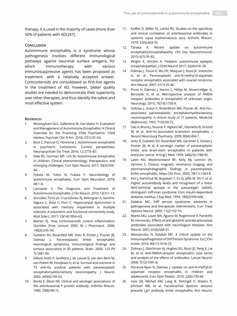
197 The use of corticosteroids in autoimmune encephalitis
therapy, it is used in the majority of cases (more than
50% of patients with AD) [47].
CONCLUSION
Autoimmune encephalitis is a syndrome whose
pathogenesis involves different immunological
pathways against neuronal surface antigens, for
which immunotherapy with various
immunosuppressive agents has been proposed as
treatment; with a relatively accepted answer.
Corticosteroids are consolidated as first-line agents
in the treatment of AD, however, better quality
studies are needed to demonstrate their superiority
over other therapies, and thus identify the safest and
most effective system.
REFERENCES
1. Mooneyham GLC, Gallentine W, Van Mater H. Evaluation
and Management of Autoimmune Encephalitis: A Clinical
Overview for the Practicing Child Psychiatrist. Child
Adolesc Psychiatr Clin N Am. 2018; 27(1):37–52.
2. Bost C, Pascual O, Honnorat J. Autoimmune encephalitis
in psychiatric institutions: Current perspectives.
Neuropsychiatr Dis Treat. 2016; 12:2775–87.
3. Dale RC, Gorman MP, Lim M. Autoimmune encephalitis
in children: Clinical phenomenology, therapeutics, and
emerging challenges. Curr Opin Neurol. 2017; 30(3):334–
44.
4. Fukata M, Yokoi N, Fukata Y. Neurobiology of
autoimmune encephalitis. Curr Opin Neurobiol. 2018;
48:1–8.
5. Lancaster E. The Diagnosis and Treatment of
Autoimmune Encephalitis. J Clin Neurol. 2016; 12(1):1-13.
6. González Torre JA, Cruz-Gómez ÁJ, Belenguer A, Sanchis-
Segura C, Ávila C, Forn C. Hippocampal dysfunction is
associated with memory impairment in multiple
sclerosis: A volumetric and functional connectivity study.
Mult Scler J. 2017; 23(14):1854–63.
7. Barnes PJ. How corticosteroids control inflammation:
Quintiles Prize Lecture 2005. Br J Pharmacol. 2006;
148(3):245–54.
8. Gultekin SH, Rosenfeld MR, Voltz R, Eichen J, Posner JB,
Dalmau J. Paraneoplastic limbic encephalitis:
neurological symptoms, immunological findings and
tumour association in 50 patients. Brain. 2000; 123 (Pt
7):1481-94.
9. Sillevis Smitt P, Grefkens J, de Leeuw B, van den Bent M,
van Putten W, Hooijkaas H, et al. Survival and outcome in
73 anti-Hu positive patients with paraneoplastic
encephalomyelitis/sensory neuronopathy. J Neurol.
2002; 249(6):745-53.
10. Bonfa E, Elkon KB. Clinical and serologic associations of
the antiribosomal P protein antibody. Arthritis Rheum.
1986; 29(8):981-5.
11. Koffler D, Miller TE, Lahita RG. Studies on the specificity
and clinical correlation of antiribosomal antibodies in
systemic lupus erythematosus sera. Arthritis Rheum.
1979; 22(5):463-70.
12. Tanaka K. Recent update on autoimmune
encephalitis/encephalopathy. Clin Exp Neuroimmunol.
2015; 6(1):70–82.
13. Wright S, Vincent A. Pediatric autoimmune epileptic
encephalopathies. J Child Neurol 2017; 32(4):418–28.
14. Dalmau J, Tüzün E, Wu HY, Masjuan J, Rossi JE, Voloschin
A, et al. Paraneoplastic anti-N-methyl-D-aspartate
receptor encephalitis associated with ovarian teratoma.
Ann Neurol. 2007; 61(1):25-36.
15. Prüss H, Dalmau J, Harms L, Höltje M, Ahnert-Hilger G,
Borowski K, et al. Retrospective analysis of NMDA
receptor antibodies in encephalitis of unknown origin.
Neurology. 2010; 75(19):1735-9.
16. Dalmau J, Graus F, Rosenblum MK, Posner JB. Anti-Hu--
associated paraneoplastic encephalomyelitis/sensory
neuronopathy. A clinical study of 71 patients. Medicine
(Baltimore). 1992; 71(2):59-72.
17. Saiz A, Bruna J, Stourac P, Vigliani MC, Giometto B, Grisold
W, et al. Anti-Hu-associated brainstem encephalitis. J
Neurol Neurosurg Psychiatry. 2009; 80(4):404-7.
18. Voltz R, Gultekin SH, Rosenfeld MR, Gerstner E, Eichen J,
Posner JB, et al. A serologic marker of paraneoplastic
limbic and brain-stem encephalitis in patients with
testicular cancer. N Engl J Med. 1999; 340(23):1788-95.
19. Lawn ND, Westmoreland BF, Kiely MJ, Lennon VA,
Vernino S. Clinical, magnetic resonance imaging, and
electroencephalographic findings in paraneoplastic
limbic encephalitis. Mayo Clin Proc. 2003; 78(11):1363-8.
20. Kim J, Namchuk M, Bugawan T, Fu Q, Jaffe M, Shi Y, et al.
Higher autoantibody levels and recognition of a linear
NH2-terminal epitope in the autoantigen GAD65,
distinguish stiff-man syndrome from insulin-dependent
diabetes mellitus. J Exp Med. 1994; 180(2):595-606.
21. Dalakas MC. Stiff person syndrome: advances in
pathogenesis and therapeutic interventions. Curr Treat
Options Neurol. 2009; 11(2):102-10.
22. Manto MU, Laute MA, Aguera M, Rogemond V, Pandolfo
M, Honnorat J. Effects of anti-glutamic acid decarboxylase
antibodies associated with neurological diseases. Ann
Neurol. 2007; 61(6):544-51.
23. Alexopoulos H, Dalakas MC. A critical update on the
immunopathogenesis of Stiff Person Syndrome. Eur J Clin
Invest. 2010; 40(11):1018-25.
24. Dalmau J, Gleichman AJ, Hughes EG, Rossi JE, Peng X, Lai
M, et al. Anti-NMDA-receptor encephalitis: case series
and analysis of the effects of antibodies. Lancet Neurol.
2008; 7(12):1091-8.
25. Florance-Ryan N, Dalmau J. Update on anti-N-methyl-D-
aspartate receptor encephalitis in children and
adolescents. Curr Opin Pediatr. 2010; 22(6):739-44.
26. Irani SR, Michell AW, Lang B, Pettingill P, Waters P,
Johnson MR, et al. Faciobrachial dystonic seizures
precede Lgi1 antibody limbic encephalitis. Ann Neurol.

198 B.L. Nahar-González, I.D. Lozada-Martinez, Y. Arevalo-Martínez et al.
2011; 69(5):892-900.
27. Sen A, Wang J, Laue-Gizzi H, Lee T, Ghougassian D,
Somerville ER. Pathognomonic seizures in limbic
encephalitis associated with anti-LGI1 antibodies. Lancet.
2014; 383(9933):2018.
28. Thompson J, Bi M, Murchison AG, Makuch M, Bien CG,
Chu K, et al. Faciobrachial Dystonic Seizures Study Group.
The importance of early immunotherapy in patients with
faciobrachial dystonic seizures. Brain. 2018; 141(2):348-
356.
29. López-Chiriboga AS, Klein C, Zekeridou A, McKeon A,
Dubey D, Flanagan EP, et al. LGI1 and CASPR2
neurological autoimmunity in children. Ann Neurol.
2018; 84(3):473-480.
30. Florance NR, Davis RL, Lam C, Szperka C, Zhou L, Ahmad
S, et al. Anti-N-methyl-D-aspartate receptor (NMDAR)
encephalitis in children and adolescents. Ann Neurol.
2009; 66(1):11-8.
31. Schmitt SE, Pargeon K, Frechette ES, Hirsch LJ, Dalmau J,
Friedman D. Extreme delta brush: a unique EEG pattern
in adults with anti-NMDA receptor encephalitis.
Neurology. 2012; 79(11):1094-100.
32. Gresa-Arribas N, Titulaer MJ, Torrents A, Aguilar E,
McCracken L, Leypoldt F, et al. Antibody titres at
diagnosis and during follow-up of anti-NMDA receptor
encephalitis: a retrospective study. Lancet Neurol. 2014;
13(2):167-77.
33. Dahm L, Ott C, Steiner J, Stepniak B, Teegen B,
Saschenbrecker S, et al. Seroprevalence of
autoantibodies against brain antigens in health and
disease. Ann Neurol. 2014; 76(1):82-94.
34. Hara M, Martinez-Hernandez E, Ariño H, Armangué T,
Spatola M, Petit-Pedrol M, et al. Clinical and pathogenic
significance of IgG, IgA, and IgM antibodies against the
NMDA receptor. Neurology. 2018; 90(16):e1386-e1394.
35. Okamoto S, Hirano T, Takahashi Y, Yamashita T, Uyama
E, Uchino M. Paraneoplastic limbic encephalitis caused by
ovarian teratoma with autoantibodies to glutamate
receptor. Intern Med. 2007; 46(13):1019-22.
36. Seki M, Suzuki S, Iizuka T, Shimizu T, Nihei Y, Suzuki N, et
al. Neurological response to early removal of ovarian
teratoma in anti-NMDAR encephalitis. J Neurol
Neurosurg Psychiatry. 2008; 79(3):324-6.
37. Monti G, Giovannini G, Marudi A, Bedin R, Melegari A,
Simone AM, et al. Anti-NMDA receptor encephalitis
presenting as new onset refractory status epilepticus in
COVID-19. Seizure. 2020; 81:18-20.
38. Burr T, Barton C, Doll E, Lakhotia A, Sweeney M. N-Methyl-
d-Aspartate Receptor Encephalitis Associated With
COVID-19 Infection in a Toddler. Pediatr Neurol. 2021;
114:75-76.
39. Panariello A, Bassetti R, Radice A, Rossotti R, Puoti M,
Corradin M, et al. Anti-NMDA receptor encephalitis in a
psychiatric Covid-19 patient: A case report. Brain Behav
Immun. 2020; 87:179-181.
40. Graus F, Titulaer MJ, Balu R, Benseler S, Bien CG, Cellucci
T, et al. A clinical approach to diagnosis of autoimmune
encephalitis. Lancet Neurol. 2016; 15(4):391-404.
41. Shin YW, Lee ST, Park KI, Jung KH, Jung KY, Lee SK, et al.
Treatment strategies for autoimmune encephalitis. Ther
Adv Neurol Disord. 2017; 11:1756285617722347.
42. Vernino S, O'Neill BP, Marks RS, O'Fallon JR, Kimmel DW.
Immunomodulatory treatment trial for paraneoplastic
neurological disorders. Neuro Oncol. 2004; 6(1):55-62.
43. Vedeler CA, Antoine JC, Giometto B, Graus F, Grisold W,
Hart IK, et al. Paraneoplastic Neurological Syndrome
Euronetwork. Management of paraneoplastic
neurological syndromes: report of an EFNS Task Force.
Eur J Neurol. 2006; 13(7):682-90.
44. Dalmau J, Lancaster E, Martinez-Hernandez E, Rosenfeld
MR, Balice-Gordon R. Clinical experience and laboratory
investigations in patients with anti-NMDAR encephalitis.
Lancet Neurol. 2011; 10(1):63-74.
45. Goldenholz DM, Wong VS, Bateman LM, Apperson M,
Oskarsson B, Akhtar S, et al. Treatment of γ-aminobutyric
acid B receptor-antibody autoimmune encephalitis with
oral corticosteroids. Arch Neurol. 2012; 69(8):1061-3.
46. Prasad P, Bhargava A, Khichar S, Gupta VK. Clinical
spectrum of autoimmune encephalitis: Case series from
Western Rajasthan. J Clin Diagnostic Res. 2017; 11(11):
OR01-OR04.
47. Nosadini M, Mohammad SS, Ramanathan S, Brilot F, Dale
RC. Immune therapy in autoimmune encephalitis: a
systematic review. Expert Rev Neurother. 2015;
15(12):1391-419.

Romanian Neurosurgery (2021) XXXV (2): pp. 199-203 DOI: 10.33962/roneuro-2021-031 www.journals.lapub.co.uk/index.php/roneurosurgery
Paediatric brain abscesses in tribal region of India. A single centre experience
Sachin Kumar Jain, Gaurav Jaiswal, Tarun Kumar Gupta, Krishna
Govind Lodha, Vishnu Kumar Lohar, Kaushal Yadav
Department of Neurosurgery, R.N.T. Medical College, Udaipur, INDIA
ABSTRACT Introduction: Brain parenchymal abscesses are relatively infrequent but potentially
serious infections in the paediatric population. Surgical intervention in addition to a
prolonged administration of antibiotics is generally appropriate management.
Aims and objective: We performed this study to assess the clinical profile, aetiology
and outcome of paediatric brain abscess which are treated surgically only by
aspiration in the tribal region of Rajasthan.
Material and method: A single-centre retrospective study was conducted over a 5
year period (2014–2019) in the department of neurosurgery in RNT medical college,
Udaipur, Rajasthan. We treated approx. 60 patients of which 25 patients treated
conservatively and 5 patient treated by craniotomy and abscess excision. So only 30
patient included in our study in which abscess treated by aspiration only. Patients<18
years of age with a confirmed intra-parenchymal abscess were included. Patient
records were reviewed for abscess location, microbiology results, Clinical features,
Surgical intervention, and outcome using the Glasgow Outcome Score at 3 months.
Result: 11 patients had an abscess in the temporal lobe and Streptococcus was the
most common causative micro-organism (n¼15). 25 patients (80%) had an
identifiable source which included: ENT infections, congenital cardiac malformations,
recent dental surgery and meningitis. The most common symptom is fever f/b
headache, seizure and vomiting. But despite previous studies seizure (10/30)
presentation is comparatively more. All 30 patients underwent aspiration.
Conclusion: In tribal regions of India ENT infections are a more common source of
brain abscess because of poor hygiene and illiteracy and their ignorance of ENT
infections and also not taking seriously to fever, headache and other health issues.
But at present, there are also decreasing trends of brain abscess by ENT infections
and rising trends by congenital heart disease which is a good sign that the health and
educational infrastructure is strengthening in the tribal region also.
INTRODUCTION A brain abscess is an intra-parenchymal collection of pus in the brain.
The incidence of brain abscess among intracranial masses varies from
1-2% in western countries, to about 8% in developing countries1. It is
still a life threatening and fatal entity and often leads to serious
disability and even death if misdiagnosed or treated improperly2. Brain
abscesses are relatively less common in the paediatric population
compared to adults; however, they remain a serious, life-threatening
infection3.
Keywords abscess,
aspiration, paediatrics,
ENT
Corresponding author: Gaurav Jaiswal
Department of Neurosurgery,
R.N.T. Medical College, Udaipur, India
Scan to access the online version

200 Sachin Kumar Jain, Gaurav Jaiswal, Tarun Kumar Gupta et al.
The causes of brain abscesses are highly variable in
children. The major predisposing conditions in
children are: an adjacent focus of infection, trauma,
haematogenous seeding, neurosurgical procedures,
cyanotic heart disease and immunocompromised
states4,5,6. Associated sites of infection commonly
include sinusitis, mastoiditis, dental infections and
chronic otitis media where aerobic bacterial
infections such as Streptococci and Staphylococci
predominate4,7
The gold standard treatment of brain abscesses
continues to be aspiration or excision combined with
antibiotic therapy.5 However the outcome has
improved dramatically in recent decades due to
improvements in diagnostic techniques and broad-
spectrum antibiotics8. Magnetic Resonance Imaging
is more sensitive at diagnosing early cerebritis9 and
has increased the prognosis of brain abscess by
improving the speed of diagnosis.
Despite all of these advances abscesses are still a
significant cause of morbidity and mortality10. In
addition, abscess growth can result in rupture of the
abscess into the ventricular system. This life-
threatening event will result in an acute
decompensation and symptoms of a purulent
meningitis11.
We performed this study to assess the clinical
profile, aetiology, and outcome of paediatric brain
abscess in the tribal region of Rajasthan.
MATERIAL AND METHOD
A single-centre retrospective study was conducted
over a 5-year period (2014–2019) in department of
neurosurgery in RNT medical college, Udaipur,
Rajasthan. We treated approx. 60 patients in which
25 patients treated conservatively and 5 patients
treated by craniotomy and abscess excision. So only
30 patients included in our study in which abscess
treated by aspiration only.
The final cohort of 30 patients comprised 20 male
patients (45.8%) and 10 female patient and had a
mean age of 6.5 ± 4 years Patients < 18 years of age
with radiological confirmed intra-parenchymal
abscess were included. Patient records were
reviewed for abscess aetiology, location, size,
microbiology results, surgical intervention, and
outcome using the Glasgow Outcome Score at 3
months.
We include the patients in which abscess treated
only by aspiration and then antibiotics. Aspiration of
abscess may be single or multiple times. Size of
abscess is more than 2.5 cm. The total antibiotic
duration was discernible from the medical notes for
all patients and the mean time was 5.8 ± 2.0weeks.
This comprised a mean of 2.9 ± 1.5 weeks
intravenous and3.0 ± 1.8 weeks oral antibiotics.
White cell count >11109/L and/or C-Reactive Protein
>5mg/L was classed as raised inflammatory markers.
Functional outcome was measured using the
Glasgow Outcome Score (GOS) at 3 months from
discharge. Those with isolated subdural empyema,
extradural abscess, or an unknown abscess location
were excluded from the study.
RESULT
Most common symptom is fever followed by
headache, seizure and vomitting. (Table-3). The
classic triad of headache, fever and focal
neurological deficit was seen in two patients (8.6%).
Drowsiness or reduced level of consciousness on
presentation was recorded in 6 patients (34.8%). Age
of patients are mostly 5-10 years. (Table-1)
The Temporal lobe was the most common
abscess location (11). The remainder of the
abscesses were in the parietal lobe (8), frontal lobe
(7), cerebellum (2), thalamus (1), pons (1) (Table 4) .22
patients had a solitary lesion at presentation and rest
8 has more than one lesion. The patient with
aspergilloma had two lesions in the same location.
Figure 1. Pre-operative scan of 10-month male child left
temporo-parietal brain abcess (original pics).
S.
N.
Age Number Percentage
1
2
3
<5 year
5-10 yr
10-18 yr
6
15
9
20%
50%
30%
Table 1.
25 patients (80 %) had an identifiable source which
include post meningitic (4), congenital cardiac
malformations (7), Otogenic and mastoditis (11), Post
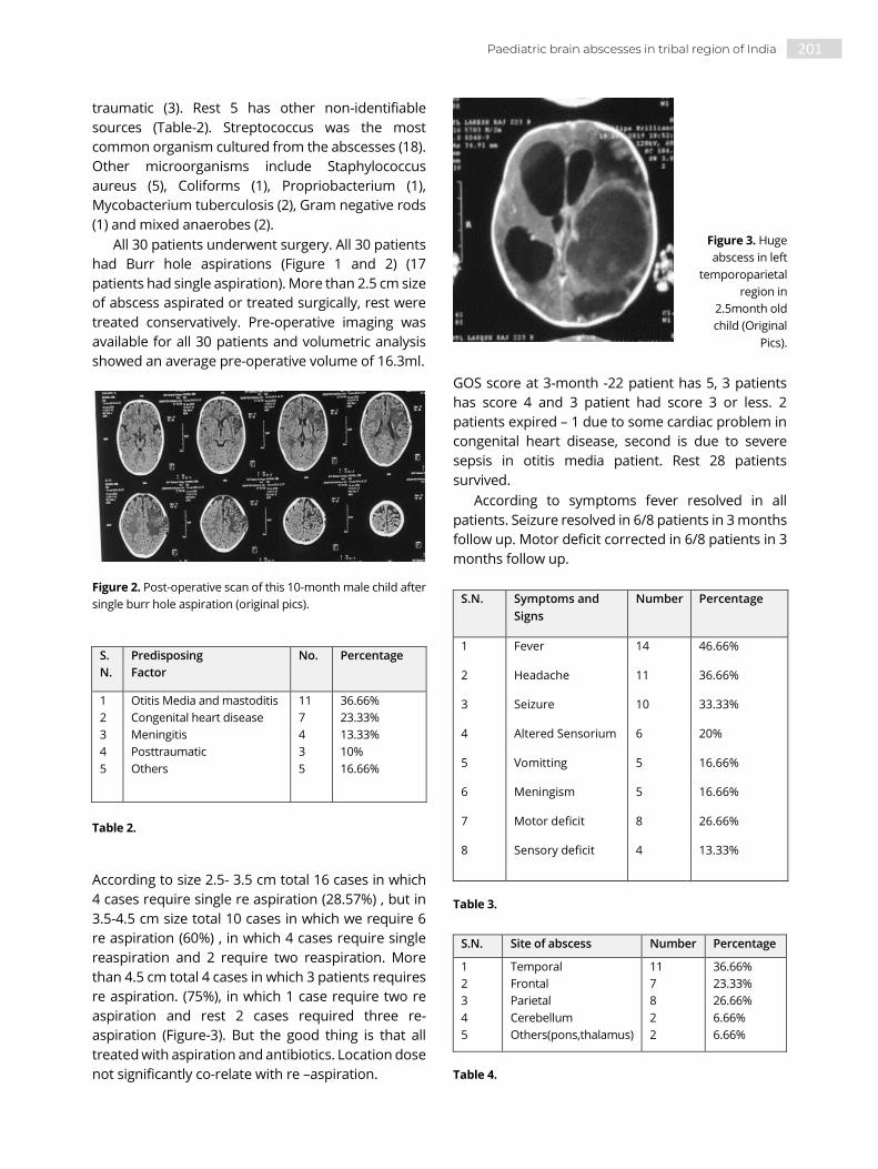
201 Paediatric brain abscesses in tribal region of India
traumatic (3). Rest 5 has other non-identifiable
sources (Table-2). Streptococcus was the most
common organism cultured from the abscesses (18).
Other microorganisms include Staphylococcus
aureus (5), Coliforms (1), Propriobacterium (1),
Mycobacterium tuberculosis (2), Gram negative rods
(1) and mixed anaerobes (2).
All 30 patients underwent surgery. All 30 patients
had Burr hole aspirations (Figure 1 and 2) (17
patients had single aspiration). More than 2.5 cm size
of abscess aspirated or treated surgically, rest were
treated conservatively. Pre-operative imaging was
available for all 30 patients and volumetric analysis
showed an average pre-operative volume of 16.3ml.
Figure 2. Post-operative scan of this 10-month male child after
single burr hole aspiration (original pics).
S.
N.
Predisposing
Factor
No. Percentage
1
2
3
4
5
Otitis Media and mastoditis
Congenital heart disease
Meningitis
Posttraumatic
Others
11
7
4
3
5
36.66%
23.33%
13.33%
10%
16.66%
Table 2.
According to size 2.5- 3.5 cm total 16 cases in which
4 cases require single re aspiration (28.57%) , but in
3.5-4.5 cm size total 10 cases in which we require 6
re aspiration (60%) , in which 4 cases require single
reaspiration and 2 require two reaspiration. More
than 4.5 cm total 4 cases in which 3 patients requires
re aspiration. (75%), in which 1 case require two re
aspiration and rest 2 cases required three re-
aspiration (Figure-3). But the good thing is that all
treated with aspiration and antibiotics. Location dose
not significantly co-relate with re –aspiration.
Figure 3. Huge
abscess in left
temporoparietal
region in
2.5month old
child (Original
Pics).
GOS score at 3-month -22 patient has 5, 3 patients
has score 4 and 3 patient had score 3 or less. 2
patients expired – 1 due to some cardiac problem in
congenital heart disease, second is due to severe
sepsis in otitis media patient. Rest 28 patients
survived.
According to symptoms fever resolved in all
patients. Seizure resolved in 6/8 patients in 3 months
follow up. Motor deficit corrected in 6/8 patients in 3
months follow up.
S.N. Symptoms and
Signs
Number Percentage
1
2
3
4
5
6
7
8
Fever
Headache
Seizure
Altered Sensorium
Vomitting
Meningism
Motor deficit
Sensory deficit
14
11
10
6
5
5
8
4
46.66%
36.66%
33.33%
20%
16.66%
16.66%
26.66%
13.33%
Table 3.
S.N. Site of abscess Number Percentage
1
2
3
4
5
Temporal
Frontal
Parietal
Cerebellum
Others(pons,thalamus)
11
7
8
2
2
36.66%
23.33%
26.66%
6.66%
6.66%
Table 4.

202 Sachin Kumar Jain, Gaurav Jaiswal, Tarun Kumar Gupta et al.
DISCUSSION
The change in practice towards early prescription of
antibiotics for oto-sinogenic infections has resulted
in a change in the infective etiologies associated with
brain abscess12. In studies from the pre-CT era ENT
infections were the cause in 38-76% of brain
abscesses in children; however this has fallen to 8-
36% .In the modern era with congenital heart disease
and immunosuppression becoming a more
commonly recognized causative factor11,12,13,14.But in
the present study, Temporal lobe abscess following
middle ear infection and mastoiditis was the
commonest finding15,16. Temporal lobe abscess
following otogenic infection has been found in
previous studies22. In tribal area due to poor hygine
and others late responding to fever and ignorance of
ear infection or maltreatment of ear infection
develop a simple meningitis and ear infection into a
brain abscess15,16.
In our study, males are more affected than
females. Males are more exposed to outdoor
activities with greater chance of ear infection and
predisposing to brain abscess 17. Regardless of the
changes in etiology, Streptococcus remains the most
commonly reported causative microbe both in this
series and those published in the literature17,18. The
presentation of brain abscesses is non-specific but
most commonly includes, headache, fever, altered
mental status, meningism and seizures5,.According
to Samull et al nowadays suggest that headache and
fever is the most common symptoms and seizures is
not so common in abscess but in this study seizure is
also common complain (10/30-30%) because
involvement of temporal lobe is more .
Those who present with altered mental status
and rapid neurological deterioration are more likely
to have an increased mortality rate19. 2 patient
expired in this study had low GCS and altered mental
status. So preoperative GCS is important for
prognosis. Multiple aspirations were associated with
a larger initial abscess volume. These will leave a
larger cavity that is slower to involute and thus leave
a nidus of infection that can re-accumulate. Half of
the patients in this series required more than one
aspiration and thus patients should be counselled
accordingly.12
Modern series report lower mortality rates than
the pre-CT era ranging from 8 to 25%20 and the large
series by Tekkok and Erbengi21 even managed 0%
mortality at the end of their study period.
Neuroimaging was done in all cases. CT scan was
done in 25 cases and MRI was done 5 cases. The only
added advantages of MRI over CT Scan are: better
differentiation of edema from necrosis, more
sensitivity in detection of early cerebritis, greater
sensitivity for early satellite lesion23. Most common
neuroimaging finding was ring enhancing lesion. This
has been reported in previous studies from India and
abroad24,25.
One reason for a low mortality rate may be an
aggressive approach towards abscess drainage and
also sepsis source control. Poor Glasgow coma scale
(GCS) at time presentation has been associated with
poor outcome. Previous studies in India, Nepal and
Pakistan have also reported poor GCS as negative
prognostic factor in their studies24,25
CONCLUSION
This study demonstrates that in the modern era with
ready access to CT imaging and broad-spectrum
antibiotics, low rates of morbidity and mortality can
be achieved. Drainage of paediatric abscesses
remain the mainstay of treatment both to relieve
mass effect and provide a microbiological diagnosis.
Patients and their families should be counselled to
expect multiple procedures.
But in this study most common predisposing
factor for abscess is otitis media than congenital
heart disease. Because in tribal region exposed to
outdoor activities with greater chance of ear
infection and predisposing to brain abscess. Also,
illiteracy and poor hygiene leads to more chances of
chronic ear discharge and brain abscess formation in
future.
So we need to educate the people of tribal region
for proper hygine and also early consult to doctor for
any ear infection. We have to educate them about
their proper health checkup and consult if child had
fever, seizure, headache and vomitting. If infant and
less than 3 yr age child had excessive irritability then
also take consultation. We can also promote
pneumococcal vaccination.
But at present there are also decreasing trends of
brain abscess by ENT infections (11/30 -36.66%) and
rising trends by congenital heart disease (7/30-
23.33%) which is a good sign that the health and
educational infrastructure is strengthening in tribal
region also.

203 Paediatric brain abscesses in tribal region of India
CONFLICT OF INTEREST There is no conflict of interest to disclose.
INFORMED CONSENT
Informed consent was obtained from all individual participants
included in this study.
REFERENCES
1. Sharma BS, Gupta SK, Khosla VK. Current concepts in the
management of pyogenic brain abscess. Neurol India.
2000;48:105–11.
2. Kao PT, Tseng HK, Liu CP, Su SC, Lee CM. Brain abscess:
Clinical analysis of 53 cases. J Microbiol Immunol Infect.
2003;36:129–36.
3. Frazier JL, Ahn ES, Jallo GI. Management of brain
abscesses in children.Neurosurg Focus2008;24:E8.
4. Brook I. Brain abscess in children: microbiology and
management.J ChildNeurol1995;10:283–8.
5. Hakan T, Ceran N, Erdem I, Berkman MZ, Goktas P.
Bacterial brain abscesses: an evaluation of 96 cases.J
Infect2006;52:359–66.
6. Tandon S, Beasley N, Swift AC. Changing trends in
intracranial abscesses secondary to ear and sinus
disease. JLO2009;123:283.
7. Shachor-Meyouhas Y, Bar-Joseph G, Guilburd JN, Lorber
A, Hadash A,Kassis I. Brain abscess in children -
epidemiology, predisposing factors and management in
the modern medicine era.Acta Paediatr2010;99:1163–7.
8. Saez-Llorens X. Brain abscess in children.Semin Pediatr
Infect Dis2003;14:108–14.
9. Mathisen GE, Johnson JP. Brain abscess.Clin Infect
Dis1997; 25:763–79.quiz 80-1.
10. Felsenstein S, Williams B, Shingadia D,et al.Clinical and
microbiologic features guiding treatment
recommendations for brain abscesses in chil-
dren.Pediatr Infect Dis J2013;32:129–35.
11. Jasonp. Sheehan,Johna. Janejr etal. Brain abscess in
children:Journal of neurosurgery 2008/24/6/E6.
12. Goodkin HP, Harper MB, Pomeroy SL. Intracerebral
abscess in children:historical trends at Children’s
Hospital Boston.Pediatrics2004;113:1765–70.
13. Samuel Halla, Shirley Yadub, Benjamin Gaastraa,
Nijaguna Mathada, Owen Sparrowa, Ryan Watersa, Aabir
Chakrabortya and Vassilios Tsitouras et al. Paediatric
brain abscesses: a single centre experience,British
journal of neurosurgery:may 2019:33(550-554).
14. Canpolat M, Ceylan O, Per H,et al.Brain abscesses in
children: results of24 children from a reference center in
Central Anatolia, Turkey.J ChildNeurol2015;30:458–67.
15. Kafle P, Sharma MR, Shilpakar SK, Sedain G, Pradhanang
A, Shrestha RK, et al. Pediatric brain abscess clinical
profile, management and outcome at tertiary care centre
in Nepal. JCMS Nepal. 2018;14:102–6.
16. Clinical Profile and Outcome of Brain Abscess in Children
from a Tertiary Care Hospital in Eastern Uttar Pradesh ,
Rajniti Prasad, John Biswas, Kulwant Singh,1 Om P.
Mishra, and Ankur Singh,Ann Indian Acad Neurol. 2020
May-Jun; 23(3): 303–307.
17. Sharma R, Mohandas K, Cooke RP. Intracranial
abscesses: changes in epi-demiology and management
over five decades in Merseyside.Infection2009;37:39–43.
18. Nathoo N, Nadvi SS, Narotam PK, van Dellen JR. Brain
abscess: manage-ment and outcome analysis of a
computed tomography era experiencewith 973
patients.World Neurosurg2011;75:716–26. discussion
612-7.
19. Seydoux C, Francioli P. Bacterial brain abscesses: factors
influencing mor-tality and sequelae.Clin Infect
Dis1992;15:394–401.
20. Auvichayapat N, Auvichayapat P, Aungwarawong S. Brain
abscess in infants and children: a retrospective study of
107 patients in northeast Thailand.J Med Assoc
Thai2007;90:1601–7
21. Tekk€ok IH, Erbengi A. Management of brain abscess in
children: review of 130 cases over a period of 21
years.Childs Nerv Syst1992;8:411–6.
22. Malik S, Joshi SM, Kandoth PW, Vengsarkar US.
Experiences with brain abscess. Indian Pediatr.
1994;31:661–6.
23. Schmidek HH. Suppurative intracranial infections. In:
Schmidek HH, Roberts DW, editors. Schmidek & Sweet
Operative Neurosurgical Techniques. Indications,
Methods, and Results. 5th ed. Philadelphia: Elsevier, Inc;
2006. pp. 1591–9.
24. Atiq M, Ahmed US, Allana SS, Chishti KN. Brain abscess in
children. Indian J Pediatr. 2006;73:401–4.
25. Singh D, Gupta V, Singh AK, Sinha S. Evolution of otogenic
brain abscess and management protocol. Indian Pediatr.
2001;38:169–73.

Romanian Neurosurgery (2021) XXXV (2): pp. 204-206 DOI: 10.33962/roneuro-2021-032 www.journals.lapub.co.uk/index.php/roneurosurgery
Lateral orbitotomy in the management of an intra-orbital lipoma. A case report
Mandour Cherkaoui1, Ait Lhaj El Houssaine2, Khouya Ali Adil2,
Gazzaz Miloudi3, E.L. Mostarchid Brahim3 1 Department of Neurosurgery, 5th Military Hospital, Guelmim,
MOROCCO 2 Department of Ophthalmology, 5th Military Hospital, Guelmim,
MOROCCO 3 Department of Neurosurgery, Military Hospital Mohammed V,
Rabat, MOROCCO
ABSTRACT Introduction: Lipomas are benign subcutaneous mass, but the intra-orbital location
is rarely reported in the literature and it can resemble a variety of other orbital
lesions.
Case report: We describe a 15-year-old girl who presented with left exophthalmia.
Orbital magnetic resonance imaging showed an encapsulated intra-conal mass
displacing the optic nerve medially, the external right muscle laterally and the globe
anteriorly. Excisional biopsy of the mass by lateral orbitotomy approach resolved the
exophthalmia, and histology revealed a primary orbital lipoma.
Conclusion: The diagnosis of an intra-orbital lipoma is not easy and the surgical
approach represents a challenge to achieve a total excision while avoiding
complications.
INTRODUCTION Lipoma is a benign mesenchymal neoplasm composed of mature
adipose tissue surrounded by a fibrous tissue capsule1. The majority is
developed in the subcutaneous soft tissue in the neck, shoulder and
back, but it is uncommon in the orbital region2. We describe a case of
exophthalmia caused by an intra-orbital lipoma and review of the
literature.
CASE REPORT
A 15-year-old Moroccan girl, admitted to our department for treatment
of a progressive left exophthalmia for 06 months. There was no history
of trauma, visual disturbances, and significant ocular or medical
histories. On ocular examination, she had visual acuity of 20/20 in both
eyes with a left axile, reducible and non-pulsatile exophthalmia. Orbital
Magnetic Resonance Imaging (MRI) revealed an encapsulated intra-
conal mass (2.5 x 1,7 x 0,5 cm) in the posterolateral compartment of the
left orbit. The mass was displacing the optic nerve medially, the external
Keywords exophthalmia,
lateral orbitotomy, lipoma, orbital
Corresponding author: Mandour Cherkaoui
Department of Neurosurgery,
5th Military Hospital, Guelmim, Morocco
Scan to access the online version

205 Lateral orbitotomy in the management of an intra-orbital lipoma
right muscle laterally and the globe anteriorly with
resultant exophthalmia. Hypointense at T1 and
hyperintense at T2 taking the contrast (Figure 1-2). A
left lateral orbitotomy approach was utilized with an
excisional biopsy (Figure 3-4). Histopathological
evaluation revealed well-encapsulated, mature
adipose tissue consistent with a primary orbital
lipoma. After surgery, exophthalmia had subsided
with a normal ocular motility.
Figure 1. Post contrast axial T1-weighted MRI showing hyper
intense lesion (arrow). Figure 2. Axial T2-weighted MRI showing hyper intense lesion
(arrow).
Figure 3. Preoperative photograph showing the incision for a
left lateral orbitotomy.
Figure 4. Intra-operative photograph showing tumor excision.
DISCUSSION
Lipoma is a benign lobulated tumor composed of
mature adipose tissue surrounded by a fibrous
tissue capsule. Frequently it is localized
subcutaneously around the torso, neck, and
proximal limbs3, but in the orbital region is
uncommon with the reported incidence of 0.6% in
adults and 2.8% in children4.
The exact etiology of lipoma formation is
unknown, but there are instances following
trauma4,5. Also some hypotheses have been
mentioned as hypertrophy theory (the growth of the
tumor is caused by obesity) and metaplasia theory
(the growth of lipoma occurs due to differentiation of
mesenchymal cells into lipoblast) 6.
Generally, orbital lipoma slow growing and often
asymptomatic until large enough to be palpable,
visible or cause mass effect. Rarely grows and
compresses the optic nerve causing disturbance in
visual function7.
The radiological appearance of orbital lipoma, on
computed tomography the tumor has a distinctive
low attenuation with a finely defined border. On MRI
it is generally hyperintense on T1-weighted imaging
and indistinct to orbital blood on T2-weighted
images, hypointense after fat suppression. However,
it is not enhanced after contrast images8, but in our
case the lesion was hyperintense.
Due to the various presentations of lipoma, the
diagnosis is not always easy and other orbital masses
should be considered in the clinical differential
diagnosis, such as dermoid cyst, fibrous
histiocytoma, schwannoma and cavernous
hemangioma9.
The recommended treatment is the surgical
excision and it is a challenge because of the complex
anatomy of the orbital structures10, 11. In our case, the
patient was operated by a lateral orbitotomy with
removal of the tumor. The long-term outcome after
surgery is seen to be excellent however, recurrence
of the tumor may occur due to incomplete excision12.
CONCLUSION
We present a case of a rare intra-orbital tumor
whose surgical approach such as lateral orbitotomy
is a real challenge for neurosurgeons.
CONSENT Written informed consent was obtained from the patient for
publication of this case report and accompanying images.
ABBREVIATIONS MRI: Magnetic resonance imaging

206 Mandour Cherkaoui, Ait Lhaj El Houssaine, Khouya Ali Adil et al.
AUTHOR’S CONTRIBUTIONS:
C M: manuscript writing; A L and A K: manuscript preparation;
B E and M G: manuscript analysis. All authors read and
approved the final manuscript.
ACKNOWLEDGEMENTS
This study received no funding
CONFLICTS OF INTEREST
The authors declare no potential conflict of interest
REFERENCES
1. Pranav Shrestha; Gulshan Bahadur Shrestha. Orbital
Lipoma as an Uncommon Cause of Unilateral Proptosis:
A Case Report. International Medical Case Reports
Journal. 2020:13 415–418.
2. Rodiah Rahmawaty Lubis. Giant Preseptal Upper Eyelid
LipomaI. A Case Report. EuroMediterranean Biomedical
Journal. 2020: 15 (13) ; 58–60.
3. Weiss S. Lipomatous tumours. Monogr Pathol 1996; 38:
207–239.
4. CPT Matthew; Chorost MC ; LTC Vladimir Yakopson, and
al. Posttraumatic Encapsulated Orbital Lipoma. Military
Medicine. 2020:185, 9/10:e1876.
5. Aust MC, Spies M, Kall S, Joukuszies A, Gohritz A, Vogt P.
Lipomas after blunt soft tissue trauma: are they real?
Analysis of 31 cases. Br J Dermatol 2007; 157(1): 92–9.
6. Kumaraswamy S, Madan N, Keerthi R, Shakti S. Lipomas
of oral cavity: case reports with review of literature. J
Maxillofac Oral Surg. 2009. 8(4):394–397.
7. Chi MJ, Roh JH, Lee JH and al. A case of orbital lipoma with
exophthalmos and visual disturbance. Jpn J Ophthalmol.
2009;53(4):442–444.
8. Shah NB, Chang WY, White VA and al. Orbital lipoma: 2
cases and review of literature. Ophthal Plast Reconstr
Surg. 2007;23(3):202–205.
9. Ali SF, Farber M, Meyer DR: Fibrolipoma of the orbit.
Ophthalmic Plast Reconstr Surg 2013; 29(3): 79–81.
10. R. Malarvizhi, K. Vasumathi, A. Anuradha and al. A Clinical
Study on Evaluation, Indications, Surgical Techniques and
Complications of Various Orbitotomies Done in a Tertiary
Care Centre. Delhi J Ophthalmol 2020; 30; 22-26.
11. Zeynel A. Karcioglu. Surgical Techniques for Orbital
Tumors. Surgical Ophthalmic Oncology. 2019-October:
pp 117-127.
12. Vasconcelos JS, Gharaibeh F, Nittinger HC. Trauma as
pathogenesis of a plantar forefoot fibrolipoma: first case
and review of the literature. Case Rep Surg. 2013:691276.

Romanian Neurosurgery (2021) XXXV (2): pp. 207-209 DOI: 10.33962/roneuro-2021-033 www.journals.lapub.co.uk/index.php/roneurosurgery
Primary calvarial cavernous haemangioma in a child. A case report
Adel Khelifa, Walid Bennabi, Lakhder Berchiche,
Abdelhalim Morsli
Neurosurgical Department of BEO University Hospital, Algiers,
ALGERIA
ABSTRACT A wide variety of bone lesions is encountered on the skull and usually with poor
clinical presentations and non-specific radiological features. Primary cavernous
hemangioma is one of these rare calvarial lesions that the symptoms are limited to a
simple subcutaneous mass frequently neglected by the patient. Although seen in all
ages from neonate to elderly; few cases were reported in the literature of patients in
the first decade of life. We report a case of four years old boy with primary calvarial
cavernous hemangioma in order to help to understand this pathology and to urge
physicians to explore any chronic subcutaneous mass at any age.
INTRODUCTION Skull hemangioma is a rare pathology with minimal clinical
presentation and non-specific images. The first known description of
this lesion in literature was in 1845 by Toynbee [1]. Skull hemangiomas
are classified into capillary and cavernous types; with cavernous type
being more frequently seen [1,2,3,4,5,6]. We present a case of four years old
boy with primary calvarial cavernous hemangioma.
CASE PRESENTATION
The patient is a four years old boy with trisomy 21. The parents
consulted for the appearance of a right fronto parietal subcutaneous
mass; of barely 1 cm of diameter. A brain CT was performed objectified
an extra axial right fronto parietal lesion of 42 x 29 mm; isodense, with
intense homogenous enhancement after contrast injection, and bone
destruction from where the lesion get through to the subcutaneous
region; there was also a circumferential osteophytic bone reaction
(Figure 1). The patient was operated and the lesion was totally removed
through a frontoparietal bone flap. In post-operative there were no
complications. Pathology study was in favor of bone cavernous
hemangioma. No supplementary management was necessary. Control
images performed nine years later, showed no residual tumor or
recidivism (Figure 2).
Keywords hemangioma,
bone tumours, skull tumours
Corresponding author: Adel Khelifa
Neurosurgical Department of
BEO University Hospital, Algiers, Algeria
.
Scan to access the online version

208 Adel Khelifa, Walid Bennabi, Lakhder Berchiche et al.
Figure 1. Preoperative brain CT; A: before injection; B: after injection; C: bone window. The images objectify an extra axial right
fronto parietal lesion; it appears spontaneously isodense (A, horizontal arrow), with intense homogenous enhancement after
contrast injection (B, horizontal arrow), and bone destruction from where the lesion get through to the subcutaneous region (C,
horizontal arrow). There was also a circumferential osteophytic reaction from the inner calvarial bone (A and C, vertical arrows).
Figure 2. Nine years post-operative MRI showing no tumor
recidivism. A: axial FLAIR sequence; B: axial T1 injected image.
DISCUSSION
Primary calvarial hemangiomas (PCHs) are rare
lesions; they account of 0.2 % of all bone neoplasm [1,3,5,6,7,8]. Yang et al. reviewed 93 cases of PCH
reported in the literature from 1845 to 2015;
according to this paper most patients were in the
fourth and the fifth decade [1,2]. Only 0.9 % of PCHs
were seen in the first decade of life as in our case [2].
Frontal bone is a predilection location for PCHs with
44.1% of all cases [1,2]. Clinical presentation depends
on the topography of the lesion. Subcutaneous mass
and headaches were the most frequent reasons for
consultation [1,2]; neurologic deficit is rarely seen [1,2,4,5,6,7,8]. Proptosis, impaired vision, facial nerve
paralysis, tinnitus, and hearing loss are seen in
orbital and skull base locations [1,2]. Bleeding from the
lesion may be seen in the epidural or subarachnoid
spaces [1,3,7]. On plane X-Rays and on CT scan, PCH is
a lytic lesion with sclerotic rim; after contrast
injection the enhancement is constant it could be
heterogeneous or homogenous as in our case [2,3].
For us those finding were sufficient to indicate the
surgery and to plan the resection strategy. Although
not specific, MRI affords supplementary information
about the relation of the lesion with the underlying
dura, parenchyma, and vascular structures; it shows
hypo to iso intense mass on T1 weighted images,
hyperintense on T2 weighted images and on FLAIR
sequences; there is a large enhancement after
gadolinium injection [1,3]. PCHs are accessible lesions
for surgical resection, thus total removal with
acceptable margin is the treatment of choice [1]; this
could be achieved through a skin incision and
craniectomy centered on the lesion. In Adult the
bone defect is correct usually with cranioplasty. As in
our case the management of many other pediatric
cases in the literature was limited to lesion removal
without bone correction due to the young age face to
future skull changing [4,8]; other teams preferred
cranioplasty [6] or remodeling [5].
CONCLUSION
Subcutaneous mass could underestimate deep
infiltrating and brain compressive lesion. This is the
case of rare lesions like calvarial cavernous
hemangioma which is seen in any age. This lesion is
associated with the best prognostic if totally
removed.
209 Primary calvarial cavernous haemangioma in a child
CONFLICTS OF INTEREST
None.
REFERENCES
1. Yang Y, Guan J, Ma W, Li Y, Xing B, Ren Z, et al. Primary
Intraosseous Cavernous Hemangioma in the Skull.
Medicine March 2016; 95 (11).
DOI:10.1097/MD.0000000000003069.
2. Cervoni L, Artico M, Delfini R. Intraosseous cavernous
hemangioma of the skull. Neurosurg. Rev 1995; 18: 61-
64.
3. Politi M, Romeike B. F.M, Papanagiotou P, Nabhan A,
Struffert T, Feiden W, et al. Intraosseous Hemangioma of
the Skull with Dural Tail Sign: Radiologic Features with
Pathologic Correlation. AJNR Am J Neuroradiol
September 2005; 26: 2049–2052.
4. Cosar M, Eser O, Aslan A, Korkmaz S, Boyaci G, Aktepe F.
Intradiploic cavernous hemangioma of the skull in a child:
a case report. Childs Nerv Syst 2008;24: 975–977.
DOI:10.1007/s00381-008-0638-0.
5. Martínez-Lage J F, Torroba M A, Pérez B C, Almagro M J,
López-Guerrero A L, Rosa P. Cavernous hemangiomas of
the cranial vault in infants: a case-based update. Childs
Nerv Syst 2010; 26: 861–865. DOI 10.1007/s00381-010-
1151-9.
6. Vural M, Acikalin M F, Adapinar B, Atasoy M A. Congenital
cavernous hemangioma of the calvaria. Neurosurg
Pediatrics 2009; 3: 41-45. DOI:
10.3171/2008.10.PEDS08161.
7. Gottfried O N, Gluf W M, Schmidt M H. Cavernous
hemangioma of the skull presenting with subdural
hematoma. Neurosurg Focus 2004 Oct 15; 17(4): ECP1.
doi:10.3171/foc.2004.17.4.7.
8. Yoshida D, Sugisaki Y, Shimura T, Teramoto A. Cavernous
hemangioma of the skull in a neonate. Child’s Nerv Syst
1999; 15: 351–353.

Romanian Neurosurgery (2021) XXXV (2): pp. 210-214 DOI: 10.33962/roneuro-2021-034 www.journals.lapub.co.uk/index.php/roneurosurgery
A solitary case of gliosarcoma an indication for TP53 mutation analysis: a non-concordant finding. Case report
Ebtesam Abdulla1, Nabeel Hameed2, Roopa Arora3 1 B.Med.Sc, MD, Neurosurgery Resident, Department of
Neurosurgery, Salmaniya Medical Complex, Manama, BAHRAIN 2 MD, AFSA, Neurosurgery Consultant, Department of Neurosurgery,
Salmaniya Medical Complex, Manama, BAHRAIN 3 MBBS, MD pathology, Histopathology Consultant, Department of
Anatomical Pathology, Manama, BAHRAIN
ABSTRACT Background: Li-Fraumeni syndrome (LFS) is a hereditary, autosomal dominant
malignancy predisposition induced by a mutant TP53. Here, we describe a
gliosarcoma (GS) case with a clinical family history impressive for an LFS case in the
absence of inherited germline TP53 mutations.
Case description: We present a 44-year-old female with a right high parietal
mass. The mass proved pathologically to be GS with an isocitrate dehydrogenase-1
(IDH1) mutation. Pedigree analysis identified five first-degree and second-degree
relatives with LFS spectrum malignancies. The patient tested positive for TP53
mutation; however, her family tested negative.
Conclusion: Of all the tumours, GSs are the least described entity with LFS. We are
highlighting the need to do a genetic survey in family members of the patient who
has been diagnosed to have gliosarcoma and other tumours consistent with LFS.
INTRODUCTION Gliosarcoma (GS) is an infrequent malignancy that is made up of glial
and sarcomatous components (3). According to the WHO classification
(2016), GS falls under the umbrella of isocitrate dehydrogenase (IDH)-
wild type Glioblastoma (GBM) (8). The distinction of primary GS is given
to GS tumours that arise without the presence of previous GBM (3).
Despite being known for over a century, many aspects of GS remain ill-
defined (7) (13). The median age for GS patients at diagnosis ranges
from 55 to 61 years (3) (7). GS has a male predominance and a
predilection for the temporal lobe (7).
Currently, GS tumours are treated in accordance with Stupp's
trimodal regimen originally designed for GBM; the regimen entails the
combination of maximum safe surgical resection, radiation therapy,
plus concomitant and adjuvant temozolomide (16). Despite the
absence of solid evidence to support Stupp's regimen for GS, several
Keywords gliosarcoma,
IDH-1 mutation, Li-Fraumeni Syndrome,
TP53
Corresponding author: Ebtesam Abdulla
Department of Neurosurgery, Salmaniya Medical Complex,
Manama, Bahrain
Scan to access the online version

211 A solitary case of gliosarcoma an indication for TP53 mutation analysis
studies have reported encouraging results (3) (4) (15)
(17). Even in the era of multimodality therapy, the
prognosis of GS tumours remains grim, with the
median survival ranging between 8.3 and 16.7 years
(5-7) (17). Factors impacting GS’s survival include age
at presentation, the extent of surgical resection, and
adjuvant radiotherapy (7).
We aim to describe a case of GS with a clinical
family history impressive for a LFS case; However, no
inherited germline TP53 mutations have been
detected.
CASE DESCRIPTION
A previously asymptomatic 44-year-old female was
introduced to our institution after experiencing a fall.
She complained of progressive weakness of her left
arm and leg over the course of one month, which
had led to a worsening ability to walk and handle
items (Figure 1). She denied any other limb weakness
or paraesthesia, speech or visual disturbances,
nausea, vomiting, or headache. Throughout this
period, she also denied any episodes of seizures or
alteration in mental status.
Figure 1. Timeline showing the course of the patient during the
follow-up and outcome. Keys: OP: operative; LLL: left lower limb;
Rx: treatment.
The patient was presented with left breast cancer
with stage III (aT3N2M0) of invasive ductal
carcinoma metastasized to the ipsilateral axillary
lymph nodes, for which she had modified radical
mastectomy and axillary clearance followed by
adjuvant radiation therapy two years back.
Pathological evaluation was negative for oestrogen
receptor, progesterone receptor, and human
epidermal growth factor receptor-2 defined by
immunohistochemistry and fluorescent in situ
hybridization.
On physical evaluation, she was grossly intact,
aside from left-sided hemiparesis (Grade II Medical
Research Council) and left-sided deep tendons
hyperreflexia. Cranial magnetic resonance imaging
(MRI) (Figure 2) verified the presence of a 3.4×3.3 cm
right high parietal mass with surrounding
hyperintensity on T2-weighted sequence,
representing vasogenic oedema. Postcontrast
images revealed thick irregular peripheral
enhancement with central hypointensity
representing central necrosis.
Right frontoparietal craniotomy and brain
tumour debulking with stereotactic computer-aided
navigation was performed. Histopathology (Figure 3)
revealed a high-grade glial neoplasm admixed with
an area comprising of spindle-shaped cells arranged
in fascicles. The glial area of the tumour was positive
for glial fibrillary acidic protein (GFAP) immune stain
and showed poor reticular network. The spindle-
shaped area was negative for GFAP and showed a
rich reticular network. The specimen was
immunoreactive for IDH-1. Based on the presence of
both glial and sarcomatous areas in the tumour, a
diagnosis of GS was established.
The patient was offered genetic counseling; her
family history revealed that her oldest sister was
diagnosed with breast cancer at 39 years old (yo) and
synovial sarcoma of the foot at 42 yo. Her other sister
was diagnosed with primary spinal osteosarcoma at
40 yo and breast cancer at 44 yo. Among her
maternal uncles, one developed leukaemia at 35 yo,
and another one had lymphoma at 55 yo. Her
paternal uncle was diagnosed with lung cancer at 50
yo. Based on the clinical family history in the context
of GS, a diagnosis of LFS was highly suspected.
Genetic testing revealed a presence of a mutated
TP53 in our patient with an absence of germline TP53
mutations in her family. The BRCA1, BRCA2, and
PTEN mutations were not identified.
Postoperative MRI (Figure 4) showed 1.3×1cm
residual tumour tissue, for which she received
adjuvant temozolomide and radiotherapy. At the
one-month follow-up, the patient endorsed focal
jerky movements in her left leg. Cranial
computerized tomography (CT) scan revealed a
further progression of the previously seen residual

212 Ebtesam Abdulla, Nabeel Hameed, Roopa Arora
tumour tissue with significant midline shift and
surrounding vasogenic oedema. Surgical extirpation
of the brain tumour was done. She had recovered
from surgery with persistent left-sided hemiparesis
and discharge on dexamethasone. One week later,
she endorsed severe headaches and episodes of
vomiting. A cranial CT scan (Figure 5) revealed
marked cerebral oedema. Her family refused the
decompressive craniectomy option. She was started
on mannitol and dexamethasone. However, two-day
after, she became unresponsive and developed a
right fixed dilated pupil for which she had an
emergency decompressive craniectomy.
Unfortunately, her clinical status progressively
deteriorated till she expired.
Figure 2. Brain MRI demonstrates a right high parietal lesion with surrounding vasogenic oedema. The mass shows isointensity on
the sagittal T1-weighted sequence (A), heterogeneous hyperintensity on the sagittal T2-weighted sequence(B), and thick irregular
peripheral enhancement in the postcontrast T1-weighted sequence (C).
Figure 3. The histopathology revealed a brain tumour tissue comprising two cells types. Area of neoplasmic glial cells depicting
moderate nuclear pleomorphism and frequent mitosis (A) admixed with area comprising of spindle shaped cells arranged in
fascicles (B). Glial area of tumour was positive for GFAP immunostain and showed a poor reticular network (C&D). Spindle shaped
area were negative for GFAP and showed a rich reticulin network (E&F).

213 A solitary case of gliosarcoma an indication for TP53 mutation analysis
Figure 4. Postoperative Brain MRI demonstrates a 2.8×1.7×2.7
cm fluid filled cystic cavity at the operative bed with 1×1.3cm
peripherally irregular enhancing lesion at its anterior-inferior
border suggestive of residual tumour tissue.
Figure 5. Cranial CT scan revealed mark cerebral oedema.
DISCUSSION
LFS is an inherited autosomal dominant cancer
predisposition triggered by a mutant TP53 (9). TP53
is a transcription factor located on chromosome 17
(17p13.1), which normally controls the cell cycle,
genomic stability, and cell death (2) (14).
Accumulation of secondary mutations over and
above the mutated TP53 attributes to the fact that
LFS is characterized by unusual patterns of cancer
incidence (11).
The Chompret Criteria allows the clinical
identification of LFS. The three clinical presentations
are taken into consideration: 1. Family presentation
of LFS cancers such as breast cancer, soft tissue
sarcoma, central nervous system cancer,
adrenocortical carcinoma, leukaemia, and
bronchoalveolar lung cancer under the age of 46 and
one first or second degree relative with an LFS cancer
under the age of 56. 2. Multiple primary tumours
related to the LFS spectrum with the first diagnosis
before the age of 46. 3. Rare cancers such as
adrenocortical carcinoma or choroid plexus
carcinoma irrespective of family history (1) (12).
Full-body MRI is the currently used screening
modality for LFS patients with high sensitivity and
specificity in detecting various tumours (10). 18F-
fluorodeoxyglucose positron emission tomography
and full-body CT scan as screening modalities are not
recommended as they increase the risk of radiation-
induced tumours (10).
Genetic testing and counseling should be offered
to the LFS patient's family. Using basic Mendelian
genetics, the probability of inheritance can be
determined, assuming that the mutant allele is
inherited in an autosomal dominant fashion. Siblings
of an affected patient have a 50% chance of having
LFS. If neither parent has LFS, the mutation is
assumed to be de novo, and the risk to siblings is
lower. However, the risk to siblings is higher than the
general population due to the possibility of germline
mosaicism (10).
In this specific instance, the clinical family history
was impressive for a LFS case according to Chompret
criteria; however, no inherited TP53 alterations have
been found except for the patient. We believe that
this lack of an absolute phenotype-genotype
concordance could be ascribed to other, as yet
unidentified mutations involved in the pathogenesis
of LFS. Further genetic studies involved in cell growth
regulation and proliferation may be efficient in
deciphering such aggregations and addressing the
genetic events involved in cancer predisposition in
families suspected to have LFS.
CONCLUSION
The description of this case highlights the
importance of genetic family counseling of patients
diagnosed to have GS and other tumors consistent
with LFS. Early identification of high-risk subjects will
provide appropriate lifelong monitoring within
clinical and psychological programs. Importantly,
sporadic cases should not be dismissed.
REFERENCES
1. Durbin A, Sonia N, Adam S, David M. Li-Fraumeni
Syndrome. University of Toronto Medical Journal.
2007;84.

214 Ebtesam Abdulla, Nabeel Hameed, Roopa Arora
2. Fischer M, Steiner L, Engeland K. The transcription factor
p53: not a repressor, solely an activator. Cell Cycle.
2014;13(19):3037-58.
3. Frandsen S, Broholm H, Larsen VA, Grunnet K, Møller S,
Poulsen HS, et al. Clinical Characteristics of Gliosarcoma
and Outcomes From Standardized Treatment Relative to
Conventional Glioblastoma. Frontiers in Oncology.
2019;9(1425).
4. Galanis E, Buckner JC, Dinapoli RP, Scheithauer BW,
Jenkins RB, Wang CH, et al. Clinical outcome of
gliosarcoma compared with glioblastoma multiforme:
North Central Cancer Treatment Group results. J
Neurosurg. 1998;89(3):425-30.
5. Han SJ, Yang I, Tihan T, Prados MD, Parsa AT. Primary
gliosarcoma: key clinical and pathologic distinctions from
glioblastoma with implications as a unique oncologic
entity. J Neurooncol. 2010;96(3):313-20.
6. Kang S-H, Park K-J, Kim C-Y, Yu MO, Park C-K, Park S-H, et
al. O6-methylguanine DNA methyltransferase status
determined by promoter methylation and
immunohistochemistry in gliosarcoma and their clinical
implications. J Neurooncol. 2011;101(3):477-86.
7. Kozak KR, Mahadevan A, Moody JS. Adult gliosarcoma:
epidemiology, natural history, and factors associated
with outcome. Neuro Oncol. 2009;11(2):183-91.
8. Louis DN, Perry A, Reifenberger G, von Deimling A,
Figarella-Branger D, Cavenee WK, et al. The 2016 World
Health Organization Classification of Tumors of the
Central Nervous System: a summary. Acta Neuropathol.
2016;131(6):803-20.
9. Mai PL, Malkin D, Garber JE, Schiffman JD, Weitzel JN,
Strong LC, et al. Li-Fraumeni syndrome: report of a clinical
research workshop and creation of a research
consortium. Cancer Genet. 2012;205(10):479-87.
10. Paixao D, Guimaraes MD, de Andrade KC, Nobrega AF,
Chojniak R, Achatz MI. Whole-body magnetic resonance
imaging of Li-Fraumeni syndrome patients: observations
from a two rounds screening of Brazilian patients. Cancer
Imaging. 2018;18.
11. Pantziarka P. Li Fraumeni Syndrome : a new hypothesis.
Oncology News. 2013;8:88-9.
12. Penkert J, Schmidt G, Hofmann W, Schubert S, Schieck M,
Auber B, et al. Breast cancer patients suggestive of Li-
Fraumeni syndrome: mutational spectrum, candidate
genes, and unexplained heredity. Breast Cancer
Research. 2018;20.
13. Salvati M, Caroli E, Raco A, Giangaspero F, Delfini R,
Ferrante L. Gliosarcomas: analysis of 11 cases do two
subtypes exist? J Neurooncol. 2005;74(1):59-63.
14. Shlien A, Baskin B, Achatz MIW, Stavropoulos DJ, Nichols
KE, Hudgins L, et al. A common molecular mechanism
underlies two phenotypically distinct 17p13.1
microdeletion syndromes. Am J Hum Genet.
2010;87(5):631-42.
15. Smith DR, Wu C-C, Saadatmand HJ, Isaacson SR, Cheng
SK, Sisti MB, et al. Clinical and molecular characteristics of
gliosarcoma and modern prognostic significance relative
to conventional glioblastoma. J Neurooncol.
2018;137(2):303-11.
16. Stupp R, Mason WP, van den Bent MJ, Weller M, Fisher B,
Taphoorn MJB, et al. Radiotherapy plus concomitant and
adjuvant temozolomide for glioblastoma. N Engl J Med.
2005;352(10):987-96.
17. Walker GV, Gilbert MR, Prabhu SS, Brown PD, McAleer
MF. Temozolomide use in adult patients with
gliosarcoma: an evolving clinical practice. J Neurooncol.
2013;112(1):83-9.

Romanian Neurosurgery (2021) XXXV (2): pp. 215-218 DOI: 10.33962/roneuro-2021-035 www.journals.lapub.co.uk/index.php/roneurosurgery
C7 contribution to the ulnar nerve. Literature review
L.P. de Macêdo1, A.U. Netto1, K. Franke1, P.V. Oliveira Eugenio2,
L.R. de Moraes Freitas2, F.A.S. de Lima Júnior2, J.V.R. de Paula3,
S.N. Bernardino4, F.H.M. de Souza5,
N.S. Almeida1, H.R.C. Azevedo-Filho6 1 Department of Neurosurgery, Hospital da Restauração, Recife,
Pernambuco, BRAZIL 2 Centro de Ciências Médicas, Universidade Federal da Paraíba, João
Pessoa, Paraíba, BRAZIL 3 Faculdade Pernambucana de Saúde, Recife, Pernambuco, BRAZIL 4 Neurophysiologist, Department of Neurology, Hospital Getúlio
Vargas, Recife, Pernambuco, BRAZIL 5 Chief of Peripheral Nerves Section, Department of Neurosurgery,
Hospital da Restauração, Recife, Pernambuco, BRAZIL 6 Chairman of Department of Neurosurgery, Hospital da
Restauração, Recife, Pernambuco, BRAZIL
ABSTRACT Introduction. Classic anatomical literature frequently describes C7 contribution to
ulnar nerve (UN) formation as a casual event. However, surgical practice and
dissections frequently reveal a recurrent presence of a lateral cord component of the
UN. This study aimed to seek through literature to establish the frequency and
degree of this contribution.
Methodology. We ran a literature review searching on Pubmed, MEDLINE, Embase
and Web of Science databases for ulnar nerve anatomy and described discursively
the results.
Results and discussion. We found 9 articles that described quantitatively and
qualitatively the contribution of C7 on the formation of the UN. The prevalence
described ranged from 2 to 100%, depending on the methodology used and
population characteristics. When present, studies described a contribution from 9,9%
to 30,4%, sending even more fibres than T1 root on average.
Conclusion. The C7 root can be considered a frequent and important component of
the ulnar nerve, explaining UN territory repercussions on C7 radiculopathies. This
knowledge is important on surgical approaches, maybe avoiding iatrogenic lesions
and negative outcomes.
INTRODUCTION The anatomical literature reports the origin of the ulnar nerve (UN) as
the medial fascicle (C8-T1), but often receiving fibers from the ventral
branch of C7. It is also mentioned its path through the axilla, initially
Keywords ulnar nerve,
brachial plexus, neuroanatomy
Corresponding author: Lívio Pereira de Macêdo
Hospital da Restauração, Recife,
Pernambuco, Brazil
Scan to access the online version
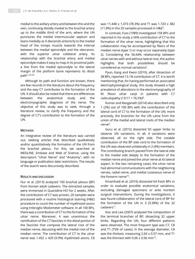
216 L.P. de Macêdo, A.U. Netto, K. Franke et al.
medial to the axillary artery and between this and the
vein, continuing distally medial to the brachial artery
up to the middle third of the arm, where the UN
punctures the medial intermuscular septum and
leans medially as it descends anteriorly to the medial
head of the triceps muscle towards the interval
between the medial epicondyle and the olecranon,
with the superior ulnar collateral artery. Its
relationship with the brachial artery and medial
epicondyle makes it easy to map in its proximal path;
a line from the medial epicondyle to the lateral
margin of the pisiform bone represents its distal
path1,2,3,4,5.
Although its path and function are known, there
are few records in the literature about the frequency
and the way C7 contributes to the formation of the
UN. It should also be noted that there are differences
between the anatomical study and the
electromyographic diagnosis of the nerve. The
objective of this study was to seek, through a
literature review, to clarify the frequency and the
degree of C7's contribution to the formation of the
UN.
METHODS
An integrative review of the literature was carried
out, seeking articles that described qualitatively
and/or quantitatively the formation of the UN from
the brachial plexus. For this, we searched at
MEDLINE, Embase and Web of Science, using the
descriptors "Ulnar Nerve" and "Anatomy", with no
language or publication date restrictions. The results
of the search were discursively described.
RESULTS AND DISCUSSION
Hur et al. (2013) analyzed 100 brachial plexus (BP)
from Korean adult cadavers. The extracted samples
were immersed in Guanidine-HCl for 2 weeks. After
the contribution of C7 was proven, 20 samples were
processed with a routine histological staining (H&E)
procedure to count the number of myelinized axons
using Imbroglio Modometer software. In all 100 BPs,
there was a contribution of C7 to the formation of the
ulnar nerve. Moreover, it was unanimous the
ramification of the C7 fascicles in the distal portion of
the fascicles that compose the lateral root of the
median nerve, decussing with the medial root of the
median nerve. The contribution of C7 to the ulnar
nerve was 1.452 ± 429 (9.9%) myelinized axons, C8
was 11.448 ± 1.473 (78.3%) and T1 was 1.720 ± 382
(11.8%) in the 20 samples processed in H&E6.
In contrast, Fuss (1989) investigated 158 BPs and
reported in his study a 56% contribution of C7 to the
formation of the ulnar nerve, highlighting that this
collaboration may be accompanied by fibers of the
median nerve (type 1) or may occur separately (type
2). Considering the 56:44% relationship between
ulnar nerves with and without lateral root, the author
highlights that both possibilities should be
considered as normal variations7.
Pyun, Kang and Kwon (2010), after dissection of
38 BPs, reported 13.1% contribution of C7. It is worth
mentioning that, for having performed an associated
electrophysiological study, this study showed a high
prevalence of alterations in the electromyography of
M. flexor ulnar carpi in patients with C7
radiculopathy (13/17 = 76.5%)8.
Kumar and Ranganath (2014) also described only
1 (2%) out of 100 BPs with the contribution of the
lateral cord of C7 to the formation of the UN; more
precisely, the branches for the UN came from the
union of the medial and lateral roots of the median
nerve9.
Guru et al. (2015) dissected 50 upper limbs to
observe UN variations. In all, 4 variations were
observed, all on the right side. The notable
contribution of the BP side cord to the formation of
the UN was observed unilaterally in 2 (4%) members.
The contributing branch passed from the lateral side
to the medial side deeply to the formation of the
median nerve and joined the ulnar nerve at its lateral
aspect. In the two remaining cases, the ulnar nerve
had abnormal communications with the neighboring
nerves, radial nerve, and medial cutaneous nerve of
the forearm nerve10.
Emamhadi et al. (2016) dissected 64 fresh BPs in
order to evaluate possible anatomical variations,
excluding damaged specimens or ante mortem
surgical interventions. During the investigations, it
was found collaboration of the lateral cord of BP for
the formation of the UN in 3 (9.38%) of the 32
cadavers11.
Koo and Lee (2007) analyzed the composition of
the terminal branches of BP, dissecting 32 upper
limbs. Regarding the UN, four different patterns
were observed. The most frequent type was C7, C8
and T1 (75% of cases). In the average diameter, C8
was the thickest, measuring 2.64 ± 0.57 mm, and T1
was the thinnest with 0.06 ± 0.56 mm12.

217 C7 contribution to the ulnar nerve
The oldest studies found, performed with even more
rudimentary cadaveric dissection techniques, were
those of Harris (1904)13 and Linell (1921)14, which
showed the presence in, respectively, 86% and 57%
of the brachial plexus of an exchange of fibers
between the medial and lateral plexus strands,
specifically the C7 fibers contributing to the
formation of the ulnar nerve.
Author,
Date
Sample
(n)
Methodol
ogy
Qualitative
Analysis
Quantitative
Analysis
Hur et al.,
2013
100 BPs H&E +
Imbroglio
Modomet
er
100 (100%,
C7)
1,452 ± 429
(9,9%)
axônios
Koo, Lee,
2007
32 BPs Cadaveric
dissection
24 (75%, C7) 1,18 ±0.59
mm
Fuss, 1989 158 BPs Cadaveric
dissection
56% (lateral
cord)
N/A
Pyun et
al., 2010
38 BPs Cadaveric
dissection
13,1%
(lateral cord)
N/A
Kumar,
Ranganat
h, 2014
50 BPs Cadaveric
dissection
1 (2%,lateral
cord)
N/A
Guru et
al., 2015
50 BPs Cadaveric
dissection
2 (4%, lateral
cord)
N/A
Emamhad
i et al.,
2016
64 BPs Cadaveric
dissection
3
(9,38%,latera
l cord)
N/A
Ramchan
dran et
al., 2006
1 BP Case
Report
1 (lateral
root of the
median)
N/A
Linnel,
1921
42 BPs Cadaveric
dissection
57%, C7 N/A
Harris,
1904
60 BPs Cadaveric
dissection
86%, C7 N/A
Figure 1. Summary of the findings of the review on C7
contribution
It is noticeable that the studies that performed
quantitative analysis, especially Hur et al. (2013) and
Koo and Lee (2007), showed a higher prevalence of
C7 contribution than the studies with cadaveric
dissection, except for those of Harris (1904) and
Linnel (1921) 6,12,13,14. This can be justified by an
eventual iatrogenic loss of the lateral contribution
during dissection or by the sophistication of
quantitative methods.
In this context, it is frequent a sub-quantification
of the contribution of C7 to the nerve during
dissection, justifying the absence of root
representation in most of the classic anatomic
literature 1,2,3,4,5. In practice, however, the
repercussions of C7 radiculopathies on
electromyographic variables in part of the UN
territory can be seen, as Pyun et al. (2010) have
shown8.
This knowledge becomes essential in
neurosurgical practice, since unadvised dissection
can cause iatrogenicity. The authors themselves
came across the contribution of the lateral cord to
the formation of the UN during surgical dissection in
the treatment of a brachial plexopathy (Figure 2),
which motivated this review.
Figure 2.
CONCLUSION
The C7 root can be considered a frequent
component of the ulnar nerve, although its
contribution may be underestimated in less
sophisticated dissection studies. There is even a high
incidence of C7 radiculopathy causing repercussions
in the nerve territory. Knowledge of this participation
is indispensable for peripheral nerve surgery,
avoiding iatrogenic lesions and possible negative
outcomes for the patient.
REFERENCES
1. Baehr M, Frotscher M. Duus Topical Diagnosis in

218 L.P. de Macêdo, A.U. Netto, K. Franke et al.
Neurology. 5th ed. Rio de Janeiro, RJ: Guanabara Koogan;
2014.
2. Ellis H. Clinical Anatomy: A Revision and Applied Anatomy
for Clinical Students. 8th ed. London: Blackwell Scientific
Publications; 1992: 209.
3. Moore KL, Dalley AF. Clinically Oriented Anatomy. 5th ed.
Baltimore: Lippincott Williams & Wilkins; 2006: 777.
4. Romanes GJ. Cunningham’s Textbook of Anatomy. 12th
ed. New York, NY: Oxford University Press; 1981: 328-
329.
5. Standring S. Gray’s Anatomy. 39th ed. Edinburgh: Elsevier
Churchill Livingston; 2005: 847-877.
6. Hur MS, Woo JS, Kim HJ, Lee KS. Frequency and Quantity
of the C7 Contribution to the Ulnar Nerve. Korean J Phys
Anthropol. 2013;26(3):101-104.
7. Fuss FK. Die Radix lateralis des Nervus ulnaris [The lateral
root of the ulnar nerve]. Acta Anat (Basel).
1989;134(3):199-205.
8. Pyun SB, Kang S, Kwon HK. Anatomical and electrophy-
siological myotome corresponding to the flexor carpi
ulnar is muscle. J Korean Med Sci. 2010; 25:454-57.
9. Kumar SV, Ranganath P. Morphology of the ulnar nerve
in axilla and arm and its variations. Int J Anat Res.
2014;2(4):677-680.
10. Guru A, Kumar N, Ravindra Shanthakumar S, et al.
Anatomical Study of the Ulnar Nerve Variations at High
Humeral Level and Their Possible Clinical and Diagnostic
Implications [published online July 12, 2015]. Anat Res Int.
doi:10.1155/2015/378063
11. Emamhadi M, Chabok SY, Samini F, et al. Anatomical
Variations of Brachial Plexus in Adult Cadavers; A
Descriptive Study. Arch Bone Jt Surg. 2016;4(3):253-258.
12. Koo JH, Lee KS. Anatomic Variations of the Spinal Origins
of the Main Terminal Branches of the Brachial Plexus.
Korean J Phys Anthropol. 2007;20(1):11-19.
13. Harris W. The True Form of the Brachial Plexus, and its
Motor Distribution. J Anat Physiol. 1904;38(Pt 4):399-422.
14. Linell EA. The Distribution of Nerves in the Upper Limb,
with reference to Variabilities and their Clinical
Significance. J Anat. 1921;55(Pt 2-3):79-112.
15. Ramachandran K, Kanakasabapathy I, Holla SJ. Multiple
variations involving all the terminal branches of the
brachial plexus and the axillary artery – a case report. Eur
J Anat. 2006;10(3):61-66.

Romanian Neurosurgery (2021) XXXV (2): pp. 219-224 DOI: 10.33962/roneuro-2021-036 www.journals.lapub.co.uk/index.php/roneurosurgery
Burr-hole craniostomy versus mini-craniotomy in the treatment of chronic subdural hematomas. Analysis of clinical results
André Tokpa1, Moussa Diallo2, Louis Kéabléon
Derou1, Yves Soress Dongo1, Bernard Fionko1,
Adéréhime Haïdara1 1 Neurosurgery Department of the Teaching Hospital of Bouaké,
CÔTE D’IVOIRE 2 Neurosurgery Department of the Teaching Hospital Grabriel
Touré, Bamako, MALI
ABSTRACT Although cases of spontaneous recovery or under medical treatment have been
reported, the treatment of chronic subdural hematoma is mainly surgical. The
optimal surgical technique for the treatment of chronic subdural hematomas is still
open to debate. The purpose of this study was to compare the clinical outcomes
between burr-hole craniostomy and craniotomy in patients with chronic subdural
hematoma. Materials and methods: we have performed a retrospective study in
patients operated for chronic subdural hematoma in the neurosurgery department
of the teaching hospital of Bouaké between July 1, 2016, and June 30, 2020. We
compared the data of patients operated by a single burr-hole craniostomy (group A)
and those operated by minicraniotomy (group B). Demographic parameters, clinical
signs, complications and neurological findings were analyzed. Fisher’s exact test, Chi-
squared, and student’s t-test were performed. Results: group A included 46 patients
and group B 55 patients. There was no significant difference between the two groups
about age (59.5 years vs 59.8 years p = 0.89), sex (man: 74% vs 78.2%, P = 0.645),
comorbidities, clinical signs on admission and location of the hematoma. There was
also no significant difference between recurrence rates (4.3% vs 3.6% p = 0.55),
postoperative complications (15.21% vs 7.27% p = 0.172) and neurological findings
between the two groups. Conclusion: patient outcomes are similar in the treatment
of chronic subdural hematomas by craniostomy and minicraniotomy.
INTRODUCTION Chronic subdural hematoma (CSDH) is a collection of aged blood that
lies between the dura mater and the arachnoid. In many patients, it
develops slowly after a mild head trauma often overlooked by them [9],
but the number of patients without a history of trauma is increasing,
probably due to anticoagulant or antiaggregant treatment [8]. Although
Keywords burr hole,
chronic subdural hematoma, craniotomy
Corresponding author: André Tokpa
Neurosurgery Department of the
Teaching Hospital of Bouaké, Côte d’Ivoire
Scan to access the online version

220 André Tokpa, Moussa Diallo, Louis Kéabléon Derou et al.
cases of spontaneous recovery [1,10] and recovery
with medical treatment (corticosteroid or
atorvastatin-based) [6] have been reported, the
treatment of CSDH is mainly surgical. Surgery for
chronic subdural hematoma itself is based on
several techniques, including evacuation through a
small bone flap associated or not with a drainage
system or through one or more burr-holes also
associated or not with a system of continuous
drainage or percutaneous twist drill craniostomy.
The optimal surgical procedure is still subject to
debate.
The purpose of this study was to compare the
clinical outcome of patients who had chronic
subdural hematoma evacuation and drainage
through a single burr hole craniostomy and those
who had chronic subdural hematoma evacuation
and drainage through a minicraniotomy.
MATERIALS AND METHOD
We carried out a retrospective study on 101 patients
who were hospitalized in the neurosurgery
department of the teaching hospital of Bouaké
between July 1, 2016 and June 30, 2020 for the
management of a chronic subdural hematoma. Due
to the retrospective nature of this study, no consent
was given and all records have been anonymised.
The diagnosis of chronic subdural hematoma was
made by CT scan without injection of contrast
medium. We compared the data of the patients who
had been operated by a burr hole craniostomy
(group A) and those of those who had been operated
by a minicraniotomy (group B). Patient data was
collected from records available in the neurosurgery
department. The following parameters were
analyzed: age, sex, history, clinical and paraclinical
signs, the etiology retained, the surgical technique
used, the complications and the clinical course.
During the postoperative period, regression of signs
and symptoms of CSDH was expected as clinical
improvement. Data were entered and statistically
analyzed using IBM SPSS 22.0. Fisher’s exact test, chi-
squared and Student’s t-test were performed to
compare the 2 groups. All the tests were bilateral.
The P-value (P) <0.05 was considered statistically
significant.
SURGICAL TECHNIQUE
In group A a single burr-hole was made while in
group B it was a minicraniotomy. The technique used
was the choice of the surgeon. All patients
(regardless of the technique) were operated under
general anesthesia with endotracheal intubation. A
skin incision of 4 to 5 centimeter was made across
the maximum thickness of CSDH. A burr hole
craniostomy of 1 centimeter diameter was
performed in group A, followed by a cruciform
opening of the dura mater and outer membrane of
CSDH after thermo coagulation. In group B, instead
of the burr hole craniostomy, a bone flap of 3
centimeter diameter was cut with the trephine.
Subsequently, like group A, the dura mater and the
outer membrane of the hematoma were incised
crosswise after their thermo coagulation. whatever
the group, rinsing of the subdural space was
performed with isotonic saline (0.9%) until the rinsing
liquid returned clear followed by drainage of the
subdural space with a closed system. by siphoning
(depending on gravity without continuous suction).
In group B, the dura is sutured around the drain
followed by placement of the bone flap. In group A,
the same closed siphoning subdural drainage
system is put in place but the dura is left open. In
bilateral subdural hematoma, surgery was done
bilaterally. Regardless of the technique used, the
inner membrane of the hematoma as well as the
arachnoid were not open. The rest of the care is
identical in both groups. It consisted of suturing the
surgical wound followed by the dressing. Patients
were strictly bedridden for 48 hours and, the
drainage bag is placed on the patient's bed. The
drains were removed in all the patients after 48
hours.
RESULTS
During our study period 102 patients were treated
for a chronic subdural hematoma, including 1 patient
treated medically (corticosteroid therapy) and 101
others treated surgically, i.e. 99%.
A total of 101 patients were included in this study,
46 in group A (patients who had burr hole
craniostomy) and 55 in group B (patients who had
mini-craniotomy). Our study population had a mean
age of 59.7 +/- 13.35 years with extremes of 24 and
100 years. Patients under the age of 65 years
accounted for 69.3% and those aged 65 years and
over represented 30.7%. There were 24 women
(23.8%) and 77 men (76.2%).
The demographic, clinical and paraclinical
characteristics of patients before their surgical

221 Burr-hole craniostomy versus mini-craniotomy in the treatment of chronic subdural hematomas
treatment are summarized in Table 1.
The clinical evolutionary aspects of the patients
are presented in Table 2.
Clinical examination at 3 months and 1 year
postoperatively showed full recovery in the 97
patients who survived.
Table 1. Characteristics of patients before their surgical treatment.
variables
Group A(burr hole
craniostomy), N =
46 (45.5%)
Group B (mini-
craniotomy),
N = 55 (54.5%)
Total (N = 101) test P value
Age (years)
59.5 +/-14.56 (min
= 24 and maxi =
100)
59.8 +/-12.396 (min =
26 and maxi = 84)
59.7 +/- 13.35
(min 24 and
maxi = 100)
t-student = 0.139 P = 0.89
gender X2 = 0.252 P = 0.645
male 34 (74) 43 (78.2) 77(76.2)
Female 12 (26) 12 (21.8) 24(23.8)
Aetiology Fisher = 2.134 P = 0.372
Head trauma 31 (67.4) 40 (72.7) 71(70.3)
Hemostatic disorder 0 2 (3.63) 2(1.98)
No cause identified 15 (32.6) 13(23.6) 28(27.72)
delay (trauma and
diagnosis (days) 31 30.7
Symptoms
headache 35(76%) 42(76.3%) 77(76.2%)
vomiting 9(19.5%) 11(20%) 20(19.8%)
behavior disorder 7(15.2%) 8(14.5%) 15(14.8%)
Epileptic seizure 1(2.1%) 1(1.8%) 2(1.9%)
mean Glasgow score
on admission
13.22 +/-2.511
min = 6 et max =
15
13.4 +/- 2.257
min = 7 et max = 15
13.32+/-2.366
Min = 6
Max = 15
Mode = 15
t-student = 0.385 P = 0.701
Motor deficit on
admission 34 (74%) 46 (83%) 80(79.2%) X2=1.438 P = 0.23
Co-morbidities Fisher = 12.51 0.27
Chronic arterial
hypertension 6(13%) 8(14.5%) 14(13.8%)
Chronic alcoholism 8(17.4%) 5(9%) 13(12.8%)
Parkinson disease 0 1(1.8%) 1(0.9%)
Hepatocellular
insufficiency 0 1(1.8%) 1(0.9%)
HIV Infection 0 2(3.6%) 2(1.9%)
tuberculous
Meningitis 1(2.1%) 0 1(0.9%)
Heart disease under
diuretics 0 1(1.8%) 1(0.9%)
Stroke and chronic
hypertension 1(2.1%) 1(1.8%) 2(1.9%)
blindness 0 2(3.6%) 2(1.9%)
prostatic
Adenocarcinoma 1(2.1%) 0 1(0.9%)
No comorbidity 27(58.7%) 34(61.8%) 61(60.4%)
location Fisher = 1.787 P = 0.455
Right 16 (15.8%) 22 (21.8%) 38(37.6%)
left 24(23.8%) 30(29.7%) 54(53.4%)
bilateral 6 (5.9%) 3 (3%) 9(8.9%)
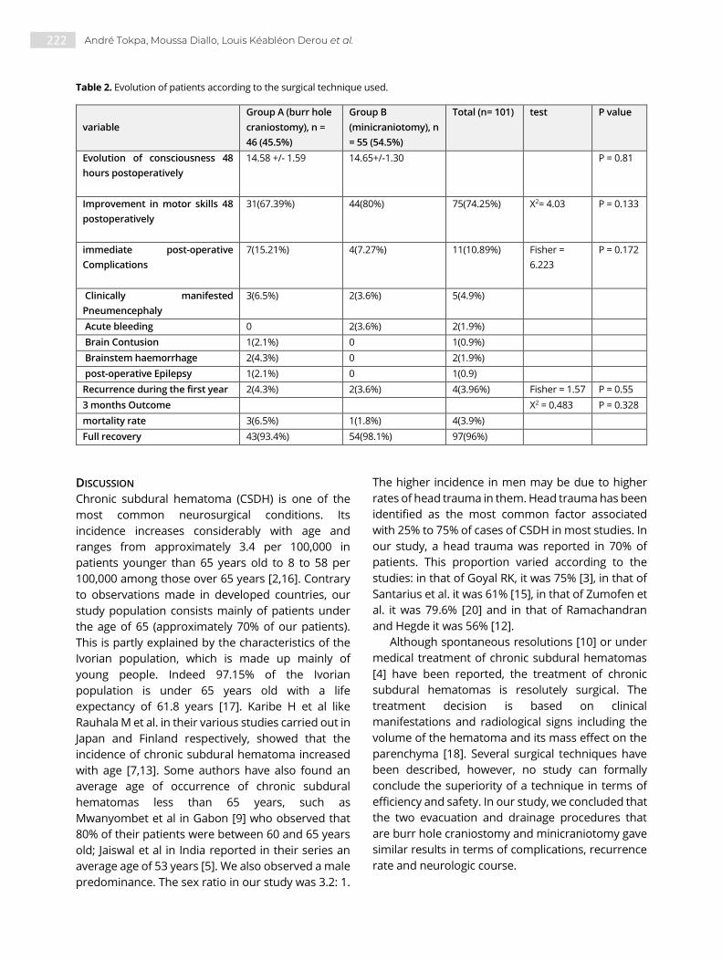
222 André Tokpa, Moussa Diallo, Louis Kéabléon Derou et al.
Table 2. Evolution of patients according to the surgical technique used.
variable
Group A (burr hole
craniostomy), n =
46 (45.5%)
Group B
(minicraniotomy), n
= 55 (54.5%)
Total (n= 101) test P value
Evolution of consciousness 48
hours postoperatively
14.58 +/- 1.59 14.65+/-1.30 P = 0.81
Improvement in motor skills 48
postoperatively
31(67.39%) 44(80%) 75(74.25%) X2= 4.03 P = 0.133
immediate post-operative
Complications
7(15.21%) 4(7.27%) 11(10.89%) Fisher =
6.223
P = 0.172
Clinically manifested
Pneumencephaly
3(6.5%) 2(3.6%) 5(4.9%)
Acute bleeding 0 2(3.6%) 2(1.9%)
Brain Contusion 1(2.1%) 0 1(0.9%)
Brainstem haemorrhage 2(4.3%) 0 2(1.9%)
post-operative Epilepsy 1(2.1%) 0 1(0.9)
Recurrence during the first year 2(4.3%) 2(3.6%) 4(3.96%) Fisher = 1.57 P = 0.55
3 months Outcome X2 = 0.483 P = 0.328
mortality rate 3(6.5%) 1(1.8%) 4(3.9%)
Full recovery 43(93.4%) 54(98.1%) 97(96%)
DISCUSSION
Chronic subdural hematoma (CSDH) is one of the
most common neurosurgical conditions. Its
incidence increases considerably with age and
ranges from approximately 3.4 per 100,000 in
patients younger than 65 years old to 8 to 58 per
100,000 among those over 65 years [2,16]. Contrary
to observations made in developed countries, our
study population consists mainly of patients under
the age of 65 (approximately 70% of our patients).
This is partly explained by the characteristics of the
Ivorian population, which is made up mainly of
young people. Indeed 97.15% of the Ivorian
population is under 65 years old with a life
expectancy of 61.8 years [17]. Karibe H et al like
Rauhala M et al. in their various studies carried out in
Japan and Finland respectively, showed that the
incidence of chronic subdural hematoma increased
with age [7,13]. Some authors have also found an
average age of occurrence of chronic subdural
hematomas less than 65 years, such as
Mwanyombet et al in Gabon [9] who observed that
80% of their patients were between 60 and 65 years
old; Jaiswal et al in India reported in their series an
average age of 53 years [5]. We also observed a male
predominance. The sex ratio in our study was 3.2: 1.
The higher incidence in men may be due to higher
rates of head trauma in them. Head trauma has been
identified as the most common factor associated
with 25% to 75% of cases of CSDH in most studies. In
our study, a head trauma was reported in 70% of
patients. This proportion varied according to the
studies: in that of Goyal RK, it was 75% [3], in that of
Santarius et al. it was 61% [15], in that of Zumofen et
al. it was 79.6% [20] and in that of Ramachandran
and Hegde it was 56% [12].
Although spontaneous resolutions [10] or under
medical treatment of chronic subdural hematomas
[4] have been reported, the treatment of chronic
subdural hematomas is resolutely surgical. The
treatment decision is based on clinical
manifestations and radiological signs including the
volume of the hematoma and its mass effect on the
parenchyma [18]. Several surgical techniques have
been described, however, no study can formally
conclude the superiority of a technique in terms of
efficiency and safety. In our study, we concluded that
the two evacuation and drainage procedures that
are burr hole craniostomy and minicraniotomy gave
similar results in terms of complications, recurrence
rate and neurologic course.
223 Burr-hole craniostomy versus mini-craniotomy in the treatment of chronic subdural hematomas
No statistically significant difference was observed in
the clinical course of patients either in the immediate
postoperative period or in the medium and long
term. In fact, there was no statistically significant
difference in the evolution of the consciousness and
motor deficit in the two groups. The occurrence of
postoperative complications were also statistically
superimposable in the two groups as were the
recurrence rates. Therefore, the two techniques had
identical efficiencies. Regan M. et al, after a
comparative study of these 2 surgical techniques,
concluded that evacuation by craniotomy was
associated with a higher re-operative rate (24.1%)
than evacuation by burr hole craniostomy with P =
0.0156 [14]. They argued that the re-operation was
mainly due to a residual chronic subdural hematoma
or secondary to one or more acute haemorrhages.
Their result was related to the lack of drainage in
their patients who had had craniotomies. Weigal
and. Al. Had shown in a study that the use of a closed
drainage system reduced the risk of recurrence of
hematoma [19] and therefore of the need for re-
operation. Santarius et al. in a randomized study
comparing two groups of patients treated for CSDH
by burr holes craniostomy, one group having had
drainage and the other not. Those with drain were
found to have a lower reoperation rate due to a
decrease in hematoma recurrence [15]. However,
Raghavan A et al. for their part found that burr hole
craniostomy was associated with a higher re-
operative rate [11]. The recurrence rate was
relatively low in our series compared to that reported
in the literature, especially the series from developed
countries. The low rate of recurrence in our series
could be explained by the characteristics of our
population which was essentially made up of young
adults with very little cerebral atrophy on the one
hand and on the other hand by a low rate of
comorbidity requiring the taking antiaggregant or
anticoagulant. Indeed, it is accepted that cerebral
atrophy and increased subdural space observed in
elderly patients make them vulnerable to the
development of chronic subdural hematomas [5].
Also according to Regan M. et al. in their series, there
was a higher incidence of postoperative
complications in the craniotomy group than in the
burr hole craniostomy group. Moreover, it clearly
appears that the duration of the procedures was
significantly longer in craniotomies than burr holes
craniostomies [14]. We were unable to compare the
duration of different surgeries in our study due to
unavailability of data.
CONCLUSION
Evacuation and drainage of chronic subdural
hematoma by burr hole craniostomy or mini-
craniotomy are comparable in terms of clinical
outcome. However, other factors not analyzed in our
study, such as surgical procedure duration and the
type of anesthesia, could help surgeons to choose
the appropriate surgical technique on a case-by-case
basis. Future prospective, randomized, multicenter
studies taking into account several factors that may
influence surgery for chronic subdural hematoma
and the vital and functional prognosis of patients are
needed.
REFERENCES
1. Baehr M, Frotscher M. Duus Topical Diagnosis in
Neurology. 5th ed. Rio de Janeiro, RJ: Guanabara Koogan;
2014.
2. Ellis H. Clinical Anatomy: A Revision and Applied Anatomy
for Clinical Students. 8th ed. London: Blackwell Scientific
Publications; 1992: 209.
3. Moore KL, Dalley AF. Clinically Oriented Anatomy. 5th ed.
Baltimore: Lippincott Williams & Wilkins; 2006: 777.
4. Romanes GJ. Cunningham’s Textbook of Anatomy. 12th
ed. New York, NY: Oxford University Press; 1981: 328-
329.
5. Standring S. Gray’s Anatomy. 39th ed. Edinburgh: Elsevier
Churchill Livingston; 2005: 847-877.
6. Hur MS, Woo JS, Kim HJ, Lee KS. Frequency and Quantity
of the C7 Contribution to the Ulnar Nerve. Korean J Phys
Anthropol. 2013;26(3):101-104.
7. Fuss FK. Die Radix lateralis des Nervus ulnaris [The lateral
root of the ulnar nerve]. Acta Anat (Basel).
1989;134(3):199-205.
8. Pyun SB, Kang S, Kwon HK. Anatomical and electrophy-
siological myotome corresponding to the flexor carpi
ulnar is muscle. J Korean Med Sci. 2010; 25:454-57.
9. Kumar SV, Ranganath P. Morphology of the ulnar nerve
in axilla and arm and its variations. Int J Anat Res.
2014;2(4):677-680.
10. Guru A, Kumar N, Ravindra Shanthakumar S, et al.
Anatomical Study of the Ulnar Nerve Variations at High
Humeral Level and Their Possible Clinical and Diagnostic
Implications [published online July 12, 2015]. Anat Res Int.
doi:10.1155/2015/378063
11. Emamhadi M, Chabok SY, Samini F, et al. Anatomical
Variations of Brachial Plexus in Adult Cadavers; A
Descriptive Study. Arch Bone Jt Surg. 2016;4(3):253-258.
12. Koo JH, Lee KS. Anatomic Variations of the Spinal Origins
of the Main Terminal Branches of the Brachial Plexus.
Korean J Phys Anthropol. 2007;20(1):11-19.

224 André Tokpa, Moussa Diallo, Louis Kéabléon Derou et al.
13. Harris W. The True Form of the Brachial Plexus, and its
Motor Distribution. J Anat Physiol. 1904;38(Pt 4):399-422.
14. Linell EA. The Distribution of Nerves in the Upper Limb,
with reference to Variabilities and their Clinical
Significance. J Anat. 1921;55(Pt 2-3):79-112.
15. Ramachandran K, Kanakasabapathy I, Holla SJ. Multiple
variations involving all the terminal branches of the
brachial plexus and the axillary artery – a case report. Eur
J Anat. 2006;10(3):61-66.

Romanian Neurosurgery (2021) XXXV (2): pp. 225-227 DOI: 10.33962/roneuro-2021-037 www.journals.lapub.co.uk/index.php/roneurosurgery
Shunt in scrotum: unusual shunt complication in an operated case of post TBM hydrocephalus
Manoj Agarwal, Sharad Pandey, Pankaj Kumar, L.N. Gupta1
Department of Neurosurgery, A.B.V.I.M.S. and Dr Ram Manohar
Lohia Hospital, New Delhi, INDIA
ABSTRACT The role of shunt placement is to divert cerebrospinal fluid (CSF) from within the
ventricles to an alternative location most commonly peritoneal space. Ventriculo
Peritoneal Shunt (VPS) is associated with many complications viz over drainage, valve
failure, breaking of catheter, catheter obstruction, coiling of catheter, spontaneous
knot formation, infection and migration of distal catheter and all of them finally
leading to obstructive hydrocephalus. One such complication is distal catheter
migration to a rare but possible site i.e. scrotum. We reported a case of scrotal
migration of shunt as a late complication of VPS insertion.
BACKGROUND
The role of shunt placement is to divert cerebrospinal fluid (CSF) from
within the ventricles to an alternative location (most commonly
peritoneal space, rarely to atria, pleural space) and from lumbar space
to peritoneal cavity.
Ventriculo Peritoneal Shunt (VPS) is associated with many
complications namely over drainage, valve failure, breaking of catheter,
catheter obstruction, coiling of catheter, spontaneous knot formation,
infection and migration of distal catheter and all of them finally leading
to obstructive hydrocephalus [1,2]. One of the rare sites of distal
catheter migration is scrotum. Scrotal migration of distal shunt catheter
is more common in children due to patent processus vaginalis [3]. We
reported a case of scrotal migration of shunt as a late complication of
VPS insertion.
CASE PRESENTATION
An eighteen months old male child, a follow up case of tubercular
meningitis (TBM) with hydrocephalus was operated by right VPMP
shunt via right Keens point in January 2020 at our tertiary care centre.
Post operatively patient's GCS improved and patient was discharged.
After approximately 03 months patient presented with gradual
increase in size of bilateral scrotum in the past 2 months, intermittent
Keywords Ventriculo Peritoneal Shunt
(VPS), complications,
distal catheter migration, scrotum
Corresponding author: Sharad Pandey
Department of Neurosurgery,
A.B.V.I.M.S. and Dr Ram Manohar Lohia Hospital, New Delhi, India
Scan to access the online version

226 Manoj Agarwal, Sharad Pandey, Pankaj Kumar et al.
fever and on & off vomiting and was admitted (Fig.1).
On local examination cough impulse was present in
the right inguinal region & a tube like structure was
palpable in the right hemiscrotum, external genitalia
was normal. Neurological examination was normal
with no focal neurological deficit, GCS was E4V5M6,
head circumference was normal for age, no signs of
shunt obstruction were present.
INVESTIGATIONS
X-ray of neck, chest, abdomen & pelvis were
conducted which showed an intact VP shunt with
extension of the peritoneal end into the right hemi
scrotum. USG of inguinoscrotal region was also
suggestive of same.
TREATMENT
Surgical intervention was planned in this rare case.
Re-exploration of the abdominal wound was done,
the peritoneal end of the shunt was identified and it
was pulled and repositioned in peritoneal cavity
under C-ARM guidance and the abdominal wound
was closed in layers. Post-operative X-ray of chest,
abdomen & pelvis showed normal position of
peritoneal end of VP shunt (Fig.2).
OUTCOME AND FOLLOW UP
The child was kept under observation and
discharged after 48 hours since the postoperative
period was uneventful and was advised for follow up
in general Surgery OPD and Neurosurgery OPD.
DISCUSSION
The most common surgery performed worldwide for
hydrocephalus is VP shunt [3]. Shunting is performed
to divert the CSF from dilated ventricles to peritoneal
cavity from where CSF is absorbed. Apart from the
morbidity associated with hydrocephalus, shunt
surgeries have their own complications which are
well known [2-6].The reported incidence of shunt-
related abdominal complications is 10-30%[7]
Various authors have reported migration of the distal
end of peritoneal catheter into the scrotum through
a patent processus vaginalis[8-11]. The processus
vaginalis may remain patent until 1 year of life in 50-
60% cases and until 2 years of life in 40-50% of cases
[3]. Rowe et al[12] have explained the aetiology of
peritoneal catheter migration, which has been
universally accepted over the years by various
authors[13]. Peritoneal cavity distension due to
draining CSF also prevents the obliteration of
processus vaginalis which had also been reported by
Ho et al [14] and Ozveren et al[15]. In the present
case, a male child had a patent processus vaginalis
which was either congenital or because of peritoneal
CSF drainage. In our case, right-sided VP shunt was
performed and it would be difficult to comment and
is another research study topic whether the side has
something to do with the shunt migration as there is
no such report in the literature comparing the right
and left patent processus vaginalis. We used
Chabbra medium pressure VP shunt in our case.
Shunt migration can occur either as an early or a late
complication. Ozveren et al[15] have reported shunt
migration within 24 h, while the average length of
time reported in other series was 6.8 months. In our
case, the time interval was approximately 3 months.
Thus this kind of shunt complication needs to be
reported and should be kept into mind in dealing
with cases who had undergone shunt insertion
surgery.
Scrotal VP shunt migration has been reported in
26 children, and the time of presentation varies from
1 month to 2.5 years (mean 3.8 months, median 1
month) after shunting, with 21 out of 26 migrations
occurring within 6 months, 88% involving the right
side [16].
Ramareddy RS describe the spontaneous
resolutionof hydrocele and retreat of migrated
peritoneal portion of VP shunt, and also discuss the
pathogenesis and management [16].
REFERENCES
1. Borkar SA, Satyarthee GD, Khan RN, et al. Spontaneous
extrusion of migrated ventriculoperitoneal shunt
catheter through chest wall: a case report. Turk
Neurosurg 2008;18:95–8.
2. Agha PP, Amendola MA, Shirazi KK, et al. Abdominal
complications of ventriculoperitoneal shunts with
emphasis on the role of imaging methods. Surg Gynecol
Obstet 1983;156:473–7.
3. Grosfeld IL, Cooney DR. Inguinal hernia after
ventriculoperitoneal shunt for hydrocephalus. J Pediatr
Surg 1974;9:311–15.
4. Patel CD, Matloub H. Vaginal perforation as a
complication of ventriculoperitoneal shunt. J Neurosurg
1973;38:761–2.
5. Redman JF, Seibert JJ. Abdominal and genitourinary
complications following ventriculoperitoneal shunts. J
Urol 1977;119:295–7.

227 Shunt in scrotum: unusual complication
6. Wakai S. Extrusion of a peritoneal catheter through the
abdominal wall in an infant. Case report. J Neurosurg
1982;57:148–9.
7. Bryant MS, Bremer AM. Abdominal complications of
ventriculoperitoneal shunts: case reports and review of
literature. Am Surg 1988;54:50–5.
8. Bristow DL, Buntain WL, James HL. Ventriculoperitoneal
(VP) shunt migration causing an acute scrotum: a case
report of Doppler evaluation. J Pediatr Surg 1978;13:538–
9.
9. Fuwa I, Matsukado Y, Itoyama Y, et al. Migration of a
dissected peritoneal shunt catheter into the scrotum.
Brain Dev 1984;6:336–8.
10. Levy SH, Cooper P, Shiffman D. Simulated testicular
torsion in a neonate: complication of
ventriculoperitoneal shunt. Urology 1977;9:174–6.
11. Ramani PS. Extrusion of abdominal catheter of
ventriculoperitoneal shunt into the scrotum. J Neurosurg
1974;40:772–3.
12. Rowe MI, Copelson LW, Clatworthy HW. The patent
processus vaginalis and inguinal hernia. J Pediatr Surg
1969;4:102–7.
13. Kita D, Hayashi Y, Kinoshita M, et al. Scrotal migration of
the peritoneal catheter of a ventriculoperitoneal shunt in
a 5-year-old male: case report. Neurol Med Chir (Tokyo)
2010;50:1122–5.
14. Ho CC, Jamaludin WJ, Goh EH, et al. Scrotal mass: a rare
complication of ventriculoperitoneal shunt. Acta Medica
(Hradec Kralove) 2011;54:81–2.
15. Ozveren MF, Kazez A, Cetin H, et al. Migration of the
abdominal catheter of a ventriculoperitoneal shunt into
the scrotum—case report. Neurol Med Chir (Tokyo)
1999;39:313–15.
16. Ramareddy RS , Alladi A . Scrotal migration of
ventriculoperitoneal catheter and hydrocele resolving
spontaneously . Indian J Neurosurg 2012;1:172-4.

Romanian Neurosurgery (2021) XXXV (2): pp. 228-233 DOI: 10.33962/roneuro-2021-038 www.journals.lapub.co.uk/index.php/roneurosurgery
Spectrum of non-traumatic craniovertebral junction disorders. Diagnosis and demonstration with magnetic resonance imaging and multidetector computed tomography
Neha Singh1, Deepak Kumar Singh2 1 Department of Radiodiagnosis and Imaging, Dr Ram Manohar
Lohia Institute of Medical Sciences, Lucknow, INDIA 2 Department of Neurosurgery, Dr Ram Manohar Lohia Institute of
Medical Sciences, Lucknow, INDIA
ABSTRACT Introduction: Craniovertebral junction is the “zone of transition” between the skull
and cervical spine so its detailed discussion is lacking in so many standard textbooks
addressing these regions. These anomalies are especially common in the Indian
subcontinent. Accurate diagnosis, probable aetiology and pre-treatment evaluation
considerably affect the prognosis of patients. The aim of this study was to classify
various Craniovertebral junction disorders according to their aetiology and to
correlate the imaging findings with the clinical profile of the patients and
histopathology whenever possible.
Methods: This prospective observational study was done in our department over the
period of one year from August 2016 to July 2017. 57 North Indian patients from all
age groups and both sex, who had imaging features suggesting craniovertebral
junction pathology were selected for the study.
Results: Our study group comprised 34 males and 23 females with a male to female
ratio of 1.47: 1. The most common age group was 11-20 years and the most common
aetiology being developmental followed by infective, autoimmune and inflammatory
disorders.
Conclusions: CVJ abnormalities are a group of treatable neurological disorders, and
are approached with much caution by clinicians. Thus, it is crucial that radiologists
should be able to make a precise diagnosis, categorize them into etiological groups,
and give precise anatomical information on MDCT and/or MRI, as this information
ultimately helps determine the management of such abnormalities and their
prognosis.
INTRODUCTION
The term craniovertebral junction (CVJ) refers to a cone shaped area,
the upper limit of which is a line joining the internal occipital
protuberance to the spheno- occipital synchondrosis& the lower limit
is inferior margin of the body of axis.[15] It is a transition site between
Keywords CVJ anomalies,
MDCT, atlanto-axial dislocation,
basilar invagination
Corresponding author: Neha Singh
Department of Radiodiagnosis and
Imaging, Dr Ram Manohar Lohia Institute of Medical Sciences,
Lucknow, India
Scan to access the online version

229 Spectrum of non-traumatic craniovertebral junction disorders
mobile cranium and relatively rigid spinal column.
Disorders of the CVJ present a dilemmatic problem
due to presence of neurologically vital structures in
the vicinity, potentially unstable articulation and
grave prognosis, if gone untreated and undetected.
Our study is an attempt to systematically classify
different CVJ abnormalities according to their
etiopathogenesis and to describe the importance of
accurate diagnosis and precise anatomical
information for pretreatment evaluation with
Multidetector computed tomography (MDCT) and/or
magnetic resonance imaging (MRI).
The main objectives of our study were:
1. To evaluate the patients with non
traumaticcraniovertebral junction disorders by
multiplanar and dynamic imaging utilizing MRI
and/ or Multidetector CT.
2. To classify various craniovertebral junction
disorders according to their etiology.
3. To correlate the imaging findings with clinical
profile of the patients.
4. To correlate radiological diagnosis with diagnosis
after medication /surgery / biopsy whenever
undertaken.
METHODS
This prospective study was done in Department of
Radiodiagnosis & Imaging, Ram Manohar Lohia
Institute of Medical sciences, Lucknow, over the
period of one year from August 2018 to July 2019.The
Permission from Ethics Committee of our institute
was taken. Written informed consent for the study
was obtained from each patient prior to the
examination.
North Indian Patients from all age groups and
both sex, who underwent MRI and / or CT for clinical
suspicion of non-traumaticCranio-vertebral junction
disorder were evaluated. Out of these, 57 patients
who had imaging features suggesting cranio-
vertebral junction pathology were selected for the
study. Contrast enhanced imaging was restricted
only to those with suspicion of infective,
inflammatory or neoplastic etiology. Dynamic
scanning in neutral, active flexion and extension
posture was done in all cases except in those with
atlanto-axial instability. Only exclusion criterion was
presence of at least one absolute contraindication
for MRI.
MR imaging was performed with a 1.5 T whole
body MR imaging system (SIGNA EXCITE GEMSOW)
and the following sequences were obtained:
1. Sagittal -T1-weighted spin echo (T1W SE), T2-
weighted turbo spin echo (T2W TSE) and T2-
weighted fat suppressed.
2. Axial- T1-weighted spin echo (T1W SE) & T2-
weighted turbo spin echo (T2W TSE).
3. Coronal - T2-weighted turbo spin echo (T2W TSE).
4. T2W TSE in flexion and extension whenever
required.
5. Contrast enhanced T1W fat saturated (FS) images
in axial, coronal and neutral sagittal planes.
Scanning parameters used for T1W SE sequence
were a repetition time (TR) of 600 ms and an echo
time (TE) of 9.7 ms; for T2W TSE sequence TR of 4,000
ms and TE of 100 ms; for T2W fat suppressed
sequence TR of 3,300 ms, TE of 110 ms. These
sequences were done with slice thickness of 4 mm
and spacing of 0.5 mm.
CT examination was done on a Multislice spiral CT
scanner (BRILLIANCE CT, Phillips medical system,
Nederland, B.V.5684 PC Best). After acquisition of
thin axial images, multiplanar reconstruction into
sagittal and coronal planes was obtained.
Craniometric measurements used for radiologic
assessment of CVJ abnormalities include
Chamberlain's line, McGregor line, McRae line,
Wackenheimclivus line, Fishgold digastric line,
Fishgoldbimastoid line, Welcher basal angle, and
atlantooccipital joint axis angle.
Detailed history of the patients was taken with
respect to age, sex, socio-economic status, clinical
profile & correlated with the spectrum of cranio-
vertebral junction finding on MRI and Multislice CT.
These patients were followed up to look for clinical
outcome after conservative or surgical treatment.
Statistical Methods: Statistical analysis was done
using ratio and percentages for age and gender
distribution. Percentage distribution was calculated
for each disease affecting the CVJ. Pearson’s chi
square test was used to establish the association
between two different anomalies.
RESULTS
Our study group comprised of 34 males and 23
females with a male to female ratio of 1.47: 1.
Maximum number of patients (21) was in the age
group between 11-20 years, which included 8
females (34.78% of all females) and 13 males (38.24%
of all males). This was followed by 21-30years age

230 Neha Singh, Deepak Kumar Singh
group (12 patients) comprising of 3 females (13.04%
of all females) and 9 males (26.47% of all males).
Figure 1. a) Sagittal T2W MRI shows Platybasia with flattened
clivus, blocked C2- C3 complex with atlanto-occipital
assimilation, AAD and BI causing compression over cervico-
medullary junction b) Sagittal T2W MR shows Osodontoideum
with AAD causing compressive myelopathy. c) Sagittal NCCT
shows shallow posterior fossa with hyoplasticclivus related to
basioccipital hypoplasia. A bony arch is noted at the tip of clivus
which is continuous laterally with occipital condyles consistent
with prebasioccipital arch. d, e, f) Sagittal dynamic NCCT shows
mobile type atlantoaxial dislocation with ossiculumterminale
causing cord compression.
Developmental cause of atlanto-axial diseases most
commonly presented in 11-20 year age group (18
patients-40.91%), followed by 21-30 year age group
(12 patients-27.27%). None of the patient of 51-60
years presented with a developmental cause, while
one of these (2.27%) presented in 61-70 years age
group. Patients with acquired causes of atlanto-axial
diseases most commonly presented in 41-50 years
age group (4 patients-30.77%) followed by 3 patients
each (23.08%) in 11-20 and 51-60 years age group
and 2 patients (15.38%) in 31-40 year age group
[Table 1].
Figure 2. a) Sagittal T2W MRI shows small posterior fossa with
hypoplasticclivus and cerebellar tonsillar herniation causing
crowding at foramen magnum with compression over
proximal cervical cord causing syringomyelia in cervico-dorsal
cord. b) Axial non contrast bone window image shows anterior
and posterior rachischisis with atlanto-axial dislocation and
hypoplastic posterior arch of atlas.
Figure 3. a) Sagittal T2W MR Image shows inflammatory
granuloma in cervical cord with marked edema involving cord
and brainstem. T1W Fat sat contrast enhanced b) sagittal
image showing contrast enhancement in atlas and axis
vertebra with erosion of dens. Enhancing granulation tissue is
noted adjacent to atlanto-axial joint with a large abscess in
paraspinal soft tissue. c) Sagittal image showing erosion of
dens with mild heterogenous contrast enhancement in
anterior atlas arch and axis vertebra. Enhancing soft tissue s/o
pannus is noted adjacent to atlanto-axial joint with increased
atlantoaxial interval. d)axial image shows a well defineddural
based lesion with strong homogenous contrast enhancement
at C1- C2 level on left side causing right lateral displacement
and compression of cord. Radiological diagnosis of
meningioma was made.
Most common presenting symptom was neck pain
seen in 33 patients (57.89%), followed by weakness
of both upper and lower limb in 24 patients (42.11%)
and neck stiffness in 23 patients (40.35%). These
were followed by fever, hemiparesis, sensory
symptoms, paraparesis, headache and upper limb
weakness. However, weakness of limbs (motor
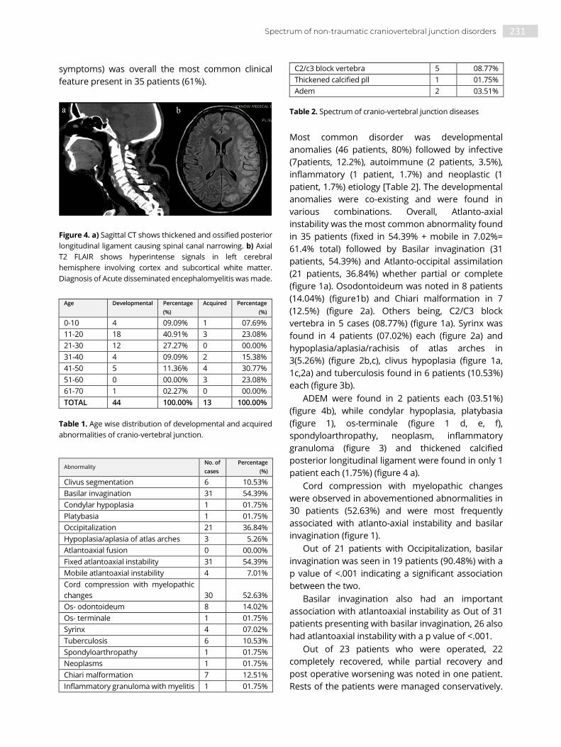
231 Spectrum of non-traumatic craniovertebral junction disorders
symptoms) was overall the most common clinical
feature present in 35 patients (61%).
Figure 4. a) Sagittal CT shows thickened and ossified posterior
longitudinal ligament causing spinal canal narrowing. b) Axial
T2 FLAIR shows hyperintense signals in left cerebral
hemisphere involving cortex and subcortical white matter.
Diagnosis of Acute disseminated encephalomyelitis was made.
Age Developmental Percentage
(%)
Acquired Percentage
(%)
0-10 4 09.09% 1 07.69%
11-20 18 40.91% 3 23.08%
21-30 12 27.27% 0 00.00%
31-40 4 09.09% 2 15.38%
41-50 5 11.36% 4 30.77%
51-60 0 00.00% 3 23.08%
61-70 1 02.27% 0 00.00%
TOTAL 44 100.00% 13 100.00%
Table 1. Age wise distribution of developmental and acquired
abnormalities of cranio-vertebral junction.
Abnormality No. of
cases
Percentage
(%)
Clivus segmentation 6 10.53%
Basilar invagination 31 54.39%
Condylar hypoplasia 1 01.75%
Platybasia 1 01.75%
Occipitalization 21 36.84%
Hypoplasia/aplasia of atlas arches 3 5.26%
Atlantoaxial fusion 0 00.00%
Fixed atlantoaxial instability 31 54.39%
Mobile atlantoaxial instability 4 7.01%
Cord compression with myelopathic
changes 30 52.63%
Os- odontoideum 8 14.02%
Os- terminale 1 01.75%
Syrinx 4 07.02%
Tuberculosis 6 10.53%
Spondyloarthropathy 1 01.75%
Neoplasms 1 01.75%
Chiari malformation 7 12.51%
Inflammatory granuloma with myelitis 1 01.75%
C2/c3 block vertebra 5 08.77%
Thickened calcified pll 1 01.75%
Adem 2 03.51%
Table 2. Spectrum of cranio-vertebral junction diseases
Most common disorder was developmental
anomalies (46 patients, 80%) followed by infective
(7patients, 12.2%), autoimmune (2 patients, 3.5%),
inflammatory (1 patient, 1.7%) and neoplastic (1
patient, 1.7%) etiology [Table 2]. The developmental
anomalies were co-existing and were found in
various combinations. Overall, Atlanto-axial
instability was the most common abnormality found
in 35 patients (fixed in 54.39% + mobile in 7.02%=
61.4% total) followed by Basilar invagination (31
patients, 54.39%) and Atlanto-occipital assimilation
(21 patients, 36.84%) whether partial or complete
(figure 1a). Osodontoideum was noted in 8 patients
(14.04%) (figure1b) and Chiari malformation in 7
(12.5%) (figure 2a). Others being, C2/C3 block
vertebra in 5 cases (08.77%) (figure 1a). Syrinx was
found in 4 patients (07.02%) each (figure 2a) and
hypoplasia/aplasia/rachisis of atlas arches in
3(5.26%) (figure 2b,c), clivus hypoplasia (figure 1a,
1c,2a) and tuberculosis found in 6 patients (10.53%)
each (figure 3b).
ADEM were found in 2 patients each (03.51%)
(figure 4b), while condylar hypoplasia, platybasia
(figure 1), os-terminale (figure 1 d, e, f),
spondyloarthropathy, neoplasm, inflammatory
granuloma (figure 3) and thickened calcified
posterior longitudinal ligament were found in only 1
patient each (1.75%) (figure 4 a).
Cord compression with myelopathic changes
were observed in abovementioned abnormalities in
30 patients (52.63%) and were most frequently
associated with atlanto-axial instability and basilar
invagination (figure 1).
Out of 21 patients with Occipitalization, basilar
invagination was seen in 19 patients (90.48%) with a
p value of <.001 indicating a significant association
between the two.
Basilar invagination also had an important
association with atlantoaxial instability as Out of 31
patients presenting with basilar invagination, 26 also
had atlantoaxial instability with a p value of <.001.
Out of 23 patients who were operated, 22
completely recovered, while partial recovery and
post operative worsening was noted in one patient.
Rests of the patients were managed conservatively.

232 Neha Singh, Deepak Kumar Singh
One of the patients expired while one patient who
presented with isolated syrinx was kept on
monitoring.
DISCUSSION
Craniovertebral junction (CVJ) is a collective term for
occipital bone, clivus, atlas, axis, supporting
ligaments and underlying part of neural axis
(medulla, spinal cord and lower cranial nerves)2. Any
process congenital, developmental or acquired,
which affects these structures, can give rise to
abnormalities of CVJ. CVJ has intricate relationship
with the major neurovascular structures which can
lead to compression over cervico-medullary junction,
lower cranial or spinal nerve and vertebral artery
[10]. Due to its complex anatomy and relationships,
CVJ disorders are difficult to treat, so, their proper
classification, accurate diagnosis and precise
anatomical information is of utmost importance as
shown in our study.
Our study group of 57 patients, revealed male to
female ratio of 1.47:1 which correlated well with the
study done by Rajshree U. Dhadve et al & N.J.M.
Mwang’ombe et al which also showed male
predilection and a ratio of 1.6:1 and 2:1
respectively.[6,13] In our study, the most common
age of presentation was 11-20 years followed by 21-
30 years (21 patients, 37%) as compared to the
studies by N.J.M. Mwangómbe et al and De
Barrorswhich had a peak in third decade.[13,5] Most
common presenting complaint in our study was neck
pain followed by limb weakness and neck stiffness
which was in accordance with most of the previous
studies.[6,14]
However. Mwang’ombe stated that most
common symptom was weakness of limbs followed
by sensory disturbances, headache and neck
pain.[13] The imaging features of cranio-vertebral
junction in first decade are confusing as ossification
is completed only after 12 years of age[11].
These composite disorders need accurate
diagnosis and precise anatomical information by
experienced radiologists so that decision for
management can be tailored on individual basis. Due
to complex anatomy of CVJ and overlap of many soft
tissue structures, plain radiographs create
limitations in diagnosis and anatomical delineation.[3]
So, we used cross-sectional imaging (MRI and CT) for
its evaluation. Due to its capability of volume
rendering and multiplanar reformations, MDCT is
“state of the art” imaging technique for evaluation of
complex osseous anatomy of CVJ. MRI, due to its high
soft tissue contrast resolution and multi-planar
capabilities, has turned into the mainstay in
radiological evaluation of this region [18].
Quantitative assessment of the CVJ uses a series of
lines and angles, as discussed in material and
method, to describe various anatomic relationships.
[14]
In our study, developmental anomalies were the
most frequent etiological group, followed by
infective, autoimmune, inflammatory and neoplastic
which correlated well with the studies done by
Bhagwati et al [1] and Kale.[10] In our study,
Atlantoaxial dislocation was most common etiology
seen in 61.4% cases which was comparable with
results produced by J.S. Chopra et al who stated that
congenital AAD accounted for 51.5–68% of all CVJ
anomalies.[3] Atlantoaxial instability was followed by
basilar invagination and occipitalization which
correlated well with study by Rajshree U. Dhadve.[6]
Compressive myelopathic changes were seen in
52.63% cases which was in accordance with studies
by Ramen Talukdar.[17] Os-odontoideum was found
in 14.02% which was in correspondence with
previous studies.[4] Os-terminale was seen in 1.75%
of the cases
Among acquired disorders, tuberculosis was
most common entity seen in 6 patients (10.5%) which
may be explained by high prevalence of the disease
in Indian subcontinent and correlated well with study
done by Ramen Talukdar.[17] Out of these 6 cases, 4
patients showed erosions and destruction of dens
and 2 of anterior atlas arch. Atlanto-axial dislocation
was noted in 5 patients and basilar invagination in
two. Prevertebral and paravertebral granulation
tissue was seen in 5 cases and frank abscess
formation in 2 cases. 3 patients showed extension of
granulation tissue/ abscess in anterior epidural
space and compressive myelopathy was seen in 2
patients. 2 cases showed involvement of the skull
base in form of marrow edema and post contrast
enhancement in clivus. Involvement of occipital bone
was not seen in any of these patients. These findings
were in accordance with study by Rajshree U.
Dhadve.[6]
CVJ involvement in Rheumatoid arthritis was
noted in one patient who showed erosions of dens
and associated fibrous pannus resulting in atlanto-
axial dislocation.

233 Spectrum of non-traumatic craniovertebral junction disorders
In our series of 59 patients, four had
Syringomyelia and two had Chiari I malformation.
The later entity was associated with platybasia,
flattened clivus, blocked vertebra and atlantoaxial
dislocation in 100 % of the cases. Scoliosis and
platybasia were seen in two patients each which was
in accordance with previous studies.[17] One patient
had neoplastic etiology of CVJ, an extra-axial dural
based meningioma. Elderly patients (between 50-70
years), who presented with complains of neck pain,
headache and walking difficulty, developmental
cause was seen in only one patient and acquired
causes in 3 patients. Bony and ligamentous
degeneration explained the above complaints in one
patient as imaging revealed ossified posterior
longitudinal ligament.
Our study showed basilar invagination in 90% of
the cases who had atlanto-occipital assimilation
indicating strong association between these two
entities. This association was first noted by by
Grawitz[9] and has since been reported frequently.
[2,12,16,19,7] Many articles have regarded atlanto-
occipital assimilation as a characteristic feature of
basilar invagination.
Similar association was also noted between
atlanto-axial instability and basilar invagination.
Previous studies state micro-trauma due to
instability related repeated cord injuries to be the
defining factor in the entire pathophysiology of
basilar invagination. [8]
REFERENCES
1. Bhagwati SN. Congenital atlantoaxial dislocation. A study
of 99 cases. Neurol India,38:317–24,1990.
2. Chamberlain WE. Basilar impression (platybasia). A
bizarre developmental anomaly of the occipital bone and
upper cervical spine with striking and misleading
neurologic manifestations. Yale J Biol Med, 11:487-96,
1939.
3. Chopra JS, Sawhney IM, Kak VK, Khosla VK.
Craniovertebral anomalies: A study of 82 cases. Br J
Neurosurg, 2:455–6, 1988.
4. Dai L, Yuan W, Ni B, Jia L. Os odontoideum: Etiology,
diagnosis, and management. World Neurosurg, 53:106-
109, 2000.
5. de Barros MC, Farias W, Ataide L, Lins S. Basilar
impression and Arnold Chiari malformation. A study of
66 Cases. J. Neurol Neurosurg Psychiatry, 31:596-605,
1968.
6. Dhadve RU, Garge SS, Vyas PD, Thakker NR, Shah SH,
Jaggi ST, Talwar IA. Multidetector Computed Tomography
and Magnetic Resonance Imaging Evaluation of
Craniovertebral junction Abnormalities. N Am J Med Sci,
7: 62-367,2015.
7. Goel A. Basilar invagination, Chiari malformation,
syringomyelia:a review. Neurol India, 57(3):235-46, 2009.
8. Goel A. Instability and basilar invagination. J
Craniovertebr Junction Spine, 3(1):1-2, 2012.
9. Grawitz P. BeitragzurLehre von der. Basilaren Impression
des Schadels. Arch Path Anat Physiol, 80:449, 1880.
10. Kale SS, Ailawadhi P, Yerramneni VK, Chandra PS, Kumar
R, Sharma BS, et al. Pediatric bony craniovertebral
junction abnormalities: Institutional experience of 10
years. J PediatrNeurosci, 6:91–5, 2011.
11. Karwacki GM, Schneider JF. Normal Ossification Patterns
of Atlas and Axis: A CT Study. Am J Neuroradiol, 33: 1882-
1887, 2012.
12. McRae DL. Bony abnormalities in the region of foramen
magnum: correlation of anatomic and neurologic
findings. ActaRadiol, 40:335-54, 1953.
13. Mwang’ombe N, Kirongo GK. Craniovertebral junction
anomalies seen at kenyatta national hospital. East
African Medical Journal, 77:162-164, 2000.
14. Sankhe SS, Kumar SK. Neuroimaging in craniovertebral
anomalies as seen in the tropics. Neuroimaging Clin N
Am, 21:879–95, 2011.
15. Smoker WR. Craniovertebral Junction: Normal Anatomy,
Craniometry, and Congenital Anomalies. RadioGraphics,
14:255-27, 1994.
16. Spillane JD, Pallis CH, Jones AM. Developmental
abnormalities in the region of the foramen magnum.
Brain, 80:11-47, 1957.
17. Talukdar R, Yalawar RS, Kumar M.Imaging In
Craniovertebral Junction (CVJ) Abnormalities. IOSR-JDMS,
14: 33-49, 2015.
18. Utz M, Khan S, O’Connor D, Meyers S. MDCT and MRI
evaluation of cervical spine trauma. Insights Imaging.
2014; 5:67–75.
19. Virchow R. Untersuchungen fiber die Entwicklung des
Schadelgrundes. Berlin: Reimer; 1857.

Romanian Neurosurgery (2021) XXXV (2): pp. 234-238 DOI: 10.33962/roneuro-2021-039 www.journals.lapub.co.uk/index.php/roneurosurgery
Possibilities of endoscopic endonasal transsphenoidal surgery in treatment of growth-hormone pituitary adenomas
Ruslan V. Aksyonov, Orest I. Palamar, Andriy P. Huk, Dmytro S.
Teslenko, Dmytro I. Okonskyi
SI “Romodanov Neurosurgery Institute” NAMS of Ukraine, Kyiv,
UKRAINE
ABSTRACT Pituitary adenomas are one of the most common primary central nervous system
tumours and have an estimated prevalence of 17%. Management of hormone-
secreting pituitary adenomas involves a multidisciplinary approach that can
incorporate surgical, medical, and/or radiation therapies. Acromegaly is a rare,
chronic disorder that mostly results from growth hormone (GH)-secreting pituitary
adenoma. We analyzed the outcomes of surgical treatment for growth hormone (GH)
pituitary adenomas based on 28 cases and determine factors that lead to biochemical
remission.
INTRODUCTION
Growth-hormone (GH) pituitary adenomas occur in 13-20% of all
hormonally active pituitary adenomas [1, 21]. Increased levels of GH
can cause the progression of cardiovascular and cerebrovascular
diseases, which leads to increased mortality [4, 5] These pituitary
adenomas are invasive to the surrounding structures around the sellar
area, which reduces the radicality and makes hormonal remission
unattainable. [6, 15]. GH- pituitary adenomas treatment includes
surgery, radiotherapy and medical therapy. Surgery allows to reduce
the GH level insulin-like growth factor 1 (IGF-1) rapidly and is the
primary method in GH pituitary adenomas [10, 11]. Currently, almost
all pituitary adenomas are removed by endoscopic endonasal route
which is highly effective and allows to achieve a high remission rate with
a low number of complications [6, 12].
MATERIAL AND METHODS
A retrospective analysis of 28 patients with GH pituitary adenomas in
the period from 2013 to 2019. All patients underwent surgical
treatment by endoscopic endonasal route. Information about patients,
tumor characteristics, clinical symptoms and biochemical remission is
shown in Table 1. Patients were divided by age and gender: men – 12
Keywords endoscopic,
endonasal, transsphenoidal,
pituitary adenomas
Corresponding author: Ruslan V. Aksyonov
SI “Romodanov Neurosurgery
Institute” NAMS of Ukraine, Kyiv, Ukraine
Scan to access the online version

235 Endoscopic endonasal transsphenoidal surgery in treatment of growth-hormone pituitary adenomas
(42.9%), women - 16 (57.1%), the average age was
40.67 years. GH pituitary adenomas were classified
by size: microadenoma (up to 10 mm) - 5 (17.9%)
patients, macroadenoma (10-39 mm) - 21 (75%)
patients, giant pituitary adenoma (> 40 mm) - 2 (7,1%)
patients. Сavernous sinus extension was classified
according Knosp scale [18]: grade 0, 1 and 2 are
defined as noninvasive tumors, grade 3 and 4 are
defined as invasive tumors.
Age (mean, range) 40,67 (18-64)
Male 12 (42,9%)
Female 16 (57,1%)
Tumor size
- Micro
- Macro
- Giant
5 (17,9%)
21 (75%)
2 (7,1%)
Cavernous sinus invasion
- No invasion
- Invasion
18 (64,3%)
10 (35,7%)
Pre-GH level (mean, range) 39, (5,7 – 252)
ng/ml
Symptoms and signs
- Acromegaly
- Visual field defect
- 6 nerve palsy
- Headache
28 (100%)
9 (32,1%)
1 (3,6%)
11 (39,3%)
Long term follow-up results
- Remission
- Persistence
- Recurrence
23 (82,1%)
3 (10,7%)
2 (7,1%)
Table 1. Patients, tumor characteristics, clinical symptoms and
biochemical remission for 28 patients.
There were 18 (64,3%) cases with noninvasive
cavernous sinus extension and 10 (35,7%) cases with
invasive tumors. All patients were tested for GH and
IGF-1 level before surgery, 3 months after surgery,
and annually. Acromegaly was diagnosed on the
basis of relevant clinical features: mean GH level >5
μg /l, plasma IGF-1 level greater than normal
appropriate to age and gender. The serum GH level
ranged from 5.7 to 252 ng / ml (at average 39), IGF-1
level ranged from 592 to 1506 ng / ml (at average
960.9). Preoperative clinical manifestations were
observed in all 28 cases. All patients had clinical signs
of acromegaly, visual impairment were found in 9
(32.1%) patients, oculomotor disorders in one
patient (3.6%), headache was observed in 11 (39.3%)
cases. All patients underwent magnetic resonance
imaging (MRI) with intravenous contrast
enhancement. The endoscopic endonasal
transsphenoidal (EET) approach was used in all
cases. Surgery was performed using an endoscopic
stand based on the HD-endoscope “Image-1HD”
(Karl Storz, Germany). Rigid endoscopes "Karl Storz"
4 mm in diameter with viewing angles of 0 and 35
degrees were used as the main tool for visualization
of the operating field. The follow-up ranged from 12
to 60 months, at average 25.4 (2.1 years) months.
The serum level of GH and IGF-1 was determined in
3 months after surgery and annually. The criteria for
endocrine remission or treatment were the lowest
serum GH level of <0.4 ng/ml after oral glucose
challenge, and subsequent normal IGF-1 levels
appropriate to age and gender.
RESULTS
Radical removal: complete resection of somatotroph
pituitary adenomas (PA) was achieved in 22 (78.6%)
cases, of which in 19 (86.4%) patients - without
invasive extension to the cavernous sinus, and in 3
(13.6%) patients - with invasive extension of Knosp 3.
Subtotal resection was in 5 (17.8%) cases, of which
non-invasive extension was found in 2 patients, in
one of which the growth of PA was prolonged. The
size of these tumors was from 30 to 40 mm. Invasive
extension with subtotal resection of somatotroph PA
Knosp 3 and Knosp 4 was revealed in 3 cases. Partial
removal in 1 (3.6%) case, where there was extension
to the cavernous sinus during Knosp 3.
Figure 1. Range of GH and IGF-1 serum level after surgery.
Postoperative radiation therapy was performed in 4
cases, in 3 of them subtotal resection was
performed, in one case due to partial removal.
Growth recurrence after radiation therapy was
observed in 2 (7.1%) cases, resulting in repeated

236 Ruslan V. Aksyonov, Orest I. Palamar, Andriy P. Huk et al.
surgery. Clinical laboratory remission (CLR) was
achieved in 23 (82.1%) patients. IGF-1 serum level
ranged from 1 to 649 (mean 378,) GH level ranged
from 0,4 to 5,7 (mean 2,9) (Fig. 1). In patients who
underwent only radiation therapy after surgery, CLR
was achieved in 2 patients. In patients who
underwent radiation therapy and repeated surgery
for a prolonged growth of PA, CLR was achieved in 1
case (out of 2 patients), which is associated with
radical removal of PA. Postoperative nasal
cerebrospinal fluid was observed in 3 (10.7%) cases,
for which lumbar drainage was inserted in all 3 cases.
Diabetes insipidus was detected in 1 (3.6%) case,
which required the assignment of replacement
therapy. There is no postoperative mortality.
DISCUSSION
Currently, in the treatment of tumors sellar region
transsphenoidal approach is generally accepted and
widely used in surgical practice [1,9,10,17]. Pituitary
adenomas of various sizes and hormonal activity can
be completely removed using EET approach [6, 22].
EET approach is currently the main method of
surgical treatment of somatotroph pituitary
adenomas [2].
The use of EET approach allows to achieve rapid
decompression of the optic nerves and chiasm,
affect the regression of acromegalic changes, reduce
the risk of cardiovascular diseases and decrease
mortality due to the rapid release of GH excess [22].
Compared to the microsurgical transsphenoidal
approach, the EET approach is effective and allows
one to achieve a larger number of totally removed
pituitary adenomas and achieve biochemical
remission surgically [6]. In our study, radical
resection was achieved in 22 (78.6%) cases, of which
19 (86.4%) patients without invasive extension to the
cavernous sinus, 3 (13.6%) patients with invasive
extension Knosp3, subtotal resection - 5 (17.8%)
cases, of which non-invasive extension was found in
2 patients. The size of these tumors was from 30 to
40 mm. Invasive extension with subtotal resection of
these tumors was found in 3 cases. Partial removal
in 1 (3.6%) case, where there was a spread to the
cavernous sinus during Knosp3. Biochemical
remission in endoscopic endonasal transsphenoidal
removal of somatotroph pituitary adenomas was
achieved in 23 (82.1%) patients.
According to the literature, biochemical remission
in surgical series can be achieved in 60% -86% of
cases [13, 16, 22]. It is noted in studies that
biochemical remission is influenced by the
invasiveness of somatotroph pituitary adenomas,
the level of preoperative GH [20]. In our series of
observations, the radical removal of somatotroph
pituitary adenomas depended on the invasive
extension of these tumors to the cavernous sinus. At
the same time the achievement of hormonal
remission depended on the degree of removal of
pituitary adenomas and their extension to the
cavernous sinus. It was noted that hormonal
remission was different depending on the size of
somatotroph pituitary adenomas (p <0.05). In
pituitary microadenomas, hormonal remission was
achieved in all patients (5 cases), in macroadenomas
- 18 (85.7%), in giant pituitary adenomas - in one (out
of two cases).
A
B
C
D
Figure 2.
A-B preoperative MRI scans with gadolinium. Macroadenoma
27х25х38 mm in size with Knosp 3B extension into the
cavernous sinus.
C-D MRI scans with gadolinium after surgery, follow-up 5 years.
GH level – 4,6 ng\ml.
Among invasive GH pituitary adenomas, biochemical
remission was achieved in 8 (72.7%) patients, among
noninvasive tumors, biochemical remission was
achieved in 16 (94.1%) patients. As a clinical case we
illustrate outcome of surgical treatment patient with
large GH pituitary adenoma (Fig. 2). Patient with
clinical manifestation of acromegaly, visual

237 Endoscopic endonasal transsphenoidal surgery in treatment of growth-hormone pituitary adenomas
disturbances, high level of GH -233 ng\ml and
increased IGF-1 - 1288 ng\ml. Preoperative MRI scans
with gadolinium shown large pituitary adenoma
27х25х38 mm in size with Knosp 3B extension into
the cavernous sinus. Endoscopic endonasal
approach was used to remove the tumor. Visual
disturbances regressed after surgery. CLR was
achieved after surgery in a few months and stay
stable during all follow-up period (5 yeas). MRI scans
with gadolinium after surgery and 5 years after
shown gross-total tumor resection and no
recurrence. Serum GH level – 4,6 ng\ml, IGF-284
ng\ml.
Recurrence and low level of hormonal remission
are associated with the volume of tumor removal, its
extension to the cavernous sinus. Thus, in 6 patients
there was incomplete resection of GH pituitary
adenoma. The size of these tumors was more than 3
cm in diameter. In 4 cases there was an invasive
extension to the cavernous sinus. In two cases, there
was a prolonged growth of tumors which were
operated on at another center.
Adjuvant therapy, which includes drugs that
reduce GH, repeated surgery or radiation therapy,
plays an important role in the long-term treatment of
patients who have not achieved hormonal remission
after surgery [8, 9]. Postoperative radiotherapy was
performed in 4 cases. Recurrence of growth after
radiation therapy was observed in 2 (7.1%) cases,
resulting in repeated surgery. Thus, hormonal
remission in the group of patients with incomplete
resection of GH-pituitary adenoma was achieved in 4
(66.7%) patients.
According to the literature, complications during
pituitary adenoma surgery occur on average in 10%
of cases, hypopituitary syndrome is manifested in
less than 10% of cases, nasal cerebrospinal fluid -
13.9%, oculomotor nerve injury- in 6% [3, 17, 19].
Postoperative nasal cerebrospinal fluid was
observed in 3 (10.7%) cases. Diabetes insipidus - 1
(3.6%) case, for which was prescribed the
replacement therapy. There is no postoperative
mortality.
CONCLUSIONS
1. Endoscopic endonasal transsphenoidal approach
in the treatment of GH pituitary adenomas is
effective, allowing to achieve hormonal remission in
82.1%, and is safe method reducing the number of
postoperative complications.
2. Cavernous sinus extension of GH pituitary
adenomas reduces the possibility of radical removal
of these tumors, clinical and laboratory remission
can be achieved in 85.7% of patients.
3. Radiotherapy can reduce the number of
recurrences, while clinical laboratory remission was
achieved in 66.7% of patients.
REFERENCES
1. Agustsson TT, Baldvinsdottir T, Jonasson JG, Olafsdottir E,
Steinthorsdottir V, Sigurdsson G, Thorsson AV, Carroll PV,
Korbonits M, Benediktsson R. The epidemiology of
pituitary adenomas in Iceland, 1955-2012: a nationwide
population-based study. Eur J Endocrinol. 2015
Nov;173(5):655-64. doi: 10.1530/EJE-15-0189. PMID:
26423473.
2. Bourdelot A, Coste J, Hazebroucq V, Gaillard S, Cazabat L,
Bertagna X, Bertherat J. Clinical, hormonal and magnetic
resonance imaging (MRI) predictors of transsphenoidal
surgery outcome in acromegaly. Eur J Endocrinol. 2004
Jun;150(6):763-71. doi: 10.1530/eje.0.1500763. PMID: 151
91345.
3. Briceno V, Zaidi HA, Doucette JA, Onomichi KB, Alreshidi
A, Mekary RA, Smith TR. Efficacy of transsphenoidal
surgery in achieving biochemical cure of growth
hormone-secreting pituitary adenomas among patients
with cavernous sinus invasion: a systematic review and
meta-analysis. Neurol Res. 2017 May;39(5):387-398. doi:
10.1080/01616412.2017.1296653. PMID: 28301972.
4. Buliman A, Tataranu LG, Ciubotaru V, Cazac TL,
Dumitrache C. The multimodal management of GH-
secreting pituitary adenomas: predictive factors,
strategies and outcomes. J Med Life. 2016;9(2):187-92.
5. Campbell PG, Kenning E, Andrews DW, Yadla S, Rosen M,
Evans JJ. Outcomes after a purely endoscopic
transsphenoidal resection of growth hormone-secreting
pituitary adenomas. Neurosurg Focus. 2010;29(4):E5.
https://doi.org/10.3171/2010.7.FOCUS10153.
6. Chen CJ, Ironside N, Pomeraniec IJ, Chivukula S, Buell TJ,
Ding D, Taylor DG, Dallapiazza RF, Lee CC, Bergsneider M.
Microsurgical versus endoscopic transsphenoidal
resection for acromegaly: a systematic review of
outcomes and complications. Acta Neurochir (Wien).
2017 Nov;159(11):2193-2207. doi: 10.1007/s00701-017-
3318-6. Epub 2017 Sep 14. PMID: 28913667; PMCID:
PMC6558977.
7. De Los Reyes KM, Gross BA, Frerichs KU, Dunn IF, Lin N,
Rincon-Torroella J, Annino DJ, Laws ER (2015) Incidence,
risk factors and management of severe post-
transsphenoidal epistaxis. J Clin Neurosci 22:116–122.
8. Ding D, Starke RM, Sheehan JP. Treatment paradigms for
pituitary adenomas: defining the roles of radiosurgery
and radiation therapy. J Neurooncol. 2014
May;117(3):445-57. doi: 10.1007/s11060-013-1262-8.
PMID: 24122025.
238 Ruslan V. Aksyonov, Orest I. Palamar, Andriy P. Huk et al.
9. Ding D, Yen CP, Starke RM, Lee CC, Sheehan JP.
Unyielding progress: recent advances in the treatment of
central nervous system neoplasms with radiosurgery
and radiation therapy. J Neurooncol. 2014
Sep;119(3):513-29. doi: 10.1007/s11060-014-1501-7.
Epub 2014 Aug 14. PMID: 25119001.
10. Gondim JA, Ferraz T, Mota I, Studart D, Almeida JP, Gomes
E et al. Outcome of surgical intrasellar growth hormone
tumor performed by a pituitary specialist surgeon in a
developing country. Surg Neurol; 2009;72(1):15-9.
https://doi.org/10.1016/j.surneu.2008.02.012.
11. Gondim JA, Schops M, Almeida JP, Albuquerque LA,
Gomes E, Ferraz T et al. Endoscopic endonasal
transsphenoidal surgery: surgical results of 228 pituitary
adenomas treated in a pituitary center. Pituitary.
2010;13(1):68-77. https://doi.org/10.1007/s11102-009-01
95-x .
12. Guinto G, Abdo M, Zepeda E, Arechiga N, Mercado M
(2012) Acromegaly: role of surgery in the therapeutic
armamentarium. Int J Endocrinol 2012:306094 J Clin
Endocrinol Metab. 2011 Sep; 96(9):2732-40.
13. Jane JA Jr, Starke RM, Elzoghby MA, Reames DL, Payne SC,
Thorner MO, Marshall JC, Laws ER Jr, Vance ML.
Endoscopic transsphenoidal surgery for acromegaly:
remission using modern criteria, complications, and
predictors of outcome. J Clin Endocrinol Metab. 2011 Sep;
96(9):2732-40.
14. Krieger MD, Couldwell WT, Weiss MH. Assessment of
long-term remission of acromegaly following surgery. J
Neurosurg. 2003 Apr;98(4):719-24. doi:
10.3171/jns.2003.98.4.0719. PMID: 12691394.
15. Mehta GU, Lonser RR. Management of hormone-
secreting pituitary adenomas. Neuro Oncol. 2017 Jun
1;19(6):762-773. doi: 10.1093/neuonc/now130. PMID:
27543627; PMCID: PMC5464431.
16. Nishioka H, Fukuhara N, Horiguchi K, Yamada S.
Aggressive transsphenoidal resection of tumors invading
the cavernous sinus in patients with acromegaly:
predictive factors, strategies, and outcomes. J Neurosurg.
2014 Sep;121(3):505-10. doi: 10.3171/2014.3.JNS132214.
Epub 2014 Jul 11. PMID: 25014437.
17. Paluzzi A, Fernandez-Miranda JC, Tonya Stefko S,
Challinor S, Snyderman CH, Gardner PA. Endoscopic
endonasal approach for pituitary adenomas: a series of
555 patients. Pituitary. 2014 Aug;17(4):307-19. doi:
10.1007/s11102-013-0502-4. PMID: 23907570.
18. Pituitary adenomas with invasion of the cavernous sinus
space: a magnetic resonance imaging classification
compared with surgical findings. Knosp E, Steiner E, Kitz
K, Matula C. Neurosurgery. 1993 Oct; 33(4):610-7;
discussion 617-8.
19. Rutkowski M, Zada G. Management of Pituitary
Adenomas Invading the Cavernous Sinus. Neurosurg Clin
N Am. 2019 Oct;30(4):445-455. doi: 10.1016/j.n
ec.2019.05.005. PMID: 31471051.
20. Takahashi JA, Shimatsu A, Nakao K, Hashimoto N. Early
postoperative indicators of late outcome in acromegalic
patients. Clin Endocrinol (Oxf). 2004 Mar;60(3):366-74.
doi: 10.1046/j.1365-2265.2003.01900.x. PMID: 15009003.
21. Tjörnstrand A, Gunnarsson K, Evert M, Holmberg E,
Ragnarsson O, Rosén T, Filipsson Nyström H. The
incidence rate of pituitary adenomas in western Sweden
for the period 2001-2011. Eur J Endocrinol. 2014
Oct;171(4):519-26. doi: 10.1530/EJE-14-0144. Epub 2014
Aug 1. PMID: 25084775.
22. Zheng Y, Chen DM, Wang Y, Mai RK, Zhu ZF. Surgical
management of growth hormone-secreting pituitary
adenomas: A retrospective analysis of 33 patients.
Medicine (Baltimore). 2020 May;99(19):e19855. doi:
10.1097/MD.0000000000019855. PMID: 32384430;
PMCID: PMC7220440.

Romanian Neurosurgery (2021) XXXV (2): pp. 239-240 DOI: 10.33962/roneuro-2021-040 www.journals.lapub.co.uk/index.php/roneurosurgery
Letter to the editor. Intracranial aneurysm: research in preclinical outcome models and human effectiveness of intraluminal devices
Tariq Janjua1, Luis Rafael Moscote-Salazar2,
Amit Agrawal3 1 Medical Director, Aneuclose LLC, MN, USA 2 Neurosurgery Department, Cartagena, COLOMBIA 3 Department of Neurosurgery, All India Institute of Medical
Sciences, Saket Nagar, Madhya Pradesh, INDIA
ABSTRACT Endovascular treatment of intracranial aneurysms has become one of the most
important preclinical research arenas. This influential progress is due to the
incredible development of new devices and catheters technologies. One of the main
outcomes for intraluminal devices used for treatment is the rate of occlusion of the
aneurysm.
Dear Editor,
The endovascular treatment of intracranial aneurysms has become
one of the most important preclinical research arenas. 1 This influential
progress is due to incredible development of new devices and catheters
technologies. One of main outcome for intraluminal devices used for
treatment is the rate of occlusion of the aneurysm. Coil embolization,
stenting, flower diverter and intrasaccular disruptors cause progressive
healing of the aneurysm.2 One of the most important challenges is
recanalization resulting in retreatment in up to 20% of cases. 2 Although
the mechanisms of aneurysm healing are poorly understood, much of
the information has been obtained from preclinical animal models. The
latter do not emulate the conditions of the healing process that occurs
in humans. 3 One recent biggest problem facing the world today is
SARS-CoV-2 epidemic and the capability of SARS virus to infect multiple
cell types and thus several organs 4 Varga et al, showed evidence of
direct viral infection of the endothelial cell and diffuse endothelial
inflammation. 5 Although the 3 cases mentioned in the report did not
include any cerebral vasculature histology though the same impact of
Keywords intracranial aneurysm,
preclinical outcome models, intraluminal devices
Corresponding author: Amit Agrawal
Department of Neurosurgery, All
India Institute of Medical Sciences, Saket Nagar,
Madhya Pradesh, India
Scan to access the online version

240 Tariq Janjua, Luis Rafael Moscote-Salazar, Amit Agrawal
virus can be there. This endotheliitis associated with
COVID-19 may have relevant implications for
preclinical investigation, usually in animal models
capable of being coronavirus-infected and in
humans receiving treatment with devices such as
stents, flow diverters, and intrasaccular disruptors.
Ravindran et al, published a systematic review
where they evaluated the published literature
regarding endothelialization after flow diverter
deployment.6 For example, after pipeline
embolization device (PED) deployment, progressive
endothelialization occurs in two forms rapidly, at the
parent artery and slowly at the aneurysmal neck. 7 At
the histopathological level, the presence of
inflammatory cells and thrombi have been
determined, all of which contribute to the
effectiveness of the devices. 8 The infection
associated with SARS-CoV-2 leads to a
hyperinflammatory response.9 The presence of a
dysregulated macrophage response that injures the
host. The role of this inflammatory response and its
effect must be established on the endothelialization
and healing process of intracranial aneurysms after
treating with endovascular devices. 9 The clinical
implication at this time is unknown with pandemic in
full force. With time, patients who are getting devices
placed for aneurysm treatment might not heal these
aneurysms fully. More research is necessary to
establish the effect of coronavirus on the healing and
endothelialization of intracranial aneurysms.
REFERENCES
1. Iosif C. Neurovascular devices for the treatment of
intracranial aneurysms: emerging and future
technologies. Expert Review of Medical Devices.
2020;17(3): 173-188. https://doi.org/10.1080/17434440.
2020.1733409.
2. Crobeddu E, Lanzino G, Kallmes DF, Cloft HJ. Review of 2
decades of aneurysm-recurrence literature, part 1:
reducing recurrence after endovascular coiling. AJNR
American journal of neuroradiology. 2013;34(2): 266-270.
https://doi.org/10.3174/ajnr.A3032.
3. Brinjikji W, Kallmes DF, Kadirvel R. Mechanisms of Healing
in Coiled Intracranial Aneurysms: A Review of the
Literature. AJNR American journal of neuroradiology.
2015;36(7): 1216-1222. https://doi.org/10.3174/ajnr.A
4175.
4. Gu J, Gong E, Zhang B, et al. Multiple organ infection and
the pathogenesis of SARS. J Exp Med. 2005;202(3): 415-
424. https://doi.org/10.1084/jem.20050828.
5. Varga Z, Flammer AJ, Steiger P, et al. Endothelial cell
infection and endotheliitis in COVID-19. Lancet (London,
England). 2020;395(10234): 1417-1418. https://doi.or
g/10.1016/s0140-6736(20)30937-5.
6. Ravindran K, Salem MM, Alturki AY, Thomas AJ, Ogilvy CS,
Moore JM. Endothelialization following Flow Diversion for
Intracranial Aneurysms: A Systematic Review. AJNR
American journal of neuroradiology. 2019;40(2): 295-301.
https://doi.org/10.3174/ajnr.A5955.
7. Ravindran K, DiStasio M, Laham R, et al. Histopathological
Demonstration of Subacute Endothelialization Following
Aneurysm Retreatment with the Pipeline Embolization
Device. World neurosurgery. 2018;118: 156-160.
https://doi.org/10.1016/j.wneu.2018.07.090.
8. Kallmes DF, Helm GA, Hudson SB, et al. Histologic
evaluation of platinum coil embolization in an aneurysm
model in rabbits. Radiology. 1999;213(1): 217-222.
https://doi.org/10.1148/radiology.213.1.r99oc16217.
9. Merad M, Martin JC. Pathological inflammation in
patients with COVID-19: a key role for monocytes and
macrophages. Nature reviews Immunology. 2020.
https://doi.org/10.1038/s41577-020-0331-4.

241 Romanian Neurosurgery
Guidelines for authors 1. ETHICS
The publication of an article in Romanian Neurosurgery is a
direct reflection of the quality of the work of the authors. The
prevention of publication malpractice is first the responsibility
of every author and also of our editorial board.
Authors must submit accurate information and sufficient
details, presenting its objective significance; unethical
behaviour is unacceptable.
Plagiarism in all its forms constitutes unethical publishing
behaviour and is unacceptable.
For Romanian Neurosurgery the publication ethics and
publication malpractice statement are consistent with the
recommendations and guidelines of the Committee on
Publication Ethics, the World Association of Medical Editors,
the International Committee of Medical Journal Editors and
Consolidated Standards of Reporting Trials.
Links:
Committee on Publication Ethics
(COPE): http://www.publicationethics.org
World Association of Medical Editors
(WAME): http://www.wame.org
International Committee of Medical Journal Editors
(ICMJE): http://www.icmje.org
2. ENCLOSED LETTER
In addition to the manuscript, the Editorial Board should
receive an enclosed letter containing the exclusive reservation
of copyright guaranteed by all authors whose manuscripts
have already been accepted. If the paper was completely or
partially published or exposed previously, a copy or a
photocopy of it should be also sent. The technical reports
should contain a declaration concerning the financial sources
that cover the costs necessary for instruments and
methodology acquisition.
In order to illustrate different cases, photos of identifiable
patients will not be published without their legal consent or
that of their legal representative. The letter containing this
consent together with the manuscripts should be sent to the
editorial office.
If the author wishes his unpublished manuscripts returned,
please note this in the enclosed letter.
3. SENDING OF MANUSCRIPTS
Authors shall ensure that the article has been "spell and
grammar checked" prior to the submission.
The manuscript sent for publishing must be submitted in
English. The manuscript will be typed without formatting, with
a 1-line space. Please enclose 1 copy of the manuscript, tables,
graphics and photos. After publishing, the paper and pictures
become the property of Romanian Neurosurgery.
There are no article processing charges (NO APCs) and no
article submission charges.
4. MANUSCRIPT ELABORATION
Paper sent for publishing should be in accordance with
international standards of manuscript submittance. These
standards are mentioned in “British Medical Journal” 1988;
296: 404-405 or in “Annals of Internal Medicine” 1988; 108:258-
265. The authors are responsible for the accuracy of the
information contained in the essay.
4.1. The title page should contain the whole title of the essay
and complete names of authors with their academical
degrees. If it is necessary, the department, the hospital or the
institution where the search has been undertaken, should be
also mentioned.
4.2. Please include an additional page containing the title of
the essay and the author responsible for correcting of any and
of all mistakes and for maintaining correspondence. The
address, phone and fax number should be included (e-mail be
available).
4.3. A summary, no longer than 300 words, should be written
on a separate page. Key-words, no more than 7, should be
listed in alphabetical order on the same page. The use of
keywords should be approved by the “Index Medicus”.
4.4. Text. The introduction should specify the purpose of the
paper. The content and the method should give a minute
account of the work methodology so that the experiment
conclusion could be reproduced and checked up on in other
centres. The experiments and medical studies performed on
human beings should respect the principles specified in “The
Declaration of Helsinki”, whereas the experiments done on
animals should be in accordance with “The Principles Charta
of Animal’s Care and Use”. The results should not contain
references to previous studies. The discussions should reflect
the main features of the research.
5. REFERENCES
- typed on 1 line;
- quoted in alphabetical order;
- explanatory footnotes are not accepted;
- unpublished data and personal papers should be quoted
inside the text and not in the bibliography;
- entries to the bibliography should appear in the following
order: the authors, the title of the essay, the title of the
periodical (abbreviated according to the list of abbreviation
specified in the “Index Medicus”), the volume number, the
page number and the data publication.
6. TABLES AND PICTURES
There should be 1 copy sent. Each table with its own title
should be submitted on separate pages. The photos,
radiographies, CT scans should be labelled on the back with
the number of each picture, corresponding with the number

242 Guidelines for authors
included inside the text, and the author’s name. The label will
be placed on the top of the picture. The drawings may be sent
on vellum paper, tracing paper or transparent paper. The use
of pictures belonging to other publications is accepted, only if
mentioning the original source. Further, the partial use of any
previously published text is accepted with the approval of its
author and editor. All pages should be consecutively
numbered, starting with the title page.
7. ABBREVIATIONS
Abbreviations should be consistently used throughout the text
and established in a fixed form from the beginning.
8. SUBMISSION
Manuscripts should be sent to the Editor:
Dr St.M. Iencean,
Romanian Neurosurgery
"Gr.T. Popa" University of Medicine and Pharmacy
Universitatii Street, No. 16, 700115, Iasi, Romania
or by e-mail to [email protected].
9. DISCLAIMER
The editors disclaim any responsibility for opinions expressed
in the papers.
10. THE REVIEW PROCESS
After the manuscript submission, the peer review process is
broken down into the following steps:
1. The Editor assigns Reviewers to the manuscript.
2. The Reviewers review the manuscript.
3. The Editor drafts a decision to be sent to the author/authors.
The review process takes between three weeks and two
months.
Copyright © 2022 FDOKUMEN