Science Journals - DukeSpace
10
REPRODUCTIVE BIOLOGY 2017 © The Authors, some rights reserved; exclusive licensee American Association for the Advancement of Science. Nrf2 inactivation enhances placental angiogenesis in a preeclampsia mouse model and improves maternal and fetal outcomes Masahiro Nezu, 1,2,3 * Tomokazu Souma, 1,2,4 * Lei Yu, 1 Hiroki Sekine, 1,3 Nobuyuki Takahashi, 2,5 Andrew Zu-Sern Wei, 4 Sadayoshi Ito, 2 Akiyoshi Fukamizu, 6 Zsuzsanna K. Zsengeller, 7,8 Tomohiro Nakamura, 9 Atsushi Hozawa, 9 S. Ananth Karumanchi, 7 Norio Suzuki, 3† Masayuki Yamamoto 1,9† Placental activation of the renin-angiotensin system (RAS) plays a key role in the pathogenesis of preeclampsia. Reactive oxygen species (ROS) are thought to affect placental angiogenesis, which is critical for preventing pre- eclampsia pathology. We examined the role of ROS in preeclampsia by genetically modifying the Keap1-Nrf2 pathway, a cellular antioxidant defense system, in a mouse model of RAS-induced preeclampsia. Nrf2 deficiency would be expected to impair cellular antioxidant responses; however, Nrf2 deficiency in preeclamptic mice im- proved maternal and fetal survival, ameliorated intra-uterine growth retardation, and augmented oxidative DNA damage. Furthermore, the placentas of Nrf2-deficient mice had increased endothelial cell proliferation with dense vascular networks. In contrast, the placentas of preeclamptic mice with overactive Nrf2 showed repressed angiogenesis, which was associated with decreased expression of genes encoding angiogenic che- mokines and cytokines. Our findings support the notion that ROS-mediated signaling is essential for maintain- ing placental angiogenesis in preeclampsia and may provide mechanistic insight into the negative results of clinical trials for antioxidants in preeclampsia. INTRODUCTION Preeclampsia is a clinical syndrome characterized by the acute onset of hypertension and damage to multiple organs (mainly kidneys, brain, and liver) in late pregnancy (1–3). It affects 3 to 8% of all pregnant women with varying severity (4), accounting for 10 to 15% of perinatal mortality and representing the major cause of intra-uterine growth retardation (5). Although preeclampsia has a marked influence on maternal and infant health, there are no effective treatments that pro- long pregnancy without resulting in adverse consequences to the fetus. The only cure for preeclampsia is delivery of the placenta. However, this is associated with iatrogenic prematurity (3, 5). Furthermore, even after delivery of the placenta, a substantial proportion of patients suffer from postpartum complications such as hypertension, cardio- myopathy, and long-term renal disease (6–9). Although the discoveries of placental-derived anti-angiogenic molecules, such as soluble FLT1 (sFLT1) [a splicing valiant of vascular endothelial growth factor (VEGF) receptor 1] (10, 11) and soluble endoglin (12, 13), have greatly facilitated the understanding of preeclampsia pathogenesis ( 14), the precise mechanism underlying the abnormal placentation in preeclampsia is still unclear, which has hindered the development of additional effec- tive strategies to prevent or treat this disorder. Angiotensin II (ANGII) is a vasoactive octapeptide that is generated by enzymatic reaction cascades, starting with the cleavage of angioten- sinogen by renin (15). ANGII activates its major cognate receptor, ANGII receptor type 1 (AT1R). Because the first report showing that the pregnancy-specific activation of AT1R signaling by transgenic ex- pression of both human angiotensinogen and human renin in mice [referred to as pregnancy-associated hypertension (PAH) mice] induces a preeclampsia-like syndrome, the renin-angiotensin system (RAS) has garnered considerable attention regarding the pathogenesis of preeclampsia (16). There are multiple lines of evidence supporting an increase in AT1R signaling in preeclampsia patients: (i) heterodi- merization of AT1R with bradykinin B2 receptor increases the respon- siveness to ANGII (17), (ii) an activating autoantibody against AT1R is detected in patients (18) and induced a preeclampsia-like phenotype in mice (19), and (iii) an oxidized form of angiotensinogen that is more efficiently cleaved by renin is frequently detected in preeclampsia patients (20). RAS-induced preeclampsia phenotypes have been char- acterized in several lines of experimental models in rodents (21–23). Augmented AT1R signaling activates NADPH (reduced form of nicotinamide adenine dinucleotide phosphate) oxidase (NOX), a ma- jor source of reactive oxygen species (ROS) (24) in the vasculature of preeclamptic patients (25, 26). In addition, tumor necrosis factor–a (TNF-a) and other proinflammatory cytokines that are increased in preeclampsia are also potent inducers of ROS production (27). Excess ROS accumulation induces oxidative stress that modifies and damages proteins, lipids, and nucleic acids, thereby causing cell death or inflam- mation (28, 29). Oxidative stress has been considered a major aggravating factor for preeclampsia pathogenesis because it induces poor placental perfusion and maternal endothelial dysfunction (3, 14, 25, 30, 31). Therefore, therapeutic strategies to reduce oxidative 1 Department of Medical Biochemistry, Tohoku University Graduate School of Medicine, Sendai 9808575, Japan. 2 Division of Nephrology, Endocrinology, and Vascular Medicine, Department of Medicine, Tohoku University Graduate School of Medicine, Sendai 9808575, Japan. 3 Division of Oxygen Biology, United Centers for Advanced Research and Translational Medicine, Tohoku University Graduate School of Medicine, Sendai 9808575, Japan. 4 Division of Nephrology-Hypertension, Northwestern University, Feinberg School of Medicine, Chicago, IL 60611, USA. 5 Division of Clinical Pharma- cology and Therapeutics, Tohoku University Graduate School of Pharmaceutical Sciences, Sendai 9808578, Japan. 6 Life Science Center of Tsukuba Advanced Research Alliance, University of Tsukuba, Tsukuba 3058577, Japan. 7 Center for Vascular Biology, Beth Israel Deaconess Medical Center and Harvard Medical School, Boston, MA 02215, USA. 8 Radikal Therapeutics Inc., Beverly, MA 02575–1626, USA. 9 Tohoku Medical Megabank Organization, Tohoku University Graduate School of Medicine, Sendai 9808573, Japan. *These authors contributed equally to this work. †Corresponding author. Email: [email protected] (N.S.); masiyamamoto@ med.tohoku.ac.jp (M.Y.) SCIENCE SIGNALING | RESEARCH ARTICLE Nezu et al., Sci. Signal. 10, eaam5711 (2017) 16 May 2017 1 of 9 on May 1, 2018 http://stke.sciencemag.org/ Downloaded from
-
Upload
khangminh22 -
Category
Documents
-
view
1 -
download
0
Transcript of Science Journals - DukeSpace

SC I ENCE S I GNAL ING | R E S EARCH ART I C L E
REPRODUCT IVE B IOLOGY
1Department of Medical Biochemistry, Tohoku University Graduate School of Medicine,Sendai 9808575, Japan. 2Division of Nephrology, Endocrinology, and Vascular Medicine,Department of Medicine, Tohoku University Graduate School of Medicine, Sendai9808575, Japan. 3Division of Oxygen Biology, United Centers for Advanced Researchand Translational Medicine, Tohoku University Graduate School of Medicine, Sendai9808575, Japan. 4Division of Nephrology-Hypertension, Northwestern University,Feinberg School of Medicine, Chicago, IL 60611, USA. 5Division of Clinical Pharma-cology and Therapeutics, Tohoku University Graduate School of PharmaceuticalSciences, Sendai 9808578, Japan. 6Life Science Center of Tsukuba Advanced ResearchAlliance, University of Tsukuba, Tsukuba 3058577, Japan. 7Center for Vascular Biology,Beth Israel Deaconess Medical Center and Harvard Medical School, Boston, MA02215, USA. 8Radikal Therapeutics Inc., Beverly, MA 02575–1626, USA. 9TohokuMedical Megabank Organization, Tohoku University Graduate School of Medicine,Sendai 9808573, Japan.*These authors contributed equally to this work.†Corresponding author. Email: [email protected] (N.S.); [email protected] (M.Y.)
Nezu et al., Sci. Signal. 10, eaam5711 (2017) 16 May 2017
2017 © The Authors,
some rights reserved;
exclusive licensee
American Association
for the Advancement
of Science.
Dow
nloaded from
Nrf2 inactivation enhances placental angiogenesisin a preeclampsia mouse model and improvesmaternal and fetal outcomesMasahiro Nezu,1,2,3* Tomokazu Souma,1,2,4* Lei Yu,1 Hiroki Sekine,1,3
Nobuyuki Takahashi,2,5 Andrew Zu-Sern Wei,4 Sadayoshi Ito,2 Akiyoshi Fukamizu,6
Zsuzsanna K. Zsengeller,7,8 Tomohiro Nakamura,9 Atsushi Hozawa,9
S. Ananth Karumanchi,7 Norio Suzuki,3† Masayuki Yamamoto1,9†
Placental activation of the renin-angiotensin system (RAS) plays a key role in the pathogenesis of preeclampsia.Reactive oxygen species (ROS) are thought to affect placental angiogenesis, which is critical for preventing pre-eclampsia pathology. We examined the role of ROS in preeclampsia by genetically modifying the Keap1-Nrf2pathway, a cellular antioxidant defense system, in a mouse model of RAS-induced preeclampsia. Nrf2 deficiencywould be expected to impair cellular antioxidant responses; however, Nrf2 deficiency in preeclamptic mice im-proved maternal and fetal survival, ameliorated intra-uterine growth retardation, and augmented oxidativeDNA damage. Furthermore, the placentas of Nrf2-deficient mice had increased endothelial cell proliferationwith dense vascular networks. In contrast, the placentas of preeclamptic mice with overactive Nrf2 showedrepressed angiogenesis, which was associated with decreased expression of genes encoding angiogenic che-mokines and cytokines. Our findings support the notion that ROS-mediated signaling is essential for maintain-ing placental angiogenesis in preeclampsia and may provide mechanistic insight into the negative results ofclinical trials for antioxidants in preeclampsia.
htt
on May 1, 2018
p://stke.sciencemag.org/
INTRODUCTIONPreeclampsia is a clinical syndrome characterized by the acute onset ofhypertension and damage to multiple organs (mainly kidneys, brain,and liver) in late pregnancy (1–3). It affects 3 to 8% of all pregnantwomen with varying severity (4), accounting for 10 to 15% of perinatalmortality and representing the major cause of intra-uterine growthretardation (5). Although preeclampsia has a marked influence onmaternal and infant health, there are no effective treatments that pro-long pregnancy without resulting in adverse consequences to the fetus.The only cure for preeclampsia is delivery of the placenta. However,this is associated with iatrogenic prematurity (3, 5). Furthermore,even after delivery of the placenta, a substantial proportion of patientssuffer from postpartum complications such as hypertension, cardio-myopathy, and long-term renal disease (6–9). Although the discoveriesof placental-derived anti-angiogenic molecules, such as soluble FLT1(sFLT1) [a splicing valiant of vascular endothelial growth factor(VEGF) receptor 1] (10, 11) and soluble endoglin (12, 13), have greatlyfacilitated the understanding of preeclampsia pathogenesis (14), the precise
mechanism underlying the abnormal placentation in preeclampsia isstill unclear, which has hindered the development of additional effec-tive strategies to prevent or treat this disorder.
Angiotensin II (ANGII) is a vasoactive octapeptide that is generatedby enzymatic reaction cascades, starting with the cleavage of angioten-sinogen by renin (15). ANGII activates its major cognate receptor,ANGII receptor type 1 (AT1R). Because the first report showing thatthe pregnancy-specific activation of AT1R signaling by transgenic ex-pression of both human angiotensinogen and human renin in mice[referred to as pregnancy-associated hypertension (PAH)mice] inducesa preeclampsia-like syndrome, the renin-angiotensin system (RAS)has garnered considerable attention regarding the pathogenesis ofpreeclampsia (16). There are multiple lines of evidence supportingan increase in AT1R signaling in preeclampsia patients: (i) heterodi-merization ofAT1Rwith bradykinin B2 receptor increases the respon-siveness to ANGII (17), (ii) an activating autoantibody against AT1Ris detected in patients (18) and induced a preeclampsia-like phenotypeinmice (19), and (iii) an oxidized form of angiotensinogen that is moreefficiently cleaved by renin is frequently detected in preeclampsiapatients (20). RAS-induced preeclampsia phenotypes have been char-acterized in several lines of experimental models in rodents (21–23).
Augmented AT1R signaling activates NADPH (reduced form ofnicotinamide adenine dinucleotide phosphate) oxidase (NOX), a ma-jor source of reactive oxygen species (ROS) (24) in the vasculature ofpreeclamptic patients (25, 26). In addition, tumor necrosis factor–a(TNF-a) and other proinflammatory cytokines that are increased inpreeclampsia are also potent inducers of ROS production (27). ExcessROS accumulation induces oxidative stress thatmodifies and damagesproteins, lipids, and nucleic acids, thereby causing cell death or inflam-mation (28, 29). Oxidative stress has been considered a majoraggravating factor for preeclampsia pathogenesis because it inducespoor placental perfusion and maternal endothelial dysfunction(3, 14, 25, 30, 31). Therefore, therapeutic strategies to reduce oxidative
1 of 9

SC I ENCE S I GNAL ING | R E S EARCH ART I C L E
http://stke.sciencD
ownloaded from
stress have been repeatedly tested in large clinical trials (32, 33). How-ever, these trials have been largely negative andhave reportedworsenedfetal outcomes, thereby leading researchers to question the causal roleof oxidative stress in the pathogenesis of preeclampsia (32, 33). Apotential underlying mechanism could be the cellular function ofROS as an essential second messenger (29, 34); thus, it is essential tobetter understand the role of ROS signaling in preeclampsia.
Nrf2 (nuclear factor erythroid 2–related factor 2) is a master tran-scriptional regulator of the cellular antioxidative stress response (28, 35).Under normal conditions, Nrf2 is constantly degraded through theubiquitin-proteasome pathway mediated by the oxidative stress sensorprotein Keap1 (Kelch-like erythroid protein with CNC homology-associated protein 1) (36). Under conditions of oxidative stress, Keap1loses its Nrf2-sequestering activity, resulting in Nrf2 stabilization.StabilizedNrf2 then translocates into the nucleus and transactivates tar-get genes that encode a battery of antioxidant proteins and detoxifyingenzymes (28, 37, 38). The placental accumulation and activation ofNrf2have been reported in preeclampsia (39–41). Nrf2 accumulation hasbeen detected in villous cytotrophoblasts (41), syncytiotrophoblasts,endothelium, and villous stromal cells (40). On the basis of the cytopro-tective functions of Nrf2 in diverse diseases, it has been highlighted as apotential therapeutic target for preeclampsia (31).
Here, we used a mouse genetic approach to elucidate the role ofROS signaling and the potential therapeutic impact of interfering withthe Keap1-Nrf2 system in preeclampsia. Unexpectedly, we found thataugmented ROS signaling as a result of genetic deletion of Nrf2improved maternal and fetal morbidity and mortality and enhancedplacental vascular plexus formation in a RAS-driven animal modelof preeclampsia. We further demonstrate a role for ROS signaling asan essential regulator of placental angiogenesis.
on May 1, 2018
emag.org/
RESULTSNrf2 deficiency reduces perinatal morbidity and mortalityof PAH miceTo gain insight into oxidative stress and preeclampsia pathology, wefirst ascertained whether the placenta is exposed to extensive oxidativestress in preeclampsia. Placental samples from patients with pre-eclampsia showed stronger immunoreactivity to malondialdehyde, alipid peroxidation marker, in comparison with normal pregnancyplacentas. This observation indicates that preeclampsia induces oxida-tive stress accumulation in the placenta, in agreement with previousreports (Fig. 1A) (42, 43).
To elucidate the pathophysiological role of oxidative stress in pre-eclampsia, we used a RAS-induced preeclampsia model, the PAHmouse(16, 44). In thismodel, the breedingof femalemice bearing ahomozygoushumanangiotensinogen transgene (hAGTTg/Tg) tomalemice harboring ahomozygous human renin transgene (hRENTg/Tg) leads to pregnancy-specific activation of the RAS and results in a preeclampsia-like pheno-type (fig. S1). We then bred these transgenic mice with systemic Keap1knockdown (Keap1KD) mice or Nrf2 knockout (Nrf2KO) mice (35, 45)and generated PAH mice with Keap1 or Nrf2 gene modification, whichresulted in Nrf2 overactivation (referred to as PAH-Keap1KD) or Nrf2loss (referred to asPAH-Nrf2KO), respectively.We referred toPAHmicewith intact Nrf2 and Keap1 gene loci as PAH-WT (wild-type) mice(fig. S1). Becausemurine renin does not cross-reactwith human angioten-sinogen (16), we used mice bearing the homozygous human angioten-sinogen transgene as controls; they are referred to as NCP (normal controlpregnancy)-WT, NCP-Nrf2KO, and NCP-Keap1KD mice (fig. S1).
Nezu et al., Sci. Signal. 10, eaam5711 (2017) 16 May 2017
To determine the effects of genetic modulation of the Keap1-Nrf2system on preeclampsia pathologies, we first investigated mortalityand morbidity of PAH mice after Keap1 or Nrf2 gene modulation. Aspreviously reported, overactivation of the RAS in PAH-WTmice led tosignificantly higher perinatal maternal and neonatal mortality rates (16).Surprisingly, against our expectation, PAH-Nrf2KO mice and theirfetuses showed the lowest mortality rate among the three PAH mousegenotypes (Fig. 1, B and C).
Overactivation of the AT1a receptor (one of the major AT1R iso-forms in rodents) is responsible for intra-uterine growth retardationin this mouse model (44, 46). Consistent with maternal and neonatalmortality rates, the fetal body weight of PAH mice inversely correlatedwith the genetic status of Nrf2 expression from embryonic day 16.5(E16.5) onward but not at E13.5 (Fig. 1, D to G, and fig. S2A). Geneticalteration of the Keap1-Nrf2 pathway did not affect fetal body weight
Fig. 1. Accumulation of oxidative stress in placentas from patients with pre-eclampsia and improvements in maternal and fetal outcomes in PAH micewith Nrf2 deficiency. (A) Lipid peroxidation status of human placentas. Immuno-histochemical detection of malondialdehyde (brown) was performed usingplacentas at 32-week gestation from normal pregnancies and those affected by pre-eclampsia. Hematoxylin (blue) was used to stain nuclei. The pictures show represent-ative data from six normal pregnancy and six preeclampsia patients. (B) Maternalmortality rate of PAH mice between E13.5 and delivery. n = 10, 24, 26, and 25 damsfor NCP-WT, PAH-Keap1KD, PAH-WT, and PAH-Nrf2KO, respectively. (C) Neonatalmortality rate in 0.5 to 7.5 days after birth. n = 38, 45, 66, and 77 neonates forNCP-WT, PAH-Keap1KD, PAH-WT, and PAH-Nrf2KO, respectively. (D and E) Fetal bodyweight at E16.5 (D) and E18.5 (E). n = 49, 69, 72, and 50 fetuses at E16.5 for NCP-WT,PAH-Keap1KD, PAH-WT, and PAH-Nrf2KO, respectively; n = 43, 53, 60, and 62 fetusesat E18.5 for NCP-WT, PAH-Keap1KD, PAH-WT, and PAH-Nrf2KO, respectively. (F andG) Representative photomicrograph of fetuses from pregnant dams of each geno-type at E16.5 (F) and E18.5 (G). Scale bars, 50 mm (A) and 5 mm (F and G). Data in bargraphs are means ± SD (B to E). *P < 0.05 and **P < 0.01 (B to E).
2 of 9
SC I ENCE S I GNAL ING | R E S EARCH ART I C L E
in normal pregnant mice (NCP-Nrf2KO and NCP-Keap1KD) atE16.5 and E18.5 (fig. S2A). These data suggest that Nrf2 specificallyaffects perinatal complications in PAHmice but not in normal preg-nant mice.
Because ROS accumulation in response to RAS activation has beenimplicated in the pathogenesis of essential hypertension (47), we eval-uated whether the Keap1-Nrf2 system affects the blood pressure ofPAH mice. The systolic blood pressure of PAH-WT mice increasedafter E13.5, as reported previously (fig. S2B) (16). Loss of Nrf2 didnot alter the systolic blood pressure of PAH mice (PAH-Nrf2KO).The systolic blood pressure of PAH-Keap1KDmice increased similarlyto that of PAH-WT and PAH-Nrf2KO mice until E16.5 and thenincreased further at E18.5 (fig. S2B). Because the systolic blood pressureincrease in PAH-Keap1KD was mild, we surmise that the observedeffects of worsened maternal and fetal/neonatal mortality and intra-uterine growth retardation in PAH-Keap1KDmicemay not be directlyrelated to blood pressure but may be related to altered ROS concentra-tions secondary to RAS activation.
on May 1, 2018
http://stke.sciencemag.org/
Dow
nloaded from
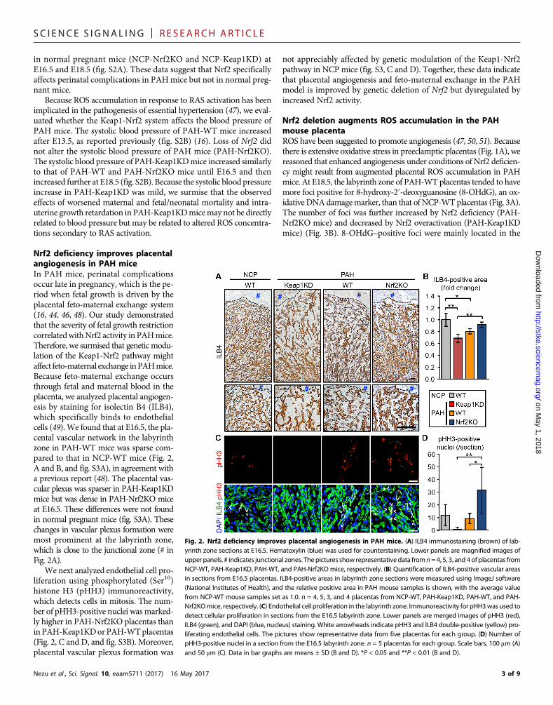
Nrf2 deficiency improves placentalangiogenesis in PAH miceIn PAH mice, perinatal complicationsoccur late in pregnancy, which is the pe-riod when fetal growth is driven by theplacental feto-maternal exchange system(16, 44, 46, 48). Our study demonstratedthat the severity of fetal growth restrictioncorrelatedwithNrf2 activity in PAHmice.Therefore, we surmised that genetic modu-lation of the Keap1-Nrf2 pathway mightaffect feto-maternal exchange inPAHmice.Because feto-maternal exchange occursthrough fetal and maternal blood in theplacenta, we analyzed placental angiogen-esis by staining for isolectin B4 (ILB4),which specifically binds to endothelialcells (49). We found that at E16.5, the pla-cental vascular network in the labyrinthzone in PAH-WT mice was sparse com-pared to that in NCP-WT mice (Fig. 2,A and B, and fig. S3A), in agreement witha previous report (48). The placental vas-cular plexus was sparser in PAH-Keap1KDmice but was dense in PAH-Nrf2KO miceat E16.5. These differences were not foundin normal pregnant mice (fig. S3A). Thesechanges in vascular plexus formation weremost prominent at the labyrinth zone,which is close to the junctional zone (# inFig. 2A).
We next analyzed endothelial cell pro-liferation using phosphorylated (Ser10)histone H3 (pHH3) immunoreactivity,which detects cells in mitosis. The num-ber of pHH3-positive nuclei was marked-ly higher in PAH-Nrf2KO placentas thaninPAH-Keap1KDorPAH-WTplacentas(Fig. 2, C and D, and fig. S3B). Moreover,placental vascular plexus formation was
Nezu et al., Sci. Signal. 10, eaam5711 (2017) 16 May 2017
not appreciably affected by genetic modulation of the Keap1-Nrf2pathway in NCP mice (fig. S3, C and D). Together, these data indicatethat placental angiogenesis and feto-maternal exchange in the PAHmodel is improved by genetic deletion of Nrf2 but dysregulated byincreased Nrf2 activity.
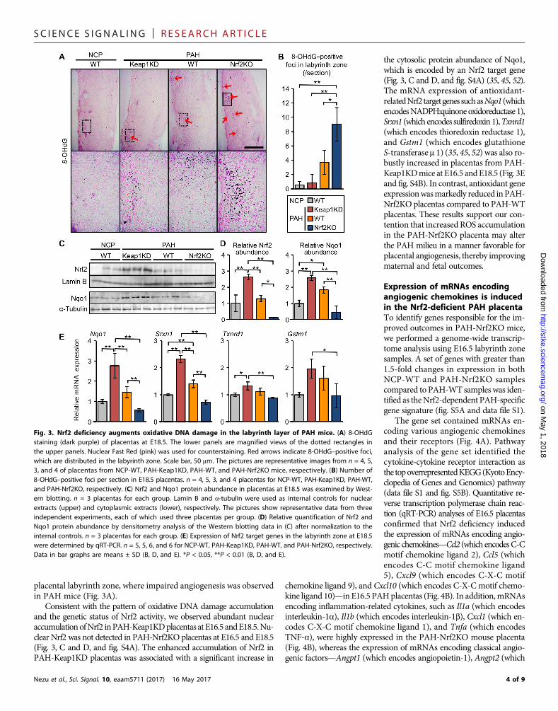
Nrf2 deletion augments ROS accumulation in the PAHmouse placentaROS have been suggested to promote angiogenesis (47, 50, 51). Becausethere is extensive oxidative stress in preeclamptic placentas (Fig. 1A), wereasoned that enhanced angiogenesis under conditions of Nrf2 deficien-cy might result from augmented placental ROS accumulation in PAHmice. At E18.5, the labyrinth zone of PAH-WTplacentas tended to havemore foci positive for 8-hydroxy-2′-deoxyguanosine (8-OHdG), an ox-idative DNAdamagemarker, than that of NCP-WTplacentas (Fig. 3A).The number of foci was further increased by Nrf2 deficiency (PAH-Nrf2KO mice) and decreased by Nrf2 overactivation (PAH-Keap1KDmice) (Fig. 3B). 8-OHdG–positive foci were mainly located in the
Fig. 2. Nrf2 deficiency improves placental angiogenesis in PAH mice. (A) ILB4 immunostaining (brown) of lab-yrinth zone sections at E16.5. Hematoxylin (blue) was used for counterstaining. Lower panels are magnified images ofupper panels. # indicates junctional zones. Thepictures show representativedata fromn=4, 5, 3, and 4 of placentas fromNCP-WT, PAH-Keap1KD, PAH-WT, and PAH-Nrf2KO mice, respectively. (B) Quantification of ILB4-positive vascular areasin sections from E16.5 placentas. ILB4-positive areas in labyrinth zone sections were measured using ImageJ software(National Institutes of Health), and the relative positive area in PAH mouse samples is shown, with the average valuefrom NCP-WT mouse samples set as 1.0. n = 4, 5, 3, and 4 placentas from NCP-WT, PAH-Keap1KD, PAH-WT, and PAH-Nrf2KOmice, respectively. (C) Endothelial cell proliferation in the labyrinth zone. Immunoreactivity for pHH3was used todetect cellular proliferation in sections from the E16.5 labyrinth zone. Lower panels are merged images of pHH3 (red),ILB4 (green), and DAPI (blue, nucleus) staining. White arrowheads indicate pHH3 and ILB4 double-positive (yellow) pro-liferating endothelial cells. The pictures show representative data from five placentas for each group. (D) Number ofpHH3-positive nuclei in a section from the E16.5 labyrinth zone. n = 5 placentas for each group. Scale bars, 100 mm (A)and 50 mm (C). Data in bar graphs are means ± SD (B and D). *P < 0.05 and **P < 0.01 (B and D).
3 of 9
SC I ENCE S I GNAL ING | R E S EARCH ART I C L E
on May 1, 2018
http://stke.sciencemag.org/
Dow
nloaded from
placental labyrinth zone, where impaired angiogenesis was observedin PAH mice (Fig. 3A).
Consistent with the pattern of oxidative DNA damage accumulationand the genetic status of Nrf2 activity, we observed abundant nuclearaccumulationofNrf2 inPAH-Keap1KDplacentas at E16.5 andE18.5.Nu-clear Nrf2 was not detected in PAH-Nrf2KO placentas at E16.5 and E18.5(Fig. 3, C and D, and fig. S4A). The enhanced accumulation of Nrf2 inPAH-Keap1KD placentas was associated with a significant increase in
Nezu et al., Sci. Signal. 10, eaam5711 (2017) 16 May 2017
the cytosolic protein abundance of Nqo1,which is encoded by an Nrf2 target gene(Fig. 3, C and D, and fig. S4A) (35, 45, 52).The mRNA expression of antioxidant-relatedNrf2targetgenessuchasNqo1 (whichencodesNADPH:quinoneoxidoreductase 1),Srxn1 (which encodes sulfiredoxin1),Txnrd1(which encodes thioredoxin reductase 1),and Gstm1 (which encodes glutathioneS-transferase m 1) (35, 45, 52) was also ro-bustly increased in placentas from PAH-Keap1KDmice at E16.5 andE18.5 (Fig. 3Eand fig. S4B). In contrast, antioxidant geneexpressionwasmarkedly reduced inPAH-Nrf2KO placentas compared to PAH-WTplacentas. These results support our con-tention that increased ROS accumulationin the PAH-Nrf2KO placenta may alterthe PAH milieu in a manner favorable forplacental angiogenesis, thereby improvingmaternal and fetal outcomes.
Expression of mRNAs encodingangiogenic chemokines is inducedin the Nrf2-deficient PAH placentaTo identify genes responsible for the im-proved outcomes in PAH-Nrf2KO mice,we performed a genome-wide transcrip-tome analysis using E16.5 labyrinth zonesamples. A set of genes with greater than1.5-fold changes in expression in bothNCP-WT and PAH-Nrf2KO samplescompared to PAH-WT samples was iden-tified as theNrf2-dependent PAH-specificgene signature (fig. S5A and data file S1).
The gene set contained mRNAs en-coding various angiogenic chemokinesand their receptors (Fig. 4A). Pathwayanalysis of the gene set identified thecytokine-cytokine receptor interaction asthe topoverrepresentedKEGG(KyotoEncy-clopedia of Genes and Genomics) pathway(data file S1 and fig. S5B). Quantitative re-verse transcription polymerase chain reac-tion (qRT-PCR) analyses of E16.5 placentasconfirmed that Nrf2 deficiency inducedthe expression of mRNAs encoding angio-genic chemokines—Ccl2 (which encodesC-Cmotif chemokine ligand 2), Ccl5 (whichencodes C-C motif chemokine ligand5), Cxcl9 (which encodes C-X-C motif
chemokine ligand 9), and Cxcl10 (which encodes C-X-C motif chemo-kine ligand 10)—in E16.5 PAHplacentas (Fig. 4B). In addition,mRNAsencoding inflammation-related cytokines, such as Il1a (which encodesinterleukin-1a), Il1b (which encodes interleukin-1b), Cxcl1 (which en-codes C-X-C motif chemokine ligand 1), and Tnfa (which encodesTNF-a), were highly expressed in the PAH-Nrf2KO mouse placenta(Fig. 4B), whereas the expression of mRNAs encoding classical angio-genic factors—Angpt1 (which encodes angiopoietin-1), Angpt2 (which
Fig. 3. Nrf2 deficiency augments oxidative DNA damage in the labyrinth layer of PAH mice. (A) 8-OHdGstaining (dark purple) of placentas at E18.5. The lower panels are magnified views of the dotted rectangles inthe upper panels. Nuclear Fast Red (pink) was used for counterstaining. Red arrows indicate 8-OHdG–positive foci,which are distributed in the labyrinth zone. Scale bar, 50 mm. The pictures are representative images from n = 4, 5,3, and 4 of placentas from NCP-WT, PAH-Keap1KD, PAH-WT, and PAH-Nrf2KO mice, respectively. (B) Number of8-OHdG–positive foci per section in E18.5 placentas. n = 4, 5, 3, and 4 placentas for NCP-WT, PAH-Keap1KD, PAH-WT,and PAH-Nrf2KO, respectively. (C) Nrf2 and Nqo1 protein abundance in placentas at E18.5 was examined by West-ern blotting. n = 3 placentas for each group. Lamin B and a-tubulin were used as internal controls for nuclearextracts (upper) and cytoplasmic extracts (lower), respectively. The pictures show representative data from threeindependent experiments, each of which used three placentas per group. (D) Relative quantification of Nrf2 andNqo1 protein abundance by densitometry analysis of the Western blotting data in (C) after normalization to theinternal controls. n = 3 placentas for each group. (E) Expression of Nrf2 target genes in the labyrinth zone at E18.5were determined by qRT-PCR. n = 5, 5, 6, and 6 for NCP-WT, PAH-Keap1KD, PAH-WT, and PAH-Nrf2KO, respectively.Data in bar graphs are means ± SD (B, D, and E). *P < 0.05, **P < 0.01 (B, D, and E).
4 of 9

SC I ENCE S I GNAL ING | R E S EARCH ART I C L E
encodes angiopoietin-2), Vegfa (which encodes vascular endothelialgrowth factor A), Kdr (which encodes kinase insert domain receptorfor VEGF), Flt1 (which encodes fetal liver tyrosine kinase 1), and Tek(which encodes endothelial tyrosine kinase receptor for angiopoietin-1)—were not significantly affected (fig. S5C). The plasma concentrations
on May 1, 2018
http://stke.sciencemag.org/
Dow
nloaded from
of PLGF2 (placenta growth factor 2) andsFLT1, both of which have been impli-cated in the pathogenesis of preeclampsia(11, 30, 53, 54), were not altered by Keap1orNrf2 genemodification in PAHmice atE16.5 (fig. S6). The chemokines and cyto-kines that showed increased expression inPAH-Nrf2KO placentas are critically im-portant for angiogenesis and placentalvascular remodeling (44, 55), suggestingthat the altered or restored expression pat-tern ofmRNAs encoding angiogenic cyto-kines and chemokinesmight play a role inthe enhanced angiogenesis in the PAH-Nrf2KOplacenta throughROSaccumula-tion in the labyrinth zone. It is also likelythat the chemokines and cytokines maycontribute to immune cell infiltration thatoccurs in preeclampsia (56, 57).
Pharmacological activation of Nrf2worsens the perinatal morbidityand mortality of PAH miceThe benefits of Nrf2 inducers have beenhighlighted as an antioxidative therapy(28). We asked whether pharmacologicalactivation of theNrf2 pathway recapitulatesthe genetic induction of Nrf2 or whetherthere is a therapeutic window for pharma-cological induction. Therefore, we treatedPAH-WT and NCP-WT mice after E13.5with an Nrf2 inducer, CDDO-Im {1-[2-cyano-3,12-dioxooleana-1,9(11)-dien-28-oyl] imidazole} (28, 52). The expression ofNrf2 target genes (Nqo1, Srxn1, Txnrd1,andGstm1)was increased in placentas fromCDDO-Im–treatedNCP-WTandPAH-WTmice (fig. S7). In PAH-WTmice, CDDO-Imtreatment increased maternal mortalityand decreased fetal body weight (Fig. 5, Aand B). In addition, PAH-WTmice treatedwith CDDO-Im experienced abortion andvaginal bleeding before dying (Fig. 5C), al-though CDDO-Im administration did notaffect the mortality or fetal body weight ofNCP-WT mice (Fig. 5, A and B).
DISCUSSIONThe mammalian placenta undergoes ex-tensive angiogenesis to develop efficientfeto-maternal exchange of oxygen, nutri-ents, and waste products (1, 14). Defectsin placental angiogenesis are believed to
Nezu et al., Sci. Signal. 10, eaam5711 (2017) 16 May 2017
play a central role in the pathogenesis of preeclampsia and fetal growthrestriction (1, 10, 11, 13, 58). Here, we used gene-modifiedmouse lines toprovide insights into the role of the Keap1-Nrf2 system in placental angi-ogenesis.Our results illustrate the essential and indispensable role ofROSasplacental proangiogenic factors in preeclampsia (Fig. 6).
Fig. 4. Nrf2 deficiency induces the expression of angiogenic and inflammatory factors in the PAH placenta.(A) Heat map of the microarray data for the labyrinth zone samples from NCP-WT, PAH-Keap1KD, PAH-WT, andPAH-Nrf2KO mice (n = 2 placentas for each group) at E16.5 shows the relative expression of genes related to an-giogenesis or inflammation, which were selected from the list of genes in the Nrf2-dependent PAH-specific genesignature (data file S1). Blue letters indicate genes for which multiple probes are included in this signature. (B) qRT-PCR analyses of the expression of mRNAs encoding angiogenic chemokines [Ccl2, Ccl5, Cxcl9, and Cxcl10; high-lighted in red letters in (A)] and inflammation-related genes (Il1a, Il1b, Cxcl1, and Tnfa) in the labyrinth zone ofPAH mice at E16.5. n = 5, 5, 6, and 5 placentas for NCP-WT, PAH-Keap1KD, PAH-WT, and PAH-Nrf2KO, respectively.Data in bar graphs are means ± SD. *P < 0.05, **P < 0.01.
Fig. 5. Pharmacological activation of Nrf2 worsens perinatal complications in PAH mice. (A) Maternal mor-tality rate of NCP-WT and PAH-WT mice treated with CDDO-Im every other day from E13.5 to E17.5. n = 3 NCP-WTdams with or without CDDO-Im administration; n = 7 PAH-WT dams with or without CDDO-Im administration. (B) Fetalbody weight of E16.5 and E18.5 embryos in NCP-WT or PAH-WT mice treated with CDDO-Im. n = 18 and 9 fetuses atE16.5 in NCP-WT and PAH-WT dams treated with vehicle, respectively; n = 15 and 23 fetuses at E16.5 in NCP-WT andPAH-WT dams treated with CDDO-Im, respectively. n = 18 and 28 fetuses at E18.5 in NCP-WT and PAH-WT dams treatedwith vehicle, respectively; n = 22 and 33 fetuses at E18.5 in NCP-WT and PAH-WT dams treated with CDDO-Im, respec-tively. (C) Placental abortion and vaginal bleeding in PAH-WT mice treated with CDDO-Im. These features were observedin every PAHmouse after CDDO-Im administration. Arrows and arrowheads indicate the fetus and placenta, respectively.Data in bar graphs are means ± SD. *P < 0.05, **P < 0.01.
5 of 9

SC I ENCE S I GNAL ING | R E S EARCH ART I C L E
on May 1, 2018
http://stke.sciencemag.org/
Dow
nloaded from
Placental angiogenesis is critical for establishing feto-maternal ex-change and is accelerated from E14.5 to delivery in mice (44, 46, 48).The roles of the placental anti-angiogenicmilieu in the pathogenesis ofpreeclampsia have been highlighted by evidence that placental-derived anti-angiogenic factors cause a preeclampsia-like phenotypeinmice (58, 59). In the PAHmodel, the plasma concentration of ANGIIis increased from E13.5 and contributes to impaired fetal growth andplacental vasculature formation (16, 44, 48). In our mouse model,disease severity correlated well with the degree of impaired placentalangiogenesis in the labyrinth zone, further supporting the pathogenicimportance of placental angiogenesis.
Genetic and pharmacological manipulation of the Keap1-Nrf2 sys-tem can reduce oxidative stress and frequently results in beneficial out-comes in various disease models (28). Nrf2 plays context-dependentroles in angiogenesis: Systemic Nrf2 deletion augments angiogenesisafter hindlimb ischemia (60), whereas endothelial cell–specific dele-tion of Nrf2 leads to impaired physiological retinal angiogenesis byreducing tip cell formation and vascular branching (61). Consistentwith the former observation, there are many lines of evidence thatsupport the notion that appropriate amounts of ROS are proangiogenicfactors that act through various mechanisms, including enhancingVEGF and hypoxia-inducible factor signaling (47, 50, 51, 62, 63). In ad-dition, physiological amounts of ROS may be important in othersystems to maintain normal cellular signaling, whereas high amountsof ROS are associated with cell toxicity (64).
Our study demonstrated that systemic deletion of Nrf2 increasedoxidative DNA damage and enhanced placental angiogenesis, as evi-denced by the higher density of the capillary plexus and the increasedproliferation of endothelial cells in the labyrinth zone of PAH-Nrf2KOmice. Conversely, genetic and pharmacological induction of Nrf2 resultedin increasedmaternalmortality andworsened fetal growth restriction, withreduced capillary density in the labyrinth zone, despite the reduction in ox-idative DNA damage. These results are consistent with a large clinical trialthat tested the ability of antioxidants topreventpreeclampsia (32); high-riskwomen that received antioxidant vitamins during pregnancy had worseoutcomes, including an increase in the number of low birth weight babiesand stillbirths. On the basis of these observations, we posit that appropriateROS accumulation in the placenta creates a placental milieu that is favor-able for endothelial proliferation and angiogenesis, thereby alleviating pre-eclampsia pathologies thatmay otherwise be associated with inappropriateamounts of ROS. Although our microarray analyses of placental labyrinthzone samples showed no significant changes in the expression of mRNAs
Nezu et al., Sci. Signal. 10, eaam5711 (2017) 16 May 2017
encoding classical angiogenic factors, we observed increased expressionof mRNAs encoding angiogenic chemokines, cytokines, and their re-ceptors in PAH-Nrf2KO mice, similar to that which occurs in thehindlimb ischemia model of Nrf2KO mice (60).
Because oxidative stress activates inflammation that produces in-tracellular and extracellular ROS (65, 66), the oxidative stress responseand inflammation pathways are tightly interconnected. Activated in-flammatory cells generate a battery of chemokines and cytokines thatfurther potentiate inflammation and can induce angiogenesis (60, 65).Furthermore, these chemokines, such as Ccl2 and Ccl5, regulate pla-cental vascularization (55, 67). We have shown that Nrf2 directly sup-presses the expression of inflammatory cytokine–encoding genes inactivated macrophages (67). These data support our contention thatinactivationofNrf2 inducesROSaccumulationandangiogenic chemokineexpression in PAH placentas, whereas Nrf2 overactivation impairsangiogenesis and enhances preeclampsia-mediated adverse outcomes.The mechanisms of excess maternal mortality in the Nrf2 overexpres-sion studies remain to be explored in future studies.
Hemeoxygenase-1 (HO-1) is an antioxidant enzyme that protects cellsfrom oxidative stress and may have protective roles against preeclampsia(68).We observed amodest induction ofHmox1mRNA (which encodesHO-1) in PAH-Keap1KD placentas compared to PAH-WT placentas(fig. S4B). However, this induction did not rescue the PAH outcomes,despite the marked reduction in oxidative DNA damage. Fetuses ofHmox1-knockout mice have extremely low birth weights and intra-uterine growth retardation (69), whereas fetuses of NCP-Nrf2KO andNCP-Keap1KDmice have normal birth weights. In addition, partial de-ficiency of HO-1 results in impaired placental angiogenesis (70, 71).Together, we speculate that HO-1may have a unique role(s) in placentalangiogenesis or systemic protective effects on the maternal endotheliumindependent of placental oxidative stress.
The plasma concentration of sFLT1 has also been suggested to corre-late with ROS accumulation, and sFLT1 has been implicated in the patho-genesis of preeclampsia in rodents and humans (11, 30, 53, 54). However,sFLT1 concentrations were not altered by Keap1 or Nrf2 gene modifica-tion in PAH mice. This observation may suggest that the induction ofsFLT1 is not fundamentally dependent on placental ROS accumulation.Simultaneous overexpression of renin and angiotensinogen induces thepreeclampsia phenotype in our mouse model by activating the angiotensinsignal during pregnancy. Also, increased ANGII sensitivity and enhancedangiotensin signaling have been reported in preeclampsia patients, al-though the circulating concentrations of renin are low compared to thosein women experiencing normal pregnancies (17–20, 72–74). To generalizeour conclusions to human preeclampsia, additional animal models, suchas sFLT1 overexpression models, are needed.
In summary, we showed that augmented ROS signaling in responseto Nrf2 deletion induced a proangiogenic milieu in the preeclampticplacenta (Fig. 6). Augmented ROS production during preeclampsiamay represent an adaptive response to hypertension and systemicvascular damage. Further studies are needed to characterize the roleof appropriate amounts of ROS signaling in preeclampsia, and theuse of additional models may lead us to a better understanding of therole of ROS signaling in humans with preeclampsia.
MATERIALS AND METHODSHuman subjectsThe Institutional Review Board at Beth Israel Deaconess MedicalCenter approved the collection and use of discarded human placentas.
Fig. 6. Schematic of Nrf2-mediated angiogenic suppression in the PAHplacenta.Overproduction of human ANGII (hANGII) stimulates AT1R signaling and creates an anti-angiogenic milieu in the labyrinth zone of PAH mice. ROS accumulation counteracts theanti-angiogenic milieu by inducing the expression of genes encoding angiogenicchemokines and cytokines. Because Nrf2 decreases ROS concentrations, Nrf2 inactivationameliorates the intra-uterine growth retardation (IUGR) and PAH complications byinducing ROS-mediated angiogenesis in PAH-Nrf2KO mice. In PAH-Keap1KD mice, pla-cental angiogenesis is further worsened due to ROS quenching by activated Nrf2.
6 of 9

SC I ENCE S I GNAL ING | R E S EARCH ART I C L E
on May 1, 2018
http://stke.sciencemag.org/
Dow
nloaded from
Immunohistochemical analysis was performed as described in theSupplementary Materials.
MiceAll mouse experiments were approved by the Regulations for AnimalExperiments and Related Activities of Tohoku University. hAGTTg/Tg
and hRENTg/Tg transgenicmice were obtained fromRIKENBioResourceCenter, Japan. Male hRENTg/Tg and female hAGTTg/Tg mice were bredto obtain PAH mice, as previously reported (fig. S1) (16). Nrf2KO(Nrf2−/−) (35) and Keap1KD (Keap1KD/KD) mice have been describedpreviously (45). Because Keap1-null mice die around weaning due tomalnutrition caused by hyperkeratosis in the digestive tract (75),Keap1KD mice with a homozygously hypomorphic mutant Keap1gene were used in this study to investigate effects of Nrf2 overactiva-tion in mice. PAH mice bearing Nrf2−/− or Keap1KD/KD alleles weregenerated as depicted in fig. S1. Homozygous hAGTTg/Tg mice withmodified Nrf2 or Keap1 genes were used as controls. Mice were back-crossed to C57BL/6J mice for more than five generations with PCRgenotyping using primers listed in table S1. Mice were maintainedin a specific pathogen–free facility.
Tissue preparation for histological analysisTissues for frozen sections were fixed with 4% paraformaldehyde(PFA) for 2 hours and embedded in an optimum cutting temperaturecompound (Sakura Finetek) after an overnight incubation in 20% su-crose. Tissues for paraffin-embedded sections were fixed with 4% PFAfor 48 hours and processed by conventional procedures. Tissues for8-OHdGstainingwere fixedwithBouin solution (DAKO) for 24 hours.
Immunohistochemistry of human placental tissuesParaffin sections (5 mm thick) were deparaffinized and incubated withcitrate buffer (DAKO, pH 6.0) for antigen retrieval. Slides were rinsedwith phosphate-buffered saline (PBS) and incubated for 15 min with3%hydrogenperoxide to quench endogenous peroxidase. To blocknon-specific antibody binding, slides were incubated at 37°C with 2.5%normal horse serum in PBS for 40 min. Slides were then incubated withrabbit anti-malondialdehydepolyclonal antibody (1:500, ab6423,Abcam)for 40 min at 37°C, rinsed in PBS, developed using ImmPRESShorseradish peroxidase (HRP) Anti-Rabbit IgG Polymer Detection Kit(Vector Laboratories), and counterstained with hematoxylin.
Immunohistochemistry of mouse tissuesForparaffin-embedded tissue sections, citrate buffer (DAKO,pH6.0)wasused for antigen retrieval. Sections were incubated with primary anti-bodies after blocking with Protein Block Serum-Free (DAKO). Primaryantibodies against pHH3 (1:300 dilution, 9701S, Cell Signaling Technol-ogy), biotinylated ILB4 (1:50, B-1205, Vector Laboratories), and 8-OHdG(1:50, MOG-20P, Japan Institute for the Control of Aging) were used.Fluorescence images were obtained using an LSM780 confocal imag-ing system (Carl Zeiss). For ILB4 and 8-OHdG staining, signals weredeveloped using diaminobenzidine (DAB, DAKO) or a DAB solu-tion containing nickel chloride hexahydrate (DAB-Ni, Wako). Sec-tions were counterstained with DAPI (4′,6-diamidino-2-phenylindole),hematoxylin, or Nuclear Fast Red (H-3403, Vector Laboratories), asappropriate.
Measurement of systolic blood pressureSystolic blood pressure was measured using a tail-cuff blood pressuresystem (BP-98a, Softron) following the manufacturer’s protocol (46).
Nezu et al., Sci. Signal. 10, eaam5711 (2017) 16 May 2017
Western blottingNuclear extracts andwhole-cell lysates of placental tissue were preparedseparately using sucrose buffer (0.25 M sucrose in 10 mMHepes-KOHbuffer). Samples were separated by SDS–polyacrylamide gel electropho-resis; transferred to polyvinylidene difluoride membranes; and immu-noblotted with antibodies against Nrf2 (1:200 dilution, in-house clone#103), Nqo1 (1:1000, ab2346, Abcam), lamin B (1:1000, M-20, SantaCruz Biotechnology), and a-tubulin (1:1000, T9026, Sigma). HRP-conjugated secondary antibodies (DAKO) and enhanced chemi-luminescence reagents (GE Healthcare) were used to detect signals.
Quantitative reverse transcription polymerasechain reactionTotal RNA was prepared using Isogen (Nippon Gene) and reverse-transcribed using random hexamers and a SuperScript III PolymeraseKit (Invitrogen). Primer information is available in table S2. Gene ex-pression was calculated on the basis of the threshold cycle values andthe efficiency of each primer set, and the data were expressed relative to18S ribosomal RNA.
Microarray analysisTotal RNA was isolated from the labyrinth zone at E16.5 using Isogen(Nippon Gene) and hybridized to whole-mouse genome microarrays(4× 44K,Agilent Technologies) following themanufacturer’s procedureafter assessing RNA quality with a Bioanalyzer (Agilent Technologies).Data were analyzed using GeneSpring software (Agilent Technologies)and submitted to GEO (Gene Expression Omnibus; accession no.GSE76669). Geneswith a greater than 1.5-fold change in average expres-sion compared to that of PAH-WT samples are listed in data file S1, andthese selected genes were further analyzed using DAVID (Database forAnnotation, Visualization, and Integrated Discovery) BioinformaticsResources 6.7 software (https://david.ncifcrf.gov) with a KEGG pathwaydatabase (www.genome.jp/kegg/pathway.html).
CDDO-Im administrationCDDO-Im (Mochida Pharmaceuticals) was dissolved in sesame oil(Wako) and orally administered to mice at 30 mmol/kg body weight atE13.5, E15.5, and E17.5 using disposable feeding needles (Fuchigami).
Statistical analysisData in bar and line graphs are means ± SD. Statistical analyses were per-formed by using R software (The R Foundation, http://r-project.org).Comparisons between groups were analyzed using Fisher’s exact test,one-way analysis of variance, or an unpaired t test, as appropriate.P valueswere adjusted byHolmmethod formultiple comparisons of Fisher’s exacttest. The Tukey-Kramer test was used as a correctionmethod formultiplecomparisons. Data were considered statistically significant at P < 0.05.
SUPPLEMENTARY MATERIALSwww.sciencesignaling.org/cgi/content/full/10/479/eaam5711/DC1Fig. S1. Mouse models used in this study.Fig. S2. Fetal body weight, systolic blood pressure, and gene expression profiles of RAScomponents in NCP and PAH mice with Keap1 or Nrf2 genetic mutation.Fig. S3. Genetic deletion of Nrf2 improves vascular plexus selectively in the PAH placenta.Fig. S4. Expression profiles of Nrf2 target genes in the placenta of PAH mice.Fig. S5. Microarray analysis to identify an Nrf2-dependent PAH-specific gene signature.Fig. S6. Genetic modification of the Keap1-Nrf2 system in PAH mice does not significantlychange the plasma concentrations of sFLT1 and PLGF2.Fig. S7. Induction of Nrf2 target gene expression in the labyrinth zone of PAH-WT mice bypharmacological activation of Nrf2.
7 of 9

SC I ENCE S I GNAL ING | R E S EARCH ART I C L E
Table S1. Primer sequences for genotyping mouse lines.Table S2. Primer sequences for qRT-PCR analysis.Data file S1. Microarray data revealed candidate genes for an Nrf2-dependent PAH-specificgene signature.
on May 1, 2018
http://stke.sciencemag.org/
Dow
nloaded from
REFERENCES AND NOTES1. A. S. Cerdeira, S. A. Karumanchi, Angiogenic factors in preeclampsia and related disorders.
Cold Spring Harb. Perspect. Med. 2, a006585 (2012).2. American College of Obstetricians and Gynecologists; Task Force on Hypertension in
Pregnancy, Hypertension in pregnancy. Report of the American College of Obstetriciansand Gynecologists’ Task Force on Hypertension in Pregnancy. Obstet. Gynecol. 122,1122–1131 (2013).
3. E. A. P. Steegers, P. von Dadelszen, J. J. Duvekot, R. Pijnenborg, Pre-eclampsia. Lancet 376,631–644 (2010).
4. A. B. Wallis, A. F. Saftlas, J. Hsia, H. K. Atrash, Secular trends in the rates ofpreeclampsia, eclampsia, and gestational hypertension, United States, 1987–2004.Am. J. Hypertens. 21, 521–526 (2008).
5. L. Duley, The global impact of pre-eclampsia and eclampsia. Semin. Perinatol. 33, 130–137(2009).
6. I. S. Patten, S. Rana, S. Shahul, G. C. Rowe, C. Jang, L. Liu, M. R. Hacker, J. S. Rhee,J. Mitchell, F. Mahmood, P. Hess, C. Farrell, N. Koulisis, E. V. Khankin, S. D. Burke,I. Tudorache, J. Bauersachs, F. del Monte, D. Hilfiker-Kleiner, S. A. Karumanchi, Z. Arany,Cardiac angiogenic imbalance leads to peripartum cardiomyopathy. Nature 485,333–338 (2012).
7. A. Goel, M. R. Maski, S. Bajracharya, J. B. Wenger, D. Zhang, S. Salahuddin, S. S. Shahul,R. Thadhani, E. W. Seely, S. A. Karumanchi, S. Rana, Epidemiology and mechanisms ofde novo and persistent hypertension in the postpartum period. Circulation 132,1726–1733 (2015).
8. B. E. Vikse, L. M. Irgens, T. Leivestad, R. Skjaerven, B. M. Iversen, Preeclampsia and the riskof end-stage renal disease. N. Engl. J. Med. 359, 800–809 (2008).
9. T. Männistö, P. Mendola, M. Vääräsmäki, M.-R. Järvelin, A.-L. Hartikainen, A. Pouta,E. Suvanto, Elevated blood pressure in pregnancy and subsequent chronic disease risk.Circulation 127, 681–690 (2013).
10. R. J. Levine, S. E. Maynard, C. Qian, K.-H. Lim, L. J. England, K. F. Yu, E. F. Schisterman,R. Thadhani, B. P. Sachs, F. H. Epstein, B. M. Sibai, V. P. Sukhatme, S. A. Karumanchi,Circulating angiogenic factors and the risk of preeclampsia. N. Engl. J. Med. 350, 672–683(2004).
11. S. E. Maynard, J.-Y. Min, J. Merchan, K.-H. Lim, J. Li, S. Mondal, T. A. Libermann,J. P. Morgan, F. W. Sellke, I. E. Stillman, F. H. Epstein, V. P. Sukhatme, S. A. Karumanchi,Excess placental soluble fms-like tyrosine kinase 1 (sFlt1) may contribute to endothelialdysfunction, hypertension, and proteinuria in preeclampsia. J. Clin. Invest. 111,649–658 (2003).
12. S. Venkatesha, M. Toporsian, C. Lam, J.-i. Hanai, T. Mammoto, Y. M. Kim, Y. Bdolah,K.-H. Lim, H.-T. Yuan, T. A. Libermann, I. E. Stillman, D. Roberts, P. A. D’Amore, F. H. Epstein,F. W. Sellke, R. Romero, V. P. Sukhatme, M. Letarte, S. A. Karumanchi, Soluble endoglincontributes to the pathogenesis of preeclampsia. Nat. Med. 12, 642–649 (2006).
13. R. J. Levine, C. Lam, C. Qian, K. F. Yu, S. E. Maynard, B. P. Sachs, B. M. Sibai, F. H. Epstein,R. Romero, R. Thadhani, S. A. Karumanchi; CPEP Study Group, Soluble endoglin and othercirculating antiangiogenic factors in preeclampsia. N. Engl. J. Med. 355, 992–1005 (2006).
14. T. Chaiworapongsa, P. Chaemsaithong, L. Yeo, R. Romero, Pre-eclampsia part 1: Currentunderstanding of its pathophysiology. Nat. Rev. Nephrol. 10, 466–480 (2014).
15. J. E. Hall, Historical perspective of the renin-angiotensin system. Mol. Biotechnol. 24,27–39 (2003).
16. E. Takimoto, J. Ishida, F. Sugiyama, H. Horiguchi, K. Murakami, A. Fukamizu,Hypertension induced in pregnant mice by placental renin and maternalangiotensinogen. Science 274, 995–998 (1996).
17. S. AbdAlla, H. Lother, A. el Massiery, U. Quitterer, Increased AT1 receptor heterodimersin preeclampsia mediate enhanced angiotensin II responsiveness. Nat. Med. 7,1003–1009 (2001).
18. G. Wallukat, V. Homuth, T. Fischer, C. Lindschau, B. Horstkamp, A. Jüpner, E. Baur,E. Nissen, K. Vetter, D. Neichel, J. W. Dudenhausen, H. Haller, F. C. Luft, Patients withpreeclampsia develop agonistic autoantibodies against the angiotensin AT1 receptor.J. Clin. Invest. 103, 945–952 (1999).
19. C. C. Zhou, Y. Zhang, R. A. Irani, H. Zhang, T. Mi, E. J. Popek, M. J. Hicks, S. M. Ramin,R. E. Kellems, Y. Xia, Angiotensin receptor agonistic autoantibodies induce pre-eclampsiain pregnant mice. Nat. Med. 14, 855–862 (2008).
20. A. Zhou, R. W. Carrell, M. P. Murphy, Z. Wei, Y. Yan, P. L. D. Stanley, P. E. Stein,F. Broughton Pipkin, R. J. Read, A redox switch in angiotensinogen modulatesangiotensin release. Nature 468, 108–111 (2010).
21. R. Dechend, P. Gratze, G. Wallukat, E. Shagdarsuren, R. Plehm, J.-H. Bräsen, A. Fiebeler,W. Schneider, S. Caluwaerts, L. Vercruysse, R. Pijnenborg, F. C. Luft, D. N. Müller, Agonistic
Nezu et al., Sci. Signal. 10, eaam5711 (2017) 16 May 2017
autoantibodies to the AT1 receptor in a transgenic rat model of preeclampsia.Hypertension 45, 742–746 (2005).
22. J. M. Denney, C. Bird, A. Gendron-Fitzpatrick, E. Sampene, I. M. Bird, D. M. Shah,Renin-angiotensin system transgenic mouse model recapitulates pathophysiologysimilar to human preeclampsia with renal injury that may be mediated through VEGF.Am. J. Physiol. Renal Physiol. 312, F445 (2017).
23. J. Bohlender, D. Ganten, F. C. Luft, Rats transgenic for human renin and humanangiotensinogen as a model for gestational hypertension. J. Am. Soc. Nephrol. 11,2056–2061 (2000).
24. A. Nguyen Dinh Cat, A. C. Montezano, D. Burger, R. M. Touyz, Angiotensin II, NADPHoxidase, and redox signaling in the vasculature. Antioxid. Redox Signal. 19, 1110–1120(2013).
25. S. Goulopoulou, S. T. Davidge, Molecular mechanisms of maternal vascular dysfunction inpreeclampsia. Trends Mol. Med. 21, 88–97 (2015).
26. R. Dechend, C. Viedt, D. N. Müller, B. Ugele, R. P. Brandes, G. Wallukat, J.-K. Park, J. Janke,P. Barta, J. Theuer, A. Fiebeler, V. Homuth, R. Dietz, H. Haller, J. Kreuzer, F. C. Luft, AT1receptor agonistic antibodies from preeclamptic patients stimulate NADPH oxidase.Circulation 107, 1632–1639 (2003).
27. B. D. LaMarca, M. J. Ryan, J. S. Gilbert, S. R. Murphy, J. P. Granger, Inflammatory cytokinesin the pathophysiology of hypertension during preeclampsia. Curr. Hypertens. Rep. 9,480–485 (2007).
28. T. Suzuki, H. Motohashi, M. Yamamoto, Toward clinical application of the Keap1–Nrf2pathway. Trends Pharmacol. Sci. 34, 340–346 (2013).
29. M. Ristow, Unraveling the truth about antioxidants: Mitohormesis explains ROS-inducedhealth benefits. Nat. Med. 20, 709–711 (2014).
30. C. A. Turpin, S. A. Sakyi, W. K. Owiredu, R. K. Ephraim, E. O. Anto, Association betweenadverse pregnancy outcome and imbalance in angiogenic regulators and oxidative stressbiomarkers in gestational hypertension and preeclampsia. BMC Pregnancy Childbirth 15,189 (2015).
31. N. Kweider, B. Huppertz, M. Kadyrov, W. Rath, T. Pufe, C. J. Wruck, A possible protectiverole of Nrf2 in preeclampsia. Ann. Anat. 196, 268–277 (2014).
32. L. Poston, A. L. Briley, P. T. Seed, F. J. Kelly, A. H. Shennan; Vitamins in Pre-eclampsia(VIP) Trial Consortium, Vitamin C and vitamin E in pregnant women at risk forpre-eclampsia (VIP trial): Randomised placebo-controlled trial. Lancet 367, 1145–1154(2006).
33. J. M. Roberts, L. Myatt, C. Y. Spong, E. A. Thom, J. C. Hauth, K. J. Leveno, G. D. Pearson,R. J. Wapner, M. W. Varner, J. M. Thorp Jr., B. M. Mercer, A. M. Peaceman, S. M. Ramin,M. W. Carpenter, P. Samuels, A. Sciscione, M. Harper, W. J. Smith, G. Saade, Y. Sorokin,G. B. Anderson; Eunice Kennedy Shriver National Institute of Child Health and HumanDevelopment Maternal-Fetal Medicine Units Network, Vitamins C and E to preventcomplications of pregnancy-associated hypertension. N. Engl. J. Med. 362, 1282–1291(2010).
34. H. Kawagishi, T. Finkel, Unraveling the truth about antioxidants: ROS and disease: Findingthe right balance. Nat. Med. 20, 711–713 (2014).
35. K. Itoh, T. Chiba, S. Takahashi, T. Ishii, K. Igarashi, Y. Katoh, T. Oyake, N. Hayashi, K. Satoh,I. Hatayama, M. Yamamoto, Y.-i. Nabeshima, An Nrf2/small Maf heterodimer mediatesthe induction of phase II detoxifying enzyme genes through antioxidant responseelements. Biochem. Biophys. Res. Commun. 236, 313–322 (1997).
36. K. Itoh, N. Wakabayashi, Y. Katoh, T. Ishii, K. Igarashi, J. D. Engel, M. Yamamoto, Keap1represses nuclear activation of antioxidant responsive elements by Nrf2 through bindingto the amino-terminal Neh2 domain. Genes Dev. 13, 76–86 (1999).
37. N. Wakabayashi, A. T. Dinkova-Kostova, W. D. Holtzclaw, M.-I. Kang, A. Kobayashi,M. Yamamoto, T. W. Kensler, P. Talalay, Protection against electrophile and oxidant stressby induction of the phase 2 response: Fate of cysteines of the Keap1 sensor modified byinducers. Proc. Natl. Acad. Sci. U.S.A. 101, 2040–2045 (2004).
38. T. Yamamoto, T. Suzuki, A. Kobayashi, J. Wakabayashi, J. Maher, H. Motohashi,M. Yamamoto, Physiological significance of reactive cysteine residues of Keap1 indetermining Nrf2 activity. Mol. Cell. Biol. 28, 2758–2770 (2008).
39. M. Løset, S. B. Mundal, M. P. Johnson, M. H. Fenstad, K. A. Freed, I. A. Lian, I. P. Eide,L. Bjørge, J. Blangero, E. K. Moses, R. Austgulen, A transcriptional profile of the decidua inpreeclampsia. Am. J. Obstet. Gynecol. 204, 84.e81–84.e27 (2011).
40. N. Acar, H. Soylu, I. Edizer, O. Ozbey, H. Er, G. Akkoyunlu, B. Gemici, I. Ustunel,Expression of nuclear factor erythroid 2-related factor 2 (Nrf2) and peroxiredoxin 6(Prdx6) proteins in healthy and pathologic placentas of human and rat. Acta Histochem.116, 1289–1300 (2014).
41. C. J. Wruck, B. Huppertz, P. Bose, L.-O. Brandenburg, T. Pufe, M. Kadyrov, Role of a fetaldefence mechanism against oxidative stress in the aetiology of preeclampsia.Histopathology 55, 102–106 (2009).
42. C. A. Hubel, M. K. McLaughlin, R. W. Evans, B. A. Hauth, C. J. Sims, J. M. Roberts, Fastingserum triglycerides, free fatty acids, and malondialdehyde are increased inpreeclampsia, are positively correlated, and decrease within 48 hours post partum.Am. J. Obstet. Gynecol. 174, 975–982 (1996).
8 of 9

SC I ENCE S I GNAL ING | R E S EARCH ART I C L E
on May 1, 2018
http://stke.sciencemag.org/
Dow
nloaded from
43. R. Madazli, A. Benian, S. Aydin, H. Uzun, N. Tolun, The plasma and placental levels ofmalondialdehyde, glutathione and superoxide dismutase in pre-eclampsia. J. Obstet.Gynaecol. 22, 477–480 (2002).
44. J. Ishida, T. Matsuoka, T. Saito-Fujita, S. Inaba, S. Kunita, F. Sugiyama, K.-i. Yagami,A. Fukamizu, Pregnancy-associated homeostasis and dysregulation: Lessons from geneticallymodified animal models. J. Biochem. 150, 5–14 (2011).
45. K. Taguchi, J. M. Maher, T. Suzuki, Y. Kawatani, H. Motohashi, M. Yamamoto, Geneticanalysis of cytoprotective functions supported by graded expression of Keap1.Mol. Cell. Biol. 30, 3016–3026 (2010).
46. T. Saito, J. Ishida, E. Takimoto-Ohnishi, S. Takamine, T. Shimizu, T. Sugaya, H. Kato,T. Matsuoka, M. Nangaku, Y. Kon, F. Sugiyama, K. Yagami, A. Fukamizu, An essential rolefor angiotensin II type 1a receptor in pregnancy-associated hypertension withintrauterine growth retardation. FASEB J. 18, 388–390 (2004).
47. D. I. Brown, K. K. Griendling, Regulation of signal transduction by reactive oxygen speciesin the cardiovascular system. Circ. Res. 116, 531–549 (2015).
48. M. Furuya, J. Ishida, S. Inaba, Y. Kasuya, S. Kimura, R. Nemori, A. Fukamizu, Impairedplacental neovascularization in mice with pregnancy-associated hypertension. Lab.Invest. 88, 416–429 (2008).
49. M. Li, N. M. J. Schwerbrock, P. M. Lenhart, K. L. Fritz-Six, M. Kadmiel, K. S. Christine,D. M. Kraus, S. T. Espenschied, H. H. Willcockson, C. P. Mack, K. M. Caron, Fetal-derivedadrenomedullin mediates the innate immune milieu of the placenta. J. Clin. Invest. 123,2408–2420 (2013).
50. M. Ushio-Fukai, Redox signaling in angiogenesis: Role of NADPH oxidase. Cardiovasc. Res.71, 226–235 (2006).
51. M. R. Abid, I. G. Schoots, K. C. Spokes, S.-Q. Wu, C. Mawhinney, W. C. Aird Vascularendothelial growth factor-mediated induction of manganese superoxide dismutaseoccurs through redox-dependent regulation of forkhead and IkB/NF-kB. J. Biol. Chem.279, 44030–44038 (2004).
52. M. Nezu, T. Souma, L. Yu, T. Suzuki, D. Saigusa, S. Ito, N. Suzuki, M. Yamamoto,Transcription factor Nrf2 hyperactivation in early-phase renal ischemia-reperfusion injuryprevents tubular damage progression. Kidney Int. 91, 387–401 (2016).
53. Z. Jiang, Y. Zou, Z. Ge, Q. Zuo, S. Y. Huang, L. Sun, A role of sFlt-1 in oxidative stress andapoptosis in human and mouse pre-eclamptic trophoblasts. Biol. Reprod. 93, 73 (2015).
54. K. B. Tam Tam, B. Lamarca, M. Arany, K. Cockrell, L. Fournier, S. Murphy, J. N. Martin Jr.,J. P. Granger, Role of reactive oxygen species during hypertension in response to chronicantiangiogenic factor (sFlt-1) excess in pregnant rats. Am. J. Hypertens. 24, 110–113 (2011).
55. E. C. Keeley, B. Mehrad, R. M. Strieter, Chemokines as mediators of neovascularization.Arterioscler. Thromb. Vasc. Biol. 28, 1928–1936 (2008).
56. J. N. Bulmer, Immune aspects of pathology of the placental bed contributing topregnancy pathology. Baillieres Clin. Obstet. Gynaecol. 6, 461–488 (1992).
57. C. W. G. Redman, Sargent IL, Immunology of pre-eclampsia. Am. J. Reprod. Immunol.63, 534–543 (2010).
58. K. Kumasawa, M. Ikawa, H. Kidoya, H. Hasuwa, T. Saito-Fujita, Y. Morioka, N. Takakura,T. Kimura, M. Okabe, Pravastatin induces placental growth factor (PGF) and amelioratespreeclampsia in a mouse model. Proc. Natl. Acad. Sci. U.S.A. 108, 1451–1455 (2011).
59. X. Fan, A. Rai, N. Kambham, J. F. Sung, N. Singh, M. Petitt, S. Dhal, R. Agrawal, R. E. Sutton,M. L. Druzin, S. S. Gambhir, B. K. Ambati, J. C. Cross, N. R. Nayak, Endometrial VEGF inducesplacental sFLT1 and leads to pregnancy complications. J. Clin. Invest. 124, 4941–4952(2014).
60. S. Ichihara, Y. Yamada, F. Liu, T. Murohara, K. Itoh, M. Yamamoto, G. Ichihara, Ablation of thetranscription factor Nrf2 promotes ischemia-induced neovascularization by enhancingthe inflammatory response. Arterioscler. Thromb. Vasc. Biol. 30, 1553–1561 (2010).
61. Y. Wei, J. Gong, R. K. Thimmulappa, B. Kosmider, S. Biswal, E. J. Duh, Nrf2 actscell-autonomously in endothelium to regulate tip cell formation and vascular branching.Proc. Natl. Acad. Sci. U.S.A. 110, E3910–E3918 (2013).
62. Y.-W. Kim, T. V. Byzova, Oxidative stress in angiogenesis and vascular disease. Blood 123,625–631 (2014).
63. M. R. Abid, K. C. Spokes, S.-C. Shih, W. C. Aird, NADPH oxidase activity selectivelymodulates vascular endothelial growth factor signaling pathways. J. Biol. Chem. 282,35373–35385 (2007).
Nezu et al., Sci. Signal. 10, eaam5711 (2017) 16 May 2017
64. K. R. Martin, J. C. Barrett, Reactiveoxygen species as double-edged swords in cellular processes:Low-dose cell signaling versus high-dose toxicity. Hum. Exp. Toxicol. 21, 71–75 (2002).
65. Y.-W. Kim, X. Z. West, T. V. Byzova, Inflammation and oxidative stress in angiogenesis andvascular disease. J. Mol. Med. 91, 323–328 (2013).
66. E. H. Kobayashi, T. Suzuki, R. Funayama, T. Nagashima, M. Hayashi, H. Sekine,N. Tanaka, T. Moriguchi, H. Motohashi, K. Nakayama, M. Yamamoto, Nrf2 suppressesmacrophage inflammatory response by blocking proinflammatory cytokine transcription.Nat. Commun. 7, 11624 (2016).
67. K. Red-Horse, P. M. Drake, S. J. Fisher, Human pregnancy: The role of chemokine networksat the fetal-maternal interface. Expert Rev. Mol. Med. 6, 1–14 (2004).
68. M. Cudmore, S. Ahmad, B. Al-Ani, T. Fujisawa, H. Coxall, K. Chudasama, L. R. Devey,S. J. Wigmore, A. Abbas, P. W. Hewett, A. Ahmed, Negative regulation of soluble Flt-1 andsoluble endoglin release by heme oxygenase-1. Circulation 115, 1789–1797 (2007).
69. H. Zhao, R. J. Wong, F. S. Kalish, N. R. Nayak, D. K. Stevenson, Effect of hemeoxygenase-1 deficiency on placental development. Placenta 30, 861–868 (2009).
70. H. Zhao, J. Azuma, F. Kalish, R. J. Wong, D. K. Stevenson, Maternal heme oxygenase1 regulates placental vasculature development via angiogenic factors in mice.Biol. Reprod. 85, 1005–1012 (2011).
71. H. Zhao, M. Ozen, R. J. Wong, D. K. Stevenson, Heme oxygenase-1 in pregnancy andcancer: Similarities in cellular invasion, cytoprotection, angiogenesis, andimmunomodulation. Front. Pharmacol. 5, 295 (2014).
72. P. August, T. Lenz, K. L. Ales, M. L. Druzin, T. G. Edersheim, J. M. Hutson, F. B. Müller,J. H. Laragh, J. E. Sealey, Longitudinal study of the renin-angiotensin-aldosterone systemin hypertensive pregnant women: Deviations related to the development ofsuperimposed preeclampsia. Am. J. Obstet. Gynecol. 163, 1612–1621 (1990).
73. H. R. Tapia, C. E. Johnson, C. G. Strong, Renin-angiotensin system in normal and inhypertensive disease of pregnancy. Lancet 2, 847–850 (1972).
74. S. D. Burke, Z. K. Zsengellér, E. V. Khankin, A. S. Lo, A. Rajakumar, J. J. DuPont, A. McCurley,M. E. Moss, D. Zhang, C. D. Clark, A. Wang, E. W. Seely, P. M. Kang, I. E. Stillman, I. Z. Jaffe,S. A. Karumanchi, Soluble fms-like tyrosine kinase 1 promotes angiotensin II sensitivityin preeclampsia. J. Clin. Invest. 126, 2561–2574 (2016).
75. N. Wakabayashi, K. Itoh, J. Wakabayashi, H. Motohashi, S. Noda, S. Takahashi, S. Imakado,T. Kotsuji, F. Otsuka, D. R. Roop, T. Harada, J. D. Engel, M. Yamamoto, Keap1-null mutationleads to postnatal lethality due to constitutive Nrf2 activation. Nat. Genet. 35, 238–245(2003).
Acknowledgments: We thank I. Hirano, A. Konuma, S. Inomata, K. Kato (Tohoku University),and the Biomedical Research Core and Center for Laboratory Animal Research at TohokuUniversity for technical support. Funding: This work was supported by grants-in-aid fromMinistry of Education, Culture, Sports, Science and Technology/Japan Society for thePromotion of Science KAKENHI (grant numbers 24249015 and 26111002 to M.Y., 26116702and 15H04691 to N.S., and 12J07924 to T.S.); by the Core Research for Evolutional Science andTechnology from the Japan Agency for Medical Research and Development (M.Y.); and bySakakibara Memorial Research Grant from the Japan Research Promotion Society forCardiovascular Diseases (N.S.). Author contributions: M.N., T.S., N.S., and M.Y. designed theresearch. M.N., T.S., L.Y., H.S., Z.K.Z., T.N., A.H., and N.S. performed the research and analyzedthe data. M.N., T.S., A.Z.-S.W., Z.K.Z., S.A.K., N.S., and M.Y. wrote the manuscript. N.T., S.I., A.F.,S.A.K., N.S., and M.Y. supervised the research. Competing interests: The authors declarethat they have no competing interests.
Submitted 12 December 2016Accepted 25 April 2017Published 16 May 201710.1126/scisignal.aam5711
Citation: M. Nezu, T. Souma, L. Yu, H. Sekine, N. Takahashi, A. Z.-S. Wei, S. Ito, A. Fukamizu,Z. K. Zsengeller, T. Nakamura, A. Hozawa, S. A. Karumanchi, N. Suzuki, M. Yamamoto, Nrf2inactivation enhances placental angiogenesis in a preeclampsia mouse model and improvesmaternal and fetal outcomes. Sci. Signal. 10, eaam5711 (2017).
9 of 9
improves maternal and fetal outcomesNrf2 inactivation enhances placental angiogenesis in a preeclampsia mouse model and
Masayuki YamamotoFukamizu, Zsuzsanna K. Zsengeller, Tomohiro Nakamura, Atsushi Hozawa, S. Ananth Karumanchi, Norio Suzuki and Masahiro Nezu, Tomokazu Souma, Lei Yu, Hiroki Sekine, Nobuyuki Takahashi, Andrew Zu-Sern Wei, Sadayoshi Ito, Akiyoshi
DOI: 10.1126/scisignal.aam5711 (479), eaam5711.10Sci. Signal.
trials of antioxidants.the placental angiogenesis that reduces the risk of preeclampsia, and help to explain the negative results of the clinical placental angiogenesis and improved fetal and maternal outcomes. These results indicate that ROS are necessary forangiogenesis, suppressed fetal growth, and worsened maternal survival. In contrast, deficiency of Nrf2 increased Unexpectedly, genetic ablation of an inhibitor of Nrf2 or treatment with an activator of Nrf2 decreased placentalmodel of preeclampsia in which the antioxidant system Nrf2 had been genetically or pharmacologically manipulated.
. used a mouseet alhowever, clinical trials have found antioxidants to be ineffective in preventing preeclampsia. Nezu formation (a process called angiogenesis) in the placenta, which would be expected to increase the risk of preeclampsia; available. Oxidative stress mediated by reactive oxygen species (ROS) has been proposed to inhibit blood vesselpregnancy, which impairs fetal growth and can result in maternal organ damage. There are few treatment options
Preeclampsia is the onset of high blood pressure and proteinuria that acutely develops after about 20 weeks ofROS against preeclampsia
ARTICLE TOOLS http://stke.sciencemag.org/content/10/479/eaam5711
MATERIALSSUPPLEMENTARY http://stke.sciencemag.org/content/suppl/2017/05/12/10.479.eaam5711.DC1
CONTENTRELATED
http://stke.sciencemag.org/content/sigtrans/11/519/eaah5770.fullhttp://stke.sciencemag.org/content/sigtrans/11/513/eaan6831.fullhttp://stke.sciencemag.org/content/sigtrans/10/500/eaal4055.fullhttp://stke.sciencemag.org/content/sigtrans/10/500/eaaq1408.fullhttp://stke.sciencemag.org/content/sigtrans/10/493/eaao7159.fullhttp://stke.sciencemag.org/content/sigtrans/10/484/eaao1186.fullhttp://stm.sciencemag.org/content/scitransmed/6/245/245ra92.fullhttp://stm.sciencemag.org/content/scitransmed/7/290/290ra88.fullhttp://stke.sciencemag.org/content/sigtrans/7/307/ra1.full
REFERENCES
http://stke.sciencemag.org/content/10/479/eaam5711#BIBLThis article cites 75 articles, 22 of which you can access for free
PERMISSIONS http://www.sciencemag.org/help/reprints-and-permissions
Terms of ServiceUse of this article is subject to the
registered trademark of AAAS.is aScience Signaling Association for the Advancement of Science. No claim to original U.S. Government Works. The title
York Avenue NW, Washington, DC 20005. 2017 © The Authors, some rights reserved; exclusive licensee American (ISSN 1937-9145) is published by the American Association for the Advancement of Science, 1200 NewScience Signaling
on May 1, 2018
http://stke.sciencemag.org/
Dow
nloaded from