Down-regulated miR-625 suppresses invasion and metastasis of gastric cancer by targeting ILK
7
Down-regulated miR-625 suppresses invasion and metastasis of gastric cancer by targeting ILK Ming Wang a , Chenglong Li a , Hui Nie a , Xin Lv a , Ying Qu a , Beiqin Yu a , Liping Su a , Jianfang Li a , Xuehua Chen a , Jingfang Ju b , Yingyan Yu a , Min Yan a , Qinlong Gu a , Zhenggang Zhu a , Bingya Liu a,⇑ a Shanghai Key Laboratory of Gastric Neoplasms, Department of Surgery, Shanghai Institute of Digestive Surgery, Ruijin Hospital, Shanghai Jiao Tong University School of Medicine, 197# Ruijin Er Road, Shanghai 200025, People’s Republic of China b Department of Pathology, Stony Brook University Medical Center, Stony Brook, NY 11794-8691, USA article info Article history: Received 23 March 2012 Revised 7 May 2012 Accepted 24 May 2012 Available online 4 June 2012 Edited by Tamas Dalmay Keywords: miRNA Invasion ILK Metastasis Gastric cancer miR-625 abstract Accumulating evidence has shown that microRNAs are involved in multiple processes in cancer development and progression. Here, we report that expression of miR-625 is significantly down-reg- ulated and negatively correlated with lymph node metastasis in gastric cancer. miR-625 significantly inhibits invasion and metastasis of gastric cancer cells both in vitro and in vivo. Moreover, we iden- tify that ILK is a direct target gene for miR-625 and knockdown of ILK has a phenocopy of overex- pression of miR-625. Taken together, our findings suggest that miR-625 plays an important role in the mechanism of tumor metastasis. Ó 2012 Federation of European Biochemical Societies. Published by Elsevier B.V. All rights reserved. 1. Introduction Gastric cancer is one of the most common malignancies and ranks the second highest in the mortality rate worldwide [1]. Although the gastric cancer research has made great progress to date, the molecular mechanism underlying gastric cancer is not fully understood yet [2]. MicroRNAs (miRNAs) are short endogenous non-coding RNAs that silence protein expression by interacting with the 3 0 -UTRs of their target gene mRNAs. Recent studies have demonstrated that miRNAs can function as tumor suppressors or oncogenes in various cancers [3]. It has been confirmed that the development and pro- gression of gastric cancer may be attributed to miRNAs [4–8]. In our previous work, by comparing the miRNA expression profile in 28 gastric cancer tissues and their adjacent non-tumor tissues, we found numerous putative miRNAs of differential expression [9,10]. Of them in addition to the well-known miRNAs, such as miR-375, miR-29c, miR-31, miR-199-3p, miR-409-3p [11] and miR-200 family, we noticed that miR-625 was rarely researched and was one of the most significantly down-regulated miRNAs in gastric cancer. In the current study, we validate the differential expression of miR-625 in gastric cancer and found that miR-625 was associated with lymphatic metastasis of human gastric cancer. Furthermore, we identify that miR-625 can regulate the invasion and metastasis of gastric cancer cells by targeting directly the inte- grin-linked kinase (ILK) gene product. 2. Materials and methods 2.1. Tissue samples and cell lines Preparation of tissue samples, gastric cell lines, immortalized normal gastric mucosal epithelial cell line (GES-1) and Human embryonic kidney cell line 293T (HEK 293T) was described in [9]. Tissue preparation and analysis were approved by the Institutional Review Board (IRB) of SJTU School of Medicine. Declaration of Hel- sinki was implemented. The seven gastric cell lines and GES-1 cells were grown in RPMI-1640, while HEK 293T was grown in DMEM (Sigma, St Louis, MO, USA) with 10% fetal bovine serum (FBS) at 37 °C in a humidified atmosphere with 5% CO 2 . 2.2. RNA isolation and qRT-PCR Total RNAs were extracted from tissue samples or cell lines using Trizol Agent (Invitrogen, Carlsbad, CA) according to the 0014-5793/$36.00 Ó 2012 Federation of European Biochemical Societies. Published by Elsevier B.V. All rights reserved. http://dx.doi.org/10.1016/j.febslet.2012.05.050 ⇑ Corresponding author. Fax: +86 21 64373909. E-mail addresses: [email protected], [email protected] (B. Liu). FEBS Letters 586 (2012) 2382–2388 journal homepage: www.FEBSLetters.org
Transcript of Down-regulated miR-625 suppresses invasion and metastasis of gastric cancer by targeting ILK
FEBS Letters 586 (2012) 2382–2388
journal homepage: www.FEBSLetters .org
Down-regulated miR-625 suppresses invasion and metastasis of gastric cancerby targeting ILK
Ming Wang a, Chenglong Li a, Hui Nie a, Xin Lv a, Ying Qu a, Beiqin Yu a, Liping Su a, Jianfang Li a,Xuehua Chen a, Jingfang Ju b, Yingyan Yu a, Min Yan a, Qinlong Gu a, Zhenggang Zhu a, Bingya Liu a,⇑a Shanghai Key Laboratory of Gastric Neoplasms, Department of Surgery, Shanghai Institute of Digestive Surgery, Ruijin Hospital, Shanghai Jiao Tong University School ofMedicine, 197# Ruijin Er Road, Shanghai 200025, People’s Republic of Chinab Department of Pathology, Stony Brook University Medical Center, Stony Brook, NY 11794-8691, USA
a r t i c l e i n f o
Article history:Received 23 March 2012Revised 7 May 2012Accepted 24 May 2012Available online 4 June 2012
Edited by Tamas Dalmay
Keywords:miRNAInvasionILKMetastasisGastric cancermiR-625
0014-5793/$36.00 � 2012 Federation of European Biohttp://dx.doi.org/10.1016/j.febslet.2012.05.050
⇑ Corresponding author. Fax: +86 21 64373909.E-mail addresses: [email protected], [email protected]
a b s t r a c t
Accumulating evidence has shown that microRNAs are involved in multiple processes in cancerdevelopment and progression. Here, we report that expression of miR-625 is significantly down-reg-ulated and negatively correlated with lymph node metastasis in gastric cancer. miR-625 significantlyinhibits invasion and metastasis of gastric cancer cells both in vitro and in vivo. Moreover, we iden-tify that ILK is a direct target gene for miR-625 and knockdown of ILK has a phenocopy of overex-pression of miR-625. Taken together, our findings suggest that miR-625 plays an important role inthe mechanism of tumor metastasis.� 2012 Federation of European Biochemical Societies. Published by Elsevier B.V. All rights reserved.
1. Introduction
Gastric cancer is one of the most common malignancies andranks the second highest in the mortality rate worldwide [1].Although the gastric cancer research has made great progress todate, the molecular mechanism underlying gastric cancer is notfully understood yet [2].
MicroRNAs (miRNAs) are short endogenous non-coding RNAsthat silence protein expression by interacting with the 30-UTRs oftheir target gene mRNAs. Recent studies have demonstrated thatmiRNAs can function as tumor suppressors or oncogenes in variouscancers [3]. It has been confirmed that the development and pro-gression of gastric cancer may be attributed to miRNAs [4–8]. Inour previous work, by comparing the miRNA expression profilein 28 gastric cancer tissues and their adjacent non-tumor tissues,we found numerous putative miRNAs of differential expression[9,10]. Of them in addition to the well-known miRNAs, such asmiR-375, miR-29c, miR-31, miR-199-3p, miR-409-3p [11] andmiR-200 family, we noticed that miR-625 was rarely researchedand was one of the most significantly down-regulated miRNAs ingastric cancer. In the current study, we validate the differential
chemical Societies. Published by E
.cn (B. Liu).
expression of miR-625 in gastric cancer and found that miR-625was associated with lymphatic metastasis of human gastric cancer.Furthermore, we identify that miR-625 can regulate the invasionand metastasis of gastric cancer cells by targeting directly the inte-grin-linked kinase (ILK) gene product.
2. Materials and methods
2.1. Tissue samples and cell lines
Preparation of tissue samples, gastric cell lines, immortalizednormal gastric mucosal epithelial cell line (GES-1) and Humanembryonic kidney cell line 293T (HEK 293T) was described in [9].Tissue preparation and analysis were approved by the InstitutionalReview Board (IRB) of SJTU School of Medicine. Declaration of Hel-sinki was implemented. The seven gastric cell lines and GES-1 cellswere grown in RPMI-1640, while HEK 293T was grown in DMEM(Sigma, St Louis, MO, USA) with 10% fetal bovine serum (FBS) at37 �C in a humidified atmosphere with 5% CO2.
2.2. RNA isolation and qRT-PCR
Total RNAs were extracted from tissue samples or cell linesusing Trizol Agent (Invitrogen, Carlsbad, CA) according to the
lsevier B.V. All rights reserved.

M. Wang et al. / FEBS Letters 586 (2012) 2382–2388 2383
manufacturer’s instructions. The expression levels of miRNAs wereassessed by the TaqMan stem-loop RT-PCR method with a mirVanamiRNA Detection Kit and gene-specific primers (Applied Biosys-tems, Foster city, CA). The U6 small nuclear RNA (RNU6B; AppliedBiosystems) was used for normalization. The relative expressionratio of miR-625 in each paired tumor and non-tumor tissue wascalculated by the 2�DDCT method. For detection of ILK and GAPDHtranscript levels, cDNA was prepared from total RNA using theSuperScript III First-Strand Synthesis System (Invitrogen, Carlsbad,CA, USA), and subsequently quantified by SYBR Green real time RT-PCR (Applied Biosystems) using oligonucleotides. The mRNAexpression levels of ILK were normalized using GAPDH mRNA lev-els. Primers of ILK for qRT-PCR were as follows: F 50-CCTGCAC-TATGCCTGTTTTTGG-30, R 50-TGTGTCCTTGTATGGAATACGGT-30.
2.3. Transient transfection
Oligonucleotides hsa-miR-625 mimics (miR-625), negative con-trol (miR-control), hsa-miR-625 inhibitors (anti-miR-625), inhibi-tor negative control (anti-miR-control) and siRNA for ILK werepurchased from GenePharma (Shanghai, PR China). Transfectionof cells with oligonucleotides was performed using Lipofectamine2000 Reagent (Invitrogen, Carlsbad, CA, USA) at a final concentra-tion of 100 nM. Transfection efficiency was monitored by qRT-PCR.
2.4. Wound-healing assay
A wound-healing assay was used to assess capacity for tumorcell motility. Briefly, cells (1 � 106/well) were seeded in six-wellplates, cultured overnight, and transfected with miR-625 mimicsor inhibitors and the respective controls. On reaching confluency,the cell layer was scratched with a sterile plastic tip and thenwashed with culture medium twice and cultured again for up to48 or 72 h with serum-reduced medium containing 1% FBS. Photoimages of the plates were taken under a microscope. The gap clo-sure was measured at 48 or 72 h and the data were summarizedbased on sextuple assays for each experiment.
2.5. Migration and invasion assays
We used a Transwell insert (24-well insert, pore size 8 lm;Corning, Inc., Corning, NY) to determine the effect of miR-625 ongastric cancer cell migration and invasion in vitro. Briefly, thetransfected cells were first starved in serum-free medium over-night, and 3 � 104 cells were resuspended in serum-free mediumand placed in the top chambers in triplicate. The lower chamberwas filled with 10% FBS as the chemoattractant and incubated for48 h for the migration assay and 72 h for the invasion assay. Forthe invasion assay, the inserts were previously coated with extra-cellular matrix gel (BD Biosciences, Bedford, MA). At the end of theexperiments, the cells on the upper surface of the membrane wereremoved, and the cells on the lower surface were fixed and stainedwith 0.1% crystal violet. Five visual fields of each insert were ran-domly chosen and counted under a light microscope.
2.6. Retrovirus production
As previously described [11], retrovirus was prepared andinfected SGC-7901 cells for stable miR-625 expression (RV-miR-625). Negative control was empty vector (vector). miR-625 expres-sion was confirmed by qRT-PCR.
2.7. Metastasis assay in vivo
SGC-7901 cells (1 � 106 cells/mouse) stably transfected withRV-miR-625 or vector were subcutaneously injected into 6-week-
old male BALB/C-nu/nu nude mice (Shanghai Laboratory AnimalCenter of China) through the tail vein. The mice were raised for60 days following injection, and tumors were measured every fourdays. Then, the mice were euthanised and the lungs were dissectedand examined histologically. All animal studies complied with pro-tocols approved by the Committee on Animal Care in Shanghai JiaoTong University School of Medicine.
2.8. Construction of the reporter gene system containing ILK 30UTR
The 30-UTRs of eight putative genes were synthesized accordingto a �50 bp genomic fragment containing the predicted miR-625binding sites. The mutant ILK 30-UTR construct was designed tomutate three intermittent nucleotides complementary to themiR-625 seed-region. Both strands were annealed and cloned intothe SpeI-MluI sites of the pMIR-REPORT miRNA expression repor-ter vector (Applied Biosystems).
2.9. Luciferase activity assay
The 293T cells were cultured in 24-well plates and transfectedwith 0.2 lg of either wide-type or mutant pMIR/ILK plasmid con-taining firefly luciferase, together with 0.01 lg of the pRL-TK vec-tor (Promega, Wisconsin, USA) containing renilla luciferase and1 lg oligonucleotides. Transfection was performed using Lipofect-amine 2000 reagent (Invitrogen). Relative luciferase activity wascalculated 36 h post-transfection by the Dual Luciferase ReporterAssay (Promega).
2.10. Immunoblot analysis
ILK protein levels were quantified by standard immunoblot pro-cedures, using rabbit anti-human ILK antibody (Bioworld, Atlanta,Georgia, USA). Protein levels were normalized to total GAPDH,using a rabbit monoclonal anti-GAPDH antibody (Abcam, UK).
2.11. Immunohistochemistry
Detection of ILK (Bioworld, Atlanta, Georgia, USA; dilution1:1000) was performed on 5 mm paraffin sections with the indi-cated antibodies, Peroxidase/DAB, Rabbit/Mouse (Dako, Denmark),according to the manufacturer’s procedure. The nuclei were coun-terstained with hematoxylin.
2.12. Statistical analysis
Data are presented as mean ± SEM. The relationship betweenthe miR-625 expression levels and clinicopathologic parameterswere analyzed using the Pearson Chi-square test, as well as therelationship between miR-625 and ILK protein levels in tissues.When comparisons were made between two different groups, sta-tistical significance was determined using the Student’s t test. Cor-relation analysis was performed with non-parametric Spearman’srank correlation test. All statistical analyses were performed usingSPSS15.0 software (SPSS Inc., USA). A two-tailed value of p < 0.05was considered to indicate a statistically significant result.
3. Results
3.1. miR-625 is aberrantly down-regulated in gastric cancer tissuesand is inversely associated with lymph node metastasis
Previous microarray results showed that miR-625 is signifi-cantly down-regulated in gastric cancer. To confirm these results,quantitative real-time RT-PCR (qRT-PCR) analysis was performedin 54 coupled samples of gastric cancer. We found that 88.24%

Table 1miR-625 expression and clinicopathological features in gastric cancer patients.
Clinicopathologic parameters miR-625 expression p-value
Low (n = 36) Mid/High (n = 18)
Age (years)660 14 12 0.054>60 22 6GenderMale 23 13 0.540Female 13 5DifferentiationHigh, middle 12 8 0.425Low 24 10LocationDistal third 11 5 0.833Middle third, proximal third 25 13Local invasionT1, T2 5 3 1.000T3, T4 31 15Lymph node metastasisNo 7 9 0.020Yes 29 9TNM stageI, II 12 8 0.425III, IV 24 10
2384 M. Wang et al. / FEBS Letters 586 (2012) 2382–2388
(45/54) of the tumor tissues showed aberrant downregulation ofmiR-625 compared with adjacent non-tumor tissues (1.74 ± 0.11vs 4.37 ± 0.10, p < 0.001; Fig. 1A). Similarly, we found that expres-sion of miR-625 was much less in seven gastric cancer cell linesthan that of immortalized normal gastric mucosal epithelial cellline GES-1 (Fig. 1B).
To further elucidate the relationship between expression levelsof miR-625 and clinicopathologic features in gastric cancer, 54 pa-tients with gastric cancer were analyzed. The cases were dividedinto miR-625 low-expression group (n = 36) and mid/high-expres-sion group (n = 18), based on miR-625 expression of tumor/non-tumor < or >0.5. The miR-625 low-expression group exhibited sig-nificantly higher lymph node metastasis rate (p = 0.020). However,the miR-625 expression levels did not show any relationship withage, gender, tumor differentiation, tumor location, local invasionand TNM stage (Table 1).
3.2. Ecptopic miR-625 inhibits the migration and invasion of gastriccancer cells in SGC-7901 and MKN-45 cells
Based on the above results, we detected whether miR-625 canchange the capacity of gastric cancer cells for migration and invasion.SGC-7901 and MKN-45 cells were selected for restoration of miR-625using transient gene transfection. As expected, transfection of miR-625 mimics into SGC-7901 cells resulted in substantially increaseof miR-625 expression compared with negative control (NC) trans-fected cells (Supplementary Fig. S1). As shown in Fig. 2A, tumor cellswith miR-625 restoration slowly closed the scratch wounds com-pared with the control (38.35 ± 0.35% vs 56.25 ± 0.25%, p = 0.006).Moreover, the cell migration and invasion assay showed that miR-625 restoration resulted in reduced migration rate (2.63 ± 0.10-fold,p = 0.007) and invasion rate (3.03 ± 0.14-fold, p = 0.005) of SGC-7901cells compared with the control (Fig. 2B). Similar results were ob-served in MKN-45 cells (Supplementary Fig. S2 and S3).
3.3. Knockdown of miR-625 promotes the migration and invasion ofGES-1 and NCI-N87 cells
To knock down expression of miR-625, high-expressed GES-1and NCI-N87 cells were selected for treatment with miR-625 inhib-itors (anti-miR-625) and inhibitors negative control (anti-NC)(Supplementary Fig. S4). In contrast, knockdown of miR-625 inGES-1 cells resulted in significant closer gap in wound-healing as-say (94 ± 0.97% vs 42.5 ± 0.55%, p = 0.006) and reduction of cellmigration (2.38 ± 0.04-fold, p = 0.001)and invasion (1.99 ± 0.09-fold, p = 0.005) in transwell assays compared with anti-NC(Fig. 2C and 2D). Similar results were observed in NCI-N87 cells(Supplementary Fig. S5 and S6). These data show that miR-625 isan important participant in the regulations of migration and inva-sion potential of gastric cancer cells.
Fig. 1. miR-625 is downregulated in gastric cancer tissues and cells. (A) qRT-PCR for mi⁄p < 0.001. (B) qRT-PCR for miR-625 in seven gastric cancer cell lines and GES-1 cells. R
3.4. Ecptopic miR-625 suppresses metastasis in nude mice
Given that miR-625 impaired migration and invasion of gastriccancer cells in vitro, we further tested whether miR-625 could af-fect the tumor metastasis in vivo. SGC-7901 cells with retrovirus-mediated miR-625 stable expression (RV-miR-625) or empty vec-tor (vector) were injected into five-week-old male nude micethrough the tail vein (Supplementary Fig. S7). The mice wereeuthanized two months after the injection. The lung metastaticnodes per mouse was much less and smaller in mice injected withSGC-7901 cells with RV-miR-625 than that in the control group(4.92 ± 1.89 vs 0.43 ± 0.49 metastatic nodes/mouse, P < 0.001)(Fig. 3). Thus, these results indicate that miR-625 can suppressmetastasis of gastric cancer cells in vivo.
3.5. miR-625 inhibits the protein expression of ILK by directly targeting30-UTR of ILK
To identify effectors of miR-625, we used TargetScan and miR-anda algorithms to predicted the mRNA targets of miR-625 andgot numerous possible candidates. Among them, we picked outthe overexpressed in cancers and metastasis-associated genes,including STC1, ILK, WSB1, FOXA1, NTRK3, SERBP1, DDX17 andSPARC, for further analysis (Supplementary Fig. S8). The 30-UTRsof these genes were cloned into the pMIR-report plasmid. Theluciferase assay showed that the activity of the reporter containingthe 30-UTR of the ILK gene was decreased by 56.9% (p = 0.002)
R-625 in 54 matched human gastric cancer tissues and adjacent non-tumor tissues.NU6B was a loading control. ⁄p < 0.05. n = 3.

Fig. 2. miR-625 regulates migration and invasion in vitro. Wound-healing assays (A), migration and invasion assays (B) after transfection with miR-625 or NC in SGC-7901cells. Wound-healing assays (C), migration and invasion assays (D) after transfection with anti-miR-625 or anti-NC in GES-1 cells. n = 3.
M. Wang et al. / FEBS Letters 586 (2012) 2382–2388 2385
following treatment with miR-625 mimics, whereas the reporteractivity containing the 30-UTRs of the other genes were not obvi-ously altered in 293T cells (Fig. 4A and 4B, SupplementaryFig. S9). However, the inhibitory effect of miR-625 mimics wasabolished in the mutant reporter (Supplementary Fig. S10). In con-trast, when miR-625 inhibitors were transfected, wild-type repor-ter activity was increased in 293T cells compared with the control(1.31 ± 0.08-fold, p = 0.008), but mutant reporter activity did notchange (Fig. 4C).
To measure the effects of miR-625 on expression levels of ILKmRNA, qRT-PCR was performed. Neither mimics nor inhibitors ofmiR-625 significantly influenced the expression levels of ILK mRNA(Supplementary Fig. S11). Next, we investigated whether miR-625modulated the protein expression of ILK using immunoblots. Weobserved that ILK protein levels were decreased when miR-625
was restored in SGC-7901 cells. A reverse result was observedwhen miR-625 was knocked down in GES-1 cells (Fig. 4D). Similarresults were observed, when miR-625 was restored in MKN-45cells and knocked down in NCI-N87 cells (Supplementary Fig. S12).
3.6. The expression of miR-625 is inversely correlated with theexpression of ILK protein
To further confirm ILK is a target of miR-625, we measured ILKprotein levels in seven gastric cancer cell lines and GES-1 cells(Fig. 4E) and investigate the relationship between expression levelsof miR-625 and protein levels of ILK. A significant inverse correla-tion was observed between the expression of miR-625 and ILK pro-tein in these cell lines (R = �0.949, p < 0.001, Fig. 4F). To detect thelevels of ILK in gastric cancer tissues, the 54 pairs of gastric cancer
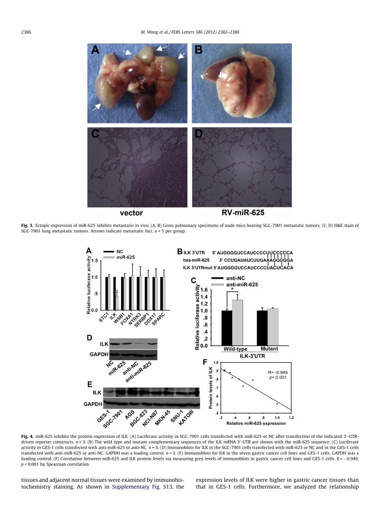
Fig. 3. Ectopic expression of miR-625 inhibits metastasis in vivo. (A, B) Gross pulmonary specimens of nude mice bearing SGC-7901 metastatic tumors. (C, D) H&E stain ofSGC-7901 lung metastatic tumors. Arrows indicate metastatic foci. n = 5 per group.
Fig. 4. miR-625 inhibits the protein expression of ILK. (A) Luciferase activity in SGC-7901 cells transfected with miR-625 or NC after transfection of the indicated 30-UTR-driven reporter constructs. n = 3. (B) The wild type and mutant complementary sequences of the ILK mRNA 30-UTR are shown with the miR-625 sequence. (C) Luciferaseactivity in GES-1 cells transfected with anti-miR-625 or anti-NC. n = 3. (D) Immunoblots for ILK in the SGC-7901 cells transfected with miR-625 or NC and in the GES-1 cellstransfected with anti-miR-625 or anti-NC. GAPDH was a loading control. n = 3. (E) Immunoblots for ILK in the seven gastric cancer cell lines and GES-1 cells. GAPDH was aloading control. (F) Correlation between miR-625 and ILK protein levels via measuring grey levels of immunoblots in gastric cancer cell lines and GES-1 cells. R = �0.949,p < 0.001 by Spearman correlation.
2386 M. Wang et al. / FEBS Letters 586 (2012) 2382–2388
tissues and adjacent normal tissues were examined by immunohis-tochemistry staining. As shown in Supplementary Fig. S13, the
expression levels of ILK were higher in gastric cancer tissues thanthat in GES-1 cells. Furthermore, we analyzed the relationship

Fig. 5. Knockdown of ILK inhibits cellular migration and invasion in SGC-7901 cells. (A, B) Immunoblots and qRT-PCR for ILK in the SGC-7901 cells transfected with si-ILK orcontrol. (C) Migration and invasion assays in SGC-7901 cells with si-ILK or control. n = 3.
M. Wang et al. / FEBS Letters 586 (2012) 2382–2388 2387
between miR-625 and ILK protein levels in tissues. The resultshows that miR-625 and ILK were negative correlated in gastriccancer tissues (p = 0.017, Supplementary Fig. S14).
3.7. Knockdown of ILK inhibits cellular migration and invasion in vitro
To confirm the effects of ILK on the migration and invasion ofSGC-7901 cells, ILK expression was knocked down by a ILK siRNA.qRT-PCR and immunoblot analyses showed that the si-ILK trans-fection of SGC-7901 cells effectively inhibited approximately87.6% of mRNA expression and 77.9% of protein expression ofILK, when compared to control groups (Fig. 5A, B). Consequently,wound-healing and transwell assays examining migration andinvasion were performed. Decreased migration (3.40 ± 0.15-fold,p = 0.002) and invasion (3.28 ± 0.20-fold, p = 0.003) cells were ob-served in SGC-7901 cells transfected with si-ILK than the controls(Fig. 5C). Similarly, when ILK was knocked down in MKN-45 cells(Supplementary Fig. S15), migration and invasion cells were de-creased (Supplementary Fig. S16). These suggest that knockdownof ILK suppresses migration and invasion in SGC-7901 cells andILK is an effective target gene of miR-625.
4. Discussion
In this study, we demonstrated the inhibitory effects of miR-625 on tumor metastasis at the clinical, animal experimental, cel-lular and molecular four different levels. We found that miR-625expression was down-regulated in gastric cancer and involved inlymphnode metastasis. Restoration of miR-625 in SGC-7901 andMKN-45 cells significantly impaired cellular migration and inva-sion in vitro and in vivo. In contrast, knockdown of miR-625 inGES-1 and NCI-N87 cells reversed the effects. Screened from eightputative genes based on bioinformatic algorithms, ILK was foundto be a target gene of miR-625. The tumor metastasis-associatedeffects of ILK were consistent with the functions of miR-625 onthe phenotypes in SGC-7901 and MKN-45 cells [12–14]. Given that
ILK regulates migration and invasion probably by the way ofLIMS1–ILK–parvin [15,16], we speculate that miR-625 may takepart in this pathway by regulating ILK.
Metastasis is a key step of tumor progression in gastric cancer,which means a poor prognosis [17]. Metastasis is a series ofsequential events, including detachment, migration, local invasion,angiogenesis, intravasation, survival in the circulatory system,extravasation, and regrowth in different organs [18–20]. SeveralmiRNAs can modulate tumor metastasis [21–23]. However, themolecular mechanisms of metastasis have not yet been fullyunderstood. Here, we show that miR-625 can regulated metastasisof gastric cancer through modulating the migration and local inva-sion at least in vitro, if not all. In vivo, we speculate that forcedmiR-625 suppressed the expression of ILK and then the motilityand extravasation of SGC-7901 cells from vessels was impaired,which contributes to reduction in lung metastasis. Further studiesbased on else metastatic steps would be required to research tofully understand the role of miR-625 in tumor metastasis.
Acknowledgments
This study was supported by Grants from National Natural Sci-ence Foundation of China (No. 30872476, No. 30900670 and No.81172324).
Appendix A. Supplementary data
Supplementary data associated with this article can be found, inthe online version, at http://dx.doi.org/10.1016/j.febslet.2012.05.050.
References
[1] Jemal, A., Bray, F., Center, M.M., Ferlay, J., Ward, E. and Forman, D. (2011)Global cancer statistics. CA Cancer J. Clin. 61, 69–90.
[2] Yuasa, Y. (2003) Control of gut differentiation and intestinal-type gastriccarcinogenesis. Nat. Rev. Cancer 3, 592–600.

2388 M. Wang et al. / FEBS Letters 586 (2012) 2382–2388
[3] Esquela-Kerscher, A. and Slack, F.J. (2006) Oncomirs - microRNAs with a role incancer. Nat. Rev. Cancer 6, 259–269.
[4] Zhang, Z., Li, Z., Gao, C., Chen, P., Chen, J., Liu, W., Xiao, S. and Lu, H. (2008) MiR-21 plays a pivotal role in gastric cancer pathogenesis and progression. Lab.Invest. 88, 1358–1366.
[5] Tsukamoto, Y. et al. (2010) MicroRNA-375 is downregulated in gastriccarcinomas and regulates cell survival by targeting PDK1 and 14–3-3zeta.Cancer Res. 70, 2339–2349.
[6] Zhu, L.H., Liu, T., Tang, H., Tian, R.Q., Su, C., Liu, M. and Li, X. (2010) MicroRNA-23a promotes the growth of gastric adenocarcinoma cell line MGC803 anddownregulates interleukin-6 receptor. FEBS J. 277, 3726–3734.
[7] Wan, H.Y., Guo, L.M., Liu, T., Liu, M., Li, X. and Tang, H. (2010) Regulation of thetranscription factor NF-kappaB1 by microRNA-9 in human gastricadenocarcinoma. Mol. Cancer 9, 16.
[8] Li, N. et al. (2012) Increased miR-222 in H. pylori-associated gastric cancercorrelated with tumor progression by promoting cancer cell proliferation andtargeting RECK. FEBS Lett 586, 722–728.
[9] Feng, R. et al. (2010) MiR-126 functions as a tumour suppressor in humangastric cancer. Cancer Lett. 298, 50–63.
[10] Li, C.L. et al. microRNA-155 is downregulated in gastric cancer cells andinvolved in cell metastasis. Oncol Rep. 27, 1960–1966.
[11] Li, C. et al. (2012). MicroRNA-409-3p regulates cell proliferation and apoptosisby targeting PHF10 in gastric cancer. Cancer Lett. 320, 189–197.
[12] Ito, R., Oue, N., Zhu, X., Yoshida, K., Nakayama, H., Yokozaki, H. and Yasui, W.(2003) Expression of integrin-linked kinase is closely correlated with invasionand metastasis of gastric carcinoma. Virchows Arch. 442, 118–123.
[13] Kim, Y.B., Lee, S.Y., Ye, S.K. and Lee, J.W. (2007) Epigenetic regulation ofintegrin-linked kinase expression depending on adhesion of gastric carcinomacells. Am. J. Physiol. Cell Physiol. 292, C857–C866.
[14] Zhao, G., Guo, L.L., Xu, J.Y., Yang, H., Huang, M.X. and Xiao, G. (2011) Integrin-linked kinase in gastric cancer cell attachment, invasion and tumor growth.World J. Gastroenterol. 17, 3487–3496.
[15] Wu, C. (2004) The PINCH-ILK-parvin complexes: assembly, functions andregulation. Biochim. Biophys. Acta 1692, 55–62.
[16] Legate, K.R., Montanez, E., Kudlacek, O. and Fassler, R. (2006) ILK, PINCH andparvin: the tIPP of integrin signalling. Nat. Rev. Mol. Cell Biol. 7, 20–31.
[17] Oue, N., Aung, P.P., Mitani, Y., Kuniyasu, H., Nakayama, H. and Yasui, W. (2005)Genes involved in invasion and metastasis of gastric cancer identified byarray-based hybridization and serial analysis of gene expression. Oncology 69(Suppl 1), 17–22.
[18] Gupta, G.P. and Massague, J. (2006) Cancer metastasis: building a framework.Cell 127, 679–695.
[19] Steeg, P.S. (2006) Tumor metastasis: mechanistic insights and clinicalchallenges. Nat. Med. 12, 895–904.
[20] Klein, C.A. (2008) Cancer. The metastasis cascade. Science 321, 1785–1787.[21] Sreekumar, R., Sayan, B.S., Mirnezami, A.H. and Sayan, A.E. (2011) MicroRNA
Control of Invasion and Metastasis Pathways. Front Genet. 2, 58.[22] Gao, P., Xing, A.Y., Zhou, G.Y., Zhang, T.G., Zhang, J.P., Gao, C., Li, H. and Shi, D.B.
(2012). The molecular mechanism of microRNA-145 to suppress invasion-metastasis cascade in gastric cancer. Oncogene.
[23] Tie, J. et al. (2010) MiR-218 inhibits invasion and metastasis of gastric cancerby targeting the Robo1 receptor. PLoS Genet. 6, e1000879.