Purification and Properties of Rabbit Skeletal Muscle Phosphorylase b Kinase
Upload
independentCategory
view
7download
0
Biochemical and Biophysical Research Communications 309 (2003) 923–928
www.elsevier.com/locate/ybbrc
BBRC
Docking and small angle X-ray scattering studiesof purine nucleoside phosphorylase
Walter Filgueira de Azevedo Jr.,a,b,* Giovanni C�eesar dos Santos,a
Denis Marangoni dos Santos,a Johnny Rizzieri Olivieri,a,b Fernanda Canduri,a,b
Rafael Guimar~aaes Silva,c Luiz Augusto Basso,c Gaby Renard,c Isabel Os�oorio da Fonseca,c
Maria Anita Mendes,b,d M�aario S�eergio Palma,b,d and Di�oogenes Santiago Santosc
a Departamento de F�ıısica, UNESP, S~aao Jos�ee do Rio Preto, SP 15054-000, Brazilb Center for Applied Toxinology, Instituto Butantan, S~aao Paulo, SP 05503-900, Brazil
c Rede Brasileira de Pesquisas em Tuberculose, Departamento de Biologia Molecular e Biotecnologia, UFRGS, Porto Alegre, RS 91501-970, Brazild Laboratory of Structural Biology and Zoochemistry, Department of Biology, Institute of Biosciences, UNESP, Rio Claro, SP 13506-900, Brazil
Received 19 August 2003
Abstract
Docking simulations have been used to assess protein complexes with some success. Small angle X-ray scattering (SAXS) is a
well-established technique to investigate protein spatial configuration. This work describes the integration of geometric docking with
SAXS to investigate the quaternary structure of recombinant human purine nucleoside phosphorylase (PNP). This enzyme catalyzes
the reversible phosphorolysis of N-ribosidic bonds of purine nucleosides and deoxynucleosides. A genetic deficiency due to mu-
tations in the gene encoding for PNP causes gradual decrease in T-cell immunity. Inappropriate activation of T-cells has been
implicated in several clinically relevant human conditions such as transplant rejection, rheumatoid arthritis, lupus, and T-cell
lymphomas. PNP is therefore a target for inhibitor development aiming at T-cell immune response modulation and has been
submitted to extensive structure-based drug design. The present analysis confirms the trimeric structure observed in the crystal. The
potential application of the present procedure to other systems is discussed.
� 2003 Elsevier Inc. All rights reserved.
Keywords: Geometric docking; SAXS; Purine nucleoside phosphorylase; Bioinformatics
Recent developments in the algorithm for protein
docking allowed the prediction of the conformation of
quaternary structures of several proteins. Among all
available algorithms for docking of biological macro-
molecules the geometric docking has been proved to
generate reasonable models of several macromolecularassemblies [1,2]. Small angle X-ray scattering (SAXS)
technique provides information on the structural char-
acteristics of macromolecules in solution at a super-
atomic scale. One of the procedures to obtain structural
information from SAXS results is based on the
comparison between structure functions of proposed
models with different configurations of monomers or
* Corresponding author. Fax: +55-17-221-2247.
E-mail address: [email protected] (W.F. de Azevedo Jr.).
0006-291X/$ - see front matter � 2003 Elsevier Inc. All rights reserved.
doi:10.1016/j.bbrc.2003.08.093
subdomains with those determined from experiments.
Even though this technique cannot guarantee the
uniqueness of the model, it is widely used and was
demonstrated to yield useful information on the struc-
ture, on structural variations, and on the quaternary
structure of a number of macromolecules of biologicalinterest [3]. The Guinier analysis of SAXS intensity
provides a structural parameter, the radius of gyration
of the macromolecule in solution, which is independent
of any a priori model. The SAXS method also yields
information on the spatial configuration of the macro-
molecular subdomains but ignores internal structural
details and dynamics features such as, vibration, rota-
tion or internal conformational changes [4].Purine nucleoside phosphorylase (PNP) catalyzes the
reversible phosphorolysis of the ribonucleosides and

Fig. 1. Flowchart describing the overall strategy to assess protein
complex conformations.
924 W.F. de Azevedo Jr. et al. / Biochemical and Biophysical Research Communications 309 (2003) 923–928
20-deoxyribonucleosides of guanine, hypoxanthine, anda number of related nucleoside compounds [5], except
adenosine. Human PNP is an attractive target for drug
design and it has been submitted to extensive structure-
based design. PNP inhibitors could be used in the fol-
lowing applications: (1) treatment of T-cell leukemia;
(2) suppression of the host versus graft response in
organ transplantation recipients; (3) treatment of sec-
ondary or xanthine gout by restricting purine catabo-lites to the more soluble nucleosides; and (4) in
combination with nucleosides to prevent their degra-
dation by PNP metabolism [6]. More recently, the
structure of human PNP has been solved using cryo-
crystallographic techniques at 2.3�AA resolution, which
allowed a redefinition of the residues involved in the
substrate binding sites [7,8]. The crystallographic
structure is a trimer, however, there is a report of di-meric structure for the human enzyme [9], which may
change the subunit interface. Since the active site is
located near the interface of two subunits, changing the
putative interactions between enzyme and inhibitors
should have a bearing on structure-based inhibitor
design.
Here we report the combination of geometric docking
simulations and SAXS studies to assess the human PNPquaternary structure in solution. The general procedure,
here described, may be used to study the spatial con-
figuration of the macromolecular subdomains of other
proteins in solution.
Materials and methods
Integration of geometric docking simulation and SAXS experiments.
In order to assess the quaternary structure of PNP a scheme was used
that involved both geometric docking simulations and SAXS experi-
ments. A flowchart describing the overall strategy is shown in Fig. 1.
This procedure was used to generate the dimeric models for PNP and
the trimeric structure was built using the crystallographic symmetry. In
order to speed up the geometric docking simulations, a parallel version
of the program GRAMM [10] was used to generate the dimeric models
for human PNP. Each step of the procedure is described in the
following sections.
Geometric docking simulations. In order to obtain the dimeric
structure of human PNP, docking simulation was performed using the
geometric recognition algorithm, which was developed to identify
molecular surface complementarity. The monomeric structure of hu-
man PNP (PDB access code: 1M73) [7] was docked against its own
structure. It generated a total of 100 dimers. The geometric recognition
algorithm is based on a geometrical approach and involves an auto-
mated procedure including: (i) a digital representation of the molecules
by three-dimensional discrete functions; (ii) the calculation of a cor-
relation function that assesses the degree of molecular surface overlap
and penetration upon relative shifts of the molecules in three dimen-
sions; and (iii) a scan of the relative orientations of the molecules [10].
The procedure is equivalent to a six-dimensional search but consid-
erably faster by design, and the computation time is only moderately
dependent on molecular size. This procedure has been applied to assess
protein–protein and protein–ligand interactions. The geometric rec-
ognition algorithm was implemented in the program GRAMM [10].
All geometric docking simulations were performed on a Beowulf
cluster, with 16 nodes (B16/AMD Athlon 1800+; BioComp, S~aao Jos�ee
do Rio Preto, SP, Brazil).
SAXS studies. X-ray scattering data were collected at room tem-
perature using Cu Ka X-rays radiation generated by a Rigaku RU300
rotating anode generator operated at 50 kV and 90mA and collimated
with a block slit system [11]. The scattering intensity was measured
using a linear position sensitive detector (CBPF-Brazil).
The SAXS measurements were performed within an angular range
defined by 0:02�AA�1
< h < 0:450�AA�1
where h ¼ ð4p sinÞ=k, 2h being the
angle between the incident and the scattered X-ray beam and k the
X-ray wavelength. The contributions to the scattering intensity from
the solvent, capillary, and air were subtracted from the total intensity.
Recombinant human PNP was expressed and purified as previously
described [12]. The SAXS measurements were carried out using human
PNP solution, which was concentrated to 12mgml�1 against 10mM
potassium phosphate buffer (pH 7.1). The counting time was 12 h. The
extrapolated experimental SAXS intensity function was desmeared to
suppress the influence from the slit collimation system yielding the
corrected intensities, IðhÞ.A structural parameter related to the overall size of the macro-
molecule, the radius of gyration Rg, was determined by using the
Guinier equation [13]
IðhÞ ¼ Ið0Þ exp"�h2R2
g
3
#: ð1Þ
Eq. (1) applies to macromolecules in the limits of a dilute solution and
small h values. More detailed information of the molecular structure
can be obtained from the distance distribution function pðrÞ, which is
related to the SAXS desmeared (free from geometrical collimation
effects) intensity IðhÞ by

W.F. de Azevedo Jr. et al. / Biochemical and Biophysical Research Communications 309 (2003) 923–928 925
pðrÞ ¼ 1
2p2
Z 1
0
IðhÞðhrÞ sinðhrÞdh: ð2Þ
The pðrÞ function is proportional to the number of pairs of electrons
separated by the distance r, which is encountered by combinations
between all the elements of the macromolecule. The radius of gyration
of macromolecules in solution is usually determined by applying Eq.
(1). The distance distribution function, pexpðrÞ, has been determined by
indirect Fourier transformation using the ITP program [11]. This
program was also used to determine the intensity IðhÞ, free from
smearing collimation effects. The theoretical function ptheoðrÞ was cal-culated using the program MULTIBODY [11], modified in order to
make molecular model building easier [4]. The program MULTI-
BODY calculates the resulting function pðrÞ of the complete set of
atomic coordinates of each structural model for the macromolecule. In
the present study we calculated the ptheoðrÞ for monomer, for the
dimers, generated by geometric docking, and for the trimer, obtained
by application of crystallographic rotations.
Correlation between geometric docking simulations and SAXS
experiments. To assess the correlation between theoretical and exper-
imental distance distribution function pexpðrÞ and ptheoðrÞ, respectively,we have calculated the linear correlation coefficient (CC), which is
defined as follows [14]:
CC ¼Pn
i jpexp;iðrÞj2 � jpexpðrÞj2� �
� jptheo;iðrÞj2 � jptheoðrÞj2� �h i
Pni jpexp;iðrÞj2 � jpexpðrÞj2
� �2 Pni jptheo;iðrÞj2 � jptheoðrÞj2� �2
� �12
;
ð3Þ
where jpexpðrÞj2 is the mean of the jpexp;iðrÞj2, jptheoðrÞj2 is the mean of
jptheo;iðrÞj2, and sums are made over all available pðrÞ. When a corre-
lation is known to be significant, CC is one conventional way of
summarizing its strength. The complex, which generates the highest
correlation coefficient, is considered the right macromolecular con-
formation. In the present work, we calculated the CC for all dimeric
models obtained from the geometric docking simulations and for the
monomeric and trimeric structures.
Results and discussion
Guinier plot (log IðhÞ versus h2) of the desmeared
scattering function is displayed in Fig. 2. The slope of
Fig. 2. Guinier plot of the SAXS intensities, IðhÞ, for human PNP. The
straight line was obtained by least-squares fitting in the region
h2 < 1:8:10�3 �AA2.
linear portion of this plot was determined to obtain theradius of gyration of human PNP. The Rg value was
29.8�AA.
PNPs from most mammalian and some of the bac-
terial sources appear to be trimeric although dimeric
quaternary structures have been proposed for the
human enzyme [9]. Analysis of the crystallographic
structures of human PNP indicates a trimeric structure
(PDB access codes: 1ULA, 1ULB, 1M73, and 1PWY)[6–8,15]. However, in a number of instances the qua-
ternary structure observed in the crystalline state is not
conserved in solution [3]. Furthermore, in the case of
human PNP the low pH used in the crystallization
condition [16] may generate differences in the spatial
configuration of the macromolecular subdomains ob-
served in the crystal when compared to the structure in
physiological pH. In addition, since the active site of thePNP is located near the interface of two subunits within
the trimer, the precise information about the biological
unit in solution is of capital importance to guide the
structure-based design of inhibitors because its target is
a structure as close as possible to the structure found in
the physiological conditions, where the drug will act. Up
to now all structure-based designs of PNP inhibitors
have used the low-resolution structures of human PNP(PDB access codes: 1ULA and 1ULB) and consider the
trimer as the target for molecular modeling studies.
Three families of theoretical models, based on the
high-resolution crystallographic structure (PDB access
code: 1M73) [7], were used to determine the theoretical
distance distribution function, ptheoðrÞ, using the pro-
gram MULTIBODY and then compared with the ex-
perimental function pðrÞ determined using the ITPprogram from SAXS data. Figs. 3A–C show structural
models and the experimental distribution function
against the theoretical distribution function for
the monomer, dimer, and trimer, of human PNP,
respectively.
The atomic coordinates for monomeric structure
were obtained from the asymmetric unit content of the
crystallographic structure of human PNP solved at 2.3�AAresolution [7]. Previous statistical analysis of low-reso-
lution docking indicated that gross structural features of
protein–protein interactions could be identified for a
significant percentage of protein complexes [1]. There-
fore, the low-resolution protocol of the GRAMM pro-
gram [10] was used to generate the dimeric models. A
total of 100 models for the dimeric structure were built,
only the complex, which generated the highest correla-tion coefficient between theoretical and experimental
distance distribution function is shown in Fig. 3B. The
trimeric structure was built applying two successive
rotations of 120� along z-axis on the atomic coordinates
of the monomer. The radii of gyration for the structural
models are 18.7, 26.5, and 28.7�AA, for the monomer,
dimer, and trimer, respectively.
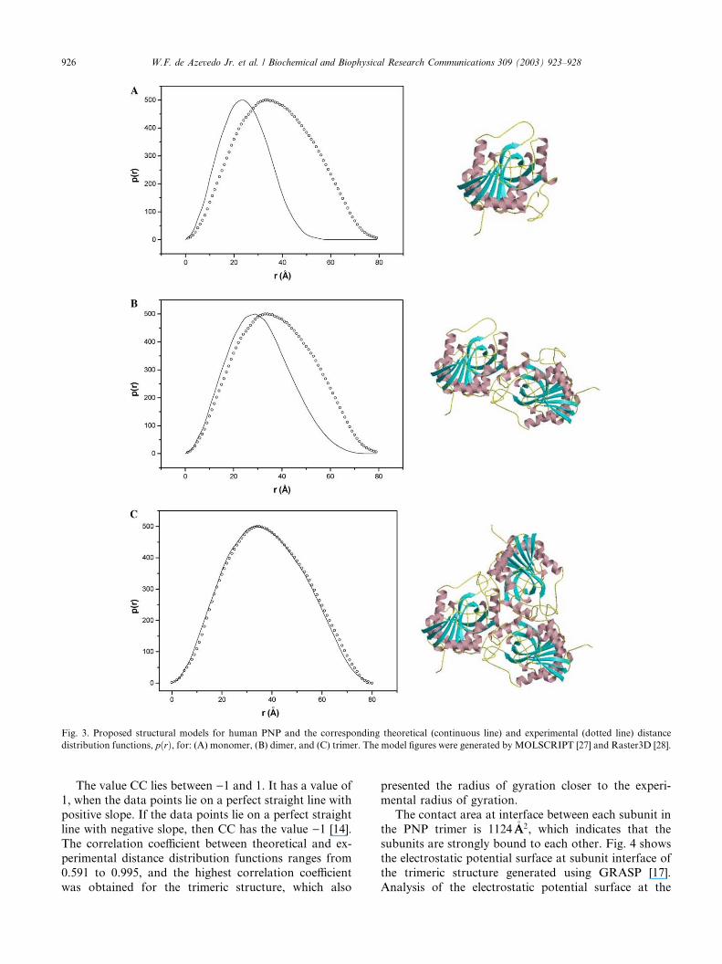
Fig. 3. Proposed structural models for human PNP and the corresponding theoretical (continuous line) and experimental (dotted line) distance
distribution functions, pðrÞ, for: (A) monomer, (B) dimer, and (C) trimer. The model figures were generated by MOLSCRIPT [27] and Raster3D [28].
926 W.F. de Azevedo Jr. et al. / Biochemical and Biophysical Research Communications 309 (2003) 923–928
The value CC lies between )1 and 1. It has a value of1, when the data points lie on a perfect straight line with
positive slope. If the data points lie on a perfect straight
line with negative slope, then CC has the value )1 [14].
The correlation coefficient between theoretical and ex-
perimental distance distribution functions ranges from
0.591 to 0.995, and the highest correlation coefficient
was obtained for the trimeric structure, which also
presented the radius of gyration closer to the experi-mental radius of gyration.
The contact area at interface between each subunit in
the PNP trimer is 1124�AA2, which indicates that the
subunits are strongly bound to each other. Fig. 4 shows
the electrostatic potential surface at subunit interface of
the trimeric structure generated using GRASP [17].
Analysis of the electrostatic potential surface at the

Fig. 4. Electrostatic potential surface at subunit interface of human PNP, calculated with GRASP [17] and shown from )10 to +10 kT. Uncharged
regions are white.
W.F. de Azevedo Jr. et al. / Biochemical and Biophysical Research Communications 309 (2003) 923–928 927
subunit interface indicates good shape complementarity
and some charge complementarity; however, most of thecontacts are hydrophobic and involve residues Tyr88,
Phe141, Phe159, Phe200, and Leu209.
The trimeric PNP structure has been extensively used
for structure-based studies of PNP inhibitors [5,6,18–
25]. However, the quaternary structure of human PNP
in solution and in physiological pH has not been pre-
viously investigated using low-resolution methods, such
as SAXS. The present analysis of the SAXS experimentsintegrated with geometric docking simulation strongly
indicates that human PNP is a trimer in solution, the
agreement found between the experimental and theo-
retical pðrÞ functions for the trimer suggests that struc-
ture in solution adopts approximately the same
conformation identified in the high-resolution crystal-
lographic structure (PDB access code: 1M73) [7]. The
radius of gyration determined for the trimeric structureis slightly smaller than that determined from the Guinier
plot (log IðhÞ versus h2) of the desmeared scattering
function. The possible reasons for this discrepancy may
be the cryogenic conditions used to solve the high-res-
olution structure of human PNP and the absence of
solvents in the theoretical model.
The integration of a high-efficient algorithm for
geometric docking with SAXS experiments allowed theinvestigation of the possible quaternary structures not
observed in the crystalline state, such as the putative
PNP dimeric structure [9]. The procedure adopted to
analyze the interaction between PNP subunits can be
used for other protein complexes. The main applications
of the present methodology are: (1) analysis of interac-
tions between biological macromolecules using struc-
tural models obtained from crystallography or NMR,(2) validation of structural models obtained from
molecular modeling [26] of complexes of biological
macromolecules, and (3) analysis of complexes of
biological macromolecules in conditions closer to the
biological environment.Geometric docking simulations may be omitted from
the strategy if the atomic coordinates for the complexes
are available. We are applying the procedure, here de-
scribed, to assess the quaternary structure of a number
of protein complexes, such as hemoglobins, PNPs, and
crotoxin.
Acknowledgments
This work was supported by grants from FAPESP (SMOLBNet,
Proc. Num. 01/07532-0), CNPq, CAPES and Instituto do Millenium
(CNPq-MCT). W.F.A. (CNPq, 300851/98-7), M.S.P. (CNPq, 500079/
90-0), and L.A.B. (CNPq, 520182/99-5) are researchers for the Bra-
zilian Council for Scientific and Technological Development.
References
[1] A. Tovchigrechko, C.A. Wells, I.A. Vakser, Docking of protein
models, Protein Sci. 11 (2002) 1888–1896.
[2] N. Jing, C. Marchand, J. Liu, R. Mitra, M.E. Hogan, Y.
Pommier, Mechanism of inhibition of HIV-1 integrase by G-
tetrad-forming oligonucleotides in vitro, J. Biol. Chem. 275 (2000)
21460–21467.
[3] D.I. Svergun, C. Barberato, M.H.J. Koch, L. Fetler, P. Vachette,
Large differences are observed between the crystal and solution
quaternary structures of allosteric aspartate transcarbamylase in
the R state, Proteins Struct. Funct. Genet. 27 (1997) 110–117.
[4] J.R. Olivieri, A.F. Craievich, The subdomain structure of human
serum albumin in solution under different pH conditions studied
by small angle X-ray scattering, Eur. Biophys. J. 24 (1995) 77–
84.
[5] J.A. Montgomery, Purine nucleoside phosphorylase: a target for
drug design, Med. Res. Rev. 13 (1993) 209–228.
[6] S.E. Ealick, Y.S. Babu, C.E. Bugg, M.D. Erion, W.C. Guida, J.A.
Montgomery, J.A. Secrist III, Application of crystallographic and
modeling methods in the design of purine nucleoside phosphor-
ylase inhibitors, Proc. Natl. Acad. Sci. USA 91 (1991) 11540–
11544.

928 W.F. de Azevedo Jr. et al. / Biochemical and Biophysical Research Communications 309 (2003) 923–928
[7] W.F. de Azevedo Jr., F. Canduri, D.M. Santos, R.G. Silva, J.S.
Oliveira, L.P.S. Carvalho, L.A. Basso, M.A. Mendes, M.S. Palma,
D.S. Santos, Crystal structure of human purine nucleoside
phosphorylase at 2.3�AA resolution, Biochem. Biophys. Res. Com-
mun. 308 (2003) 545–552.
[8] D.M. Santos, F. Canduri, J.H. Pereira, M.V.B. Dias, R.G. Silva,
M.A. Mendes, M.S. Palma, L.A. Basso, W.F. de Azevedo, D.S.
Santos, Crystal structure of human purine nucleoside phosphor-
ylase complexed with acyclovir, Biochem. Biophys. Res. Com-
mun. 308 (2003) 553–559.
[9] A.S. Lewis, B.A. Lowy, Human erythrocytes purine nucleoside
phosphorylase: molecular weight and physical properties, J. Biol.
Chem. 254 (1979) 9927–9932.
[10] E. Katchalski-Katzir, I. Shariv, M. Eisenstein, A.A. Friesem, C.
Aflalo, I.A. Vakser, Molecular surface recognition: determination
of geometric fit between proteins and their ligands by correlation
techniques, Proc. Natl. Acad. Sci. USA 89 (1992) 2195–2199.
[11] O. Glatter, in: O. Glatter, O. Kratky (Eds.), Small Angle X-Ray
Scattering, Academic Press, London, 1982.
[12] R.G. Silva, L.P. Carvalho, J.S. Oliveira, C.A. Pinto, M.A.
Mendes, M.S. Palma, L.A. Basso, D.S. Santos, Cloning, overex-
pression, and purification of functional human purine nucleoside
phosphorylase, Protein Expr. Purif. 27 (2003) 158–164.
[13] A. Guinier, G. Fournet, Small-Angle Scattering of X-rays, Wiley,
New York, 1955.
[14] W.H. Press, S.A. Teukolsky, W.T. Vetterling, B.P. Flannery,
Numerical Recipes in FORTRAN. The Art of Scientific Com-
puting, second ed., Cambridge University Press, New York, 1992.
[15] S.E. Ealick, S.A. Rule, D.C. Carter, T.J. Greenhough, V. Babu,
W.J. Cook, J. Habash, J.R. Helliwell, J.D. Stoeckler, R.E. Parks
Jr., F. Chen, C.E. Bugg, Three-dimensional structure of human
erythrocytic purine nucleoside phosphorylase at 3.2�AA resolution,
J. Biol. Chem. 265 (1990) 1812–1820.
[16] W.J. Cook, S.E. Ealick, C.E. Bugg, J.D. Stoeckler, R.E. Parks Jr.,
Crystallization and preliminary X-ray investigation of human
erythrocytic purine nucleoside phosphorylase, J. Biol. Chem. 256
(1981) 4079–4080.
[17] A. Nicholls, K. Sharp, B. Honig, Protein folding and association:
insights from the interfacial and thermodynamic properties of
hydrocarbons, Proteins Struct. Funct. Genet. 11 (1991) 281–296.
[18] P.W.K. Woo, C.R. Kostlan, J.C. Sircar, M.K. Dong, R.B.
Gilbertsen, Inhibitors of human purine nucleoside phosphorylase.
Synthesis and biological activities of 8-amino-3-benzylhypoxan-
thine and related analogues, J. Med. Chem. 35 (1992) 1451–1457.
[19] J.-W. Chern, H.-Y. Lee, C.-S. Chen, Nucleosides. 5. Synthesis of
guanine and formycin B derivatives as potential inhibitors of
purine nucleoside phosphorylase, J. Med. Chem. 36 (1993) 1024–
1031.
[20] M.D. Erion, S. Niwas, J.D. Rose, S. Ananthan, M. Allen, J.A.
Secrist III, Y.S. Babu, C.E. Bugg, W.C. Guida, S.E. Ealick, J.A.
Montgomery, Structure-based design of inhibitors of purine
nucleoside phosphorylase. 3. 9-Arylmethyl derivatives of 9-
deazaguanine substituted on the methylene group, J. Med. Chem.
36 (1993) 3771–3783.
[21] J.A. Secrist III, S. Niwas, J.D. Rose, Y.S. Babu, C.E. Bugg, M.D.
Erion, W.C. Guida, S.E. Ealick, J.A. Montgomery, Structure-
based design of inhibitors of purine nucleoside phosphorylase. 2.
9-Alicyclic and 9-heteroalicyclic derivatives of 9-deazaguanine, J.
Med. Chem. 36 (1993) 1847–1854.
[22] W.C. Guida, R.D. Elliot, H.J. Thomas, J.A. Secrist III, Y.S.
Babu, C.E. Bugg, M.D. Erion, S.E. Ealick, J.A. Montgomery,
Structure-based design of inhibitors of purine nucleoside phos-
phorylase. 4. A study of phosphate mimics, J. Med. Chem. 37
(1994) 1109–1114.
[23] S. Niwas, P. Chand, V.P. Pathak, J.A. Montgomery, Structure-
based design of inhibitors of purine nucleoside phosphorylase. 5.
9-Deazahypoxanthines, J. Med. Chem. 37 (1994) 2477–2480.
[24] P.E. Morris, A.J. Elliott, S.P. Walton, C.H. Williams, J.A.
Montgomery, Synthesis and biological activity of a novel class of
purine nucleoside phosphorylase inhibitors, Nucleosides Nucleo-
tides Nucleic Acids 19 (2000) 379–404.
[25] A. Andricopulo, R.A. Yunes, Structure–activity relationships for
a collection of structurally diverse inhibitors of purine nucleoside
phosphorylase, Chem. Pharm. Bull. 49 (2001) 10–17.
[26] A. Sali, T.L. Blundell, Comparative protein modelling by satis-
faction of spatial restraints, J. Mol. Biol. 234 (1993) 779–815.
[27] P.J. Kraulis, MOLSCRIPT: a program to produce both detailed
and schematic plots of proteins, J. Appl. Cryst. 24 (1991) 946–950.
[28] E.A. Merritt, D.J. Bacon, Raster3D: photorealistic molecular
graphics, Methods Enzimol. 277 (1997) 505–524.
Copyright © 2022 FDOKUMEN