Divergent functions of the Arabidopsis mitochondrial SCO proteins: HCC1 is essential for COX...
17
ORIGINAL RESEARCH ARTICLE published: 25 March 2014 doi: 10.3389/fpls.2014.00087 Divergent functions of the Arabidopsis mitochondrial SCO proteins: HCC1 is essential for COX activity while HCC2 is involved in the UV-B stress response Iris Steinebrunner 1 *, Uta Gey 1 , Manuela Andres 1 , Lucila Garcia 2 and Daniel H. Gonzalez 2 1 Department of Biology, Technische Universität Dresden, Dresden, Germany 2 Instituto de Agrobiotecnología del Litoral (CONICET-UNL), Universidad Nacional del Litoral, Santa Fe, Argentina Edited by: Philippe Giegé, Centre National de la Recherche Scientifique, France Reviewed by: Xia Wu, University of Washington, USA Monika Weronika Murcha, University of Western Australia, Australia *Correspondence: Iris Steinebrunner, Department of Biology, Technische Universität Dresden, Helmholtzstr. 10, 01062 Dresden, Germany e-mail: iris.steinebrunner@ tu-dresden.de The two related putative cytochrome c oxidase (COX) assembly factors HCC1 and HCC2 from Arabidopsis thaliana are Homologs of the yeast Copper Chaperones Sco1p and Sco2p. The hcc1 null mutation was previously shown to be embryo lethal while the disruption of the HCC2 gene function had no obvious effect on plant development, but increased the expression of stress-responsive genes. Both HCC1 and HCC2 contain a thioredoxin domain, but only HCC1 carries a Cu-binding motif also found in Sco1p and Sco2p. In order to investigate the physiological implications suggested by this difference, various hcc1 and hcc2 mutants were generated and analyzed. The lethality of the hcc1 knockout mutation was rescued by complementation with the HCC1 gene under the control of the embryo-specific promoter ABSCISIC ACID INSENSITIVE 3. However, the complemented seedlings did not grow into mature plants, underscoring the general importance of HCC1 for plant growth. The HCC2 homolog was shown to localize to mitochondria like HCC1, yet the function of HCC2 is evidently different, because two hcc2 knockout lines developed normally and exhibited only mild growth suppression compared with the wild type (WT). However, hcc2 knockouts were more sensitive to UV-B treatment than the WT. Complementation of the hcc2 knockout with HCC2 rescued the UV-B-sensitive phenotype. In agreement with this, exposure of wild-type plants to UV-B led to an increase of HCC2 transcripts. In order to corroborate a function of HCC1 and HCC2 in COX biogenesis, COX activity of hcc1 and hcc2 mutants was compared. While the loss of HCC2 function had no significant effect on COX activity, the disruption of one HCC1 gene copy was enough to suppress respiration by more than half compared with the WT. Therefore, we conclude that HCC1 is essential for COX function, most likely by delivering Cu to the catalytic center. HCC2, on the other hand, seems to be involved directly or indirectly in UV-B-stress responses. Keywords: SCO (synthesis of cytochrome c oxidase), mitochondria, copper chaperone, COX complex, UV-B stress, plant growth and development, BN-PAGE, Arabidopsis thaliana INTRODUCTION Mitochondrial (mt) biogenesis requires the assembly of pro- teins synthesized in two different compartments. In addition, due to the essential redox nature of many processes that take place in mitochondria, the formation of active components also requires the synthesis, transport and insertion of a set of co- factors (Herrmann and Funes, 2005; Barrientos et al., 2009; Kim et al., 2012). Among these, copper is one of the compo- nents which is required for the activity of cytochrome c oxi- dase (COX or complex IV; Cobine et al., 2006). COX contains two copper centers located in the subunits COX1 and COX2. Insertion of copper into COX is an intricate process that requires the participation of several mt proteins that function in either its delivery or redox reactions related with the assembly pro- cess (Herrmann and Funes, 2005; Cobine et al., 2006). The occurrence of COX assembly factors in prokaryotes suggests that some of them were already present in the endosymbiont that originated the mitochondrion while others are more recent acquisitions. A family of proteins that has been related with copper inser- tion into COX, particularly into the COX2 subunit, is the SCO family. SCO proteins are of prokaryotic origin and usually con- tain a transmembrane domain and a soluble domain that contains redox-active cysteines and a histidine presumably involved in cop- per binding (Banci et al., 2011). The fact that the soluble domain contains a thioredoxin fold has prompted some authors to pos- tulate that SCO proteins do not act in the direct transfer of copper to COX but rather in the reduction of the COX2 cys- teines involved in copper binding (Balatri et al., 2003; Abriata et al., 2008). SCO proteins were first analyzed in Saccharomyces (S.) cerevisiae mutants defective in COX assembly (hence their name, Synthesis of Cytochrome c Oxidase; Schulze and Rödel, 1988). However, current evidence of the presence of SCO proteins in bacteria that do not contain COX-like proteins (Arnesano et al., www.frontiersin.org March 2014 | Volume 5 | Article 87 | 1
-
Upload
independent -
Category
Documents
-
view
1 -
download
0
Transcript of Divergent functions of the Arabidopsis mitochondrial SCO proteins: HCC1 is essential for COX...

ORIGINAL RESEARCH ARTICLEpublished: 25 March 2014
doi: 10.3389/fpls.2014.00087
Divergent functions of the Arabidopsis mitochondrial SCOproteins: HCC1 is essential for COX activity while HCC2 isinvolved in the UV-B stress responseIris Steinebrunner1*, Uta Gey1, Manuela Andres1, Lucila Garcia2 and Daniel H. Gonzalez2
1 Department of Biology, Technische Universität Dresden, Dresden, Germany2 Instituto de Agrobiotecnología del Litoral (CONICET-UNL), Universidad Nacional del Litoral, Santa Fe, Argentina
Edited by:
Philippe Giegé, Centre National de laRecherche Scientifique, France
Reviewed by:
Xia Wu, University of Washington,USAMonika Weronika Murcha,University of Western Australia,Australia
*Correspondence:
Iris Steinebrunner, Department ofBiology, Technische UniversitätDresden, Helmholtzstr. 10, 01062Dresden, Germanye-mail: [email protected]
The two related putative cytochrome c oxidase (COX) assembly factors HCC1 and HCC2from Arabidopsis thaliana are Homologs of the yeast Copper Chaperones Sco1p andSco2p. The hcc1 null mutation was previously shown to be embryo lethal while thedisruption of the HCC2 gene function had no obvious effect on plant development, butincreased the expression of stress-responsive genes. Both HCC1 and HCC2 contain athioredoxin domain, but only HCC1 carries a Cu-binding motif also found in Sco1p andSco2p. In order to investigate the physiological implications suggested by this difference,various hcc1 and hcc2 mutants were generated and analyzed. The lethality of the hcc1knockout mutation was rescued by complementation with the HCC1 gene under thecontrol of the embryo-specific promoter ABSCISIC ACID INSENSITIVE 3. However, thecomplemented seedlings did not grow into mature plants, underscoring the generalimportance of HCC1 for plant growth. The HCC2 homolog was shown to localize tomitochondria like HCC1, yet the function of HCC2 is evidently different, because twohcc2 knockout lines developed normally and exhibited only mild growth suppressioncompared with the wild type (WT). However, hcc2 knockouts were more sensitive toUV-B treatment than the WT. Complementation of the hcc2 knockout with HCC2 rescuedthe UV-B-sensitive phenotype. In agreement with this, exposure of wild-type plants toUV-B led to an increase of HCC2 transcripts. In order to corroborate a function of HCC1and HCC2 in COX biogenesis, COX activity of hcc1 and hcc2 mutants was compared.While the loss of HCC2 function had no significant effect on COX activity, the disruptionof one HCC1 gene copy was enough to suppress respiration by more than half comparedwith the WT. Therefore, we conclude that HCC1 is essential for COX function, most likelyby delivering Cu to the catalytic center. HCC2, on the other hand, seems to be involveddirectly or indirectly in UV-B-stress responses.
Keywords: SCO (synthesis of cytochrome c oxidase), mitochondria, copper chaperone, COX complex, UV-B stress,
plant growth and development, BN-PAGE, Arabidopsis thaliana
INTRODUCTIONMitochondrial (mt) biogenesis requires the assembly of pro-teins synthesized in two different compartments. In addition,due to the essential redox nature of many processes that takeplace in mitochondria, the formation of active components alsorequires the synthesis, transport and insertion of a set of co-factors (Herrmann and Funes, 2005; Barrientos et al., 2009;Kim et al., 2012). Among these, copper is one of the compo-nents which is required for the activity of cytochrome c oxi-dase (COX or complex IV; Cobine et al., 2006). COX containstwo copper centers located in the subunits COX1 and COX2.Insertion of copper into COX is an intricate process that requiresthe participation of several mt proteins that function in eitherits delivery or redox reactions related with the assembly pro-cess (Herrmann and Funes, 2005; Cobine et al., 2006). Theoccurrence of COX assembly factors in prokaryotes suggeststhat some of them were already present in the endosymbiont
that originated the mitochondrion while others are more recentacquisitions.
A family of proteins that has been related with copper inser-tion into COX, particularly into the COX2 subunit, is the SCOfamily. SCO proteins are of prokaryotic origin and usually con-tain a transmembrane domain and a soluble domain that containsredox-active cysteines and a histidine presumably involved in cop-per binding (Banci et al., 2011). The fact that the soluble domaincontains a thioredoxin fold has prompted some authors to pos-tulate that SCO proteins do not act in the direct transfer ofcopper to COX but rather in the reduction of the COX2 cys-teines involved in copper binding (Balatri et al., 2003; Abriataet al., 2008). SCO proteins were first analyzed in Saccharomyces(S.) cerevisiae mutants defective in COX assembly (hence theirname, Synthesis of Cytochrome c Oxidase; Schulze and Rödel,1988). However, current evidence of the presence of SCO proteinsin bacteria that do not contain COX-like proteins (Arnesano et al.,
www.frontiersin.org March 2014 | Volume 5 | Article 87 | 1

Steinebrunner et al. Arabidopsis SCO protein functions
2005; Banci et al., 2007) suggests that some members of the familymay have different or additional functions. In support of this, S.cerevisiae contains two SCO genes, but only SCO1 is essential forCOX assembly, while mutations in SCO2 do not have a significanteffect (Glerum et al., 1996).
Also higher eukaryotes like humans and seed plants con-tain more than one SCO gene. However, the duplication eventsthat led to this divergence seem to have occurred independently(Attallah et al., 2011). Accordingly, the functional consequencesof duplication also seem to differ. Unlike the case in S. cere-visiae mentioned above, both human SCO proteins participate inCOX assembly, but they are not redundant and fulfill differentroles (Leary et al., 2004). In plants, knockout (KO) mutations ofthe Arabidopsis (A.) thaliana HCC1 gene caused embryo lethal-ity, possibly due to defects in COX assembly (Attallah et al.,2011; Steinebrunner et al., 2011). This hypothesis is supportedby its localization in mitochondria (Steinebrunner et al., 2011)and the presence of a Cu-binding motif. A HCC2 mutationaltered the expression of genes related to copper homeostasisand stress responses, but contrary to the KO of HCC1, didnot show a strong phenotype change compared with the wildtype (WT) under normal growth conditions (Attallah et al.,2011).
In the present work, we reinforce the argument that the twoproteins have divergent functions. Mutants with only one intactHCC1 copy showed diminished COX activity, corroborating thatHCC1 is indeed required for complex IV assembly. The HCC2loss-of-function, on the other hand, did not impair COX activ-ity, but reduced the tolerance to UV-B stress. We summarizeour data in a working model, showing how the two proteinsmight function in plant mitochondria. While HCC1 directlyaffects COX performance, HCC2 seems to be important for UV-Bstress response, possibly by directly or indirectly participating inreactive oxygen (ROS) defense mechanisms.
MATERIALS AND METHODSPLANT MATERIAL AND CULTURE CONDITIONSOf the plant lines used in this work, the following were generatedpreviously or obtained from public seed collections, GABI-Kat(German Plant Genomics Research Program—Kölner ArabidopsisT-DNA lines; Rosso et al., 2003) and NASC (NottinghamArabidopsis Stock Center; (Scholl et al., 2000)), respectively: hcc1(GABI-Kat 923A11; termed hcc1-2 in Steinebrunner et al., 2011),hcc2-1 (GABI-Kat 843H01), hcc2-2 (GABI-Kat 640A10), mt-gk(NASC ID N16263; Nelson et al., 2007), and HCC1:GUS line1 (Steinebrunner et al., 2011). The ecotype Columbia was usedas the WT which is also the background of all the mutantsused in this study. Sterilized seeds were imbibed overnightat 4◦C and then grown on 0.8% (w/v) agar plates (pH 5.7)with 1x Murashige and Skoog basal salts (MS) and 1% (w/v)sucrose. Antibiotics were added when applicable at 25 μg mL−1
(kanamycin, hygromycin) and 50 μg mL−1 (sulfadiazine). Forexperiments with adult plants or seed production, seedlings weretransferred to soil (Einheitserde, type P, Pätzer, Sinntal-Jossa,Germany) mixed with sand 10:1. The plant growth chamber con-ditions were 16 h of photosynthetically active radiation (PAR)(100–150 μmol photons m−2 s−1, determined with a quantum
sensor LI-190SA from LI-COR) per day, 35% humidity and 24◦C(light)/21◦C (dark).
BIOINFORMATIC ANALYSESPrimers for cloning and genotyping were selected with thehelp of the SeqViewer tool on the Arabidopsis InformationResource database (Rhee et al., 2003). Protein sequences wereobtained from the UNIPROT database (www.uniprot.org).Sequence alignment was done with the ClustalO 1.2.0 align-ment tool (Sievers et al., 2011; www.ebi.ac.uk/Tools/mhsa/clustalo/). Prediction of transmembrane domains and disul-fide bonds was performed with TMPred (Hofmann and Stoffel,1993; www.ch.embnet.org/software/TMPRED_form.html) andDiANNA (Ferrè and Clote, 2006; clavius.bc.edu/∼clotelab/DiANNA/), respectively. Sources for microarray data were theAtGenExpress visualization tool (www.weigelworld.org/expviz/expviz.jsp) and the Arabidopsis eFP Browser (Winter et al., 2007).
GENOTYPIC CHARACTERIZATION OF hcc2-1 AND hcc2-2The seeds for the lines hcc2-1 and hcc2-2 were germinated onMS agar containing sulfadiazine (sul). For genotyping of theseand any other mutants used in this work, DNA was isolated asdescribed elsewhere (Steinebrunner et al., 2011). DNA from resis-tant progeny was subjected to PCR analysis to identify homozy-gous mutants. The hcc2-1 and hcc2-2 alleles were detected with theprimer pair 8409/Sco2E1F (330 bp) and 8409/Sco2E5R (570 bp),respectively. The presence of the intact HCC2 gene was checkedwith the primer pair Sco2E1F/Sco2E5R. The primer sequences arelisted in Supplementary Table 1.
GENERATION OF GUS REPORTER LINESThe ABI3:GUS and HCC2:GUS constructs were generated byGateway cloning (Life Technologies). The ABI3 (ABSCISIC ACIDINSENSITIVE 3) promoter region (5 kb) comprised the sequenceupstream of the ABI3 start codon and was amplified with theprimer pair ABI3F/ABI3R. The HCC2 promoter region con-sisted of 1532 bp upstream of the start codon of HCC2 andwas amplified with the primer pair AtSco2PF/AtSco2PR. Allprimer sequences contained attB recombination sites and arelisted in Supplementary Table 1. Each attB-flanked PCR prod-uct was recombined with the donor vector pDONR221 (LifeTechnologies) to generate entry clones. The pMDC163 vector(Curtis and Grossniklaus, 2003) which carries the GUS (glu-curonidase) reporter gene, served as the destination vector foreach promoter region present in the entry clone. Plant trans-formants resulting from Agrobacterium-mediated transforma-tion were selected on MS agar plates containing hygromycin(hyg). In addition, the presence of the ABI3:GUS construct inthe transgenic lines was confirmed by PCR (primers ABI3P-F/pMDC163_GUS-R; Supplementary Table 1).
GUS ACTIVITY STAININGPlant material was immersed in 90% (v/v) acetone at −20◦Cfor 1 h. After aspirating the acetone, three washes with50 mM sodium phosphate buffer (pH 7.0) followed. Incubationin 5-bromo-4-chloro-3-indolyl-ß-D-glucuronide (x-gluc) solu-tion [1 mM x-gluc; 50 mM phosphate buffer pH 7.0, 10 mM
Frontiers in Plant Science | Plant Physiology March 2014 | Volume 5 | Article 87 | 2

Steinebrunner et al. Arabidopsis SCO protein functions
potassium ferrocyanide, 10 mM potassium ferricyanide, 0.2%(v/v) triton X-100] was done overnight at 37◦C. The plant mate-rial was transferred to 70% (v/v) ethanol and stored at 4◦C.
COMPLEMENTATION OF hcc1 WITH ABI3:HCC1For the Gateway cloning (Life Technologies) of the ABI3:HCC1construct, PCR products of the ABI3 promoter region and theHCC1 sequence which both contained attB attachment sites forrecombination with suitable donor vectors, were generated. Thesame ABI3 promoter region as described for the ABI3:GUS con-struct was used and recombined into the plasmid pDONR221P1-P5r (Life Technologies). The complete coding sequence ofHCC1 without the stop codon was amplified (primer pairHCC1F/HCC1R) and recombined into the plasmid pDONR221P5-P2 (Life Technologies). The HCC1 cDNA U19562 (Yamadaet al., 2003) served as the PCR template. The promoterless desti-nation vector pGWB516 (Nakagawa et al., 2007) for the two entryclones ABI3 promoter and HCC1 contained four consecutivemyc epitope sequences. Therefore, the final ABI3:HCC1 constructcoded for a HCC1 fusion protein that was C-terminally labeledwith a 4x myc-tag. Hemizygous HCC1/hcc1 mutants were trans-formed with the construct. The progeny were selected on sul andhyg and then genotyped: the HCC1 transgene was detected withthe primers E5F/HCC1E5-6R or E5F/E7R, the genomic HCC1allele with the primers E5F/HCC1I6R or E5F/E7R and the hcc1allele with the primers E5F/8409. All primer sequences are listedin Supplementary Table 1.
RT-PCR ANALYSISTotal RNA was extracted from leaves using the peqGOLD plantRNA kit (Peqlab). For reverse transcription, 2 μg of RNA wereincubated in a total volume of 9 μL for 5 min at 65◦C and thenplaced on ice for 2 min. The reverse transcriptase M-MuLV (NewEngland Biolabs), oligo(dT) primers, buffer and dNTPs wereadded in a final volume of 20 μL according to the manufacturer’sinstructions. The reaction was incubated at 37◦C for 60 min andstopped at 70◦C for 10 min. The cDNA for the housekeepinggene UBC21 (ubiquitin-conjugating enzyme 21) was amplifiedwith the primers UBCF/UBCR and for the HCC2 gene with theprimers Sco2E1F/Sco2E5R.
COMPLEMENTATION OF hcc2-2 WITH HCC2:HCC2For the complementation of the hcc2 KO mutant, the entireHCC2 coding sequence was amplified from A. thaliana cDNA(primers Sco2F/Sco2R; Supplementary Table 1) and fused tothe Snap-tag (Keppler et al., 2003). The fusion construct wasunder the control of the identical HCC2 promoter region usedfor the HCC2:GUS construct. The Gateway technology (LifeTechnologies) was applied to clone the HCC2 promoter regionand the HCC2 cDNA into the donor plasmid pDONR P1-P4and pDONR P4r-P3r, respectively. The cloning of the Snap-tag entry clone is described elsewhere (Steinebrunner et al.,2011). The three entry clones were recombined into the des-tination vector pGWB516 (Nakagawa et al., 2007). The Snap-tag sequence included a stop codon to avoid fusion withthe 4x myc-tag present in pGWB516. The final HCC2-Snapsequence coded for a HCC2 protein with a C-terminal 19.6-kDa
Snap-tag. Homozygous hcc2-2 mutants were transformed withagrobacteria containing the HCC2:HCC2-Snap construct, here-after referred to as HCC2:HCC2. Transformed progeny wereselected on sul and hyg. The presence of the HCC2 cDNAwas confirmed by the detection of a 569-bp amplicon withthe primers Sco2E1F/Sco2E5R. The two lines 1–4 and 2–6used in this work were homozygous for the HCC2:HCC2 con-struct and are abbreviated as line 1 and line 2, respectively,hereafter.
GENERATION OF HCC2-mRFP OVEREXPRESSORSA 1.5-kb HCC2 gene fragment comprising the sequence from thestart codon to 28 nt before the stop codon was amplified withthe primers HCC2-BamHIF/HCC2-SalIR, introducing BamHIand SalI restriction sites. The PCR product was cloned into thepENTR 3C dual selection vector (Life Technologies) digestedwith BamHI and XhoI. Using this clone, the HCC2 fragmentplus the att recombination sites from pENTR 3C were ampli-fied by PCR with the oligonucleotides AHL1F/AHL2R. Finally,the PCR product was transferred into the destination vectorpGWB554 (Nakagawa et al., 2007) by the Gateway cloning system(Life Technologies). This binary vector contained the constitutivecauliflower mosaic virus 35S promoter for the expression of thetarget protein C-terminally fused to monomeric RFP (mRFP).The sequence of the final construct in pGWB554 coded fora HCC2-mRFP fusion protein of 58.1 kDa (30.8 kDa plus thetag of 27.3 kDa). Transformed agrobacteria were used to trans-form the homozygous kanamycin-resistant Arabidopsis mt-gk line(Nelson et al., 2007). This line expresses GFP targeted to mito-chondria (mt-GFP). Transformed plants were selected on MSagar containing kanamycin and hyg. The presence of the HCC2-mRFP construct was validated by PCR, using genomic DNAand the primer pair HCC2-BamHIF/mCherryR1. In addition,the elevated expression of HCC2 transcripts in the individ-ual lines was corroborated by qRT-PCR analysis of RNA fromroots of 20-day-old seedlings (primer combination HCC2-RT-F/HCC2-RT-R).
CONFOCAL LASER SCANNING MICROSCOPY (CLSM)For CLSM, the upright Zeiss LSM 780 equipped with waterimmersion objectives (C-Apochromat 10x/1.20 W Korr M27 orC-Apochromat 63x/1.20 W Korr M27) was used. Roots of 8- to9-day-old transgenic seedlings co-expressing HCC2-mRFP andmt-GFP which had been selected on hyg-containing MS agarplates were imaged in water. GFP and mRFP were excited with the488-nm and 561-nm laser, respectively. Imaging was done in thechannel mode choosing 491–552 nm for GFP detection and 587–631 nm for mRFP detection. GFP and mRFP were co-imaged byunidirectional scanning, switching tracks every line. The powerof each laser was always kept below the saturation of pixels withthe help of the range indicator. The pinhole was set to one airyunit. Bright field-type images were acquired with the transmittedlight detector. Crosstalk between channels was ruled out with tis-sue sections in which individual cells in the same image showedeither signals in the GFP or in the mRFP detection channel, butnot in both. The images were analyzed with the Zeiss Zen 2010software.
www.frontiersin.org March 2014 | Volume 5 | Article 87 | 3

Steinebrunner et al. Arabidopsis SCO protein functions
PREPARATION OF MITOCHONDRIA AND OTHER SUBCELLULARFRACTIONSMitochondrial crude fractions (MCFs) as well as the fractions SII,PI, and PII were prepared from etioled 10- to 13-day-old seedlingsas described previously (Steinebrunner et al., 2011) with slightmodifications. The MS medium contained 1% (w/v) sucrose.Baffled flasks were used for better aeration and shaken at 40 rpm.Ethylenediaminetetraacetic acid (EDTA) was added to the grind-ing buffer to a final concentration of 2 mM. The MCFs wereresuspended in 60 μL washing buffer per gram of fresh weight.The HCC1/hcc1 hemizygotes and the hcc2-2 KO mutants weregrown in the presence of sul. Protein concentrations were deter-mined in duplicates using the bicinchoninic acid protein assay kitfrom Pierce.
SDS-PAGE AND WESTERN BLOT ANALYSISPreparation of 10% SDS gels and protein electrophoresis wascarried out according to Laemmli (1970). The “PageRuler PlusPrestained Protein Ladder” (Thermo Scientific) was applied asa molecular weight marker. Proteins were transferred onto apolyvinylidene difluoride (PVDF) membrane (Millipore), incu-bated with primary antibodies, and detected with horseradishperoxidase-conjugated secondary antibodies and the ECL PlusWestern blotting detection reagents (GE Healthcare). Primarypolyclonal antibodies were diluted in 5% (w/v) nonfat drymilk 1:5000 against mRFP (Rockland), 1:2000 against COX2(cytochrome c oxidase subunit 2; Agrisera), 1:10,000 against RbcL(large subunit of Rubisco; Agrisera) and 1:10,000 against VDAC1(voltage-dependent anion-selective channel protein; Agrisera).Membranes were stripped between consecutive primary antibodyincubations with stripping buffer [62.5 mM Tris pH 6.7, 2% (w/v)SDS, 100 mM β-mercaptoethanol] for 30 min at 55◦C.
MEASUREMENT OF COX ACTIVITYAs the purity of the different MCFs can vary, the citrate syn-thase activity (CSA) was chosen as an mt marker to calibratethe content of mitochondria in each MCF. The CSA was deter-mined spectrophotometrically in a 96-well plate. Each well wassuccessively filled with 130 μL TE-buffer (50 mM Tris/HCl, 2 mMEDTA, pH 8.0), 10 μL 10 mM oxaloacetate, 30 μL 5 mM acetyl-coenzyme A and 10 μL 10 mM 5,5′-dithiobis-2-nitrobenzoic acid.The reaction was started by the addition of 100 μg of MCF perwell. Increase in the absorption at 412 nm was followed in aTECAN InfiniteM200 plate reader at 25◦C. Measurements wereperformed in duplicates or triplicates. The slope of the linearphase of the reaction was used to determine the activity. TheCSA in the WT MCF was used to recalculate the mt proteinamounts in the other MCFs (WT CSA/sample CSA × sampleprotein amount = calibrated sample protein amount). Theseadjusted protein concentrations were used for the COX activitymeasurements and in-gel COX activity stainings.
Measurements of COX activity in isolated mitochondria wereperformed as described by Sweetlove et al. (2007), using a Clarkelectrode (STRATHKELVIN Oxygen Meter 782 connected to aMitocell MT200 chamber). Briefly, 500 μL of mitochondria reac-tion buffer [0.3 mM sucrose, 10 mM TES/KOH pH 7.5, 10 mMKH2PO4, 3 mM MgSO4, 10 mM NaCl, 0.1% (w/v) bovine serum
albumin] were supplemented with 200 μg of MCF followed byaddition of 10 μL 0.5 M sodium ascorbate and 25 μL 1 mMreduced cytochrome c [from horse heart; prepared without usingtrichloroacetic acid (TCA); Sigma-Aldrich]. The reaction wasstarted by lysing the mitochondria with 2.5 μL 10% (v/v) TritonX-100. The linear decline of oxygen concentration in the cham-ber was used to calculate COX activity in comparison withthe WT.
BLUE NATIVE GEL ELECTROPHORESIS (BN-PAGE) AND IN-GEL COXACTIVITY STAININGThe method of BN-PAGE was performed as described (Schäggerand von Jagow, 1991; Schägger, 2001). For the analysis, 150 μgmitochondria (MCF) from etiolated plants were lysed withdigitonin (detergent:protein ratio of 4:1), and the lysate wasapplied to 3–13% BN gradient gels. The protein mixture ofthe “Gel Filtration Calibration Kit High Molecular Weight” (GEHealthcare) was used as a native molecular weight marker. In-gelCOX activity staining was performed modified as described byThomas et al. (1976). BN gels were equilibrated in COX buffer(75 mg/mL sucrose, 50 mM potassium phosphate buffer, pH 7.4)for 1 h followed by replacement with fresh COX buffer containing1 mg/mL diaminobenzidine (Fluka) and 1 mg/mL cytochrome c(from horse heart; prepared without using TCA; Sigma-Aldrich).The formation of brown precipitates was followed over time andphotographically documented.
QUANTITATIVE REVERSE TRANSCRIPTASE-PCR (qRT-PCR) ANALYSISOF HCC2 EXPRESSIONAerial tissue (30 mg each) was frozen in liquid nitrogen at var-ious time points after a 60-min UV-B irradiation. RNA wasprepared with TRIzol reagent (Life Technologies) according tothe instructions of the manufacturer. First strand cDNA synthesiswas performed using oligo dTV(20) primers and M-MuLV (RNaseH−) reverse transcriptase (New England Biolabs) in a finalvolume of 20 μL according to the manufacturer’s instructions.Quantitative PCR (qPCR) was performed with 2 μL of cDNAsynthesis reaction, using primers specific for HCC2 (HCC2-RT-F/HCC2-RT-R) or PP2AA3 (protein phosphatase 2A sub-unit A3) (PP2AA3F/PP2AA3R) (Supplementary Table 1). Bothprimer pairs spanned two introns each. The qPCR was carried outin a 20 μL final volume, containing 1x of DyNAmo flash SYBRgreen qPCR kit (Thermo Scientific) and run in triplicates in aMastercycler ep realplex apparatus (Eppendorf). Relative tran-script levels were calculated by the comparative CT method (Livakand Schmittgen, 2001). Expression values were calibrated to tran-script levels of the reference gene PP2AA3 as recommended byCzechowski et al. (2005).
UV-B TREATMENTSFor the HCC2 promoter activity studies, the HCC2:GUS seedlingswere selected on hyg-containing MS agar plates. The 7-day-oldseedlings were exposed to a 1-h UV-B treatment (Sankyo DenkiG15T8E lamps; 1.9 μmol m−2s−1, determined with a LightScoutUV meter from Spectrum Technologies) supplemented withPAR [Osram L15W/840 (daywhite) fluorescent lamps; 28 μmolm−2s−1]. Then the plates with the seedlings were returned to the
Frontiers in Plant Science | Plant Physiology March 2014 | Volume 5 | Article 87 | 4

Steinebrunner et al. Arabidopsis SCO protein functions
plant growth chamber and stained for GUS activity 3 and 24 hlater, respectively.
For UV-B stress tests on soil, seedlings were grown withoutantibiotics on MS agar plates for 7 days and then transferred tosoil. For each genotype, 30 plants for the control and 30 plants forthe treatment were planted. All plants were cultivated as describedunder “Plant material and culture conditions,” except that after10 days on soil the treated plants were irradiated daily for 1 h withUV-B light (1.9 μmol m−2s−1) supplemented with PAR (28 μmolm−2s−1). The daily UV-B treatment started 6 h after the begin-ning of the light period. Pictures were taken on day 1 (rightbefore the first UV-B treatment) and weekly (right after the UV-Bexposure) until day 21 (=final UV-B irradiation).
GENE IDENTIFIERSThe loci of the genes from this study are provided in parenthe-ses: ABI3 (At3g24650), HCC1 (At3g08950), HCC2 (At4g39740),PP2AA3 (At1g13320), UBC21 (At5g25760).
RESULTSA. THALIANA CONTAINS TWO SCO PROTEINS, BUT ONLY ONE OFTHEM CARRIES THE HIGHLY CONSERVED CU-BINDING MOTIFThe family of SCO-like proteins is highly conserved amongprokaryotes and eukaryotes (Arnesano et al., 2005), in whicheukaryotes frequently contain at least two of these proteins.Figure 1 shows an alignment of the yeast S. cerevisiae, humanand Arabidopsis SCO proteins. Experimental studies which weremainly performed in yeast unraveled the role of SCO1 in cop-per transport to the COX complex (Schulze and Rödel, 1988;Rentzsch et al., 1999) and structural analysis revealed the spe-cial importance of the CxxxC motif and one histidine residue forcopper ligation (Nittis et al., 2001). The members of the proteinfamily in different organisms almost perfectly meet the typicalfeatures such as a single transmembrane domain, a high conserva-tion in the C-terminal part, and the Cu-binding motif. However,the latter one is not conserved in HCC2 (Figure 1). Interestingly,one SCO protein containing and one lacking this conserved Cu-binding domain can be found in other higher plants as well(Supplementary Figure 1).
These structural characteristics give hints that HCC2 is rathernot involved in copper transport and might have a different func-tion in plants. Therefore, we compared the effects of HCC1 andHCC2 loss-of-functions on COX and plant growth performance.
HCC1 IS ESSENTIAL FOR PLANT DEVELOPMENTHCC1 was shown to be essential for embryo development,because T-DNA hcc1 KO mutants (Figure 2A) died as embryos,mostly at the heart stage (Attallah et al., 2011; Steinebrunneret al., 2011). Previous HCC1:GUS analyses revealed HCC1 pro-moter activity in seedlings and adult plants, indicating that HCC1might have a function beyond the embryonic stage (Attallahet al., 2011; Steinebrunner et al., 2011). In order to find out ifHCC1 is essential for later stages of plant development as well,a partial complementation strategy was employed. For this, apromoter was needed that would be active during the entireembryo development, but not at any other developmental stage.The ABI3 promoter was described to meet these criteria (Parcy
et al., 1994; Devic et al., 1996). To confirm this, transgenicreporter lines were generated, expressing GUS under the con-trol of the ABI3 promoter. The GUS activity was checked intwo ABI3:GUS lines at different time points after seedling ger-mination (Figure 2B). In 2-day-old ABI3:GUS seedlings, strongGUS staining was visible in cotyledons and in the vasculatureof the hypocotyl and root. Eight days after germination of theseeds, the GUS staining was still very strong. After 21 days,however, the GUS activity had considerably faded and onlyweak staining of the vasculature in older leaves, the hypocotyland roots remained. The activity of the HCC1 promoter, onthe other hand, was still highly active in the apex and youngleaf tissue, substantiating a role of HCC1 in post-embryonicdevelopment.
The GUS analyses further demonstrated that the ABI3 pro-moter was still active at early stages of seedling growth. Thepresence of the GUS protein in young seedlings had been reportedbefore by Parcy et al. (1994), although in their analysis, the GUStranscript had disappeared by day 4, and the GUS protein, whichis more stable, by day 7. For sure, the ABI3 promoter activ-ity was suppressed at day 21 after seed germination. Therefore,the ABI3 promoter was selected for the partial complementationapproach and fused with the HCC1 cDNA. Of the two previ-ously characterized T-DNA KO mutants hcc1-1 (Attallah et al.,2011; Steinebrunner et al., 2011) and hcc1-2 (Steinebrunner et al.,2011), the T-DNA allele hcc1-2 (Figure 2A) was chosen for thetransformation with ABI3:HCC1.
Since the embryo-specific complementation had to be per-formed in the hemizygous mutant background, nine genotypeswere possible as the outcome of the selfing cross (Table 1), assum-ing that the loci of HCC1/hcc1 and the transgene were not linked.However, as the hcc1 KO is embryo lethal, the expected numberof viable genotypes in the offspring was only eight.
In order to narrow down the search for the rescued hcc1KO mutant, the seeds from the described cross were germi-nated both on sul and hyg to eliminate all the seedlings thatdid not carry the hcc1 mutation and the transgene (Table 1).Thus, 40% of the progeny should get purged. After 21 days ofgrowth, indeed 36% of the 243 offspring plantlets displayed anantibiotic-sensitive phenotype (Table 2). The remaining 155 sur-viving seedlings could be divided into two phenotypes. One group(45%) had developed several sets of true leaves and a long root,while the other group (19%) had grown only two true leaves andvery short roots (Figure 2C; Table 2). The observed percentagesof the phenotypes matched the expected percentages (Table 2),indicating that the stunted phenotype represented the comple-mented hcc1 KO mutation. As expected, the genotyping of thetwo phenotypes (Figure 2C) by PCR confirmed that the dwarfplantlets were in fact rescued KO mutants.
In order to rule out that the presence of the transgene exerteda negative effect on plant growth, seeds from an ABI3:HCC1-complemented hemizygous selfing cross were germinated onMS agar for 13 days without antibiotics. Thirty-four seedlingswere photographed and subsequently genotyped. Now only twodifferent phenotypes appeared: seedlings with long roots orwith short roots (Figure 2D). Again, the short roots belongedto the rescued KO mutants. The long roots were grown by
www.frontiersin.org March 2014 | Volume 5 | Article 87 | 5

Steinebrunner et al. Arabidopsis SCO protein functions
FIGURE 1 | Protein sequence alignment of SCO proteins from S.
cerevisiae (ScSCO1 and ScSCO2), human (HsSCO1 and HsSCO2),
and A. thaliana (AtHCC1 and AtHCC2). The alignment wasperformed with protein sequences from the UNIPROT database(accession codes are given) using the ClustalO 1.2.0 alignment tool(Sievers et al., 2011). The consensus sequence is shown below (“∗”
conserved in all sequences, “.” conserved in some sequences, “:”homologous in all sequences). The predicted transmembrane segments(TMPred; Hofmann and Stoffel, 1993) are shown in blue. The aminoacids involved in Cu-binding which includes the CxxxC motif as wellas one histidine residue (Rentzsch et al., 1999; Nittis et al., 2001) aremarked in red, if present.
all other genotypes, regardless of the presence of the trans-gene. The phenotype and genotype comparison also showed thatthe disruption of one HCC1 gene copy did not impair plantgrowth.
The hcc1 KO phenotype became visible as early as day 13after germination (Figure 2D). The KO mutants did not developbeyond the development of two tiny true leaves and died aftertransfer to soil (data not shown).
Frontiers in Plant Science | Plant Physiology March 2014 | Volume 5 | Article 87 | 6

Steinebrunner et al. Arabidopsis SCO protein functions
FIGURE 2 | Embryo-specific complementation of the hcc1 knockout. (A)
A schematic diagram of the hcc1 allele is shown. The exons 4–7 and introns4–6 of HCC1 are denoted by gray boxes and solid lines, respectively. Thelabeled arrows represent the primers used for genotyping. The gray and blackarrow colors denote a binding site in a coding and noncoding region,respectively. Primer HCC1E5-6R spans the exon5-exon6 boundary. SUL,sulfadiazine resistance cassette; TAA, stop codon. (B) Seedlings selected onhyg from two ABI3:GUS lines (3 and 6) and the HCC1:GUS line 1 werestained for GUS activity at the indicated days after germination. The stainingpattern was similar in both ABI3:GUS lines and representative images of line3 are shown. The scale bars equal 1 mm each. (C) Progeny from a selfingcross of the hemizygous hcc1 mutant complemented with ABI3:HCC1 (line
“Nils”) were grown on MS agar with hyg and sul for 21 days. The plants werephotographed and then used for DNA isolation to determine their genotypes.The scale bar (=1 cm) is valid for both panels. (D) Progeny from a selfingcross of the hemizygous hcc1 mutant complemented with ABI3:HCC1 (line“Nils”) were grown on an MS agar plate without antibiotics for 13 days.Before each seedling was sacrificed for DNA isolation, the seedlings (samplesize = 34) were laid out on agar plates, numbered consecutively andphotographed. With the genotyping results at hand, representative images ofthe WT and the hemizygous HCC1/hcc1 mutant, both with or without theABI3:HCC1 transgene, and of the ABI3:HCC1-complemented hcc1/hcc1mutant were assembled for the overview. The scale bar equals 5 mm and isvalid for all panels.
Table 1 | Punnett square of the selfing cross of the
ABI3:HCC1-complemented hemizygous hcc1 mutant.
HA Ha ha hA
HA HHAA HHAa HhAa HhAA
Ha HHAa HHaa Hhaa HhAa
ha HhAa Hhaa hhaa hhAa
hA HhAA HhAa hhAa hhAA
The genotypes of the offspring seedlings sensitive to the antibiotic selection
of sul (HHAa, HHAA), hyg (Hhaa), or both (HHaa) are highlighted in blue. The
genotypes of the double-resistant WT phenotypes are labeled in green. The
genotypes of the double-resistant KO phenotypes are indicated in red. The
embryo-lethal genotype is marked in black. H, HCC1; h, hcc1; A, ABI3:HCC1;
a, no ABI3:HCC1.
The complementation of the hcc1 KO mutation withthe ABI3:HCC1 construct demonstrated that HCC1 is notonly essential for embryonic development, but also for plantdevelopment in general.
THE DISRUPTION OF THE HCC2 GENE HAS MILD ADVERSE EFFECTS ONPLANT GROWTHThe function of HCC1 is obviously crucial for plant devel-opment. GUS activity analyses of HCC2:GUS transgenic lines(Supplementary Figure 2; Attallah et al., 2011) show stainingof mainly the same tissues as observed in HCC1:GUS lines(Attallah et al., 2011; Steinebrunner et al., 2011), suggestingsimilar functions. However, the structural differences (Figure 1)rather pointed to different functions. To resolve this issue, aKO mutant with a T-DNA insertion in intron 2 of the HCC2gene (Salk_008313) was studied previously (Attallah et al., 2011).Plants homozygous for the insertion behaved like WT, except fora delayed development of inflorescences (Attallah et al., 2011).
In this study, we analyzed two other T-DNA insertion mutantsof the HCC2 gene. One of the two new mutants, hcc2-1, borea T-DNA insertion in intron 1, while the other one, hcc2-2,had the T-DNA inserted exactly between exon 3 and intron 3(Figure 3A). RT-PCR analysis of cDNA from homozygous hcc2-1and hcc2-2 mutants with primers spanning the T-DNA inser-tions yielded no HCC2 transcripts, suggesting that both mutants
www.frontiersin.org March 2014 | Volume 5 | Article 87 | 7

Steinebrunner et al. Arabidopsis SCO protein functions
Table 2 | Segregation analysis of a selfing cross of the
ABI3:HCC1-complemented hemizygous hcc1 mutant.
Phenotype (Genotype) Percentage Percentage
expected (%) observed (%)
Sensitive (HHAA, HHAa, HHaa, Hhaa) 40 36
Resistant, long root (HhAa, HhAA) 40 45
Resistant, short root (hhAa, hhAA) 20 19
The seeds (T2 generation) from a selfing cross of the ABI3:HCC1-complemented
hemizygous hcc1 mutant (line “Nils”) were germinated on MS agar containing
both sul and hyg. After 21 days the seedlings (n = 243) were sorted by pheno-
type that was either sensitive or resistant to the antibiotic double selection. The
resistant seedlings were subdivided into two groups with long or short roots.
The expected percentages for each phenotype group were calculated based on
the assumptions that the transgene ABI3:HCC1 and the HCC1/hcc1 loci were
not linked and that the transgene was only inserted once into the genome.
Because of the embryo-lethality of the allele combination hhaa, the total num-
ber of seedling-viable allele combinations was 15 (see Table 1). The deviations
of the expected from the observed values were statistically insignificant (p >
0.05; chi-square = 2.5; degrees of freedom = 2). The colors refer to the coding
in Table 1. H, HCC1; h, hcc1; A, ABI3:HCC1; a, no ABI3:HCC1.
were KOs (Figure 3B). The phenotypes of the homozygous hcc2seedlings were indistinguishable from the WT (data not shown).However, after 31 days of growth a slight, but statistically sig-nificant (p < 0.01) reduction in primary shoot lengths becameapparent in comparison with the WT (Figure 3C). In order toconfirm that this phenotype was caused by the disruption of theHCC2 gene, the rescue of the shoot phenotype was pursued bycomplementing the KO with a functional HCC2 cDNA fused tothe native HCC2 promoter region. In addition, the HCC2 cDNAwas fused to the Snap-tag (Keppler et al., 2003) to produce aC-terminally tagged HCC2 protein. Two HCC2-complementedlines were included in the shoot length analysis and both linesgrew primary stems like the WT (Figure 3C), demonstrating thatthe shoot growth retardation was indeed caused by the HCC2loss-of-function.
The hcc2-2 KO mutant was selected as the background forthe rescue experiment for two reasons. First, segregation anal-ysis results available on the website of the GABI-Kat T-DNAmutant collection (Rosso et al., 2003; Kleinboelting et al., 2012)implied that the hcc2-1 mutant contained at least one more T-DNA insertion locus. Second, qRT-PCR analysis of the hcc2-1 KOmutant with primers downstream of the known T-DNA inser-tion revealed about 20-fold higher HCC2 transcript levels thanWT (data not shown; for HCC2 primer sequences see Attallahet al., 2011). The accumulation of the partial HCC2 transcriptscan be explained by the presence of the suitably oriented 35Spromoter in the T-DNA (Figure 3A). These partial transcriptsprobably do not give rise to functional HCC2 proteins, becausethe hcc2-1 KO mutants are phenotypically identical to the hcc2-2KOs (Figure 3C). Nevertheless, all further studies were conductedsolely with the hcc2-2 KO mutant.
The analysis of the two hcc2 KO mutants corroborated that theimportance of HCC2 for plant development, unlike the functionof HCC1, is limited under normal growth conditions.
HCC2 IS LOCALIZED IN MITOCHONDRIASeveral programs predict the localization of both plant SCO pro-teins in mitochondria (SUBA database; Heazlewood et al., 2007).Different experimental approaches confirmed the mt localizationof HCC1. HCC1 was detected in mt fractions by mass spec-trometry (Dunkley et al., 2006) and by Western blot analyses,using a Snap-tagged version of HCC1 (Steinebrunner et al., 2011).However, there were no experimental data available for HCC2.
Initially, the same Snap-tagging approach was attempted forHCC2 to prove its presence in mitochondria. However, it wasimpossible to detect the biotinylated HCC2-Snap protein in mito-chondria (data not shown). One explanation for the detectionfailure could be that HCC2 is less abundant than HCC1. In fact,studies in the yeast S. cerevisiae revealed that there are 1.7 timesmore Sco1p than Sco2p molecules per cell (Ghaemmaghami et al.,2003).
As an alternative strategy to investigate the predicted local-ization, HCC2 was C-terminally fused to mRFP and expressedunder the control of the 35S promoter. The construct was trans-formed into a transgenic line which expressed GFP targeted tomitochondria (mt-GFP) (Nelson et al., 2007).
Roots of various transgenic lines co-expressing HCC2-mRFPand mt-GFP were imaged by confocal laser scanning microscopy(CSLM). Numerous dot-like and often fast-moving structureswere visible in the cytosol, using the GFP detection channel(Figure 4A, top panel). The diameter of these GFP signals wasapproximately 0.5 μm, matching the size of Arabidopsis mito-chondria (Nelson et al., 2007). When detecting the mRFP inthe same cells, the fluorescence signal pattern was very similar(Figure 4A, middle panel), suggesting co-localization. Indeed, theGFP and mRFP signals nicely overlapped (Figure 4A, bottompanel), suggesting that HCC2 is localized in mitochondria. NomRFP signal was detectable in any other compartments inside oroutside of cells.
In order to confirm the localization results, Western blot anal-yses of different fractions obtained during the preparation ofmitochondria (M) from HCC2-mRFP expressing lines and theWT control were performed (Figure 4B). Identity of the differentfractions was confirmed by the detection of COX2 and RbcL as mtand chloroplastic marker proteins, respectively. The COX2 andRbcL proteins were absent in the supernatant fraction II which isexpected to contain soluble proteins and to be organelle-free. Thechloroplast marker RbcL was present in all pellet fractions (PI,PII, M). The mt marker COX2, on the other hand, was faintlydetectable in PII, but strongly enriched in the mt fraction (M).
Immunological detection using an mRFP antibody yielded aweak signal in the PII fraction and a strong signal in the mt frac-tion of the HCC2-mRFP overexpressing line, but not of the WTwhich served as a control for the specificity of the antibody. Themolecular weight (∼57 kDa) of the signal corresponded to thetheoretical size of HCC2 (31 kDa minus 1 kDa signal peptide)fused to mRFP (27 kDa). The molecular weight of the signal alsodemonstrated that the HCC2-mRFP protein exists in planta andthat the fluorescence detected by CLSM was not originating fromfree mRFP.
The signal pattern for COX2 and HCC2-mRFP matched, indi-cating the mt localization of HCC2-mRFP. The missing signal for
Frontiers in Plant Science | Plant Physiology March 2014 | Volume 5 | Article 87 | 8

Steinebrunner et al. Arabidopsis SCO protein functions
FIGURE 3 | Characterization of hcc2 T-DNA mutants. (A) Theschematic diagram of the HCC2 gene shows the insertion sites ofT-DNA for the hcc2-1 and hcc2-2 mutant, respectively, the orientationand location of primers (solid arrows), the start (ATG) and stop (TAA)codon. Exons (gray boxes) and introns (solid lines) are drawn to scale.The white arrows denote 35S promoters. SUL, sulfadiazine resistancecassette. (B) RT-PCR analysis of HCC2 expression in homozygoushcc2 mutants and the WT. Water instead of cDNA was used as a
negative control. The amplification of a UBC21 DNA fragment servedas evidence for the presence of cDNA in a reaction. (C) The primarystems of 38-day-old plants were measured. The average length(=24 cm) of WT stems was normalized to 100%. Values representmeans ± standard deviations (SD). Columns with different letters aresignificantly different from each other (p < 0.01; one-way analysis ofvariance; Tukey test). The diagram combines the data from twoindependent experiments. The sample sizes (n) are indicated.
HCC2-mRFP in the fraction PI, which showed a strong signal forRbcL, argues against an additional localization of this protein inchloroplasts.
Our data obtained by two independent methods giveexperimental evidence for the localization of HCC2 in plantmitochondria.
COX ACTIVITY IS SEVERELY AFFECTED IN HCC1/hcc1 HEMIZYGOTES,BUT NOT IN hcc2 KO MUTANTSSCO proteins are hypothesized to function in the copper trans-port and/or assembly of the COX complex in eukaryotes. Indeed,previous results using diaminobenzidine staining showed noCOX activity in hcc1-1/hcc1-1 embryos (Attallah et al., 2011).However, since these embryos do not develop into adult plants,it is impossible to further investigate the COX complex assemblyin the KO background. Therefore, hemizygous HCC1/hcc1 plantswere chosen to see if the deletion of one HCC1 gene copy wassufficient to cause COX alterations. In order to analyze a pos-sible influence of HCC2 on COX function, the hcc2-2 KO wasincluded in the study. From these mutants as well as the WT,mt fractions were prepared and the amount of mitochondriain these fractions was equilibrated by measuring CSA (data notshown).
In S. cerevisiae, a lack of Sco1p results in not only missing COXactivity, but also destabilization of the complex and degradation
of the COX subunits (Paret et al., 2000). Therefore, the proteinlevel of COX2 was previously investigated in the hcc2 Salk KOmutant and found to be unaffected (Attallah et al., 2011). TheWestern blot analysis was also done with the hcc2-2 KO to confirmthat COX2 levels were independent of the presence of functionalHCC2 proteins. Clearly, the amount of COX2 was not reduced inthe hcc2-2 KO compared with the WT (Supplementary Figure 3),providing a first hint that HCC2 is not directly involved in COXassembly.
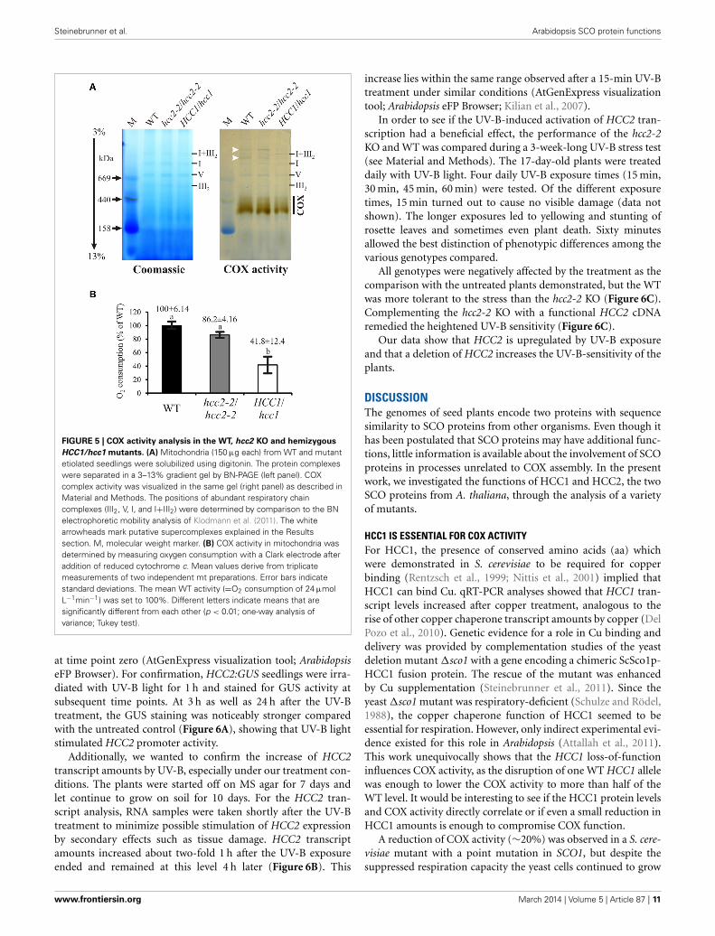
For a more direct analysis of a link between the HCC proteinsand COX, its activity was measured by two different approaches.First, blue native gel electrophoresis (BN-PAGE) allowed us tostudy COX activity in conjunction with the molecular organi-zation of the complex (Figure 5A). The in-gel staining revealedCOX activity in a broad molecular range of about 300–450 kDa(Figure 5A, COX), in which the strongest band was visible in thelower range of the stained area. The activity in the hcc2-2 KOmutant was very similar to that in the WT, whereas the stain-ing intensity seemed to be weaker in the hemizygous HCC1/hcc1plants. The molecular organization of the complex in this molec-ular weight range did not seem to be different among the threegenotypes.
Interestingly, also two light-brownish bands in the range above1 MDa could be detected in the WT and the hcc2-2 KO, butnot in the HCC1/hcc1 mutant (Figure 5A; white arrowheads).
www.frontiersin.org March 2014 | Volume 5 | Article 87 | 9

Steinebrunner et al. Arabidopsis SCO protein functions
FIGURE 4 | Subcellular localization analysis of HCC2-mRFP. (A) Fivedifferent transgenic lines co-expressing HCC2-mRFP and GFP targeted tomitochondria (mt-GFP) were imaged by laser scanning microscopy, andco-localization of HCC2-mRFP and mt-GFP was found in each line. Arepresentative cell (line 3) is shown, in which co-localization of the greenmt-GFP (top panel) and the red HCC2-mRFP signals (middle panel) isdepicted as yellow signals after merging the two images (overlay). Theoverlay also includes a bright-field image of the cell. The image was acquiredwith a resolution of 1024 × 1024 pixels and a pixel dwell of 3.15 μs. Scale
bars correspond to 10 μm each. (B) Mitochondria (M), supernatant (SII) andpellet fractions (PI, PII) were prepared as described in Material and Methodsfrom light-grown WT seedlings or HCC2-mRFP overexpressors (line 5)selected on hyg. The indicated fractions (50 μg each) obtained during thepreparations were subjected to 10% SDS-PAGE. Western blot analyses of thesame membrane were performed with antibodies directed against mRFP,cytochrome c oxidase subunit 2 (COX2) and the large subunit of Rubisco(RbcL). The total protein in the gel was visualized after the transfer bycolloidal Coomassie staining.
These bands might represent COX associations with other com-plexes of the respiratory chain (so-called supercomplexes), whichwere described in several organisms (Schägger, 2002) includingthe plants potato, spinach and asparagus (reviewed in Dudkinaet al., 2008). In Arabidopsis, only respiratory chain supercom-plexes consisting of complex I and III have been described sofar (Eubel et al., 2003; Klodmann et al., 2011). However, theseprevious studies differed strongly in their experimental designfrom our analysis, since they used suspension cell culture as start-ing material and harsher lysis conditions of the mitochondria.This might also explain why we detected activity deriving frommonomeric—and possibly dimeric—COX at a slightly highermolecular weight range (up to 450 kDa) than in these publica-tions (at 200–230 kDa).
Whether the higher molecular weight complexes we detectedindeed contain COX and under which physiological conditionsthese supercomplexes are present, remains to be elucidated.
Although the BN results gave hints that the COX activity wasreduced in the HCC1/hcc1 mutant, but not in the hcc2-2 KO,
the staining was not suitable for quantification of the enzymeactivity. Therefore, COX activity measurements were performedin isolated mitochondria, using a Clark electrode as the secondapproach (Figure 5B). Our results showed that the activity wasonly minimally reduced in the hcc2-2 KO (∼14%) compared withthe WT, and the difference was not statistically significant. In thehemizygous HCC1/hcc1 mutant, however, it was suppressed byalmost 60% relative to the WT.
These data validate the proposition that HCC1 is crucial forCOX function, since the deletion of one HCC1 gene copy alreadyleads to a severe drop in COX activity. Furthermore, the data sug-gest that the homozygous knockout of HCC2 does not affect COXactivity and strengthen the hypothesis that this protein is involvedin a different cellular pathway in Arabidopsis.
HCC2 KO MUTANTS ARE MORE SENSITIVE TO UV-B STRESSPublic databases of microarray data indicated that HCC2 isinduced by UV-B light. HCC2 transcript levels were approxi-mately two times higher 1 h and 3 h after UV-B irradiation than
Frontiers in Plant Science | Plant Physiology March 2014 | Volume 5 | Article 87 | 10
Steinebrunner et al. Arabidopsis SCO protein functions
FIGURE 5 | COX activity analysis in the WT, hcc2 KO and hemizygous
HCC1/hcc1 mutants. (A) Mitochondria (150 μg each) from WT and mutantetiolated seedlings were solubilized using digitonin. The protein complexeswere separated in a 3–13% gradient gel by BN-PAGE (left panel). COXcomplex activity was visualized in the same gel (right panel) as described inMaterial and Methods. The positions of abundant respiratory chaincomplexes (III2, V, I, and I+III2) were determined by comparison to the BNelectrophoretic mobility analysis of Klodmann et al. (2011). The whitearrowheads mark putative supercomplexes explained in the Resultssection. M, molecular weight marker. (B) COX activity in mitochondria wasdetermined by measuring oxygen consumption with a Clark electrode afteraddition of reduced cytochrome c. Mean values derive from triplicatemeasurements of two independent mt preparations. Error bars indicatestandard deviations. The mean WT activity (=O2 consumption of 24 μmolL−1min−1) was set to 100%. Different letters indicate means that aresignificantly different from each other (p < 0.01; one-way analysis ofvariance; Tukey test).
at time point zero (AtGenExpress visualization tool; ArabidopsiseFP Browser). For confirmation, HCC2:GUS seedlings were irra-diated with UV-B light for 1 h and stained for GUS activity atsubsequent time points. At 3 h as well as 24 h after the UV-Btreatment, the GUS staining was noticeably stronger comparedwith the untreated control (Figure 6A), showing that UV-B lightstimulated HCC2 promoter activity.
Additionally, we wanted to confirm the increase of HCC2transcript amounts by UV-B, especially under our treatment con-ditions. The plants were started off on MS agar for 7 days andlet continue to grow on soil for 10 days. For the HCC2 tran-script analysis, RNA samples were taken shortly after the UV-Btreatment to minimize possible stimulation of HCC2 expressionby secondary effects such as tissue damage. HCC2 transcriptamounts increased about two-fold 1 h after the UV-B exposureended and remained at this level 4 h later (Figure 6B). This
increase lies within the same range observed after a 15-min UV-Btreatment under similar conditions (AtGenExpress visualizationtool; Arabidopsis eFP Browser; Kilian et al., 2007).
In order to see if the UV-B-induced activation of HCC2 tran-scription had a beneficial effect, the performance of the hcc2-2KO and WT was compared during a 3-week-long UV-B stress test(see Material and Methods). The 17-day-old plants were treateddaily with UV-B light. Four daily UV-B exposure times (15 min,30 min, 45 min, 60 min) were tested. Of the different exposuretimes, 15 min turned out to cause no visible damage (data notshown). The longer exposures led to yellowing and stunting ofrosette leaves and sometimes even plant death. Sixty minutesallowed the best distinction of phenotypic differences among thevarious genotypes compared.
All genotypes were negatively affected by the treatment as thecomparison with the untreated plants demonstrated, but the WTwas more tolerant to the stress than the hcc2-2 KO (Figure 6C).Complementing the hcc2-2 KO with a functional HCC2 cDNAremedied the heightened UV-B sensitivity (Figure 6C).
Our data show that HCC2 is upregulated by UV-B exposureand that a deletion of HCC2 increases the UV-B-sensitivity of theplants.
DISCUSSIONThe genomes of seed plants encode two proteins with sequencesimilarity to SCO proteins from other organisms. Even though ithas been postulated that SCO proteins may have additional func-tions, little information is available about the involvement of SCOproteins in processes unrelated to COX assembly. In the presentwork, we investigated the functions of HCC1 and HCC2, the twoSCO proteins from A. thaliana, through the analysis of a varietyof mutants.
HCC1 IS ESSENTIAL FOR COX ACTIVITYFor HCC1, the presence of conserved amino acids (aa) whichwere demonstrated in S. cerevisiae to be required for copperbinding (Rentzsch et al., 1999; Nittis et al., 2001) implied thatHCC1 can bind Cu. qRT-PCR analyses showed that HCC1 tran-script levels increased after copper treatment, analogous to therise of other copper chaperone transcript amounts by copper (DelPozo et al., 2010). Genetic evidence for a role in Cu binding anddelivery was provided by complementation studies of the yeastdeletion mutant �sco1 with a gene encoding a chimeric ScSco1p-HCC1 fusion protein. The rescue of the mutant was enhancedby Cu supplementation (Steinebrunner et al., 2011). Since theyeast �sco1 mutant was respiratory-deficient (Schulze and Rödel,1988), the copper chaperone function of HCC1 seemed to beessential for respiration. However, only indirect experimental evi-dence existed for this role in Arabidopsis (Attallah et al., 2011).This work unequivocally shows that the HCC1 loss-of-functioninfluences COX activity, as the disruption of one WT HCC1 allelewas enough to lower the COX activity to more than half of theWT level. It would be interesting to see if the HCC1 protein levelsand COX activity directly correlate or if even a small reduction inHCC1 amounts is enough to compromise COX function.
A reduction of COX activity (∼20%) was observed in a S. cere-visiae mutant with a point mutation in SCO1, but despite thesuppressed respiration capacity the yeast cells continued to grow
www.frontiersin.org March 2014 | Volume 5 | Article 87 | 11

Steinebrunner et al. Arabidopsis SCO protein functions
FIGURE 6 | Response of hcc2 mutants and WT to UV-B treatment. (A)
HCC2:GUS seedlings were stained for GUS activity 3 h or 24 h after a 1-htreatment with UV-B light. Untreated seedlings were stained in parallel tothe 3-h time point. Two GUS lines (lines 3 and 7) were included in theexperiment, and three seedlings were photographed per time point. Hereseedlings of the line 3 are shown. The experiment was repeated onceproviding the same staining pattern. The scale bars equal 1 mm each. (B)
The relative amounts of HCC2 transcripts were analyzed in 14-day-old WTseedlings by qRT-PCR which were either treated with UV-B for 1 h (whitebars) or left untreated (black bars). RNA was isolated from leavesharvested at the indicated time points. The amounts of HCC2 transcriptsat time point 1 h of the untreated control were set to 1. PP2AA3 transcriptlevels were used as a reference for normalization. Error bars representstandard deviations calculated from the normalized HCC2 expression
values of two independent RNA preparations of the same UV-B assay.Different letters denote values which are statistically significantly differentfrom each other (p < 0.05; one-way analysis of variance; Tukey test). HCC2levels shown in this graph are representative of two independent UV-Bexperiments. (C) Day 1 marks the beginning of a UV-B stress test in which17-day-old WT plants, hcc2-2 KOs and complemented hcc2-2 KOs (line 2)either served as untreated controls (top row) or were irradiated daily withUV-B light for 1 h (bottom row). The phenotypes of the same plants areshown after they had grown for 21 days in the absence of any UV-B light(-UV-B) or exposed daily to UV-B (+UV-B). Any plants which died during theUV-B stress test were removed. At day 21, the shoots were cut forunobstructed view of the rosettes. The scale bars equal 5 cm each and arevalid for all panels. This UV-B assay was conducted three times showingthe same phenotypic responses.
on nonfermentable carbon sources like the WT (Lode, 2001).Similarly, the strong effect on COX activity did not seem to affectplant growth. Plants with only one intact HCC1 copy lookedphenotypically indistinguishable from the WT (Figure 2D). In
agreement with this, COX-deprived mutants of the green algaChlamydomonas reinhardtii continued to grow like the WT whenkept in the light (Colin et al., 1995; Remacle et al., 2010).Apparently, the reduced production of ATP is compensated by
Frontiers in Plant Science | Plant Physiology March 2014 | Volume 5 | Article 87 | 12

Steinebrunner et al. Arabidopsis SCO protein functions
photosynthesis, or other metabolic pathways such as glycolysismay contribute some ATP. The latter process might be enhancedby the presence of sucrose in our growth media. In future studiesit might therefore be interesting to compare the adenylate statusof the different mutant lines under various growth conditions.
However, COX activity is linked to many important processesother than the provision of ATP. For example, it maintains themt membrane potential necessary for the import of many mtproteins from the cytosol (reviewed in Chacinska et al., 2009).In addition, the synthesis of ascorbate is dependent on COXactivity. The oxidation of L-galactono-γ-lactone to ascorbate byL-galactono-γ-lactone dehydrogenase uses cytochrome c as anelectron acceptor. Therefore, a continuous electron flow fromcytochrome c to complex IV is prerequisite for ascorbate biosyn-thesis (Bartoli et al., 2000). Ascorbate affects growth, because lowlevels correlate with low cell division rates (Kerk and Feldman,1995). In addition, ascorbate is a co-factor for prolyl hydroxy-lases (Smirnoff, 2000) which produce hydroxyproline-rich glyco-proteins relevant for cell wall structure (Showalter, 2001). Thisfunction might be the reason why HCC1 promoter activity isnot only high in metabolically active cell types, but also in tri-chome support cells (Steinebrunner et al., 2011) which needmechanical reinforcements. This function in ascorbate synthesiscould also explain why Arabidopsis mutants without functionalcytochrome c (Welchen et al., 2012) and HCC1 (Attallah et al.,2011; Steinebrunner et al., 2011) die as embryos. For the hcc1KO embryos, the predominant time point of their developmentalarrest was pinned to the heart stage (Steinebrunner et al., 2011).At this stage, the embryos start growing anisodiametrically, form-ing two symmetric protrusions as they transit into the torpedostage. The cell wall plays a pivotal role for these local cell divisionsand expansions as proposed for the cell wall mutant cyt1. Theaffected CYT1 gene codes for the precursor of cell wall polysac-charides and ascorbate (reviewed in Smirnoff, 2000) and theknockout mutant also stops growing as a heart-shaped embryo(Lukowitz et al., 2001).
POSSIBLE REDOX ROLES OF HCC2The homolog HCC2 does not contain the conserved Cu-binding motif, contradicting a function as a copper chaperone.However, its promoter activity overlapped with the HCC1 pro-moter activity (Attallah et al., 2011; Steinebrunner et al., 2011;Supplementary Figure 2), hinting at similar functions. However,the determined WT levels of COX activity in the hcc2-2 KOmutant provided compelling evidence that HCC2 is not requiredfor COX function.
No effect on respiration efficiency was also documented forthe deletion of SCO2 in yeast (Glerum et al., 1996). Instead, anindirect involvement of Sco2p in COX assembly was postulated.The two cysteines of the Cu-binding motif in the thioredoxindomain of Sco2p could possibly maintain the proper redox stateof Sco1p, allowing Sco1p to bind and release Cu (Banci et al.,2007). In human cells it could indeed be shown that SCO2 actsas thiol-disulfide oxidoreductase for SCO1 (Leary et al., 2009).
However, HCC2 strikingly lacks these cysteines as well as thehistidine residue contributing to Cu binding. The general occur-rence of a SCO homolog that bears no conserved amino acids
relevant to Cu binding seems to be a common feature in plants(Supplementary Figure 1), arguing against its function in metaltransfer. But what could be such a plant-specific function?
The results presented here suggest a role in the UV-B stressresponse, however, it is not clear if HCC2 specifically responds toUV-B or if it fulfills a general function as an antioxidant throughits thioredoxin-like fold. In favor of a UV-B-specific function isthe finding that (i) HCC2 is upregulated by UV-B treatment andthat (ii) HCC2 is not upregulated by UV-A or by other factorscausing oxidative stress such as paraquat (Kilian et al., 2007).These data should be confirmed and complemented with phe-notypic studies in the future. Preliminary studies indicate thathcc2 KO plants contain higher lipid peroxidation levels underbasal growth conditions, suggesting that HCC2 could serve as aregulator of ROS levels.
The homologous yeast proteins Sco1p and Sco2p were bothshown to reside in the inner mt membrane and to protrudetheir catalytically active C-terminal domain into the intermem-brane space (IMS) (Krummeck, 1992; Lode et al., 2002). Thedata of our work do not allow conclusions about the submito-chondrial localization of HCC2, but nicely prove its presence inmitochondria.
Nevertheless, the analysis tool TMPred predicts for HCC2 onetransmembrane helix from aa 66 to 82 with an inside-outside ori-entation, arguing for a localization of the active domain of HCC2in the mt IMS space. This subcompartment represents a suitablesite for a protein with an oxidoreductive function, because theIMS contains the highest abundance of cysteine-rich proteins inthe cell (Herrmann and Funes, 2005).
MODEL FOR HCC1 AND HCC2 FUNCTIONIncorporating our experimental data, we propose the follow-ing model (Figure 7). HCC1 is essential for plant life, becauseit ensures COX function, most conceivably through delivery ofthe co-factor Cu to complex IV. HCC2, on the contrary, is notan essential plant protein, but important nonetheless. As a puta-tive oxidoreductase, HCC2 could maintain the proper redox stateof redox-sensitive proteins, such as HCC1, although COX activ-ity was not significantly suppressed in the hcc2-2 KO. However,the HCC2 activity is possibly compensated in the mutant by anmt oxidoreductase of redundant function, or HCC2 may only beimportant under certain conditions, such as UV-B stress.
High UV-B fluence rates—as used in our experiments—triggerthe production of ROS (reviewed in Mackerness et al., 2001 andHideg et al., 2013) which cause oxidative damage of cell compo-nents. The damage leads to the release of more ROS, amplifyingthe original ROS levels. HCC2 could protect against ROS dam-age by exerting the oxidoreductive function of its thioredoxindomain, protecting the plant indirectly against UV-B. However,as a caveat for this hypothesis it must be considered that despitethe fact that HCC2 contains a thioredoxin fold, the typical thiore-doxin motif CxxC directly involved in catalysis (reviewed inMeyer et al., 2012) is missing. Nevertheless, HCC2 is a verycysteine-rich protein with five cysteines remaining in the pro-tein after the putative signal peptide (aa 1–29) is cleaved off. Inaddition, there is a CGC-motif (aa 102–104) which is predictedto form a disulfide bond with C253 by the DiANNA software.
www.frontiersin.org March 2014 | Volume 5 | Article 87 | 13

Steinebrunner et al. Arabidopsis SCO protein functions
FIGURE 7 | Model for the role of HCC1 and HCC2 in plant mitochondria.
We propose different functions (dashed arrows) for the two SCO proteins inmitochondria of A. thaliana indicated by solid purple circles. HCC1 is anessential plant protein and is most likely involved in copper transport to COX(complex IV) of the respiratory chain (A). HCC2 might have rather a slightinfluence on COX activity by either directly or indirectly supporting HCC1
function (B). In fact it is more likely that HCC2 functions as a thioredoxin,converting oxidized (ox.) proteins back to their reduced (red.) forms, ordetoxifies reactive oxygen species (ROS) in mitochondria (C). This function isof special importance under UV-B stress which leads to increased ROSdamage in cells and organelles, e.g., by lipid and protein peroxidation. Thefigure was partially adapted from Nawkar et al. (2013).
Additionally, any of the other cysteines could exert a redox-activefunction. Alternatively, HCC2 may not act directly as an oxi-doreductase, but may modify the activity of other redox proteinspresent in the mitochondrial IMS.
In conclusion, plants have evolved two different SCO proteinswith specific functions in COX assembly and stress responses.Even though our results indicate that HCC1 and HCC2 have com-pletely separate roles, the presence of both proteins in mitochon-dria and the conservation in structure does not rule out that theyexert their functions through partially overlapping pathways. Theresults highlight the role of mitochondria in many physiologicalresponses and indicate that plants have adapted preexisting pro-teins to serve additional functions related to their specific needs.
AUTHOR CONTRIBUTIONSIris Steinebrunner conceived of the study, characterized mutantlines (HCC2:HCC2, hcc2-1, hcc2-2, hcc1 lines complemented withABI3:HCC1 or ABI3:GUS), did the confocal microscopy and seg-regation analyses and drafted the manuscript. Uta Gey conductedthe COX activity experiments, the BN-PAGE and Western blotanalyses, performed the sequence alignments and participated inthe writing of the manuscript. Manuela Andres did the UV-Bexperiments and analyzed the GUS expression controlled by theHCC2 promoter in response to UV-B light and by the ABI3 pro-moter. Lucila Garcia generated the 35S:HCC2-mRFP lines and did
the qRT-PCR analyses. Daniel H. Gonzalez advised the study andparticipated in the writing. All authors read and approved thefinal manuscript.
ACKNOWLEDGMENTSWe are very grateful for the help of the following students:René Morgenstern (cloning of ABI3:GUS, ABI3:HCC1, andHCC2:HCC2 constructs), Annelie Müller (contributions to thecharacterization of hcc1-complemented lines and establishmentof the UV-B assay) and Marlen Landschreiber (RT-PCR andHCC2:GUS analyses). We would like to thank Gerhard Rödelfor his ongoing support of the project and helpful comments.Silke Hilbig is thanked for her technical assistance. The LightMicroscopy Facility of the Biotechnology Center TU Dresdenprovided excellent training and technical assistance with theCSLM imaging. The T-DNA mutants were generated in thecontext of the GABI-Kat program and provided by BerndWeisshaar (Max Planck Institute for Plant Breeding Research,Cologne, Germany). The cDNA clone U19562 was donated byAthanasios Theologis, Joseph Ecker, and Ronald Davis to theArabidopsis Biological Resource Center who provided the cloneto us. Many thanks to the following people for providing mate-rials: Elena Taverna (mRFP antibody), Mark Curtis (pMDC163),Francois Parcy (ABI3 promoter region), and Tsuyoshi Nakagawa(pGWB516, pGWB553, pGWB554).
Frontiers in Plant Science | Plant Physiology March 2014 | Volume 5 | Article 87 | 14

Steinebrunner et al. Arabidopsis SCO protein functions
FUNDINGThe grant STE 1455/5-1 from the German Research Foundation(DFG) awarded to Iris Steinebrunner supported the researchstays of Uta Gey and Lucila Garcia in Daniel H. Gonzalez’sand Iris Steinebrunner’s laboratory, respectively, which resultedin data presented in this study. This work was also fundedby Daniel H. Gonzalez’s grants PICT1035 and PICT1203 fromthe National Agency for the Promotion of Science of Argentina(ANPCyT). Daniel H. Gonzalez is a member of the NationalResearch Council of Argentina (CONICET) which co-fundedUta Gey’s stay at the Universidad Nacional del Litoral (UNL);Lucila Garcia is a CONICET fellow at UNL. The publica-tion fee for this article was covered by the DFG and theOpen Access Publication Funds of the Technische UniversitätDresden.
SUPPLEMENTARY MATERIALThe Supplementary Material for this article can be found onlineat: http://www.frontiersin.org/journal/10.3389/fpls.2014.00087/abstract
Supplementary Table 1 | Primer details. ∗The attachment site sequences
necessary for recombination were omitted. Only the gene-specific
sequences are listed.
Supplementary Figure 1 | Protein sequence alignment
of SCO (HCC) proteins from plants: Arabidopsis thaliana (At), Oryza
sativa subsp. japonica (Os), Brassica rapa subsp. pekinensis (Br), Glycine
max (Gm), and Solanum lycopersicum (Sl). Protein sequences were
retrieved from the UNIPROT database (UNIPROT numbers are given). The
sequence alignment was performed using the ClustalO 1.2.0 alignment
tool (Sievers et al., 2011). The consensus is quoted below the sequences
(“*” conserved in all sequences, “.” partially conserved, “:” homology
in all sequences). The amino acids involved in Cu-binding which includes
the CxxxC motif as well as one histidine residue (Rentzsch et al., 1999;
Nittis et al., 2001) are marked in red, if present. Proteins containing these
conserved residues are titled “HCC1,” whereas those missing the motif
are named “HCC2.” If more than one sequence in the UNIPROT database
met this criterion, they were distinguished by different letters (a, b, c).
Supplementary Figure 2 | HCC2:GUS studies of different developmental
stages. Various tissues from HCC2:GUS transgenic lines were stained for
GUS activity: Ovules (A,B), embryo (C), cotyledon (D,E), roots (F,G),
leaves (H,I), flower buds (J), flower (K) and silique (L). The tissues
stemmed from 7-day-old seedlings (D–G) and 4-week-old plants (A–C,
H–L), respectively. Exemplary guard cells are indicated by arrows in (E).
Staining of guard and trichome support cells was only observed in
younger leaves. ch, chalaza; f, funiculus; r, root; h, hypocotyl; tsc, trichome
support cells; c, connective; az, abscission zone.
Supplementary Figure 3 | Analysis of steady-state COX2 levels in the WT
and hcc2-2 KO mutants. MCF (50 μg each, normalized to WT CSA levels)
prepared from liquid cultures of etiolated seedlings (WT and
hcc2-2/hcc2-2 mutants) were separated in a 15% SDS gel for Western
blot analysis. Immunological detection of COX2 and VDAC1 was
performed successively on the same membrane. VDAC1 was detected as
the loading control for equal amounts of mt protein. The Western blot
analysis was performed three times with two independent sets of protein
extracts, producing the same results.
REFERENCESAbriata, L. A., Banci, L., Bertini, I., Ciofi-Baffoni, S., Gkazonis, P., Spyroulias, G. A.,
et al. (2008). Mechanism of Cu(A) assembly. Nat. Chem. Biol. 4, 599–601. doi:10.1038/nchembio.110
A.-H.-Mackerness, S., John, C. F., Jordan, B., and Thomas, B. (2001). Early signal-ing components in ultraviolet-B responses: distinct roles for different reactiveoxygen species and nitric oxide. FEBS Lett. 489, 237–242. doi: 10.1016/S0014-5793(01)02103-2
Arnesano, F., Banci, L., Bertini, I., and Martinelli, M. (2005). Ortholog search ofproteins involved in copper delivery to cytochrome c oxidase and functionalanalysis of paralogs and gene neighbors by genomic context. J. Proteome Res. 4,63–70. doi: 10.1021/pr049862f
Attallah, C. V., Welchen, E., Martin, A. P., Spinelli, S. V., Bonnard, G., Palatnik, J. F.,et al. (2011). Plants contain two SCO proteins that are differentially involved incytochrome c oxidase function and copper and redox homeostasis. J. Exp. Bot.62, 4281–4294. doi: 10.1093/jxb/err138
Balatri, E., Banci, L., Bertini, I., Cantini, F., and Ciofi-Baffoni, S. (2003).Solution structure of Sco1: a thioredoxin-like protein involved incytochrome c oxidase assembly. Structure 11, 1431–1443. doi: 10.1016/j.str.2003.10.004
Banci, L., Bertini, I., Cavallaro, G., and Ciofi-Baffoni, S. (2011). Seeking the deter-minants of the elusive functions of Sco proteins. FEBS J. 278, 2244–2262. doi:10.1111/j.1742-4658.2011.08141.x
Banci, L., Bertini, I., Cavallaro, G., and Rosato, A. (2007). The functions ofSco proteins from genome-based analysis. J. Proteome Res. 6, 1568–1579. doi:10.1021/pr060538p
Barrientos, A., Gouget, K., Horn, D., Soto, I. C., and Fontanesi, F. (2009).Suppression mechanisms of COX assembly defects in yeast and human: insightsinto the COX assembly process. Biochim. Biophys. Acta 1793, 97–107. doi:10.1016/j.bbamcr.2008.05.003
Bartoli, C. G., Pastori, G. M., and Foyer, C. H. (2000). Ascorbate biosynthesis inmitochondria is linked to the electron transport chain between complexes IIIand IV. Plant Physiol. 123, 335–344. doi: 10.1104/pp.123.1.335
Chacinska, A., Koehler, C. M., Milenkovic, D., Lithgow, T., and Pfanner, N. (2009).Importing mitochondrial proteins: machineries and mechanisms. Cell 138,628–644. doi: 10.1016/j.cell.2009.08.005
Cobine, P. A., Pierrel, F., and Winge, D. R. (2006). Copper trafficking to the mito-chondrion and assembly of copper metalloenzymes. Biochim. Biophys. Acta1763, 759–772. doi: 10.1016/j.bbamcr.2006.03.002
Colin, M., Dorthu, M. P., Duby, F., Remacle, C., Dinant, M., Wolwertz, M. R., et al.(1995). Mutations affecting the mitochondrial genes encoding the cytochromeoxidase subunit I and apocytochrome b of Chlamydomonas reinhardtii. Mol.Gen. Genet. 249, 179–184. doi: 10.1007/BF00290364
Curtis, M. D., and Grossniklaus, U. (2003). A gateway cloning vector set for high-throughput functional analysis of genes in planta. Plant Physiol. 133, 462–469.doi: 10.1104/pp.103.027979
Czechowski, T., Stitt, M., Altmann, T., Udvardi, M. K., and Scheible, W. R.(2005). Genome-wide identification and testing of superior reference genesfor transcript normalization in Arabidopsis. Plant Physiol. 139, 5–17. doi:10.1104/pp.105.063743
Del Pozo, T., Cambiazo, V., and Gonzalez, M. (2010). Gene expression profilinganalysis of copper homeostasis in Arabidopsis thaliana. Biochem. Biophys. Res.Commun. 393, 248–252. doi: 10.1016/j.bbrc.2010.01.111
Devic, M., Albert, S., and Delseny, M. (1996). Induction and expression of seed-specific promoters in Arabidopsis embryo-defective mutants. Plant J. 9, 205–215.doi: 10.1046/j.1365-313X.1996.09020205.x
Dudkina, N. V., Sunderhaus, S., Boekema, E. J., and Braun, H. P. (2008). The higherlevel of organization of the oxidative phosphorylation system: mitochondrialsupercomplexes. J. Bioenerg. Biomembr. 40, 419–424. doi: 10.1007/s10863-008-9167-5
Dunkley, T. P., Hester, S., Shadforth, I. P., Runions, J., Weimar, T., Hanton, S. L.,et al. (2006). Mapping the Arabidopsis organelle proteome. Proc. Natl. Acad. Sci.U.S.A. 103, 6518–6523. doi: 10.1073/pnas.0506958103
Eubel, H., Jansch, L., and Braun, H. P. (2003). New insights into the respiratorychain of plant mitochondria. Supercomplexes and a unique composition ofcomplex II. Plant Physiol. 133, 274–286. doi: 10.1104/pp.103.024620
Ferrè, F., and Clote, P. (2006). DiANNA 1.1: an extension of the DiANNA webserver for ternary cysteine classification. Nucleic Acids Res. 34, W182–W185. doi:10.1093/nar/gkl189
www.frontiersin.org March 2014 | Volume 5 | Article 87 | 15

Steinebrunner et al. Arabidopsis SCO protein functions
Ghaemmaghami, S., Huh, W. K., Bower, K., Howson, R. W., Belle, A., Dephoure,N., et al. (2003). Global analysis of protein expression in yeast. Nature 425,737–741. doi: 10.1038/nature02046
Glerum, D. M., Shtanko, A., and Tzagoloff, A. (1996). SCO1 and SCO2act as high copy suppressors of a mitochondrial copper recruitmentdefect in Saccharomyces cerevisiae. J. Biol. Chem. 271, 20531–20535. doi:10.1074/jbc.271.34.20531
Heazlewood, J. L., Verboom, R. E., Tonti-Filippini, J., Small, I., and Millar, A.H. (2007). SUBA: the Arabidopsis Subcellular Database. Nucleic Acids Res. 35,D213–D218. doi: 10.1093/nar/gkl863
Herrmann, J. M., and Funes, S. (2005). Biogenesis of cytochrome oxidase-sophisticated assembly lines in the mitochondrial inner membrane. Gene 354,43–52. doi: 10.1016/j.gene.2005.03.017
Hideg, E., Jansen, M. A., and Strid, A. (2013). UV-B exposure, ROS, and stress:inseparable companions or loosely linked associates? Trends Plant. Sci. 18,107–115. doi: 10.1016/j.tplants.2012.09.003
Hofmann, K., and Stoffel, W. (1993). TMbase: a database of membrane spanningprotein segments. Biol. Chem. Hoppe-Seyler 374, 166.
Keppler, A., Gendreizig, S., Gronemeyer, T., Pick, H., Vogel, H., and Johnsson, K.(2003). A general method for the covalent labeling of fusion proteins with smallmolecules in vivo. Nat. Biotechnol. 21, 86–89. doi: 10.1038/nbt765
Kerk, N. M., and Feldman, N. J. (1995). A biochemical model for the initiationand maintenance of the quiescent center: implications for organization of rootmeristems. Development 121, 2825–2833.
Kilian, J., Whitehead, D., Horak, J., Wanke, D., Weinl, S., Batistic, O., et al. (2007).The AtGenExpress global stress expression data set: protocols, evaluation andmodel data analysis of UV-B light, drought and cold stress responses. Plant J.50, 347–363. doi: 10.1111/j.1365-313X.2007.03052.x
Kim, H. J., Khalimonchuk, O., Smith, P. M., and Winge, D. R. (2012).Structure, function, and assembly of heme centers in mitochondrial respira-tory complexes. Biochim. Biophys. Acta 1823, 1604–1616. doi: 10.1016/j.bbamcr.2012.04.008
Kleinboelting, N., Huep, G., Kloetgen, A., Viehoever, P., and Weisshaar, B.(2012). GABI-Kat SimpleSearch: new features of the Arabidopsis thalianaT-DNA mutant database. Nucleic Acids Res. 40, D1211–D1215. doi:10.1093/nar/gkr1047
Klodmann, J., Senkler, M., Rode, C., and Braun, H. P. (2011). Defining the pro-tein complex proteome of plant mitochondria. Plant Physiol. 157, 587–598. doi:10.1104/pp.111.182352
Krummeck, G. (1992). Das mitochondriale SCO1 Protein von Saccharomyces cere-visiae: Untersuchungen zur Expression, Topologie und Funktion. Ph.D. thesis,Ludwig Maximilian University Munich.
Laemmli, U. K. (1970). Cleavage of structural proteins during the assembly of thehead of bacteriophage T4. Nature 227, 680–685. doi: 10.1038/227680a0
Leary, S. C., Kaufman, B. A., Pellecchia, G., Guercin, G. H., Mattman, A., Jaksch, M.,et al. (2004). Human SCO1 and SCO2 have independent, cooperative functionsin copper delivery to cytochrome c oxidase. Hum. Mol. Genet. 13, 1839–1848.doi: 10.1093/hmg/ddh197
Leary, S. C., Sasarman, F., Nishimura, T., and Shoubridge, E. A. (2009). HumanSCO2 is required for the synthesis of CO II and as a thiol-disulphide oxidore-ductase for SCO1. Hum. Mol. Genet. 18, 2230–2240. doi: 10.1093/hmg/ddp158
Livak, K. J., and Schmittgen, T. D. (2001). Analysis of relative gene expressiondata using real-time quantitative PCR and the 2(-Delta Delta C(T)) method.Methods 25, 402–408. doi: 10.1006/meth.2001.1262
Lode, A. (2001). Assemblierung der Cytochrom c Oxidase: molekulare und biochemi-sche Charakterisierung des mitochondrialen Sco1p aus Saccharomyces cerevisiaeund homologer Proteine. Ph.D. thesis, Technische Universität Dresden.
Lode, A., Paret, C., and Rödel, G. (2002). Molecular characterization ofSaccharomyces cerevisiae Sco2p reveals a high degree of redundancy with Sco1p.Yeast 19, 909–922. doi: 10.1002/yea.883
Lukowitz, W., Nickle, T. C., Meinke, D. W., Last, R. L., Conklin, P. L., andSomerville, C. R. (2001). Arabidopsis cyt1 mutants are deficient in a mannose-1-phosphate guanylyltransferase and point to a requirement of N-linked glycosy-lation for cellulose biosynthesis. Proc. Natl. Acad. Sci. U.S.A 98, 2262–2267. doi:10.1073/pnas.051625798
Meyer, Y., Belin, C., Delorme-Hinoux, V., Reichheld, J. P., and Riondet, C. (2012).Thioredoxin and glutaredoxin systems in plants: molecular mechanisms,crosstalks, and functional significance. Antioxid. Redox Signal. 17, 1124–1160.doi: 10.1089/ars.2011.4327
Nakagawa, T., Suzuki, T., Murata, S., Nakamura, S., Hino, T., Maeo, K., et al. (2007).Improved Gateway binary vectors: high-performance vectors for creation offusion constructs in transgenic analysis of plants. Biosci. Biotechnol. Biochem.71, 2095–2100. doi: 10.1271/bbb.70216
Nawkar, G. M., Maibam, P., Park, J. H., Sahi, V. P., Lee, S. Y., and Kang, C. H.(2013). UV-induced cell death in plants. Int. J. Mol. Sci. 14, 1608–1628. doi:10.3390/ijms14011608
Nelson, B. K., Cai, X., and Nebenführ, A. (2007). A multicolored set of in vivoorganelle markers for co-localization studies in Arabidopsis and other plants.Plant J. 51, 1126–1136. doi: 10.1111/j.1365-313X.2007.03212.x
Nittis, T., George, G. N., and Winge, D. R. (2001). Yeast Sco1, a protein essential forcytochrome c oxidase function is a Cu(I)-binding protein. J. Biol. Chem. 276,42520–42526. doi: 10.1074/jbc.M107077200
Parcy, F., Valon, C., Raynal, M., Gaubier-Comella, P., Delseny, M., and Giraudat, J.(1994). Regulation of gene expression programs during Arabidopsis seed devel-opment: roles of the ABI3 locus and of endogenous abscisic acid. Plant Cell 6,1567–1582.
Paret, C., Lode, A., Krause-Buchholz, U., and Rödel, G. (2000). The P(174)Lmutation in the human hSCO1 gene affects the assembly of cytochrome coxidase. Biochem. Biophys. Res. Commun. 279, 341–347. doi: 10.1006/bbrc.2000.3949
Remacle, C., Coosemans, N., Jans, F., Hanikenne, M., Motte, P., and Cardol, P.(2010). Knock-down of the COX3 and COX17 gene expression of cytochromec oxidase in the unicellular green alga Chlamydomonas reinhardtii. Plant Mol.Biol. 74, 223–233. doi: 10.1007/s11103-010-9668-6
Rentzsch, A., Krummeck-Weiss, G., Hofer, A., Bartuschka, A., Ostermann, K., andRödel, G. (1999). Mitochondrial copper metabolism in yeast: mutational analy-sis of Sco1p involved in the biogenesis of cytochrome c oxidase. Curr. Genet. 35,103–108. doi: 10.1007/s002940050438
Rhee, S. Y., Beavis, W., Berardini, T. Z., Chen, G., Dixon, D., Doyle, A., et al. (2003).The Arabidopsis Information Resource (TAIR): a model organism databaseproviding a centralized, curated gateway to Arabidopsis biology, researchmaterials and community. Nucleic Acids Res. 31, 224–228. doi: 10.1093/nar/gkg076
Rosso, M. G., Li, Y., Strizhov, N., Reiss, B., Dekker, K., and Weisshaar, B. (2003).An Arabidopsis thaliana T-DNA mutagenized population (GABI-Kat) for flank-ing sequence tag-based reverse genetics. Plant Mol. Biol. 53, 247–259. doi:10.1023/B:PLAN.0000009297.37235.4a
Schägger, H. (2001). Blue-native gels to isolate protein complexes from mitochon-dria. Methods Cell Biol. 65, 231–244. doi: 10.1016/S0091-679X(01)65014-3
Schägger, H. (2002). Respiratory chain supercomplexes of mitochondria and bacte-ria. Biochim. Biophys. Acta 1555, 154–159. doi: 10.1016/S0005-2728(02)00271-2
Schägger, H., and von Jagow, G. (1991). Blue native electrophoresis for isolation ofmembrane protein complexes in enzymatically active form. Anal. Biochem. 199,223–231. doi: 10.1016/0003-2697(91)90094-A
Scholl, R. L., May, S. T., and Ware, D. H. (2000). Seed and molecular resources forArabidopsis. Plant Physiol. 124, 1477–1480. doi: 10.1104/pp.124.4.1477
Schulze, M., and Rödel, G. (1988). SCO1, a yeast nuclear gene essential for accu-mulation of mitochondrial cytochrome c oxidase subunit II. Mol. Gen. Genet.211, 492–498. doi: 10.1007/BF00425706
Showalter, A. M. (2001). Arabinogalactan-proteins: structure, expression and func-tion. Cell. Mol. Life Sci. 58, 1399–1417. doi: 10.1007/PL00000784
Sievers, F., Wilm, A., Dineen, D., Gibson, T. J., Karplus, K., Li, W., et al. (2011). Fast,scalable generation of high-quality protein multiple sequence alignments usingClustal Omega. Mol. Syst. Biol. 7, 539. doi: 10.1038/msb.2011.75
Smirnoff, N. (2000). Ascorbic acid: metabolism and functions of a multi-facetted molecule. Curr. Opin. Plant Biol. 3, 229–235. doi: 10.1016/S1369-5266(00)80070-9
Steinebrunner, I., Landschreiber, M., Krause-Buchholz, U., Teichmann, J., andRödel, G. (2011). HCC1, the Arabidopsis homologue of the yeast mitochondrialcopper chaperone SCO1, is essential for embryonic development. J. Exp. Bot.62, 319–330. doi: 10.1093/jxb/erq269
Sweetlove, L. J., Taylor, N. L., and Leaver, C. J. (2007). Isolation of intact, functionalmitochondria from the model plant Arabidopsis thaliana. Methods Mol. Biol.372, 125–136. doi: 10.1007/978-1-59745-365-3_9
Thomas, P. E., Ryan, D., and Levin, W. (1976). An improved staining proce-dure for the detection of the peroxidase activity of cytochrome P-450 onsodium dodecyl sulfate polyacrylamide gels. Anal. Biochem. 75, 168–176. doi:10.1016/0003-2697(76)90067-1
Frontiers in Plant Science | Plant Physiology March 2014 | Volume 5 | Article 87 | 16

Steinebrunner et al. Arabidopsis SCO protein functions
Welchen, E., Hildebrandt, T. M., Lewejohann, D., Gonzalez, D. H., and Braun, H.P. (2012). Lack of cytochrome c in Arabidopsis decreases stability of Complex IVand modifies redox metabolism without affecting Complexes I and III. Biochim.Biophys. Acta 1817, 990–1001. doi: 10.1016/j.bbabio.2012.04.008
Winter, D., Vinegar, B., Nahal, H., Ammar, R., Wilson, G. V., and Provart, N.J. (2007). An “Electronic Fluorescent Pictograph” browser for exploring andanalyzing large-scale biological data sets. PLoS ONE 2:e718. doi: 10.1371/jour-nal.pone.0000718
Yamada, K., Lim, J., Dale, J. M., Chen, H., Shinn, P., Palm, C. J., et al. (2003).Empirical analysis of transcriptional activity in the Arabidopsis genome. Science302, 842–846. doi: 10.1126/science.1088305
Conflict of Interest Statement: The authors declare that the research was con-ducted in the absence of any commercial or financial relationships that could beconstrued as a potential conflict of interest.
Received: 02 December 2013; paper pending published: 07 January 2014; accepted: 24February 2014; published online: 25 March 2014.Citation: Steinebrunner I, Gey U, Andres M, Garcia L and Gonzalez DH (2014)Divergent functions of the Arabidopsis mitochondrial SCO proteins: HCC1 is essen-tial for COX activity while HCC2 is involved in the UV-B stress response. Front. PlantSci. 5:87. doi: 10.3389/fpls.2014.00087This article was submitted to Plant Physiology, a section of the journal Frontiers inPlant Science.Copyright © 2014 Steinebrunner, Gey, Andres, Garcia and Gonzalez. This is anopen-access article distributed under the terms of the Creative Commons AttributionLicense (CC BY). The use, distribution or reproduction in other forums is permit-ted, provided the original author(s) or licensor are credited and that the originalpublication in this journal is cited, in accordance with accepted academic practice.No use, distribution or reproduction is permitted which does not comply with theseterms.
www.frontiersin.org March 2014 | Volume 5 | Article 87 | 17