Keywords: Avian embryo Cell guidance Cell migration Chemotaxis Ciliary ganglion Electroporation
Upload
independentCategory
view
2download
0
2759Research Article
IntroductionChemokine receptors are seven-transmembrane-domain G-protein-coupled receptors (GPCRs) that are widely expressed on many celltypes of the body and responsible for guiding migrating cells alonga chemokine gradient. A highly regulated network of chemokinereceptors and chemokines is responsible for directing inflammatoryand homeostatic cell trafficking (Moser et al., 2004). Homing ofantigen-bearing dendritic cells to secondary lymphoid organs forinstance, depends on the chemokine receptor CCR7. In the T-cellzone of lymphoid organs, where the two ligands of CCR7,CCL19/ELC and CCL21/SLC are expressed (Luther et al., 2000),incoming dendritic cells present their peptide antigens to naive Tcells, thereby inducing an immune response. In addition to itsexpression on mature dendritic cells, CCR7 is also expressed onsubpopulations of T cells, B cells, NK cells and thymocytes(Campbell et al., 2003; Muller et al., 2003). Homing of dendriticcells and naive T cells is strongly affected by the lack of eitherCCR7 or its ligands, and consequently results in a defect in initiatingprimary immune responses (Forster et al., 1999; Gunn et al., 1999;Luther et al., 2000; Ohl et al., 2004). Moreover, CCR7 is alsoexpressed in breast, gastric, non-small-cell lung, and oesophagealsquamous cancer, as well as in chronic lymphocytic leukemia(reviewed by Balkwill, 2004; Zlotnik, 2006). CCR7-expressingcancer cells respond to CCL19 and CCL21 and metastasize to thedraining lymph nodes and the lung (Muller et al., 2001).Consequently, understanding how chemokine receptors transmitsignals resulting in cell migration and identifying ways of
modulating chemotaxis or receptor silencing is of interest for futuretherapeutic strategies.
Chemokine receptor signaling is initiated by activation of theBordetella pertussis toxin-sensitive Gi family of heterotrimeric Gproteins (Thelen, 2001). Chemokine-bound receptors trigger the Gαsubunits to exchange GTP for GDP, leading to the dissociation ofthe Gα subunit from the βγ heterodimer. Whereas the Gα subunititself appears not to be required for chemotaxis, the released Gβγsubunit transduces signals leading to cell locomotion (Neptune andBourne, 1997). Gβγ activates phospholipase Cβ (PLCβ) isoformsleading to the formation of inositol (1,4,5)-trisphosphate and atransient rise in the concentration of intracellular free calcium. Gβγalso activates phosphoinositide 3-kinases (PI3Ks) and downstreampleckstrin-homology-domain-containing proteins, such as proteinkinase B (PKB)/Akt or guanine-nucleotide exchange factors andmitogen-activated protein kinases (MAPKs) (Thelen, 2001;Wymann et al., 2003). Despite its well-characterized biologicalfunction, CCR7 signaling has not yet been characterized in depth.So far, CCR7 triggering has been shown to induce receptorphosphorylation, β-arrestin (official symbol ARRB1) recruitment,phosphorylation of PKB and extracellular regulated kinases 1 and2 (Erk1 and Erk2; official symbols MAPK3 and MAPK1) and tomobilize intracellular calcium (Bardi et al., 2001; Kohout et al.,2004; Otero et al., 2006; Riol-Blanco et al., 2005; Sanchez-Sanchezet al., 2004; Scandella et al., 2004; Willimann et al., 1998). Of note,despite similar CCR7 binding affinities, G-protein activation andchemotactic properties of CCL19 and CCL21 (Willimann et al.,
The chemokine receptor CCR7, together with its ligands CCL19and CCL21, is responsible for the correct homing andtrafficking of dendritic cells and lymphocytes to secondarylymphoid tissues. Moreover, cancer cells can utilize CCR7 tometastasize to draining lymph nodes. However, information onCCR7 signaling leading to cell migration or receptor traffickingis sparse. Using novel CCR7 deletion mutants with successivetruncations of the intracellular C-terminus and a mutant withimpaired G-protein coupling, we identified distinct motifsresponsible for various aspects of CCR7 signal transduction.Deleting a Ser/Thr motif at the tip of the intracellular tail ofCCR7 resulted in an impaired chemokine-mediated activationof Erk1/2 kinases. Interestingly, deleting an additional adjacentmotif restored the ability of CCL19-mediated Erk1/2
phosphorylation, suggesting the presence of a regulatory motif.Both the Ser/Thr and the regulatory motif are dispensable forsignaling events leading to cell migration and receptortrafficking. A CCR7 mutant lacking virtually the complete C-terminus readily bound CCL19 and was internalized, but wasunable to activate the G protein and to transmit signals requiredfor cell migration, mobilization of [Ca2+]i and Erk1/2 activation.Finally, G-protein coupling was critical for [Ca2+]i mobilization,Erk1/2 phosphorylation and chemotaxis, but not for CCR7trafficking.
Key words: CCR7 signaling, Cell migration, Chemokine receptor,Receptor trafficking
Summary
Distinct motifs in the chemokine receptor CCR7regulate signal transduction, receptor trafficking andchemotaxisCarolina Otero1,*, Petra S. Eisele1,*, Karin Schaeuble1,2,*, Marcus Groettrup1,2 and Daniel F. Legler2,3,‡
1Department of Biology, Division of Immunology, University of Konstanz, Konstanz, Germany2Biotechnology Institute Thurgau (BITg) at the University of Konstanz, Unterseestrasse 47, CH-8280 Kreuzlingen, Switzerland3Zukunftskolleg, University of Konstanz, Konstanz, Germany*These authors contributed equally to this work‡Author for correspondence (e-mail: [email protected])
Accepted 30 April 2008Journal of Cell Science 121, 2759-2767 Published by The Company of Biologists 2008doi:10.1242/jcs.029074
Jour
nal o
f Cel
l Sci
ence
JCS ePress online publication date 29 July 2008
2760
1998), CCL19 induces stronger receptor phosphorylation and β-arrestin recruitment than CCL21 (Kohout et al., 2004). Moreover,CCL19, unlike CCL21, induces rapid endocytosis of CCR7 (Bardiet al., 2001; Otero et al., 2006). We have shown recently, that CCR7is internalized through clathrin-coated pits together with CCL19and transported to early endosomes (Otero et al., 2006).Subsequently, CCR7 recycles back to the plasma membrane toparticipate again in chemokine gradient sensing, whereas CCL19is sorted to lysosomes for degradation (Otero et al., 2006). Themolecular mechanism underlying this observation has not yet beenelucidated.
Crucial motifs of chemokine receptors involved in migration andendocytosis have been mapped to the intracellular loops and the C-terminus. For instance, the Asp-Arg-Tyr (DRY) motif in the secondintracellular loop is generally believed to be responsible for G-protein activation. Whereas G-protein activation is essential forchemotaxis, it is still a matter of debate whether and how G-proteinactivation is involved in GPCR endocytosis (Vilardaga et al., 2001).For GPCR internalization, two main consensus signals within theC-terminus have been described (Bonifacino and Traub, 2003; Neelet al., 2005). A tyrosine-based (Yxxφ or NPxY) and a di-Leu/Ile-Leu/Leu-Ile motif have been identified which interact with theclathrin adaptor protein AP2. The di-Leu motif is essential forinternalization and trafficking of many chemokine receptors,including CXCR2, CXCR3, CXCR4 and CCR5 (reviewed by Neelet al., 2005). However, CCR7 does not contain such knownendocytosis motifs and domains responsible for delivering differentsignals remain to be identified.
In the present study, we searched for distinct motifs within thechemokine receptor CCR7 that regulate signal transduction, receptortrafficking, and cell migration. To this end, we generated three C-terminal truncation mutants of CCR7, where the intracellular tailwas gradually removed. In addition, we also mutated the conservedDRY motif in the second intracellular loop of the receptor. Usingthis strategy, we identified segregated motifs in CCR7 that areresponsible for distinct aspects of CCR7 signal transduction, suchas ligand induced mobilization of intracellular calcium, Erkphosphorylation, cell migration and receptor trafficking.
ResultsCCR7 and its mutants are expressed to a similar degree at theplasma membraneIn order to identify distinct motifs within the chemokine receptorCCR7 that regulate signal transduction, receptor trafficking, andcell migration, we generated three truncation mutants of CCR7lacking parts or the entire C-terminal tail and a mutant with amodified DRY motif in the second intracellular loop of the receptor(Fig. 1A). The cytoplasmic C-terminal part of CCR7 was definedin accordance with the TOPO_DOM prediction by theUniProtKB/Swiss-Prot (www.expasy.org/uniprot) entry for humanCCR7 (accession number P32248). In the mutant MT1 (residues1-334), all except the three most proximal amino acids of thepredicted C-terminal tail were removed, and Lys was replaced byArg to prevent a putative modification by ubiquitin. The last 34amino acids were removed in the MT2 (residues 1-345) mutant,whereas in MT3 (residues 1-355) only the last 24 amino acids weredeleted (Fig. 1A). Within these last 24 amino acids of CCR7(residues 365-378), threonine and serine clusters were reported tobe phosphorylated after ligand binding (Kohout et al., 2004),indicating a possible role in receptor signaling and/or traffickingfor these amino acids. These three constructs were used to establish
stable cloned cell lines in HEK293 cells and in pre-B 300-19 cellsthat both do not express endogenous CCR7. Moreover, to analyzethe role of the highly conserved DRY motif of CCR7 (residues 153-155) with respect to receptor trafficking and signaling we createda mutated CCR7 receptor where we replaced the DRY sequenceby DNY. This single mutation has been reported to disrupt the G-protein coupling of the chemokine receptors CCR5 and CXCR4(Berchiche et al., 2007; Lagane et al., 2005). The DNY constructwas transfected into 300-19 cells and stable cell clones wereestablished. Cell surface staining of CCR7 with a specific antibodywas assessed by flow cytometry (Fig. 1B and Fig. 2) and confocalmicroscopy (data not shown), which revealed that all CCR7 mutantsare expressed at the plasma membrane of stably transfected celllines. In addition, we also quantified the ratio of intracellularcompared with extracellular protein by flow cytometry and foundno difference between wild-type CCR7 and its mutants (data notshown), indicating that all mutants were not trapped within the ERor heavily misfolded. These results provide clear evidence that theC-terminus of CCR7 is not required for proper insertion of thereceptor into the plasma membrane.
CCR7 and its mutants equally bind its ligand CCL19To investigate whether all four CCR7 mutants are able to interactwith their ligand, we performed a binding assay with CCL19.Therefore, 300-19 cells stably transfected with wild-type receptor
Journal of Cell Science 121 (16)
NH2
DRY
MT1: RFRMT2: KFRNDIFKLFKDLGMT3: KFRNDIFKLFKDLGCLSQEQLRQWwt: KFRNDIFKLFKDLGCLSQEQLRQWSSCRHIRRSSMSVEAETTTTFSP
A
B
Cou
nts
CCR7 surface expression100 101 102 103 104
0
20
40
60
80
100
101 102 103 104 101 102 103 104 101 102 103 104
CCR7wt MT1 MT2 MT3
334 345 355 378
Fig. 1. CCR7 C-terminal deletion mutants reach the plasma membrane.(A) Scheme of CCR7 and its C-terminal deletion mutants. The cytoplasmicC-terminus of CCR7 depicted is based on the prediction by Swiss-Prot (SwissInstitute of Bioinformatics, Basel, Switzerland). Amino acids according toaccession number P32248 are indicated. MT1 to MT3 represent C-terminaltruncation mutants of CCR7. In the case of MT1, a Lys was replaced by Arg.Potential phosphorylation sites (S, T) and potential ubiquitylation sites (K) arehighlighted in bold. The conserved DRY motif in the second intracellular loopwas mutated to DNY by site-directed mutagenesis. (B) CCR7 and its C-terminal deletion mutants are equally expressed at the plasma membrane.HEK293 cells stably expressing wild-type (wt) CCR7, MT1, MT2 or MT3were stained with a FITC-conjugated CCR7-specific antibody and cell surfaceexpression was monitored by flow cytometry. Dashed lines representuntransfected cells stained with the same antibody. FITC-labeled isotypecontrol staining on transfected cells revealed the same results. The experimenthas been repeated twice yielding similar results.
Jour
nal o
f Cel
l Sci
ence

2761CCR7 signaling by distinct motifs
or the CCR7 mutants were incubated with biotinylated CCL19 andFITC-labeled streptavidin. In parallel, CCR7 surface expression wasmeasured by flow cytometry using a CCR7-specific antibody. Asshown in Fig. 2, all CCR7 mutants were able to bind CCL19,demonstrating that neither the C-terminal tail of the receptor northe point mutation of the DRY motif is required for chemokinebinding.
The membrane proximal part of the C-terminal tail of CCR7and the DRY motif are crucial for cell migrationNext, we investigated whether motifs in the C-terminal tail of CCR7or the DRY motif are required for signaling leading to cell migration.To this end, we tested the chemotactic abilities of 300-19 cells stablyexpressing CCR7 or mutants thereof in TranswellTM chemotaxisassays. CCR7-expressing cells readily migrated in a dose-dependentmanner in response to both CCL19 and CCL21 as expected (Fig.3). Cells expressing truncated CCR7 that lacks parts of theintracellular tail (MT2 and MT3), migrated towards CCL19 andCCL21, comparable with 300-19 cells that express wild-typeCCR7. This somewhat unexpected finding indicates that the Ser/Thrcontaining motifs (Ser356-Ser357, Ser364-Ser365 and Thr372-Thr373-Thr374-Thr375) at the end of the C-terminus, which werepostulated as putative targets for GRK-mediated phosphorylation(Kohout et al., 2004), are not critical for eliciting signals leadingto cell migration. In marked contrast, MT1, which lacks the entireintracellular tail, was unable to induce a chemotactic response toCCL19 and CCL21 (Fig. 3). This points to an important role of theproximal part of the cytoplasmic CCR7 C-terminus (residues 335-344) in signal transduction of chemotactic processes. As expected,300-19 cells stably transfected with the DNY mutant were unableto migrate, confirming the key role of G-protein coupling in themigration process (Fig. 3).
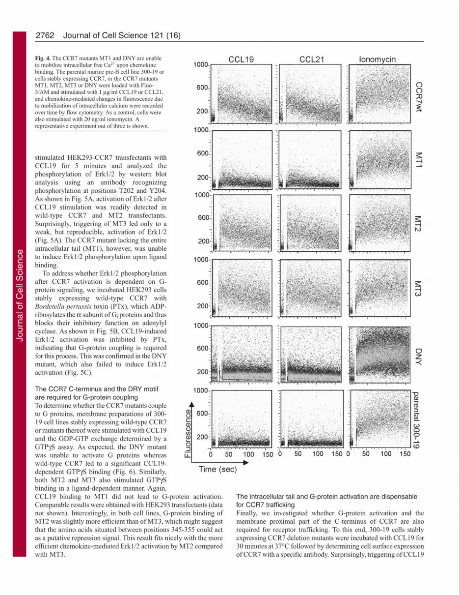
Chemokine-mediated mobilization of intracellular calciumdepends on the membrane proximal part of the C terminusand the DRY motif of CCR7The mobilization of intracellular calcium is an early event afterchemokine binding to its receptors. To examine whether CCR7mutants are able to release Ca2+ from intracellular stores upon ligandbinding, cells were loaded with Fluo-3/AM and chemokine-mediated [Ca2+]i changes were monitored by flow cytometry. MT2
and MT3 both mobilized Ca2+ in response to CCL19 and CCL21at levels comparable with wild-type CCR7 (Fig. 4). By contrast,CCL19 and CCL21 were unable to induce changes in [Ca2+]i incells expressing the CCR7 mutants MT1 and DNY (Fig. 4). Allcell lines responded to ionomycin, indicating comparable dyeloading of the cells (Fig. 4). Similarly to their role in cell migration,the DRY motif and residues 335-345 of CCR7 are indispensablefor calcium signaling.
Chemokine-mediated triggering of the CCR7 mutants MT1 andDNY does not lead to Erk1/2 activationSignal transduction by chemokine receptors also results in atransient activation of the MAPKs Erk1 and Erk2. Thus, we
101 102 103 1040
20
40
60
80
100
101 102 103 104 101 102 103 104 101 102 103 104
101 102 103 1040
20
40
60
80
100
101 102 103 104 101 102 103 104 101 102 103 104 101 102 103 104
101 102 103 104
Cou
nts
CCR7 surface expression
CCR7wt MT1 MT2 MT3
Cou
nts
CCL19-biotin binding
DNY
10
100
0
Fig. 2. CCL19 binds equally to allCCR7 variants. 300-19 pre-Bcells stably transfected with wild-type CCR7 or the CCR7 mutantswere incubated withmonobiotinylated CCL19 (1μg/ml) for 20 minutes at 4°C.CCR7-bound CCL19-biotin wasthen detected with streptavidin-FITC and monitored by flowcytometry (upper panel). CCR7cell surface staining wasperformed with a FITC-labeledCCR7-specific antibody (lowerpanel). Dashed lines representuntransfected cells stained withthe same antibody. FITC-labeledisotype control staining ontransfected cells revealed thesame results. Results arerepresentative of two experiments.
ng/ml CCL210
20
40
60
0 30 300 3000
CCR7wt
DNY
MT2MT3
MT1Spe
cific
mig
ratio
n (%
)
A
B
MT3
0
20
40
60
Spe
cific
mig
ratio
n (%
)
ng/ml CCL190 30 300 3000
CCR7wt
DNYMT1
MT2
Fig. 3. The membrane proximal part of the C-terminus and the DRY motif ofCCR7 are crucial for cell migration. Chemotaxis of 300-19 cells stablytransfected with wild-type CCR7, MT1-3 or DNY mutants was assessed inTranswellTM chemotaxis assays. After 3 hours of incubation at 37°C, cellsmigrated in response to graded concentrations of CCL19 (A) or CCL21 (B) tothe lower chamber were collected and counted by flow cytometry. Meanvalues ± s.d. of three independent experiments are depicted as percentage ofinput cells.
Jour
nal o
f Cel
l Sci
ence
2762
stimulated HEK293-CCR7 transfectants withCCL19 for 5 minutes and analyzed thephosphorylation of Erk1/2 by western blotanalysis using an antibody recognizingphosphorylation at positions T202 and Y204.As shown in Fig. 5A, activation of Erk1/2 afterCCL19 stimulation was readily detected inwild-type CCR7 and MT2 transfectants.Surprisingly, triggering of MT3 led only to aweak, but reproducible, activation of Erk1/2(Fig. 5A). The CCR7 mutant lacking the entireintracellular tail (MT1), however, was unableto induce Erk1/2 phosphorylation upon ligandbinding.
To address whether Erk1/2 phosphorylationafter CCR7 activation is dependent on G-protein signaling, we incubated HEK293 cellsstably expressing wild-type CCR7 withBordetella pertussis toxin (PTx), which ADP-ribosylates the α subunit of Gi proteins and thusblocks their inhibitory function on adenylylcyclase. As shown in Fig. 5B, CCL19-inducedErk1/2 activation was inhibited by PTx,indicating that G-protein coupling is requiredfor this process. This was confirmed in the DNYmutant, which also failed to induce Erk1/2activation (Fig. 5C).
The CCR7 C-terminus and the DRY motifare required for G-protein couplingTo determine whether the CCR7 mutants coupleto G proteins, membrane preparations of 300-19 cell lines stably expressing wild-type CCR7or mutants thereof were stimulated with CCL19and the GDP-GTP exchange determined by aGTPγS assay. As expected, the DNY mutantwas unable to activate G proteins whereaswild-type CCR7 led to a significant CCL19-dependent GTPγS binding (Fig. 6). Similarly,both MT2 and MT3 also stimulated GTPγSbinding in a ligand-dependent manner. Again,CCL19 binding to MT1 did not lead to G-protein activation.Comparable results were obtained with HEK293 transfectants (datanot shown). Interestingly, in both cell lines, G-protein binding ofMT2 was slightly more efficient than of MT3, which might suggestthat the amino acids situated between positions 345-355 could actas a putative repression signal. This result fits nicely with the moreefficient chemokine-mediated Erk1/2 activation by MT2 comparedwith MT3.
The intracellular tail and G-protein activation are dispensablefor CCR7 traffickingFinally, we investigated whether G-protein activation and themembrane proximal part of the C-terminus of CCR7 are alsorequired for receptor trafficking. To this end, 300-19 cells stablyexpressing CCR7 deletion mutants were incubated with CCL19 for30 minutes at 37°C followed by determining cell surface expressionof CCR7 with a specific antibody. Surprisingly, triggering of CCL19
Journal of Cell Science 121 (16)
Fig. 4. The CCR7 mutants MT1 and DNY are unableto mobilize intracellular free Ca2+ upon chemokinebinding. The parental murine pre-B cell line 300-19 orcells stably expressing CCR7, or the CCR7 mutantsMT1, MT2, MT3 or DNY were loaded with Fluo-3/AM and stimulated with 1 μg/ml CCL19 or CCL21,and chemokine-mediated changes in fluorescence dueto mobilization of intracellular calcium were recordedover time by flow cytometry. As a control, cells werealso stimulated with 20 ng/ml ionomycin. Arepresentative experiment out of three is shown.
Jour
nal o
f Cel
l Sci
ence
2763CCR7 signaling by distinct motifs
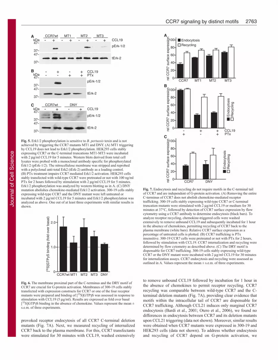
provoked receptor endocytosis of all CCR7 C-terminal deletionmutants (Fig. 7A). Next, we measured recycling of internalizedCCR7 back to the plasma membrane. For this, CCR7 transfectantswere stimulated for 30 minutes with CCL19, washed extensively
to remove unbound CCL19 followed by incubation for 1 hour inthe absence of chemokines to permit receptor recycling. CCR7recycling was comparable between wild-type CCR7 and the C-terminal deletion mutants (Fig. 7A), providing clear evidence thatmotifs within the intracellular tail of CCR7 are dispensable forCCR7 trafficking. Although CCL21 induces only marginal CCR7endocytosis (Bardi et al., 2001; Otero et al., 2006), we found nodifferences in endocytosis between CCR7 and its deletion mutantsupon CCL21 triggering (data not shown). Moreover, similar resultswere obtained when CCR7 mutants were expressed in 300-19 andHEK293 cells (data not shown). To address whether endocytosisand recycling of CCR7 depend on G-protein activation, we
- + - + CCL19CCR7wt DNY
kDa47
3747
37
pErk-1/2
tErk-2
- + - + CCL19kDa47
3747
37
pErk-1/2
tErk-2
- - + + PTx
A
B
C
- + - + CCL19CCR7wt
kDa47
3747
37
pErk-1/2
tErk-2
- + - +MT1 MT2 MT3
Fig. 5. Erk1/2 phosphorylation is sensitive to B. pertussis toxin and is notachieved by triggering the CCR7 mutants MT1 and DNY. (A) MT1 triggeringby CCL19 does not lead to Erk1/2 phosphorylation. HEK293 cells stablyexpressing CCR7 or the C-terminal truncations MT1-MT3 were incubatedwith 2 μg/ml CCL19 for 5 minutes. Western blots derived from total celllysates were probed with a monoclonal antibody specific for phosphorylatedErk1/2 (pErk-1/2). The nitrocellulose membrane was stripped and reprobedwith a polyclonal anti-total Erk2 (tErk-2) antibody as a loading control.(B) PTx treatment impairs CCR7 mediated Erk1/2 activation. HEK293 cellsstably transfected with wild-type CCR7 were pretreated or not with 100 ng/mlPTx for 2 hours followed by stimulation with 2 μg/ml CCL19 for 5 minutes.Erk1/2 phosphorylation was analyzed by western blotting as in A. (C) DNYmutation abolishes chemokine-mediated Erk1/2 activation. 300-19 cells stablyexpressing wild-type CCR7 and the DNY mutant were left untreated orincubated with 2 μg/ml CCL19 for 5 minutes and Erk1/2 phosphorylation wasanalyzed as above. One out of at least three experiments with similar results isshown.
[35S
]GT
PyS
bin
ding
(fo
ld o
ver
basa
l)
CCR7wt MT1 MT2 MT3 DNY0
1.0
2.0
Fig. 6. The membrane proximal part of the C-terminus and the DRY motif ofCCR7 are crucial for G-protein activation. Membranes of 300-19 cells stablytransfected with expression constructs for CCR7 or one of the four receptormutants were prepared and binding of [35S]GTPγS was assessed in response tostimulation with CCL19 (5 μg/ml). Results are expressed as fold over basal[35S]GTPγS binding in the absence of chemokine. Values represent the mean ±s.e.m. of three experiments.
rel C
CR
7 su
rfac
e ex
pres
sion
0
80
100
40
60
20
A
rel C
CR
7 su
rfac
e ex
pres
sion
0
80
100
40
60
20
B
End
ocyt
osis
Rec
yclin
g
+- + CCL19-+ + PTx
rel C
CR
7 su
rfac
e ex
pres
sion
0
80
100
40
60
20
C
CCR7 DNY
EndocytosisRecycling
CCR7 MT1 MT2 MT3
End
ocyt
osis
Rec
yclin
g
Fig. 7. Endocytosis and recycling do not require motifs in the C-terminal tailof CCR7 and are independent of G-protein activation. (A) Removing the entireC-terminus of CCR7 does not abolish chemokine-mediated receptortrafficking. 300-19 cells stably expressing wild-type CCR7 or C-terminaltruncation mutants were stimulated with 2 μg/ml CCL19 or medium for 30minutes at 37°C, followed by detection of CCR7 surface expression by flowcytometry using a CCR7 antibody to determine endocytosis (black bars). Toanalyze receptor recycling, chemokine-triggered cells were washedextensively to remove unbound CCL19 and subsequently incubated for 1 hourin the absence of chemokines, permitting recycling of CCR7 back to theplasma membrane (white bars). Relative CCR7 surface expression as apercentage of untreated cells is plotted. (B) CCR7 trafficking is PTxinsensitive. 300-19 CCR7 cells were pretreated or not with PTx for 2 hours,followed by stimulation with CCL19. CCR7 internalization and recycling weredetermined by flow cytometry as described above. (C) The DRY motif isdispensable for CCR7 trafficking. 300-19 cells stably expressing wild-typeCCR7 or the DNY mutant were incubated with 2 μg/ml CCL19 for 30 minutesfor internalization assays. CCR7 endocytosis and recycling were assessed asoutlined in A. Values represent the mean ± s.e.m. of three experiments.
Jour
nal o
f Cel
l Sci
ence
2764
incubated 300-19 cells expressing wild-type CCR7 with PTx. Gαi
inhibition influenced neither CCL19-induced CCR7 internalizationnor recycling (Fig. 7B). Studies on the DNY mutant confirmed thatCCR7 internalization and recycling were independent of G-proteinactivation (Fig. 7C).
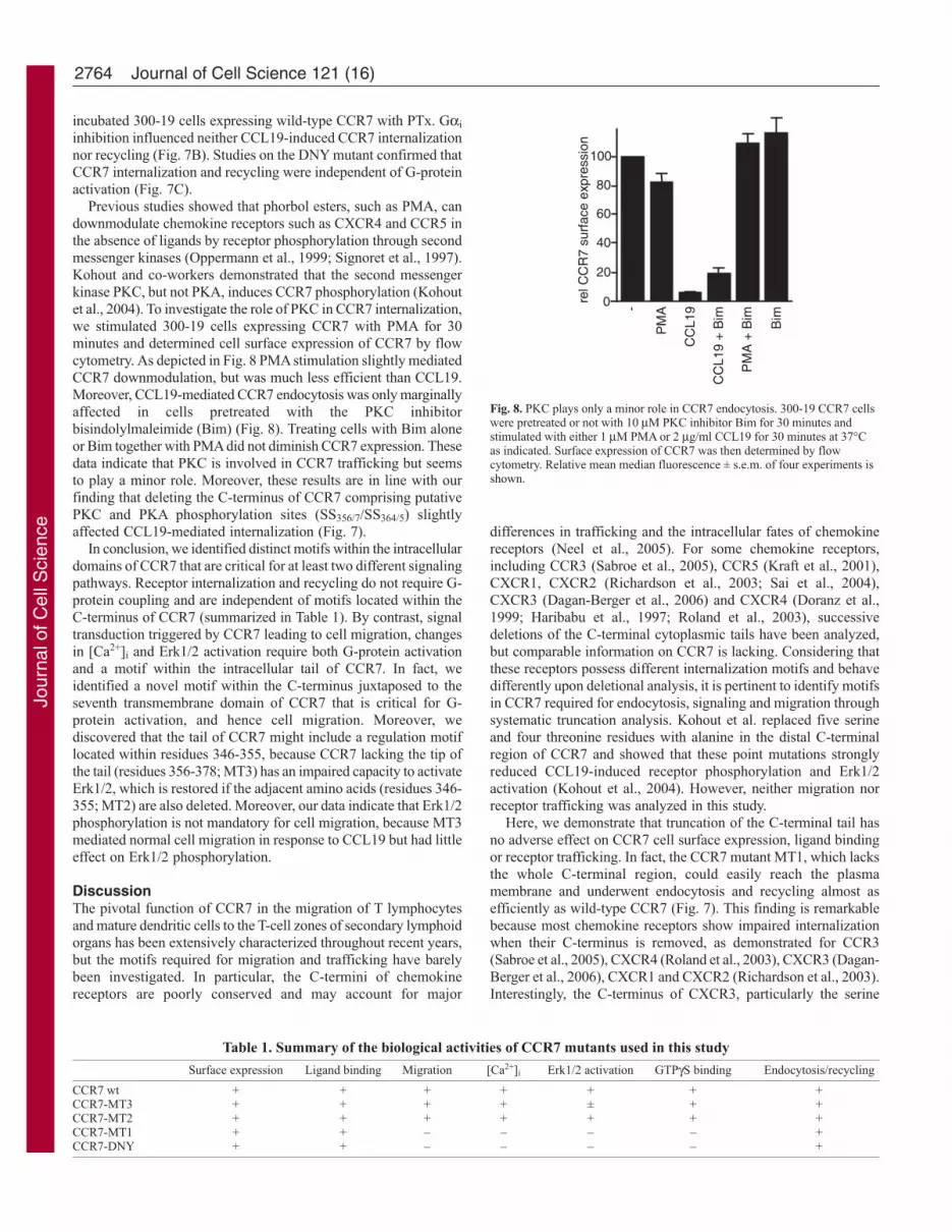
Previous studies showed that phorbol esters, such as PMA, candownmodulate chemokine receptors such as CXCR4 and CCR5 inthe absence of ligands by receptor phosphorylation through secondmessenger kinases (Oppermann et al., 1999; Signoret et al., 1997).Kohout and co-workers demonstrated that the second messengerkinase PKC, but not PKA, induces CCR7 phosphorylation (Kohoutet al., 2004). To investigate the role of PKC in CCR7 internalization,we stimulated 300-19 cells expressing CCR7 with PMA for 30minutes and determined cell surface expression of CCR7 by flowcytometry. As depicted in Fig. 8 PMA stimulation slightly mediatedCCR7 downmodulation, but was much less efficient than CCL19.Moreover, CCL19-mediated CCR7 endocytosis was only marginallyaffected in cells pretreated with the PKC inhibitorbisindolylmaleimide (Bim) (Fig. 8). Treating cells with Bim aloneor Bim together with PMA did not diminish CCR7 expression. Thesedata indicate that PKC is involved in CCR7 trafficking but seemsto play a minor role. Moreover, these results are in line with ourfinding that deleting the C-terminus of CCR7 comprising putativePKC and PKA phosphorylation sites (SS356/7/SS364/5) slightlyaffected CCL19-mediated internalization (Fig. 7).
In conclusion, we identified distinct motifs within the intracellulardomains of CCR7 that are critical for at least two different signalingpathways. Receptor internalization and recycling do not require G-protein coupling and are independent of motifs located within theC-terminus of CCR7 (summarized in Table 1). By contrast, signaltransduction triggered by CCR7 leading to cell migration, changesin [Ca2+]i and Erk1/2 activation require both G-protein activationand a motif within the intracellular tail of CCR7. In fact, weidentified a novel motif within the C-terminus juxtaposed to theseventh transmembrane domain of CCR7 that is critical for G-protein activation, and hence cell migration. Moreover, wediscovered that the tail of CCR7 might include a regulation motiflocated within residues 346-355, because CCR7 lacking the tip ofthe tail (residues 356-378; MT3) has an impaired capacity to activateErk1/2, which is restored if the adjacent amino acids (residues 346-355; MT2) are also deleted. Moreover, our data indicate that Erk1/2phosphorylation is not mandatory for cell migration, because MT3mediated normal cell migration in response to CCL19 but had littleeffect on Erk1/2 phosphorylation.
DiscussionThe pivotal function of CCR7 in the migration of T lymphocytesand mature dendritic cells to the T-cell zones of secondary lymphoidorgans has been extensively characterized throughout recent years,but the motifs required for migration and trafficking have barelybeen investigated. In particular, the C-termini of chemokinereceptors are poorly conserved and may account for major
differences in trafficking and the intracellular fates of chemokinereceptors (Neel et al., 2005). For some chemokine receptors,including CCR3 (Sabroe et al., 2005), CCR5 (Kraft et al., 2001),CXCR1, CXCR2 (Richardson et al., 2003; Sai et al., 2004),CXCR3 (Dagan-Berger et al., 2006) and CXCR4 (Doranz et al.,1999; Haribabu et al., 1997; Roland et al., 2003), successivedeletions of the C-terminal cytoplasmic tails have been analyzed,but comparable information on CCR7 is lacking. Considering thatthese receptors possess different internalization motifs and behavedifferently upon deletional analysis, it is pertinent to identify motifsin CCR7 required for endocytosis, signaling and migration throughsystematic truncation analysis. Kohout et al. replaced five serineand four threonine residues with alanine in the distal C-terminalregion of CCR7 and showed that these point mutations stronglyreduced CCL19-induced receptor phosphorylation and Erk1/2activation (Kohout et al., 2004). However, neither migration norreceptor trafficking was analyzed in this study.
Here, we demonstrate that truncation of the C-terminal tail hasno adverse effect on CCR7 cell surface expression, ligand bindingor receptor trafficking. In fact, the CCR7 mutant MT1, which lacksthe whole C-terminal region, could easily reach the plasmamembrane and underwent endocytosis and recycling almost asefficiently as wild-type CCR7 (Fig. 7). This finding is remarkablebecause most chemokine receptors show impaired internalizationwhen their C-terminus is removed, as demonstrated for CCR3(Sabroe et al., 2005), CXCR4 (Roland et al., 2003), CXCR3 (Dagan-Berger et al., 2006), CXCR1 and CXCR2 (Richardson et al., 2003).Interestingly, the C-terminus of CXCR3, particularly the serine
Journal of Cell Science 121 (16)
Table 1. Summary of the biological activities of CCR7 mutants used in this study
Surface expression Ligand binding Migration [Ca2+]i Erk1/2 activation GTPγS binding Endocytosis/recycling
CCR7 wt + + + + + + +CCR7-MT3 + + + + ± + +CCR7-MT2 + + + + + + +CCR7-MT1 + + – – – – +CCR7-DNY + + – – – – +
rel C
CR
7 su
rfac
e ex
pres
sion
0
80
100
40
60
20
-
CC
L19
PM
A
CC
L19
+ B
im
PM
A +
Bim
Bim
Fig. 8. PKC plays only a minor role in CCR7 endocytosis. 300-19 CCR7 cellswere pretreated or not with 10 μM PKC inhibitor Bim for 30 minutes andstimulated with either 1 μM PMA or 2 μg/ml CCL19 for 30 minutes at 37°Cas indicated. Surface expression of CCR7 was then determined by flowcytometry. Relative mean median fluorescence ± s.e.m. of four experiments isshown.
Jour
nal o
f Cel
l Sci
ence

2765CCR7 signaling by distinct motifs
clusters therein, was shown to be involved in CXCL9- and CXCL10-mediated receptor internalization, whereas CXCL11-mediatedCXCR3 endocytosis depends on a motif within the third intracellularloop of the receptor (Colvin et al., 2004). Serial C-terminaltruncation of CCR5 resulted in progressive loss of cell surfaceexpression (Venkatesan et al., 2001) or impaired ligand-inducedinternalization (Kraft et al., 2001). Despite the normal traffickingof MT1 in HEK293 and 300-19 cell transfectants, CCR7 lackingits whole C-terminus was not able to transmit signals leading tocell migration (Fig. 3). A previous study showed that a cluster ofSer/Thr at the tip of the tail of CCR7 is phosphorylated upon ligandbinding, suggesting that this region may be important for CCR7internalization (Kohout et al., 2004). Our MT2 mutant lacks allpossible phosphorylation sites of the C-terminus, whereas in MT3the above-mentioned Ser/Thr motifs were deleted, leaving oneputative phosphorylation site (Ser348). CCL19-induced receptorendocytosis and recycling occurred in both MT2 and MT3expressing cells, and even in cells expressing the tailless mutantMT1, providing clear evidence that the intracellular tail of CCR7does not include major signaling motifs for receptor trafficking.Moreover, CCR7 trafficking does not require G-protein coupling,as inhibiting G-protein activation by either PTx treatment or by theDRY to DNY mutation, did not abrogate receptor internalizationor recycling (Fig. 7). In this respect, CCR7 behaves like otherchemokine receptors as CXCR3, CXCR4 and CCR5 internalizationfor instance does not depend on G-protein activation (Amara et al.,1997; Colvin et al., 2004; Lagane et al., 2005) Noteworthy, deletionof the Ser/Thr motifs (MT3) at the C-terminus significantly impairedchemokine-mediated Erk1/2 activation (Fig. 5). This finding is inagreement with the previous observation that replacing Ser356-Ser357, Ser364-Ser365, Ser378and Thr373-Thr374-Thr375-Thr376with Ala resulted in diminished Erk1/2 phosphorylation (Kohoutet al., 2004). Interestingly, this phenotype is completely recoveredif another ten amino acids are deleted. CCL19-induced Erk1/2phosphorylation is completely restored in the MT2 mutant,indicating that residues 346-355 may contain a novel regulatorymotif. Moreover, CCR7 signaling mediated by MT2 and MT3 alsosuggests that Erk1/2 activation is not required for migration or forinducing changes in [Ca2+]i. The removal of potentialphosphorylation sites within the C-terminus of CCR7 in the mutantsMT2 and MT3 did not impair chemotaxis (Fig. 3), indicating thatthe Ser356-Ser357, Ser364-Ser365 and Thr372-Thr373-Thr374-Thr375 putative targets for GRK phosphorylation (Kohout et al.,2004) are not required for chemotactic signal transduction.
Truncation of the C-terminus of CCR7 led to the discovery ofother exciting findings. Deletion of the entire C-terminus of CCR7resulted in the loss of chemotactic activity (Fig. 3). Chemotaxisdepends on the activation of G proteins, and chemokine-mediatedG-protein activation is believed to exclusively depend on the highlyconserved DRY motif within the second intracellular loop. Indeed,modifying the DRY motif completely abolished CCL19-mediatedG-protein activation (Fig. 6), cell migration (Fig. 3), [Ca2+]i
mobilization (Fig. 4) and Erk1/2 phosphorylation (Fig. 5), but didnot affect receptor trafficking (Fig. 7). As the impaired migrationof MT1 is due to the inability to activate the G protein (Fig. 6), itis tempting to speculate that the C-terminus contains an additionalunidentified second motif critical for G-protein activation. This G-protein activation motif of CCR7 must be located within residues335-345 (sequence: NDIFKLFKDLG), because MT2 readilyactivated the G protein upon chemokine binding. To our knowledge,such a G-protein activation motif located in the C-terminus of a
chemokine receptor has not been identified. However, removingthe C-terminus of CXCR4 resulted in an even higher G-proteinactivation in response to CXCL12 compared with cells expressingwild-type CXCR4 (Haribabu et al., 1997).
So far, we have been unable to identify a motif responsible forCCR7 trafficking. We demonstrate that neither G-protein couplingnor the intracellular tail contribute to CCR7 trafficking. Mostcommonly, tyrosine- or leucine-based internalization motifs cantarget membrane receptors to clathrin-coated pits. The C-terminaltail of CXCR2, CXCR4 and CCR5 for instance contains di-Leumotifs (LLKIL, LKIL and LL, respectively) which are determinantsfor receptor endocytosis upon ligand binding (Fan et al., 2001; Kraftet al., 2001; Orsini et al., 1999). The C-terminal tail of CCR7contains five leucine residues, but they are not arranged in pairsand hence do not represent a bona fide internalization motif.Moreover, MT1 was efficiently endocytosed upon ligand bindingalthough none of these leucines was present (Fig. 7). Ubiquitylationof the C-terminus also seems not to be the main driving force forCCR7 endocytosis, because in MT1 the remaining lysine wasreplaced by arginine. Thus, further studies on the intracellular loopsare required to identify the motif involved in CCR7 trafficking.
In summary, the present study reveals that endocytosis andrecycling signals are not contained within the C-terminus of CCR7,whereas signal transduction leading to cell migration relys on a tenresidue motif adjacent to the seventh transmembrane domain andon G-protein activation. Deleting a Ser/Thr motif at the distal partof the tail of CCR7 resulted in an impaired chemokine-mediatedactivation of Erk1/2 kinases. Interestingly, deleting an additionaladjacent motif restored the ability of CCL19-mediated Erk1/2phosphorylation, indicating the presence of a regulatory motif. Boththe Ser/Thr motif and the regulatory motif are dispensable forchemotactic signals. Moreover, we identified a motif in the C-terminus of CCR7 juxtaposed to the plasma membrane that is criticalfor G-protein activation and cell migration.
Materials and MethodsAntibodies and reagentsAntibodies were obtained from the following sources: FITC-coupled mouse anti-human CCR7 (clone 150503; R&D Systems Inc., Minneapolis, MN), mouse anti-HA, (Sigma, Buchs, Switzerland), streptavidin-FITC (Jackson ImmunoResearchLaboratories), phospho-Erk-1/2, total Erk-2 (Santa Cruz Biotechnology Inc, SantaCruz, CA), and HRP-conjugated secondary anti-mouse and anti-rabbit antibodies(DAKO, Hamburg, Germany). Similar results were obtained using rat anti-humanCCR7 (clone 3D12, BD Biosciences PharMingen, San Diego, CA) (data not shown).Recombinant human chemokines CCL19 and CCL21 were purchased from PromoCell(Heidelberg, Germany). Bordetella pertussis toxin was purchased from Calbiochem(La Jolla, CA). Ionomycin, bisindolylmaleimide (Bim) and PMA were from Sigma.[35S]GTPγS was from Amersham Biosciences (Little Chalfont, Buckinghamshire,UK). Monobiotinylated CCL19 was from RMF Dictagene SA (Epalinges,Switzerland).
Construction of expression plasmidsThe cloning of pcDNA3-CCR7-HA has been described (Otero et al., 2006). C-terminaldeletion mutants of CCR7, MT1 (aa 1-334), MT2 (aa 1-345) and MT3 (1-355) weregenerated by PCR using full-length CCR7 (aa 1-378) as template. PCR primers areas follows: CCR7-MTse 5�-ATA AAG CTT CGT CAT GGA CCT GGG G-3� (HindIIIrestriction site underlined), CCR7-MT1as 5�-ATA GAA TTC CGC GGA ACC TGACGC C-3� (EcoRI restriction site underlined), CCR7-MT2as (5�-ATA GAA TTC CGCCCA GGT CCT TGA AG-3�) and CCR7-MT3as (5�-ATA GAA TTC CCC CACTGC CGG AGC TG-3�). PCR fragments were cloned into pcDNA3-HA, revealingC-terminal-tagged CCR7 mutants. In CCR7-MT1, a K332R substitution wasintroduced. The CCR7-DNY mutant was generated by site-directed mutagenesis usingpcDNA3-CCR7-HA as template. Briefly, a first PCR was performed with the primerCCR7se2 (5�-ATA GAA TTC CGT CAT GGA CCT GGG GAA AC-3�; EcoRI siteunderlined) and the primer DRYas (5�-GGC CAC GTA GTT GTC AAT GCT GAT-3�; modification underlined). A second PCR was performed with the primers DRYse(5�-ATC AGC ATT GAC AAC TAC GTG GCC-3�; modification underlined) andCCR7as (5�-TAT GCG GCC GCT GGG GAG AAG GTG GTG-3�; NotI site
Jour
nal o
f Cel
l Sci
ence

2766
underlined). Then, both PCR products were mixed and a third PCR was performedwith the primers CCR7as and CCR7se2 and the amplification product was clonedinto pcDNA3-HA. HA-tagged CCR7 behaves like wild-type CCR7 in terms ofchemotaxis, Erk activation, Calcium mobilization and receptor trafficking (Otero etal., 2006).
Cell lines and transfectionThe human embryonic kidney cell line HEK293 was grown in DMEM (Invitrogen,Basel, Switzerland) supplemented with 10% (v/v) fetal bovine serum. HEK293 cellswere stably transfected with FuGENE6 (Roche, Basel, Switzerland). Cell clones wereestablished by limiting dilution in the presence of 0.8 mg/ml of G-418 (Invitrogen).The murine pre-B cell line 300-19 (Legler et al., 1998; Willimann et al., 1998) wasgrown in RPMI1640 (Invitrogen) with 10% (v/v) fetal bovine serum, 50 μM β-mercaptoethanol and 2 mM nonessential amino acids. Transfection was performedby electroporation and stable cell lines were generated by limiting dilution as described(Legler et al., 1998; Willimann et al., 1998).
CCL19-bio binding assayCCR7-transfected 300-19 cells were incubated with monobiotinylated CCL19 (1μg/ml) for 20 minutes at 4°C. Cells were extensively washed with PBS, 2% FBS, 5mM EDTA and incubated with streptavidin-FITC (1 μg/ml) for 20 minutes, washedand fluorescence was monitored by flow cytometry (FACScan IITM or LSR IITM)using CellQuestTM or FACSDivaTM software (BD Biosciences). Data were analyzedwith the FlowJoTM software (Tree Star, San Carlos, CA).
ChemotaxisCell migration (1�105 cells in 100 μl) in response to graded concentrations of CCL19and CCL21 was measured in 24-well TranswellTM chambers (Corning Costar,Cambridge, MA) through a polycarbonate filter of 5 μm pore size. After 3 hours ofincubation at 37°C, a 600 μl aliquot of cells that migrated to the bottom chamberwas counted by flow cytometry acquiring events for a fixed time period of 60 secondsand the number of migrated cells were expressed as percent of input cells.
Western blottingCells (2�106) were lysed with 0.5 ml of 1% Triton X-100 in 150 mM NaCl, 50 mMHEPES, 0.1 M EGTA, 2 mM MgCl2 and 10% glycerol. Proteins from total cell lysateswere resolved by SDS-PAGE and transferred to Protran nitrocellulose membranes(Schleicher & Schuell, Dassel, Germany). Membranes were blocked with PBScontaining 5% low-fat dry milk and incubated with Erk-specific antibodies overnightat 4°C (anti-phospho-Erk-1/2) or for 1 hour at room temperature (anti-total Erk-2)on a rocking plate. After washing, HRP-conjugated secondary antibodies were detectedusing enhanced chemiluminescence (Pierce/Socochim, Lausanne, Switzerland).
Analysis of chemokine-mediated changes in intracellular free Ca2+
concentrationsCells (106/ml in 145 mM NaCl, 5 mM KCl, 1 mM Na2HPO4, 1 mM MgCl2, 5 mMglucose, 1 mM CaCl2 and 10 mM HEPES, pH 7.5) were loaded with 1.5 μl/ml Fluo-3/AM (4 mM in DMSO) for 30 minutes at 37°C. Samples were divided into 500 μlaliquots and changes in intracellular calcium concentrations in response to 1 μg/mlof chemokines or 0.02 μg/ml of ionomycin were acquired by FACS analysis. Changesin the fluorescence of Fluo-3 were recorded by flow cytometry for a period of 150seconds.
CCR7 traffickingTo investigate receptor internalization, 300-19 transfectants in suspension (106/ml)or HEK293 cells derived from a 90% confluent six-well plates, respectively, werestimulated with 2 μg/ml chemokine or 1 μM PMA for 30 minutes at 37°C. Whereindicated, cells were pre-treated with 10 μM Bim for 30 minutes at 37°C as described(Oppermann et al., 1999). Cell surface expression of CCR7 after ligand-mediatedendocytosis was measured by flow cytometry using a CCR7-specific antibody. Forrecycling experiments, internalized receptor (2 μg/ml of chemokine for 30 minutesat 37°C) was allowed to recycle back to the plasma membrane by washing off unboundchemokines twice with warm PBS following incubation in chemokine-free mediumfor 1 hour at 37°C. Surface expression of CCR7 was thereafter assessed by flowcytometry.
Pertussis toxin treatment300-19 cells (106/ml) or HEK293 cells (one well of a six-well plate) expressing CCR7were preincubated with 100 ng/ml of PTx for 2 hours at 37°C prior to the respectiveassay. PTx was maintained in the medium throughout the experiments to ensurecomplete inhibition of Gαi.
GTPγS binding assayCell transfectants (2�106/assay point) were washed twice with PBS and serum-starvedfor 3 hours. Membrane fractions were prepared from cells resuspended in 1 mlmembrane buffer (20 mM HEPES, 6 mM MgCl2, 1 mM EGTA, pH 7.2) supplementedwith protease inhibitors (Roche), by incubation on ice for 10 minutes, and subsequentsqueezing the cells six times through a syringe (G25). The supernatant of a first, low-
speed centrifugation step (500 g, 10 minutes, 4°C) was again spun down at a higherspeed (20,000 g, 30 minutes, 4°C) and the resulting membrane pellet was resuspendedin GTPγS assay buffer (50 mM HEPES, 100 mM NaCl, 10 mM MgCl2, 1 mM EGTA,0.1% BSA, pH 7.2) supplemented with protease inhibitors. GTPγS binding wasassessed in the presence and absence of CCL19 (5 μg/ml) by adding [35S]GTPγS(0.5 nM) and GDP (10 μM, Sigma) to 200 μg of membrane preparations. After 30minutes at 37°C, each sample was sucked onto a GF/C filter (Whatman, Maidstone,UK). Filters were washed four times with washing buffer (50 mM HEPES, 5 mMMgCl2) and dried at 60°C. Scintillation fluid (rotiszinteco; Roth, Karlsruhe, Germany)was added and scintillography performed with a Beckman LS 6000IC counter(Fullerton, CA).
This work was supported by the German Research Foundation (DFG,TR-SFB 11 TP-C9 and GR 1517/8-1), the Thurgauische Stiftung fürWissenschaft und Forschung, the State Secretariat for Education andResearch and the Thurgauische Krebsliga. D.F.L. is a recipient of acareer development award from the Prof. Dr Max Cloëtta Foundation.The authors declare no competing financial interests.
ReferencesAmara, A., Gall, S. L., Schwartz, O., Salamero, J., Montes, M., Loetscher, P., Baggiolini,
M., Virelizier, J. L. and Arenzana-Seisdedos, F. (1997). HIV coreceptor downregulationas antiviral principle: SDF-1alpha-dependent internalization of the chemokine receptorCXCR4 contributes to inhibition of HIV replication. J. Exp. Med. 186, 139-146.
Balkwill, F. (2004). Cancer and the chemokine network. Nat. Rev. Cancer 4, 540-550.Bardi, G., Lipp, M., Baggiolini, M. and Loetscher, P. (2001). The T cell chemokine
receptor CCR7 is internalized on stimulation with ELC, but not with SLC. Eur. J.Immunol. 31, 3291-3297.
Berchiche, Y. A., Chow, K. Y., Lagane, B., Leduc, M., Percherancier, Y., Fujii, N.,Tamamura, H., Bachelerie, F. and Heveker, N. (2007). Direct assessment of CXCR4mutant conformations reveals complex link between receptor structure and G(alpha)(i)activation. J. Biol. Chem. 282, 5111-5115.
Bonifacino, J. S. and Traub, L. M. (2003). Signals for sorting of transmembrane proteinsto endosomes and lysosomes. Annu. Rev. Biochem. 72, 395-447.
Campbell, D. J., Kim, C. H. and Butcher, E. C. (2003). Chemokines in the systemicorganization of immunity. Immunol. Rev. 195, 58-71.
Colvin, R. A., Campanella, G. S., Sun, J. and Luster, A. D. (2004). Intracellular domainsof CXCR3 that mediate CXCL9, CXCL10, and CXCL11 function. J. Biol. Chem. 279,30219-30227.
Dagan-Berger, M., Feniger-Barish, R., Avniel, S., Wald, H., Galun, E., Grabovsky, V.,Alon, R., Nagler, A., Ben-Baruch, A. and Peled, A. (2006). Role of CXCR3 carboxyl-terminus and third intracellular loop in receptor-mediated migration, adhesion andinternalization in response to CXCL11. Blood 107, 3821-3831.
Doranz, B. J., Orsini, M. J., Turner, J. D., Hoffman, T. L., Berson, J. F., Hoxie, J. A.,Peiper, S. C., Brass, L. F. and Doms, R. W. (1999). Identification of CXCR4 domainsthat support coreceptor and chemokine receptor functions. J. Virol. 73, 2752-2761.
Fan, G. H., Yang, W., Sai, J. and Richmond, A. (2001). Phosphorylation-independentassociation of CXCR2 with the protein phosphatase 2A core enzyme. J. Biol. Chem.276, 16960-16968.
Forster, R., Schubel, A., Breitfeld, D., Kremmer, E., Renner-Muller, I., Wolf, E. andLipp, M. (1999). CCR7 coordinates the primary immune response by establishingfunctional microenvironments in secondary lymphoid organs. Cell 99, 23-33.
Gunn, M. D., Kyuwa, S., Tam, C., Kakiuchi, T., Matsuzawa, A., Williams, L. T. andNakano, H. (1999). Mice lacking expression of secondary lymphoid organ chemokinehave defects in lymphocyte homing and dendritic cell localization. J. Exp. Med. 189,451-460.
Haribabu, B., Richardson, R. M., Fisher, I., Sozzani, S., Peiper, S. C., Horuk, R., Ali,H. and Snyderman, R. (1997). Regulation of human chemokine receptors CXCR4.Role of phosphorylation in desensitization and internalization. J. Biol. Chem. 272, 28726-28731.
Kohout, T. A., Nicholas, S. L., Perry, S. J., Reinhart, G., Junger, S. and Struthers, R.S. (2004). Differential desensitization, receptor phosphorylation, beta-arrestin recruitment,and ERK1/2 Activation by the two endogenous ligands for the CC chemokine receptor7. J. Biol. Chem. 279, 23214-23222.
Kraft, K., Olbrich, H., Majoul, I., Mack, M., Proudfoot, A. and Oppermann, M. (2001).Characterization of sequence determinants within the carboxyl-terminal domain ofchemokine receptor CCR5 that regulate signaling and receptor internalization. J. Biol.Chem. 276, 34408-34418.
Lagane, B., Ballet, S., Planchenault, T., Balabanian, K., Le Poul, E., Blanpain, C.,Percherancier, Y., Staropoli, I., Vassart, G., Oppermann, M. et al. (2005). Mutationof the DRY motif reveals different structural requirements for the CC chemokine receptor5-mediated signaling and receptor endocytosis. Mol. Pharmacol. 67, 1966-1976.
Legler, D. F., Loetscher, M., Roos, R. S., Clark-Lewis, I., Baggiolini, M. and Moser,B. (1998). B cell-attracting chemokine 1, a human CXC chemokine expressed in lymphoidtissues, selectively attracts B lymphocytes via BLR1/CXCR5. J. Exp. Med. 187, 655-660.
Luther, S. A., Tang, H. L., Hyman, P. L., Farr, A. G. and Cyster, J. G. (2000).Coexpression of the chemokines ELC and SLC by T zone stromal cells and deletion ofthe ELC gene in the plt/plt mouse. Proc. Natl. Acad. Sci. USA 97, 12694-12699.
Journal of Cell Science 121 (16)
Jour
nal o
f Cel
l Sci
ence

2767CCR7 signaling by distinct motifs
Moser, B., Wolf, M., Walz, A. and Loetscher, P. (2004). Chemokines: multiple levels ofleukocyte migration control. Trends Immunol. 25, 75-84.
Muller, A., Homey, B., Soto, H., Ge, N., Catron, D., Buchanan, M. E., McClanahan,T., Murphy, E., Yuan, W., Wagner, S. N. et al. (2001). Involvement of chemokinereceptors in breast cancer metastasis. Nature 410, 50-56.
Muller, G., Hopken, U. E. and Lipp, M. (2003). The impact of CCR7 and CXCR5 onlymphoid organ development and systemic immunity. Immunol. Rev. 195, 117-135.
Neel, N. F., Schutyser, E., Sai, J., Fan, G. H. and Richmond, A. (2005). Chemokinereceptor internalization and intracellular trafficking. Cytokine Growth Factor Rev. 16,637-658.
Neptune, E. R. and Bourne, H. R. (1997). Receptors induce chemotaxis by releasing thebetagamma subunit of Gi, not by activating Gq or Gs. Proc. Natl. Acad. Sci. USA 94,14489-14494.
Ohl, L., Mohaupt, M., Czeloth, N., Hintzen, G., Kiafard, Z., Zwirner, J., Blankenstein,T., Henning, G. and Forster, R. (2004). CCR7 governs skin dendritic cell migrationunder inflammatory and steady-state conditions. Immunity 21, 279-288.
Oppermann, M., Mack, M., Proudfoot, A. E. and Olbrich, H. (1999). Differential effectsof CC chemokines on CC chemokine receptor 5 (CCR5) phosphorylation andidentification of phosphorylation sites on the CCR5 carboxyl terminus. J. Biol. Chem.274, 8875-8885.
Orsini, M. J., Parent, J. L., Mundell, S. J., Benovic, J. L. and Marchese, A. (1999).Trafficking of the HIV coreceptor CXCR4. Role of arrestins and identification of residuesin the c-terminal tail that mediate receptor internalization. J. Biol. Chem. 274, 31076-31086.
Otero, C., Groettrup, M. and Legler, D. F. (2006). Opposite fate of endocytosed CCR7and its ligands: recycling versus degradation. J. Immunol. 177, 2314-2323.
Richardson, R. M., Marjoram, R. J., Barak, L. S. and Snyderman, R. (2003). Role ofthe cytoplasmic tails of CXCR1 and CXCR2 in mediating leukocyte migration,activation, and regulation. J. Immunol. 170, 2904-2911.
Riol-Blanco, L., Sanchez-Sanchez, N., Torres, A., Tejedor, A., Narumiya, S., Corbi,A. L., Sanchez-Mateos, P. and Rodriguez-Fernandez, J. L. (2005). The chemokinereceptor CCR7 activates in dendritic cells two signaling modules that independentlyregulate chemotaxis and migratory speed. J. Immunol. 174, 4070-4080.
Roland, J., Murphy, B. J., Ahr, B., Robert-Hebmann, V., Delauzun, V., Nye, K. E.,Devaux, C. and Biard-Piechaczyk, M. (2003). Role of the intracellular domains ofCXCR4 in SDF-1-mediated signaling. Blood 101, 399-406.
Sabroe, I., Jorritsma, A., Stubbs, V. E., Xanthou, G., Jopling, L. A., Ponath, P. D.,Williams, T. J., Murphy, P. M. and Pease, J. E. (2005). The carboxyl terminus of thechemokine receptor CCR3 contains distinct domains which regulate chemotacticsignaling and receptor down-regulation in a ligand-dependent manner. Eur. J. Immunol.35, 1301-1310.
Sai, J., Fan, G. H., Wang, D. and Richmond, A. (2004). The C-terminal domain LLKILmotif of CXCR2 is required for ligand-mediated polarization of early signals duringchemotaxis. J. Cell Sci. 117, 5489-5496.
Sanchez-Sanchez, N., Riol-Blanco, L., De La Rosa, G., Puig-Kroger, A., Garcia-Bordas,J., Martin, D., Longo, N., Cuadrado, A., Cabanas, C., Corbi, A. L. et al. (2004).Chemokine receptor CCR7 induces intracellular signaling that inhibits apoptosis of maturedendritic cells. Blood 104, 619-625.
Scandella, E., Men, Y., Legler, D. F., Gillessen, S., Prikler, L., Ludewig, B. andGroettrup, M. (2004). CCL19/CCL21-triggered signal transduction and migration ofdendritic cells requires prostaglandin E2. Blood 103, 1595-1601.
Signoret, N., Oldridge, J., Pelchen-Matthews, A., Klasse, P. J., Tran, T., Brass, L. F.,Rosenkilde, M. M., Schwartz, T. W., Holmes, W., Dallas, W. et al. (1997). Phorbolesters and SDF-1 induce rapid endocytosis and down modulation of the chemokinereceptor CXCR4. J. Cell Biol. 139, 651-664.
Thelen, M. (2001). Dancing to the tune of chemokines. Nat. Immunol. 2, 129-134.Venkatesan, S., Petrovic, A., Locati, M., Kim, Y. O., Weissman, D. and Murphy, P.
M. (2001). A membrane-proximal basic domain and cysteine cluster in the C-terminaltail of CCR5 constitute a bipartite motif critical for cell surface expression. J. Biol. Chem.276, 40133-40145.
Vilardaga, J. P., Frank, M., Krasel, C., Dees, C., Nissenson, R. A. and Lohse, M. J.(2001). Differential conformational requirements for activation of G proteins and theregulatory proteins arrestin and G protein-coupled receptor kinase in the G protein-coupled receptor for parathyroid hormone (PTH)/PTH-related protein. J. Biol. Chem.276, 33435-33443.
Willimann, K., Legler, D. F., Loetscher, M., Roos, R. S., Delgado, M. B., Clark-Lewis,I., Baggiolini, M. and Moser, B. (1998). The chemokine SLC is expressed in T cellareas of lymph nodes and mucosal lymphoid tissues and attracts activated T cells viaCCR7. Eur. J. Immunol. 28, 2025-2034.
Wymann, M. P., Zvelebil, M. and Laffargue, M. (2003). Phosphoinositide 3-kinasesignalling-which way to target? Trends Pharmacol. Sci. 24, 366-376.
Zlotnik, A. (2006). Chemokines and cancer. Int. J. Cancer 119, 2026-2029.
Jour
nal o
f Cel
l Sci
ence
Copyright © 2022 FDOKUMEN