Generic Priors Yield Competition Between Independently-Occurring Preventive Causes
Upload
independentCategory
view
0download
0
of April 15, 2014.This information is current as
Migratory SpeedIndependently Regulate Chemotaxis andDendritic Cells Two Signaling Modules That The Chemokine Receptor CCR7 Activates in
Sánchez-Mateos and José Luis Rodríguez-FernándezAlberto Tejedor, Shuh Narumiya, Angel L. Corbí, Paloma Lorena Riol-Blanco, Noelia Sánchez-Sánchez, Ana Torres,
http://www.jimmunol.org/content/174/7/40702005; 174:4070-4080; ;J Immunol
Referenceshttp://www.jimmunol.org/content/174/7/4070.full#ref-list-1
, 34 of which you can access for free at: cites 73 articlesThis article
Subscriptionshttp://jimmunol.org/subscriptions
is online at: The Journal of ImmunologyInformation about subscribing to
Permissionshttp://www.aai.org/ji/copyright.htmlSubmit copyright permission requests at:
Email Alertshttp://jimmunol.org/cgi/alerts/etocReceive free email-alerts when new articles cite this article. Sign up at:
Print ISSN: 0022-1767 Online ISSN: 1550-6606. Immunologists All rights reserved.Copyright © 2005 by The American Association of9650 Rockville Pike, Bethesda, MD 20814-3994.The American Association of Immunologists, Inc.,
is published twice each month byThe Journal of Immunology
by guest on April 15, 2014
http://ww
w.jim
munol.org/
Dow
nloaded from
by guest on April 15, 2014
http://ww
w.jim
munol.org/
Dow
nloaded from
The Chemokine Receptor CCR7 Activates in Dendritic CellsTwo Signaling Modules That Independently RegulateChemotaxis and Migratory Speed1
Lorena Riol-Blanco,2* Noelia Sanchez-Sanchez,2* Ana Torres,† Alberto Tejedor,† Shuh Narumiya,§
Angel L. Corbı,* Paloma Sanchez-Mateos,‡ and Jose Luis Rodrıguez-Fernandez3*
CCR7 is necessary to direct dendritic cells (DCs) to secondary lymphoid nodes and to elicit an adaptative immune response.Despite its importance, little is known about the molecular mechanisms used by CCR7 to direct DCs to lymph nodes. In additionto chemotaxis, CCR7 regulates the migratory speed of DCs. We investigated the intracellular pathways that regulate CCR7-dependent chemotaxis and migratory speed. We found that CCR7 induced a Gi-dependent activation of MAPK members ERK1/2,JNK, and p38, with ERK1/2 and p38 controlling JNK. MAPK members regulated chemotaxis, but not the migratory speed, ofDCs. CCR7 induced activation of PI3K/Akt; however, these enzymes did not regulate either chemotaxis or the speed of DCs. CCR7also induced activation of the GTPase Rho, the tyrosine kinase Pyk2, and inactivation of cofilin. Pyk2 activation was independentof Gi and Src and was dependent on Rho. Interference with Rho or Pyk2 inhibited cofilin inactivation and the migratory speedof DCs, but did not affect chemotaxis. Interference with Rho/Pyk2/cofilin inhibited DC migratory speed even in the absence ofchemokines, suggesting that this module controls the speed of DCs and that CCR7, by activating its components, induces anincrease in migratory speed. Therefore, CCR7 activates two independent signaling modules, one involving Gi and a hierarchy ofMAPK family members and another involving Rho/Pyk2/cofilin, which control, respectively, chemotaxis and the migratory speedof DCs. The use of independent signaling modules to control chemotaxis and speed can contribute to regulate the chemotacticeffects of CCR7. The Journal of Immunology, 2005, 174: 4070–4080.
D endritic cells (DCs)4 migrate from peripheral tissues tolymph nodes to prime T lymphocytes in these areas andelicit an immune response (1, 2). The migration of DCs
toward T cell areas requires up-regulation of CCR7, an event thattakes place during the process of maturation of DCs (3–5). The twoligands of CCR7, CCL19 and CCL21, are expressed by stromalcells in the T cell zone of the lymph nodes (6–10). Moreover,CCL19 is also expressed by mature DCs (6, 11, 12), and CCL21is expressed by endothelial cells of afferent lymphatic vessels andhigh endothelial veins (8–10). Mice deficient in CCL19, CCL21,or CCR7 show defective DC traffic and altered immune responses(13–15). Recently, it has also been suggested that CCR7 expressed
in different metastatic cell lines may direct these cells to theirniches (5, 16, 17).
Chemokine receptors confer upon cells the ability to detect andmove directionally toward a chemotactic stimulus. Often in addi-tion to chemotaxis, chemokine receptors regulate the migratoryspeed of the cells (also called random motility or nondirectionalspeed) (18, 19). In this regard, it is not clear whether the signalingpathways that regulate chemotaxis and migratory speed overlap orare independent of each other.
Although there are common rules, it is emerging that there is ahigh level of cell context and cell type variability in the signalingthat is regulated by chemokine receptors. Generally, chemokinereceptors relay intracellular signals that regulate chemotaxisthrough the Gi subfamily of G proteins (20). These receptors reg-ulate a variety of signaling molecules, including MAPK familymembers (21–23). Mammalian cells contain three major classes ofMAPKs, ERK1/2, JNK, and p38. These molecules are importantregulators of chemotaxis and/or random motility in a variety of celltypes (23–25). Chemokine receptors may also activate PI3K andthe downstream effector Akt, which play a central role in regula-tion of the chemotactic response in leukocytes and other cells (21,23, 26–30). Other mediators of chemokine receptors include ki-nases, such as proline-rich tyrosine kinase 2 (Pyk2) (21, 22, 31).Pyk2 binds a variety of signaling molecules, including tyrosinekinases such as Src, that impinge upon this protein in several signalingpathways (32–35). It has been shown that Pyk2 regulates leukocytemotility, although the mechanism involved is unknown (36–38).
The actin cytoskeleton is crucially involved in the regulation ofchemotaxis and other motile-related functions. In this regard, re-organization of the actin cytoskeleton is an early response to CCR7and other chemokine receptor stimulation (17, 23, 39). Actin organi-zation is regulated by Rho GTPases and downstream effectors, in-cluding the serine/threonine kinase Rho-associated kinase (ROCK)
*Centro de Investigaciones Biologicas, Consejo Superior de Investigaciones Cientı-ficas, †Laboratorio de Medicina Experimental, and ‡Servicio de Immunologia, Hos-pital Gregorio Maranon, Madrid, Spain; and §Department of Pharmacology, Facultyof Medicine, Kyoto University, Kyoto, Japan
Received for publication August 2, 2003. Accepted for publication January 19, 2005.
The costs of publication of this article were defrayed in part by the payment of pagecharges. This article must therefore be hereby marked advertisement in accordancewith 18 U.S.C. Section 1734 solely to indicate this fact.1 This work was supported in part by Grants BFI-2001-228, CAM08.3/0040/2001.1,and PI021058; a grant awarded by the Fundacion Ramon Areces (to J.L.R.F.); andGrant SAF 2002-04615-C02-02 (to P.S.M.). J.L.R.F. was supported in part by Fondode Investigacion Sanitaria and Ramon y Cajal contracts. L.R.B. was supported by ascholarship associated with Grant PI021058. N.S.S. is the recipient of a fellowship(Formacion del Profesorado Universitario) conferred by the Ministerio de Educacion(Spain).2 L.R.-B. and N.S.-S. contributed equally to this work.3 Address correspondence and reprint requests to Dr. Jose Luis Rodrıguez-Fernandez,Centro de Investigaciones Biologicas, Consejo Superior de Investigaciones Cientıficas,C/Ramiro de Maeztu 9, 28040 Madrid, Spain. E-mail address: [email protected] Abbreviations used in this paper: DC, dendritic cell; PLL, poly-L-lysine; PRNK,Pyk2-related nonkinase; PTX, pertussis toxin; Pyk2, proline-rich tyrosine kinase 2;RBD, Rho binding domain; ROCK, Rho-associated kinase.
The Journal of Immunology
Copyright © 2005 by The American Association of Immunologists, Inc. 0022-1767/05/$02.00
by guest on April 15, 2014
http://ww
w.jim
munol.org/
Dow
nloaded from

(40–42). ROCK phosphorylates and activates LIM kinase-1, a serine/threonine kinase that phosphorylates Ser-3 and inhibits cofilin, an ac-tin-binding protein that, through its ability to depolymerize and severactin filaments, is essential for regulating cell motility (43–47).
Despite the cardinal importance of CCR7 in the immune re-sponse, little is known about the signals induced from this receptorand how they are integrated to regulate the motile functions ofDCs. Because stimulation of CCR7 induces both chemotaxis andan increase in the migratory speed of DCs, we investigated themechanisms by which these two functions could be regulated inDCs. We used the following strategy. First, we identified signalingmolecules that are stimulated by CCR7. Second, using selectiveinhibitors and/or dominant negative constructs, we investigated theroles that these molecules played in the CCR7-dependent regula-tion of the chemotaxis and migratory speed of DCs. By using thisapproach we found that CCR7 activates in human DCs two inde-pendent signaling modules, one involving Gi and a hierarchy ofMAPK family members that regulate chemotaxis and another in-volving Rho/Pyk2/cofilin that regulates the migratory speed ofDCs. Because interference with Rho/Pyk2/cofilin also inhibited thebasal migratory speed even in the absence of chemokines, thisindicates that this is an intrinsic module that regulates the basalspeed of DCs and that CCR7 stimulates its components to causeenhancement of the migratory speed. The independent regulationof chemotaxis and migratory speed could be a mechanism thatcontributes to modulating the chemotactic effects of CCR7.
Materials and MethodsReagents and materials
GM-CSF (Leucomax) was obtained from Schering-Plough. IL-4 andCCL21 were purchased from R&D Systems. CCL19 was obtained fromPeproTech. TNF-� was obtained from Alexis Biochemicals. The anti-vinculin and the anti-�-actin mAbs, BSA, fibronectin, wortmannin,LY294002, and pertussis toxin (PTX) were purchased from Sigma-Al-drich. [�-32P]ATP (5000 Ci/mmol) was obtained from Hartmann Analytic.Texas Red phalloidin was purchased from Molecular Probes. Protein G-agarose was obtained from Roche. UO126, PP2, SB203580, and SP60012were obtained from Calbiochem. The anti-RhoA (26C4), anti-Pyk2 (C-19),anti-Src, anti-ERK2, and anti-phospho-cofilin Abs were purchased fromSanta Cruz Biotechnology. The anti-phospho-ERK, anti-phospho-p38, andanti-phospho-JNK Abs were obtained from Cell Signaling. The agarose-conjugated Rhotekin-Rho binding domain (RBD) was obtained from Up-state Biotechnology. C3-exoenzyme was prepared as previously described(48). Blocking anti-human CCR7 mAb (49), anti-phospho-Akt1, and anti-Akt1 were purchased from BD Pharmingen.
Cells, culture conditions, and treatment with inhibitors
Human PBMC were isolated from buffy coats over a Lymphoprep (Ny-comed), and monocytes obtained from this preparation were used to deriveDCs (50–52). Briefly, monocytes were resuspended at 0.5–1 � 106
cells/ml and cultured in complete medium containing GM-CSF (1000U/ml) and IL-4 (1000 U/ml). Cells were cultured for 6–7 days, with cy-tokine addition every second day, then GM-CSF, IL-4, and 50 ng/mlTNF-� were added for an additional 72-h period. Analysis by flow cytom-etry showed that the treatment yielded a homogeneous population of ma-ture DCs (CD1a�-, CD14low/�-, HLA-DRhigh-, CCR7�-, and CD83�-ex-pressing cells). When inhibitors were used, cells were pretreated with theseagents for 1–2 h, as indicated in the figure legends. Inhibitors, at the in-dicated final concentrations, were also present during the adhesion, che-motaxis, or videomicroscopy assays. Inhibitor treatment at the dose useddid not affect the viability or expression of CCR7 by the cells.
Measurement of chemotaxis and migratory speed of DCs
Chemotaxis in response to chemokines was determined by measuring thenumber of cells migrating through a polycarbonate filter (5-�m pore size)in 24-well Transwell chambers (Costar Europe). The upper chamber in-cluded 1 � 105 DCs diluted in 100 �l of RPMI 1640 medium and 0.1%BSA, and the lower chamber contained 600 �l of the same medium withor without chemokines (at 200 ng/ml, unless otherwise indicated). DCs thatmigrated to the bottom chamber (after 2 h at 37°C) were counted by flow
cytometry using CellQuest software (BD Biosciences). In the experimentsperformed with GFP-expressing cells, the percentage of GFP-migratedcells was referred to as the total number of GFP-expressing cells in theinput. To selectively measure the chemotactic response, independently ofmigratory speed, we used the chemotactic index, defined as the fold in-crease over basal (in the absence of chemokines). Migratory speed wasmeasured by videomicroscopy. DCs suspended in 0.1% BSA in RPMI1640 were allowed to attach to fibronectin (0.25 �g/ml)-coated plasticdishes for 30 min, then movement was recorded using a video cameraattached to an Axiovert microscope (Zeiss) for 2 h. Tracks of individualcells (30 or more cells) were measured, and the data are presented asmicrons per hour. In some experiments we also assessed the random mo-tility of DCs using a checkerboard setting where we included chemokinesin the upper and lower chambers of the Transwells and then counted thenumber of DCs that migrated to the bottom chambers. When the inhibitorswere used, they were included in the upper and bottom chambers of the Tr-answells during the chemotaxis assays or checkerboard settings or in the me-dium of the cells when the migratory speed was analyzed by videomicroscopy.
Immunofluorescence and cytometry
Immunofluorescence was determined as previously described (32, 42, 52).For cytometric analysis, DCs were preincubated with human poly-IgGs (50�g/ml) and then incubated at 4°C with the appropriate mAb. The cells weresubsequently analyzed on a FACScan cytometer using CellQuest software.The mAbs used were purchased from BD Biosciences.
Expression constructs and nucleoporation
The pEGFP-C1 (GFP) expression vector was obtained from BD Clontech.The pEGFP-N19Rho, pcDNA3, and PRNK plasmids have been describedpreviously (53, 54). DCs were transfected with the Amaxa nucleoporatorsystem following the manufacturer’s instructions. After nucleoporation,DCs were maintained in complete medium, and experiments were per-formed 18 h after transfection.
Cell adhesion assays
Adhesion assays were performed as previously described (32). Briefly,96-well, flat-bottom plates were precoated with 0.25 �g/ml fibronectin in PBSand blocked with 1% BSA in PBS. DCs (50 � 103 cells/well) were added towells containing, or not, CCL19 or CCL21 (both at 200 ng/ml) or PMA (at 50ng/ml). Incubation of plates continued for 60 min at 37°C. Cells were stainedwith crystal violet and quantified as described previously (32).
Cell lysis and immunoprecipitation
To examine total or phosphorylated ERK1/2, p38, JNK, �-actin, cofilin,Pyk2, and PRNK, DCs (300 � 103 cells) were extracted by boiling inSDS-PAGE sample buffer (100 mM Tris-HCl (pH 6.8), 0.05 mM sodiumorthovanadate, 0.5 mM EDTA, 3% SDS, 1 mM EDTA, 2% 2-ME, and 5%glycerol). To analyze total and phosphorylated Akt, DCs were solubilizedin Akt lysis buffer (20 mM Tris-HCl (pH 7.5), 120 mM NaCl, 1% NonidetP-40, 10% glycerol, 1 mM sodium pyrophosphate, 20 mM sodium fluoride,and 1 mM sodium orthovanadate plus a protease inhibitor mixture (Sigma-Aldrich)) and subsequently extracted in SDS-PAGE sample buffer. Afterextraction, all samples were separated in SDS-PAGE and analyzed byWestern blot. When Src kinase and Pyk2 activities were examined, DCswere lysed in Src-Pyk2 lysis buffer (10 mM Tris-HCl (pH 7.65), 5 mMEDTA, 50 mM NaCl, 30 mM sodium pyrophosphate, 50 mM sodium flu-oride, 2 mM sodium orthovanadate, and 1% Triton X-100 plus a proteaseinhibitor mixture). Pyk2 or Src was then immunoprecipitated with proteinG-agarose-conjugated anti-Pyk2 or anti-Src Abs. Immunoprecipitates werewashed with lysis buffer and either used for in vitro kinase reactions (seebelow) or extracted in SDS-PAGE sample buffer.
In vitro kinase assays of Src and Pyk2
Pyk2 and Src kinase assays were performed as previously described (32,55). Briefly, Pyk2 and Src immunoprecipitates were washed in lysis buffer,then dissolved in kinase buffer (20 mM HEPES and 3 mM MnCl2, pH7.35). The reactions, which were started by adding 10 �Ci of [�-32P]ATP,were conducted at 30°C for 15 min and stopped on ice by adding 10 mMEDTA. Finally, pellets were extracted in SDS-PAGE sample buffer andanalyzed by SDS-PAGE. After fixing and drying the gels, autoradiographywas conducted at �80°C.
Rho activity
DCs (1 � 106 cells) were dissolved in Rho lysis buffer (25 mM HEPES(pH 7.5), 150 mM NaCl, 1% Igepal CA-630, 10 mM MgCl2, 1 mM EDTA,
4071The Journal of Immunology
by guest on April 15, 2014
http://ww
w.jim
munol.org/
Dow
nloaded from
2% glycerol, 5 mM sodium fluoride, and 0.2 mM sodium orthovanadate).Active Rho (Rho-GTP) was isolated directly on glutathione-agarose beadsusing GST-tagged Rhotekin-RBD. The bead pellets were washed in Rholysis buffer, suspended in SDS-PAGE sample buffer, and boiled. Sampleswere separated by SDS-PAGE and subjected to Western blotting with ananti-RhoA Ab.
Western blot analysis
Samples fractionated by SDS-PAGE were electrotransferred to mem-branes. After blocking with 5% nonfat milk protein in PBS, filters wereincubated with Abs dissolved in 1� PBS plus 0.05% Tween 20 solutioncontaining 5% BSA and 50 mM sodium fluoride. Membranes were incu-bated with suitable peroxidase-conjugated secondary Abs, and immunore-active bands were visualized using ECL reagents.
Statistical analysis
Statistical analysis was performed using a two-tailed Student’s t test (Mi-crosoft Excel software). A value of p � 0.05 was considered significant.
ResultsCCL19 and CCL21 induce chemotaxis and increase themigratory speed of DCs
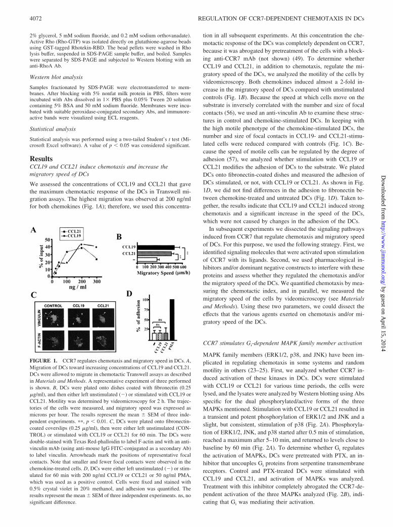
We assessed the concentrations of CCL19 and CCL21 that gavethe maximum chemotactic response of the DCs in Transwell mi-gration assays. The highest migration was observed at 200 ng/mlfor both chemokines (Fig. 1A); therefore, we used this concentra-
tion in all subsequent experiments. At this concentration the che-motactic response of the DCs was completely dependent on CCR7,because it was abrogated by pretreatment of the cells with a block-ing anti-CCR7 mAb (not shown) (49). To determine whetherCCL19 and CCL21, in addition to chemotaxis, regulate the mi-gratory speed of the DCs, we analyzed the motility of the cells byvideomicroscopy. Both chemokines induced almost a 2-fold in-crease in the migratory speed of DCs compared with unstimulatedcontrols (Fig. 1B). Because the speed at which cells move on thesubstrate is inversely correlated with the number and size of focalcontacts (56), we used an anti-vinculin Ab to examine these struc-tures in control and chemokine-stimulated DCs. In keeping withthe high motile phenotype of the chemokine-stimulated DCs, thenumber and size of focal contacts in CCL19- and CCL21-stimu-lated cells were reduced compared with controls (Fig. 1C). Be-cause the speed of motile cells can be regulated by the degree ofadhesion (57), we analyzed whether stimulation with CCL19 orCCL21 modifies the adhesion of DCs to the substrate. We platedDCs onto fibronectin-coated dishes and measured the adhesion ofDCs stimulated, or not, with CCL19 or CCL21. As shown in Fig.1D, we did not find differences in the adhesion to fibronectin be-tween chemokine-treated and untreated DCs (Fig. 1D). Taken to-gether, the results indicate that CCL19 and CCL21 induced strongchemotaxis and a significant increase in the speed of the DCs,which were not caused by changes in the adhesion of the DCs.
In subsequent experiments we dissected the signaling pathwaysinduced from CCR7 that regulate chemotaxis and migratory speedof DCs. For this purpose, we used the following strategy. First, weidentified signaling molecules that were activated upon stimulationof CCR7 with its ligands. Second, we used pharmacological in-hibitors and/or dominant negative constructs to interfere with theseproteins and assess whether they regulated the chemotaxis and/orthe migratory speed of the DCs. We quantified chemotaxis by mea-suring the chemotactic index, and in parallel, we measured themigratory speed of the cells by videomicroscopy (see Materialsand Methods). Using these two parameters, we could dissect theeffects that the various agents exerted on chemotaxis and/or mi-gratory speed of the DCs.
CCR7 stimulates Gi-dependent MAPK family member activation
MAPK family members (ERK1/2, p38, and JNK) have been im-plicated in regulating chemotaxis in some systems and randommotility in others (23–25). First, we analyzed whether CCR7 in-duced activation of these kinases in DCs. DCs were stimulatedwith CCL19 or CCL21 for various time periods, the cells werelysed, and the lysates were analyzed by Western blotting using Absspecific for the dual phosphorylated/active forms of the threeMAPKs mentioned. Stimulation with CCL19 or CCL21 resulted ina transient and potent phosphorylation of ERK1/2 and JNK and aslight, but consistent, stimulation of p38 (Fig. 2A). Phosphoryla-tion of ERK1/2, JNK, and p38 started after 0.5 min of stimulation,reached a maximum after 5–10 min, and returned to levels close tobaseline by 60 min (Fig. 2A). To determine whether Gi regulatesthe activation of MAPKs, DCs were pretreated with PTX, an in-hibitor that uncouples Gi proteins from serpentine transmembranereceptors. Control and PTX-treated DCs were stimulated withCCL19 and CCL21, and activation of MAPKs was analyzed.Treatment with this inhibitor completely abrogated the CCR7-de-pendent activation of the three MAPKs analyzed (Fig. 2B), indi-cating that Gi was mediating their activation.
FIGURE 1. CCR7 regulates chemotaxis and migratory speed in DCs. A,Migration of DCs toward increasing concentrations of CCL19 and CCL21.DCs were allowed to migrate in chemotactic Transwell assays as describedin Materials and Methods. A representative experiment of three performedis shown. B, DCs were plated onto dishes coated with fibronectin (0.25�g/ml), and then either left unstimulated (�) or stimulated with CCL19 orCCL21. Motility was determined by videomicroscopy for 2 h. The trajec-tories of the cells were measured, and migratory speed was expressed asmicrons per hour. The results represent the mean � SEM of three inde-pendent experiments. ��, p � 0.01. C, DCs were plated onto fibronectin-coated coverslips (0.25 �g/ml), then were either left unstimulated (CON-TROL) or stimulated with CCL19 or CCL21 for 60 min. The DCs weredouble-stained with Texas Red-phalloidin to label F-actin and with an anti-vinculin mAb (using anti-mouse IgG FITC-conjugated as a secondary Ab)to label vinculin. Arrowheads mark the positions of representative focalcontacts. Note that smaller and fewer focal contacts were observed in thechemokine-treated cells. D, DCs were either left unstimulated (�) or stim-ulated for 60 min with 200 ng/ml CCL19 or CCL21 or 50 ng/ml PMA,which was used as a positive control. Cells were fixed and stained with0.5% crystal violet in 20% methanol, and adhesion was quantified. Theresults represent the mean � SEM of three independent experiments. ns, nosignificant difference.
4072 REGULATION OF CCR7-DEPENDENT CHEMOTAXIS IN DCs
by guest on April 15, 2014
http://ww
w.jim
munol.org/
Dow
nloaded from

FIGURE 2. CCR7 induces PTX-sensitive activation of ERK1/2, JNK, and p38 and hierarchy of MAPK family members. A, DCs (300,000 cells) werestimulated for the indicated times with CCL21 or CCL19 (both at 200 ng/ml), then lysed and analyzed by SDS-PAGE, followed by Western blotting with Absagainst phospho-ERK1/2, phospho-JNK, or phospho-p38. To show equal loading, the membrane was stripped and probed with an Ab against p38. B, Control orPTX-treated (100 ng/ml; 120 min) DCs were either left unstimulated (�) or stimulated with CCL19 or CCL21 (both at 200 ng/ml) and subsequently lysed andsubjected to Western blot, as described in A, with Abs against phospho-ERK1/2, phospho-JNK, or phospho-p38. To show equal loading, the membrane wasstripped and probed with an Ab against ERK2. C, DCs were left untreated (control) or were pretreated for 60 min with UO126 (5 �M), SP60012 (30 �M), orSB203580 (20 �M) to inhibit phosphorylation/activation of ERK1/2, JNK, or p38, respectively. Then DCs were either left unstimulated (�) or stimulated with200 ng/ml CCL21 or CCL19 for 5 min. The cells were lysed and analyzed by Western blotting with Abs against phospho-ERK1/2, phospho-JNK, or phospho-p38.To show equal loading, the membrane was stripped, and respective blots were reprobed with Abs against ERK2, JNK, and p38. D, DCs were left untreated (control)or were pretreated with UO126, SB203580, or SP600125, as described in C, to inhibit phosphorylation of ERK1/2, p38, or JNK, respectively. Cells were lysedand subjected to Western blot with Abs against phospho-JNK, phospho-ERK1/2, or phospho-p38. To show equal loading, the membranes were stripped, and therespective blots were reprobed with Abs against �-actin. A–D, A representative experiment of three performed is shown. E, Cells were washed in RPMI 1640medium, then either treated with vehicle (Control) or pretreated with PTX (100 ng/ml) for 120 min. a, Migratory speed in the absence (�) or the presence (�)of CCL19 was measured by videomicroscopy. The results represent the mean � SEM of three independent experiments. ns, no significant difference. b, DCmigration against no stimulus (�) or against 200 ng/ml CCL21 or CCL19 was measured in Transwell assays. The chemotactic response of the cells was expressedas a chemotactic index (CI). The results represent the mean � SEM of three independent experiments. �, p � 0.05; ��, p � 0.01. F, a, DCs, untreated (Control)or pretreated with UO126, SB203580, or SP600125, as described in C, were plated onto dishes coated with fibronectin (0.25 �g/ml), and the motility ofunstimulated control (�) or CCL19-stimulated (�) DCs was followed by video microscopy. The results represent the mean � SEM of three independentexperiments. ns, no significant difference. b, DCs were left untreated (Control) or were pretreated with UO126, SB203500, or SP600125, as described in C. ThenDC migration against no stimulus (�) or against CCL21 or CCL19 was measured in Transwell assays. The chemotactic response of the cells was expressed asa chemotactic index (CI). The results represent the mean � SEM of three independent experiments. The CI values corresponding to the CCL21- or CCL19-stimulated DCs in the presence of UO126, SB203500, and SP600125 were compared with their corresponding CCL19- or CCL21-stimulated untreated controlvalues. �, p � 0.05; ��, p � 0.01. G, DCs were left untreated (control) or were pretreated with UO126, SB203500, SP600125, or the indicated combination ofthese inhibitors, as described in C. Chemotaxis toward CCL21 was analyzed in Transwell assays. Data are presented as the percent inhibition of migrationcompared with migration measured in the absence of any inhibitor. The results are representative of two independent experiments.
4073The Journal of Immunology
by guest on April 15, 2014
http://ww
w.jim
munol.org/
Dow
nloaded from

Hierarchy among MAPK family members in CCR7-mediatedstimulation of DCs
To analyze the possible relationship among MAPK family mem-bers after stimulation of CCR7, we used pharmacological agents toinhibit these kinases. DCs were pretreated with UO126,SB203580, and SP600125 to inhibit ERK1/2, p38, and JNK, re-spectively. As expected, treatment with the inhibitors blunted theincrease in the phosphorylation of all MAPKs induced by stimu-lation with CCL19 or CCL21 (Fig. 2C). We analyzed the possiblerelationships among ERK1/2, JNK, and p38 using the inhibitors ofthese enzymes. Inhibition of ERK1/2 (with UO126) or p38 (withSB203580) completely or partially blunted JNK activity, respec-tively (Fig. 2D; see also Fig. 7). In contrast, inhibition of JNK(with SP600125) did not affect either ERK1/2 or p38 activation,indicating that JNK was downstream of ERK1/2 and p38 (Fig. 2D;see also Fig. 7). Finally, because inhibition of p38 phosphorylation(by treating DCs with SB203580; Fig. 2C) did not affect activationof ERK1/2 (Fig. 2D, middle panel), and inhibition of ERK1/2 (bytreating DCs with U0126) did not blunt p38 activation (Fig. 2D,upper panel), this implies that p38 and ERK1/2 are activated in-dependently of each other. Taken together, the data indicate thatCCR7-stimulated activation of MAPKs takes place with a specifichierarchy, where p38 and ERK1/2 are activated independently, andboth regulate the activity of JNK (see Fig. 7).
MAPKs regulate CCR7-dependent chemotaxis, but not themigratory speed, of DCs
We analyzed the role of MAPKs in regulation of the motile func-tions of DCs. Because MAPKs were inhibited by PTX, we ana-lyzed the involvement of Gi proteins in regulation of CCR7-de-pendent chemotaxis and migratory speed. PTX treatment did notaffect the migratory speed of DCs in either the absence (not shown)or the presence of chemokines (Fig. 2Ea). PTX treatment did notaffect the adhesion of cells to the substrate (not shown). However,inhibition of Gi completely blunted the ability of DCs to chemotaxtoward CCL19 or CCL21 (Fig. 2Eb). Next, we examined the effectof inhibition of ERK1/2, p38, or JNK on migratory speed andchemotaxis. Inhibition of any of these MAPKs did not affect themigratory speed of DCs in the absence (not shown) or the presenceof CCL19 or CCL21 (Fig. 2Fa). The inhibitors did not affect theadhesion of DCs to the substrate (not shown). In contrast, inhibi-tion of MAPKs blunted the ability of DCs to chemotax towardCCL19 or CCL21 (Fig. 2Fb). The strongest inhibition of chemo-taxis was observed when the activity of ERK1/2 was inhibited, andless potent inhibitory effects were observed when JNK or p38 wasblocked (Fig. 2Fb). To determine whether MAPK members exerta synergistic effect on chemotaxis, we treated DCs with combina-tions of two or three inhibitors and analyzed the chemotactic re-sponses of the cells (Fig. 2G). The results of these experiments,which are presented as percent inhibition with respect to the max-imum migration obtained in the presence of CCL21, showed thatthe effects exerted by MAPKs were not synergistic (Fig. 2G). In-terestingly, although simultaneous inhibition of the three MAPKsled to almost 75% inhibition of the chemotactic response of thecells, this response was not completely abrogated (Fig. 2G), sug-gesting that, apart from MAPKs, additional molecules can regulateCCR7-mediated chemotaxis (see Figs. 2G and 7). Taken collec-tively, the results indicate that ERK1/2, JNK, and p38 regulate, ina Gi-dependent and nonsynergistic manner, CCR7-dependent che-motaxis, but not the migratory speed, of DCs.
PI3K/Akt does not regulate CCR7-dependent chemotaxis normigratory speed of DCs
Searching for additional molecules that could regulate chemotaxis,and because PI3K/Akt kinase has been involved in regulating che-motaxis in several cell types (23), we examined whether stimula-tion of CCR7 induces activation of the PI3K effector Akt. For thispurpose, we performed Western blots with Abs against phosphor-ylated/active forms of Akt. As shown in Fig. 3A, chemokines in-
FIGURE 3. PI3K/Akt does not regulate CCR7-mediated chemotaxis ormigratory speed in DCs. A, DCs, untreated (control) or pretreated withLY294002 (100 �M; 60 min), were either left unstimulated (�) or stim-ulated with CCL21 or CCL19 for an additional 60 min. Then DCs werelysed and subjected to Western blot with Abs against active/phosphory-lated Akt. To show equal loading, the membrane was stripped and reprobedwith an anti-�-actin Ab. B, a, Control DCs or DCs pretreated withLY294002, as described in A, were plated onto dishes coated with fi-bronectin (0.25 �g/ml), and migratory speed was analyzed by videomi-croscopy. The results represent the mean � SEM of three independentexperiments. ns, no significant difference. b, Chemotaxis against CCL19 orCCL21 in control or LY294002 treated DCs was measured in Transwellassays. The chemotactic response of the cells was expressed as a chemo-tactic index (CI). The results are representative of three independent ex-periments. C, DC, untreated (control) or pretreated with LY204002, asdescribed in A, were either left unstimulated (�) or stimulated with CCL19or CCL21 for 5 min. Cells were lysed and subjected to SDS-PAGE andWestern blot with Abs against phospho-ERK1/2. To show equal loading,the membrane was stripped and reprobed with an anti-�-actin Ab.
4074 REGULATION OF CCR7-DEPENDENT CHEMOTAXIS IN DCs
by guest on April 15, 2014
http://ww
w.jim
munol.org/
Dow
nloaded from

duced a potent increase in the phosphorylation of Akt.Because persistent inhibition of PI3K for long periods (e.g., af-
ter transfection with dominant negative PI3K) leads to prohibitivelevels of cell death, we used pharmacological inhibitors to analyzethe involvement of PI3K/Akt in the regulation of the motility ofDCs. To inhibit PI3K/Akt, we pretreated the cells with two struc-turally different PI3K inhibitors, LY294002 (Fig. 3A) and wort-mannin (not shown). The inhibitors completely abrogated the in-crease in phosphorylation of Akt elicited by CCL19 or CCL21(Fig. 3A and not shown). However, surprisingly, neither migratoryspeed in the absence (not shown) or the presence of chemokines(Fig. 3Ba) nor chemotaxis (Fig. 3Bb), was altered in inhibitor-treated DCs compared with untreated control DCs. Because Akthas been shown to be upstream of ERK1/2 (28), which, as dem-onstrated above (Fig. 2Fb), plays an important role in the regula-tion of CCR7-dependent chemotaxis, we analyzed whether PI3K/Akt was upstream of ERK1/2. As shown in Fig. 3C, inhibition ofPI3K/Akt with LY294002 did not blunt ERK1/2 activation. Insummary, our results show that PI3K/Akt does not regulate CCR7-dependent chemotaxis nor migratory speed of DCs.
Rho regulates migratory speed, but not CCR7-dependentchemotaxis
Because CCR7 regulates actin organization (17), and the latter canbe controlled by the small GTPase Rho (40), we studied whetherCCR7 induces activation of Rho. We stimulated cells with CCL21,DCs were lysed, and active Rho was pulled down using GST-tagged RBD bound to glutathione-agarose beads. As observed inFig. 4A, stimulation of CCR7 induced activation of Rho. To de-termine whether Rho was regulating CCR7-dependent chemotaxisand/or migratory speed, DCs were pretreated for 12 h with the C3exoenzyme, a selective Rho inhibitor. We have shown previouslythat this treatment leads to efficient inhibition of Rho in leukocytes(35, 48). Videomicroscopic analysis showed that the basal migra-tory speed was potently inhibited in DCs treated with C3 exoen-zyme (Fig. 4Ba). The migratory speed was also inhibited in C3-treated cells when they were stimulated with chemokines (Fig.4Ba). When we transfected DCs with GFP vector or with a dom-inant negative Rho (pEGFP-N19Rho) (35), the migratory speedwas also drastically inhibited in cells that overexpressed pEGFP-N19Rho (not shown). Adhesion experiments suggested that theobserved differences in motility were not due to the effects of theRho inhibitors (C3 exoenzyme or N19Rho-GFP) on the adhesionof DCs to substrate (not shown). Taken together, the results pointout the crucial role of Rho in regulation of the migratory speed ofDCs in both unstimulated and cells stimulated by chemokines.
A parallel analysis of the chemotactic response to CCL19 orCCL21 chemokines of DCs treated with C3-exoenzyme, showedthat inhibition of Rho had only a slight effect on the chemotacticresponse of the cells (Fig. 4Bb). Similarly, analysis of the chemotaxisresponse of DCs that express the dominant negative Rho (N19Rho-GFP) demonstrated that interfering with Rho had no effect on thechemotaxis of DCs toward CCL19 or CCL21 (not shown).
Finally, consistent with the lack of a regulatory role for Rho onthe regulation of CCR7-dependent chemotaxis, pretreatment ofDCs with the C3 exoenzyme to inhibit Rho did not block activa-tion of the chemotactic regulator ERK1/2 (Fig. 2Fb) upon stimu-lation of DCs with CCL19 or CCL21 (Fig. 4C). This result em-phasizes that the signals relayed from CCR7 that regulatedmigratory speed are independent of those that regulate chemotaxis.In summary, the results indicate that Rho regulates CCR7-depen-dent migratory speed, but not chemotaxis, in DCs.
CCL19 and CCL21 induce Rho-dependent and Gi and Src-independent stimulation of tyrosine kinase activity of Pyk2
Because tyrosine kinase Pyk2 has been involved in the signalingfrom chemokine receptors (21, 34, 37), we examined whetherstimulation of DCs with CCL19 and CCL21 modulates the activityof this kinase. DCs were stimulated with chemokines for differenttime periods and lysed. Then Pyk2 was immunoprecipitated withspecific anti-Pyk2 Abs, and immunoprecipitates were used to per-form in vitro kinase reactions. As shown in Fig. 5A, CCL19 and
FIGURE 4. Rho regulates the migratory speed, but not the chemotaxis,of DCs. A, DCs were either left unstimulated (�) or stimulated withCCL21 (200 ng/ml) for 0.5 min. Then DCs were lysed, and active Rho(Rho-GTP) was pulled down using GST-tagged RBD bound to glutathio-ne-agarose beads (see Materials and Methods). The level of RhoA isolatedon the beads was assessed by Western blotting with an anti-RhoA Ab. Toshow equal loading, an aliquot of the lysed cells before the pulldown wasused to perform a Western blot with the anti-RhoA Ab. The results arerepresentative of three independent experiments. B, DCs were either leftuntreated (control) or pretreated with C3 exoenzyme (50 ng/ml; 12 h), thenwere either left unstimulated (�) or stimulated (�) with CCL19 (200 ng/ml). a, DCs were plated onto fibronectin-coated dishes (0.25 �g/ml), andthe motility of the DCs was followed by videomicroscopy. The resultsrepresent the mean � SEM of three independent experiments. �, p � 0.05;��, p � 0.01. b, The chemotactic response of the cells was expressed as achemotactic index (CI). The results represent the mean � SEM of threeindependent experiments. ns, no significant difference. C, DCs untreated(control) or pretreated with C3 exoenzyme, as described in B, were eitherleft unstimulated (�) or stimulated with CCL19 or CCL21 for 5 min. DCswere then lysed and subjected to SDS-PAGE and Western blot with Absagainst phospho-ERK1/2. To show equal loading, the membrane wasstripped, and the blot was reprobed with anti-�-actin Ab.
4075The Journal of Immunology
by guest on April 15, 2014
http://ww
w.jim
munol.org/
Dow
nloaded from

CCl21 induced a transient increase in the activity of Pyk2 withsimilar kinetics. Autophosphorylation of Pyk2 increased as earlyas 0.5 min after the addition of chemokines to the cells, reached amaximum after 5–10 min, and returned to basal levels after 60 min(Fig. 5A). To determine whether Gi proteins mediate the CCR7-dependent stimulation of Pyk2, we pretreated DCs with PTX. Asshown in Fig. 5B, pretreatment with PTX did not inhibit the acti-vation of Pyk2 induced by CCL19 or CCL21, indicating that suchactivation is not mediated by Gi proteins. Because we observed
that stimulation of DCs with chemokines also induced activationof Rho (Fig. 4A), we determined whether Rho was upstream ofPyk2. Treatment with C3 exoenzyme abrogated the activation ofPyk2 induced by CCL19 or CCL21 (Fig. 5C), indicating that Rhowas mediating this activation.
Because Src family members can regulate the activity of Pyk2(34),we also analyzed whether Src was mediating Pyk2 activation.Pretreatment of DCs with PP2, a selective inhibitor of the Srcfamily, did not prevent the CCR7-dependent stimulation of Pyk2,indicating that Src was not mediating the chemokine-induced stim-ulation of Pyk2 (Fig. 5D). The lower basal activity of Pyk2 ob-served in PP2-pretreated cells (Fig. 5D) is in agreement with pre-vious findings that Pyk2 constitutively associates with Src (34).Consistent with the lack of a role for Src in the activation of Pyk2,stimulation of DCs with CCL19 or CCL21 failed to induce acti-vation of this tyrosine kinase (not shown). Taken together, theresults demonstrate that CCR7 induces stimulation of Pyk2 activ-ity downstream of Rho, which is not mediated by Gi or Src.
Pyk2 regulates the migratory speed, but not the chemotaxis, of DCs
To examine the role of Pyk2 in regulation of CCR7-mediated che-motaxis or migratory speed, we transfected DCs with a dominantnegative Pyk2 construct (PRNK) (53). Overexpression of PRNK(Fig. 5F) lead to a potent inhibition of migratory speed in theabsence or presence of chemokines (Fig. 5Ea), but failed to affectthe chemotactic response of DCs to CCL21 (Fig. 5Eb). Thechanges in motility were not related to differences in adhesion tothe substrate, because such adhesion was not altered by overex-pression of PRNK (not shown). Finally, consistent with the inde-pendence of the pathways regulating chemotactic and migratoryspeed, overexpression of PRNK did not inhibit stimulation of thechemotactic regulator ERK1/2 (Fig. 5F) nor did inhibition ofERK1/2 block activation of Pyk2 (Fig. 5G). In summary, the re-sults indicate that the tyrosine kinase Pyk2 regulates CCR7-depen-dent migratory speed, but not chemotaxis, in DCs.
CCR7 induces phosphorylation/inactivation of cofilin that ismediated by Pyk2
Because cell motility involves regulation of the actin cytoskeleton,and the actin-severing protein cofilin regulates actin organization,we analyzed whether CCR7 induces changes in the activity of thisprotein. We used an Ab that recognizes phosphorylated Ser-3, cor-responding to an inactive form of cofilin. Stimulation of DCs withCCL21 induced a transient phosphorylation/inactivation of cofilin.The level of phosphorylation increased after 0.5 min of stimulationwith chemokines, reached a maximum after 5–10 min, and re-turned to basal levels after 60 min (Fig. 6A). Inhibition of Rho bytreating cells with C3 exoenzyme blunted the CCR7-dependentphosphorylation/inactivation of cofilin, indicating that the proteinwas downstream of Rho (Fig. 6B). Because the time course ofPyk2 activity paralleled that of cofilin, we analyzed whether cofilinwas downstream of Pyk2. DCs were transfected with the dominantnegative Pyk2 (PRNK), then phosphorylation of cofilin was com-pared in vector- and PRNK-transfected cells. Most interestingly,overexpression of PRNK blunted the increase in phosphorylationof cofilin (Fig. 6C), implying that cofilin is downstream of Pyk2.In summary, the results indicate that CCR7 induces phosphoryla-tion/inactivation of cofilin that is mediated by Rho and Pyk2.
DiscussionDespite the importance of the chemokine receptor CCR7 in regu-lation of the migration of DCs to the lymph node and, conse-quently, for the immune response (15), little is known about thesignaling pathways triggered and the mechanism(s) by which this
FIGURE 5. CCR7 stimulates Rho-dependent and Gi-independent acti-vation of the tyrosine kinase Pyk2. A, Time course of CCL21- and CCL19-stimulated Pyk2 activity in DCs. DCs were stimulated for the indicatedtimes with 200 ng/ml CCL19 or CCL21 and lysed. Lysates were incubatedwith anti-Pyk2 Abs to precipitate Pyk2. Kinase activities in the resultingimmunoprecipitates were measured by in vitro kinase (ivk) reaction (seeMaterials and Methods). Pyk2 immunoprecipitates were also analyzed bySDS-PAGE, followed by transfer to membranes and Western blotting withanti-Pyk2 Abs (WB). B–D, DCs were either left untreated (control) orpretreated PTX (100 ng/ml; B), C3 exoenzyme (50 ng/ml; 16 h; C), or PP2(20 �M; D). Then DCs were suspended in RPMI 1640 and stimulated for5 min with either CCL21 or CCL19. Pyk2 was immunoprecipitated, and invitro kinase activity was determined. E, DCs were transfected with emptyvector or with a dominant negative Pyk2 (PRNK). a, The migratory speedof DCs was followed by videomicroscopy. The results represent themean � SEM of three independent experiments. �, p � 0.05; ��, p � 0.01.b, The chemotactic index (CI) in response to CCL21 was determined. Theresults are representative of three independent experiments. F, DCs trans-fected with vector or PRNK were either left unstimulated (�) or stimulatedfor 5 min with CCL19 or CCL21. Cells were lysed and subjected to SDS-PAGE and Western blotting with Abs against phospho-ERK1/2. The mem-brane was stripped, and blots were reprobed with anti-Pyk2 Abs that rec-ognize endogenous Pyk2 and PRNK. A representative blot of threeexperiments performed is shown. G, DCs were either left untreated (con-trol) or pretreated with UO126 (5 �M). Then DCs were suspended inRPMI 1640 and stimulated for 5 min with CCL21. Pyk2 was immunopre-cipitated, and Pyk2 in vitro kinase activity was determined (ivk). Pyk2immunoprecipitates were also analyzed by SDS-PAGE, followed by trans-fer to membranes and Western blotting with anti-Pyk2 Abs (WB).
4076 REGULATION OF CCR7-DEPENDENT CHEMOTAXIS IN DCs
by guest on April 15, 2014
http://ww
w.jim
munol.org/
Dow
nloaded from

receptor regulates the motile functions of DCs. Stimulation ofCCR7 with CCL19 and CCL21 induces a chemotactic response aswell as an increase in the migratory speed of DCs, as shown byvideomicroscopy (Fig. 1) and checkerboard analysis (19) (notshown). This simultaneous stimulation of chemotaxis and migra-tory speed has been observed for several chemokine receptors(18); however, to date it has not been determined whether similaror different signaling pathways regulate both processes.
To analyze the signaling components that regulate CCR7-de-pendent chemotaxis and migratory speed of DCs, we used the fol-lowing strategy. First, we identified molecules that were activatedafter stimulation of CCR7. Second, we used dominant negativeconstructs and pharmacological inhibitors to analyze the hierarchyamong the signaling molecules identified. Third, we assessed theeffect(s) that inhibition of molecules activated by CCR7 exerted oneither the chemotactic response or the migratory speed of DCs. Todissect the effects on chemotaxis from those on migratory speed,we determined, in parallel, the chemotactic index in Transwellassays and the migratory speed using videomicroscopy. The mi-gratory speed data obtained by videomicroscopy were corrobo-rated using a checkerboard setting analysis with similar results (notshown). Because the migratory speed study was performed in DCs
that were plated on fibronectin-coated dishes, the results apply tomature DCs that are motile on this substrate. Additional studiesneed to be performed to confirm that these findings can be ex-tended to other substrates.
Using the experimental strategy described, we found that inDCs, CCR7 regulates two signaling modules, one formed by Gi
and a specific hierarchy of MAPK family members that regulateschemotaxis (see below) and another formed by Rho/Pyk2/cofilinthat regulates the migratory speed of DCs (Fig. 7). For the modulethat regulates the migratory speed (Rho/Pyk2/cofilin) we made aninteresting observation. When we analyzed the effect that interfer-ence with these molecules exerted on DC motility, we observedinhibition of migratory speed in both the absence (not shown) andthe presence of chemokines (Fig. 4B, a and b, and Fig. 5E, a andb). This result suggests that Rho/Pyk2/cofilin regulate the basalmotility of DCs independently of the presence of chemokines.Therefore, the stimulation of CCR7 by its ligands leads to an in-crease in the activity of these signaling molecules and conse-quently to an increase in the migratory speed of the cells (see Figs.4A, 5A, and 6A). The stimulation by CCR7 ligands of the intrinsicmigratory speed axis behaves as an accelerator system that in-creases the speed at which DCs move toward the maximum con-centrations of CCL19 and CCL21. In the in vivo context, thisregulatory process would have the obvious advantage of more rap-idly directing DCs to lymph node regions.
FIGURE 6. CCR7 induced transient phosphorylation of cofilin that ismediated by Rho and Pyk2. A, Time course of CCL21-stimulated phos-phorylation of cofilin. DCs were stimulated for the indicated times with200 ng/ml CCL21 and lysed. Lysates were used to perform Western blot-ting with the anti-Ser-3-phosphorylated cofilin. Blots were striped and re-blotted with anti-�-actin to show equal loading. B, Untreated DCs (control)or DCs pretreated with C3 (50 ng/ml; 12 h) were either left unstimulated(�) or stimulated with CCL21 for 0.5 min. DCs were then lysed andsubjected to SDS-PAGE and Western blotting with Abs against phospho-cofilin. To show equal loading, the membrane was stripped, and respectiveblots were reprobed with Abs against �-actin. C, DCs were transfectedwith empty vector (VECTOR) or with a dominant negative form of Pyk2(PRNK). Vector- or PRNK-transfected DCs were either left unstimulated(�) or stimulated for 0.5 min with CCL21. Cells were lysed and subjectedto SDS-PAGE and Western blotting with Abs against phospho-cofilin. Toshow equal loading and expression of PRNK, the membrane was stripped,and blots were reprobed with an anti-Pyk2 Ab (C19) that recognizes the Cterminus of both Pyk2 and PRNK.
FIGURE 7. Model explaining CCR7-mediated independent regulationof chemotaxis and migratory speed in DCs. CCR7 induces Gi-mediatedactivation of p38, ERK1/2, and JNK, with ERK1/2 and p38 upstream ofJNK. Together, these molecules constitute a signaling module that regu-lates CCR7-dependent chemotaxis. An additional unknown molecule(s),denoted X, participates in the regulation of chemotaxis, because simulta-neous inhibition of the three MAPKs did not completely abrogate chemo-taxis (see Fig. 2G). CCR7 also regulates Rho and Pyk2 activation andcofilin phosphorylation/inactivation. Rho is upstream of Pyk2, and the lat-ter is upstream of cofilin. Src can phosphorylate Pyk2, but does not mediatethe effects of CCR7. Rho/Pyk2/cofilin constitute a module that regulatesthe intrinsic or basal migratory speed of DCs. Stimulation of CCR7 acti-vates the signaling components of this module, resulting in enhanced mo-tility. CCR7 also stimulates PI3K/Akt; however, these molecules do notregulate chemotaxis or migratory speed, but are involved in regulating DCsurvival (70). Also shown are inhibitors of the molecules tested (see textfor additional details).
4077The Journal of Immunology
by guest on April 15, 2014
http://ww
w.jim
munol.org/
Dow
nloaded from
Importantly, we found that the signaling molecules that regulatechemotaxis and migratory speed were regulated independently ofeach other, as shown by the fact that selective interference withcomponents of the chemotactic module did not affect migratoryspeed and vice versa (Fig. 7 and see below). The independence ofboth modules was corroborated by the experimental demonstrationthat selective inhibition of a key signaling regulator of chemotaxis(i.e., ERK1/2) failed to affect a key signaling component of themigratory speed (i.e., Pyk2; see Fig. 5G). In contrast, overexpres-sion of a dominant negative Pyk2 construct (PRNK), which inhib-ited migratory speed (Fig. 5E), did not blunt stimulation of thechemotactic regulator ERK1/2 (Fig. 5F). Finally, we also observedthat interference with the signaling molecules identified did notaffect adhesion of cells to substrate (Fig. 1D and not shown), sug-gesting that the proteins identified do not exert their effects on theadhesive function of the DCs.
Regarding the specific molecules that regulate chemotaxis, wefound that MAPK family members played a crucial role. In thisregard, CCR7 induced a Gi-dependent activation of ERK1/2, p38,and JNK (Fig. 2, A and B). We found that upon stimulation ofCCR7, ERK1/2 and p38 were activated independently of eachother. However, the use of selective inhibitors showed that bothkinases regulated JNK activation (Fig. 2D). ERK1/2, p38, andJNK played an important role in the regulation of CCR7-dependentchemotaxis, because simultaneous inhibition of these three kinasesblunted almost 75% of the chemotactic response of the cells (Fig. 2,F and G). These experiments also showed that the effects of MAPKswere not synergistic (Fig. 2G). Interestingly, because inhibition ofthese enzymes did not completely blunt CCR7-dependent chemo-taxis, this implies that additional unidentified molecules could be reg-ulating this process. In contrast to results obtained in other cell types(24, 25), the three MAPKs played no role in regulation of the migra-tory speed of DCs (Fig. 2Fa), indicating the importance of cell contextin the regulation of chemotaxis.
Interestingly, we found that PI3K/Akt regulated neither CCR7-dependent chemotaxis nor migratory speed in DCs (Fig. 3). Pre-viously it has been shown that PI3K/Akt plays a role in the reg-ulation of both functions in several cell types (23, 26–28, 30, 58,59). However, it is emerging that in certain cell settings, includingDCs, chemotaxis can take place even when PI3K/Akt is com-pletely inhibited (60–64). In this regard, a similar lack of a reg-ulatory role for PI3K/Akt on migration has been reported for Tcells (60, 64–66). Furthermore, Dumstrei et al. (67) suggestedrecently that PI3K does not regulate chemotaxis in primordialgerm cells, and Lacalle et al. (68) showed that PTEN (Phosphataseand tensin homolog deleted on chromosome 10), an enzyme thatdegrades the products of PI3K, does not regulate directed motilityin HL60 cells. A similar lack of control of PI3K/Akt on cell mi-gration has been observed for other chemokine receptors differentfrom CCR7, including CXCR3 (60) and CCR4 (64). Of note, al-though the lack of a regulatory role for PI3K/Akt in chemotaxis isnot exclusive of DCs or CCR7 receptor, however, because it hasbeen reported that, apart from DCs, CCR7 does not control che-motaxis in T cells, it is possible that this receptor may be coupledto the machinery that regulate chemotaxis independently of PI3K/Akt at least in the two cell types indicated. In this regard, weobserved that inhibition of PI3K/Akt by treating DCs withLY294002 specifically blunted CXCR4-dependent, but not CCR7-dependent, chemotaxis DCs (not shown). Additional studies willbe required to clarify whether CCR7 uses preferentially PI3K/Akt-independent mechanisms to regulate cell migration. Taken to-gether, the results emphasize the context dependence of the regu-lation of motile functions and indicate that there are alternativepathways that regulate chemotaxis which do not require PI3K/Akt
activation (60–64, 69). Because we have shown previously thatCCR7 induces activation of PI3K/Akt, and these molecules areinvolved in inhibiting apoptosis of DCs, we suggest that regulationof apoptosis, instead of motility, could be the main role of CCR7-stimulated activation of PI3K/Akt in DCs (70).
We identified Rho/Pyk2/cofilin as the components of a modulethat regulates CCR7-dependent migratory speed based on the fol-lowing data. 1) We observed that stimulation of CCR7 inducedactivation of Rho (Fig. 4A). We also observed that inhibition ofRho with C3 exoenzyme (Fig. 4B) or a dominant negative form ofRho (N19Rho; not shown) inhibited the motility of DCs, but didnot affect chemotaxis. In agreement with our results, Rho has beenshown to regulate basal motility in HL60 cells (71). Furthermore,a low basal motility is also observed in lymphocytes deficient inp115Rho, a guanine nucleotide exchange factor for Rho, whichmaintains this GTPase in an active form (72). 2) We observed thatPyk2 is activated after stimulation of CCR7 in a Gi-independent andRhoA-dependent manner (Fig. 5). We suggest that Pyk2 is involvedin regulation of the migratory speed of DCs, because overexpressiona dominant negative Pyk2 (PRNK) reduced the motility of the cells,but did not affect chemotaxis (Fig. 5). 3) Finally, CCR7-dependentactivation of the chemotactic regulator ERK1/2 was not affected byinhibition of Rho or Pyk2, emphasizing that these molecules regulateonly the migratory speed of DCs (Figs. 4 and 5).
Pyk2 is a kinase activated by integrin and chemokine receptorsthat regulate cell motile functions (22, 27, 31, 35). The potentialrelevance of the activation of Pyk2 by these receptors is under-scored by previous reports that Pyk2 regulates the migratory speedof leukocytes (36–38). However, the mechanism(s) by which Pyk2regulates cell speed is unknown. In this regard, we made the im-portant observation that stimulation of CCR7 induced phosphory-lation/inactivation of cofilin (Fig. 6). Overexpression of the dominantnegative Pyk2 (PRNK) completely abolished the phosphorylation/in-activation of cofilin, suggesting that Pyk2 is upstream of this molecule(Fig. 6C). Because cofilin can regulate cell motility through its abilityto depolymerize and sever actin filaments at the leading edge (43–47),our results imply that cofilin may mediate the effects of Pyk2 on cellmotility. This is an important finding, because, to the best of ourknowledge, this is the first report suggesting that cofilin could mediatethe effects of Pyk2 on cell motility.
The finding that CCR7 transmit signals that modulate chemo-taxis and migratory speed is relevant for regulation of the functionsof DCs in vivo (73). As mentioned above, when CCR7-expressingDCs move from tissues toward lymph nodes, an increase in thespeed of the cells can allow them to reach these regions morerapidly. Moreover, once in the nodes, where the cells are sur-rounded by chemokines, the increased motility of the DCs canenhance the likelihood of encountering Ag-specific T cells at thisimportant meeting point for DCs and T cells (73). It is possible thaton the way to lymph nodes or in the nodes, the speed of DCs couldbe enhanced by environmental factors that stimulate receptors thatcontrol signaling components of the migratory speed module.
Our data are consistent with recent models suggesting that thesignaling module that controls chemotaxis is separated from otherbiochemical modules that regulate motility (74). In this regard, itwould be interesting to analyze whether the modular mechanismsthat we observed for CCR7 could be extended to other chemokinereceptors and leukocytes. The knowledge that independent signal-ing modules regulate CCR7-dependent chemotaxis and migratoryspeed may open the possibility of a more selective intervention tomodulate CCR7-dependent immune response in both pathologicaland normal states.
4078 REGULATION OF CCR7-DEPENDENT CHEMOTAXIS IN DCs
by guest on April 15, 2014
http://ww
w.jim
munol.org/
Dow
nloaded from

AcknowledgmentsWe thank Julia Villarejo and Isabel Trevino for their help, Dr. Ivan Dikicfor the PRNK construct, and Dr. Sanchez-Madrid for the dominant nega-tive Rho (pEGFP-N19Rho).
DisclosuresThe authors have no financial conflict of interest.
References1. Banchereau, J., and R. M. Steinman. 1998. Dendritic cells and the control of
immunity. Nature 392:245.2. Banchereau, J., F. Briere, C. Caux, J. Davoust, S. Lebecque, Y.-J. Liu,
B. Pulendran, and K. Palucka. 2000. Immunobiology of dendritic cells. Annu.Rev. Immunol. 18:767.
3. Dieu, M. C., B. Vanbervliet, A. Vicari, J. M. Bridon, E. Oldhaam, S. Ait-Yahia,F. Briere, A. Zlotnik, S. Lebecque, and C. Caux. 1998. Selective recruitment ofimmature and mature dendritic cells by distinct chemokines expressed in differentanatomical sites. J. Exp. Med. 188:373.
4. Sallusto, F., P. Schaerli, P. Loetscher, C. Schaniel, D. Lenig, C. R. Mackay,S. Qin, and A. Lanzavecchia. 1998. Rapid and coordinated switch in chemokinereceptor expression during dendritic cell maturation. Eur. J. Immunol. 28:2760.
5. Allavena, P., A. Sica, A. Vecchi, M. Locati, S. Sozzani, and A. Mantovani. 2000.The chemokine receptor switch paradigm and dendritic cell migration: its sig-nificance in tumor tissues. Immunol. Rev. 177:141.
6. Luther, S. A., H. L. Tang, P. L. Hyman, A. G. Farr, and J. G. Cyster. 2000.Coexpression of chemokines ELC and SLC by T zone stromal cells and deletionof the ELC gene in the plt/plt mouse. Proc. Natl. Acad. Sci. USA 97:12694.
7. Ngo, V. N., H. L. Tang, and J. G. Cyster. 1998. Epstein-Barr virus-induced molecule1 ligand chemokine is expressed by dendritic cells in lymphoid tissues and stronglyattracts naive T cells and activated B cells. Eur. J. Immunol. 188:181.
8. Gunn, M. D., K. Tangemann, C. Tam, J. G. Cyster, S. D. Rosen, andL. T. Williams. 1998. A chemokine expressed in lymphoid high endothelialvenules promotes the adhesion and chemotaxis of naive T lymphocytes. Proc.Natl. Acad. Sci. USA 95:258.
9. Stein, J. V., A. Rot, Y. Luo, M. Narasimhaswamy, H. Nakano, M. D. Gunn,A. Matsuzawa, E. J. Quackenbush, M. E. Dorf, and U. H. von Andrian. 2000. TheCC chemokine thymus-derived chemotactic agent 4 (TCA-4, secondary lym-phoid tissue chemokine, 6Ckine, exodus-2) triggers lymphocyte function-associ-ated antigen 1-mediated arrest of rolling T lymphocytes in peripheral lymph nodehigh endothelial venules. J. Exp. Med. 191:61.
10. Warnock, R. A., J. J. Campbell, M. E. Dorf, A. Matsuzawa, L. M. McEvoy, andE. C. Butcher. 2000. The role of chemokines in the microenvironmental control of Tversus B cell arrest in Peyer’s path high endothelial venules. J. Exp. Med. 191:77.
11. Ansel, K. M., V. N. Ngo, P. L. Hyman, S. A. Luther, R. Forster, J. D. Sedgwick,J. L. Browning, M. Lipp, and J. G. Cyster. 2000. A chemokine driven positivefeedback loop organizes lymphoid follicles. Nature 406:309.
12. Baekkevold, E. S., T. Yamanaka, R. T. Palframan, H. S. Carlsen, F. P. Reinholdt,U. H. von Andrian, P. Brandtzaeg, and G. Haraldsen. 2001. The CCR7 ligandELC (CCL19) is transcytosed in high endothelial venules and mediates T-cellrecruitment. J. Exp. Med. 193:1105.
13. Gunn, M. D., S. Kyuwa, C. Tam, T. Kakiuchi, A. Matsuwa, L. T. Williams, andH. Nakano. 1999. Mice lacking expression of secondary lymphoid organ che-mokine have defects in lymphocyte homing and dendritic cells localisation.J. Exp. Med. 189:451.
14. Nakano, H., and M. D. Gunn. 2001. Gene duplication at the chemokine locus onmouse chromosome 4: multiple strain specific haplotypes and the deletion ofsecondary lymphoid organ chemokine and EBI-1 ligand chemokine genes in theplt mutation. J. Immunol. 1. 66:361.
15. Forster, R., A. Schubel, D. Breitfeld, E. Kremmer, I. Renner-Muller, E. Wolf, andM. Lipp. 1999. CCR7 coordinates the primary immune response by establishingfunctional microenvironments in secondary lymphoid organs. Cell 99:23.
16. Moore, M. A. 2001. The role of chemoattraction in cancer metastases. BioEssays23:674.
17. Muller, A., B. Homey, H. Soto, N. Ge, D. Catron, M. E. Buchanan,T. McClanahan, E. Murphy, W. Yuan, S. N. Wagner, et al. 2001. Involvement ofchemokine receptors in breast cancer metastasis. Nature 410:50.
18. Campbell, J. J., S. Qin, K. B. Bacon, C. R. Mackay, and E. C. Butcher. 1996.Biology of chemokine and classical chemoattractant receptors: differential re-quirement for adhesion-triggering versus chemotactic responses in lymphoidcells. J. Cell Biol. 134:255.
19. Kellermann, S. A., S. Hudak, E. R. Oldham, Y.-J. Liu, and L. M. McEvoy. 1999.The CC chemokine receptor-7 ligands 6Ckine and macrophage inflammatoryprotein-3� are potent chemoattractants for in vitro and in vivo-derived dendriticcells. J. Immunol. 162:3859.
20. Thelen, M. 2001. Dancing to the tune of chemokines. Nat. Immunol. 2:129.21. Ganju, R. K., S. A. Brubaker, J. Meyer, P. Dutt, Y. Yang, S. Qin, W. Newman,
and J. Groopman. 1998. The �-chemokine, stromal cell-derived factor-1�, bindsto the transmembrane G-protein-coupled CXCR-4 receptor and activates multiplesignal transduction pathways. J. Biol. Chem. 273:23169.
22. Ganju, R. K., P. Dutt, L. Wu, W. Newman, H. Avraham, S. Avraham, andJ. E. Groopman. 1998. �-Chemokine receptor CCR5 signals via the novel ty-rosine kinase RAFTK. Blood 91:791.
23. Wong, M. M., and E. N. Fish. 2003. Chemokines: attractive mediators of theimmune response. Semin. Immunol. 15:5.
24. Klenke, R. L., S. Cai, A. L. Giannini, P. J. Gallagher, P. de Lorerolle, andD. A. Cheresh. 1997. Regulation of cell motility by mitogen-activated proteinkinase. J. Cell Biol. 137:481.
25. Huang, C., Z. Rajfur, C. Borchers, M. D. Schaller, and K. Jacobson. 2003. JNKphosphorylates paxillin and regulates cell migration. Nature 424:219.
26. Sotsios, Y., and S. G. Ward. 2000. Phosphoinositide 3-kinase: a key biochemicalsignal for cell migration in response to chemokines. Immunol. Rev. 177:217.
27. Wang, J. F., I. W. Park, and J. E. Groopman. 2000. Stromal cell-derived factor-1�stimulates tyrosine phosphorylation of multiple focal adhesion proteins and in-duces migration of hematopoietic progenitor cells: roles of phosphoinositide-3kinase and protein kinase C. Blood 95:2505.
28. Curnock, A. P., M. K. Logan, and S. G. Ward. 2002. Chemokine signalling:pivoting around multiple phosphoinositide 3-kinases. Immunology 105:125.
29. Devreotes, P., and C. Janetopoulos. 2003. Eukaryotic chemotaxis: Distinctionbetween directional sensing and polarization. J. Biol. Chem. 278:20445.
30. Wang, J., P. Herzmark, O. D. Weiner, S. Srinivasan, G. Servant, andH. R. Bourne. 2002. Lipid products of PI3Ks maintain persistent cell polarity anddirected polarity in neutrophils. Nat. Cell. Biol. 4:513.
31. Ganju, R. K., S. A. Brubaker, R. D. Chernock, S. Avraham, and J. E. Groopman.2000. �-Chemokine receptor CCR5 signals through SHP1, SHP2, and Syk.J. Biol. Chem. 275:17263.
32. Rodrıguez-Fernandez, J. L., M. Gomez, A. Luque, N. Hogg, F. Sanchez-Madrid,and C. Cabanas. 1999. The interaction of activated integrin lymphocyte function-associated antigen 1 with ligand intercellular adhesion molecule 1 induces acti-vation and redistribution of focal adhesion kinase and proline-rich tyrosine kinase2 in T lymphocytes. Mol. Biol. Cell 10:1891.
33. Schlaepfer, D. D., C. R. Hauck, and D. J. Sieg. 1999. Prog. Biophys. Mol. Biol.71:435.
34. Avraham, H., S.-Y. Park, K. Schinkmann, and S. Avraham. 2000. RAFTK/Pyk-2mediated cellular signalling. Cell. Signal. 12:123.
35. Rodriguez-Fernandez, J. L., L. Sanchez-Martin, M. Rey,M. Vicente-Manzanares, S. Narumiya, J. Teixido, F. Sanchez-Madrid, and C.Cabanas. 2001. Rho and ROCK modulate the tyrosine kinase PYK2 in T-cellsthrough regulation of the activity of the integrin LFA-1. J. Biol. Chem.276:40518.
36. Guinamard, R., M. Okigaki, J. Schlessinger, and J. V. Ravetch. 2000. Absence ofmarginal zone B cells in Pyk-2-deficient mice defines their role in the humoralresponse. Nat. Immunol. 1:31.
37. Gismondi, A., J. Jacobelli, R. Strippoli, F. Mainiero, A. Soriani, L. Cifaldi,M. Piccoli, L. Frati, and A. Santoni. 2003. Proline-rich tyrosine kinase 2 and Racactivation by chemokine and integrin receptors controls NK cell transendothelialmigration. J. Immunol. 170:3065.
38. Okigaki, M., C. Davis, M. Falasca, S. Harroch, D. P. Felsenfeld, M. P. Sheetz,J. Schlessinger, and S. P. Palecek. 2003. Pyk2 regulates multiple signaling eventscrucial for macrophage morphology and migration. Proc. Natl. Acad. Sci. USA100:10740.
39. Vicente-Manzanares, M., and F. Sanchez-Madrid. 2004. Role of the cytoskeletonduring leukocyte responses. Nat. Rev. Immunol. 4:1.
40. Bishop, A. L., and A. Hall. 2000. Rho GTPases and their effector proteins. Bio-chem. J. 348:241.
41. Higgs, H. N., and T. D. Pollard. 2001. Regulation of actin filament networkformation through Arp2/3 complex: activation by a diverse array of proteins.Annu. Rev. Biochem. 70:649.
42. Riol-Blanco, L., T. Iglesias, N. Sanchez-Sanchez, G. De la Rosa, L. Sanchez-Ruiloba, N. Cabrera-Poch, A. Torres, I. Longo, J. Garcıa-Bordas, N. Longo, et al.2004. The neuronal protein Kidins220 localizes in a raft compartment at theleading edge of motile immature dendritic cells. Eur. J. Immunol. 34:108.
43. Chen, J., D. Godt, K. Gunsalus, I. Kiss, M. Goldberg, and F. A. Laski. 2001.Cofilin/ADF is required for cell motility during Drosophila ovary developmentand oogenesis. Nat. Cell. Biol. 3:204.
44. Ono, S. 2003. Regulation of actin filament dynamics by actin depolymerizingfactor/cofilin and actin-interacting protein 1: new blades for twisted filaments.Biochemistry 42:13363.
45. Pollard, T. D., and G. G. Borisy. 2003. Cellular motility driven by assembly anddisassembly of actin filaments. Cell 112:453.
46. Bailly, M., and G. E. Jones. 2003. Polarised migration: cofilin holds the front.Curr. Biol. 13:R128
47. Rogers, S. L., U. Wiedemann, N. Stuurman, and R. D. Vale. 2003. Molecularrequirements for actin-based lamella formation in Drosophila S2 cells. J. CellBiol. 162:1079.
48. Morii, N., and Narumiya, S. 1995. Preparation of native and recombinant Clos-tridium botulinum C3 ADP-ribosyltransferase and identification of Rho proteinsby ADP-ribosylation. Methods Enzymol. 256:196.
49. Sallusto, F., D. Lenig, R. Forster, M. Lipp, and A. Lanzavecchia. 1999. Twosubsets of memory T lymphocytes with distinct homing potentials and effectorfunctions. Nature 401:708.
50. Romani, N., S. Gruner, E. Brang, B. Kampgen, G. Lenz, P. Trockenbacher,O. Konwalinka, R. M. Fritsch, G. Steinman, and Schuler. 1994. Proliferatingdendritic cells progenitors in human blood. J. Exp. Med. 180:83.
51. Sallusto, F., and A. Lanzavecchia. 1994. Efficient presentation of soluble antigenby cultured human dendritic cells is maintained by granulocyte/macrophage col-ony-stimulating factor plus interleukin-4 and down regulated by tumor necrosisfactor �. J. Exp. Med. 179:1109.
52. Relloso, M., A. Puig-Kroger, O. Muniz Pello, J. L. Rodrıguez-Fernandez,G. de la Rosa, N. Longo, J. Navarro, M. A. Munoz-Fernandez, P. Sanchez-Mateos, and Corbi, A. L. 2002. DC-SIGN (CD209) Expression is IL-4 dependent
4079The Journal of Immunology
by guest on April 15, 2014
http://ww
w.jim
munol.org/
Dow
nloaded from

and is negatively regulated by IFN, TGF-�, and anti-Inflammatory agents. J. Im-munol. 168:2634.
53. Ivankovik-Dikic, I., E. Gronroos, A. Blaukat, B.-U. Barth, and I. Dikic. 2000.Pyk2 and FAK regulate neurite outgrowth induced by growth factors and inte-grins. Nat. Cell. Biol. 2:574.
54. del Pozo, M. A., M. Vicente-Manzanares, R. Tejedor, J. M. Serrador, and F.Sanchez-Madrid. 1999. Rho GTPases control migration and polarization of ad-hesion molecules and cytoskeletal ERM components in T lymphocytes. Eur.J. Immunol. 29:3609.
55. Rodrıguez-Fernandez, J. L., and E. Rozengurt. 1996. Bombesin, bradykinin, va-sopressin, and phorbol esters rapidly and transiently activate Src family tyrosinekinases in Swiss 3T3 cells. J. Biol. Chem. 271:27895.
56. Rodrıguez-Fernandez, J. L., B. Geiger, D. Salomon, and A. Ben-Ze’ev. 1993.Suppression of vinculin expression by antisense transfection confers changes incell morphology, motility, and anchorage-dependent growth of 3T3 cells. J. CellBiol. 122:1285.
57. Palecek, S. P., J. C. Loftus, M. H. Ginsberg, D. A. Lauffenburger, andA. F. Horwitz. 1997. Integrin-ligand binding properties govern cell migrationspeed through cell-substratum adhesiveness. Nature 385:537.
58. Sotsios, Y., G. C. Whittaker, J. Westwick, and S. G. Ward. 1999. The CXCchemokine stromal cell-derived factor activates a Gi-coupled phosphoinositide3-kinase in T-lymphocytes. J. Immunol. 163:5954.
59. Chung, C. Y., S. Funamoto, and R. A. Firtel. 2001. Signaling pathways control-ling cell polarity and chemotaxis. Trends Biochem. Sci. 26:557.
60. Smit, M. J., P. Verdijk, E. M. H. van der Raaij-Helmer, M. Navis,P. J. Hensbergen, R. Leurs, and C. P. Tensen. 2003. CXCR3-mediated chemo-taxis of human T cells is regulated by a Gi-and phospholipase C-dependent path-way and not via activation of MEK/p44/p42 MAPK nor Akt/PI-3 kinase. Blood102:1959.
61. Chodniewicz, D., and D. V. Zhelev. 2003. Chemoattractant receptor-stimulatedF-actin polymerization in the human neutrophils is signaled by 2 distinct path-ways. Blood 101:1181.
62. Chodniewicz, D., and D. V. Zhelev. 2003. Novel pathways of F-actin polymer-ization in the human neutrophils. Blood 102:2251.
63. Scandella, E., Y. Men, D. F. Legler, S. Gillessen, L. Prikler, B. Ludewig, andM. Groettrup. 2004. CCL19/CCL21-triggered signal transduction and migrationof dendritic cells requires prostaglandin E2. Blood 103:1595.
64. Ward, S. G. 2004. Do phosphoinositide 3-kinases direct lymphocyte navigation?Trends Immunol. 25:67.
65. Cinamon, G., V. Shinder, and R. Alon. 2001. Shear forces promote lymphocytemigration across vascular endothelium bearing apical chemokines. Nat. Immunol.2:515.
66. Constantin, G., M. Majeed, C. Giagulli, L. Piccio, J. Y. Kim, E. C. Butcher, andC. Laudanna. 2000. Chemokines trigger immediate �2 integrin affinity and mo-bility changes: differential regulation and roles in lymphocyte arrest under flow.Immunity 13:759.
67. Dumstrei, K., R. Mennecke, and E. Raz. 2004. Signaling pathways controllingprimordial germ cell migration in zebrafish. J. Cell Sci. 117:4787.
68. Lacalle, R. A., C. Gomez-Mouton, D. F. Barber, S. Jimenez-Baranda, E. Mira, C.Martınez-A, A. C. Carrera, and S. Manes. 2004. PTEN regulates motility but notdirectionality during leukocyte chemotaxis. J. Cell Sci. 117:6207.
69. Cronshaw, D. G., C. Owen, Z. Brown, and S. G. Ward. 2004. Activation ofphosphoinositide 3-kinases by the CCR4 ligand macrophage-derived chemokineis a dispensable signal for T lymphocyte chemotaxis. J. Immunol. 172:7761.
70. Sanchez-Sanchez, N., L. Riol-Blanco, G. de la Rosa, J. Garcıa-Bordas, D. Martın,N. Longo, A. Cuadrado, C. Cabanas, P. Sanchez-Mateos, and J. L. Rodrıguez-Fernandez. 2004. Chemokine receptor CCR7 induces intracellular signaling thatinhibits apoptosis of mature dendritic cells. Blood 104:619.
71. Xu, J., F. Wang, A. Van Keymeulen, P. Herzmark, A. Straight, K. Kelly,Y. Takuwa, N. Sugimoto, T. Mitchison, and H. R. Bourne. 2003. Divergentsignals and cytoskeletal assemblies regulate self-organizing polarity in neutro-phils. Cell 114:201.
72. Girkontaite, I., K. Missy, V. Sakk, A. Harenberg, K. Tedford, T. Potzel,K. Pfeffer, and K. D. Fischer. 2001. Lsc is required for marginal zone B cells,regulation of lymphocyte motility and immune responses. Nat. Immunol. 2:855.
73. Mempel, T. R., S. E. Henrickson, and U. H. von Andrian. 2004. T-cell primingby dendritic cells in lymph nodes occurs in three distinct phases. Nature 427:154.
74. Iglesias, P. A., and A. Levchenko. 2002. Modelling the cell’s guidance system.Science STKE 148:re12.
4080 REGULATION OF CCR7-DEPENDENT CHEMOTAXIS IN DCs
by guest on April 15, 2014
http://ww
w.jim
munol.org/
Dow
nloaded from
Copyright © 2022 FDOKUMEN