Functional sexual dimorphism of the nucleolar organizer regions in the tuberomamillary nucleus
Anthropologia Integra vol. 5 no. 2/2014 7
ANTHROPOLOGIA INTEGRA5/2014/2ČASOPIS PRO OBECNOU ANTROPOLOGII A PŘÍBUZNÉ OBORYJOURNAL FOR GENERAL ANTHROPOLOGY AND RELATED DISCIPLINES
Distal Part of the Human Hand: Study of Form Variability and Sexual Dimorphism Using Geometric Morphometrics
Miroslav králík1 – stanislav katina1,2 – Petra urbanová1
1 Department of Anthropology, Faculty of Science, Masaryk University, Kotlářská 2, 611 37, Brno, Czech Republic. 2 Department of Mathematics and Statistics, Faculty of Science, Masaryk University, Kotlářská 2, 611 37, Brno, Czech Republic.
dIstÁlnÍ ČÁst lIdskÉ rukY: studIe VarIaBIlItY forMY a seXuÁlnÍho dIMorfIsMu MetodaMI GeoMetrIckÉ MorfoMetrIe
ABSTRAKT Tradičně se lidská ruka studuje prostřednictvím měření a srovnávání jednotlivých segmentů (prstů, článků prstů, záprstních kos-tí) bez zohlednění jejich vzájemných prostorových souvislostí. Cílem této studie je výzkum vnitro-populační variability formy lidské ruky jako celku v oblasti tříčlánkových prstů se zvláštním zaměřením na sexuální dimorfismus a vztah mezi velikostí a tvarem ruky. Pravé ruce 99 žen a 70 mužů, převážně vysokoškolských studentů, byly ve standardizované poloze z palmární strany zaznamenány běžným stolním skenerem. Na každém dvourozměrném snímku bylo umístěno 16 význačných bodů a byla studována variabilita mezi těmito konfiguracemi prostřednic-tvím metod geometrické morfometrie. Pro lepší pochopení vnitřních vzorců variability tvaru byl tvarový prostor rozložen na afinní a neafinní podprostor, které byly dále analyzovány samostatně. Převážná část celkové variability byla spojena s afinními tvarovými rozdíly, které jsou identické v celé studované oblasti ruky. Jejich větší část představovala střižnou změnu v proximodistálním směru, menší část pak napínání odpovídající změnám v šířce ruky. Toto napínání také silně korelovalo s velikostí ruky. Mezipohlavní rozdíly představovala afinní změna, ve které byla proximodistální střižná změna neoddělitelně spojena se změnami v relativní šířce ruky. Lokální neafinní pohlavní rozdíly byly zaznamenány v určitých článcích prstů a mohou souviset s rozdíly v poměrech délek prstů. Domníváme se, že oddělení globálních a lokálních sexuálně dimorfních znaků ruky může pomoci blíže ozřejmit původ/počátek jejich dimorfismu v rané ontogenezi – čím lokálnější znak, tím později se v ontogenezi zakládá. Dimorfní znaky lokální a globální by proto mohly být ovlivněny odlišnými ontogenetickými faktory.
Received 10th of April 2014; accepted 20th of October 2014
KLÍČOVÁ SLOVA Lidská ruka; tvar; variabilita formy; sexuální dimorfi smus; proximodistální střižná variabilita ruky
ABSTRACT Traditionally, the human hand has been analyzed by measuring and comparing individual segments (fingers, phalanges, meta-carpals) without considering their mutual spatial relationships. The present study aimed to analyze intra-population variability of the human hand form in the region of fingers as whole with special focus given to sexual dimorphism and the association between shape and size. Right hands of 99 females and 70 males, mostly college students, were scanned with a document scanner in standardized position from the pal-mar side. For each image 2-D coordinates of 16 landmarks were recorded and variability between configurations of landmarks were studied using geometric morphometrics. To understand patterns of variability, shape spaces were decomposed into affine and non-affine subspaces and further studied separately. The prevalence of the total variability was associated with affine shape change identical for the whole studied region of the hand. Its major portion was represented by shearing in proximodistal direction and to a lesser extent by straining in the relative hand width. The strain also strongly correlated with size of the hand. Intersexual differences were represented by affine change in which the proximodistal shearing was inextricably tied with the differences in relative hand width. Local non-affine sex differences were found in specific phalanges and might be associated with differences in finger length ratios. We presume that separating global and local sexually dimorphic features of the hand might shed light on the origin/onset of the dimorphism during early ontogeny – the more local the feature, the later diffe-rentiated, and thus influenced by different ontogenetic factors than global features.
KEY WORDS Human hand; shape; form variability; sexual dimorphism; hand proximodistal shear variability
Anthropologia Integra vol. 5 no. 2/20148
Distal Part of the Human Hand: Study of Form Variability and Sexual Dimorphism
Variability in hand form (size and shape) within modern Homo sapiens are relatively subtle in comparison, for instance, between hands of humans and great apes, but could still have significance in terms of strength, dexterity, and skills in some contexts. For instance, certain professions, sports, and physi-cal occupations benefit from hands with particular character-istics (Napier 1993, p. 24; Fallahi – Jadidian 2011). Upon comparing the results of previously published studies it is evident, that the size and shape aspects of the hand vary both between and within human populations. Apart from the theoretical importance in human biology and paleoan-thropology (e.g. Tocheri et al. 2008; Rolian et al. 2010), the practical use of knowledge of variability of the human hand can be found in the working/ergonomic (e.g. Bolstad et al. 2001; Nag et al. 2003; Balakrishnan – Yeow 2008), sporting (e.g. Barut et al. 2008), clinical (e.g. Boz et al. 2004; for review of clinical application of metacarpophalangeal pattern profile analysis, abbreviated as MCPP analysis, see Poznanski – Gart-man 1997), forensic (reviewed in Kanchan – Krishan 2011), biometric (e.g. Yörük et al. 2006), palaeoanthropological (e.g. Snow 2006; Nelson et al. 2006) and other fields. The form of the hand is usually assessed by anthropometry. An overview of the methodology for measuring the hand was published by Bayer and Gray (1933). Since then, however, hand anthropometry has developed both in methods and ap-plications. Dimensions measured on the skin surface of liv-ing individuals, on hand bones (e.g. Poznanski et al. 1972; Lewis 2001) and on records of both (contours, photographs, 2D-scans, radiographs, virtual 3D models) express the size of the hand and its parts. Various combinations of size measure-ments are used to express a wide range of shape features of the hand. Among the most important hand features are hand length and hand width (Bayer – Gray 1933), their proportion – hand index (e.g. Jelínek 1950; Pospíšil 1959; Procházková et al. 1994; Agnihotri et al. 2006), MCPP analysis (Poznanski et al. 1972; Garn et al. 1972), digital formulae (e.g. Drobný 1959; Lewis 1996; reviewed in Peters et al. 2002), and finger-length ratios (e.g. Snihur – Hampson 2011), particularly the ratio between the length of the 2nd and 4th finger or the 2D:4D ratio (reviewed in Manning 2002; McIntyre 2006). In addition to individual level, the hand form exhibits sexual dimorphism, although there are substantial differences in its extent both among the features and among human popula-tions. For instance, in adults average sexual dimorphism (mean difference between males and females) in the length of the hand reaches lower values, most frequently between 5 and 10 %, while in the width of hand it is most often be-tween 10 and 15 % (Pospíšil 1959; Bláha et al. 1986; Imrhan et al. 1993; Kulaksiz – Gözil 2002; Imrhan – Contreras 2005; Agnihotri et al. 2006; Klamklay et al. 2008; Mandahawi et al. 2008; Imrhan et al. 2009). According to sources available to us, effect sizes (standardized mean differences equivalent to Cohen d) for sexual dimorphism in adults ranges mostly from 1.0 to 2.5 (in 15 out of 20 published samples, median
IntroductIon is 1.6; largest d value was 3.46 – derived from Klamklay et al. 2008 – males: mean 191.1 mm, SD 7.16 mm, females: mean 166.1 mm, SD 7.3 mm in college student from lower south-ern Thailand) for right hand length and mostly from 0.34 to 3.33 (in 8 out of 12 published samples, median is 2.23, largest d value was 3.33 – derived from Chuan et al. 2010 – males: mean 9 cm, SD 0.58 cm, females: mean 7 cm, SD 0.6 cm in Singaporean citizen) for right hand width. This suggests that different dimensions of the hand vary between sexes in dif-ferent degrees, which implies differences in the proportions and thus in the shape of the hand. Many studies have reported systematic differences in the hand index between the sexes; on average, males have always had relatively wider hands than females (Jelínek 1950; Pospíšil 1959; Procházková et al. 1994; Kulaksiz – Gözil 2002). Hand index, however, also differs be-tween populations, although it seems that the average inter-population differences are not as large as the variability within populations. The most recent study of hand proportions in Turkish population (Barut et al. 2014) confirmed significant differences between males and females in several hand ratios (hand index, third digit length/hand length, palmar length/palmar width) even after controlling for Body Mass Index. Most studies of the 2D:4D ratio recorded differences between males and females, with females on average having a higher 2D:4D ratio than males (reviewed in Peters et al. 2002). These differences have been attributed to the (putative) action of prenatal androgens in males (Manning et al. 2000, 2007), however, empirical evidence for a direct causal link between prenatal androgens and the 2D:4D ratio in humans is still controversial (cf. Lutschmaya et al. 2004; Hickey et al. 2010; Ventura et al. 2013). Zheng and Cohn (2011) showed in their interventional experimental study in mice paws that activity of receptors for androgens is higher in the 4th than the 2nd fin-ger. Since males produce higher levels of prenatal testosterone they have relatively longer 4th finger and hence lower 2D:4D ratio than females. Similar (homologous) mechanism could also work in humans. However, within-population sexual di-morphism of this feature is rather low, especially compared to variability among individuals of the same sex and to the aver-age differences between populations. This implies that uncon-trolled population/ethnic composition of compared male and female subsamples within a population sample might have a major (and unpredictable) effect on the recorded sexual di-morphism, especially in samples derived from large modern urban populations. The pattern of finger-lengths combined with the pattern of the distal extent of metacarpals (given mainly, but not completely, by their lengths) create a base for the pattern of relative posi-tions of tips of individual fingers in the proximodistal direc-tion, also called distal finger extent pattern or digital formula (assessed as a categorical variable). Most populations are generally dominated by the radial form (with the tip of the 2nd finger projected more distally than the tip of the 4th fin-ger), but in varying frequencies. In most published samples, the radial form was more common in females than in males, i.e. in females, the index finger projects more distally than
Anthropologia Integra vol. 5 no. 2/2014 9
M. Králík, S. Katina, P. Urbanová
the ring finger at a greater frequency than in males (Crhák 1957; Drobný 1959; Pospíšil 1959; Lewis 1996; Robertson et al. 2008). In ulnar form, the distribution in sexes is the oppo-site. Although the direction of this trend is the same in most studied human populations, the differences between them in terms of frequencies of the digital formulae are significant (Pe-ters et al. 2002, for review). One of the most comprehensive approaches to investigat-ing hand variability is MCPP analysis, in which lengths of all metacarpals and phalanges are measured on postero-anterior (dorso-palmar) hand radiographs (Poznanski et al. 1972). Applications of MCPP analysis to study normal vari-ability have suggested a relatively complex linkage among in-dividual components of the hand bones. For instance, when a sample of a North-American population (Garn et al. 1972) and that of a Welsh population (Lewis 2001) was compared, then males and females of the Welsh sample generally had shorter hand bones than those of the reference sample (Lew-is 2001). Intersexual differences in the Welsh sample were smaller than in the reference sample. Differences between these two samples in the lengths of metacarpals and distal phalanges were similar for both sexes, but differed in each ray (finger). In contrast, differences between the two samples in the lengths of the proximal and middle phalanges were similar in all rays (fingers), but differed between the sexes; in these rows Welsh men had shorter bones compared to the male reference sample than females to the female reference sample (Lewis 2001). Despite the abundance of approaches and results, the pat-terns of inter- and intra-population variability of the human hand form, the factors or biological reasons that determine these patterns, the mechanisms of action of these factors dur-ing development and growth, and the functional implications of these variability have still not been adequately addressed. One of the reasons could be the fact that various aspects of the hand form have been studied in isolation, i.e. patterns between length, width aspects and relative proximodistal position is disregarded. Moreover, the hand has been sys-tematically studied apart from the rest of the body (for some exceptions see Saino et al. 2006; Kratochvíl – Flegr 2009). The scarce evidence suggests that hand shape respects some gen-eral properties of the body constitution, which can be further ascribed to a thermally adapted morphology as predicted by Allen’s ecogeographic rule (Lazenby – Smashnuk 1999) or corresponds to Kretschmer’s somatotypes (Kühnel 1932). Kratochvíl and Flegr (2009) studied the dependence of the 2D:4D ratio on the original lengths of the fingers in three Czech samples and found an allometric relationship—with increasing length of the fingers (the 4th finger) 2D:4D ratio decreased. They hold that sex differences in the 2D:4D ratio are largely caused by the fact that males are on average larger (have longer fingers) than females. Based on these findings it can be supposed, that body constitution and size dependence might explain part of the diversity in hand shape between hu-man populations, and even part of the inter-individual vari-ability within populations, and possibly sexual dimorphism.
Our intention was to develop and directly apply new method-ology based on geometric morphometrics (review in Book-stein 1991; Dryden and Mardia 1999) which would allow studying hand form as a whole while decomposing it into various features in a single analysis. The aim of this study was to explore intra-population shape variability of distal part of the human hand in the region of fingers, with a special fo-cus on the nature of intersexual differences and the extent to which the size variability of the hand contribute to the sexual dimorphism in its shape.
Goals
subjectsSubjects in the original sample (100 females and 75 males) were recruited predominantly from a population of college students in the cities of Brno and Ostrava (Czech Republic). All subjects were informed about anonymity of the research and notified about its purpose and conduct in detail. All sub-jects participated voluntarily. After excluding low-quality scans (see below), 169 subjects remained (99 females and 70 males). All were healthy individuals without apparent pathol-ogies of the hand. The age in females ranged between 18 to 39 years (average = 23.9; SD = 3.57) and 17 to 31 years in males (average = 23.5 years; SD = 3.31). The difference in age be-tween males and females was not statistically significant (Per-mutation two-sample t-test, with 999 replications, p-value = 0.715).
Methods
Hand scanningIn order to record two-dimensional morphology of the hu-man hand in palmar view, we employed a readily available commercial device—a flat document scanner Canon Ca-noScan 4200. To reduce the influence of motion in joints, the hand was recorded in standardised position of fingers and wrist. The position and direction of movement of the scanner lamp were also standardized (see Technical note below). Sub-jects sitting in front of the scanner were asked to put the palm of their right hand on the central part of the scanner desk. By doing so, fingers rested in adduction and the thumb was partially extended (“radial abduction”) in the same plane on the glass. The longitudinal axis of the hand and the forearm were oriented parallel to the longitudinal axis of the scanner. Scanning was performed without the hand being covered by the top of the scanner. In a working environment with stan-dard quality of diffuse daylight this technique permits the capture of a sufficient contrast between the edges of the hand and a dark background. The scanning lamp moved along the hand axis from fingertips to wrist, which means in proximal direction with respect to the scanned hand.
MaterIals and Methods

Anthropologia Integra vol. 5 no. 2/201410
Hands were recorded as digital images (TIFF format, 24 co-lours, 150 dpi, 100 % size). In each individual, the right hand was scanned. Finally, six subjects (5 male and 1 female) were excluded from the sample, because they did not have their fin-gers perfectly together (in adduction) on the images. Before the next step, digital images were cropped selecting the ap-propriate area (Fig. 1). (Technical note: The standardization of hand position to the direction in which the light beam moves is fundamental be-cause three-dimensional objects registered by a flat document scanner with a variable orientation to the lamp movement provide differing images. Distances between points along the direction of motion of the lamp are still recorded in the same way regardless of the distance between the points and the desk of the scanner. In contrast, distances which are parallel to the desk and perpendicular to the direction of the lamp motion decrease considerably, but variably in different scanners, with the height of the points (object) above the scanner desk. It is therefore appropriate that the measured points or landmarks are located closest to the scanner, preferably directly on the glass, and the position of the hand toward movement of the lamp should be standardized. It is possible that similar differ-ences also apply to photocopiers. This effect might be a source of recording errors in studies of 2D:4D ratio where the ratio was measured on photocopies and the position of the hand were not standardized.)
Data collectionWhile scanning, it was difficult to standardize the position of the thumb. At the same time, we did not find suitable land-marks on the palm. For these reasons, we limited the scope of the studied region to fingers region. A set of 16 points (land-marks) was acquired for each digital image (Fig. 1). Land-marks 1–4 are located at points wherein an imaginary axis of the distal phalanx of a given finger crosses the finger‘s dis-tal outline. Landmarks 5–8 lie within the mediodistal flexion crease of a given finger in the centre of the radioulnar width.
Fig. 1. Position of landmarks as acquired on the digital images of the human hand (a), Procrustes shape coordinates of the studied sample (b), and the Procrustes mean shape captured in not deformed TPS grid (c).
If the flexion crease is not present as a thin line but forms a flexion zone spread proximodistally then the point is lo-cated at its proximal border. Landmarks 9–12 are located at proximal borders of flexion creases (zones) between proximal and middle phalanges in the centre of the radioulnar width of the crease. Landmarks 13–16 are defined as points placed at proximal borders of corresponding metacarpophalangeal flexion creases in the centre of the radioulnar width.For each point, x and y Cartesian coordinates were recorded using tpsDig 1.40 software (Rohlf 2004).
statistical analysis of data
Shape standardizationStatistical shape analysis of 16 landmarks was performed with R software (R Development Core Team 2011) in a two-dimen-sional plane. From the viewpoint of geometric morphomet-rics, coordinates of an object constitute a (so called) configura-tion space (Fig. 1a). Landmark coordinates were transformed to Procrustes shape coordinates (PSC) using generalized Pro-crustes analysis (Dryden – Mardia 1999). This results in PSC, which were finally averaged across the pooled sample (Fig. 1b). These shape coordinates belong into shape space. Additionally, they were augmented by natural logarithm of centroid size (ln(CS); abbreviated as log CS; Mitteroecker et al. 2004) and belong into the form space (size-and-shape space).
Mean sex differencesMean male and female shapes were visualized by means of thin-plate splines (TPS) grids and the differences were mag-nified by 4, 8, and 12 in both directions, female-to-male and male-to-female; i.e. TPS grids were k-fold magnified (ex-trapolated), k = 4, 8, and 12; to ease the visualisation and in-terpretation. This visualisation was performed the same way in shape space and size-adjusted shape space (see next para-graphs for details).
Distal Part of the Human Hand: Study of Form Variability and Sexual Dimorphism

Anthropologia Integra vol. 5 no. 2/2014 11
Principal components analysisUsing methods of shape-space principal components analy-sis (PCA, in morphometrics also called relative warp analy-sis, RWA) of approximate tangent coordinates (synonyms: centered PSC, Procrustes fit coordinates), variability was decomposed into orthogonal components and, subsequent-ly, these components of shape variability (PCs, or relative warps, RWs) were examined (Bookstein 1991).Using methods of form-space PCA of centered PSC and log CS, variability was decomposed into orthogonal com-ponents and, subsequently, these components of size-and-shape variability were examined. In other words, interpreta-tion of the PCs follows that of Mitteroecker et al. (2004) and Katina (2007), where the effects of each PC can be visualized (i.e. back-projected into the configuration space). Usually, the PC1 reflects the shape changes associated with log CS (allometry—linear or linearised characterization of the de-pendence of shape on size, here size is represented by log CS). The association of PC1 with CS was calculated by Pear-son product-moment correlation coefficient of PC1 scores and log CS.Residuals of form space PC1 model in the form of centered PSC (equivalent to the residuals of multivariate linear regres-sion model of centered PSC on log CS), were then calculated to analyse size-adjusted (non-allometric) shape variability by size-adjusted PCA, where the variability was decomposed into orthogonal components and, subsequently, these com-ponents of shape variability were examined. Linear discriminat analysisTo explore the nature of the differences between males and females, a linear discriminant analysis (LDA) of centered PSC in shape space, form space, and size-adjusted shape space was used.
Affine and non-affine subspacesThe shape space, form space, and size-adjusted shape space were decomposed to affine and non-affine subspaces. In other words, PSC were decomposed to affine and non-af-fine components (Rohlf – Bookstein 2003) and evaluated by PCA, LDA, TPS grids, and percentages (proportions) of decomposed parts. The affine subspace was calculated as an orthogonal subspace of pure bending, which is equivalent to the residuals of a multivariate linear regression model of centered PSC on Procrustes mean shape. The affine compo-nent is a subspace of no bending, the non-affine component is a subspace of pure bending, where bending can be under-stood locally (i.e. local bending, bending on small scale) and globally (i.e. global bending, bending on large scale (Katina 2012)). In total, we used nine PCAs and nine LDAs models in: 1) shape space, 2) affine subspace of the shape space, 3) non-affine subspace of the shape space, 4) form space, 5) af-fine subspace of the form space, 6) non-affine subspace of the form space, 7) size-adjusted shape space, 8) affine sub-space of size-adjusted shape space, 9) non-affine subspace of the size-adjusted shape space. Only those with biologically
plausible interpretation were used in results and discussion. In all PCA models (for our data), the first two PCs covered sufficient amount of variability. In all LDA models (for our data), the first linear discriminant (LD) covered 100 % of variability.
Visualization of shape differencesThe shapes estimated as the Procrustes mean shape plus PC, resp. LD, loadings of all types of PCA, resp. LDA (target), were visualized as TPS grids where the Procrustes mean shape was used as the source. The differences between the two were magnified k times in the range of particular PC, resp. LD scores, in both directions of the particular PC (k specified in the figure captions means k-fold TPS grid ex-trapolation to ease the visualisation and interpretation). Ad-ditionally, male and female PC1 and PC2 scores were super-imposed with 95% empirical tolerance ellipses comprising 95% of PC1 and PC2 scores variability separately for both sexes (based on bivariate normality assumption). For both sexes, linear regression line of regression of PC2 on PC1 scores was visualised as well (to ease the interpretation). Note: In the whole sample, the PC1 and PC2 are orthogo-nal and the regression line is identical with x-axis and it is unreasonable to calculate it. But separately for females and males, we may expect different biological signal in the rela-tionship of PC1 and PC2 for each sex.
Treating rows separatelyThe set of 16 landmarks was divided row-by-row to four subsets (row 4: landmarks 1–4, row 3: landmarks 5–8, row 3: landmarks 9–12, and row 1: landmarks 13–16). Since the hand forms by distal outgrowth in rows during early devel-opment, it was important to see row-related shape compo-nents and their relationship to the overall shape as well as the row interrelations. Therefore, mutual multivariate asso-ciation between centered PSC of these subsets was explored by two-block partial least squares (PLS) of cross-block co-variance matrix. This association was measured by Pearson product-moment correlation coefficient of singular warp (SW) scores of two blocks together with 95% empirical con-fidence intervals (CI).
Visualization of rows associationsThe shapes estimated as the Procrustes mean shape plus SW loadings of a particular block (targets) were visualized in TPS grids where the Procrustes mean shape was used as the source. The differences between the two where magnified k times in the range of particular SW1 and SW2 scores. The TPS grids should be understood as a mutual shape change of the first block directly related to the shape change of the second block (Rohlf – Corti 2000).
Statistical testsThe following permutation tests were performed (signifi-cance level α=0.05; 999 replications):
M. Králík, S. Katina, P. Urbanová
Anthropologia Integra vol. 5 no. 2/201412
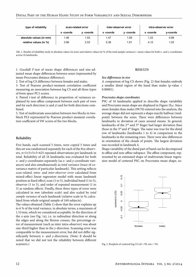
1. Goodall F-test of mean shape differences and size-ad-justed mean shape differences between sexes (represented by mean Procrustes distance difference).2. Test of log CS difference between females and males. 3. Test of Pearson product-moment correlation coefficient measuring an association between log CS and all three types of form space PC1 scores.4. Paired t-test of difference in proportion of variances ex-plained by non-affine component between each pair of rows and for each direction (x and y) and for both directions com-bined.5. Test of multivariate association between two blocks in two-block PLS represented by Pearson product-moment correla-tion coefficient of SW scores of the two blocks.
reliability
Five hands, each scanned 5 times, were copied 5 times and this set was randomized separately for each of the five observ-ers, n=5×5×5×5=625 repeated observations per landmark in total. Reliability of all 16 landmarks was evaluated for both x- and y-coordinates separately (as x- and y-coordinate vari-ances) and also simultaneously as total variance (trace of co-variance matrix of particular landmark). This setting reflects scan-related, intra- and inter-observer error calculated from mixed-effect linear regression model with mean landmark position as fixed effect; scan (1 to 5), individual hand (1 to 5), observer (1 to 5), and order of repeated measurement (1 to 5) as random effects. Finally, these three types of error were calculated in mm (absolute scale) and also scaled by total sample variance of each landmark (relative scale in %; calcu-lated from whole original sample of 169 subjects). The values obtained (Table 1) show that the error explains up to 6 % of the total variance, in absolute terms, a maximum of 1.53 mm, which we considered acceptable. In the direction of the x-axis (see Fig. 1a), i.e. in radioulnar direction or along the edges and along the flexion creases, the percentage er-ror of measurement (such as inter intra-observer) was about one-third higher than in the y-direction. Scanning error was comparable to the measurement error, but did not differ sig-nificantly between x- and y-directions. (Note: It should be noted that we did not test the reliability between different scanners.)
Tab. 1. Results of reliability study in absolute values (in mm) and relative values (in % of the total sample variance)—mean values for both x- and y-coordinates across 16 landmarks.
Sex differences in sizeA comparison of log CS shows (Fig. 2) that females embody a smaller distal region of the hand than males (p-value < 0.00001).
Procrustes shape coordinatesPSC of 16 landmarks applied to describe shape variability and Procrustes mean shape are displayed in Figure 1b,c. Since more females than males (99:70) entered into the analysis, the average shape did not represent a shape exactly halfway (mid-point) between the sexes. There were differences between landmarks in deviation of cases around means. In general, landmarks of the 2nd and 5th finger had larger deviation than those in the 3rd and 4th finger. The same was true for the distal row of landmarks (landmarks 1 to 4) in comparison to the landmarks in the remaining rows. There were also differences in orientation of the clouds of points. The largest deviation was recorded in landmark 4. Shape variability of the distal part of hand can be decomposed to affine and non-affine subspace. The affine component, rep-resented by an estimated shape of multivariate linear regres-sion model of centered PSC on Procrustes mean shape, ex-
results
Fig. 2. Boxplots of centered log CS (nf = 99, nm = 70).
Distal Part of the Human Hand: Study of Form Variability and Sexual Dimorphism
type of reliability scan-related error inter-observer error intra-observer error
x -coords y -coords x -coords y -coords x -coords y-coords
absolute values (in mm) 1.46 1.53 1.47 1.09 1.22 0.89
relative values (in %) 3.96 2.53 5.38 1.81 4.12 1.03
females males
cent
ered
ln (C
S)
Anthropologia Integra vol. 5 no. 2/2014 13
press the majority (99.97 %) of shape variance. The non-affine component, represented by the residuals of a multivariate linear regression model of centered PSC on Procrustes mean shape, express the rest of shape variability.
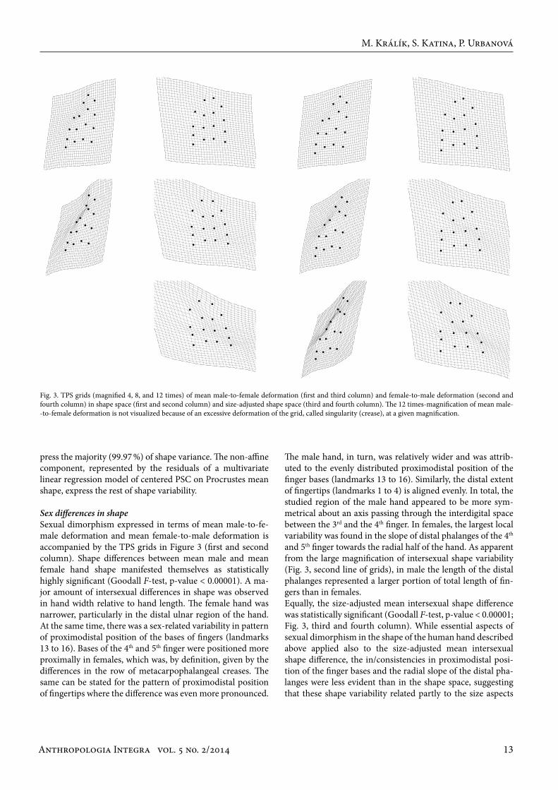
Sex differences in shape Sexual dimorphism expressed in terms of mean male-to-fe-male deformation and mean female-to-male deformation is accompanied by the TPS grids in Figure 3 (first and second column). Shape differences between mean male and mean female hand shape manifested themselves as statistically highly significant (Goodall F-test, p-value < 0.00001). A ma-jor amount of intersexual differences in shape was observed in hand width relative to hand length. The female hand was narrower, particularly in the distal ulnar region of the hand. At the same time, there was a sex-related variability in pattern of proximodistal position of the bases of fingers (landmarks 13 to 16). Bases of the 4th and 5th finger were positioned more proximally in females, which was, by definition, given by the differences in the row of metacarpophalangeal creases. The same can be stated for the pattern of proximodistal position of fingertips where the difference was even more pronounced.
The male hand, in turn, was relatively wider and was attrib-uted to the evenly distributed proximodistal position of the finger bases (landmarks 13 to 16). Similarly, the distal extent of fingertips (landmarks 1 to 4) is aligned evenly. In total, the studied region of the male hand appeared to be more sym-metrical about an axis passing through the interdigital space between the 3rd and the 4th finger. In females, the largest local variability was found in the slope of distal phalanges of the 4th and 5th finger towards the radial half of the hand. As apparent from the large magnification of intersexual shape variability (Fig. 3, second line of grids), in male the length of the distal phalanges represented a larger portion of total length of fin-gers than in females.Equally, the size-adjusted mean intersexual shape difference was statistically significant (Goodall F-test, p-value < 0.00001; Fig. 3, third and fourth column). While essential aspects of sexual dimorphism in the shape of the human hand described above applied also to the size-adjusted mean intersexual shape difference, the in/consistencies in proximodistal posi-tion of the finger bases and the radial slope of the distal pha-langes were less evident than in the shape space, suggesting that these shape variability related partly to the size aspects
Fig. 3. TPS grids (magnified 4, 8, and 12 times) of mean male-to-female deformation (first and third column) and female-to-male deformation (second and fourth column) in shape space (first and second column) and size-adjusted shape space (third and fourth column). The 12 times-magnification of mean male--to-female deformation is not visualized because of an excessive deformation of the grid, called singularity (crease), at a given magnification.
M. Králík, S. Katina, P. Urbanová
Anthropologia Integra vol. 5 no. 2/201414
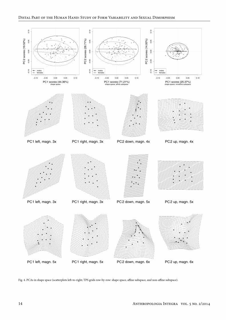
Fig. 4. PCAs in shape space (scatterplots left-to-right; TPS grids row-by-row: shape space, affine subspace, and non-affine subspace).
Distal Part of the Human Hand: Study of Form Variability and Sexual Dimorphism
Anthropologia Integra vol. 5 no. 2/2014 15
of the hand. Difference can be found also in the location of the most proximodistal shape compression in males (highest density of the grid). While in the shape space (Fig. 3, second column), the compression was located in the proximal pha-lanx of the 2nd finger, proximal and middle phalanges of the 3rd and 4th finger and middle phalanx of the 5th finger; after size adjustment (Fig. 3, fourth column) the maximum proxi-modistal compression relocated completely into the middle row of the phalanges. After size adjustment, sex differences in the relative width of the distal row of phalanges were lower. In the size-adjusted shape space, variability in the proportion of distal phalanges to the total finger length contributed to sex differences to a higher extent than in the shape space. Fi-nally, in males 2nd and 5th fingers more converged more dis-tally (possessed less parallel direction) in the size-adjusted shape space than in the shape space. In general, it appeared that the overall shape dimorphism after the size adjustment changed little, while the local sex-related differences changed to a larger extent.
Shape space variabilityIn contrast to the above-described direct assessment of dif-ferences between mean male and mean female shape, PCA allowed an analysis of major directions of variability in the sample. PCA performed on aligned coordinates of shape space (Fig. 4, first scatter-plot, upper row of grids) revealed that 44.36 % of the total variance (PC1) corresponded to a global change in the radioulnar asymmetry, accompanied by a change in the distal extent pattern of the finger bases (landmarks 13 to 16) and fingertips (landmarks 1 to 4). The only more local variability was observable in the region of the proximal and medial phalanges of the 5th finger. The PC2 accounting for 19.52 % of the total variance was associated with mostly global shape modifications in relative hand width. In positive values of the PC2, the 4th finger position coupled with the relative hand widening shifted itself more distally, accompanied by shortening of medial phalanges in relation to the total finger lengths and relative shortening of proximal and medial phalanges of the 5th finger. Negative values of PC2 were characterized by narrowing of the hand with a concurrent increment of slope of the distal phalanges of the 4th and 5th finger radially and shifting of the whole 4th finger more proximally. The scatter-plot of shape space PCA (Fig. 4) shows that the greater proportion of males was in the positive side of the PC2 axis, while for females the reverse was true. When being observed in the affine subspace of the shape space, the visualizations of shape differences on PCs are fair-ly similar to those of the whole shape space (Fig. 4, second scatter-plot, middle row of grids). Affine or uniform com-ponents express those shape differences that can be char-acterized as uniform stretching or compressing the space in orthogonal directions. The affine changes leave parallel lines parallel and affect the local space precisely the same way everywhere, and thus the changes are considered global (Slice 2005, p. 30–31, for review). When the shape change is
affine, the proportional lengths of the parallel lines remain unchanged. Comparing the position of males and females on the plot of PC1/PC2 scores (Fig. 4) in the affine subspace (and also in the full shape space), it can be observed that male and female hands of an identical proximodistal shear level (e.g zero value on the PC1) differ in their relative width level (PC2) so that the female hand is relatively thinner. Centroids for males and females differed at a similar level on PC1 and PC2, female hands are on average relatively more thin and, at the same time, more proximally sheared on their ulnar sides. Shape variability in the non-affine subspace of shape space (Fig. 4, third scatter-plot, lower row of grids) corresponded mostly to alterations in ratio between finger lengths. Shape changes in PC1 accounting for 25.37 % of the total variance described changes in the relative length of the 5th finger, caused by variability in its proximal and middle phalanges, accompanied by changes in proximodistal position of the 3rd and 4th finger (given predominantly by the position of the metacarpophalangeal crease, i.e. the distal extent of a given metacarpal). PC2 accounting for 14.00 % of the total vari-ance of non-affine subspace was associated with complex modifications composed of changes in relative width of the proximal and distal part of the studied region, accompanied by changes in proportions between lengths of the proximal and distal row of phalanges. Together with these changes, relative length of the 5th and 4th fingers to the remaining two fingers changed, which was accompanied by (or com-pensated with) an opposite change in the length of meta-carpals: the longer the 5th finger was, the shorter was the 5th metacarpal (the less distal was the position of landmark 16). Scatter-plot of PC scores (Fig. 4) shows that, in the non-af-fine subspace, distributions of values for males and females completely overlapped.
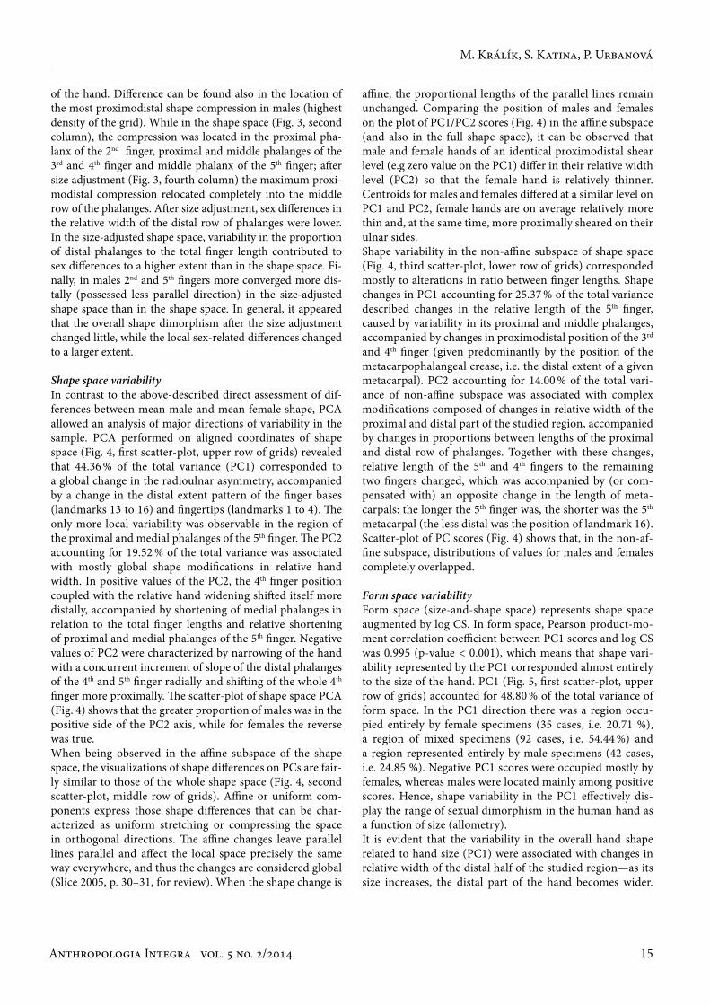
Form space variabilityForm space (size-and-shape space) represents shape space augmented by log CS. In form space, Pearson product-mo-ment correlation coefficient between PC1 scores and log CS was 0.995 (p-value < 0.001), which means that shape vari-ability represented by the PC1 corresponded almost entirely to the size of the hand. PC1 (Fig. 5, first scatter-plot, upper row of grids) accounted for 48.80 % of the total variance of form space. In the PC1 direction there was a region occu-pied entirely by female specimens (35 cases, i.e. 20.71 %), a region of mixed specimens (92 cases, i.e. 54.44 %) and a region represented entirely by male specimens (42 cases, i.e. 24.85 %). Negative PC1 scores were occupied mostly by females, whereas males were located mainly among positive scores. Hence, shape variability in the PC1 effectively dis-play the range of sexual dimorphism in the human hand as a function of size (allometry). It is evident that the variability in the overall hand shape related to hand size (PC1) were associated with changes in relative width of the distal half of the studied region—as its size increases, the distal part of the hand becomes wider.
M. Králík, S. Katina, P. Urbanová

Anthropologia Integra vol. 5 no. 2/201416
Fig. 5. PCAs in form space (scatterplots left-to-right; TPS grids row-by-row: shape space, affine subspace, and non-affine subspace).
Distal Part of the Human Hand: Study of Form Variability and Sexual Dimorphism

Anthropologia Integra vol. 5 no. 2/2014 17
However, local shape variability in PC1 were also substantial and affected particular rows of phalanges with each row ex-pressed in the total hand size differently. The larger the hand, the more important the contribution of middle phalanges of the 2nd, 3rd, and 4th fingers. The larger the hand, the smaller the ratio of the length of the distal phalanx of the 2nd finger to the length of the whole finger. Moreover, a small hand was linked to a tendency towards convergence of the distal phalanges of the 3rd and 4th fingers, which represented the overall tendency of small hands to incline (or even curve) the 2nd and the 3rd finger in the ulnar direction. Moreover, on large hands, bases of the 3rd and 4th finger are placed rela-tively closer to each other (landmarks 14 to 15) than each of them to the 2nd and 5th finger, respectively (landmarks 14 to 13 and 15 to 16). Variability represented by PC2 in form space closely resemble PC1 in shape space (compare Fig. 4). In the affine subspace of form space (Fig. 5, second scatter-plot, middle row of grids), PC1 accounting for 62.24 % of the total variance of affine subspace and the Pearson prod-uct-moment correlation coefficient between PC1 scores and log CS was 0.996 (p-value < 0.001). Shape changes in affine subspace were represented by shear deformation of the ra-dial and ulnar parts of the hand in the proximodistal direc-tion, in the opposite direction for each of these parts. At the same time, in PC1 this shape change included narrowing/widening of the relative hand width—hands characterized by a displacement of the ulnar part proximally are relatively narrower and vice versa. In the PC1 direction, the distribu-tions of male and female specimens in the affine subspace were similar to that of full form space. Again, a relatively wide shape represented a large, i.e. male hand, while a thin shape represented a small hand, i.e. female hand. Shape variability associated with PC2 of the affine subspace of form space also described a shear deformation, and the shear was even more pronounced than in the PC1, while any substantial altera-tions in relative width of the hand were not evident.In non-affine subspace of form space (Fig. 5, third scatter-plot, lower row of grids), the Pearson product-moment cor-relation coefficient between PC1 scores and log CS was 0.999 (p-value < 0.001). Shape variability expressed by PC1 corre-sponded to that which was described for the full form space, except the relative width and shear changes. Variability rep-resented by PC2 in the non-affine component of form space resembled variability on PC1 in the non-affine component of shape space (compare Fig. 4). Comparing each sex separately in the affine subspace (and also in full form space), we can see (Fig. 5) that distributions for males and females are similar and have similar slopes that are (contrary to the pooled distribution) not strictly parallel to the x-axis. Moreover, distributions for males and females are shifted from each other more in the x- than in y-direction. A female hand of a particular relative width (e.g. zero value on the PC1) had a higher level of proximal shear (PC2) in its ulnar region than a male hand of identical relative width. In the non-affine subspace, the sexes differed only in their position on the PC1 but not on the PC2.
Size-adjusted shape space variabilityEssentially, shape changes in the full size-adjusted shape space and both subspaces were very similar to the changes in the shape space (Fig. 4) and its respective subspaces, with the only difference that (after the same magnification) these shape changes were more pronounced. Therefore, variability of the human hand in size-adjusted (non-allometric) shape space are not displayed. While PC1 accounted for 44.66 % of the total variance PC2 accounted for 19.53 % of the total variance. Affine subspace also explained 99.94 % of the total variance of size-adjusted shape space. These shape changes represented mutual shifts of individual fingers in proximodistal direction combined with a change in relative hand width.
Correlations with the total shape space Affine component correlated strongly with the total shape space; in non-affine components, in contrast, the correla-tions are relatively moderate as the Pearson product-moment correlation coefficient reaches 5-times lower values. Of the non-affine components, only PC1 by form space PCA yield-ed a statistically highly significant correlation with the total shape (r = 0.995; p-value < 0.0001).
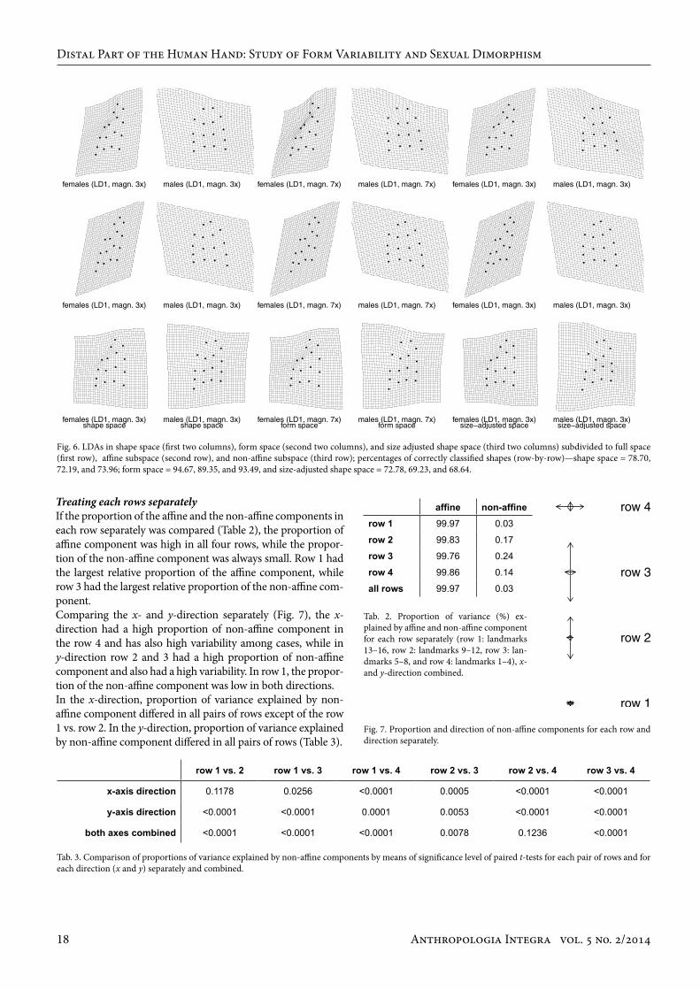
Linear discriminant analysisContrary to the PCA, LDA searches for a linear combination of variables which best separates defined groups, in our case males and females, to model differences between them. In all three morphometric spaces (shape space, form space, size-adjusted shape space) and their subspaces, changes of the shape of the hand along the first discriminant axis (LD1) were very similar (Fig. 6). They differed, however, in the per-centage of correctly classified cases. While in the form space the discrimination was high (the highest for the full space: 94.67 % of correctly classified cases according to sex, 94.29 % in females and 94.95 % in males); in the shape space and the size-adjusted shape space combined the overall classification rate was moderate and unbalanced if males and females were considered separately (72.88 % of correctly classified cases according to sex, 61.43 % in females and 80.98 % in males).LD1 shape differences between sexes essentially correspond-ed to those described above for mean male-to-female and mean female-to-male deformations. In the affine subspaces, the differences were characterized by shear changes accom-panied with changes in relative radioulnar width. Female hand was thinner, with the more distal position of its radial side and the more proximal position of its ulnar side giv-en by overall shear change. The male hand was wider, with a more proximal position of its radial side and a more distal position of its ulnar side given by overall shear change. It is worth noting that in non-affine subspaces, sex dif-ferences in the pattern of distal extent of fingertips corre-sponded to these sex differences in the affine subspace while sex differences in the pattern of distal extent of finger bases (landmarks 13–16) were exactly the opposite, i.e. a more dis-tal position of ulnar side in females and more proximal in males.
M. Králík, S. Katina, P. Urbanová
Anthropologia Integra vol. 5 no. 2/201418
Treating each rows separatelyIf the proportion of the affine and the non-affine components in each row separately was compared (Table 2), the proportion of affine component was high in all four rows, while the propor-tion of the non-affine component was always small. Row 1 had the largest relative proportion of the affine component, while row 3 had the largest relative proportion of the non-affine com-ponent. Comparing the x- and y-direction separately (Fig. 7), the x-direction had a high proportion of non-affine component in the row 4 and has also high variability among cases, while in y-direction row 2 and 3 had a high proportion of non-affine component and also had a high variability. In row 1, the propor-tion of the non-affine component was low in both directions. In the x-direction, proportion of variance explained by non-affine component differed in all pairs of rows except of the row 1 vs. row 2. In the y-direction, proportion of variance explained by non-affine component differed in all pairs of rows (Table 3).
Fig. 6. LDAs in shape space (first two columns), form space (second two columns), and size adjusted shape space (third two columns) subdivided to full space (first row), affine subspace (second row), and non-affine subspace (third row); percentages of correctly classified shapes (row-by-row)—shape space = 78.70, 72.19, and 73.96; form space = 94.67, 89.35, and 93.49, and size-adjusted shape space = 72.78, 69.23, and 68.64.
Fig. 7. Proportion and direction of non-affine components for each row and direction separately.
Tab. 2. Proportion of variance (%) ex-plained by affine and non-affine component for each row separately (row 1: landmarks 13–16, row 2: landmarks 9–12, row 3: lan-dmarks 5–8, and row 4: landmarks 1–4), x- and y-direction combined.
Tab. 3. Comparison of proportions of variance explained by non-affine components by means of significance level of paired t-tests for each pair of rows and for each direction (x and y) separately and combined.
Distal Part of the Human Hand: Study of Form Variability and Sexual Dimorphism
row 1 vs. 2 row 1 vs. 3 row 1 vs. 4 row 2 vs. 3 row 2 vs. 4 row 3 vs. 4
x-axis direction 0.1178 0.0256 <0.0001 0.0005 <0.0001 <0.0001
y-axis direction <0.0001 <0.0001 0.0001 0.0053 <0.0001 <0.0001
both axes combined <0.0001 <0.0001 <0.0001 0.0078 0.1236 <0.0001
affine non-affinerow 1 99.97 0.03
row 2 99.83 0.17
row 3 99.76 0.24
row 4 99.86 0.14
all rows 99.97 0.03
Anthropologia Integra vol. 5 no. 2/2014 19
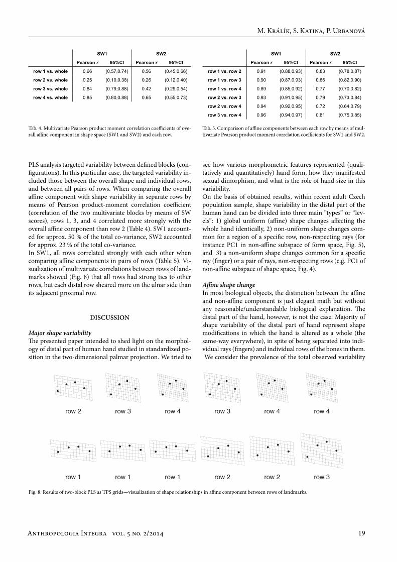
PLS analysis targeted variability between defined blocks (con-figurations). In this particular case, the targeted variability in-cluded those between the overall shape and individual rows, and between all pairs of rows. When comparing the overall affine component with shape variability in separate rows by means of Pearson product-moment correlation coefficient (correlation of the two multivariate blocks by means of SW scores), rows 1, 3, and 4 correlated more strongly with the overall affine component than row 2 (Table 4). SW1 account-ed for approx. 50 % of the total co-variance, SW2 accounted for approx. 23 % of the total co-variance. In SW1, all rows correlated strongly with each other when comparing affine components in pairs of rows (Table 5). Vi-sualization of multivariate correlations between rows of land-marks showed (Fig. 8) that all rows had strong ties to other rows, but each distal row sheared more on the ulnar side than its adjacent proximal row.
Tab. 4. Multivariate Pearson product moment correlation coefficients of ove-rall affine component in shape space (SW1 and SW2) and each row.
Tab. 5. Comparison of affine components between each row by means of mul-tivariate Pearson product moment correlation coefficients for SW1 and SW2.
Fig. 8. Results of two-block PLS as TPS grids—visualization of shape relationships in affine component between rows of landmarks.
Major shape variabilityThe presented paper intended to shed light on the morphol-ogy of distal part of human hand studied in standardized po-sition in the two-dimensional palmar projection. We tried to
dIscussIon
M. Králík, S. Katina, P. Urbanová
SW1 SW2
Pearson r 95%Cl Pearson r 95%Cl
row 1 vs. whole 0.66 (0.57,0.74) 0.56 (0.45,0.66)
row 2 vs. whole 0.25 (0.10,0.38) 0.26 (0.12,0.40)
row 3 vs. whole 0.84 (0.79,0.88) 0.42 (0.29,0.54)
row 4 vs. whole 0.85 (0.80,0.88) 0.65 (0.55,0.73)
SW1 SW2
Pearson r 95%Cl Pearson r 95%Cl
row 1 vs. row 2 0.91 (0.88,0.93) 0.83 (0.78,0.87)
row 1 vs. row 3 0.90 (0.87,0.93) 0.86 (0.82,0.90)
row 1 vs. row 4 0.89 (0.85,0.92) 0.77 (0.70,0.82)
row 2 vs. row 3 0.93 (0.91,0.95) 0.79 (0.73,0.84)
row 2 vs. row 4 0.94 (0.92,0.95) 0.72 (0.64,0.79)
row 3 vs. row 4 0.96 (0.94,0.97) 0.81 (0.75,0.85)
see how various morphometric features represented (quali-tatively and quantitatively) hand form, how they manifested sexual dimorphism, and what is the role of hand size in this variability. On the basis of obtained results, within recent adult Czech population sample, shape variability in the distal part of the human hand can be divided into three main “types” or “lev-els”: 1) global uniform (affine) shape changes affecting the whole hand identically, 2) non-uniform shape changes com-mon for a region of a specific row, non-respecting rays (for instance PC1 in non-affine subspace of form space, Fig. 5), and 3) a non-uniform shape changes common for a specific ray (finger) or a pair of rays, non-respecting rows (e.g. PC1 of non-affine subspace of shape space, Fig. 4).
Affine shape changeIn most biological objects, the distinction between the affine and non-affine component is just elegant math but without any reasonable/understandable biological explanation. The distal part of the hand, however, is not the case. Majority of shape variability of the distal part of hand represent shape modifications in which the hand is altered as a whole (the same-way everywhere), in spite of being separated into indi-vidual rays (fingers) and individual rows of the bones in them. We consider the prevalence of the total observed variability
Anthropologia Integra vol. 5 no. 2/201420
in shape of the human hand associated with a global uniform shape change, which is almost identical in every point of the distal hand, to be the most important finding. Major portion of this shape change is represented by almost pure shear in proximodistal direction, which leaves the Cartesian x-coordi-nate (width of hand) invariant and alters the y-coordinate by a translation that is a multiple of x-coordinate (Fig. 4, changes in PC1 of affine subspace). We propose to name this essential aspect of hand morphological variability as hand proximodis-tal shear variability. Minor portion of affine shape variabil-ity is represented by almost pure change (strain change) in the ratio between proximodistal and radioulnar dimensions, i.e. the relative hand width (Fig. 4, changes in PC2 of affine subspace). Affine variability always take place as combination of shear change (i.e. proximodistal shear change) and strain change (i.e. radioulnar expansion). The degree of variability covered by the affine component states in evidence, that distal part of human hand constitutes a structurally unifying system where position, size, and shape of each and every part is structurally interconnected with all the others. Thus we only need to know, theoretically, the coor-dinate system and the position of two points to be able to re-construct position of all the remaining points in a hand, while leaving aside of non-affine shape variability. It is evident that the proximodistal position of all points on the finger depends on the position of the respective metacar-pal bones to the same degree. For instance, when a metacar-pal grows by 1 mm, all points lying distally on the respective finger (phalanges) would automatically move about 1 mm distally. The affine shear variability in the metacarpal row (specifically in the location of the metacarpal heads) would be sufficient for the distal region of the hand to be strongly interrelated in the sense of affine shear variability, without any need for variability in the fingers themselves. If this was the case, we would not have to explain the affine variability in the region of fingers, but the affine variability in the metacarpal row would still remain unexplained. Our results have shown that affine variability in the metacarpal row does not suffi-ciently explain the strong affine component of the hand. With the affine shear change in the distal hand region, an affine strain change (stretching/dilation), named radioulnar strain change, simultaneously occurs in the fingers themselves, i.e. changes in the relative length/width proportions that cannot be explained just by change in the metacarpal row. Affine changes in the fingers occur by themselves. It is there-fore a change, where the shear and strain variability are intri-cately linked rather than a passive change in the position of fingers merely due to an affine change in metacarpals. Even this, however, is only part of the story. We tested how the rows of landmarks differed in their proportions of non-affine com-ponent of shape variability. If only the proximal region of the hand (represented by row 1; landmarks 13–16) caused the af-fine shear changes in the distal region of the hand, the propor-tion of the non-affine component should increase in the distal direction and it should be the highest on the fingertips (row 4). According to our results, row 1 is actually the least affected
by the non-affine component. Row 4 has the second lowest proportion of the non-affine component in the direction of the x-axis (landmarks 1–4), which contradicts the assumption that the non-affine component increases in the distal direc-tion. Moreover, rows in more distal positions (row 3 and 4) achieve greater correlation with the overall affine component than rows 1 and 2.Analysis of variability common for pairs of rows showed that each subsequent row is strongly tied to previous rows (Table 5). Given the strong overall affine component in the whole hand, strong affine shearing change common in each pair of rows could be expected. The rate of shearing shared by both rows grows with distance between the two rows, i.e. shearing in row 1 corresponds to more pronounced shearing in row 2, to even more pronounced shearing in row 3 and most pro-nounced shearing in row 4. This means not only that all rows are strongly uniformly interconnected, but also that each sub-sequent distal row adds its own complement to the shearing of the proximal row (Fig. 8). The proximodistal shear variability is associated with large al-terations in proximodistal extent of the fingertips and also in the digital formula, while the finger lengths (measured as dis-tance from the fingertips to the centre of the metacarpophalan-geal flexion crease—landmark distances 1–13, 2–14, 3–15, and 4–16), and therefore the finger length ratios, remain unchanged. The major variability in the digital formula are therefore played out as an affine change without needing to alter the digit ratios. In terms of prevailing within-population variability, the digital formula is principally a different trait to the digit ratios. (Note: The finger lengths in our model of hand proximodistal shear variability are not vectors oriented perfectly in the direction of the y-axis, but they slightly deviate in relation to the radio-ulnar position of a given finger and also its curvature, so the fingers are not perfectly parallel. Therefore even within affine shape change slight changes might appear in the finger length ratios.) It is also important to keep in mind that despite the overall prevalence of the affine component even when rows are considered separately, row landmarks in our study statistically differed in the proportion of variance (%) explained by affine and non-affine component. Variability between whole shape and the individual rows differed for each row, as well as the co-variance between individual rows when compared in pairs. Affine shape variability in the relative hand width (Fig. 4, changes in PC1 of affine subspace) is significant mainly be-cause it is dependent on size. A large hand is relatively wide in proportion to its length. This claim agrees with a general trend in hominids in which cross-sectional diaphyseal di-mensions in weight-bearing skeletal elements increase with the body mass (e.g. Ruff 2000). This relationship of a biologi-cal property (e.g. shape) with size is known as allometry which is regarded as a ubiquitous phenomenon in biology (reviewed shortly in Strauss 1993; Corruccini 1987). Despite the fact the human hand is not directly used for locomotion for a long time in human evolution, it is similarly affected by the same principles. It might be regarded as a pleiotropic effect of genes which forms both hand and foot (Rolian et al. 2010).
Distal Part of the Human Hand: Study of Form Variability and Sexual Dimorphism
Anthropologia Integra vol. 5 no. 2/2014 21
Non-affine variabilityUnlike affine component explaining the vast majority of the shape variability in our sample, non-affine component, seen as a change consistently affecting several landmarks of a row or a ray, or even all landmarks changing their positions si-multaneously, but not uniformly (so-called global bending), in various directions, represented a minority. Still, it would be incorrect to consider the non-affine variability of the hand shape as unimportant.One type of non-affine variability represents changes, which are due to a congruent change in one row region. We might therefore agree with previous studies claiming that hand bones of each row are mutually interconnected (Lewis 1996, 2001) and that bone length in each of the autopodium rows is probably influenced by separate genetic factors (Takai 1978). Local changes linked to particular rows are associated with hand size (PC1 in non-affine subspace of form space, Fig. 5) and only this type of local (non-affine) shape changes cor-relates highly significantly with the total shape. Regarding the relationship between the size and non-affine variabil-ity, larger hands are represented by relatively longer middle row phalanges and a generally wider distal region of the hand (Fig. 5, changes in PC1 of non-affine subspace). This is consistent with the theory that bipedalism evolved in an ancestor, whose locomotor pattern included a proportion of knuckle walking (Richmond et al. 2001, for review). Elonga-tion of the middle phalanx is one of the possible adaptations to knuckle-walking which in sum of all middle rows provides large area to reduce the pressure exerted to unit area and to increase stability of the locomotion (Richmond et al. 2001, p. 95). In the visualization (Fig. 5) it is apparent, that the non-affine size change concerns mainly the middle phalanges of the 2nd, 3rd, and 4th fingers, i.e. those that are most loaded, but less in the 5th finger. Nevertheless, it is clear that with the in-creasing size of the hand, the length of the fifth finger aligns with the other fingers and the middle phalanx approaches the proximodistal level of the middle phalanges of the other fingers. If it is demonstrated in future studies that these row-related non-affine shape variability correlates with size and weight variables of the human body, then this allometric de-pendence of the human hand could be considered another vestige of knuckle-walking in human evolution.In landmark rows 1, 2, and 3 in the direction of the x-axis (radioulnar), the proportion of non-affine components is gen-erally low and similar in the order of magnitude (although the differences are still statistically significant). In row 4, it is substantially higher. This may be partly due to slight inconsis-tencies in standardizing hand position during scanning since each subject could have put his/her fingers together with dif-ferent strength/pressure and intensity and unevenly between different fingers. Since the adduction/abduction of fingers takes place in metacarpophalangeal joints and change in the finger axis angle is identical for the whole finger, the effect of standardisation inconsistencies should continuously increase distally with the distance from the axis of rotation. Instead, we observed a significant increase in the proportions of the non-
affine component in row 4 only. It could therefore be a real biological phenomenon, resulting from the fact that the distal phalanx is distally free and not functionally tied to any adjoin-ing element.Local changes which are much less related to the size variabil-ity are contrastingly represented by changes, which separate individual fingers, or more precisely each finger represents a separate unit of shape variability. On a small scale, these local changes affect both the digital formula and the finger ratios while, in contrast to the affine changes described above, they can change both at the same time to different degrees (see Fig. 4). Note also that each of the phalanges may contribute to the total finger length by a different amount, or even change in a direction opposite to the other phalanges. For instance, while the 3rd finger relatively elongates (Fig. 4, PC2 non-affine subspace), its distal phalanx becomes relatively shorter and contributes less to the total length.
Developmental contextDuring ontogeny the hand undergoes forelimb bud formation and outgrowth regulated by apical ectodermal ridge (AER) (Nissim – Tabin 2004, p. 148; Hu – He 2008, for review), dif-ferentiation and separation of cartilaginous primordia of the five fingers, segmentation of the primordia trough interzones under the regulation of zone of polarizing activity (ZPA) and the creation of individual cartilaginous phalanges (Garn et al. 1975) and synovial joints connecting them (Zakany – Duboule 2007; Hu – He 2008), a complex process of diaphyseal ossifica-tion (Mall 1906), continuous and heterogenous prenatal (Ma-las et al. 2006) and postnatal growth (Hajniš 1970; Procház-ková et al. 1994; Trivers et al. 2006; Mayhew 2012), cessation of growth with complex epiphyseal fusion pattern and skeletal maturation during puberty and adolescence (Garn et al. 1961), and even further changes during adulthood (Harris et al. 1992). Moreover, in various developmental processes substan-tial interindividual variability were observed. There is a wide scope and time for the action of various internal and external regionally specific or even disrupting factors that may affect hand shape. This process can occur differently in each person. Between the end of embryonic phase and adulthood, the size of the hand changes by two orders of magnitude (from several millimetres to several hundreds of millimetres), but the pro-portionality (shape) does not change nearly as dramatically. A common response when a person sees the hand of a human fetus for the first time is surprise at how “mature” it looks. Mi-croscopic study (Garn et al. 1975) showed that despite the va-riety of developmental processes, proportions between lengths of phalanges and metacarpals approach nearly adult values already in embryos of 15–44 mm of crown-rump length (ca. day 55–68, hand length approx. 2 mm). Throughout the entire subsequent development it is still the human hand, only grows and, finally, becomes a hundred times larger. The general simi-larity of the fetal and adult hand proportions, however, does not exclude slight changes in the shape and proportions of the hand during development, as well as individual differences in these changes (cf. Králík et al. 2014).
M. Králík, S. Katina, P. Urbanová
Anthropologia Integra vol. 5 no. 2/201422
Interpreting our results ontogenetically, we hypothesize that the strong uniform ties are caused by the action of AER dur-ing differentiation and outgrowth of the limb bud and finger enchondral primordia. Places formed approximately at the same time are located in the same proximodistal position and are congruously differentiated. During the subsequent devel-opment, these places probably react congruously in response to central regulatory factors (growth hormones, sex steroids etc.) and grow uniformly, or non-uniformly, but similarly in all people, in spite of the division of the hand into 19 separate growth units (metacarpals and phalanges). As a consequence, strong affine shape variability and strong ties between autopo-dial rows persist even into adulthood. Ray related variability are established through the individual reaction of individual fingers during and/or after their antero-posterior differentia-tion and separation of rays caused by the action of ZPA. Fi-nally, non-affine shape variability located in individual bones might result in the process of ray segmentation. It should be noted, that the biggest local shape variability are localised in the proximal phalanx of the 5th finger, which we consider to be closest to the embryonic ZPA of the entire studied region of the hand. Shape variability in this region are also least congru-ent with the global affine shape variability—this also applies to sexual differences.
Sexual dimorphismThe essence of sexual dimorphism of the shape of the human hand lies in pronounced uniform (affine) change. Part of this change is represented by the hand proximodistal shear vari-ability where females are characterized by the distal position of the radial half of the hand and at the same time by the po-sition of the ulnar part of the hand placed more proximally than in males.Another form of sexual dimorphism is also realized largely as an affine shape change—differences in relative hand width. This finding is in congruence with the knowledge from tra-ditional anthropometry and metacarpal studies that sex dif-ferences (in percentages) in the hand and metacarpal widths are, on average, more pronounced than that in the hand and metacarpal lengths (e.g. Khanpetch et al. 2012). Dimorphism in relative hand width can be partly explained by allometry, since the size of the hand is correlated with changes in rela-tive hand width—large hands (regardless of sex) are relatively wider. Female hand is relatively thin and the male hand is relatively broad. This corresponds to the results from tradi-tional morphometry for the hand index (e.g. Pospíšil 1959; Bláha et al. 1986; Imrhan et al. 1993). Dimorphism in hand width and the hand index has been recorded even in new-borns (Procházková et al. 1994). Although the entire hand (in both males and females) during the postnatal period becomes relatively narrower (due to more intensive growth in length than in width), the difference in the relative width between males and females persists and even increases in puberty. An important aspect of hand sex dimorphism is the fact, that the sex-related differences result from both affine changes, the hand proximodistal shear change and radioulnar strain
change in relative hand width—as the ulnar side of the hand is sheared more distally, it becomes relatively wider. As females are smaller and on average they have smaller hands (thinner hands), the extent of the shift is greater than in males. How-ever, this shear-strain relationship does not explain all dimor-phism of the shear type. The ulnar part of the female hand is more sheared in the proximal direction than a male hand of the same relative width. This change is associated with a dif-ference between the sexes in the digital formula as described in previous studies.Other aspects of sexual dimorphism in the human hand are rather localized. Underlining differences in various regions of hand, the local differences are mostly limited to a single or several phalanges in a single row. In terms of whole fin-gers, visualizing LDA in non-affine subspace (Fig. 6) showed that these sex differences represent mainly changes in length proportions between the fingers. The changes in these length proportions chiefly affect the 2D:4D ratio. In LDA results, the non-affine sex differences in the proximal ulnar area of the studied hand region have the opposite effect compared to af-fine sex differences. According to the generally accepted model of mammalian sexual development, sex-related development of genitals, brain, and body (including the hand) is mediated by organi-zational effect of prenatal sex hormones from developing go-nads (Blecher – Erickson 2007, for review). In human male, in the period of mid-gestational peak of testosterone production (from 10th to 18th week according to the McIntyre 2006; from 12th to 17th week according to Wilson – Davies 2007; from 8th to 12th week according to Manning 2012), fingers and indi-vidual phalanges are separated completely. It is assumed, that interactions between the homeobox genes (HOXa and HOXd, which are involved both in the formation of the external geni-tals and the fingers; Kondo et al. 1997) and the sex specific levels of steroid hormones lead to the emergence of sexual di-morphism in both genitals and finger length ratios (Manning 2012, for review)—non-affine sex differences in our approach, which have been reported already in prenatal period (Malas et al. 2006; Galis et al. 2010). However, prevalence of the affine components in the sexual dimorphism of the human hand (and hence variability in digi-tal formula) can hardly be explained by an organizational ef-fect of prenatal sex hormones. The activity of AER, epithelial structure responsible for regulating distal outgrowth of the forelimb, falls before differentiation of the gonads and long before the peak of prenatal production of the sex steroids. Sex differences at this time would have to be directly caused by the sexual differences in genes encoded on the sex chromosomes. A number of studies has demonstrated the existence of sexual dimorphism in human embryos before gonadal differentia-tion and hence independence on the hormonal environment (Blecher – Erickson 2007, for review), which led to formula-tion of a revised model of mammalian sexual differentiation (Arnold 2009). According to this model, genetic factors from sex chromosomes and hormonal factors interact synergically to increase sex differences or counteract to decrease them. It
Distal Part of the Human Hand: Study of Form Variability and Sexual Dimorphism
Anthropologia Integra vol. 5 no. 2/2014 23
is possible that the differences we found between the affine and non-affine component of hand sexual dimorphism reflect different action of genetic and hormonal factors affecting dif-ferentiation of the hand during different stages of embryogen-esis. The legitimacy of this interpretation could be confirmed, for instance, by comparing the affine components of hand shape variability between individuals with different aberra-tions of sex chromosomes. We believe that the human hand can be understood as a mor-phological “palimpsest” written by several stages of morpho-genesis. Geometric morphometrics might be suitable for rec-ognizing these stages in the hand morphology by extraction of shape variability at different scales and analysing their mu-tual interconnections. Quantification of these shape variabil-ity might be suitable for investigating factors and processes (e.g. gradients in the developing hand identified by molecular geneticists, cf. Mayhew 2012) that participate in hand differ-entiation not only in pathology studies but also within the normal range of variability.
We would like to thank all the volunteers who participated in the study and at the same time to all fellow colleagues and students who had assisted in the study. In particular we thank Yvetta Vrublová (University of Ostrava) and Michal Živný (University of Ostrava) for great help in organizing the research. We also express gratitude to Ladislav Nejman (The University of Queensland, School of Social Science, Australia) for proofreading the text. The study was finan-cially supported by Grant Agency of the Czech Republic, project No 305/05/P303. Statistical analysis was supported by CZ.1.07/2.3.00/20.0181.
acknowledGeMents
Agnihotri, A. K. – Purwar, B. – Jeebun, N. – Agnihotri, S. (2006): Determina-tion of Sex by Hand Dimensions. Internet Journal of Forensic Science, 1.
Arnold, A. P. 2009. The organizational–activational hypothesis as the foun-dation for a unified theory of sexual differentiation of all mammalian tissues. Hormones and Behavior, 55, 570–578.
Balakrishnan, V. – Yeow, P. H. P. (2008): Hand Anthropometry and SMS Sat-isfaction. Journal of Applied Sciences, 8, 816–822.
Barut, Ç, – Demirel, P. – Kıran, S. (2008): Evaluation of hand anthropometric measurements and grip strength in basketball, volleyball and handball players. Anatomy, 2, 55–59.
Barut, C. – Dogan, A. – Buyukuysal, M. (2014): Anthropometric aspects of hand morphology in relation to sex and to body mass in Turkish popu-lation sample. HOMO – Journal of Comparative Human Biology, 65, 338–348.
Bayer, L. M. – Gray, H. (1933): The hand: Method of measurement. American Journal of Physical Anthropology, 17, 379–415.
Bláha, P. et al. (1986): Anthropometric Studies of the Czechoslovak Popula-tion from 6 to 55 Years: Czechoslovak spartakiade 1985. Part I, section 2. Praha: Ústřední štáb Československé spartakiády 1985, ÚV ČSTV, ÚNZ hlavního města Prahy. 357 p.
Blecher, S. R. – Erickson, R. P. (2007): Genetics of sexual development: A new paradigm. American Journal of Medical Genetics Part A, 143A, 3054–3068.
references
M. Králík, S. Katina, P. Urbanová
Bolstad, G. – Benum, B. – Rokne, A. (2001): Anthropometry of Norwegian light industry and office workers. Applied Ergonomics, 32, 239–246.
Bookstein, F. (1991): Morphometric Tools for Landmark Data. Geometry and Biology, New York: Cambridge University Press.
Boz, C. – Ozmenoglu, M. – Altunayoglu, V. – Velioglu, S. – Alioglu, Z. (2004): Individual risk factors for carpal tunnel syndrome: an evaluation of body mass index, wrist index and hand anthropometric measurements. Clini-cal Neurology and Neurosurgery, 106, 294–299.
Chuan, T. K. – Hartono, M. – Kumar, N. (2010): Anthropometry of the Sin-gaporean and Indonesian populations. International Journal of Industrial Ergonomics, 40, 757–766.
Corruccini, R. S. (1987): Shape in morphometrics: Comparative analyses. American Journal of Physical Anthropology, 73, 289–303.
Crhák, L. (1957): Příspěvek k poznání morfologie dlaně opavských chlapců. Přírodovědný sborník ostravského kraje, 57, 329–441.
Drobný, I. (1959): Relatívna dĺžka prstov detí horného Liptova. Acta Faculta-tis Rerum Naturalium Universitatis Comenianae, 3, 339–346.
Dryden, I. – Mardia, K. (1999): Statistical shape analysis. Chisester: John Wi-ley & Sons.
Fallahi, A. A. – Jadidian, A. A. (2011): The Effect of Hand Dimensions, Hand Shape and Some Anthropometric Characteristics on Handgrip Strength in Male Grip Athletes and Non-Athletes. Journal of Human Kinetics, 29, 151–159.
Galis, F. – Ten Broek, C. M. A. – Van Dongen, S. – Wijnaendts, L. C. (2010): Sexual Dimorphism in the Prenatal Digit Ratio (2D:4D). Archives of Sexual Behavior, 39, 57–62.
Garn, S. M. – Burdi, A. R. – Babler, W. J. – Stinson, S. (1975): Early prenatal attainment of adult metacarpal-phalangeal rankings and proportions. American Journal of Physical Anthropology, 43, 327–332.
Garn, S. M. – Nagy, J. M. – Hertzog, K. P. – Poznanski, A. K. (1972): Metacar-pophalangeal Length in Evaluation of Skeletal Malformation. Radiology, 105, 375–381.
Garn, S. M. – Rohmann, C. G. – Apfelbaum, B. (1961): Complete epiphy-seal union of the hand. American Journal of Physical Anthropology, 19, 365–372.
Hajniš, K. (1970): The growth of certain external dimensions of the hand and the operation period of its congenital defects. Anthropologie, 8, 49–59.
Harris, E. F. – Aksharanugraha, K. – Behrents, R. G. (1992): Metacarpo-phalangeal length changes in humans during adulthood: a longitudinal study. American Journal of Physical Anthropology, 87, 263–275.
Hickey, M. – Doherty, D. A. – Hart, R. – Norman, R. J. – Mattes, E. – Atkin-son, H. C. – Sloboda, D. M. (2010): Maternal and umbilical cord andro-gen concentrations do not predict digit ratio (2D:4D) in girls: A prospec-tive cohort study. Psychoneuroendocrinology, 35, 1235–1244.
Hu, J. – He, L. (2008): Patterning mechanisms controlling digit development. Journal of Genetics and Genomics, 35, 517–524.
Imrhan, S. N. – Nguyen, M-T. – Nguyen, N-N. (1993): Hand anthropometry of Americans of Vietnamese origin. International Journal of Industrial Ergonomics, 12, 281–287.
Imrhan, S. N. – Contreras, M. G. (2005): Hand anthropometry in a sample of Mexicans in the US Mexico border region. In: Lockhart, T. – Fernandez, J. E., eds., Proceedings of the XIX Annual Occupational Ergonomics and Safety Conference, Las Vegas, Nevada, USA, 589–593,
Imrhan, S. N. – Sarder, M. D. – Mandahawi, N. (2009): Hand anthropometry in Bangladeshis living in America and comparisons with other popula-tions. Ergonomics, 52, 987–998.
Jelínek, J. (1950): Příspěvek k poznání morfologie dlaně řeckých dětí. Zprávy anthropologické společnosti (Brno), 3, 25–28.
Kanchan, T. – Krishan, K. (2011): Anthropometry of hand in sex determina-tion of dismembered remains – A review of literature. Journal of Forensic and Legal Medicine, 18, 14–17.
Katina, S. (2007): Shape analysis in the light of simplicial depth estimators. In: Barber, S. – Baxter, P. D. – Mardia, K. V., eds., Systems Biology & Statisti-cal Bioinformatics, Leeds: Leeds University Press, 51–54.
Katina, S. (2012): Detection of non-affine shape outliers for matched-pair shape data. Tatra Mountains Mathematical Publications, 51(1), 83–90.
Khanpetch, P. – Prasitwattanseree, S. – Case, D. T. – Mahakkanukrauh, P. (2012): Determination of sex from the metacarpals in a Thai population. Forensic Science International, 217, 229.e1–229.e8.

Anthropologia Integra vol. 5 no. 2/201424
Klamklay, J. – Sungkhapong, A. –Yodpijit, N. – Patterson, P. E. (2008): An-thropometry of the southern Thai population. International Journal of Industrial Ergonomics, 38, 111–118.
Kondo, T. – Zakany, J. – Innis, J. W. – Duboule, D. (1997): Of fingers, toes and penises. Nature, 390, 29–29.
Králík, M. – Gronkiewicz, S. – Ingrová, P. – Kozieł, S. (2014): Changes in digit ratio during puberty: X-ray sample from the Wrocław Longitudinal Study of Twins. The Dolní Věstonice Studies, vol. 20: Academy of Sciences of the czech republic and Masaryk University, 187–189.
Kratochvíl, L. – Flegr, J. (2009): Defferences in the 2nd to 4th digit length ratio in humans reflect shifts along the common allometric line. Biology Letters, 5, 643–646.
Kulaksiz, G. – Gözil, R. (2002): The effect of hand preference on hand an-thropometric measurements in healthy individuals. Annals of Anatomy, 184, 257–265.
Kühnel, G. (1932): Die Konstitutionsform der Hand. Zeitschrift für die gesam-te Neurologie und Psychiatrie, 141, 98–131.
Lazenby, R. – Smashnuk, A. (1999): Osteometric variation in the Inuit second metacarpal: a test of Allen’s Rule. International Journal of Osteoarchaeol-ogy, 9, 182–188.
Lewis, S. (1996): Morphological aspects of male and female hands. Annals of Human Biology, 23, 491–494.
Lewis, S. (2001): Metacarpophalangeal pattern profile analysis of a sample drawn from a North Wales population. Annals of Human Biology, 28, 589–593.
Lutchmaya, S. – Baron-Cohen, S. – Raggatt, P. – Knickmeyer, R. – Manning, J. T. (2004): 2nd to 4th digit ratios, fetal testosterone and estradiol. Early Human Development, 77, 23–28.
Malas, M. A. – Dogan, S. – Evcil, E. H. – Desdicioglu, K. (2006): Fetal de-velopment of the hand, digits and digit ratio (2D : 4D). Early Human Development, 82, 469–475.
Mall, F. P. (1906): On ossification centers in human embryos less than one hundred days old. American Journal of Anatomy, 5, 433–458.
Mandahawi, N. – Imrhan, S. – Al-Shobaki, S. – Sarder, B. (2008): Hand an-thropometry survey for the Jordanian population. International Journal of Industrial Ergonomics, 38, 966–976.
Manning, J. T. (2002): Digit Ratio: A Pointer to Fertility, Behavior and Health. New York: Rutgers University Press.
Manning, J. T. (2012): Chapter 48 Sex Differences and Age Changes in Digit Ratios: Implications for the Use of Digit Ratios in Medicine and Biology. In: Preedy, V. R., ed., Handbook of Anthropometry: Physical Measures of Human Form in Health and Disease, New York: Springer. p 841–851.
Manning, J. T. – Barley, L. – Walton, J. – Lewis-Jones, D. I. – Trivers, R. L. – Singh, D. – Thornhill, R. – Rohde, P. – Bereczkei, T. – Henzi, P. – Soler, M. – Szwed, A. (2000): The 2nd : 4th digit ratio, sexual dimorphism, population differences, and reproductive success: evidence for sexually antagonistic genes? Evolution and Human Behavior, 21, 163–183.
Manning, J. T. – Churchill, A. J. G. – Peters, M. (2007): The effects of sex, ethnicity, and sexual orientation on self-measured digit ratio (2D : 4D). Archives of Sexual Behavior, 36, 223–233.
Mayhew, T. M. (2012): Cross-sectional data on soft tissue morphometry of the growing hand and fingers of dextral individuals 5–65 years old. Jour-nal of Anatomy, 221, 373–381.
McIntyre, M. H. (2006): The use of digit ratios as markers for perinatal andro-gen action. Reproductive Biology and Endocrinology, 4, 10.
Mitteroecker, P. – Gunz, P. – Bernhard, M. – Schaefer, K. – Bookstein, F. L. (2004): Comparison of cranial ontogenetic trajectories among great apes and humans. Journal of Human Evolution, 46, 679–698.
Nag, A. – Nag, P. K. – Desai, H. (2003): Hand anthropometry of Indian wom-en. Indian Journal of Medical Research, 117, 260–269.
Napier, J. R. (1993): Hands. Chichester: Princeton University Press.Nelson, E. C. – Manning, J. T. – Sinclair, A. G. M. (2006): Using the length of
the 2nd to 4th digit ratio (2D:4D) to sex cave art hand stencils: factors to consider. Before Farming 2006, 1–7.
Nissim, S. – Tabin, C. (2004): Development of the Limbs. In: Epstein, C. J. – Erickson, R. P. – Wynshaw-Boris, A., eds., Inborn Errors of Development: The Molecular Basis of Clinical Disorders of Morphogenesis, Oxford, New York: Oxford University Press, 148–167.
Peters, M. – Mackenzie, K. – Bryden, P. (2002): Finger length and distal finger
extent patterns in humans. American Journal of Physical Anthropology, 117, 209–217.
Pospíšil, M. F. (1959): Morfologie ruky Lužických Srbů. Acta Facultatis Rerum Naturalium Universitatis Comenianae, 3, 311–325.
Poznanski, A. K. – Gartman, S. (1997): A bibliography covering the use of metacarpophalangeal pattern profile analysis in bone dysplasias, conge-nital malformation syndromes, and other disorders. Pediatric Radiology, 27, 358–365.
Poznanski, A. K. – Nagy, J. M. – Garn, S. M. – Gall, J. C. (1972): Metacar-pophalangeal Pattern Profiles in Evaluation of Skeletal Malformations. Radiology, 104, 1–11.
Procházková, J. – Zlámalová, H. – Doležal, A. (1994): Dimensions of the hand in newborns in Prague. In: Hajniš K, ed., Growth and Ontogene-tic Development in Man, Prague: Department of Anthropology, Charles University, 7–14.
R Development Core Team. (2011): R: A language and environment for stati-stical computing. Vienna, Austria: R Foundation for Statistical Compu-ting. Retrieved from http://www.R-project.org.
Richmond, B. G. – Begun, D. R. – Strait, D. S. (2001): Origin of human bipe-dalism: The knuckle-walking hypothesis revisited. American Journal of Physical Anthropology, 116(S33), 70–105.
Robertson, J. – Zhang, W. – Liu, J. J. – Muir, K. R. – Maciewicz, R. A. – Doher-ty, M. (2008): Radiographic assessment of the index to ring finger ratio (2D : 4D) in adults. Journal of Anatomy, 212, 42–48.
Rohlf, F. J. (2004): tpsDig, digitize landmarks and outlines, version 1.40. New York: Department of Ecology and Evolution, State University of New York at Stony Brook.
Rohlf, F. J. – Bookstein, F. L. (2003): Computing the Uniform Component of Shape Variation. Systematic Biology, 52, 66–69.
Rohlf, F. J. – Corti, M. (2000): Use of Two-Block Partial Least-Squares to Stu-dy Covariation in Shape. Systematic Biology, 49, 740–753.
Rolian, C. – Lieberman, D. E. – Hallgrímsson, B. (2010): The Coevolution of Human Hands and Feet. Evolution, 64, 1558–1568.
Ruff, C. B. (2000): Body size, body shape, and long bone strength in modern humans. American Journal of Physical Anthropology, 38, 269–290.
Saino, N. – Romano, M. – Innocenti, P. (2006): Length of index and ring fingers differentially influence sexual attractiveness of men’s and women’s hands. Behavioral Ecology and Sociobiology, 60, 447–454.
Slice, D. E. (2005): Modern Morphometrics. In: Slice, D. E., ed., Modern Mor-phometrics in Physical Anthropology, New York: Springer, 1–45.
Snihur, A. W. K. – Hampson, E. (2011): Individual differences in 2D:4D di-git-ratios and otoacoustic emissions: Do they share a common develop-mental origin? Personality and Individual Differences, 51, 406–411.
Snow, D. R. (2006): Sexual dimorphism in Upper Palaeolithic hand stencils. Antiquity, 80, 390–404.
Strauss, R. E. (1993): The study of allometry since Huxley. Introductory essay for the re-publication of: Problems of Relative Growth, J.S. Hu-xley (1932). In: Huxley, J. S. –Churchill, F. B., eds., Problems of Relative Growth, Johns Hopkins University Press, 47–75.
Takai, S. (1978): Metacarpal and Phalangeal Lengths Are Influenced by Row--Related Factor. Human Biology, 50, 51–56.
Tocheri, M. W. – Orr, C. M. – Jacofsky, M. C. – Marzke, M. W. (2008): The evolutionary history of the hominin hand since the last common ances-tor of Pan and Homo. Journal of Anatomy, 212, 544–562.
Trivers, R. – Manning, J. – Jacobson, A. (2006): A longitudinal study of digit ratio (2D:4D) and other finger ratios in Jamaican children. Hormones and Behavior, 49, 150–156.
Ventura, T. – Gomes, M. C. – Pita, A. – Neto, M. T. – Taylor, A. (2013): Digit ratio (2D:4D) in newborns: Influences of prenatal testosterone and ma-ternal environment. Early Human Development, 89, 107–112.
Wilson, C. A. – Davies, D. C. (2007): The control of sexual differentiation of the reproductive system and brain. Reproduction, 133, 331–359.
Yörük, E. – Dutağaci, H. – Sankur, B. (2006): Hand biometrics. Image and Vision Computing, 24, 483–497.
Zakany, J. – Duboule, D. (2007): The role of Hox genes during vertebrate limb development. Current Opinion in Genetics and Development, 17, 359–366.
Zheng, Z. – Cohn, M. J. (2011): Developmental basis of sexually dimorphic digit ratios. Proceedings of the National Academy of Sciences, 108(39), 16289–16294.
Distal Part of the Human Hand: Study of Form Variability and Sexual Dimorphism
Anthropologia Integra vol. 5 no. 2/2014 25
authors
M. Králík, S. Katina, P. Urbanová
Miroslav Králík is a biological anthropologist. His research focuses on human sexual dimorphism in physical features and behavior, human ontogenetic adaptations, developmental stress factors, hu-man traces in archaeology, and fingerprints (dermatoglyphics) in archaeological artefacts.Contact: Miroslav Králík, Department of Anthropology, Faculty of Science, Masaryk University, Kotlářská 2, 611 37, Brno, Czech Re-public; Phone: +420 549 49 49 66; e-mail: [email protected].
Stanislav Katina is an expert at mathematics, probability, and ma-thematical statistics, specialized in shape and image analysis. His re-search activities are involving statistical analyses in different fields of biology, biological and clinical anthropology.
Petra Urbanová is a biological anthropologist, interested in forensic and virtual anthropology. Her research focuses on human osteology, geometric morphometrics, statistical shape analysis and multivari-ate statistics, 3D technology and non-invasive imaging techniques, living person identification.

Copyright © 2022 FDOKUMEN