Disruption of the ASTN2/TRIM32 locus at 9q33.1 is a risk factor in males for autism spectrum...
17
Disruption of the ASTN2/TRIM32 locus at 9q33.1 is a risk factor in males for autism spectrum disorders, ADHD and other neurodevelopmental phenotypes Anath C. Lionel 1,2,4, { , Kristiina Tamemimies 1,2,5, { , Andrea K. Vaags 1,2,6,10, { , Jill A. Rosenfeld 11 , Joo Wook Ahn 12 , Daniele Merico 1,2 , Abdul Noor 13,14 , Cassandra K. Runke 16,17 , Vamsee K. Pillalamarri 18,19 , Melissa T. Carter 3 , Matthew J. Gazzellone 1,2,4 , Bhooma Thiruvahindrapuram 1,2 , Christina Fagerberg 20,21 , Lone W. Laulund 20,21 , Giovanna Pellecchia 1,2 , Sylvia Lamoureux 1,2 , Charu Deshpande 12 , Jill Clayton-Smith 22 , Ann C. White 23 , Susan Leather 24 , John Trounce 25 , H. Melanie Bedford 26 , Eli Hatchwell 27 , Peggy S. Eis 27 , Ryan K.C. Yuen 1,2 , Susan Walker 1,2 , Mohammed Uddin 1,2 , Michael T. Geraghty 28,29 , Sarah M. Nikkel 28,29 , Eva M. Tomiak 28,29 , Bridget A. Fernandez 30 , Noam Soreni 31 , Jennifer Crosbie 15 , Paul D. Arnold 2,15 , Russell J. Schachar 15 , Wendy Roberts 32,33 , Andrew D. Paterson 2 , Joyce So 14,34 , Peter Szatmari 34,35 , Christina Chrysler 35 , Marc Woodbury-Smith 35 , R. Brian Lowry 7,8 , Lonnie Zwaigenbaum 36 , Divya Mandyam 1,2 , John Wei 1,2 , Jeffrey R. MacDonald 1,2 , Jennifer L. Howe 1,2 , Thomas Nalpathamkalam 1,2 , Zhuozhi Wang 1,2 , Daniel Tolson 37 , David S. Cobb 37 , Timothy M. Wilks 37 , Mark J. Sorensen 38 , Patricia I. Bader 39 , Yu An 40 , Bai-Lin Wu 40,41,42 , Sebastiano Antonino Musumeci 43,44,45 , Corrado Romano 43,44,45 , Diana Postorivo 46 , Anna M. Nardone 46 , Matteo Della Monica 47 , Gioacchino Scarano 47 , Leonardo Zoccante 48 , Francesca Novara 49 , Orsetta Zuffardi 49,50 , Roberto Ciccone 49 , Vincenzo Antona 51 , Massimo Carella 52 , Leopoldo Zelante 52 , Pietro Cavalli 53 , Carlo Poggiani 54 , Ugo Cavallari 53 , Bob Argiropoulos 9 , Judy Chernos 6,55 , Charlotte Brasch-Andersen 20,21 , Marsha Speevak 14,56 , Marco Fichera 43,44,45,57 , Caroline Mackie Ogilvie 12 , Yiping Shen 41,42,58 , Jennelle C. Hodge 16,17 , Michael E. Talkowski 18,19 , Dimitri J. Stavropoulos 13,14 , Christian R. Marshall 1,2,4 and Stephen W. Scherer 1,2,4, ∗ 1 The Centre for Applied Genomics, 2 Program in Genetics and Genome Biology and 3 Division of Clinical and Metabolic Genetics, The Hospital for Sick Children, Toronto, ON, Canada M5G 1L7, 4 Department of Molecular Genetics, McLaughlin Centre, University of Toronto, Toronto, ON, Canada M5G 1L7, 5 Center of Neurodevelopmental Disorders, Department of Women’s and Children’s Health, Karolinska Institutet, Stockholm 113 30, Sweden, 6 Cytogenetics Laboratory, 7 Department of Medical Genetics, 8 Department of Pediatrics, Alberta Children’s Hospital, Calgary, AB, Canada T3B 6A8, 9 Alberta Children’s Hospital Research Institute for Child and Maternal Health, University of Calgary, Calgary, AB, Canada, T3B 6A8, 10 Department of Anatomical Pathology and Cytopathology, Calgary Laboratory Services, Calgary, Canada, AB T2L 2K8, 11 Signature Genomic Laboratories, PerkinElmer, Inc., Spokane, WA 99207, USA, 12 Cytogenetics Department and Clinical Genetics, Guy’s and St Thomas’ NHS Foundation Trust, London, SE1 9RT, UK, 13 Cytogenetics Laboratory, Department of Pediatric Laboratory Medicine, Hospital for Sick Children, Toronto, ON, Canada, M5G 1X8, 14 Department of Laboratory Medicine and Pathobiology, 15 Department of Psychiatry, University of Toronto, Toronto, ON, Canada, M5S 1A1, Copy Edited by: K.R. Language used: US # The Author 2013. Published by Oxford University Press. All rights reserved. For Permissions, please email: [email protected] † These authors contributed equally to this work. ∗ To whom correspondence should be addressed at: The Centre for Applied Genomics, The Hospital for Sick Children, 686 Bay Street, Peter Gilgan Centre for Research and Learning, Room 139800, Toronto, ON, Canada M5G 1L7. Tel: +1 4168137613; Fax: +1 4168138319; Email: [email protected] Human Molecular Genetics, 2013, Vol. 22, No. ? 1–17 doi:10.1093/hmg/ddt669 Advance Access published on xxx 5 10 15 20 25 30 35 40 45 50 55 60 65 70 75 80 85 90 95 100 105 110 115 120
-
Upload
independent -
Category
Documents
-
view
2 -
download
0
Transcript of Disruption of the ASTN2/TRIM32 locus at 9q33.1 is a risk factor in males for autism spectrum...

Disruption of the ASTN2/TRIM32 locus at 9q33.1 is arisk factor in males for autism spectrum disorders,ADHD and other neurodevelopmental phenotypes
Anath C. Lionel1,2,4,{, Kristiina Tamemimies1,2,5,{, Andrea K. Vaags1,2,6,10,{, Jill A. Rosenfeld11,
Joo Wook Ahn12, Daniele Merico1,2, Abdul Noor13,14, Cassandra K. Runke16,17,
Vamsee K. Pillalamarri18,19, Melissa T. Carter3, Matthew J. Gazzellone1,2,4,
Bhooma Thiruvahindrapuram1,2, Christina Fagerberg20,21, Lone W. Laulund20,21,
Giovanna Pellecchia1,2, Sylvia Lamoureux1,2, Charu Deshpande12, Jill Clayton-Smith22,
Ann C. White23, Susan Leather24, John Trounce25, H. Melanie Bedford26, Eli Hatchwell27,
Peggy S. Eis27, Ryan K.C. Yuen1,2, Susan Walker1,2, Mohammed Uddin1,2, Michael T. Geraghty28,29,
Sarah M. Nikkel28,29, Eva M. Tomiak28,29, Bridget A. Fernandez30, Noam Soreni31,
Jennifer Crosbie15, Paul D. Arnold2,15, Russell J. Schachar15, Wendy Roberts32,33,
Andrew D. Paterson2, Joyce So14,34, Peter Szatmari34,35, Christina Chrysler35,
Marc Woodbury-Smith35, R. Brian Lowry7,8, Lonnie Zwaigenbaum36, Divya Mandyam1,2,
John Wei1,2, Jeffrey R. MacDonald1,2, Jennifer L. Howe1,2, Thomas Nalpathamkalam1,2,
Zhuozhi Wang1,2, Daniel Tolson37, David S. Cobb37, Timothy M. Wilks37, Mark J. Sorensen38,
Patricia I. Bader39, Yu An40, Bai-Lin Wu40,41,42, Sebastiano Antonino Musumeci43,44,45,
Corrado Romano43,44,45, Diana Postorivo46, Anna M. Nardone46, Matteo Della Monica47,
Gioacchino Scarano47, Leonardo Zoccante48, Francesca Novara49, Orsetta Zuffardi49,50,
Roberto Ciccone49, Vincenzo Antona51, Massimo Carella52, Leopoldo Zelante52, Pietro Cavalli53,
Carlo Poggiani54, Ugo Cavallari53, Bob Argiropoulos9, Judy Chernos6,55,
Charlotte Brasch-Andersen20,21, Marsha Speevak14,56, Marco Fichera43,44,45,57,
Caroline Mackie Ogilvie12, Yiping Shen41,42,58, Jennelle C. Hodge16,17, Michael E. Talkowski18,19,
Dimitri J. Stavropoulos13,14, Christian R. Marshall1,2,4 and Stephen W. Scherer1,2,4,∗
1The Centre for Applied Genomics, 2Program in Genetics and Genome Biology and 3Division of Clinical and Metabolic
Genetics, The Hospital for Sick Children, Toronto, ON, Canada M5G 1L7, 4Department of Molecular Genetics, McLaughlin
Centre, University of Toronto, Toronto, ON, Canada M5G 1L7, 5Center of Neurodevelopmental Disorders, Department of
Women’s and Children’s Health, Karolinska Institutet, Stockholm 113 30, Sweden, 6Cytogenetics Laboratory, 7Department
of Medical Genetics, 8Department of Pediatrics, Alberta Children’s Hospital, Calgary, AB, Canada T3B 6A8, 9Alberta
Children’s Hospital Research Institute for Child and Maternal Health, University of Calgary, Calgary, AB, Canada, T3B 6A8,10Department of Anatomical Pathology and Cytopathology, Calgary Laboratory Services, Calgary, Canada, AB T2L 2K8,11Signature Genomic Laboratories, PerkinElmer, Inc., Spokane, WA 99207, USA, 12Cytogenetics Department and Clinical
Genetics, Guy’s and St Thomas’ NHS Foundation Trust, London, SE1 9RT, UK, 13Cytogenetics Laboratory, Department of
Pediatric Laboratory Medicine, Hospital for Sick Children, Toronto, ON, Canada, M5G 1X8, 14Department of Laboratory
Medicine and Pathobiology, 15Department of Psychiatry, University of Toronto, Toronto, ON, Canada, M5S 1A1,
Copy Edited by: K.R.Language used: US
# The Author 2013. Published by Oxford University Press. All rights reserved.For Permissions, please email: [email protected]
†These authors contributed equally to this work.
∗To whom correspondence should be addressed at: The Centre for Applied Genomics, The Hospital for Sick Children, 686 Bay Street, Peter Gilgan Centre forResearch and Learning, Room 139800, Toronto, ON, Canada M5G 1L7. Tel: +1 4168137613; Fax: +1 4168138319; Email: [email protected]
Human Molecular Genetics, 2013, Vol. 22, No. ? 1–17doi:10.1093/hmg/ddt669Advance Access published on xxx
5
10
15
20
25
30
35
40
45
50
55
60
65
70
75
80
85
90
95
100
105
110
115
120

16Department of Laboratory Medicine and Pathology, 17Department of Medical Genetics, Mayo Clinic, Rochester, MN
55905, USA, 18Center for Human Genetic Research, Massachusetts General Hospital, Department of Genetics,19Departmentof Neurology,HarvardMedicalSchool, HarvardUniversity,Boston,MA02114,USA, 20DepartmentofClinical
Genetics, 21Department of Paediatrics, Odense University Hospital, Odense DK-5000, Denmark, 22Genetic Medicine,
Manchester Academic Health Sciences Centre, St Mary’s Hospital, Manchester M13 9WL, UK, 23Sussex Community NHS
Trust, Brighton General Hospital, Brighton BN2 3EW, UK, 24Community Paediatrics, Lewisham Healthcare NHS Trust,
London SE13 6LH, UK, 25Brighton and Sussex University Hospital NHS Trust, Brighton BN2 5BE, UK, 26Genetics Program,
North York General Hospital, Toronto, ON, Canada M2K 1E1, 27Population Diagnostics, Inc., Melville, NY 11747, USA,28DepartmentofPediatrics, 29DepartmentofGenetics,Children’s HospitalofEasternOntario,UniversityofOttawa,Ottawa,
ON, Canada K1H 8L1, 30Disciplines of Genetics and Medicine, Memorial University of Newfoundland, St. John’s, NL,
Canada A1B 3V6, 31Anxiety Treatment and Research Center St. Joseph’s Healthcare, Hamilton, ON, Canada L8P 3B6,32Autism Research Unit, The Hospital for Sick Children, Toronto, ON, Canada M5G 1X8, 33Bloorview Research Institute,
University of Toronto, Toronto, ON, Canada M4G 1R8, 34Centre for Addiction and Mental Health, University of Toronto,
Toronto, ON, Canada M5T 1R8, 35Department of Psychiatry and Behavioural Neurosciences, Offord Centre for Child
Studies, McMaster University, Hamilton, ON, Canada L8S 4K1, 36Department of Pediatrics, University of Alberta,
Edmonton, AB, Canada T5G 0B7, 37Developmental Pediatrics and Pediatric Genetics, Madigan Army Medical Center,
Tacoma, WA 98431, USA, 38Kalispell Regional Medical Center, Kalispell, MT 59901, USA, 39Northeast Indiana Genetic
Counseling Center, Fort Wayne, IN 46845, USA, 40Institutes of Biomedical Sciences, Children’s Hospital and MOE Key
Laboratory of Contemporary Anthropology, Fudan University, Shanghai, 200032, China, 41Department of Pathology,
Harvard Medical School 42Department of Laboratory Medicine, Children’s Hospital Boston, Boston, MA 02115, USA,43Units of Neurology, 44Unit of Pediatrics, 45Unit of Medical Genetics, IRCCS Oasi Maria SS, Troina 94018, Italy,46Department of Medical Genetics, Tor Vergata University of Rome, Rome 00133, Italy, 47Medical Genetics Department,
Gaetano Rummo General Hospital, Benevento 82100, Italy, 48Child Neuropsychiatry Unit, Department of Life Science and
Reproduction G.B. Rossi Hospital, University of Verona, Verona 37126, Italy, 49Department of Molecular Medicine,
University of Pavia, 27100 Pavia, Italy, 50IRCCS C. Mondino National Institute of Neurology Foundation, 27100 Pavia, Italy,51Department of Sciences for Health Promotion and Mother and Child Care, University of Palermo, 90127 Palermo, Italy,52Medical Genetics Unit, IRCCS Casa Sollievo della Sofferenza, San Giovanni Rotondo (FG) 71013, Italy, 53Genetics Unit,54Neonatal Intensive Care Unit, A.O. Istituti Ospitalieri di Cremona, Cremona 26100, Italy, 55Department of Medical
Genetics, University of Calgary, Calgary, AB, Canada T2N 1N4, 56Department of Genetics, Trillium Health Partners, Credit
Valley Hospital Site, Mississauga, ON, Canada L5M 2N1, 57Medical Genetics, University of Catania, Catania 95131, Italy
and 58Shanghai Children’s Medical Center, Shanghai Jiaotong University School of Medicine, Shanghai 200127, China
Received September 17, 2013; Revised December 13, 2013; Accepted December 23, 2013
Rare copy number variants (CNVs) disrupting ASTN2 or both ASTN2 and TRIM32 have been reported at 9q33.1 bygenome-wide studies in a few individuals with neurodevelopmental disorders (NDDs). The vertebrate-specificastrotactins, ASTN2 and its paralog ASTN1, have key roles in glial-guided neuronal migration during brain devel-opment. To determine the prevalence of astrotactin mutations and delineate their associated phenotypic spec-trum, we screened ASTN2/TRIM32 and ASTN1 (1q25.2) for exonic CNVs in clinical microarray data from 89 985individuals across 10 sites, including 64 114 NDD subjects. In this clinical dataset, we identified 46 deletionsand 12 duplications affecting ASTN2. Deletions of ASTN1 were much rarer. Deletions near the 3′ terminus ofASTN2, which would disrupt all transcript isoforms (a subset of these deletions also included TRIM32), were sig-nificantly enriched in the NDD subjects (P 5 0.002) compared with 44 085 population-based controls. Frequentphenotypes observed in individuals with such deletions include autism spectrum disorder (ASD), attention deficithyperactivity disorder (ADHD), speech delay, anxiety and obsessive compulsive disorder (OCD). The 3′-terminalASTN2 deletions weresignificantly enriched compared with controls in maleswith NDDs, but not in females.Uponquantifying ASTN2 human brain RNA, we observed shorter isoforms expressed from an alternative transcriptionstart site of recent evolutionary origin near the 3′ end. Spatiotemporal expression profiling in the human brainrevealed consistently high ASTN1 expression while ASTN2 expression peaked in the early embryonic neocortexand postnatal cerebellar cortex.Our findingsshed newlight onthe roleof the astrotactins inpsychopathologyandtheir interplay in human neurodevelopment.
2 Human Molecular Genetics, 2013, Vol. 22, No. ?
125
130
135
140
145
150
155
160
165
170
175
180
185
190
195
200
205
210
215
220
225
230
235
240

INTRODUCTION
Genomic studies driven by the recent advances in microarrayand next-generation sequencing technology have begun touncover the architecture of genetic risk for autism spectrum dis-order (ASD) (1,2). Rapid implementation of these genome-widescreening methods in the clinical diagnostic and research set-tings has facilitated the identification of etiologic variants insome 15% of ASD cases (2). Particularly prominent amongthese genetic findings have been the detection of rare de novoand inherited copy number variants (CNVs) and single-nucleotide variants (SNVs) impacting genes encodingcell-adhesion and scaffolding proteins at the neuronal synapseincluding those from the neurexin (3–5), neuroligin (6),SHANK (7–10), contactin (11–14) and contactin-associated(14–16) protein families. The parallel discoveries of rare muta-tions affecting several of these and other synaptic genes in con-ditions such as schizophrenia and intellectual disability (ID)have highlighted the disruption of synaptic homeostasis as akey overarching etiologic factor underlying clinically diverseNDDs (17–20).
In addition to disruption of synaptic pathways, dysfunction ofproteins participating in embryonic neuronal migration has beenlinked to the etiology of several neurocognitive disorders (21).Notable examples include the disruption of key signaling mole-cules that stimulate neuronal migration such as BDNF deletionsin patients with behavioral disorders (22), reelin (RELN) as a riskfactor for several NDDs including ASD and schizophrenia (23),and the implication of neuregulin (NRG1) and its receptorERBB4 in risk for schizophrenia (24). The NRG1/ERBB4complex is a key facilitator of neuronal migration along radialglial fibers during cortical development of the cerebrum andcerebellum.
Another well-characterized molecule of critical functionalrelevance to glial-guided neuronal migration is the integralmembrane protein astrotactin 1 (ASTN1), which forms adhe-sions between neurons and astroglia as a neuronal cell-surfaceantigen (25–27). Mouse Astn1 is highly expressed in migratinggranule neuron cells in the cerebellum and also in other brainregions featuring formation of laminar structures via glial-guided neuronal migration including the cerebral cortex, hippo-campus and olfactory bulb (28). Astn1 null mice exhibit impairedmigration of cerebellar granule cells, smaller cerebellar size,reduced glial-neuron binding, abnormal Purkinje cell morph-ology and poorer balance and co-ordination in behavioralassays compared with wild-type (29). A second member of theastrotactin protein family, astrotactin 2 (ASTN2), has recentlybeen found to interact with ASTN1 in the neuronal membraneand regulate its expression on the neuronal surface, thus mediat-ing the formation and release of neuronal-glial adhesions duringmigration (30).
Rare CNVs affecting ASTN2 or both ASTN2 and TRIM32, asmall gene nested within an intron of ASTN2 and transcribedfrom the opposite strand, at the 9q33.1 locus were the most intri-guing findings in our recent genome-wide rare CNV scan forshared risk factors between ASD and ADHD (31). These raregenetic events were significantly enriched in individuals fromthe ADHD and ASD cohorts (exonic CNVs in 5/597 probands)(Supplementary Material, Fig. S1) compared with a collectionof 2357 population-based controls, in which they were absent.
Other genome-wide scans have also detected very rare exonicCNVs at the ASTN2/TRIM32 locus in a handful of individualswith diverse neurodevelopmental diagnoses (SupplementaryMaterial, Fig. S1) including 3 with ASD (32), 2 with schizophre-nia (one patient also had epilepsy) (33), 2 with Tourette syn-drome (34), 10 with ID (35,36) and 1 with bipolar disorder(37). All of these CNVs impacted one or more exons ofASTN2, while a subset also encompassed TRIM32. There havebeen no reports to date of mutations at the ASTN1 locus at 1q25.2.
The intriguing preliminary human genetic findings and thewell-established functions of the astrotactins in mammalianbrain development highlight ASTN1 and ASTN2 as promisingcandidate risk genes for NDDs. We exploited the availabilityof massive clinical microarray databases to screen systematical-ly for novel mutations affecting these two genetic loci. Wesought to elucidate their prevalence and role in human psycho-pathology, investigate their patterns of transmission and pene-trance, and delineate their associated clinical phenotype.
RESULTS
Rare CNV findings at ASTN2/TRIM32 and ASTN1 regions
We examined microarray data from 89 985 individuals referredfor postnatal genetic testing across 10 different sites, including64 114 NDD subjects (Table 1 and see Materials and Methods).We identified three individuals (two deletions and one duplica-tion) with exonic CNVs overlapping ASTN1 and 58 individualswithCNVsimpactingexonsof ASTN2 (Figs 1 and 2, Table 2; Sup-plementary Material, Fig. S2). One individual with an exonicASTN2 deletion (patient 18) was obtained from the DECIPHERdatabase and was not included in the CNV counts and enrichmentanalysis, since it was not part of data from the 10 molecular diag-nostic sites. The exonic ASTN2 CNVs used in the analysesincluded 46 deletions (patients 1–17 and 19–47 in Fig. 1 andTable 2) and 12 duplications (patients 25 and 48–58 in Supple-mentary Material, Fig. S2 and Table 2). Except for Patient 3,who possessed a whole gene deletion completely overlappingall ASTN2 transcript isoforms, all other individuals had partialdeletions or duplications of ASTN2. One individual was seen topossess both a deletion and duplication at the ASTN2 locus(patient 25). 27 of 46 of the deletions but none of the duplicationsalso affected TRIM32. There were no exonic CNVs in the clinicaldataset that impacted only TRIM32 without simultaneouslyaffecting one or more exons of ASTN2. However, such CNVscould have gone undetected due to being smaller than the reso-lution of the microarray platforms.
We were able to determine inheritance for 20 of the exonicASTN2 deletions (Supplementary Material, Table S1). The dele-tions were inherited from the mother in 10 (50%) and from thefather in 8 (40%) individuals. In two individuals (10%), the dele-tions arose de novo (patients 6 and 36). Although caution isrequired given the small numbers involved, this observed rateof de novo deletions in the cases (2/20) was significantlyhigher (one-sided binomial test P ¼ 0.017) than the expectedgenome-wide background rate of 1% for de novo deletions inthe general population. The latter rate was derived from findingsin control individuals by previous work (38–40), which usedmicroarrays of similar resolution to those in this study. Therehave also been recent reports of de novo ASTN2 deletions in
Human Molecular Genetics, 2013, Vol. 22, No. ? 3
245
250
255
260
265
270
275
280
285
290
295
300
305
310
315
320
325
330
335
340
345
350
355
360

individuals with ID (35,36). Inheritance testing was performedfor 7 of the individuals with exonic duplications. These eventswere maternally inherited in five (71%) individuals and paternal-ly inherited in the other two (29%). Both deletions at the ASTN1locus were found to be de novo, and the duplication was seen tobe paternally inherited.
Other rare CNV findings in the individuals with exonic var-iants at ASTN2 or ASTN1 were inspected and categorized (Sup-plementary Material, Table S1) based on American College ofMedical Genetics guidelines for CNV interpretation (41). Ofthe 46 individuals with ASTN2 exonic deletions, only one hadanother CNV that could be classified as ‘Pathogenic’ or ‘Uncer-tain clinical significance: likely pathogenic (UCS-LP)’. Malepatient 16 had a 2.5 Mb microdeletion at the 22q11.2 Velocar-diofacial syndrome locus. None of the 12 individuals withASTN2 duplications had other CNVs in the pathogenic orUCS-LP categories. One of the individuals with a de novo dele-tion overlapping ASTN1 (Patient 61) had an additional de novodeletion on chromosome 1 in the UCS-LP category.
Exonic deletions affecting multiple isoforms of ASTN2and/or TRIM32 are significantly enriched in NDD cases
The majority of the individuals with exonic ASTN2 CNVs (40/46with deletions and 9/12 with duplications) belonged to the casesubset of 64 114 with NDD phenotypes (Table 2). We observedsignificant enrichment of ASTN2 exonic deletions (P ¼ 0.01;OR ¼ 2.691; 95% CI ¼ 1.134–7.767) in the NDD cases com-pared with the 25 871 non-NDD cases in the clinical cohort butnot for exonic duplications (P ¼ 0.531; OR ¼ 1.211; 95%CI ¼ 0.302–6.954). Two of the three individuals with CNVsoverlapping ASTN1 presented with NDD phenotypes (Table 2).
Upon inspection of microarray data from 44 085 control indi-viduals (Supplementary Material, Table S2 and Materials and
Methods), we discovered 18 exonic deletions and five duplica-tions at the ASTN2 locus, one deletion exonic solely toTRIM32 and one deletion affecting ASTN1 (Figs 1 and 2, Supple-mentary Material, Fig. S2 and Table S3). The frequency ofexonic ASTN2 duplications in controls did not differ significant-ly from that of either the NDD or non-NDD case cohorts(Table 3). The relative frequency of exonic ASTN2 deletions inNDD cases compared with controls was above the thresholdfor significance (P ¼ 0.084). In a secondary analysis, weobserved a strong enrichment of deletions around the 3′ end ofASTN2 (Fig. 1) that disrupted multiple transcript isoforms ofASTN2 in NDD cases versus controls (P ¼ 0.002; OR ¼3.714; 95% CI ¼ 1.41–12.356) but not in non-NDD casesversus controls (Table 3). To test the robustness of this secondaryanalysis, we performed a permutation-based multiple test cor-rection. After permuting 70 000 times the case–control labelsof the 64 114 NDD cases and 44 085 controls, we found only133 of 70 000 permutations with an FET P-value of ≤0.002, cor-responding to a type I error estimate of 0.0019 that is almost iden-tical to the real test P-value. Considering the expanded set of testsfor losses and gains overlapping the multiple isoform or longisoform region or any of the two (2 × 3 ¼ 6 tests), we found457 of 70 000 permutations with at least one test with FETP-value ≤ 0.002, corresponding to a multiple test permutation-corrected association P-value of 0.007. This indicates that thesignificant enrichment we observe is robust to the type of mul-tiple tests that were performed. Deletions near the 5′ end ofASTN2 that affected only the long ASTN2 transcript isoform(NM_014010) were not enriched in NDD or non-NDD casesversus controls (Table 3). Deletions affecting TRIM32 were sig-nificantly enriched in NDD cases (P ¼ 0.019; OR ¼ 2.636; 95%CI ¼ 1.043–7.916) but not in non-NDD cases compared withcontrols (Table 3).
Table 1. Clinical case cohorts
Cohorta Total no.of cases
Total no. exonicASTN1 CNVsb
Total no. exonicASTN2 CNVsb
No. of NDD individuals(males/females)
No. of exonic ASTN2CNVs in NDD individuals
Alberta Children’s Hospital 1619 0 1 (1 loss) 1170 (675/495) 1 (1 loss)BBGRE 14 847 2 (1 loss, 1 gain) 3 (3 losses) 9650 (6486/3164) 1 (1 loss)Boston Children’s Hospital 7320 1 (1 loss) 8 (6 losses, 2 gains) 6623 (4152/2471) 6 (5 losses, 1 gain)Credit Valley Hospital 3552 0 2 (1 loss, 1 gain) 3098 (2055/1043) 2 (1 loss, 1 gain)Hospital for Sick Children 7411 0 5 (5 losses) 4863 (3267/1596) 5 (5 losses)Italian diagnostic labsc 6626 0 6 (3 losses, 3 gains) 5568 (3272/2296) 6 (3 losses, 3 gains)Mayo Clinic 19 131 0 7 (6 losses, 1 gain) 11 208 (7282/3926) 7 (6 losses, 1 gain)Odense University Hospital 551 0 2 (2 losses) 289 (182/107) 2 (2 losses)Signature Genomics 26 973 0 17 (13 losses, 4 gains) 19 690 (11 617/8073)d 13 (11 losses, 2 gains)The Centre for Applied Genomicse 1955 0 7 (6 losses, 1 gain) 1955 (1450/505) 6 (5 losses, 1 gain)Total 89 985 3 (2 losses, 1 gain) 58 (46 losses, 12 gains) 64 114 (40 438/23 676) 49 (40 losses, 9 gains)
BBGRE, brain and body genetic resource exchange database; NDD, neurodevelopmental disorders.aTen different molecular diagnostic sites that contributed clinical microarray data for this study. Further descriptions are available in the following references:Tropeano et al. (71) for BBGRE data, Chen et al. (97) for Boston Children’s Hospital data, Hodge et al. (53) for Mayo Clinic data and Rosenfeld et al. (98) for SignatureGenomics data. The microarray platforms utilized at each site and their corresponding number of probes within ASTN1 and ASTN2 are summarized in SupplementaryMaterial, Table S9.bAll CNVs in the clinical cohorts ,6 Mb that overlapped one of more exons of ASTN1 or ASTN2 were included in the counts above.cItalian cohort includes data from individuals tested at five different molecular diagnostic sites: Cremona, Pavia, San Giovanni Rotondo, Tor Vergata and Troina.dSex distribution of the signature cohort was extrapolated from that found in a sampling cross-section of the data by Ernst et al. (22).eThe Centre for Applied Genomics cohort includes 415 Canadian individuals with ADHD (Lionel et al. (31)) genotyped on the Affymetrix 6.0 (n ¼ 248) and theAffymetrix CytoScan HD (n ¼ 167), 174 individuals with OCD genotyped on the Illumina Omni2.5M-quad and 1366 Canadian individuals with ASD (Sato et al. (7))genotyped on one of the following microarray platforms: Affymetrix 6.0, Agilent 1M, Illumina 1M or Affymetrix CytoScan HD.
4 Human Molecular Genetics, 2013, Vol. 22, No. ?
365
370
375
380
385
390
395
400
405
410
415
420
425
430
435
440
445
450
455
460
465
470
475
480

The functional impact of two-independent deletions affectingmultiple ASTN2 isoforms on ASTN2 expression was tested inlymphoblast cell lines from six individuals with such deletionsincluding Patients 14 and 22 (Supplementary Material,Fig. S3A). Expression was significantly lower in ASTN2 deletioncarriers (Supplementary Material, Fig. S3B) compared with ex-pression levels in nine individuals with two copies of ASTN2.
Exonic deletions affecting multiple isoforms of ASTN2and/or TRIM32 are significantly enriched in male NDDbut not in female NDD individuals
We observed a difference in sex-specific frequencies for ASTN2exonic deletions among the NDD cases, with an excess of such
events in male cases compared with female cases (two-tailedP ¼ 0.003; OR ¼ 3.32; 95% CI ¼ 1.377–9.672), but not forduplications (P ¼ 0.168; OR ¼ 4.684; 95% CI ¼ 0.628–207.702). We did not see a sex-specific difference in deletion fre-quencies among the controls. The sex-specific difference wasalso observed for the deletions at the 3′ end of ASTN2 (Fig. 1)that disrupted multiple transcript isoforms of the gene. Suchdeletions were enriched in the male NDD cases compared withmale controls (P ¼ 0.005; OR ¼ 8.509; 95% CI ¼ 1.381–350.06). On the contrary, the frequency of these events infemale NDD cases did not differ relative to female controls(Table 3). We tested the robustness of the enrichment in malecases using a permutation-based multiple test correction. Afterpermuting 70 000 times, the case–control labels of the
Figure 1. Exonic deletions found at the Q4ASTN2/TRIM32 locus in clinical and control cohorts. Exonic deletions identified in 46 of 89 985 cases and 19 of 44 085 controlsare depicted. Filled red bars represent deletions detected in individuals with NDD phenotypes. Empty red bars denote deletions in cases without known NDD phe-notypes (from available clinical information) and in controls. Shaded gray region denotes the critical region defined by deletions that disrupt multiple isoforms ofASTN2. Numbers adjacent to the bars are the randomized sample identifiers of individuals with the deletions and correlate with information in Table 2, SupplementaryMaterial, Tables S1 and S3. Gender information was not available for the two control individuals markedwith ∗ at the bottom of the figure. Dashed purple lines intersectdeletions that overlap exons shared by multiple ASTN2 isoforms and dashed green lines intersect those affecting only the long isoform. Dashed vertical black lineintersects deletions that overlap an exon of TRIM32. Genomic locations and coordinates are based on hg18 (NCBI36). Information about genes and transcript isoformswas obtained from the RefSeq database. The three transcript isoforms of ASTN2 possessing different numbers of exons are depicted including the long isoform(NM_014010) and two shorter isoforms (NM_198186 and NM_001184735). The three other shorter isoforms of the gene (NM_198187, NM_198188 andNM_001184734) have the same number and location of exons as NM_198186 but differ slightly in the length of their first and terminal exons and UTRs.
Human Molecular Genetics, 2013, Vol. 22, No. ? 5
485
490
495
500
505
510
515
520
525
530
535
540
545
550
555
560
565
570
575
580
585
590
595
600
individuals with sex information, we found only 668 of 70 000permutations with an FET P-value of ≤0.005. This correspondsto a multiple-test corrected P-value (type I error estimate) of0.009 and provides evidence that such deletions are indeedNDD risk factors in male individuals.
We specifically tested the significance of the effect of sex withrespect to the NDD case versus control association results. Thelogistic regression model interaction analysis (interaction coef-ficient P-value: 0.094) and the Tarone’s test for odds ratio hetero-geneity (P-value: 0.061) supported a trend for the interaction ofsex with NDD phenotype in individuals with deletions affectingmultiple ASTN2 isoforms. These results were observed to berobust upon reassessment after randomly re-assigning sex tosubjects within each of the case and control cohorts: logistic re-gression (768/70 000 permutations ≤ 0.094; multiple test cor-rected P-value ¼ 0.011) and Tarone’s test (4877/70 000permutations ≤ 0.006, multiple test corrected P-value ¼0.069). We additionally confirmed these trends by re-testingthe Fisher’s exact test for enrichment of deletions affecting mul-tiple isoforms in males (4045/70 000 permutations ≤ 0.005;multiple test corrected P-value ¼ 0.058), showing that the sexlabel permutation (in cases and controls separately) decreasesthe significance of the association. Overall, these resultssuggest sex as a key modifier of the association of 3′-terminalASTN2 deletions to NDD risk, but an expanded cohort is requiredto conclusively prove this trend.
Rare SNV findings at ASTN2/TRIM32 and ASTN1
Screening of additional Canadian ASD individuals by whole-exome sequencing (WES) and whole-genome sequencing(WGS) identified several rare (present at ,1% frequency inthe 1000 genomes data) missense coding variants (Supplemen-tary Material, Tables S4–S6) at the three genes of interest:ASTN1 (seven SNVs in eight individuals), ASTN2 (five SNVsin five individuals) and TRIM32 (one SNV in one individual).No nonsense or frame-shift mutations were detected in the
three genes. Seventy-seven percent of the 13 SNVs were pre-dicted to be damaging by at least one of either the SIFT or Poly-Phen software programs. However, these variants were all foundto be inherited from parents with no reported ASD phenotypes(Supplementary Material, Table S4–S6). There were no denovo or loss of function mutations reported at ASTN1, ASTN2or TRIM32 in published exome sequencing data from ASD indi-viduals (42–45).
In addition, we accessed the NIH National Heart, Lung andBlood Institute (NHLBI)-ESP database to evaluate the presenceof loss-of-function mutations in TRIM32 and ASTN2. TRIM32has one stop-gain (rs199664043) supported only by 1 of13 005 allele, which affects only 41 of 653 amino acids.ASTN2 has only one putative splice variant, also supportedonly by 1 of 13 005 allele, potentially affecting the last twocoding exons of isoform NM_198188.2, but not the other iso-forms. We can conclude that loss-of-function events disruptingthe gene products of TRIM32 and ASTN2 are extremely rareand almost never observed.
Clinical phenotypes observed in individuals with rare CNVsof interest
The reasons for referral for genetic testing, together with moredetailed clinical phenotypes when available, were obtained(Supplementary Material, Table S1) for the 61 individualswith exonic CNVs at the ASTN2/TRIM32 and ASTN1 loci(Table 2). Given the enrichment of exonic ASTN2 deletions incases relative to controls, we examined the clinical features ofindividuals with such CNVs for phenotypic trends. The majorcommonality among these individuals was some form of anNDD phenotype, which was observed in 87% of ASTN2 deletioncases (n ¼ 41). The most common NDD diagnoses werespeech–language delay (n ¼ 18), ASD (n ¼ 12), ADHD (n ¼9), generalized developmental delay (DD) (n ¼ 9), anxiety(n ¼ 9), obsessive compulsive disorder (OCD) (n ¼ 6) andlearning disability (n ¼ 8) (Table 2). Gross motor delay was
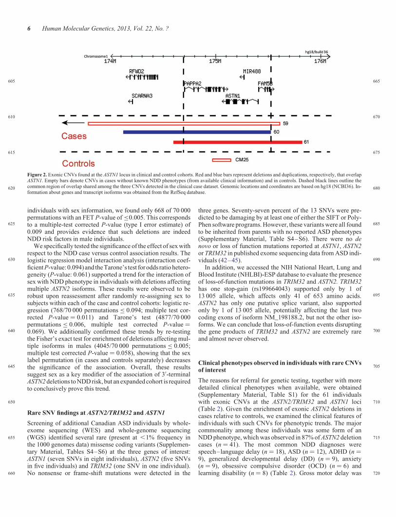
Figure 2. Exonic CNVs found at the ASTN1 locus in clinical and control cohorts. Red and blue bars represent deletions and duplications, respectively, that overlapASTN1. Empty bars denote CNVs in cases without known NDD phenotypes (from available clinical information) and in controls. Dashed black lines outline thecommon region of overlap shared among the three CNVs detected in the clinical case dataset. Genomic locations and coordinates are based on hg18 (NCBI36). In-formation about genes and transcript isoforms was obtained from the RefSeq database.
6 Human Molecular Genetics, 2013, Vol. 22, No. ?
605
610
615
620
625
630
635
640
645
650
655
660
665
670
675
680
685
690
695
700
705
710
715
720

Table 2. Genetic and clinical information for individuals with rare CNVs of interest
Case no. Sex Sitea CNV coordinates (hg18) CNV size CNV NDD phenotypesb
1 M TCAG chr9:117 954 428–118 356 243 401 816 Loss ADHD, Anx, LD, SD2 M OUH chr9:118,055 333–118 646 904 591 572 Loss DD, MD3 M ACH chr9:118 069 649–119 679 670 1 610 022 Loss Chiari I malformation4 M MC chr9:118 130 121–119 029 857 899 737 Loss Hydrocephalus, Mac5 M HSC chr9:118 164 272–118 358 705 194 434 Loss ADHD, ASD, DD, LD, Mic, MD, OCD, SD6 M SG chr9:118 291 060–118 661 674 370 615 Loss Anx, ASD, LD, Mac, MD, OCD, SD7 M SG chr9:118 327 395–118 595 433 268 039 Loss ASD, DD8 M CVH chr9:118 342 936–118 685 436 342 501 Loss DD, seizures9 M BCH chr9:118 358 646–118 459 563 100 918 Loss Anx, DD, Mac10 M ITA chr9:118 358 837–118 728 270 369 434 Loss ASD, SD11 M HSC chr9:118 390 436–118 524 432 133 997 Loss DD, ID, LD, MD, SD12 M SG chr9:118 395 767–118 520 501 124 735 Loss ADHD, SD, CNS disorder13 M SG chr9:118 395 767–118 595 433 199 667 Loss Hydrocephalus14 M TCAG chr9:118 407 129–118 523 510 116 382 Loss Anx, ASD, SD15 M SG chr9:118 421 170–118 683 092 261 923 Loss Behavioral problems16 M SG chr9:118 430 585–118 569 556 138 972 Loss DD, MD, seizures, SD17 M SG chr9:118 430 585–118 610 907 180 323 Loss DD, MD, SD18 M DEC chr9:118 440 935–118 584 415 143 481 Loss ID, Mac19 M MC chr9:118 459 294–118 616 407 157 114 Loss Chronic static encephalopathy20 M BCH chr9:118 469 713–118 524 312 54 600 Loss DD, LD, tics21 M TCAG chr9:118 479 893–118 627 637 147 745 Loss ADHD, Anx, OCD, tics22 M TCAG chr9:118 480 042–118 570 447 90 406 Loss ADHD, ASD, Mac, OCD, SD, seizures23 M TCAG chr9:118 481 308–118 654 031 172 724 Loss Anx, OCD24 M BCH chr9:118 488 204–118 558 274 70 071 Loss ASD, seizures25 M BCH chr9:118 488 204–118 700 657 212 454 Loss –
chr9:118 814 591–118 867 559 52 969 Gain26 M TCAG chr9:118 493 276–118 670 608 177 333 Loss ADHD, Anx, LD27 M BBG chr9:118 497 759–118 661 673 163 915 Loss –28 M MC chr9:118 502 294–118 616 407 114 114 Loss ADHD, Anx, ASD, Mac, SD29 M SG chr9:118 530 143–118 569 556 39 414 Loss Mic30 M MC chr9:118 541 180–118 685 465 144 286 Loss Dizziness, dysgraphia, migraines31 M BCH chr9:118 572 937–118 637 250 64 314 Loss DCD, DD, MD, SD32 M ITA chr9:118 580 317–118 814 591 234 275 Loss ASD, structural brain anomaly33 M SG chr9:118 580 891–118 630 518 49 628 Loss DD34 M HSC chr9:118 616 347–118 907 058 290 712 Loss Anx, ASD, hydrocephalus, LD, Mac, MD, structural brain anomaly35 M SG chr9:118 620 063–118 781 101 161 039 Loss ASD36 M BCH chr9:118 743 266–118 991 977 248 712 Loss ASD, DD37 M ITA chr9:118 840 027–118 935 027 95 001 Loss Behavioral problems, ID, OCD, SD38 M SG chr9:118 874 947–119 109 618 234 672 Loss –39 F SG chr9:118 199 243–118 248 950 49 708 Loss ADHD, DD, Mic, SD40 F BBG chr9:118 202 805–118 227 359 24 555 Loss MD, plagiocephaly, SD41 F HSC chr9:118 202 811–118 459 563 256 753 Loss Mac42 F HSC chr9:118 390 436–118 524 432 133 997 Loss ID, MD, SD, seizures43 F SG chr9:118 481 080–118 610 907 129 828 Loss –44 F BBG chr9:118 497 759–118 661 673 163 915 Loss –45 F OUH chr9:118 608 198–118 669 889 61 692 Loss ADHD, MD, SD46 F MC chr9:118 728 240–118 992 036 263 797 Loss –47 F MC chr9:118 829 818–118 890 615 60 798 Loss Septo-optic dysplasia48 M TCAG chr9:118 263 609–118 308 641 45 033 Gain Anx, ASD, MD, seizures49 M BCH chr9:118 899 840–121 893 169 2 993 330 Gain ADHD, ASD, DD, LD50 M ITA chr9:118 916 018–119 875 655 959 638 Gain ASD, ID, LD, Mic, MD, SD51 M ITA chr9:118 934 968–119 903 304 968 337 Gain Anx, ID, LD, MD, SD52 M MC chr9:118 934 968–121 071 351 2 136 384 Gain Mac53 M SG chr9:118 982 145–121 054 922 2 072 778 Gain Encephalocele54 M SG chr9:118 982 145–121 054 922 2 072 778 Gain LD55 M SG chr9:118 991 976–119 281 150 289 175 Gain –56 M ITA chr9:119 042 541–119 297 292 254 752 Gain ADHD, ID, LD, MD, Mic, SD57 M SG chr9:119 115 694–119 500 725 385 032 Gain –58 F CVH chr9:118 899 870–120 282 623 1 382 754 Gain Craniosynostosis, ID59 F BBG chr1:169 964 282–175 595 424 5 631 143 Loss –60 M BBG chr1:174 117 247–175 518 085 1 400 839 Gain Anx, ASD, LD, Mac, MD61 M BCH chr1:174 591 306–175 817 067 1 225 762 Loss Agenesis of corpus callosum, DD, Mic, seizures
aMolecular diagnostic testing site of origin of the case: ACH, Alberta Children’s Hospital; BBG, Brain and Body Genetic Resource Exchange (BBGRE); BCH, BostonChildren’s Hospital; CVH, Credit Valley Hospital; DEC, DECIPHER database; HSC, The Hospital for Sick Children; ITA, Italian diagnostic labs; MC, Mayo Clinic; OUH,Odense University Hospital; SG, Signature Genomics; TCAG, The Centre for Applied Genomics.bNDD trait abbreviations: ADHD, attention deficit hyperactivity disorder; Anx, anxiety; ASD, autism spectrum disorder; CNS, central nervous system; DCD, developmentalcoordination disorder; DD, developmental delay; ID, intellectual disability; LD, learning disability; Mac; macrocephaly; MD, motor delay; Mic; microcephaly; NDD;neurodevelopmental disorder; OCD, obsessive compulsive disorder; SD, speech delay.‘–’ indicates non-NDD cases (no NDD terms were present in their reasons for referral forgenetic testing).
Human Molecular Genetics, 2013, Vol. 22, No. ? 7
725
730
735
740
745
750
755
760
765
770
775
780
785
790
795
800
805
810
815
820
825
830
835
840

present in 12 cases and fine motor delay in 6 cases. In addition, awide range of dysmorphic features were present in ASTN2 dele-tion carriers, although macrocephaly was the only feature foundto be common in more than 10% of the cases (n ¼ 7). Phenotypicinformation was available for 12 (8 mothers and 4 fathers) of the17 parents with exonic ASTN2 deletions. Seventy-five percent ofthe paternal carriers and 50% of the maternal carriers reportedsome form of neurodevelopmental trait (primarily anxiety, de-pression, learning disabilities, dyslexia and in some casesformal adult diagnoses of ASD or ADHD), though usuallymilder than those seen in the probands.
Alternative transcription start site of ASTN2 shorterisoforms is of recent evolutionary origin
Given the location-dependent penetrance observed for theASTN2 exonic deletions, we assessed the average nucleotideconservation of all exons in ASTN2 (Fig. 3A) and TRIM32across vertebrates, placental mammals and primates (Fig. 3B).Exons with coding sequence were well-conserved across all ver-tebrate species. Interestingly, the conservation was much lowerbetween humans and other vertebrates for the exons unique to theshorter isoforms (exons 1B and 5C in Fig. 3A). Even though thispattern was partially driven by the UTR regions of these uniqueexons, their conservation was also lower than the conservation inthe UTRs present in the long isoform or in TRIM32 (Supplemen-tary Material, Fig. S4). Additionally, we did not find any evi-dence from EST databases for the presence of shorter3′-terminal Astn2 isoforms in mouse. These observationssuggest that the unique exons of the shorter isoforms (and the al-ternative transcription site in exon 1B) are of more recent evolu-tionary origin and are potentially derived in or just prior to theprimate lineage. In support of this claim, both exons 1B and5C contain transposable elements. Recent research has revealedthe ability of transposable elements to contribute novel regula-tory elements and thus give rise to new transcript isoforms (46).
Although there was no evident difference in the average nu-cleotide conservation between the coding exons of ASTN2, theamino acid alignment of eight ASTN2 protein orthologsrevealed that the C-terminal end exhibits a greater degree of con-servation compared with the rest of the protein (SupplementaryMaterial, Fig. S5 and S6). This section of the protein correspondsto the 3′-terminal region of the gene exhibiting the strongest en-richment of exonic deletions in male NDD cases (Fig. 1) andencodes the fibronectin type III domain (Supplementary Mater-ial, Fig. S6).
Expression of ASTN2 transcript isoforms in the human brain
The expression profile of ASTN2 and its different isoforms in thehuman brain has not been described previously. Therefore, weperformed reverse transcriptase–PCR (RT–PCR) analysis ofthe ASTN2 isoforms in human brain samples using primerswith locations as depicted in Figure 3A. We detected abundantexpression for primers amplifying exons present in all isoforms(Fig. 3C, ASTN2 all) and for exons specific to the long ASTN2isoform (NM_014010) (Fig. 3C, ASTN2 long isoform) in allthe brain regions. The shorter transcripts (NM_198187,NM_198188, NM_001184734 and NM_001184735) wereexpressed at a lower level (Fig. 3C, ASTN2 shorter isoforms).T
ab
le3.
Res
ult
sof
CN
Ven
rich
men
tan
alysi
sof
AS
TN
2/T
RIM
32
locu
s
CN
Vty
pe
Tota
lN
DD
dat
aset
Mal
eN
DD
Fem
ale
ND
DT
ota
lnon-N
DD
dat
aset
ND
Dca
ses
(N¼
64
114)
Contr
ols
(N¼
44
085)
P-v
aluea
ND
Dm
ales
(N¼
40
438)
Mal
eco
ntr
ols
(N¼
14
953)b
P-v
aluea
ND
Dfe
mal
es(N¼
23
676)
Fem
ale
contr
ols
(N¼
18
218)b
P-v
aluea
Non-N
DD
case
s(N¼
25
871)
Contr
ols
(N¼
44
085)
P-v
aluea
Exonic
AST
N2
CN
Vs
49
23
0.0
79
42
13
0.3
49
77
0.7
78
823
0.9
33
Exonic
AST
N2
gai
ns
95
0.4
63
83
0.6
57
11
0.8
11
35
0.6
19
Exonic
AST
N2
loss
es40
18
0.0
84
34
10
0.3
28
66
0.7
73
618
0.9
27
Exonic
loss
esaf
fect
ing
only
long
AST
N2
isofo
rm
13
13
0.8
76
11
90.9
76
23
0.8
83
413
0.9
24
Exonic
loss
esaf
fect
ing
mult
iple
AST
N2
isofo
rms
27
50.0
02
23
10.0
05
43
0.6
42
50.7
98
Exonic
loss
esaf
fect
ing
TR
IM32
c23
60.0
19
22
20.0
24
13
0.9
64
46
0.5
4
ND
D,neu
rodev
elopm
enta
ldis
ord
ers.
aP
-val
ues
are
from
one-
sided
Fis
her
’sex
act
test
.V
alues
inbold
are
signifi
cant
atth
resh
old
of
P,
0.0
5.
bG
ender
info
rmat
ion
was
avai
lable
for
33
171
contr
ol
indiv
idual
s(S
upple
men
tary
Mat
eria
l,T
able
S2),
and
this
subse
tw
asuse
dfo
rth
ese
x-s
pec
ific
enri
chm
ent
anal
ysi
s.cA
lldel
etio
ns
whic
haf
fect
edT
RIM
32
inth
eca
ses
also
over
lapped
exon(s
)of
AST
N2.O
ne
of
the
exonic
TR
IM32
del
etio
ns
inth
eco
ntr
ols
(CF
14)
did
not
over
lap
anA
ST
N2
exon.
8 Human Molecular Genetics, 2013, Vol. 22, No. ?
845
850
855
860
865
870
875
880
885
890
895
900
905
910
915
920
925
930
935
940
945
950
955
960

We did not detect any expression when using primers specific forNM_198186. In addition, we measured the mRNA expressionratios of the isoforms in whole brain and fetal brain and observeda change in the ratios between the developmental stages. In thefetal brain, the shorter isoforms accounted for �40% of thetotal ASTN2 expression, which decreased to 20% in the adultbrain (Fig. 3D). This suggests a functional role for the shorter iso-forms during early development.
Spatiotemporal expression patterns of ASTN1, ASTN2and TRIM32 in human brain
We analyzed the pattern of ASTN2, ASTN1 and TRIM32 genelevel expression in different brain regions during human
development by utilizing the comprehensive gene expressiondata from the BrainSpan database (47). Overall, ASTN2 isexpressed at a moderate level throughout development and exhi-bits an increase in the late prenatal period and during postnataldevelopment in many of the brain regions (Fig. 4A). Thehighest level of ASTN2 expression was observed in the cerebellarcortex (CBC), where the expression increase during infancy andearly childhood is in concordance with the expression patternpreviously reported in mice (30). Genes exhibiting similar ex-pression patterns in the CBC (as quantified by Pearson correl-ation, Supplementary Material, Table S7) were more enrichedin gene ontology (GO) terms such as synaptic transmission andplasticity (Supplementary Material, Fig. S7A and Table S7).Interestingly, during the prenatal period, ASTN2 has a dynamic
Figure 3. Relative expression of ASTN2 transcript isoforms and exon conservation. (A) Schematic presentation of the ASTN2 transcript isoforms. Red arrows denotelocation of the primers used for the RT–PCR and/or qRT–PCR assays (Supplementary Material, Table S10). The ‘∗’ symbol denotes exons with variable length indifferent isoforms. (B) Conservation profile of ASTN2and TRIM32 exons across vertebrates, placental mammals and primates. (C) The expression of ASTN2 transcriptisoforms (long, shorter and all) in eight different human brain regions. ACTB was used as a control gene. (D) Quantification of ASTN2 isoform expression levels intriplicate from adult brain and fetal brain RNA samples by qRT–PCR (standard curve method). The expression was normalized using ACTB as a housekeeping gene,and the expression ratio is relative to the expression from all ASTN2 isoforms (mean ratio+ standard deviation). The results were replicated using GAPDH as a house-keeping gene.
Human Molecular Genetics, 2013, Vol. 22, No. ? 9
965
970
975
980
985
990
995
1000
1005
1010
1015
1020
1025
1030
1035
1040
1045
1050
1055
1060
1065
1070
1075
1080

spike in the expression �12–13 gestational weeks in the neocor-tical regions (frontal cortex, parietal cortex and occipital cortex)(Fig. 4A). This expression pattern is enriched in neuronal devel-opment GO terms such as ‘axonogenesis’ and ‘neuron differen-tiation’ (Supplementary Material, Fig. S7B and Table S7). Thedynamic expression pattern of ASTN2 together with the biologic-al annotation of genes with similar expression patterns suggeststhat ASTN2 could have an important role in both prenatal andpostnatal brain development.
In contrast to ASTN2, ASTN1 has a high and steady level of ex-pression across different brain regions suggesting a constant andfundamental role in human brain development (Fig. 4B). The ex-pression pattern of TRIM32 differs from the astrotactins and ismarked by high expression during early prenatal development,
a decrease after birth followed by an increase during adolescencein all the brain regions studied (Supplementary Material,Fig. S8). Similar to ASTN2, TRIM32 has highest levels of expres-sion in the CBC.
DISCUSSION
By leveraging information from multiple diagnostic laboratoriesto aid in the clinical interpretation of rare variants, we detectedspecific enrichment of exonic ASTN2/TRIM32 deletions inNDD cases compared with either non-NDD cases or with con-trols. Follow-up clinical characterization revealed a spectrumof NDD diagnoses, with ASD, ADHD, OCD and speech and
Figure 4. Expression profiles of ASTN2 and ASTN1 across human brain development. The gene level expression profiles of (A) ASTN2 and (B) ASTN1 across devel-opmental time points in nine regions of the human brain; amygdala (AMY), cerebellar cortex (CBC), diencephalon (DIE), frontal cortex (FC), hippocampus (HIP),occipital cortex (OC), parietal cortex (PC), temporal cortex (TC) and ventral forebrain (VF).
10 Human Molecular Genetics, 2013, Vol. 22, No. ?
1085
1090
1095
1100
1105
1110
1115
1120
1125
1130
1135
1140
1145
1150
1155
1160
1165
1170
1175
1180
1185
1190
1195
1200

language delay being the most common. A similarly diverserange of phenotypic outcomes from deletions at a single locushave been reported before for NRXN1 (48–50), GPHN (51),MBD5 (52,53), and other regions (19,54), which could reflectsome shared genetic risk for different NDDs, consistent withoverlap of clinical phenotypes often observed across diagnosticboundaries in psychiatry (55–57). In addition to the heterogen-eity of their clinical presentation, ASTN2/TRIM32 deletionsexhibit reduced penetrance, as demonstrated by their predomin-antly inherited nature and their presence in a small number ofcontrol individuals. SNPs within ASTN2 have been highlightedby recent genome-wide association studies (GWAS) in risk forADHD (58), schizophrenia (59,60), bipolar disorder (60), mi-graine without aura (61), cognitive decline and reduced hippo-campal volume (62). These reports of common risk factors atthis gene complement our rare variant findings and lendsupport to the etiological contributions of inherited ASTN2 var-iants to a range of neurodevelopmental conditions. While severalof the NDD risk gene discovery efforts to date have focused on denovo events, the role of inherited rare variants has been less thor-oughly explored. It is likely that a combination of both de novoand inherited risk factors contribute to the genetic architectureof different NDDs. The presence of deletions in controls mightalso reflect the absence of stringent psychiatric screening ofthe control individuals or could arise from false-positive calls,since experimental validation was not possible due to unavail-ability of DNA from control individuals. Importantly, we notethat the relatively lower resolution of the clinical microarrayplatforms compared with those used for the controls (Supple-mentary Material, Fig. S9) could imply an underestimation ofsmaller CNVs at ASTN2, ASTN1 and TRIM32 in the patientcohorts.
The striking enrichment in the NDD cases compared with thecontrols, of exonic deletions at the 3′ end of ASTN2 (correspond-ing to multiple ASTN2 isoforms), defines a critical region forpathogenicity that has several functional implications. As sug-gested by our lymphoblast expression analysis, such deletionscould disrupt the expression of all transcript isoforms ofASTN2 and potentially lead to complete haploinsufficiency ofthe protein. This may have more severe phenotypic conse-quences than those deletions affecting only the long transcriptisoform. Similar trends of greater penetrance of deletionsimpacting multiple isoforms of a gene, relative to those overlap-ping a single isoform, have recently been observed at other locisuch as NRXN1 (49,50,63), NRXN3 (5) and AUTS2 (64) in con-nection with risk for NDDs. Our findings also highlight the im-portance of the C-terminal end of the protein, which encodesthe fibronectin III domain and was observed to be the most con-served part of the protein in our cross-species analysis. Interest-ingly, this domain is also found in other genes implicated inNDDs such as the contactins (13). In addition, our data suggestthat the shorter transcript isoforms are of functional importanceespecially during early brain development. This claim is sup-ported by our mRNA quantification assays in which the shorterisoforms account for 40% of the total expression of the gene infetal brain. Our evolutionary conservation analyses suggestthat the first exon shared by the shorter ASTN2 isoforms is ofmore recent evolutionary origin and could have a function spe-cific to primates. There is no indication from previous functionalwork (30) or from mouse EST databases for existence of shorter
3′-terminal Astn2 transcript isoforms in the mouse. Taken to-gether, the recent emergence of the shorter isoforms comprisinga functionally important domain and their widespread expres-sion in the human brain could indicate primate-specific involve-ment in neurodevelopment.
Several of the deletions at the 3′ end of ASTN2 also encompassTRIM32, a small two-exon gene nested within an intron of thelong isoform of ASTN2, which is transcribed from the oppositestrand to ASTN2. The first exons of TRIM32 and the ASTN2shorter isoforms are only 39 bp apart. TRIM32 encodes an ubi-quitin ligase and is expressed at high levels in a wide range oftissues, including the brain, muscle and skeletal tissue (65).Homozygous point mutations in this gene have been implicatedin autosomal recessive disorders such as limb-girdle musculardystrophy (66,67) and Bardet–Biedl syndrome (68). The latteris a heterogeneous multi-system disorder presenting with retin-opathy, obesity and cognitive impairment, among other symp-toms. TRIM32 expression levels in the mouse neocortex havebeen shown to determine the post-differentiation fates of neuron-al stem cells (69) and the TRIM32 protein is a key regulator ofneural differentiation (70). It is unlikely that disruption ofTRIM32 alone, and not ASTN2, could be primarily responsiblefor the observed neurodevelopmental phenotypes in our studygiven the absence of such diagnoses in individuals with hetero-zygous TRIM32 missense mutations (66,68) and in a limb-girdlemuscular dystrophy patient who possessed both a TRIM32 non-sense mutation and a deletion of the entire gene (67). The expres-sion of ASTN2 is also notably higher than that of TRIM32 in thebrain relative to other body tissues (33). Furthermore, we did notdetect any deletions in the large clinical dataset that overlappedTRIM32 alone but did observe several deletions at both the 3′ and5′ ends of ASTN2 that do not affect TRIM32. Given the very closegenomic proximity of TRIM32 and ASTN2, and the finding thatdeletions impacting both genes exhibit stronger enrichment incases versus controls than those affecting either gene alone,the most conservative interpretation of our data would suggestthat the joint disruption of both genes potentially contributes torisk for a spectrum of NDD phenotypes.
We observed evidence for a sex-specific effect for the pheno-typic expression of exonic deletions affecting ASTN2 or bothASTN2 and TRIM32 (Table 3). These mutations were significant-ly in excess in the males compared with the females within theNDD case cohort, whereas controls did not exhibit this differ-ence. Deletions affecting the 3′ end of ASTN2 and/or TRIM32were also significantly enriched in male NDD cases comparedwith male controls, whereas the difference in frequencies ofsuch events between female cases and female controls was non-significant. These findings suggest greater penetrance in malesrelative to females of the NDD phenotypes linked to ASTN2/TRIM32 deletions, although a larger cohort is required to conclu-sively prove the significance of this sex-specific effect and therole of such deletions in females. Similar sex-specific effectshave been recently reported for CNVs at other autosomal loci in-cluding SHANK1 deletions in ASD risk (7) and 16p13.11 dele-tions and duplications in NDD risk (71). The discovery ofadditional autosomal loci with male-biased penetrance (72), to-gether with risk genes on the X chromosome (6,73), could helpexplain the molecular basis of the skewed sex ratios observedin the prevalence of NDDs such as ADHD, ASD and ID. Al-though the biological basis of this sex-modulated penetrance is
Human Molecular Genetics, 2013, Vol. 22, No. ? 11
1205
1210
1215
1220
1225
1230
1235
1240
1245
1250
1255
1260
1265
1270
1275
1280
1285
1290
1295
1300
1305
1310
1315
1320

unknown, theories involving sex hormones have been proposed(74). Interestingly, there is some evidence that ASTN2 could beregulated by estrogen (75).
The extreme rarity of exonic deletions affecting the ASTN1gene is intriguing, especially in comparison with the relativelyhigher rate of such mutations at ASTN2. It is also striking thatall the ASTN1 deletions in the cases were of de novo origin andoverlapped multiple genes, in contrast to exonic ASTN2 dele-tions, which were predominantly of an inherited nature (only10% of such deletions in families with parental DNA availablefor testing were de novo) and localized to ASTN2/TRIM32.These observations suggest stronger selective pressure againstdisruption of ASTN1, relative to ASTN2, and are consistentwith results from functional characterization of the two proteinsin the mouse brain. Although mouse ASTN1 and ASTN2 areboth integral neuronal membrane proteins with similar domainstructures, they have been shown to play non-redundant andcomplementary roles during neuronal migration (30). TheC-terminus of mouse ASTN1 is exposed on the neuronalsurface, enabling it to act as the inter-cellular linker molecule dir-ectly binding neurons to glia during migration. On the otherhand, the C-terminus of ASTN2 was not detected on the neuronalsurface. Rather than directly participating in neuron-glial adhe-sion, ASTN2 reportedly functions as a regulatory molecule byforming a complex with ASTN1 in the neuronal membrane,thus controlling ASTN1 surface expression levels and indirectlymodulating the rate of neuronal migration. Presumably, the dis-ruption of the key ligand enabling cerebellar neuron-glialbinding would be more deleterious for neuronal migration rela-tive to that of an indirect regulator of the process.
The differing functions of the two proteins are also reflected inthe spatiotemporal mRNA expression profiles of ASTN1 andASTN2. Both astrotactin genes are expressed in the developinghuman brain with highest overall expression during late prenataldevelopment and early childhood. In contrast to ASTN1, whichhas a very static and high expression pattern during developmentacross different regions, ASTN2 exhibits a wider range of expres-sion levels suggesting region specific roles across differentphases of human brain development. These findings, taken to-gether with previous work (30), are in line with a more funda-mental, constant role for ASTN1 protein, regulated at specifictime points by varying ASTN2 protein levels.
Comparison of the human brain expression profile of ASTN2with previous work in mice revealed both intriguing differencesand similarities. The striking prenatal spike in ASTN2 expressionin the neocortical regions, towards the end of the first trimester,has not been reported in the embryonic mouse brain (30). Thisfinding might indicate transcriptional regulation patterns or add-itional functions of the protein and/or transcript isoforms specificto primates. Interestingly, this period in the developmental time-line of the human cerebral cortex is marked by extensive neuron-al migration, increasing axonal outgrowth and the formation ofthe early synapses (47,76). Several genes previously implicatedin risk for ASD (and also other NDDs) such as DOCK4, andNRCAM also exhibit increasing cortical expression around thistime point (15,77). In concordance with the experimental workfrom mouse (30), we observe the highest ASTN2 expression inthe cerebellar cortex shortly after birth. Several studies investi-gating the neuropathology of ASD (78,79) and ADHD (80–82) have consistently highlighted the cerebellum as a major
region of interest. Reported neuroanatomical abnormalitiesinclude disrupted neuronal migration in the cerebellar cortex(79) as well as reduction in volume of the cerebellar vermis(83), which has been linked to repetitive and stereotyped behav-ior in ASD (84). Furthermore, the number of Purkinje cells, oneof the main cell types in the cerebellum, has been found to bedecreased by up to 50% in individuals with ASD (79). Interest-ingly, the Astn1 KO mouse has reduced cerebellar volume,slower neuronal migration rates and abnormal development ofthe Purkinje cells compared with wild type (29). Astn2 has alsobeen shown to be very highly expressed in the cerebellum ingeneral, and in the Purkinje cells in particular, during both em-bryonic and postnatal development (30).
While the role of the astrotactins in neuronal migration is wellestablished, their potential involvement in other brain develop-mental processes remains to be elucidated. Our gene ontologyanalyses revealed that many of the genes with expression pat-terns similar to ASTN2 are involved in synapse-related biologicalprocesses. The presence of both astrotactins at high levels in thepostnatal and adult brain, well after the completion of most neur-onal migration, also suggests that ASTN2 and ASTN1 might po-tentially act together as a receptor system for synapse guidance inaddition to their role in neuronal migration. Interestingly, recentstudies show that genes important for embryonic neuronal mi-gration such as PAFAH1B1 (LIS1) and RELN also participatein guiding and maintaining the synapses in postnatal brain devel-opment (23,85). CNTNAP2, another ASD risk gene, encodes aneuronal trans-membrane protein with important roles indiverse processes including neuron-glia interactions during mi-gration, maintenance of the connectivity and synchronization ofneuronal circuits and the clustering of K+ channels in myelin-ated axons (86).
Our data emphasize the need to characterize rare CNV (andother genetic variants) in the context of large case and controlcohorts, in order to extract meaningful genotype and phenotypedata necessary for proper clinical genetic interpretation. For theASTN2/TRIM32 locus, we show that a neurodevelopmentalphenotype ensues preferentially in male patients when deletionsof ASTN2 impact all its transcript isoforms. Functional dissec-tion of the influences gender has on the ASTN2 isoformsduring brain development may also inform on new treatmentstrategies in psychopathology.
MATERIALS AND METHODS
Clinical case cohorts
The clinical case cohorts utilized for this study are summarized inTable 1. These comprise a total of 89 985 postnatal patientsamples submitted for clinical microarray testing to 10 differentmolecular diagnostic centers in Canada, Denmark, Italy, the UKand the USA. The reasons for referral for clinical microarraytesting were systematically assessed at each of the study sitesand the number of NDD cases was tabulated based on the presenceof one or more of the following phenotypes: ADHD, ASD, behav-ioral disorders, cognitive impairment, developmental delay, ID,learning disability, macrocephaly, microcephaly, neurologicaldisorders, OCD, psychoses, seizures and speech/language disor-ders. The remaining patients in the clinical cohorts, whosereasons for referral for genetic testing did not contain any of the
12 Human Molecular Genetics, 2013, Vol. 22, No. ?
1325
1330
1335
1340
1345
1350
1355
1360
1365
1370
1375
1380
1385
1390
1395
1400
1405
1410
1415
1420
1425
1430
1435
1440

NDD terms listed above, were counted as non-NDD cases. Thegender composition of each clinical dataset was also tabulatedin order to test for sex-biased effects. Individuals with CNVsthat spanned one or more exons of the ASTN2/TRIM32 (Fig. 1)or ASTN1 (Fig. 2) genetic loci at 9q33.1 and 1q25.2, respectively,were included in the analysis and are listed in Table 2. To avoidbias in the CNV burden analysis by inclusion of large-scale multi-genic chromosomal abnormalities, CNVs .6 Mb in the clinicaldataset were excluded. Independent validation was performedfor the 71% (44/62) of the CNVs presented in Table 2 for whichDNA was available, using one of the following methods: quanti-tative PCR (qPCR), multiplex ligation-dependent probe amplifi-cation (MLPA), fluorescence in situ hybridization (FISH) or bya second microarray platform (Supplementary Material,Table S1). All attempted assays revealed true positive CNVs.To obtain standardized clinical information from the individualswith the CNVs of interest, a phenotype checklist was sent to refer-ring physicians for completion, based on physical and/or psycho-logical examination, as well as review of the subject’s medicalhistory. The frequencies of specific features were tabulated (Sup-plementary Material, Table S1). In addition to the clinical cohortsdescribed above, we inspected the DECIPHER database (https://decipher.sanger.ac.uk) for individuals with exonic deletionsand duplications, smaller than 6 Mb, that overlapped ASTN2(Supplementary Material, Table S8). We were able to obtain add-itional phenotype details from one individual (patient 18), whopossessed an exonic ASTN2/TRIM32 deletion. Clinical informa-tion from this individual was included in the phenotypesummary but this case was not counted in the CNV counts and en-richment analysis described in the Results since it was not part ofdata from the 10 molecular diagnostic sites. This study wasapproved by the Research Ethics Board at the Hospital for SickChildren, Toronto.
Control cohorts
For the purpose of statistical testing of findings in the casecohorts, we compiled exonic CNV findings at the ASTN2/TRIM32 and ASTN1 regions in high resolution microarray datafrom 44 085 individuals from population-based control cohortsand studies of individuals with non-NDDs such as diabetes (Sup-plementary Material, Table S2). These included individualsfrom different control datasets analyzed by us (14,31,51,87–90), published genome-wide CNV data (91,92) and publisheddata from controls that were inspected for CNVs affectingASTN2 (33,34). Gender information was available for 33 171control individuals (Supplementary Material, Table S2), andthis subset was used for the sex-specific enrichment analysis.Fisher’s one-sided exact test was used to test for enrichment ofCNVs in cases versus non-NDD cases and versus controls witha significance threshold of P , 0.05. A challenge in combiningdata across multiple case and control cohorts is the heterogeneityof microarray platforms and the resulting differences in probecoverage. We compiled probe numbers from the different micro-array platforms featured among the clinical and control datasetsin the ASTN2/TRIM32 and ASTN1 regions (Supplementary Ma-terial, Table S9). The array platforms used for the control datasethad much higher probe densities on average, both genome wideand in the specific regions of interest, than those constituting theclinical dataset (Supplementary Material, Table S9 and Fig. S9).
The higher resolution of the control CNV dataset relative to thecases decreases the likelihood of spurious enrichment findingsdriven by false negatives in the controls and provides a conserva-tive estimate of the significance and effect size of our findings.
Mutation screening of ASTN1, ASTN2 and TRIM32 fromASD exome sequencing data
Exons and splice sites of ASTN1, ASTN2 and TRIM32 wereinspected for single-nucleotide changes and small indels in next-generation WES and WGS data from Canadian individuals (ofEuropean ancestry) with ASD (Supplementary Material,Tables S4–S6). ASTN1 was screened in 338 individuals, whileASTN2 and TRIM32 were inspected in 182 individuals. The sub-jects screened for mutations in ASTN2 and TRIM32 by WES andWGS were distinct from those in whom the two genes wereinspected using Sanger sequencing in our previous study (31).Data generation and SNV analysis were performed as previouslydescribed for the WES (51) and WGS (93) data. Rare SNVsdetected in the WES and WGS data that were present at ,1%frequency in the 1000 genomes data (94) were confirmed by bi-directional Sanger sequencing. Inheritance testing of such var-iants was also conducted when parental DNA was available.We also examined the published results of four recent ASDexome sequencing studies (42–45) for variants at ASTN1,ASTN2 and TRIM32. In addition, we accessed the NHLBIexome sequencing database to evaluate the presence ofloss-of-function mutations in TRIM32 and ASTN2.
ASTN2 expression analysis using RT–PCR and quantitativereverse transcriptase–PCR
The expression of ASTN2 mRNA was analyzed by RT–PCR froma panel of eight different human brain RNA samples (adult wholebrain, adult cerebellum, adult caudate nucleus, adult amygdala,adult hippocampus, adult corpus callosum, adult thalamus andfetal whole brain purchased from BD Biosciences, Clontech andAMSBIO). In addition, expression was measured in lymphoblastcell lines from six individuals with ASTN2 deletions and nine indi-viduals with two copies of ASTN2 (detailed description in Supple-mentary Material, Fig. S3). For all the RNA samples, cDNA wassynthesized using Superscript III First strand Synthesis Supermix(Invitrogen, Carlsbad, CA, USA) with 1 mg of poly (A+) or totalDNase I treated RNA as a template. RT–PCR was performedunder standard PCR conditions using 10 ng of cDNA as template.Eight primer pairs were used to amplify different transcripts ofASTN2 (Supplementary Material, Table S10). In total, ASTN2has six different isoforms which including the long isoform(NM_014010) and shorter isoforms (NM_198186, NM_198187,NM_198188, NM_001184734 and NM_001184735). To quantifythe expression ratio between the transcript isoforms, we used fivesuitable amplicons for quantitative reverse transcriptase–PCR(qRT–PCR) (Supplementary Material, Table S10). The qRT–PCR assay was performed using Brilliant III SYBRw GreenPCR Master Mix (Agilent, Santa Clara, CA, USA) in a total reac-tion volume of 15 ml, containing 5 ng of cDNA templates. Thereactions were amplified using the Mx3005P qPCR system(Agilent, Santa Clara, CA, USA). The expression ratios were cal-culated after determining the exact quantities of each isoform
Human Molecular Genetics, 2013, Vol. 22, No. ? 13
1445
1450
1455
1460
1465
1470
1475
1480
1485
1490
1495
1500
1505
1510
1515
1520
1525
1530
1535
1540
1545
1550
1555
1560

using the standard curve method, normalized against ACTB orGAPDH expression levels.
Nucleotide and amino acid conservation analysis of ASTN2
The nucleotide conservation scores for each base present in allASTN2 and TRIM32 exons were computed using the PhyloPprogram based on alignment between 46 vertebrate species in-cluding 23 placental mammals and eight primates. Theaverage score was calculated for each exon and for the uniqueUTRs. The amino acid sequences of 1:1 ASTN2 orthologsfrom eight species were downloaded from Ensembl, and the mul-tiple sequence alignment was carried out using ClustalW2. Afterthe alignment, the amino acid conservation was quantified usingScorecons server (95). To check for the presence of the shorterASTN2 isoforms in mouse, we screened different databases in-cluding AceView, Ensembl and FANTOM.
Spatiotemporal expression analysis of ASTN2, ASTN1 andTRIM32 in the human brain
To analyze the expression pattern of ASTN2, ASTN1 and TRIM32during human brain development, we utilized expression datafrom the BrainSpan database (www.brainspan.org) (47). Thisdataset contains extensive transcriptome profiles for 16 brainregions from 41 individuals. The age range of the subjects spansfrom 8 postconception weeks to 40 years (56–13 720 postconcep-tion days). Full sample information is available on the BrainSpanwebsite. The data were quantile normalized and the gene level ex-pression was averaged across donors for each time point in the fol-lowing regions: the frontal cortex (FC), parietal cortex (PC),temporal cortex (TC), occipital cortex (OC), hippocampus(HIP), amygdala (AMY), ventral forebrain (VF), diencephalon(DIE) and cerebellar cortex (CBC). The expression levels ofASTN1, ASTN2 and TRIM32 in the different brain regions wereplotted across age range using the R package ggplot2. Smoothcurve lines were computed by loess using a span of 0.4. The Brain-Span dataset was also used to identify genes with expression pat-terns similar to that of ASTN2, and these genes were used for thegene set enrichment analysis (90,96) (Supplementary Material,Table S7).
SUPPLEMENTARY MATERIAL
Supplementary Material is available at HMG online.
ACKNOWLEDGEMENTS
We thank the patients and their families, clinicians and diagnos-tic lab personnel who participated in this study. We also thank DrMaria Tropeano, the CHOP CNV team and members of Dr EvanEichler’s group for sharing gender information for the controldatasets.
Conflict of Interest statement. J.A.R. is an employee of SignatureGenomics Laboratories, a subsidiary of Perkin Elmer Inc. E.H.and P.S.E. are employees of Population Diagnostics, Inc.S.W.S. is on the Scientific Advisory Board of Population
Diagnostics, Inc and is a founding scientist of YouNique Gen-omics, both of which could use data from this study.
FUNDING
This work was supported by grants from the University of TorontoMcLaughlin Centre, NeuroDevNet, Genome Canada and theOntario Genomics Institute, the Canadian Institutes for Health Re-search (CIHR), the Canadian Institute for Advanced Research, theCanada Foundation for Innovation, the Government of Ontario,Autism Speaks and The Hospital for Sick Children Foundation.A.C.L. was supported by a NeuroDevNet doctoral fellowship.K.T. holds a postdoctoral fellowship from the Swedish ResearchCouncil. S.W.S. holds the GlaxoSmithKline-CIHR Chair inGenome Sciences at the University of Toronto and The Hospitalfor Sick Children. D.T., D.S.C and T.M.W. are US militaryservicemembersand thisworkwaspreparedaspartof theirofficialduties. The views expressed in this article are those of the authorsand do not necessarily reflect the official policy or position of theDepartment of the Army, Department of the Navy, Department ofDefense, nor the US Government. Title 17, USC § 105 providesthat ‘Copyright protection under this title is not available for anywork of the U.S. Government.’ Title 17, USC § 101 defines aUS Government work as a work prepared by a military servicemember or employee of the US Government as part of thatperson’s official duties. Control datasets were obtained, alongwith permission for use, from the database of Genotypes and Phe-notypes (dbGaP) found at http://www.ncbi.nlm.nih.gov/gapthrough accession numbers phs000143.v1.p1 (Starr CountyHealth Studies’ Genetics of Diabetes Study), phs000091.v2.p1(GENEVA NHS/HPFS Diabetes study), phs000169.v1.p1(Whole Genome Association Study of Visceral Adiposity in theHABC Study), phs000303.v1.p1 (Genetic Epidemiology of Re-fractive Error in the KORA Study), phs000404.v1.p1(COGEND; The Genetic Architecture of Smoking and SmokingCessation) and phs000086.v2.p1 (DCCT-EDIC Clinical Trialand Follow-up of Persons with Type 1 Diabetes). The StarrCounty Health Studies Genetics of Diabetes Study was supportedby the National Institute of Diabetes and Digestive and KidneyDiseases (NIDDK) and the NIDDK Central Repositories.Support for the GWAS of Gene and Environment Initiatives inType 2 Diabetes was provided through the NIH Genes, Environ-ment and Health Initiative [GEI] (U01HG004399). The humansubjects participating in the GWAS derive from The Nurses’Health Study and Health Professionals’ Follow-up Study andthese studies are supported by National Institutes of Health(NIH) grants CA87969, CA55075 and DK58845. Assistancewith phenotype harmonization and genotype cleaning, as well aswith general study coordination, was provided by the Gene Envir-onmentAssociationStudies,GENEVACoordinatingCenter (U01HG004446) and the National Center for Biotechnology Informa-tion. Support for genotyping, which was performed at the BroadInstitute of MIT and Harvard, was provided by the NIH GEI(U01HG004424). Support for the ‘CIDR Visceral AdiposityStudy’ was provided through the Division of Aging Biology andthe Division of Geriatrics and Clinical Gerontology, National In-stitute on Aging. Assistance with phenotype harmonization andgenotype cleaning, as well as with general study coordination,was provided by Health ABC Study (HABC) Investigators. The
14 Human Molecular Genetics, 2013, Vol. 22, No. ?
1565
1570
1575
1580
1585
1590
1595
1600
1605
1610
1615
1620
1625
1630
1635
1640
1645
1650
1655
1660
1665
1670
1675
1680

KORA dataset was obtained from the NEI Refractive Error Col-laboration (NEIREC) Database, support for which was providedby the National Eye Institute. Support for genotyping of theCOGEND samples, which was performed at the Center for Inher-ited Disease Research (CIDR), was provided by 1 X01HG005274-01. Assistance with genotype cleaning of theCOGEND samples, as well as with general study coordination,was provided by the Gene Environment Association Studies(GENEVA) Coordinating Center (U01HG004446). Support forthe collection of COGEND datasets and samples was providedby the Collaborative Genetic Study of Nicotine Dependence(COGEND; P01 CA089392) and the University of WisconsinTransdisciplinary Tobacco Use Research Center (P50DA019706, P50 CA084724). The DCCT-EDIC Research Groupis sponsored through research contracts from the National Instituteof Diabetes, Endocrinology and Metabolic Diseases of theNIDDK and the NIH. The contents of this article are solely the re-sponsibility of the authors and do not necessarily represent the of-ficial views of the NIDDK or the NIH.
REFERENCES
1. Scherer, S.W. and Dawson, G. (2011) Risk factors for autism: translatinggenomic discoveries into diagnostics. Hum. Genet., 130, 123–148.
2. Devlin, B. and Scherer, S.W. (2012) Genetic architecture in autism spectrumdisorder. Curr. Opin. Genet. Dev., 22, 229–237.
3. Szatmari, P., Paterson, A.D., Zwaigenbaum, L., Roberts, W., Brian, J., Liu,X.Q., Vincent, J.B., Skaug, J.L., Thompson, A.P., Senman, L. et al. (2007)Mapping autism risk loci using genetic linkage and chromosomalrearrangements. Nat. Genet., 39, 319–328.
4. Gauthier, J., Siddiqui, T., Huashan, P., Yokomaku, D., Hamdan, F.,Champagne, N., Lapointe, M., Spiegelman, D., Noreau, A., Lafreniere, R.et al. (2011) Truncating mutations in NRXN2 and NRXN1 in autismspectrum disorders and schizophrenia. Hum. Genet., 130, 563–573.
5. Vaags, A.K., Lionel, A.C., Sato, D., Goodenberger, M., Stein, Q.P., Curran,S., Ogilvie, C., Ahn, J.W., Drmic, I., Senman, L. et al. (2012) Rare deletionsat the neurexin 3 locus in autism spectrum disorder. Am. J. Hum. Genet., 90,133–141.
6. Jamain, S., Quach, H., Betancur, C., Rastam, M., Colineaux, C., Gillberg,I.C., Soderstrom, H., Giros, B., Leboyer, M., Gillberg, C. et al. (2003)Mutations of the X-linked genes encoding neuroligins NLGN3 and NLGN4are associated with autism. Nat. Genet., 34, 27–29.
7. Sato, D., Lionel, A.C., Leblond, C.S., Prasad, A., Pinto, D., Walker, S.,O’Connor, I., Russell, C., Drmic, I.E., Hamdan, F.F. et al. (2012) SHANK1deletions in males with autism spectrum disorder. Am. J. Hum. Genet., 90,879–887.
8. Berkel, S., Marshall, C.R., Weiss, B., Howe, J., Roeth, R., Moog, U., Endris,V., Roberts, W., Szatmari, P., Pinto, D. et al. (2010) Mutations in theSHANK2 synaptic scaffolding gene in autism spectrum disorder and mentalretardation. Nat. Genet., 42, 489–491.
9. Durand, C.M., Betancur, C., Boeckers, T.M., Bockmann, J., Chaste, P.,Fauchereau, F., Nygren, G., Rastam, M., Gillberg, I.C., Anckarsater, H. et al.(2007) Mutations in the gene encoding the synaptic scaffolding proteinSHANK3 are associated with autism spectrum disorders. Nat. Genet., 39,25–27.
10. Moessner, R., Marshall, C.R., Sutcliffe, J.S., Skaug, J., Pinto, D., Vincent, J.,Zwaigenbaum, L., Fernandez, B., Roberts, W., Szatmari, P. et al. (2007)Contribution of SHANK3 mutations to autism spectrum disorder.Am. J. Hum. Genet., 81, 1289–1297.
11. Roohi, J., Montagna, C., Tegay, D.H., Palmer, L.E., DeVincent, C.,Pomeroy, J.C., Christian, S.L., Nowak, N. and Hatchwell, E. (2009)Disruption of contactin 4 in three subjects with autism spectrum disorder.J. Med. Genet., 46, 176–182.
12. van Daalen, E., Kemner, C., Verbeek, N.E., van der Zwaag, B., Dijkhuizen,T., Rump, P., Houben, R., van ’t Slot, R., de Jonge, M.V., Staal, W.G. et al.(2011) Social responsiveness scale-aided analysis of the clinical impact ofcopy number variations in autism. Neurogenetics, 12, 315–323.
13. Burbach, J.P. and van der Zwaag, B. (2009) Contact in the genetics of autismand schizophrenia. Trends Neurosci., 32, 69–72.
14. Prasad, A., Merico, D., Thiruvahindrapuram, B., Wei, J., Lionel, A.C., Sato,D., Rickaby, J., Lu, C., Szatmari, P., Roberts, W. et al. (2012) A discoveryresource of rare copy number variations in individuals with autism spectrumdisorder. G3 (Bethesda), 2, 1665–1685.
15. Pagnamenta, A.T., Bacchelli, E., de Jonge, M.V., Mirza, G., Scerri, T.S.,Minopoli, F., Chiocchetti, A., Ludwig, K.U., Hoffmann, P., Paracchini, S.et al. (2010) Characterization of a family with rare deletions in CNTNAP5and DOCK4 suggests novel risk loci for autism and dyslexia. Biol.
Psychiatry, 68, 320–328.16. Bakkaloglu, B., O’Roak, B.J., Louvi, A., Gupta, A.R., Abelson, J.F.,
Morgan, T.M., Chawarska, K., Klin, A., Ercan-Sencicek, A.G., Stillman,A.A. et al. (2008) Molecular cytogenetic analysis and resequencing ofcontactin associatedprotein-like 2 in autism spectrumdisorders. Am. J. Hum.
Genet., 82, 165–173.
17. Ramocki, M.B. and Zoghbi, H.Y. (2008) Failure of neuronal homeostasisresults in common neuropsychiatric phenotypes. Nature, 455, 912–918.
18. Toro, R., Konyukh, M., Delorme, R., Leblond,C., Chaste, P., Fauchereau, F.,Coleman, M., Leboyer, M., Gillberg, C. and Bourgeron, T. (2010) Key rolefor gene dosage and synaptic homeostasis in autism spectrum disorders.Trends Genet., 26, 363–372.
19. Guilmatre, A., Dubourg, C., Mosca, A.L., Legallic, S., Goldenberg, A.,Drouin-Garraud, V., Layet, V., Rosier, A., Briault, S., Bonnet-Brilhault, F.et al. (2009) Recurrent rearrangements in synaptic and neurodevelopmentalgenes and shared biologic pathways in schizophrenia, autism, and mentalretardation. Arch. Gen. Psychiatry, 66, 947–956.
20. Grant, S.G. (2012) Synaptopathies: diseases of the synaptome. Curr. Opin.
Neurobiol., 22, 522–529.21. Valiente, M. and Marin, O. (2010) Neuronal migration mechanisms in
development and disease. Curr. Opin. Neurobiol., 20, 68–78.
22. Ernst, C., Marshall, C.R., Shen, Y., Metcalfe, K., Rosenfeld, J., Hodge, J.C.,Torres, A., Blumenthal, I., Chiang, C., Pillalamarri, V. et al. (2012) Highlypenetrant alterations of a critical region including BDNF in humanpsychopathology and obesity. Arch. Gen. Psychiatry, 69, 1238–1246.
23. Folsom, T.D. and Fatemi, S.H. (2013) The involvement of Reelin inneurodevelopmental disorders. Neuropharmacology, 68, 122–135.
24. Banerjee, A., Macdonald, M.L., Borgmann-Winter, K.E. and Hahn, C.G.(2010) Neuregulin 1-erbB4 pathway in schizophrenia: From genes to aninteractome. Brain Res. Bull., 83, 132–139.
25. Fishell, G. and Hatten, M.E. (1991) Astrotactin provides a receptor systemfor CNS neuronal migration. Development, 113, 755–765.
26. Edmondson, J.C., Liem, R.K., Kuster, J.E. and Hatten, M.E. (1988)Astrotactin: a novel neuronal cell surface antigen that mediatesneuron-astroglial interactions in cerebellar microcultures. J. Cell Biol., 106,505–517.
27. Stitt, T.N. and Hatten, M.E. (1990) Antibodies that recognize astrotactinblock granule neuron binding to astroglia. Neuron, 5, 639–649.
28. Zheng, C., Heintz, N. and Hatten, M.E. (1996) CNS gene encodingastrotactin, which supports neuronal migration along glial fibers. Science,272, 417–419.
29. Adams, N.C., Tomoda, T., Cooper, M., Dietz, G. and Hatten, M.E. (2002)Mice that lack astrotactin have slowed neuronal migration. Development,129, 965–972.
30. Wilson, P.M., Fryer, R.H., Fang, Y. and Hatten, M.E. (2010) Astn2, a novelmember of the astrotactin gene family, regulates the trafficking of ASTN1during glial-guided neuronal migration. J. Neurosci., 30, 8529–8540.
31. Lionel, A.C., Crosbie, J., Barbosa, N., Goodale, T., Thiruvahindrapuram, B.,Rickaby, J., Gazzellone, M., Carson, A.R., Howe, J.L., Wang, Z. et al. (2011)Rare copy number variation discovery and cross-disorder comparisonsidentify risk genes for ADHD. Sci. Transl. Med., 3, 95ra75. Q1
32. Glessner, J.T., Wang, K., Cai, G., Korvatska, O., Kim, C.E., Wood, S.,Zhang, H., Estes, A., Brune, C.W., Bradfield, J.P. et al. (2009) Autismgenome-wide copy number variation reveals ubiquitin and neuronal genes.Nature, 459, 569–573.
33. Vrijenhoek, T., Buizer-Voskamp, J.E., van der Stelt, I., Strengman, E.,Sabatti, C., Geurts van Kessel, A., Brunner, H.G., Ophoff, R.A. and Veltman,J.A. (2008) Recurrent CNVs disrupt three candidate genes in schizophreniapatients. Am. J. Hum. Genet., 83, 504–510.
34. Fernandez, T.V., Sanders, S.J., Yurkiewicz, I.R., Ercan-Sencicek, A.G.,Kim, Y.S., Fishman, D.O., Raubeson, M.J., Song, Y., Yasuno, K., Ho, W.S.et al. (2012) Rare copy number variants in tourette syndrome disrupt genes
Human Molecular Genetics, 2013, Vol. 22, No. ? 15
1685
1690
1695
1700
1705
1710
1715
1720
1725
1730
1735
1740
1745
1750
1755
1760
1765
1770
1775
1780
1785
1790
1795
1800

in histaminergic pathways and overlap with autism. Biol. Psychiatry, 71,392–402.
35. Bernardini, L., Alesi, V., Loddo, S., Novelli, A., Bottillo, I., Battaglia, A.,Digilio, M.C., Zampino, G., Ertel, A., Fortina, P. et al. (2010)High-resolution SNP arrays in mental retardation diagnostics: how much dowe gain? Eur. J. Hum. Genet., 18, 178–185.
36. Vulto-van Silfhout, A.T., Hehir-Kwa, J.Y., van Bon, B.W.,Schuurs-Hoeijmakers, J.H., Meader, S., Hellebrekers, C.J., Thoonen, I.J., deBrouwer, A.P., Brunner, H.G., Webber, C. et al. (2013) Clinical significanceof de novo and inherited copy-number variation. Hum. Mutat., 34,1679–1687.
37. Grozeva, D., Kirov, G., Ivanov, D., Jones, I.R., Jones, L., Green, E.K., StClair, D.M., Young, A.H., Ferrier, N., Farmer, A.E. et al. (2010) Rare copynumber variants: a point of rarity in genetic risk for bipolar disorder andschizophrenia. Arch. Gen. Psychiatry, 67, 318–327.
38. Xu, B., Roos, J.L., Levy, S., van Rensburg, E.J., Gogos, J.A. andKarayiorgou, M. (2008) Strong association of de novo copy numbermutations with sporadic schizophrenia. Nat. Genet., 40, 880–885.
39. Levy, D., Ronemus, M., Yamrom, B., Lee, Y.H., Leotta, A., Kendall, J.,Marks, S., Lakshmi, B., Pai, D., Ye, K. et al. (2011) Rare de novo andtransmitted copy-number variation in autistic spectrum disorders. Neuron,70, 886–897.
40. Sanders, S.J., Ercan-Sencicek, A.G., Hus, V., Luo, R., Murtha, M.T.,Moreno-De-Luca, D., Chu, S.H., Moreau, M.P., Gupta, A.R., Thomson, S.A.et al. (2011) Multiple recurrent de novo CNVs, including duplications of the7q11.23 Williams syndrome region, are strongly associated with autism.Neuron, 70, 863–885.
41. Kearney, H.M., Thorland, E.C., Brown, K.K., Quintero-Rivera, F. andSouth, S.T. (2011) American College of Medical Genetics standards andguidelines for interpretation and reporting of postnatal constitutional copynumber variants. Genet. Med., 13, 680–685.
42. Sanders, S.J., Murtha, M.T., Gupta, A.R., Murdoch, J.D., Raubeson, M.J.,Willsey, A.J., Ercan-Sencicek, A.G., DiLullo, N.M., Parikshak, N.N., Stein,J.L. et al. (2012) De novo mutations revealed by whole-exome sequencingare strongly associated with autism. Nature, 485, 237–241.
43. Neale, B.M., Kou, Y., Liu, L., Ma’ayan, A., Samocha, K.E., Sabo, A., Lin,C.F., Stevens, C., Wang, L.S., Makarov, V. et al. (2012) Patterns and ratesof exonic de novo mutations in autism spectrum disorders. Nature, 485,242–245.
44. O’Roak, B.J., Vives, L., Girirajan, S., Karakoc, E., Krumm, N., Coe, B.P.,Levy, R., Ko, A., Lee, C., Smith, J.D. et al. (2012) Sporadic autism exomesreveal a highly interconnected proteinnetworkof de novo mutations.Nature,485, 246–250.
45. Iossifov, I., Ronemus, M., Levy, D., Wang, Z., Hakker, I., Rosenbaum, J.,Yamrom, B., Lee, Y.H., Narzisi, G., Leotta, A. et al. (2012) De novo genedisruptions in children on the autistic spectrum. Neuron, 74, 285–299.
46. Jacques, P.E., Jeyakani, J. and Bourque, G. (2013) The majority ofprimate-specific regulatory sequences are derived from transposableelements. PLoS Genet., 9, e1003504.
47. Kang, H.J., Kawasawa, Y.I., Cheng, F., Zhu, Y., Xu, X., Li, M., Sousa, A.M.,Pletikos, M., Meyer, K.A., Sedmak, G. et al. (2011) Spatio-temporaltranscriptome of the human brain. Nature, 478, 483–489.
48. Duong, L., Klitten, L.L., Moller, R.S., Ingason, A., Jakobsen, K.D., Skjodt,C., Didriksen, M., Hjalgrim, H., Werge, T. and Tommerup, N. (2012)Mutations in NRXN1 in a family multiply affected with brain disorders:NRXN1 mutations and brain disorders. Am. J. Med. Genet. B.Neuropsychiatr. Genet., 159B, 354–358.
49. Dabell, M.P., Rosenfeld, J.A., Bader, P., Escobar, L.F., El-Khechen, D.,Vallee, S.E., Dinulos, M.B., Curry, C., Fisher, J., Tervo, R. et al. (2013)Investigation of NRXN1 deletions: clinical and molecular characterization.Am. J. Med. Genet. A, 161, 717–731.
50. Curran, S., Ahn, J.W., Grayton, H., Collier, D. and Ogilvie, C.M. (2013)NRXN1 deletions identified by array comparative genome hybridisation in aclinical case series – further understanding of the relevance of NRXN1 toneurodevelopmental disorders. Journal of Molecular Psychiatry, 1, 4.
51. Lionel, A.C., Vaags, A.K., Sato, D., Gazzellone, M.J., Mitchell, E.B., Chen,H.Y., Costain, G., Walker, S., Egger, G., Thiruvahindrapuram, B. et al.(2013) Rare exonic deletions implicate the synaptic organizer Gephyrin(GPHN) in risk for autism, schizophrenia and seizures. Hum. Mol. Genet.,22, 2055–2066.
52. Talkowski, M.E., Mullegama, S.V., Rosenfeld, J.A., van Bon, B.W., Shen,Y., Repnikova, E.A., Gastier-Foster, J., Thrush, D.L., Kathiresan, S.,Ruderfer, D.M. et al. (2011) Assessment of 2q23.1 microdeletion syndrome
implicates MBD5 as a single causal locus of intellectual disability, epilepsy,and autism spectrum disorder. Am. J. Hum. Genet., 89, 551–563.
53. Hodge, J.C., Mitchell, E., Pillalamarri, V., Toler, T.L., Bartel, F., Kearney,H.M., Zou, Y.S., Tan, W.H., Hanscom, C., Kirmani, S. et al. (2013)Disruption of MBD5 contributes to a spectrum of psychopathology andneurodevelopmental abnormalities. Mol. Psychiatry. Q2
54. Talkowski, M.E., Rosenfeld, J.A., Blumenthal, I., Pillalamarri, V., Chiang,C., Heilbut, A., Ernst, C., Hanscom, C., Rossin, E., Lindgren, A.M. et al.
(2012) Sequencing chromosomal abnormalities revealsneurodevelopmental loci that confer risk across diagnostic boundaries. Cell,149, 525–537.
55. Adam, D. (2013) Mental health: On the spectrum. Nature, 496, 416–418.56. Hofvander, B., Delorme, R., Chaste, P., Nyden, A., Wentz, E., Stahlberg, O.,
Herbrecht, E., Stopin, A., Anckarsater, H., Gillberg, C. et al. (2009)Psychiatric and psychosocial problems in adults with normal-intelligenceautism spectrum disorders. BMC Psychiatry, 9, 35.
57. Craddock, N. and Owen, M.J. (2010) The Kraepelinian dichotomy – going,going...but still not gone. Br. J. Psychiatry, 196, 92–95.
58. Lesch, K.P., Timmesfeld, N., Renner, T.J., Halperin, R., Roser, C., Nguyen,T.T., Craig, D.W., Romanos, J., Heine, M., Meyer, J. et al. (2008) Moleculargenetics of adult ADHD: converging evidence from genome-wideassociation and extended pedigree linkage studies. J. Neural Transm., 115,1573–1585.
59. Glessner, J.T., Reilly, M.P., Kim, C.E., Takahashi, N., Albano, A., Hou, C.,Bradfield, J.P., Zhang, H., Sleiman, P.M., Flory, J.H. et al. (2010) Strongsynaptic transmission impact by copy number variations in schizophrenia.Proc. Natl. Acad. Sci. USA, 107, 10584–10589.
60. Wang, K.S., Liu, X.F. and Aragam, N. (2010) A genome-wide meta-analysisidentifies novel loci associated with schizophrenia and bipolar disorder.Schizophr. Res., 124, 192–199.
61. Freilinger, T., Anttila, V., de Vries, B., Malik, R., Kallela, M., Terwindt,G.M., Pozo-Rosich, P., Winsvold, B., Nyholt, D.R., van Oosterhout, W.P.et al. (2012) Genome-wide association analysis identifies susceptibility locifor migraine without aura. Nat. Genet., 44, 777–782.
62. Bis, J.C., DeCarli, C., Smith, A.V., van der Lijn, F., Crivello, F., Fornage, M.,Debette, S., Shulman, J.M., Schmidt, H., Srikanth, V. et al. (2012) Commonvariants at 12q14 and 12q24 are associated with hippocampal volume. Nat.
Genet., 44, 545–551.63. Schaaf, C.P., Boone, P.M., Sampath, S., Williams, C., Bader, P.I., Mueller,
J.M., Shchelochkov, O.A., Brown, C.W., Crawford, H.P., Phalen, J.A. et al.
(2012) Phenotypic spectrum and genotype-phenotype correlations ofNRXN1 exon deletions. Eur. J. Hum. Genet., 20, 1240–1247.
64. Beunders, G., Voorhoeve, E., Golzio, C., Pardo, L.M., Rosenfeld, J.A.,Talkowski, M.E., Simonic, I., Lionel, A.C., Vergult, S., Pyatt, R.E. et al.
(2013) Exonic deletions in AUTS2 cause a syndromic form of intellectualdisability and suggest a critical role for the C terminus. Am. J. Hum. Genet.,92, 210–220.
65. Kudryashova, E., Wu, J., Havton, L.A. and Spencer, M.J. (2009) Deficiencyof the E3 ubiquitin ligase TRIM32 in mice leads to a myopathy with aneurogenic component. Hum. Mol. Genet., 18, 1353–1367.
66. Frosk, P., Weiler, T., Nylen, E., Sudha, T., Greenberg, C.R., Morgan, K.,Fujiwara, T.M. and Wrogemann, K. (2002) Limb-girdle muscular dystrophytype 2H associated with mutation in TRIM32, a putative E3-ubiquitin-ligasegene. Am. J. Hum. Genet., 70, 663–672.
67. Neri, M., Selvatici, R., Scotton, C., Trabanelli, C., Armaroli, A., De Grandis,D., Levy, N., Gualandi, F. and Ferlini, A. (2013) A patient with limb girdlemuscular dystrophy carries a TRIM32 deletion, detected by a novel CGHarray, in compound heterozygosis with a nonsense mutation. Neuromuscul.
Disord., 23, 478–482.
68. Chiang, A.P., Beck, J.S., Yen, H.J., Tayeh, M.K., Scheetz, T.E., Swiderski,R.E., Nishimura, D.Y., Braun, T.A., Kim, K.Y., Huang, J. et al. (2006)Homozygosity mapping with SNP arrays identifies TRIM32, an E3 ubiquitinligase, as a Bardet-Biedl syndrome gene (BBS11). Proc. Natl. Acad. Sci.
USA, 103, 6287–6292.69. Schwamborn, J.C., Berezikov, E. and Knoblich, J.A. (2009) The
TRIM-NHL protein TRIM32 activates microRNAs and preventsself-renewal in mouse neural progenitors. Cell, 136, 913–925.
70. Sato, T., Okumura, F., Kano, S., Kondo, T., Ariga, T. and Hatakeyama, S.(2011) TRIM32 promotes neural differentiation through retinoic acidreceptor-mediated transcription. J. Cell Sci., 124, 3492–3502.
71. Tropeano, M., Ahn, J.W., Dobson, R.J., Breen, G., Rucker, J., Dixit, A., Pal,D.K., McGuffin, P., Farmer, A., White, P.S. et al. (2013) Male-biased
16 Human Molecular Genetics, 2013, Vol. 22, No. ?
1805
1810
1815
1820
1825
1830
1835
1840
1845
1850
1855
1860
1865
1870
1875
1880
1885
1890
1895
1900
1905
1910
1915
1920

autosomal effect of 16p13.11 copy number variation in neurodevelopmentaldisorders. PLoS ONE, 8, e61365.
72. Girirajan, S., Rosenfeld, J.A., Coe, B.P., Parikh, S., Friedman, N., Goldstein,A., Filipink, R.A., McConnell, J.S., Angle, B., Meschino, W.S. et al. (2012)Phenotypic heterogeneity of genomic disorders and rare copy-numbervariants. N. Engl. J. Med., 367, 1321–1331.
73. Noor, A., Whibley, A., Marshall, C.R., Gianakopoulos, P.J., Piton, A.,Carson, A.R., Orlic-Milacic, M., Lionel, A.C., Sato, D., Pinto, D. et al.(2010) Disruption at the PTCHD1 Locus on Xp22.11 in Autism spectrumdisorder and intellectual disability. Sci. Transl. Med., 2, 49ra68.Q3
74. Werling, D.M. and Geschwind, D.H. (2013) Sex differences in autismspectrum disorders. Curr. Opin. Neurol., 26, 146–153.
75. Qin, X.Y., Kojima, Y., Mizuno, K., Ueoka, K., Muroya, K., Miyado, M.,Zaha, H., Akanuma, H., Zeng, Q., Fukuda, T. et al. (2012) Identification ofnovel low-dose bisphenol a targets in human foreskin fibroblast cells derivedfrom hypospadias patients. PLoS ONE, 7, e36711.
76. Pescosolido, M.F., Yang, U., Sabbagh, M. and Morrow, E.M. (2012)Lighting a path: genetic studies pinpoint neurodevelopmental mechanismsin autism and related disorders. Dialogues Clin. Neurosci., 14, 239–252.
77. Sakurai, T. (2012) The role of NrCAM in neural development anddisorders--beyond a simple glue in the brain. Mol. Cell Neurosci., 49,351–363.
78. Hampson, D.R., Gholizadeh, S. and Pacey, L.K. (2012) Pathways to drugdevelopment for autism spectrum disorders. Clin. Pharmacol. Ther., 91,189–200.
79. Fatemi, S.H., Aldinger, K.A., Ashwood, P., Bauman, M.L., Blaha, C.D.,Blatt, G.J., Chauhan, A., Chauhan, V., Dager, S.R., Dickson, P.E. et al.(2012) Consensus paper: pathological role of the cerebellum in autism.Cerebellum, 11, 777–807.
80. Hart, H., Radua, J., Mataix-Cols, D. and Rubia, K. (2012) Meta-analysis offMRI studies of timing in attention-deficit hyperactivity disorder (ADHD).Neurosci. Biobehav. Rev., 36, 2248–2256.
81. O’Halloran, C.J., Kinsella, G.J. and Storey, E. (2012) The cerebellum andneuropsychological functioning: a critical review. J. Clin. Exp.Neuropsychol., 34, 35–56.
82. van Ewijk, H., Heslenfeld, D.J., Zwiers, M.P., Buitelaar, J.K. andOosterlaan, J. (2012) Diffusion tensor imaging in attention deficit/hyperactivity disorder: a systematic review and meta-analysis. Neurosci.Biobehav. Rev., 36, 1093–1106.
83. Anagnostou, E. and Taylor, M.J. (2011) Review of neuroimaging in autismspectrum disorders: what have we learned and where we go from here. Mol.Autism., 2, 4.
84. Pierce, K. and Courchesne, E. (2001) Evidence for a cerebellar role inreduced exploration and stereotyped behavior in autism. Biol. Psychiatry,49, 655–664.
85. Sudarov, A., Gooden, F., Tseng, D., Gan, W.B. and Ross, M.E. (2013) Lis1controls dynamics of neuronal filopodia and spines to impact synaptogenesisand social behaviour. EMBO Mol. Med., 5, 591–607.
86. Penagarikano, O., Abrahams, B.S., Herman, E.I., Winden, K.D., Gdalyahu,A., Dong, H., Sonnenblick, L.I., Gruver, R., Almajano, J., Bragin, A. et al.
(2011) Absence of CNTNAP2 leads to epilepsy, neuronal migrationabnormalities, and core autism-related deficits. Cell, 147, 235–246.
87. Marshall, C.R., Noor, A., Vincent, J.B., Lionel, A.C., Feuk, L., Skaug, J.,Shago, M., Moessner, R., Pinto, D., Ren, Y. et al. (2008) Structuralvariation of chromosomes in autism spectrum disorder. Am. J. Hum. Genet.,82, 477–488.
88. Silversides, C.K., Lionel, A.C., Costain, G., Merico, D., Migita, O., Liu, B.,Yuen, T., Rickaby, J., Thiruvahindrapuram, B., Marshall, C.R. et al. (2012)Rare copy number variations in adults with tetralogy of Fallot implicatenovel risk gene pathways. PLoS Genet., 8, e1002843.
89. Costain, G., Lionel, A.C., Merico, D., Forsythe, P., Russell, K., Lowther, C.,Yuen, T., Husted, J., Stavropoulos, D.J., Speevak, M. et al. (2013)Pathogenic rare copy number variants in community-based schizophreniasuggest a potential role for clinical microarrays. Hum. Mol. Genet., 22,4485–4501.
90. Pinto, D., Pagnamenta, A.T., Klei, L., Anney, R., Merico, D., Regan, R.,Conroy, J., Magalhaes, T.R., Correia, C., Abrahams, B.S. et al. (2010)Functional impact of global rare copy number variation in autism spectrumdisorders. Nature, 466, 368–372.
91. Cooper, G.M., Coe, B.P., Girirajan, S., Rosenfeld, J.A., Vu, T.H., Baker, C.,Williams, C., Stalker, H., Hamid, R., Hannig, V. et al. (2011) A copy numbervariation morbidity map of developmental delay. Nat. Genet., 43, 838–846.
92. Shaikh, T.H., Gai, X., Perin, J.C., Glessner, J.T., Xie, H., Murphy, K.,O’Hara, R., Casalunovo, T., Conlin, L.K., D’Arcy, M. et al. (2009)High-resolution mapping and analysis of copy number variations in thehuman genome: a data resource for clinical and research applications.Genome Res., 19, 1682–1690.
93. Jiang, Y.-h., Yuen, RyanK.C., Jin, X., Wang, M., Chen, N., Wu, X., Ju, J.,Mei, J., Shi, Y., He, M. et al. (2013) Detection of Clinically Relevant GeneticVariants in Autism Spectrum Disorder by Whole-Genome Sequencing.Am. J. Hum. Genet., 93, 249–263.
94. Abecasis, G.R., Altshuler, D., Auton, A., Brooks, L.D., Durbin, R.M., Gibbs,R.A., Hurles, M.E. and McVean, G.A. (2010) A map of human genomevariation from population-scale sequencing. Nature, 467, 1061–1073.
95. Valdar, W.S. (2002) Scoring residue conservation. Proteins, 48, 227–241.
96. Merico, D., Isserlin, R., Stueker, O., Emili, A. and Bader, G.D. (2010)Enrichment map: a network-based method for gene-set enrichmentvisualization and interpretation. PLoS ONE, 5, e13984.
97. Chen, X., Shen, Y., Zhang, F., Chiang, C., Pillalamarri, V., Blumenthal, I.,Talkowski, M., Wu, B.L. and Gusella, J.F. (2013) Molecular analysis of adeletion hotspot in the NRXN1 region reveals the involvement of shortinverted repeats in deletion CNVs Am. J. Hum. Genet., 92, 375–386.
98. Rosenfeld, J.A., Coe, B.P., Eichler, E.E., Cuckle, H. and Shaffer, L.G. (2013)Estimates of penetrance for recurrent pathogenic copy-number variations.Genet. Med., 15, 478–481.
Human Molecular Genetics, 2013, Vol. 22, No. ? 17
1925
1930
1935
1940
1945
1950
1955
1960
1965
1970
1975
1980
1985
1990
1995
2000
2005
2010
2015
2020
2025
2030
2035
2040