Dispensable role of protein 4.1B/DAL-1 in rodent adrenal medulla regarding generation of...
10
Dispensable role of protein 4.1B/DAL-1 in rodent adrenal medulla regarding generation of pheochromocytoma and plasmalemmal localization of TSLC1 Nobuhiko Ohno a , Nobuo Terada a , Masayuki Komada b , Sei Saitoh a , Frank Costantini c , Virgilio Pace d , Paul-Georg Germann e , Klaus Weber f , Hisashi Yamakawa g , Osamu Ohara g , Shinichi Ohno a, ⁎ a Department of Anatomy and Molecular Histology, Interdisciplinary Graduate School of Medicine and Engineering, University of Yamanashi, Chuo, Yamanashi 409-3898, Japan b Department of Biological Sciences, Tokyo Institute of Technology, Yokohama, Kanagawa 226-8501, Japan c Department of Genetics and Development, Columbia University Medical Center, New York, NY 10032, USA d R&D, Dompé pha.r.ma SpA, 67100 L'Aquila, Italy e Nycomed GmbH, Gebäude F 21 / 4.10, Byk Guldenstr 2, D-75467 Konstanz, Germany f Department of Pathology, RCC Ltd, CH-4452 Itingen, Switzerland g Department of Human Gene Research, Kazusa DNA Research Institute, Kisarazu, Chiba 292-0818, Japan abstract article info Article history: Received 30 August 2008 Received in revised form 6 December 2008 Accepted 6 January 2009 Available online 20 January 2009 Keywords: Adrenal medulla Chromaffin cell Pheochromocytoma Protein 4.1B TSLC1 Protein 4.1B is a membrane skeletal protein expressed in various organs, and is associated with tumor suppressor in lung cancer-1 (TSLC1) in vitro. Although involvement of 4.1B in the intercellular junctions and tumor-suppression was suggested, some controversial results posed questions to the general tumor- suppressive function of 4.1B and its relation to TSLC1 in vivo. In this study, the expression of 4.1B and its interaction with TSLC1 were examined in rodent adrenal gland, and the involvement of 4.1B in tumorigenesis and the effect of 4.1B deficiency on TSLC1 distribution were also investigated using rodent pheochromo- cytoma and 4.1B-knockout mice. Although plasmalemmal immunolocalization of 4.1B was shown in chromaffin cells of rodent adrenal medulla, expression of 4.1B was maintained in developed pheochromo- cytoma, and morphological abnormality or pheochromocytoma generation could not be found in 4.1B- deficient mice. Furthermore, molecular interaction and colocalization of 4.1B and TSLC1 were observed in mouse adrenal gland, but the immunolocalization of TSLC1 along chromaffin cell membranes was not affected in the 4.1B-deficient mice. These results suggest that the function of 4.1B as tumor suppressor might significantly differ among organs and species, and that plasmalemmal retention of TSLC1 would be maintained by molecules other than 4.1B interacting in rodent chromaffin cells. © 2009 Elsevier B.V. All rights reserved. 1. Introduction Proteins associated with membrane skeletons are essential for structural integrity via cellular morphogenesis and intercellular adhesion, and also clearly related to intracellular signal transduction [1–4]. Protein 4.1B (4.1B), which is also known as differentially expressed in adenocarcinoma of the lung-1 (DAL-1), is one of the proteins associated with the membrane skeleton. It was originally identified as a molecule composing the junctional complex between axons and myelinating cells in the paranodal and juxtaparanodal regions of the peripheral and central nervous systems [5–7]. The distribution of 4.1B along cell membranes in organs outside the nervous system, such as kidney, intestine and pancreatic islets, suggest that 4.1B would also be involved in intercellular junctions via transmembranous molecules in different organs [8–11]. On the other hand, 4.1B was identified as a molecule whose expression decreased in non-small cell lung carcinoma [12]. Several studies in different cancers such as non-small cell lung carcinoma, meningioma, ependymoma, breast cancer, colorectal carcinoma and prostate cancer supported the tumor-suppressive function of 4.1B in general [13–17]. Tumor suppressor in lung cancer-1 (TSLC1) is another tumor suppressor responsible for the development of various cancers, and is also known as IGSF4/Necl-2/SgIGSF/RA175/SynCAM1. Direct binding of 4.1B to TSLC1 was postulated to mediate the interaction between TSLC1 and actin membrane skeletons, and impairment of the 4.1B- TSLC1 system was found in lung and breast cancers [18–23]. Although these results showed significant involvement of 4.1B in multi-organ carcinogenesis and related tumor-suppressive functions of 4.1B and TSLC1, genetic loss of 4.1B in mice did not result in abnormal development or higher frequency of tumor generation [24]. In addition, reduced expression of TSLC1, but not 4.1B, was found in the generation of rat hepatocellular carcinoma [25]. These discordant results pose questions concerning the general tumor-suppressive function of 4.1B, as well as its relationship with TSLC1 in vivo. In the present study, expression of 4.1B and its interaction with TSLC1 were examined in rodent adrenal gland. The involvement of 4.1B in tumorigenesis and the effect of 4.1B deficiency on TSLC1 Biochimica et Biophysica Acta 1793 (2009) 506–515 ⁎ Corresponding author. Tel.: +81 55 273 6743; fax: +81 55 273 6743. E-mail address: [email protected] (S. Ohno). 0167-4889/$ – see front matter © 2009 Elsevier B.V. All rights reserved. doi:10.1016/j.bbamcr.2009.01.005 Contents lists available at ScienceDirect Biochimica et Biophysica Acta journal homepage: www.elsevier.com/locate/bbamcr
-
Upload
independent -
Category
Documents
-
view
4 -
download
0
Transcript of Dispensable role of protein 4.1B/DAL-1 in rodent adrenal medulla regarding generation of...
Biochimica et Biophysica Acta 1793 (2009) 506–515
Contents lists available at ScienceDirect
Biochimica et Biophysica Acta
j ourna l homepage: www.e lsev ie r.com/ locate /bbamcr
Dispensable role of protein 4.1B/DAL-1 in rodent adrenal medulla regardinggeneration of pheochromocytoma and plasmalemmal localization of TSLC1
Nobuhiko Ohno a, Nobuo Terada a, Masayuki Komada b, Sei Saitoh a, Frank Costantini c, Virgilio Pace d,Paul-Georg Germann e, Klaus Weber f, Hisashi Yamakawa g, Osamu Ohara g, Shinichi Ohno a,⁎a Department of Anatomy and Molecular Histology, Interdisciplinary Graduate School of Medicine and Engineering, University of Yamanashi, Chuo, Yamanashi 409-3898, Japanb Department of Biological Sciences, Tokyo Institute of Technology, Yokohama, Kanagawa 226-8501, Japanc Department of Genetics and Development, Columbia University Medical Center, New York, NY 10032, USAd R&D, Dompé pha.r.ma SpA, 67100 L'Aquila, Italye Nycomed GmbH, Gebäude F 21 / 4.10, Byk Guldenstr 2, D-75467 Konstanz, Germanyf Department of Pathology, RCC Ltd, CH-4452 Itingen, Switzerlandg Department of Human Gene Research, Kazusa DNA Research Institute, Kisarazu, Chiba 292-0818, Japan
⁎ Corresponding author. Tel.: +81 55 273 6743; fax: +E-mail address: [email protected] (S. Ohno).
0167-4889/$ – see front matter © 2009 Elsevier B.V. Aldoi:10.1016/j.bbamcr.2009.01.005
a b s t r a c t
a r t i c l e i n f oArticle history:
Protein 4.1B is a membran Received 30 August 2008Received in revised form 6 December 2008Accepted 6 January 2009Available online 20 January 2009Keywords:Adrenal medullaChromaffin cellPheochromocytomaProtein 4.1BTSLC1
e skeletal protein expressed in various organs, and is associated with tumorsuppressor in lung cancer-1 (TSLC1) in vitro. Although involvement of 4.1B in the intercellular junctions andtumor-suppression was suggested, some controversial results posed questions to the general tumor-suppressive function of 4.1B and its relation to TSLC1 in vivo. In this study, the expression of 4.1B and itsinteraction with TSLC1 were examined in rodent adrenal gland, and the involvement of 4.1B in tumorigenesisand the effect of 4.1B deficiency on TSLC1 distribution were also investigated using rodent pheochromo-cytoma and 4.1B-knockout mice. Although plasmalemmal immunolocalization of 4.1B was shown inchromaffin cells of rodent adrenal medulla, expression of 4.1B was maintained in developed pheochromo-cytoma, and morphological abnormality or pheochromocytoma generation could not be found in 4.1B-deficient mice. Furthermore, molecular interaction and colocalization of 4.1B and TSLC1 were observed inmouse adrenal gland, but the immunolocalization of TSLC1 along chromaffin cell membranes was notaffected in the 4.1B-deficient mice. These results suggest that the function of 4.1B as tumor suppressor mightsignificantly differ among organs and species, and that plasmalemmal retention of TSLC1 would bemaintained by molecules other than 4.1B interacting in rodent chromaffin cells.
© 2009 Elsevier B.V. All rights reserved.
1. Introduction
Proteins associated with membrane skeletons are essential forstructural integrity via cellular morphogenesis and intercellularadhesion, and also clearly related to intracellular signal transduction[1–4]. Protein 4.1B (4.1B), which is also known as differentiallyexpressed in adenocarcinoma of the lung-1 (DAL-1), is one of theproteins associated with the membrane skeleton. It was originallyidentified as a molecule composing the junctional complex betweenaxons and myelinating cells in the paranodal and juxtaparanodalregions of the peripheral and central nervous systems [5–7]. Thedistribution of 4.1B along cell membranes in organs outside thenervous system, such as kidney, intestine and pancreatic islets,suggest that 4.1B would also be involved in intercellular junctionsvia transmembranous molecules in different organs [8–11]. On theother hand, 4.1B was identified as a molecule whose expressiondecreased in non-small cell lung carcinoma [12]. Several studies in
81 55 273 6743.
l rights reserved.
different cancers such as non-small cell lung carcinoma, meningioma,ependymoma, breast cancer, colorectal carcinoma and prostate cancersupported the tumor-suppressive function of 4.1B in general [13–17].Tumor suppressor in lung cancer-1 (TSLC1) is another tumorsuppressor responsible for the development of various cancers, andis also known as IGSF4/Necl-2/SgIGSF/RA175/SynCAM1. Direct bindingof 4.1B to TSLC1 was postulated to mediate the interaction betweenTSLC1 and actin membrane skeletons, and impairment of the 4.1B-TSLC1 systemwas found in lung and breast cancers [18–23]. Althoughthese results showed significant involvement of 4.1B in multi-organcarcinogenesis and related tumor-suppressive functions of 4.1B andTSLC1, genetic loss of 4.1B in mice did not result in abnormaldevelopment or higher frequency of tumor generation [24]. Inaddition, reduced expression of TSLC1, but not 4.1B, was found inthe generation of rat hepatocellular carcinoma [25]. These discordantresults pose questions concerning the general tumor-suppressivefunction of 4.1B, as well as its relationship with TSLC1 in vivo.
In the present study, expression of 4.1B and its interaction withTSLC1 were examined in rodent adrenal gland. The involvement of4.1B in tumorigenesis and the effect of 4.1B deficiency on TSLC1
507N. Ohno et al. / Biochimica et Biophysica Acta 1793 (2009) 506–515
distribution were also investigated using rodent pheochromocytomaspecimens and our 4.1B-knockout mouse model.
2. Materials and methods
2.1. Derivation of rodent pheochromocytoma and 4.1B-knockout mice
All animal experiments were performed in accordance with theguidelines of the Animal Care and Use Committee, University ofYamanashi. Rat spontaneous pheochromocytomas were obtainedfrom Wistar and Sprague–Dawley rats, as described previously [26].A mouse model of multiple endocrine neoplasia type 2B (MEN2B) wasproduced by substituting methionine at the codon 919 of Retoncogene to threonine, as described previously [27]. The MEN2Bmice were inbred on C57BL/6J background, and used for histologicalanalyses. For 4.1B-knockout mice, DNA fragments located upstreamand downstream of the 4.1B gene exons 2 and 3 were obtained fromgenomic DNA of 129/Sv embryonic stem cells by PCR using TaKaRa LATaq (TaKaRa Bio, Otsu, Shiga, Japan). The fragments were directlycloned and sequenced with a TOPO XL PCR cloning kit (Invitrogen,Carlsbad, CA) to be N99.8% identical to the original 129/Sv mousegenomic sequences. The fragments were then subcloned to thePGKneolox2DTA vector [28], and flanked a neoR expression cassette(Fig. 1A). This targeting vector replaces a 2.8-kb genomic fragmentcontaining exons 2 and 3. The lineated construct was electroporatedinto 129/S4-derived AK7 embryonic stem (ES) cells [28], and colonieswere selected with G418. Homologous recombination events werescreened by the PCR, using a set of primers corresponding to the Neogene and a genomic sequence outside the targeting construct. ES cellswere injected into blastocysts of C57BL/6 mice, and the chimeric micegenerated were crossed with C57BL/6 mice for several generations.
2.2. Genotyping of 4.1B-knockout mice by Southern blotting and PCR
First, we genotyped wild type and heterozygote mice obtainedfrom the chimeric mice. For genotyping, Southern blotting wasperformed with the digoxigenin application system according to themanufacturer's instructions (Roche Diagnostic Systems, Basel, Swit-
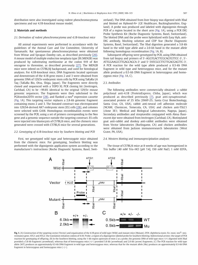
Fig. 1. (A) Construction of the targeting vector (Vector) and organization of the 4.1B gene of wresistance gene; ATG1 and ATG2: the translation initiation codons of 4.1B; Probe: a region of areaction for genotyping of offspring. (B) In the Southern blotting, using the 1-kb region upstprovided a 5.8-kb fragment (arrowhead), whereas that of heterozygous mice (+/−) providedallele (WT) produces an approximately 0.5-kb DNA fragment in wild type and heterozygousfragment in heterozygous and homozygous mice (−/−).
zerland). The DNA obtained from liver biopsy was digested with XbaIand blotted on Hybond-N+ (GE Healthcare, Buckinghamshire, Eng-land). A probe was produced and labeled with digoxigenin throughPCR of a region located in the short arm (Fig. 1A), using a PCR DIGProbe Synthesis Kit (Roche Diagnostic Systems, Basel, Switzerland).The blotted DNA and the probe were hybridized with Easy Hyb, anti-DIG antibody, blocking solution and CDP Star (Roche DiagnosticSystems, Basel, Switzerland). The XbaI digestion generated a 5.8-kbband in the wild type allele and a 2.0-kb band in the mutant allelefollowing homologous recombination (Fig. 1A, B).
Subsequent offspring were genotyped by PCR, using DNA obtainedfrom tail biopsy and primers of 5′-AGGTGACTTCGCAGTGTTCC-3′, 5′-ATTGGAAGCCTTGAGCAGCA-3′ and 5′-TATCGCCTTCTTGACGAGTTC-3′.A PCR reaction for the wild type allele produced a 0.5-kb DNAfragment in wild type and heterozygous mice, and for the mutantallele produced a 0.5-kb DNA fragment in heterozygous and homo-zygous mice (Fig. 1A, C).
2.3. Antibodies
The following antibodies were commercially obtained: a rabbitpolyclonal anti-4.1B (ProteinExpress, Chiba, Japan), which wasproduced as described previously [7], goat anti-synaptosomal-associated protein of 25 kDa (SNAP-25; Santa Cruz Biotechnology,Santa Cruz, CA, USA), rabbit anti-neural cell adhesion molecule(NCAM; Chemicon, Temecula, CA, USA) and chicken anti-TSLC1(clone 3E1; Medical and Biological Laboratories, Nagoya, Japan).Secondary antibodies and streptavidin conjugated with Alexa fluor-escent dye were obtained from Invitrogen (Carlsbad, CA). Biotinylatedgoat anti-rabbit and donkey anti-rabbit antibodies were obtainedfrom Vector laboratories (Burlingame, CA) and chicken antibodieswere obtained from Jackson immunoresearch laboratories (WestGrave, PA, USA).
2.4. Western blotting and immunoprecipitation analyses
The tissue of C57BL/6 mice at 8 weeks of age was homogenized inTris buffer (40 mM Tris–HCl [pH 7.4], 150 mM NaCl, 1 mM EDTA,
ild type (Wild) and mutant mice (Mutant). DTA: diphtheria toxin; Ex: exon; neoR: neo-digoxigenin-labeled probe for Southern blotting; bidirectional arrows: the target of PCRream of exon 2 as a probe, the genomic DNA of wild type mice (+/+) digested with XbaI5.8-kb (arrowhead) and 2.0-kb (arrow) fragments. (C) The PCR reaction for wild typemice, whereas that for the mutant allele (Mu) produces an approximately 0.5-kb DNA
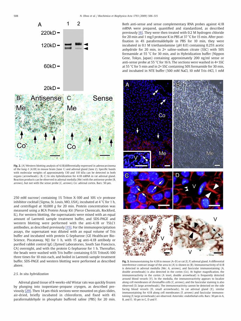
Fig. 2. (A) Western blotting analysis of 4.1B/differentially expressed in adenocarcinomaof the lung-1 (4.1B) in mouse brain (lane 1) and adrenal gland (lane 2). Specific bandswith molecular weights of approximately 130 and 110 kDa can be detected in bothorgans (arrowheads). (B, C) In situ hybridization for 4.1B mRNA in rat adrenal gland.Reaction products can be observed in adrenal medulla (Me) with the antisense probe (B,arrows), but not with the sense probe (C, arrows). Co: adrenal cortex. Bars: 50 μm.
Fig. 3. Immunostaining for 4.1B in mouse (A–D) or rat (E, F) adrenal gland. A differentialinterference contrast image of the area in (A) is shown in (B). Immunoreactivity of 4.1Bis detected in adrenal medulla (Me; A, arrows), and fascicular immunostaining (A,double arrowheads) is also detected in the cortex (Co). At higher magnification, theimmunoreactivity in the cortex (A inset, double arrowhead) is frequently detectedaround blood vessels (V). In the medulla, the immunoreactivity appears to localizealong cell membranes of chromaffin cells (C, arrows), and the fascicular staining is alsoobserved (D, large arrowheads). The immunoreactivity cannot be detected on the sidefacing blood vessels (D, small arrowheads). In rat adrenal gland (E), similarimmunostaining for 4.1B along cell membranes (F, arrows) and fascicular immunos-taining (F, large arrowheads) are observed. Asterisks: endothelial cells. Bars: 30 μm in A,B, and E; 10 μm in C, D and F.
508 N. Ohno et al. / Biochimica et Biophysica Acta 1793 (2009) 506–515
250 mM sucrose) containing 1% Triton X-100 and 10% v/v proteaseinhibitor cocktail (Sigma, St. Louis, MO, USA), incubated at 4 °C for 1 h,and centrifuged at 10,000 g for 20 min. Protein concentration wasmeasured using a BCA Protein Assay Kit (Pierce Chemicals, Rockford,IL). For western blotting, the supernatants were mixed with an equalamount of Laemmli sample treatment buffer, and SDS-PAGE andwestern blotting were performed with the anti-4.1B or TSLC1antibodies, as described previously [15]. For the immunoprecipitationassays, the supernatant was diluted with an equal volume of Trisbuffer and incubated with protein G-Sepharose (GE Healthcare Bio-Science, Piscataway, NJ) for 1 h, with 15 μg anti-4.1B antibody orpurified rabbit control IgG (Zymed Laboratories, South San Francisco,CA) overnight, and with the protein G-Sepharose for 1 h. Thereafter,the beads were washed with Tris buffer containing 0.5% TritonX-100three times for 10 min each, and boiled in Laemmli sample treatmentbuffer. SDS-PAGE and western blotting were performed as describedabove.
2.5. In situ hybridization
Adrenal gland tissue of 8-weeks-oldWistar rats was quickly frozenby plunging into isopentane–propane cryogen, as described pre-viously [29]. Then 14 μm-thick sections were mounted on glass slides,air-dried, briefly incubated in chloroform, and fixed with 4%paraformaldehyde in phosphate buffered saline (PBS) for 20 min.
Both anti-sense and sense complementary RNA probes against 4.1BmRNA were prepared, quantified and standardized, as describedpreviously [6]. They were then treated with 0.2 M hydrogen chloridefor 20 min and 1 mg/l protease K in PBS at 37 °C for 15 min. After post-fixation in 4% paraformaldehyde in PBS for 10 min, they wereincubated in 0.1 M triethanolamine (pH 8.0) containing 0.25% aceticanhydride for 20 min, in 2× saline-sodium citrate (SSC) with 50%formamide at 55 °C for 30 min, and in Hybridization buffer (NipponGene, Tokyo, Japan) containing approximately 200 ng/ml sense oranti-sense probe at 55 °C for 16 h. The sections were washed in 4× SSCat 55 °C for 5 min and in 2× SSC containing 50% formamide for 30 min,and incubated in NTE buffer (500 mM NaCl, 10 mM Tris–HCl, 1 mM
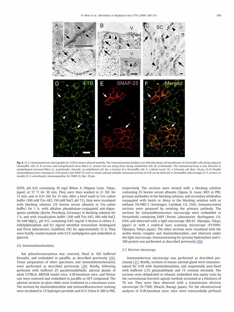
Fig. 4. (A–C) Immunoelectron micrographs for 4.1B in mouse adrenal medulla. The immunoreaction products are detected along cell membranes of chromaffin cells facing adjacentchromaffin cells (A, B, arrows) and unmyelinated nerve fibers (C, arrows) but not along those facing endothelial cells (B, arrowheads). The immunoreactivity is also detected inunmyelinated neuronal fibers (C, arrowheads). Asterisk: an endothelial cell. Nu: a nucleus of a chromaffin cell; V: a blood vessel; SC: a Schwann cell. Bars: 50 μm. (D–F) Doubleimmunofluorescence staining for 4.1B (green) and SNAP-25 (red) in mouse adrenal medulla. Immunoreactivity of 4.1B can be detected in chromaffin cells strongly (D–F, arrows) orweakly (D–F, arrowheads) immunopositive for SNAP-25. Bar: 20 μm.
509N. Ohno et al. / Biochimica et Biophysica Acta 1793 (2009) 506–515
EDTA, pH 8.0) containing 10 mg/l RNase A (Nippon Gene, Tokyo,Japan) at 37 °C for 30 min. They were then washed in 2× SSC for15 min, and in 0.2× SSC for 15 min. After a brief wash in Tris–salinebuffer (100 mM Tris–HCl, 150 mM NaCl, pH 7.5), they were incubatedwith blocking solution (2% bovine serum albumin in Tris–salinebuffer) for 1 h, with alkaline phosphatase-conjugated anti-digox-igenin antibody (Roche, Penzberg, Germany) in blocking solution for1 h, and with visualization buffer (100 mM Tris–HCl, 100 mM NaCl,50 mM MgCl2, pH 9.5) containing 0.05 mg/ml 5-bromo-4-chloro-3-indolylphosphate and 0.1 mg/ml nitroblue tetrazolium (Kirkegaardand Perry laboratories, Guildford, UK) for approximately 12 h. Theywere briefly counterstained with 0.1% methylgreen and embedded inglycerol.
2.6. Immunohistochemistry
Rat pheochromocytoma was resected, fixed in 10% bufferedformalin, and embedded in paraffin, as described previously [26].Tissue preparation of other specimens and immunohistochemistrywere performed as described previously [30]. Briefly, followingperfusion with buffered 2% paraformaldehyde, adrenal glands ofadult C57BL/6, MEN2B model mice, 4.1B-knockout mice, and Wistarrats were removed and embedded in paraffin or OCT compound. Theadrenal sections on glass slides were irradiated in a microwave oven.The sections for diaminobenzidine and immunofluorescence stainingwere incubated in 1% hydrogen peroxide and 0.1% Triton X-100 in PBS,
respectively. The sections were treated with a blocking solutioncontaining 2% bovine serum albumin (Sigma, St. Louis, MO) in PBS,primary antibodies in the blocking solution, and secondary antibodiesconjugated with biotin or Alexa in the blocking solution with orwithout TO-PRO-3 (Invitrogen, Carlsbad, CA, USA). Immunocontrolsections were prepared by omitting the primary antibody. Thesections for immunofluorescence microscopy were embedded inVectashield containing DAPI (Vector Laboratories. Burlingame, CA,USA) and observed with a light microscope (BX-61; Olympus, Tokyo,Japan) or with a confocal laser scanning microscope (FV1000;Olympus, Tokyo, Japan). The other sections were visualized with theavidin–biotin complex and diaminobenzidine, and observed underthe light microscope. Immunostaining for tyrosine hydroxylase and S-100 protein was performed as described previously [26].
2.7. Electron microscopy
Immunoelectron microscopy was performed as described pre-viously [31]. Briefly, sections of mouse adrenal gland were immunos-tained for 4.1B with diaminobenzidine, and sequentially post-fixedwith buffered 2.5% glutaraldehyde and 1% osmium tetroxide. Thesections were dehydrated in ethanol, embedded into epoxy resin bythe conventional inverted capsule method, sectioned at a thickness of70 nm. They were then observed with a transmission electronmicroscope (H-7500; Hitachi, Ibaragi, Japan). For the ultrastructuralanalyses of 4.1B-knockout mice, mice were transcardially perfused

Fig. 5. (A,B) Western blotting (A, lanes a–d) and immunoprecipitation analyses (B, lanes 4.1, Co) for tumor suppressor in lung cancer-1 (TSLC1) in mouse adrenal glands. The westernblotting for TSLC1 shows single bands (A, arrowhead) in adrenal gland (A, lane a), kidney (A, lane b), and lung (A, lane c), but not in heart (A, lane d), although actin is detected in allorgans. A band (B, arrowhead) can be detected following immunoprecipitation with the anti-4.1B/differentially expressed in adenocarcinoma of the lung-1 (4.1B) antibody (B, lane4.1) but not with the control IgG (B, lane Co). (C, D) Immunostaining for TSLC1 in mouse adrenal gland. The immunostained section (D) is also shown as a differential interferencecontrast image (C). The immunoreactivity of TSLC1 is observed in adrenal medulla (Me; C, D, arrows), and appears to localize along cell membranes of chromaffin cells (D, inset,arrows). Co: adrenal cortex. Bars: 50 μm. (E–H) Double immunofluorescence staining for 4.1B (green) and TSLC1 (red) in mouse adrenal medulla. Cellular nuclei are labeled withTOPRO3 (blue). Immunoreactivity of TSLC colocalizes with that of 4.1B on cell membranes of chromaffin cells (E–H, arrows). Bar: 20 μm.
510 N. Ohno et al. / Biochimica et Biophysica Acta 1793 (2009) 506–515
with 4% paraformaldehyde and 0.5% glutaraldehyde in 0.1 M phos-phate buffer, and the collected adrenal gland specimens wereembedded, sectioned, stained and analyzed as described previously[32]. All statistical comparisons were conducted utilizing a two-sidedStudent's t-test, and difference with pb0.05 was consideredsignificant.
3. Results
Two isoforms of 4.1B are reported to be expressed in rat braintissue [7]. In the western blot analyses of 4.1B using rat adrenal glandand brain tissue, two bands with similar molecular weights weredetected in both organs (Fig. 2A). The lower band corresponds to asmaller isoform lacking the 5′ terminal region [33]. In situ hybridiza-tion for 4.1B mRNA was also conducted in the rat adrenal gland, andhybridization signals for 4.1B could be detected in the adrenal medullawith an antisense probe (Fig. 2B), but not with a sense probe (Fig. 2C).In the immunohistochemical analysis, immunoreactivity of 4.1B wasmainly localized in the adrenal medulla (Fig. 3A, B), and fascicularimmunostaining was also observed in the adrenal cortex (Fig. 3A). Thefascicular immunostaining was frequently observed around bloodvessels (Fig. 3A), and considered to represent unmyelinated nervefibers [9]. There was no immunostaining for 4.1B in the immunocon-trol sections (data not shown). At higher magnification, the immunor-eactivity appeared to distribute along the cell membranes ofchromaffin cells in the adrenal medulla (Fig. 3C). The fascicularimmunostaining similar to that detected in cortex was also observedamong the chromaffin cells (Fig. 3D). In the chromaffin cells, theimmunoreactivity of 4.1B was not detected on the side facing theblood vessels, which was characterized by spaces delimited byendothelial cells (Fig. 3D). Similar findings were also observed in ratadrenal glands as well (Fig. 3E, F). Immunoelectron microscopy
showed that the immunoreaction products for 4.1B were localizedalong cell membranes of chromaffin cells not in contact with theendothelial cells (Fig. 4A, B). The immunoreaction products were alsoobserved on the unmyelinated axolemma (Fig. 4C). In the doubleimmunofluorescence staining with SNAP-25, which predominantlyimmunolocalizes in noradrenergic cells of the adrenal medulla [34],the immunoreactivity of 4.1B was observed in both adrenergic andnoradrenergic cells of the mouse adrenal medulla (Fig. 4D–F). Theseresults suggest that 4.1B in rodent adrenal medulla is localized alongcell membranes of all chromaffin cells.
TSLC1 expression was also examined and compared with 4.1B inthe mouse adrenal glands. TSLC1 expression has been reportedpreviously in kidney and lung tissue [35]. In western blot analyses ofmouse adrenal glands using an anti-TSLC1 antibody, bands with thesame molecular weight as those in mouse kidney and lung tissuewere observed (Fig. 5A). Additionally, a band could be detected bywestern blotting against TSLC1 following the immunoprecipitationassay with the anti-4.1B antibody (Fig. 5B). The immunostaining forTSLC1 was predominantly observed in the adrenal medulla, and itappeared to immunolocalize along cell membranes of chromaffincells (Fig. 5C, D). There was no immunostaining for TSLC1 in theimmunocontrol sections (data not shown). Double immunofluores-cence labeling for 4.1B and TSLC1 confirmed that 4.1B and TSLC1 co-localized on cell membranes of chromaffin cells (Fig. 5E–H). Theseresults suggest that in the mouse adrenal medulla, TSLC1 interactswith 4.1B, and is distributed along the cell membranes of chromaffincells.
Given the expression of 4.1B and its interaction with TSLC1, therelationship between the loss of 4.1B and tumorigenesis wasexamined in the adrenal medulla. First, loss of 4.1B expression intumors was investigated immunohistochemically using the ratspontaneous pheochromocytoma [26]. In rat pheochromocytomas
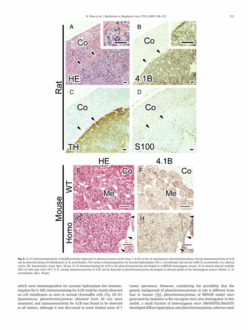
Fig. 6. (A–D) Immunostaining for 4.1B/differentially expressed in adenocarcinoma of the lung-1 (4.1B) in the rat spontaneous pheochromocytoma. Strong immunoreactivity of 4.1Bcan be detected along cell membranes (A, B, arrowheads). The tumor is immunopositive for tyrosine hydroxylase (TH; C, arrowheads) but not for S100 (D, arrowheads). Co: adrenalcortex. HE: hematoxylin–eosin. Bars: 30 μm. (E–H) Immunostaining for 4.1B in the pheochromocytoma developed in a MEN2B homozygous mouse. As in normal adrenal medulla(Me) of wild type mice (WT; E, F), strong immunoreactivity of 4.1B can be detected in pheochromocytoma developed in adrenal gland of the homozygous mouse (Homo; G, H,arrowheads). Bars: 50 μm.
511N. Ohno et al. / Biochimica et Biophysica Acta 1793 (2009) 506–515
which were immunopositive for tyrosine hydroxylase but immuno-negative for S-100, immunostaining for 4.1B could be clearly observedon cell membranes as seen in normal chromaffin cells (Fig. 6A–D).Spontaneous pheochromocytomas obtained from 20 rats wereexamined, and immunoreactivity for 4.1B was found to be detectedin all tumors, although it was decreased in some limited areas of 5
tumor specimens. However, considering the possibility that thegenetic background of pheochromocytomas in rats is different fromthat in human [36], pheochromocytomas of MEN2B model micegenerated by mutation in Ret oncogene were also investigated. In thismodel, a small fraction of heterozygous mice (Met919Thr/Met919)developed diffuse hyperplasia and pheochromocytoma, whereas most
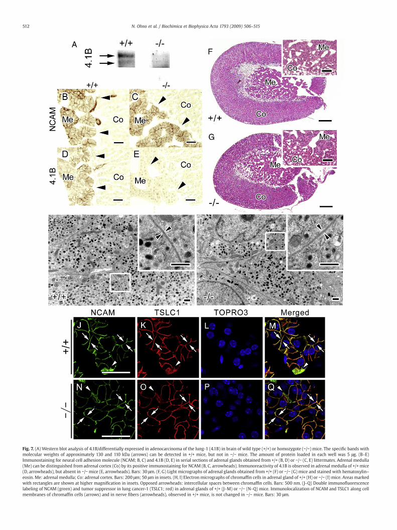
Fig. 7. (A) Western blot analysis of 4.1B/differentially expressed in adenocarcinoma of the lung-1 (4.1B) in brain of wild type (+/+) or homozygote (−/−) mice. The specific bands withmolecular weights of approximately 130 and 110 kDa (arrows) can be detected in +/+ mice, but not in −/− mice. The amount of protein loaded in each well was 5 μg. (B–E)Immunostaining for neural cell adhesion molecule (NCAM; B, C) and 4.1B (D, E) in serial sections of adrenal glands obtained from +/+ (B, D) or −/− (C, E) littermates. Adrenal medulla(Me) can be distinguished from adrenal cortex (Co) by its positive immunostaining for NCAM (B, C, arrowheads). Immunoreactivity of 4.1B is observed in adrenal medulla of +/+ mice(D, arrowheads), but absent in −/−mice (E, arrowheads). Bars: 30 μm. (F, G) Light micrographs of adrenal glands obtained from +/+ (F) or −/− (G) mice and stained with hematoxylin–eosin. Me: adrenal medulla; Co: adrenal cortex. Bars: 200 μm; 50 μm in insets. (H, I) Electron micrographs of chromaffin cells in adrenal gland of +/+ (H) or −/− (I) mice. Areas markedwith rectangles are shown at higher magnification in insets. Opposed arrowheads: intercellular spaces between chromaffin cells. Bars: 500 nm. (J–Q) Double immunofluorescencelabeling of NCAM (green) and tumor suppressor in lung cancer-1 (TSLC1; red) in adrenal glands of +/+ (J–M) or −/− (N–Q) mice. Immunolocalization of NCAM and TSLC1 along cellmembranes of chromaffin cells (arrows) and in nerve fibers (arrowheads), observed in +/+ mice, is not changed in −/− mice. Bars: 30 μm.
512 N. Ohno et al. / Biochimica et Biophysica Acta 1793 (2009) 506–515
513N. Ohno et al. / Biochimica et Biophysica Acta 1793 (2009) 506–515
homozygous mice (Met919Thr/Met919Thr) developed pheochromo-cytomas at the ages of 6–10 months [27]. Therefore, immunohisto-chemistry for 4.1B was performed in the adrenal medulla of 5homozygote and 3 wild type (Met919/Met919) MEN2B model mice at6 months of age. There was no histological abnormality in the adrenalgland of any of the wild type mice (Fig. 6E). By contrast, in the adrenalmedulla of all homozygous mice, normal polyhedral secretory cellswere replaced by abnormal cells with higher density of nuclei and lessvolume of basophilic cytoplasm, and their mass mostly compressedthe surrounding adrenal cortex (Fig. 6G). This morphological appear-ance was suggestive of pheochromocytoma development in most ofthe homozygous mice, as reported previously [27]. In the pheochro-mocytomas of the homozygotes, the immunostaining of 4.1B was alsostrongly observed in all examined tumors, as obtained in normaladrenal medulla of wild type (Fig. 6F, H).
Adrenal glands of 4.1B-knockout mice were further examined forthe development of tumors in the adrenal medulla. The 4.1B-deficientmice were born and developed normally without any apparentdifference from wild type littermates, as reported previously inanother 4.1B-knockout model [24]. First, lack of 4.1B expression wasconfirmed with western blotting in brain tissue, and bands reflectingthe expression of 4.1B in brain tissue of wild type mice were notobserved in those of homozygous mice (Fig. 7A). The expression of4.1B protein was also immunohistochemically examined on cryosec-tions of adrenal glands. In the adrenal medulla, immunoreactivity of4.1B, which was clearly observed inwild type mice, was not present inthe homozygous mice, as determined by immunostaining for NCAM[37] (Fig. 7B–E). However, the histological and ultrastructural analysesof adrenal glands in the 4.1B-knockout mice revealed no significantdifference between the homozygous mice and wild type littermates;the sizes of the cortex and medulla were not significantly different,and no apparent changes indicating abnormal proliferation ofchromaffin cells could be observed in the adrenal medulla up to theage of 18 months (Fig. 7F, G). In addition, although 4.1B is mainlyimmunolocalized along cell membranes of chromaffin cells, theirultrastructures were not changed in the knockoutmice (Fig. 7H, I), andthe intercellular spaces were not significantly different in thequantitative comparison (Mean±SD; 15.6±1.3 nm for wild typemice; 15.7±1.6 nm for knockout mice; N=40, pN0.05). Consideringthe potential involvement of 4.1B in the linkage of TSLC1 to cellmembranes via the actin cytoskeleton, immunostaining for TSLC1wasalso investigated in the adrenal medulla. Immunoreactivity of TSLC1was colocalized with that of NCAM along cell membranes ofchromaffin cells without any translocation or decrease, and thusimmunostaining of TSLC1 appeared to be unchanged in mutant andwild type littermates of 4.1B-knockout mice (Fig. 7J–Q).
4. Discussion
In the present study, localization of 4.1B along cell membranes, andits co-localization and association with TSLC1, were found for the firsttime in rodent adrenal medulla in vivo. These results support thepreviously reported presence of 4.1B mRNA in the adrenal gland ofmouse fetus [38]. The adrenal medulla is composed of chromaffincells, and stimulated to secrete catecholamines through elaboratemechanisms upon a “fight or flight” response [39]. The intercellularspaces among adrenal chromaffin cells are believed to be crucial forthe transport of the secreted catecholamines and also for intercellularcommunication [40]. Although expression of some cell adhesionmolecules such as NCAM and L1 are reported in the adrenal medulla,the precise structural and molecular basis for maintaining thosestructures are not well understood. TSLC1 expression was alreadyreported in several organs, including testes, kidneys, and lungs[35,41,42], and TSLC1 is known to be involved in Ca2+-independentintercellular junctions, which is critical in spermatogenesis of mousetestes [35,43,44]. Given that 4.1B is involved in the intercellular
junctional complex through interaction with transmembranousproteins such as Caspr in the nervous system [5], our results suggesta physiological association between 4.1B and TSLC1 in the adrenalmedulla as well as in cancer cells [23], and also the potentialinvolvement of 4.1B in the intercommunication among adjacentchromaffin cells. At the same time, other new functions of the TSLC1–4.1B interaction should be also evaluated in further studies. Forexample, NCAM-deficiency resulted in impaired granule traffickingrather than morphological abnormalities [37], although a significantdifference in the density of secretary granules could not be found inour quantitative comparison (data not shown).
In the present study, immunoreactivity of 4.1B in chromaffin cellswas not detected on the side of endothelial cells. Expression of 4.1Bhas been found in different epithelial cells, such as mammary,intestinal and renal epithelial cells [8,11,15,45,46]. In all of thesecells, immunostaining for 4.1B was observed along lateral and basalplasma membranes directly facing adjacent cells or extracellularmatrices. From these findings, it was considered that 4.1B wouldstabilize molecules for attachment to adjacent structures, includingthose against extracellular matrices [47]. This hypothesis is alsosupported by the finding that deletion of the intracellular domain ofCaspr resulted in its impaired binding to 4.1B and abnormaldistribution in axonal cytoplasm [48]. On the other hand, adrenalmedulla is one of the organs with sinusoidal capillaries, and thebasement membranes are located between the fenestrated sinusoidalendothelial cells and chromaffin cells [49]. In the pancreatic isletswhere endocrine cells also have sinusoidal capillaries and basementmembranes on the side of endothelial cells [50], similar findings havebeen observed [51]. Plasma membranes of β-cell on the side ofendothelial cells had less 4.1B immunoreactivity compared to thosefacing adjacent endocrine cells. These findings suggest that thedistribution of 4.1B is significantly influenced by the molecularmechanisms of adhesion, which would be distinct on the side ofbasement membrane facing endothelial cells in chromaffin cells. Themolecular complex of intercellular and/or cell-extracellular junctionsin adrenal medulla should be examined in future studies.
Although 4.1B has been implicated in carcinogenesis in severalorgans, its expression was well maintained in rodent pheochromocy-toma, and also 4.1B-knockout mice did not developed pheochromo-cytoma by our examination. The possibility that some unknownisoforms are initiated from unidentified initiation sites but undetect-able with our antibody could not be excluded. However, known 4.1Bgene products from the two initiation sites, which are expressed inbrain and adrenal glands, are eliminated by the genetic mutation inour knockout mice [33,52]. Our results were consistent with theprevious observation that a murine 4.1B-knockout model does notproduce increased incidence of cancer [24]. FERM and U2 domains of4.1B are thought to be essential for growth suppression of menin-gioma-derived cells [53], and post-translational methylation ofarginine residues is a proposed pathway of 4.1B-induced apoptosis[54]. However, the mechanism by which 4.1B influences thecarcinogenesis in various organs is not well known. Our resultssuggest that loss of 4.1B is not essential for the generation ofpheochromocytoma in the rodent adrenal gland, and one possibleexplanation for this might be that rodent adrenal glands lack suchmolecules related to the tumor suppressive functions of 4.1B in otherorgans. Another possibility is that the tumor-suppressive function of4.1B varies in different species (including humans), as we found thatimmunoreactivity of 4.1B was significantly decreased in humanpheochromocytomas compared with the residual part of the normaladrenal medulla (Ohno et al. unpublished data). This possibility wouldbe supported by the distinctive immunolocalization of human 4.1B inblood vessel walls [13], where immunoreactivity of rodent 4.1B couldnot be observed. Recently, some reports suggested that 4.1B wasmainly involved in the progression or metastatic process of cancers,rather than cancer generation [17,55]. Compared to several key
514 N. Ohno et al. / Biochimica et Biophysica Acta 1793 (2009) 506–515
molecules associated with hereditary pheochromocytoma, such asRet, Menin and NF1 [56,57], other genes involved in the generationand behavior of sporadic pheochromocytomas are still poorly under-stood. Further studies on the role of 4.1B in the progression ormetastasis of pheochromocytoma would be essential to elucidate thefunctional significance of 4.1B in the adrenal medulla.
In our present investigation, TSLC1 distributionwas not affected bygenetic ablation of 4.1B in mice. TSLC1 contains a domain essential forits efficient binding to 4.1B, and deletion of the domain causesdiminished binding to 4.1B [23]. However, although inhibition of actinpolymerization resulted in reduced immunoreactivity of 4.1B andTSLC1 along cell membranes [23], dispensability of 4.1B on theplasmalemmal localization of TSLC1 in vivo has remained unclear.TSLC1 also contains the PSD-95, Discs Large, and Zona Occludens 1(PDZ) binding motif in its C-terminal region which mediates thebinding to membrane-associated guanylate kinase homologs(MAGuK) [58]. Our results in 4.1B-knockout mice showing that 4.1Bis not essential to the localization of TSLC1 along cell membranessuggest that the PDZ domain in TSLC1 would compensate for thelinkage to the actin membrane skeleton via other cytoskeletalmolecules associated with MAGuKs. Further studies on TSLC1-bindingproteins including MAGuKs in adrenal medulla are needed to revealthe interaction network responsible for the plasmalemmal distribu-tion of TSLC1, which is considered to be crucial for its tumor-suppressive function [22].
Another possible explanation for the lack of obvious phenotypeand unaffected distribution of TSLC1 is the compensatory effect ofother molecules in the protein 4.1 family. The protein 4.1 family iscomposed of four homologous proteins, 4.1R, 4.1N, 4.1G and 4.1B. Theyare characterized by three highly conservative domains; the FERMdomain, the spectrin-actin binding domain and the C-terminaldomain [52,59]. Multiple members of protein 4.1 family can beexpressed in single cells and localize in same areas of different organs,which lead to the hypothesis of compensation among family members[6,10,31,60–62]. In our studies, we found that immunoreactivity of4.1N is detected along intercellular membranes of chromaffin cells inmouse adrenal gland (Ohno et al., unpublished data). By ourobservation, 4.1N did not co-immunoprecipitated with TSLC-1, andthe immunoreactivity of 4.1N did not significantly change in the 4.1B-knockout mice. However, single binding proteins can interact withdifferent members of protein 4.1 family via highly conserved domains[5,47], and 4.1B is considered to associate with TSLC1 via the FERMdomain [5,23,52,59]. Further studies, such as genetic ablation of 4.1Nin 4.1B knockout mice, would be necessary to examine thecompensatory role among protein 4.1 family more precisely. Inaddition, potential expression of protein 4.1 family isoforms whichcould not be detected in our paradigm should be explored in futurestudies.
In conclusion, although expression and plasmalemmal localizationof 4.1B were shown in chromaffin cells of rodent adrenal medulla, theexpression of 4.1B wasmaintained in developed pheochromocytomas,and generation of pheochromocytomas in 4.1B-deficient mice couldnot be detected in our studies. Furthermore, interaction andcolocalization of 4.1B and TSLC1 were observed in the adrenalmedulla, but 4.1B was not essential for the immunolocalization ofTSLC1 along chromaffin cell membranes. These results suggest thatthe function of 4.1B as a tumor suppressor might differ significantlyamong organs and species, and that retention of TSLC1 on the cellmembranes would be maintained by interacting molecules other than4.1B in rodent chromaffin cells.
Acknowledgements
This workwas partially supported by a grant from Japan Society forthe Promotion of Science (#18590181) to N.T and a grant from the
National Cancer Institute (5P01CA023767) to F.C. We thank Dr.Christopher Nelson for his careful editing of this manuscript.
References
[1] V.M. Braga, Cell–cell adhesion and signalling, Curr. Opin. Cell Biol. 14 (2002)546–556.
[2] C. Jamora, E. Fuchs, Intercellular adhesion, signalling and the cytoskeleton, Nat.Cell Biol. 4 (2002) E101–E108.
[3] M. Perez-Moreno, C. Jamora, E. Fuchs, Sticky business: orchestrating cellularsignals at adherens junctions, Cell 112 (2003) 535–548.
[4] C. Revenu, R. Athman, S. Robine, D. Louvard, The co-workers of actin filaments:from cell structures to signals, Nat. Rev. Mol. Cell Biol. 5 (2004) 635–646.
[5] N. Deisenko-Nehrbass, K. Oguievetskaia, L. Goutebroze, T. Galvez, H. Yamakawa, O.Ohara, M. Carnaud, J.A. Girault, Protein 4.1B associates with both Caspr/paranodinand Caspr2 at paranodes and juxtaparanodes of myelinated fibres, Eur. J. Neurosci.17 (2003) 411–416.
[6] R. Ohara, H. Yamakawa, M. Nakayama, O. Ohara, Type II brain 4.1 (4.1B/KIAA0987),a member of the protein 4.1 family, is localized to neuronal paranodes, Brain Res.Mol. Brain Res. 85 (2000) 41–52.
[7] H. Yamakawa, O. Ohara, Comparison of mRNA and protein levels of four membersof the protein 4.1 family: the type II brain 4.1/4.1B/KIAA0987 is the mostpredominant member of the protein 4.1 family in rat brain, Gene 248 (2000)137–145.
[8] M. Ramez, M. Blot-Chabaud, F. Cluzeaud, S. Chanan, M. Patterson, L.D. Walensky, S.Marfatia, A.J. Baines, J.A. Chasis, J.G. Conboy, N. Mohandas, P. Gascard, Distinctdistribution of specific members of protein 4.1 gene family in the mouse nephron,Kidney Int. 63 (2003) 1321–1337.
[9] N. Terada, N. Ohno, H. Yamakawa, T. Baba, Y. Fujii, O. Ohara, S. Ohno, Protein 4.1Blocalizes on unmyelinated axonal membranes in the mouse enteric nervoussystem, Neurosci. Lett. 366 (2004) 15–17.
[10] N. Terada, N. Ohno, H. Yamakawa, T. Baba, Y. Fujii, Z. Zea, O. Ohara, S. Ohno,Immunohistochemical study of protein 4.1B in the normal and W/W(v) mouseseminiferous epithelium, J. Histochem. Cytochem. 52 (2004) 769–777.
[11] N. Terada, N. Ohno, H. Yamakawa, G. Seki, Y. Fujii, T. Baba, O. Ohara, S. Ohno,Immunoelectron microscopic localization of protein 4.1B in proximal S1 and S2tubules of rodent kidneys, Med. Electron Microsc. 37 (2004) 45–51.
[12] Y.K. Tran, O. Bogler, K.M. Gorse, I. Wieland, M.R. Green, I.F. Newsham, A novelmember of the NF2/ERM/4.1 superfamily with growth suppressing properties inlung cancer, Cancer Res. 59 (1999) 35–43.
[13] D.H. Gutmann, J. Donahoe, A. Perry, N. Lemke, K. Gorse, K. Kittiniyom, S.A. Rempel,J.A. Gutierrez, I.F. Newsham, Loss of DAL-1, a protein 4.1-related tumor suppressor,is an important early event in the pathogenesis of meningiomas, Hum. Mol. Genet.9 (2000) 1495–1500.
[14] A.L. Charboneau, V. Singh, T. Yu, I.F. Newsham, Suppression of growth andincreased cellular attachment after expression of DAL-1 in MCF-7 breast cancercells, Int. J. Cancer 100 (2002) 181–188.
[15] N. Ohno,N. Terada, S.Murata, H. Yamakawa, I.F. Newsham, R. Katoh, O. Ohara, S. Ohno,Immunolocalization of protein4.1B/DAL-1during neoplastic transformation ofmouseand human intestinal epithelium, Histochem. Cell Biol. 122 (2004) 579–586.
[16] P.K. Singh, D.H. Gutmann, C.E. Fuller, I.F. Newsham, A. Perry, Differentialinvolvement of protein 4.1 family members DAL-1 and NF2 in intracranial andintraspinal ependymomas, Mod. Pathol. 15 (2002) 526–531.
[17] S.Y. Wong, H. Haack, J.L. Kissil, M. Barry, R.T. Bronson, S.S. Shen, C.A. Whittaker, D.Crowley, R.O. Hynes, Protein 4.1B suppresses prostate cancer progression andmetastasis, Proc. Natl. Acad. Sci. U. S. A. 104 (2007) 12784–12789.
[18] G. Heller, K.M. Fong, L. Girard, S. Seidl, A. End-Pfutzenreuter, G. Lang, A.F. Gazdar,J.D. Minna, C.C. Zielinski, S. Zochbauer-Muller, Expression andmethylation patternof TSLC1 cascade genes in lung carcinomas, Oncogene 25 (2006) 959–968.
[19] G. Heller, J. Geradts, B. Ziegler, I. Newsham, M. Filipits, E.M. Markis-Ritzinger, D.Kandioler, W. Berger, W. Stiglbauer, D. Depisch, R. Pirker, C.C. Zielinski, S.Zochbauer-Muller, Downregulation of TSLC1 and DAL-1 expression occursfrequently in breast cancer, Breast Cancer Res. Treat. 103 (2007) 283–291.
[20] S.S. Houshmandi, E.I. Surace, H.B. Zhang, G.N. Fuller, D.H. Gutmann, Tumorsuppressor in lung cancer-1 (TSLC1) functions as a glioma tumor suppressor,Neurology 67 (2006) 1863–1866.
[21] M. Kuramochi, H. Fukuhara, T. Nobukuni, T. Kanbe, T. Maruyama, H.P. Ghosh, M.Pletcher, M. Isomura, M. Onizuka, T. Kitamura, T. Sekiya, R.H. Reeves, Y. Murakami,TSLC1 is a tumor-suppressor gene in human non-small-cell lung cancer, Nat.Genet. 27 (2001) 427–430.
[22] Y. Murakami, Involvement of a cell adhesion molecule, TSLC1/IGSF4, in humanoncogenesis, Cancer Sci. 96 (2005) 543–552.
[23] M. Yageta, M. Kuramochi, M. Masuda, T. Fukami, H. Fukuhara, T. Maruyama, M.Shibuya, Y. Murakami, Direct association of TSLC1 and DAL-1, two distinct tumorsuppressor proteins in lung cancer, Cancer Res. 62 (2002) 5129–5133.
[24] C. Yi, J.H. McCarty, S.A. Troutman, M.S. Eckman, R.T. Bronson, J.L. Kissil, Loss of theputative tumor suppressor band 4.1B/Dal1 gene is dispensable for normal develop-ment and does not predispose to cancer, Mol. Cell. Biol. 25 (2005) 10052–10059.
[25] T. Tsujiuchi, E. Sugata, T. Masaoka, M. Onishi, H. Fujii, K. Shimizu, K. Honoki,Expression and DNA methylation patterns of Tslc1 and Dal-1 genes inhepatocellular carcinomas induced by N-nitrosodiethylamine in rats, Cancer Sci.98 (2007) 943–948.
[26] V. Pace, E. Perentes, P.G. Germann, Pheochromocytomas and ganglioneuromas inthe aging rats: morphological and immunohistochemical characterization,Toxicol. Pathol. 30 (2002) 492–500.

515N. Ohno et al. / Biochimica et Biophysica Acta 1793 (2009) 506–515
[27] C.L. Smith-Hicks, K.C. Sizer, J.F. Powers, A.S. Tischler, F. Costantini, C-cellhyperplasia, pheochromocytoma and sympathoadrenal malformation in amouse model of multiple endocrine neoplasia type 2B, EMBO J. 19 (2000)612–622.
[28] M. Komada, P. Soriano, Hrs, a FYVE finger protein localized to early endosomes, isimplicated in vesicular traffic and required for ventral folding morphogenesis,Genes Dev. 13 (1999) 1475–1485.
[29] Z. Li, N. Ohno, N. Terada, S. Ohno, Immunolocalization of serum proteins in livingmouse glomeruli under various hemodynamic conditions by “in vivo cryotechni-que”, Histochem. Cell Biol. 126 (2006) 399–406.
[30] N. Ohno, N. Terada, S. Ohno, Histochemical analyses of living mouse liver underdifferent hemodynamic conditions by “in vivo cryotechnique”, Histochem. CellBiol. 126 (2006) 389–398.
[31] N. Terada, N. Ohno, H. Yamakawa, O. Ohara, X. Liao, T. Baba, S. Ohno,Immunohistochemical study of a membrane skeletal molecule, protein 4.1G, inmouse seminiferous tubules, Histochem. Cell Biol. 124 (2005) 303–311.
[32] N. Ohno, N. Terada, S. Saitoh, S. Ohno, Extracellular space in mouse cerebellarcortex revealed by in vivo cryotechnique, J. Comp. Neurol. 505 (2007) 292–301.
[33] P. Gascard, M.K. Parra, Z. Zhao, V.R. Calinisan, W. Nunomura, S.A. Rivkees, N.Mohandas, J.G. Conboy, Putative tumor suppressor protein 4.1B is differentiallyexpressed in kidney and brain via alternative promoters and 5′ alternativesplicing, Biochim. Biophys. Acta 1680 (2004) 71–82.
[34] R. Kannan, N.J. Grant, D. Aunis, K. Langley, SNAP-25 is differentially expressed bynoradrenergic and adrenergic chromaffin cells, FEBS Lett. 385 (1996) 159–164.
[35] T. Shingai, W. Ikeda, S. Kakunaga, K. Morimoto, K. Takekuni, S. Itoh, K. Satoh, M.Takeuchi, T. Imai, M. Monden, Y. Takai, Implications of nectin-like molecule-2/IGSF4/RA175/SgIGSF/TSLC1/SynCAM1 in cell-cell adhesion and transmembraneprotein localization in epithelial cells, J. Biol. Chem. 278 (2003) 35421–35427.
[36] A. Fritz, A. Walch, K. Piotrowska, M. Rosemann, E. Schaffer, K. Weber, A. Timper, G.Wildner, J. Graw, H. Hofler, M.J. Atkinson, Recessive transmission of a multipleendocrine neoplasia syndrome in the rat, Cancer Res. 62 (2002) 3048–3051.
[37] S.A. Chan, L. Polo-Parada, L.T. Landmesser, C. Smith, Adrenal chromaffin cellsexhibit impaired granule trafficking in NCAM knockout mice, J. Neurophysiol. 94(2005) 1037–1047.
[38] M. Parra, P. Gascard, L.D. Walensky, J.A. Gimm, S. Blackshaw, N. Chan, Y. Takakuwa,T. Berger, G. Lee, J.A. Chasis, S.H. Snyder, N. Mohandas, J.G. Conboy, Molecular andfunctional characterization of protein 4.1B, a novel member of the protein 4.1family with high level, focal expression in brain, J. Biol. Chem. 275 (2000)3247–3255.
[39] A.G. Garcia, A.M. Garcia-De-Diego, L. Gandia, R. Borges, J. Garcia-Sancho, Calciumsignaling and exocytosis in adrenal chromaffin cells, Physiol. Rev. 86 (2006)1093–1131.
[40] I. Benedeczky, P. Somogyi, Ultrastructure of the adrenal medulla of normal andinsulin-treated hamsters, Cell Tissue Res. 162 (1975) 541–550.
[41] S. Kikuchi, D. Yamada, T. Fukami, T. Maruyama, A. Ito, H. Asamura, Y. Matsuno, M.Onizuka, Y. Murakami, Hypermethylation of the TSLC1/IGSF4 promoter isassociated with tobacco smoking and a poor prognosis in primary nonsmall celllung carcinoma, Cancer 106 (2006) 1751–1758.
[42] T. Wakayama, K. Ohashi, K. Mizuno, S. Iseki, Cloning and characterization of anovel mouse immunoglobulin superfamily gene expressed in early spermatogeniccells, Mol. Reprod. Dev. 60 (2001) 158–164.
[43] W.L. van der, M.J. Arends, O.E. Chausiaux, P.J. Ellis, U.C. Lange, M.A. Surani, N.Affara, Y. Murakami, D.J. Adams, A. Bradley, Loss of TSLC1 causes male infertilitydue to a defect at the spermatid stage of spermatogenesis, Mol. Cell. Biol. 26(2006) 3595–3609.
[44] D. Yamada, M. Yoshida, Y.N. Williams, T. Fukami, S. Kikuchi, M. Masuda, T.Maruyama, T. Ohta, D. Nakae, A. Maekawa, T. Kitamura, Y. Murakami, Disruption ofspermatogenic cell adhesion and male infertility in mice lacking TSLC1/IGSF4, an
immunoglobulin superfamily cell adhesion molecule, Mol. Cell. Biol. 26 (2006)3610–3624.
[45] R. Kuns, J.L. Kissil, I.F. Newsham, T. Jacks, D.H. Gutmann, L.S. Sherman, Protein 4.1Bexpression is induced inmammary epithelial cells during pregnancy and regulatestheir proliferation, Oncogene 24 (2005) 6502–6515.
[46] N. Terada, N. Ohno, S. Saitoh, G. Seki, M. Komada, T. Suzuki, H. Yamakawa, M.Soleimani, S. Ohno, Interaction of membrane skeletal protein, protein 4.1B andp55, and sodium bicarbonate cotransporter1 in mouse renal S1–S2 proximaltubules, J. Histochem. Cytochem. 55 (2007) 1199–1206.
[47] J.H. McCarty, A.A. Cook, R.O. Hynes, An interaction between {alpha}v{beta}8integrin and Band 4.1B via a highly conserved region of the Band 4.1 C-terminaldomain, Proc. Natl. Acad. Sci. U. S. A. 102 (2005) 13479–13483.
[48] L. Gollan, H. Sabanay, S. Poliak, E.O. Berglund, B. Ranscht, E. Peles, Retention of acell adhesion complex at the paranodal junction requires the cytoplasmic regionof Caspr, J. Cell Biol. 157 (2002) 1247–1256.
[49] R.E. Coupland, Electron microscopic observations on the structure of the ratadrenal medulla. I. The ultrastructure and organization of chromaffin cells in thenormal adrenal medulla, J. Anat. 99 (1965) 231–254.
[50] P.E. Lacy, W.S. Hartroft, Electron microscopy of the islets of Langerhans, Ann. N. Y.Acad. Sci. 82 (1959) 287–301.
[51] N. Terada, N. Ohno, H. Yamakawa, T. Baba, Y. Fujii, G. Christofori, O. Ohara, S. Ohno,Protein 4.1B in mouse islets of Langerhans and beta-cell tumorigenesis,Histochem. Cell Biol. 120 (2003) 277–283.
[52] M. Parra, S. Gee, N. Chan, D. Ryaboy, I. Dubchak, N. Mohandas, P.D. Gascard, J.G.Conboy, Differential domain evolution and complex RNA processing in a family ofparalogous EPB41 (protein 4.1) genes facilitate expression of diverse tissue-specific isoforms, Genomics 84 (2004) 637–646.
[53] V.A. Robb, M.A. Gerber, E.K. Hart-Mahon, D.H. Gutmann, Membrane localization ofthe U2 domain of Protein 4.1B is necessary and sufficient for meningioma growthsuppression, Oncogene 24 (2005) 1946–1957.
[54] W. Jiang, I.F. Newsham, The tumor suppressor DAL-1/4.1B and proteinmethylation cooperate in inducing apoptosis in MCF-7 breast cancer cells,Mol. Cancer 5 (2006) 4.
[55] T. Cavanna, E. Pokorna, P. Vesely, C. Gray, D. Zicha, Evidence for protein 4.1B actingas a metastasis suppressor, J. Cell Sci. 120 (2007) 606–616.
[56] C. Jimenez, G. Cote, A. Arnold, R.F. Gagel, Review: should patients with apparentlysporadic pheochromocytomas or paragangliomas be screened for hereditarysyndromes? J. Clin. Endocrinol. Metab. 91 (2006) 2851–2858.
[57] A. Karagiannis, D.P. Mikhailidis, V.G. Athyros, F. Harsoulis, Pheochromocytoma: anupdate on genetics and management, Endocr. Relat Cancer 14 (2007) 935–956.
[58] H. Fukuhara, M. Masuda, M. Yageta, T. Fukami, M. Kuramochi, T. Maruyama, T.Kitamura, Y. Murakami, Association of a lung tumor suppressor TSLC1 with MPP3,a human homologue of Drosophila tumor suppressor Dlg, Oncogene 22 (2003)6160–6165.
[59] C.X. Sun, V.A. Robb, D.H. Gutmann, Protein 4.1 tumor suppressors: getting a FERMgrip on growth regulation, J. Cell. Sci. 115 (2002) 3991–4000.
[60] L.D. Walensky, S. Blackshaw, D. Liao, C.C. Watkins, H.U. Weier, M. Parra, R.L.Huganir, J.G. Conboy, N. Mohandas, S.H. Snyder, A novel neuron-enriched homologof the erythrocyte membrane cytoskeletal protein 4.1, J. Neurosci. 19 (1999)6457–6467.
[61] F. Delhommeau, C. Vasseur-Godbillon, P. Leclerc, P.O. Schischmanoff, L. Croisille, P.Rince, M. Morinière, E.J. Benz, G. Tchernia, G. Tamagnini, L. Ribeiro, J. Delaunay, F.Baklouti, A splicing alteration of 4.1R pre-mRNA generates 2 protein isoforms withdistinct assembly to spindle poles in mitotic cells, Blood 100 (2002) 2629–2636.
[62] Z.T. Shi, V. Afzal, B. Coller, D. Patel, J.A. Chasis, M. Parra, G. Lee, C. Paszty, M. Stevens,L. Walensky, L.L. Peters, N. Mohandas, E. Rubin, J.G. Conboy, Protein 4.1R-deficientmice are viable but have erythroid membrane skeleton abnormalities, J. Clin.Invest. 103 (1999) 331–340.


![Usefulness of [18F]-DA and [18F]-DOPA for PET imaging in a mouse model of pheochromocytoma](https://static.fdokumen.com/doc/165x107/6325a7d9852a7313b70e9a7d/usefulness-of-18f-da-and-18f-dopa-for-pet-imaging-in-a-mouse-model-of-pheochromocytoma.jpg)