Diphenyl diselenide modulates oxLDL-induced cytotoxicity in macrophage by improving the redox...
8
Research paper Diphenyl diselenide modulates oxLDL-induced cytotoxicity in macrophage by improving the redox signaling Marcos Raniel Straliotto a , Mariana Appel Hort a , Bianca Fiuza a , João Batista Teixeira Rocha b , Marcelo Farina a , Gustavo Chiabrando c , Andreza Fabro de Bem a, * a Departamento de Bioquímica, Centro de Ciências Biológicas, Universidade Federal de Santa Catarina, 88040900 Florianópolis, SC, Brazil b Departamento de Química, Centro de Ciências Naturais e Exatas, Universidade Federal de Santa Maria, 97105900 Santa Maria, RS, Brazil c Centro de Investigaciones en Bioquímica Clínica e Inmunología (CIBICI-CONICET), Faculdad de Ciencias Químicas, Universidad Nacional de Córdoba, Ciudad Universitaria, 5000 Córdoba, Argentina article info Article history: Received 26 November 2012 Accepted 9 April 2013 Available online xxx Keywords: Diphenyl diselenide Selenium Atherosclerosis Macrophage Glutathione peroxidase LDL oxidation Inflammation Oxidative stress abstract It has been reported that oxidized LDLs (oxLDL) are involved in the pathogenesis of atherosclerosis, and that macrophages as well as other cells of the arterial wall can oxidize LDL in vitro, depending on the balance between intracellular prooxidant generation and antioxidant defense efficiency. Because of their potential beneficial role in preventing atherosclerosis and other oxidative stress-related diseases, orga- noselenium compounds such as diphenyl diselenide (PhSe) 2 , are receiving increased attention. In the present work, we investigated the mechanisms underlying the protective effect exerted by (PhSe) 2 on oxLDL-mediated effects in murine J774 macrophage-like cells. (PhSe) 2 pretreatment reduced atherogenic signaling triggered by oxLDL in macrophages in vitro, namely: ROS generation, disturbance of NO ho- meostasis, activation of matrix metalloproteinase, foam cell formation, and mitochondrial dysfunction. Moreover, the redox signaling effects of (PhSe) 2 presented herein were accompanied by a down- regulation of NF-kB-binding activity. The relatively strong performance of (PhSe) 2 makes it an ideal candidate for further, expanded trials as a new generation of antioxidants for preventing atherosclerotic lesion. Ó 2013 Elsevier Masson SAS. All rights reserved. 1. Introduction Accumulation of modified low-density lipoprotein (LDL), such as oxidized LDL (oxLDL), in the arterial wall, and the recruitment of monocytes to the subendothelial space are known to be the main early events in the development of atherosclerosis [1]. Blood-circulating monocytes adhere to the endothelium, migrate to the intima, and differentiate to macrophages. Macro- phages express receptors (CD36 and SR-A) that bind and internalize oxidized forms of LDL, and these processes give rise to the arterial foam cell, a hallmark of arterial lesion [2e6]. Macrophage activation and foam cell formation are involved in both the initiation and the progression and ultimate instability of advanced lesions. Macro- phages taking up oxLDL could modify the production of inflam- matory mediators, such as cytokines, proteases, reactive oxygen and nitrogen species (ROS and RNS), metalloproteinases (MMPs) [7e9] and other factors through oxidative sensitive signaling pathways [10]. In oxLDL-stimulated macrophages, the intracellular ROS/RNS are generated by several pathways, including the NADPH oxidase (NOX) system, the lipoxygenase/cyclooxygenase system, inducible NO synthase (iNOS), and mitochondrial respiratory chain [11]. Moreover, intracellular ROS generated by oxLDL stimulation can activate a wide variety of proinflammatory and proapoptotic pathways that are regulated by transcription factors, such as nu- clear factor-kB (NF-kB) [12,13]. These events will culminate in the death of macrophages by activation of apoptosis. Recent clinical studies have suggested an important anti- atherogenic role for the antioxidant enzyme glutathione peroxi- dase (GPx) [14], which uses glutathione to reduce hydrogen peroxide to water and lipid peroxides to their respective alcohols [15], and it also acts as a peroxynitrite reductase [16]. Therefore, the investigation of antiatherogenic properties of synthetic organo- selenium compounds with GPx-mimetic activity has attracted considerable attention. In this scenario, we have been studying the pharmacological properties of diphenyl diselenide (PhSe) 2 , a * Corresponding author. Tel.: þ55 48 37215565; fax: þ55 48 37219672. E-mail address: [email protected] (A.F. de Bem). Contents lists available at SciVerse ScienceDirect Biochimie journal homepage: www.elsevier.com/locate/biochi 0300-9084/$ e see front matter Ó 2013 Elsevier Masson SAS. All rights reserved. http://dx.doi.org/10.1016/j.biochi.2013.04.008 Biochimie xxx (2013) 1e8 Please cite this article in press as: M.R. Straliotto, et al., Diphenyl diselenide modulates oxLDL-induced cytotoxicity in macrophage by improving the redox signaling, Biochimie (2013), http://dx.doi.org/10.1016/j.biochi.2013.04.008
-
Upload
independent -
Category
Documents
-
view
0 -
download
0
Transcript of Diphenyl diselenide modulates oxLDL-induced cytotoxicity in macrophage by improving the redox...

at SciVerse ScienceDirect
Biochimie xxx (2013) 1e8
Contents lists available
Biochimie
journal homepage: www.elsevier .com/locate/biochi
Research paper
Diphenyl diselenide modulates oxLDL-induced cytotoxicity inmacrophage by improving the redox signaling
Marcos Raniel Straliotto a, Mariana Appel Hort a, Bianca Fiuza a,João Batista Teixeira Rocha b, Marcelo Farina a, Gustavo Chiabrando c,Andreza Fabro de Bema,*
aDepartamento de Bioquímica, Centro de Ciências Biológicas, Universidade Federal de Santa Catarina, 88040900 Florianópolis, SC, BrazilbDepartamento de Química, Centro de Ciências Naturais e Exatas, Universidade Federal de Santa Maria, 97105900 Santa Maria, RS, BrazilcCentro de Investigaciones en Bioquímica Clínica e Inmunología (CIBICI-CONICET), Faculdad de Ciencias Químicas, Universidad Nacional de Córdoba,Ciudad Universitaria, 5000 Córdoba, Argentina
a r t i c l e i n f o
Article history:Received 26 November 2012Accepted 9 April 2013Available online xxx
Keywords:Diphenyl diselenideSeleniumAtherosclerosisMacrophageGlutathione peroxidaseLDL oxidationInflammationOxidative stress
* Corresponding author. Tel.: þ55 48 37215565; faxE-mail address: [email protected] (A.F. d
0300-9084/$ e see front matter � 2013 Elsevier Mashttp://dx.doi.org/10.1016/j.biochi.2013.04.008
Please cite this article in press as: M.R. Straliothe redox signaling, Biochimie (2013), http:
a b s t r a c t
It has been reported that oxidized LDLs (oxLDL) are involved in the pathogenesis of atherosclerosis, andthat macrophages as well as other cells of the arterial wall can oxidize LDL in vitro, depending on thebalance between intracellular prooxidant generation and antioxidant defense efficiency. Because of theirpotential beneficial role in preventing atherosclerosis and other oxidative stress-related diseases, orga-noselenium compounds such as diphenyl diselenide (PhSe)2, are receiving increased attention. In thepresent work, we investigated the mechanisms underlying the protective effect exerted by (PhSe)2 onoxLDL-mediated effects in murine J774 macrophage-like cells. (PhSe)2 pretreatment reduced atherogenicsignaling triggered by oxLDL in macrophages in vitro, namely: ROS generation, disturbance of �NO ho-meostasis, activation of matrix metalloproteinase, foam cell formation, and mitochondrial dysfunction.Moreover, the redox signaling effects of (PhSe)2 presented herein were accompanied by a down-regulation of NF-kB-binding activity. The relatively strong performance of (PhSe)2 makes it an idealcandidate for further, expanded trials as a new generation of antioxidants for preventing atheroscleroticlesion.
� 2013 Elsevier Masson SAS. All rights reserved.
1. Introduction
Accumulation of modified low-density lipoprotein (LDL), such asoxidized LDL (oxLDL), in the arterial wall, and the recruitment ofmonocytes to the subendothelial space are known to be the mainearly events in the development of atherosclerosis [1].
Blood-circulating monocytes adhere to the endothelium,migrate to the intima, and differentiate to macrophages. Macro-phages express receptors (CD36 and SR-A) that bind and internalizeoxidized forms of LDL, and these processes give rise to the arterialfoam cell, a hallmark of arterial lesion [2e6]. Macrophage activationand foam cell formation are involved in both the initiation and theprogression and ultimate instability of advanced lesions. Macro-phages taking up oxLDL could modify the production of inflam-matory mediators, such as cytokines, proteases, reactive oxygenand nitrogen species (ROS and RNS), metalloproteinases (MMPs)
: þ55 48 37219672.e Bem).
son SAS. All rights reserved.
tto, et al., Diphenyl diselenid//dx.doi.org/10.1016/j.biochi.2
[7e9] and other factors through oxidative sensitive signalingpathways [10].
In oxLDL-stimulated macrophages, the intracellular ROS/RNSare generated by several pathways, including the NADPH oxidase(NOX) system, the lipoxygenase/cyclooxygenase system, inducible�NO synthase (iNOS), and mitochondrial respiratory chain [11].Moreover, intracellular ROS generated by oxLDL stimulation canactivate a wide variety of proinflammatory and proapoptoticpathways that are regulated by transcription factors, such as nu-clear factor-kB (NF-kB) [12,13]. These events will culminate in thedeath of macrophages by activation of apoptosis.
Recent clinical studies have suggested an important anti-atherogenic role for the antioxidant enzyme glutathione peroxi-dase (GPx) [14], which uses glutathione to reduce hydrogenperoxide to water and lipid peroxides to their respective alcohols[15], and it also acts as a peroxynitrite reductase [16]. Therefore, theinvestigation of antiatherogenic properties of synthetic organo-selenium compounds with GPx-mimetic activity has attractedconsiderable attention. In this scenario, we have been studying thepharmacological properties of diphenyl diselenide (PhSe)2, a
emodulates oxLDL-induced cytotoxicity in macrophage by improving013.04.008
M.R. Straliotto et al. / Biochimie xxx (2013) 1e82
simple diaryl diselenide with high GPx-mimetic activity [17]. Inaddition to its GPx-like activity, we recently demonstrated that(PhSe)2 is able to promote the nuclear translocation of the Nuclear(erythroid-derived 2)-related factor (Nrf2) increasing the expres-sion of enzymes related to antioxidant defenses, as gamma-glutamylcysteine synthetase (GGCS) and GPx [18].
This compound was very efficient in reducing the human LDLoxidation and Cu2þ-induced lipid peroxidation in rat aortic slices[19] and it decreased hypercholesterolemia and oxidative stress incholesterol-fed rabbits [20]. Furthermore, the treatment with(PhSe)2 reduced the atherosclerotic lesion in hypercholesterolemicLDLr �/� mice by modulating pathways related to antioxidant andanti-inflammatory responses [21]. In the present study, we there-fore aimed at determining whether pretreatment with (PhSe)2 at-tenuates oxLDL-induced effects inmurine J774A.1macrophage cellsand, if so, whether the mechanisms underlying the process involvethe redox signaling pathway.
2. Materials and methods
2.1. Materials
Diphenyl diselenide (PhSe)2 was synthesized according topublished methods [22]. Analysis of the 1H NMR and 13C NMRspectra showed that the compound obtained presented analyticaland spectroscopic data in full agreement with its assigned struc-ture. The chemical purity of (PhSe)2 (99.9%) was determined by GC/HPLC. All other chemicals were of analytical grade and obtainedfrom standard commercial suppliers.
2.2. LDL isolation and oxidation
This study was approved by our Ethic Committee at FederalUniversity of Santa Catarina (n� 943/11). LDL was isolated fromfresh human plasma by discontinuous density-gradient ultracen-trifugation as previously described [19] and the protein concen-tration in LDL solution was determined by Lowry [23]. LDL isolatedsamples (1 mg of protein/mL) were oxidized at 37 �C in the pres-ence of 10 mMCuSO4 for 16 h, to produce oxLDL. Then EDTA 1.5 mMwas added and the samples were dialyzed against 148 mM phos-phate buffer for 24 h at 4 �C. LDL oxidation was monitored byfollowing the thiobarbituric acid-reactive substances (TBARS)production.
2.3. Cell culture
Murine J774A.1 macrophage cells were obtained from theAmericanType Culture Collection (ATCC, Rockville, MD, USA). Thesecells were maintained with Dulbecco’s Modified Eagle Medium(DMEM) supplemented with 2 mM glutamine, 10 mM HEPES,100 units/mL penicillin, 100 mg/mL streptomycin and 10% fetalbovine serum (FBS) in a 5% CO2 humidified atmosphere at 37 �C.The exposition of cells with (PhSe)2 and/or oxLDL was carried out inDMEM without FBS.
2.4. Measurement of reactive oxygen species (ROS) production
Intracellular ROS production was detected using the non-fluorescent cell permeating compound, 20,70-dichlorodihydro-fluorescein diacetate (DCFH-DA). In order to evaluated the protec-tive effect of (PhSe)2 on ROS production induced by oxLDL,macrophage (2.0 � 105 cells/well) seeded in 24 well plates werefirstly stimulated to an increasing concentration curve of oxLDL (25,50,100 and 200 mg/mL) for 1 h. After that, cells were incubated withDCFH-DA (10 mM) for 30 min at 37 �C, washed twice with PBS,
Please cite this article in press as: M.R. Straliotto, et al., Diphenyl diselenidthe redox signaling, Biochimie (2013), http://dx.doi.org/10.1016/j.biochi.2
harvested and collected for immediate determination of ROS gen-eration by flow cytometry (FACS Canto II, BD Bioscience, UnitedStates). The protective effect of (PhSe)2 was also evaluated by flowcytometry by pretreating cells for 24 h with (PhSe)2 (1 mM) orvehicle before oxLDL (100 mg/mL) stimulation for 1 h. The resultswere expressed as percentage of control (non-stimulated cells;100%).
2.5. Measurement of mitochondrial membrane potential (DJm)
The mitochondrial membrane potential (DJm) was assessedusing the lipophilic cationic probe fluorochrome JC-1 [24]. Inpresence of physiological mitochondrial membrane potentials, JC-1forms aggregates that fluoresce with an emission peak at 588 nm.Loss of membrane potential favors the monomeric form of JC-1,which has an emission peak at 530 nm. To examine the effect of(PhSe)2 on modifications in DJm induced by oxLDL, macrophageswere plated into 24well plates (2.0� 105 cells/well) and pretreatedwith (PhSe)2 (1 mM) or vehicle for 24 h, followed by stimulationwith oxLDL (100 mg/mL) or FCCP 1 mM (a positive control) foradditional 3 h. After, macrophages were incubated with JC-1 (5 mM)for 20 min at 37 �C and JC-1 fluorescence intensity was measuredusing a fluorometric microplate reader (Tecan, Grödig/Salzburg,Austria) with excitation at 488 nm and an emission at 525 nm and590 nm. Mitochondrial membrane potential was inferred from theratio of fluorescence intensity red/green. The images were acquiredfrom three randomly chosen fields using an inverted epifluor-escence microscope (Olympus IX70).
2.6. Cell viability assay
The cell viability in the presence or absence of (PhSe)2 wasmeasured by MTT assay as described by Siqueira and cols [25].Macrophage (1 � 104 cells/well) seeded in 96 well plates werepretreated with (PhSe)2 (1 mM) or vehicle for 24 h, followed bystimulation with oxLDL (100 or 200 mg/mL) for additional 24 h.After this, 200 mL of MTT solution (0.5 mg/mL) was added andincubated for 2 h. Then, theMTTwas removed and 200 mL of DMSO/well was added to dissolve the intracellular crystalline formazanproduct. The absorbance at 550 nm was read spectrophotometri-cally using a microplate reader. The results were expressed as apercentage of the absorbance of non-treated cells.
2.7. Foam cell formation assay
Foam cell formation assay was performed with the Oil Red Ostaining method [26]. Macrophages (2.5 � 105 cells/well) plated incoverslip in 12 wells plate were pretreated with (PhSe)2 (1 mM) orvehicle for 24 h, and then stimulated with oxLDL (100 mg/mL) foradditional 3 h. Following oxLDL incubation, cells were fixed with 4%paraformaldehyde and stained by 0.3% Oil Red O for 10 min. He-matoxylin was used as counterstaining. Images of cells were ac-quired using confocal microscopy (Leica DMI6000 B Microscope)using a 546 nm filter set. Ten images were captured from eachgroup and the total pixels intensity was determined using NIHImage J 1.36b imaging software (National Institutes of Health,Bethesda, MD, USA), and lipid content was expressed as opticaldensity (OD).
2.8. MMP activity measurement
Matrix metalloproteinases (MMPs) are a family of zinc-dependent endopeptidases, capable of degrading all the molecu-lar components of extracellular matrix. To evaluate the activity ofMMP-9, we used two different techniques, gelatin zymography and
emodulates oxLDL-induced cytotoxicity in macrophage by improving013.04.008

Fig. 1. (PhSe)2 decreases the oxLDL-induced ROS production in macrophages. (A) Macrophages were exposed to different concentrations of oxLDL (25e200 mg/mL) for 1 h. (B) Themacrophages were pretreated with (PhSe)2 or vehicle for 24 h and exposure to oxLDL (100 mg/mL) for additional 1 h. DCFH-DA (10 mM) was added to the medium for 30 min and theamount of ROS was determinate by flow cytometry. The ROS amount was expressed as percentage of increase of control. (C) Representative flow cytometer histogram. Each barrepresents the mean � SEM of at least three independent experiments. *P < 0.05; **P < 0.01; ***P < 0.001 indicates the difference when compared with control; ##P < 0.01 indicatesthe difference when compared with oxLDL exposure cells (one-way ANOVA, Duncan’s test).
Fig. 2. (PhSe)2 prevents oxLDL-mediated mitochondrial dysfunction. Macrophages were pretreated 24 h with (PhSe)2 (1 mM) or vehicle and then exposed to oxLDL (100 mg/mL) foradditional 3 h. FCCP (1 mM) was utilized as a positive control. (A) The change in mitochondrial membrane potential (DJm) was assessed based on the signal intensity from JC-1monomeric and aggregate fluorescence (Magnification 400�). (B) JC-1 fluorescence was quantified fluorometrically. Each bar represents the mean � SEM of the ratio (red/greenfluorescence) of at least three independent experiments. ***P < 0.001 indicates the difference when compared with control; ##P < 0.01 indicates the difference when comparedwith oxLDL exposure cells (one-way ANOVA, Duncan’s test).
M.R. Straliotto et al. / Biochimie xxx (2013) 1e8 3
Please cite this article in press as: M.R. Straliotto, et al., Diphenyl diselenidemodulates oxLDL-induced cytotoxicity in macrophage by improvingthe redox signaling, Biochimie (2013), http://dx.doi.org/10.1016/j.biochi.2013.04.008

Fig. 3. OxLDL-induced cytotoxicity was prevent by (PhSe)2. Macrophages were treated24 h with (PhSe)2 1 mM or vehicle and then exposed to oxLDL (100 mg/mL or 200 mg/mL) for additional 24 h. The cell viability was determined by MTT assay and expressedas percentage of control. Each bar represents the mean � SEM of at least three inde-pendent experiments. ***P < 0.001 indicates the difference when compared withcontrol; #P < 0.05 indicates the difference when compared with oxLDL exposure cells(one-way ANOVA, Duncan’s test).
M.R. Straliotto et al. / Biochimie xxx (2013) 1e84
the activity of collagenase IV. For these assays, cells (2.5 � 105 cells/well) were seeded in 12 wells plate and then preincubated with(PhSe)2 (1 mM) or vehicle for 24 h, followed by stimulation withoxLDL (100 mg/mL) for additional 24 h.
2.8.1. Gelatin zymographySupernatants (50 mg/mL of total protein) were subjected to SDS-
PAGE in 7.5% (w/v) polyacrylamide gels copolymerized with 1.5%gelatin as a substrate as previously described [27]. After electro-phoresis, the gels were stained with Coomassie Brilliant Blue.Proteolytic activity in a particular gel location yielded a clear bandagainst the blue background of the stained gelatin. The MMP-9were identified by molecular size using high-molecular mass(92 kDa) standards (Bio-Rad, Hercules, CA, USA). Gels were scannedand the intensity of the bands was quantified using Quantity Onesoftware (Bio-Rad, Hercules, CA, USA). The results were expressedas intensity of MMP-9 relative to control.
2.8.2. EnzChek Gelatinase/Collagenase assay kitThe activity of gelatinase or collagenase IV in conditioned su-
pernatants was determinate using an Enzymatic Gelatinase/Colla-genase fluorometric assay kit (Molecular Probes, USA). Briefly,conditioned supernatants (50 mg/mLof protein)were incubatedwithMMP specific peptide substrate, based on the instructions includedwith the kit. The cleavage of substrates by MMP (gelatinases orcollagenase IV) result in increased fluorescence signal measuredusing a fluorescence microplate reader (Tecan, Grödig/Salzburg,Austria)with excitationat 495nmandemissionat515nm.Datawerereported as the collagenase IV activity relative to the control.
2.9. iNOS expression
The expression of iNOS was evaluated by western blot analysis.Macrophages (5.0� 105 cells/well) were seeded in 6 wells plate andthen preincubated with (PhSe)2 (1 mM) or vehicle for 24 h, followedby stimulationwith oxLDL (100 mg/mL or 200 mg/mL) for additional24 h. Proteins were subjected to 7.5% SDS-PAGE, transferred tonitrocellulose membranes and blocked with 5% low fat milk in50 mM PBS for 1 h. Membranes were then probed at room tem-perature, for 1 hwith rabbit polyclonal anti-iNOS antibody (1:2000,Santa Cruz Biotechnology, USA). For the detection, immunocom-plexed membranes were washed in PBS containing 0.6% Tween 20,
Please cite this article in press as: M.R. Straliotto, et al., Diphenyl diselenidthe redox signaling, Biochimie (2013), http://dx.doi.org/10.1016/j.biochi.2
and probed with goat anti-rabbit antibody at room temperature for1 h (1:5000, Santa Cruz Biotechnology, USA). After extensivewashing, blots were visualized using the Perkin Elmer ECL system.Equal loading of gel lanes was verified by probing the blots for b-actin content. The intensity of the bands was quantified usingQuantity One software (Bio-Rad, Hercules, CA, USA).
2.10. Nitrite assay
Nitrite production, an indicator of �NO production, was measuredin the conditioned culture medium by the Griess reaction [28]. Mac-rophages (2.0 � 105 cells/well) were cultured in 24 well plates andpreincubated with (PhSe)2 (1 mM) or vehicle for 24 h and then stim-ulated with oxLDL (100 or 200 mg/mL) for additional 24 h. The su-pernatantswere collected to quantification of nitrite. Briefly,100 mL ofsupernatants were mixed with 100 mL of Griess reagent (equal vol-umes of 1% (w/v) sulphanilic acid in 30% (v/v) acetic acid and 0.1% (w/v)N-(1-naphthyl) ethylenediamine in60% (v/v) acetic acid), incubatedat room temperature for 30 min in the dark, and the absorbance at540nmwasmeasured inamicroplate reader (Tecan, Grödig/Salzburg,Austria). The amount of nitrite was calculated from a standard curveusing freshly prepared sodium nitrite in culture medium and the re-sults were expressed as percentage of control (non-treated cells).
2.11. Immunofluorescence (immunohistochemistry)
The effect of (PhSe)2 on oxLDL-induced NF-kB nuclear trans-location was evaluated by immunofluorescence. Macrophages(2.5 � 105 cells/well) seeded in coverslips in 12 wells plate werepreincubated with (PhSe)2 (1 mM) or vehicle for 24 h, followed bystimulation with oxLDL (100 mg/mL) or vehicle for additional 1 h.After which cells were fixed with 4% paraformaldehyde andblocked with 4% of albumin in 0.05% (v/v) Tween 20-PBS (PBS-T20)for 15 min. The nuclear translocation of NF-kB was evaluated byimmunofluorescence using an anti-NF-kB rabbit monoclonal p-65antibody (1:100, Santa Cruz Biotechnology, USA) in PBS-T20 for24 h at 4 �C, followed by FITC conjugated donkey anti-rabbit IgGsecondary antibody (1:100, Santa Cruz Biotechnology, USA) for 1 h.Cell nuclei were stained with Hoechst 33258. The images wereacquired from eight to ten randomly chosen fields using a fluo-rescent microscope (Olympus BX41).
2.12. Statistical analysis
All data are expressed as mean � SEM from at least three inde-pendent experiments. Differences betweenmean values ofmultiplegroups were analyzed by one-way analysis of variance (ANOVA)followed by the post-hoc Duncan multiple range test when P wassignificant. Statistical significance was considered at P < 0.05.
3. Results
3.1. (PhSe)2 prevents the oxLDL-induced ROS production,mitochondrial dysfunction and cell death
As expected, the exposition of J774A.1macrophage cells for 1 h tooxLDL (25e200 mg/mL) caused a concentration-dependent(b ¼ 0.861, P < 0.001) increase on intracellular ROS production(Fig. 1A). The pretreatment of macrophages with (PhSe)2, signifi-cantly attenuated the oxLDL-inducedROSproduction (Fig.1B andC).
Mitochondrial dysfunction and the ensuing bioenergeticimpairment are likely to promote ion pump failure and loss ofmitochondrial membrane integrity. Mitochondrial membrane po-tential (DJm) was analyzed by JC-1 fluorescence. OxLDL (100 mg/mL) significantly induced a decrease in DJm, by reducing the
emodulates oxLDL-induced cytotoxicity in macrophage by improving013.04.008
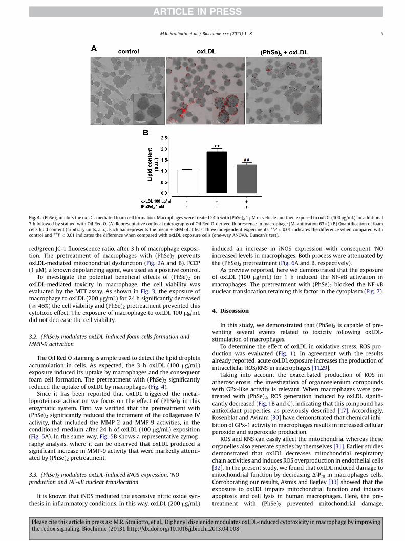
Fig. 4. (PhSe)2 inhibits the oxLDL-mediated foam cell formation. Macrophages were treated 24 h with (PhSe)2 1 mM or vehicle and then exposed to oxLDL (100 mg/mL) for additional3 h followed by stained with Oil Red O. (A) Representative confocal micrographs of Oil Red O-derived fluorescence in macrophage (Magnification 63�). (B) Quantification of foamcells lipid content (arbitrary units, a.u.). Each bar represents the mean � SEM of at least three independent experiments. **P < 0.01 indicates the difference when compared withcontrol and ##P < 0.01 indicates the difference when compared with oxLDL exposure cells (one-way ANOVA, Duncan’s test).
M.R. Straliotto et al. / Biochimie xxx (2013) 1e8 5
red/green JC-1 fluorescence ratio, after 3 h of macrophage exposi-tion. The pretreatment of macrophages with (PhSe)2 preventsoxLDL-mediated mitochondrial dysfunction (Fig. 2A and B). FCCP(1 mM), a known depolarizing agent, was used as a positive control.
To investigate the potential beneficial effects of (PhSe)2 onoxLDL-mediated toxicity in macrophage, the cell viability wasevaluated by the MTT assay. As shown in Fig. 3, the exposure ofmacrophage to oxLDL (200 mg/mL) for 24 h significantly decreased(y 46%) the cell viability and (PhSe)2 pretreatment prevented thiscytotoxic effect. The exposure of macrophage to oxLDL 100 mg/mLdid not decrease the cell viability.
3.2. (PhSe)2 modulates oxLDL-induced foam cells formation andMMP-9 activation
The Oil Red O staining is ample used to detect the lipid dropletsaccumulation in cells. As expected, the 3 h oxLDL (100 mg/mL)exposure induced its uptake by macrophages and the consequentfoam cell formation. The pretreatment with (PhSe)2 significantlyreduced the uptake of oxLDL by macrophages (Fig. 4).
Since it has been reported that oxLDL triggered the metal-loproteinase activation we focus on the effect of (PhSe)2 in thisenzymatic system. First, we verified that the pretreatment with(PhSe)2 significantly reduced the increment of the collagenase IVactivity, that included the MMP-2 and MMP-9 activities, in theconditioned medium after 24 h of oxLDL (100 mg/mL) exposition(Fig. 5A). In the same way, Fig. 5B shows a representative zymog-raphy analysis, where it can be observed that oxLDL produced asignificant increase in MMP-9 activity that were markedly attenu-ated by (PhSe)2 pretreatment.
3.3. (PhSe)2 modulates oxLDL-induced iNOS expression, �NOproduction and NF-kB nuclear translocation
It is known that iNOS mediated the excessive nitric oxide syn-thesis in inflammatory conditions. In this way, oxLDL (200 mg/mL)
Please cite this article in press as: M.R. Straliotto, et al., Diphenyl diselenidthe redox signaling, Biochimie (2013), http://dx.doi.org/10.1016/j.biochi.2
induced an increase in iNOS expression with consequent �NOincreased levels in macrophages. Both process were attenuated bythe (PhSe)2 pretreatment (Fig. 6A and B, respectively).
As preview reported, here we demonstrated that the exposureof oxLDL (100 mg/mL) for 1 h induced the NF-kB activation inmacrophages. The pretreatment with (PhSe)2 blocked the NF-kBnuclear translocation retaining this factor in the cytoplasm (Fig. 7).
4. Discussion
In this study, we demonstrated that (PhSe)2 is capable of pre-venting several events related to toxicity following oxLDL-stimulation of macrophages.
To determine the effect of oxLDL in oxidative stress, ROS pro-duction was evaluated (Fig. 1). In agreement with the resultsalready reported, acute oxLDL exposure increases the production ofintracellular ROS/RNS in macrophages [11,29].
Taking into account the exacerbated production of ROS inatherosclerosis, the investigation of organoselenium compoundswith GPx-like activity is relevant. When macrophages were pre-treated with (PhSe)2, ROS generation induced by oxLDL signifi-cantly decreased (Fig. 1B and C), indicating that this compound hasantioxidant properties, as previously described [17]. Accordingly,Rosenblat and Aviram [30] have demonstrated that chemical inhi-bition of GPx-1 activity in macrophages results in increased cellularperoxide and superoxide production.
ROS and RNS can easily affect the mitochondria, whereas theseorganelles also generate species by themselves [31]. Earlier studiesdemonstrated that oxLDL decreases mitochondrial respiratorychain activities and induces ROS overproduction in endothelial cells[32]. In the present study, we found that oxLDL induced damage tomitochondrial function by decreasing DJm in macrophages cells.Corroborating our results, Asmis and Begley [33] showed that theexposure to oxLDL impairs mitochondrial function and inducesapoptosis and cell lysis in human macrophages. Here, the pre-treatment with (PhSe)2 prevented mitochondrial damage,
emodulates oxLDL-induced cytotoxicity in macrophage by improving013.04.008

Fig. 5. (PhSe)2 modulates oxLDL triggered MMP-9 activity. Macrophages were treated24 h with (PhSe)2 1 mM or vehicle and then exposed to oxLDL (100 mg/mL) for addi-tional 24 h. (A) The collagenase IV activity in the supernatant was measured using anEnzChek Gelatinase/Collagenase Assay kit. (B) Zymography analysis of MMP-9 activityin cell supernatant. Upper panel: representative zymography assays of MMP-9 gelat-inolytic activity. Lower panel: densitometry analysis from zymography assays. Resultswere expressed as relative intensity to control. Each bar represents mean � SEM of atleast three independent experiments. ***P < 0.001 compared with control and##P < 0.01; ###P < 0.001 indicates the difference when compared with oxLDL exposurecells (one-way ANOVA, Duncan’s test).
Fig. 6. Effect of (PhSe)2 on oxLDL-induced iNOS expression and �NO production.Macrophages were treated 24 h with (PhSe)2 1 mM or vehicle and then exposed tooxLDL (100 mg/mL or 200 mg/mL) for additional 24 h. (A) Representative western blotanalysis of iNOS. b-actin was used as a load control. (B) Nitrite levels in the conditionedculture medium and expressed as percentage of control. Each bar represents themean � SEM of at least three independent experiments. *P < 0.05; ***P < 0.001compared with control and #P < 0.05; ###P < 0.001 indicates the difference whencompared with oxLDL (200 mg/mL) exposure cells (one-way ANOVA, Duncan’s test).
M.R. Straliotto et al. / Biochimie xxx (2013) 1e86
recovering the DJm (Fig. 2) and this effect could be in part attrib-uted to its antioxidant properties. In agreement with this, recentstudies have demonstrated that (PhSe)2 restores the mitochondrialdysfunction caused by acetaminophen [34] and by methylmercury[35] inmouse brain. This ROS overproduction can be responsible fortriggering different pathways related to cell death [36], which canoccur either by mitochondrial dysfunction or by downregulation ofanti-apoptotic proteins [37]. In accordance with this, here the ROSproduction and mitochondrial dysfunction promoted by oxLDLwere associated with a decrease in macrophage viability, whichwas prevented by (PhSe)2 treatment (Fig. 3). These evidencessupport the hypothesis that (PhSe)2 could prevent the cytotoxiceffects mediated by oxLDL in macrophage by inhibit ROSgeneration.
NF-kB and Activator protein-1 (AP-1) are two redox-sensitivetranscription factors that can be activated during various
Please cite this article in press as: M.R. Straliotto, et al., Diphenyl diselenidthe redox signaling, Biochimie (2013), http://dx.doi.org/10.1016/j.biochi.2
inflammatory disease processes [38]. The activation of these tran-scription factors can up regulate several major proinflammatorymediators such as iNOS, MMPs, CD36 receptor and others [36,39e41]. The NF-kB pathway is an attractive candidate to mediate theeffects of ROS because the interaction between the inhibitoryprotein IkB and NF-kB is regulated by protein kinases that containseveral redox-sensitive cysteine residues in critical kinase domains[42]. In addition, H2O2 stimulation enhanced the nuclear trans-location of AP-1 and NF-kB subunits [43]. Wiesner et al. [44]demonstrated, by exposure of macrophages to the minimallyoxidized LDL (mmLDL) and lipopolysaccharide (LPS), that thecooperative engagement of NF-kB and AP-1 results in additive/synergistic increased transcription of inflammatory cytokineswithin atherosclerotic lesions. Here, we showed that the exposureof macrophages to oxLDL can rapidly (1 h) lead to the activation of
emodulates oxLDL-induced cytotoxicity in macrophage by improving013.04.008

Fig. 7. (PhSe)2 modulates oxLDL-induced NF-kB nuclear translocation. Macrophageswere treated 24 h with (PhSe)2 1 mM or vehicle and then exposed to oxLDL (100 mg/mL)for additional 1 h. Nuclear translocation of NF-kB was evaluated by immunofluores-cence (green). Nuclear counterstaining was made with Hoechst 33258 (blue). Arepresentative merge of the two fluorescence images (green and blue) is shown(Magnification 1000�). The representative images were from three independentexperiments.
Scheme 1. Effects of (PhSe)2 on macrophage oxLDL-induced toxicity. Based on theresults appointed in this study, we conclude that the (PhSe)2 decreased the ROSgeneration and LDL uptake in macrophages. Moreover, the pretreatment with theorganoselenium compound prevented the decrease in DJm and the consequently celldeath induced by oxLDL. In addition, the NF-kB translocation to the nucleus wasblocked by (PhSe)2, diminishing the transcription of genes such as CD36 receptor, iNOSand MMP-9, resulting in a decreasing expression of these proteins and �NO production.
M.R. Straliotto et al. / Biochimie xxx (2013) 1e8 7
NF-kB, promoting its translocation to the nucleus (Fig. 7) whereasthe (PhSe)2 treatment led to a downregulation of NF-kB-bindingactivity. Consistent with this, we recently demonstrated that theoxLDL-mediated increase in TNF-a and MCP-1, inflammatory me-diators trigged by the NF-kB activation, was prevented by (PhSe)2 inmacrophage [21].
It has been documented that macrophages express high levels ofMMPs, mainly theMMP-2 andMMP-9, in response to oxidative andinflammatory stimulus. These MMPs are highly expressed invulnerable regions of atherosclerotic plaques and are involved inthe remodeling processes associated with atherogenesis and pla-que rupture [45,46]. Herein, we showed by two different method-ological approaches that oxLDL induced a markedly increase inspecific MMP-9 and collagenase IV activities, which was attenuatedby the pretreatment with (PhSe)2 (Fig. 5). We elected to evaluateonly the specific activity of MMP-9 because its increase is moreevident than the others MMPs isoforms [47]. Then, based on ourresults we can’t discard that the increase in collagenase IV activitycould be a reflex of the sum of theMMP-2 andMMP-9 activities andthat pretreatment with (PhSe)2 was able of attenuating bothMMPs.
Nitric oxide (�NO) is known to counteract the atherogenic pro-cess bymediating vasorelaxation, reducing vascular smoothmusclecell proliferation, and decreasing platelet aggregation [48]. A recentstudy shows that oxLDL enhances the expression of iNOS and �NOproduction in macrophages [49]. In our experimental condition,exposure of macrophages to oxLDL caused a significant increase inboth iNOS protein expression and �NO levels, while pretreatmentwith (PhSe)2 prevented it (Fig. 6A and B). Consistent with theseobservations, Shin et al. [50] reported that another diselenide, bis-(3-hydroxyphenyl) diselenide (DSE-B), potently inhibits �NO pro-duction and decreases iNOS protein levels in macrophages stimu-lated with LPS. Recently, Rupil et al. [51] showed that (PhSe)2inhibits the �NO production in a dose-dependent mode in perito-neal macrophages activated by LPS and this effect was correlatedwith a decrease in iNOS expression.
The internalization of oxLDL in macrophages occurs throughmechanisms involving different cell surface receptors; in thiscontext, the CD36 has an important role [52]. In this study, wedemonstrated that (PhSe)2 efficiently decreased oxLDL uptake and
Please cite this article in press as: M.R. Straliotto, et al., Diphenyl diselenidthe redox signaling, Biochimie (2013), http://dx.doi.org/10.1016/j.biochi.2
consequent foam cell formation (Fig. 4). This effect can be explainedby the inhibition of the activation of NF-kB by the organoseleniumcompound with consequent decrease in the expression of CD36,resulting in less uptake of oxLDL.
Taken together, the effects of (PhSe)2 here presented, arepossibly related to its antioxidant property. We believe that, thedownregulation in oxLDL-mediated free radical generation couldprovide a favorable cellular environment that avoid NF-kB activa-tion and the consequent cell death. This potent antioxidant effect of(PhSe)2 may be explained in part to its ability in decomposingorganic hydroperoxides of oxLDL via GPx-mimetic activity. By theother hand, this organoselenium compound could be acting viaNrf2, which increases the expression of phase II antioxidant genesleading to an improvement in cellular redox environment [53]. Thishypothesis is supported by our recent study that reported animprovement in glutathione antioxidant system mediated by(PhSe)2 via Nrf2 activation [18].
5. Conclusion
In this study, we provide evidence that (PhSe)2 protects mac-rophages against atherogenic signaling triggered by oxLDL byinhibiting ROS generation and ROS-induced inflammatory path-ways modulated by NF-kB (Scheme 1). Our findings suggest that(PhSe)2 might be a candidate agent for further developments in theprevention of cardiovascular diseases.
Conflicts of interest
None.
Acknowledgments
This work was supported by grants from the Brazilian in-stitutions Conselho Nacional de Desenvolvimento Científico eTecnológico (CNPq), Coordenação de Aperfeiçoamento de Pessoalde Nível Superior (CAPES) and Fundação de Apoio à Pesquisa doEstado de Santa Catarina (FAPESC). Marcos Raniel Straliotto was arecipient of the grant CAPES. Gustavo Chiabrando is member of theResearch Career of CONICET (Consejo de Investigaciones Científicasy Técnicas de la República Argentina). Andreza Fabro de Bem, João
emodulates oxLDL-induced cytotoxicity in macrophage by improving013.04.008

M.R. Straliotto et al. / Biochimie xxx (2013) 1e88
Batista Teixeira Rocha and Marcelo Farina are supported byresearch fellowships from CNPq-Brazil.
References
[1] G.K. Hansson, A.K. Robertson, C. Soderberg-Naucler, Inflammation andatherosclerosis, Annu. Rev. Pathol. 1 (2006) 297e329.
[2] R. Ross, Atherosclerosis e an inflammatory disease, N. Engl. J. Med. 340 (1999)115e126.
[3] A.J. Lusis, Atherosclerosis, Nature 407 (2000) 233e241.[4] P. Libby, Inflammation in atherosclerosis, Nature 420 (2002) 868e874.[5] Y. Yamada, T. Doi, T. Hamakubo, T. Kodama, Scavenger receptor family pro-
teins: roles for atherosclerosis, host defence and disorders of the centralnervous system, Cell. Mol. Life Sci. 54 (1998) 628e640.
[6] W.J. de Villiers, E.J. Smart, Macrophage scavenger receptors and foam cellformation, J. Leukoc. Biol. 66 (1999) 740e746.
[7] G.K. Hansson, P. Libby, U. Schonbeck, Z.Q. Yan, Innate and adaptive immunityin the pathogenesis of atherosclerosis, Circ. Res. 91 (2002) 281e291.
[8] G.K. Hansson, Inflammation, atherosclerosis, and coronary artery disease,N. Engl. J. Med. 352 (2005) 1685e1695.
[9] X.P. Xu, S.R. Meisel, J.M. Ong, S. Kaul, B. Cercek, T.B. Rajavashisth, B. Sharifi,P.K. Shah, Oxidized low-density lipoprotein regulates matrixmetalloproteinase-9 and its tissue inhibitor in human monocyte-derivedmacrophages, Circulation 99 (1999) 993e998.
[10] H. Geng, A. Wang, G. Rong, B. Zhu, Y. Deng, J. Chen, R. Zhong, The effects of ox-LDL in human atherosclerosis may be mediated in part via the toll-like re-ceptor 4 pathway, Mol. Cell. Biochem. 342 (2010) 201e206.
[11] I. Levitan, S. Volkov, P.V. Subbaiah, Oxidized LDL: diversity, patterns ofrecognition, and pathophysiology, Antioxid. Redox Signal. 13 (2010) 39e75.
[12] M. Karin, The beginning of the end: IkappaB kinase (IKK) and NF-kappaBactivation, J. Biol. Chem. 274 (1999) 27339e27342.
[13] F. Robbesyn, V. Garcia, N. Auge, O. Vieira, M.F. Frisach, R. Salvayre, A. Negre-Salvayre, HDL counterbalance the proinflammatory effect of oxidized LDL byinhibiting intracellular reactive oxygen species rise, proteasome activation,and subsequent NF-kappaB activation in smooth muscle cells, FASEB J. 17(2003) 743e745.
[14] S. Blankenberg, H.J. Rupprecht, C. Bickel, M. Torzewski, G. Hafner, L. Tiret,M. Smieja, F. Cambien, J. Meyer, K.J. Lackner, Glutathione peroxidase 1 activityand cardiovascular events in patients with coronary artery disease, N. Engl. J.Med. 349 (2003) 1605e1613.
[15] L. Flohe, Glutathione peroxidase, Basic Life Sci. 49 (1988) 663e668.[16] H. Sies, Glutathione and its role in cellular functions, Free Radic. Biol. Med. 27
(1999) 916e921.[17] C.W. Nogueira, J.B. Rocha, Diphenyl diselenide a Janus-Faced molecule, J. Braz.
Chem. Soc. 21 (2010) 2055e2071.[18] A.F. de Bem, B. Fiuza, P. Calcerrada, P. Brito, G. Peluffo, T. Dinis, M. Trujillo,
J.T. Rocha, R. Radi, L. Almeida, Protective effect of diphenyl diselenide againstperoxynitrite-mediated endothelial cell death: a comparison with ebselen,Nitric Oxide 31 (2013) 20e30.
[19] A.F. de Bem, M. Farina, L. Portella Rde, C.W. Nogueira, T.C. Dinis,J.A. Laranjinha, L.M. Almeida, J.B. Rocha, Diphenyl diselenide, a simple gluta-thione peroxidase mimetic, inhibits human LDL oxidation in vitro, Athero-sclerosis 201 (2008) 92e100.
[20] A.F. de Bem, L. Portella Rde, E. Colpo, M.M. Duarte, A. Frediane, P.S. Taube,C.W. Nogueira, M. Farina, E.L. da Silva, J.B. Teixeira Rocha, Diphenyl diselenidedecreases serum levels of total cholesterol and tissue oxidative stress incholesterol-fed rabbits, Basic Clin. Pharmacol. Toxicol. 105 (2009) 17e23.
[21] M.A. Hort, M.R. Straliotto, P.M. Netto, J.B. da Rocha, A.F. de Bem, R.M. Ribeiro-do-Valle, Diphenyl diselenide effectively reduces atherosclerotic lesions inLDLr �/� mice by attenuation of oxidative stress and inflammation,J. Cardiovasc. Pharmacol. 58 (2011) 91e101.
[22] C. Paulmier, Selenium reagents and intermediates, in: Organic Synthesis,Pergamon, Oxford, 1986.
[23] O.H. Lowry, N.J. Rosebrough, A.L. Farr, R.J. Randall, Protein measurement withthe Folin phenol reagent, J. Biol. Chem. 193 (1951) 265e275.
[24] M. Reers, T.W. Smith, L.B. Chen, J-aggregate formation of a carbocyanine as aquantitative fluorescent indicator of membrane potential, Biochemistry 30(1991) 4480e4486.
[25] J.M. Siqueira, A.C. Gazola, M.R. Farias, L. Volkov, N. Rivard, A.J. de Brum-Fer-nandes, R.M. Ribeiro-do-Valle, Evaluation of the antitumoral effect ofdihydrocucurbitacin-B in both in vitro and in vivo models, Cancer Chemother.Pharmacol. 64 (2009) 529e538.
[26] R. Koopman, G. Schaart, M.K. Hesselink, Optimisation of oil red O stainingpermits combination with immunofluorescence and automated quantificationof lipids, Histochem. Cell. Biol. 116 (2001) 63e68.
[27] D.E. Kleiner, W.G. Stetler-Stevenson, Quantitative zymography: detection ofpicogram quantities of gelatinases, Anal. Biochem. 218 (1994) 325e329.
[28] K. Schulz, S. Kerber, M. Kelm, Reevaluation of the Griess method for deter-mining NO/NO2
� in aqueous and protein-containing samples, Nitric Oxide 3(1999) 225e234.
[29] T. Deng, K. Xu, L. Zhang, X. Zheng, Dynamic determination of Ox-LDL-inducedoxidative/nitrosative stress in single macrophage by using fluorescent probes,Cell Biol. Int. 32 (2008) 1425e1432.
Please cite this article in press as: M.R. Straliotto, et al., Diphenyl diselenidthe redox signaling, Biochimie (2013), http://dx.doi.org/10.1016/j.biochi.2
[30] M. Rosenblat, M. Aviram, Macrophage glutathione content and glutathioneperoxidase activity are inversely related to cell-mediated oxidation of LDL:in vitro and in vivo studies, Free Radic. Biol. Med. 24 (1998) 305e317.
[31] J.L. Perfettini, T. Roumier, G. Kroemer, Mitochondrial fusion and fission in thecontrol of apoptosis, Trends Cell. Biol. 15 (2005) 179e183.
[32] T. Matsunaga, K. Iguchi, T. Nakajima, I. Koyama, T. Miyazaki, I. Inoue, S. Kawai,S. Katayama, K. Hirano, S. Hokari, T. Komoda, Glycated high-density lipopro-tein induces apoptosis of endothelial cells via a mitochondrial dysfunction,Biochem. Biophys. Res. Commun. 287 (2001) 714e720.
[33] R. Asmis, J.G. Begley, Oxidized LDL promotes peroxide-mediated mitochon-drial dysfunction and cell death in human macrophages: a caspase-3-independent pathway, Circ. Res. 92 (2003) e20e29.
[34] M.H. da Silva, E.J. da Rosa, N.R. de Carvalho, F. Dobrachinski, J.B. da Rocha,J.L. Mauriz, J. Gonzalez-Gallego, F.A. Soares, Acute brain damage induced byacetaminophen in mice: effect of diphenyl diselenide on oxidative stress andmitochondrial dysfunction, Neurotoxicity Res. 21 (2012) 334e344.
[35] D.F. Meinerz, M.T. de Paula, B. Comparsi, M.U. Silva, A.E. Schmitz, H.C. Braga,P.S. Taube, A.L. Braga, J.B. Rocha, A.L. Dafre, M. Farina, J.L. Franco, T. Posser,Protective effects of organoselenium compounds against methylmercury-induced oxidative stress in mouse brain mitochondrial-enriched fractions,Braz. J. Med. Biol. Res. 44 (2011) 1156e1163. Revista brasileira de pesquisasmedicas e biologicas/Sociedade Brasileira de Biofisica. [et al.].
[36] H.C. Ou, F.P. Chou, W.H. Sheu, S.L. Hsu, W.J. Lee, Protective effects of magnololagainst oxidized LDL-induced apoptosis in endothelial cells, Arch. Toxicol. 81(2007) 421e432.
[37] S. Tan, Y. Sagara, Y. Liu, P. Maher, D. Schubert, The regulation of reactiveoxygen species production during programmed cell death, J. Cell. Biol. 141(1998) 1423e1432.
[38] M. Karin, T. Takahashi, P. Kapahi, M. Delhase, Y. Chen, C. Makris, D. Rothwarf,V. Baud, G. Natoli, F. Guido, N. Li, Oxidative stress and gene expression: theAP-1 and NF-kappaB connections, Biofactors 15 (2001) 87e89.
[39] R. Terkeltaub, C.L. Banka, J. Solan, D. Santoro, K. Brand, L.K. Curtiss, OxidizedLDL induces monocytic cell expression of interleukin-8, a chemokine with T-lymphocyte chemotactic activity, Arterioscler Thromb. 14 (1994) 47e53.
[40] C. Napoli, Oxidation of LDL, atherogenesis, and apoptosis, Ann. N. Y Acad. Sci.1010 (2003) 698e709.
[41] C. Maziere, J.C. Maziere, Activation of transcription factors and gene expres-sion by oxidized low-density lipoprotein, Free Radic. Biol. Med. 46 (2009)127e137.
[42] D. Impellizzeri, E. Esposito, E. Mazzon, I. Paterniti, R. Di Paola, P. Bramanti,V.M. Morittu, A. Procopio, E. Perri, D. Britti, S. Cuzzocrea, The effects of apolyphenol present in olive oil, oleuropein aglycone, in an experimentalmodel of spinal cord injury in mice, Biochem. Pharmacol. 83 (2012) 1413e1426.
[43] W.C. Chiu, C.J. Chen, T.S. Lee, Z.J. Chen, P.H. Ke, A.N. Chiang, Oxidativestress enhances AP-1 and NF-kappaB-mediated regulation of beta(2)-glycoprotein I gene expression in hepatoma cells, J. Cell. Biochem. 111(2010) 988e998.
[44] P. Wiesner, S.H. Choi, F. Almazan, C. Benner, W. Huang, C.J. Diehl, A. Gonen,S. Butler, J.L. Witztum, C.K. Glass, Y.I. Miller, Low doses of lipopolysaccharideand minimally oxidized low-density lipoprotein cooperatively activate mac-rophages via nuclear factor kappa B and activator protein-1: possible mech-anism for acceleration of atherosclerosis by subclinical endotoxemia, Circ. Res.107 (2010) 56e65.
[45] I.M. Loftus, A.R. Naylor, S. Goodall, M. Crowther, L. Jones, P.R. Bell,M.M. Thompson, Increased matrix metalloproteinase-9 activity in unstablecarotid plaques. A potential role in acute plaque disruption, Stroke 31 (2000)40e47.
[46] Z.S. Galis, J.J. Khatri, Matrix metalloproteinases in vascular remodeling andatherogenesis: the good, the bad, and the ugly, Circ. Res. 90 (2002) 251e262.
[47] A. Radhika, S.S. Jacob, P.R. Sudhakaran, Influence of oxidatively modified LDLon monocyteemacrophage differentiation, Mol. Cell. Biochem. 305 (2007)133e143.
[48] R.P. Patel, A. Levonen, J.H. Crawford, V.M. Darley-Usmar, Mechanisms of thepro- and anti-oxidant actions of nitric oxide in atherosclerosis, Cardiovasc.Res. 47 (2000) 465e474.
[49] K. Persson, L. Sauma, A. Safholm, L. Xu, W. Li, X.M. Yuan, LDL and UV-oxidizedLDL induce upregulation of iNOS and NO in unstimulated J774 macrophagesand HUVEC, APMIS 117 (2009) 1e9.
[50] K.M. Shin, L. Shen, S.J. Park, J.H. Jeong, K.T. Lee, Bis-(3-hydroxyphenyl) dis-elenide inhibits LPS-stimulated iNOS and COX-2 expression in RAW 264.7macrophage cells through the NF-kappaB inactivation, J. Pharm. Pharmacol.61 (2009) 479e486.
[51] L.L. Rupil, A.F. de Bem, G.A. Roth, Diphenyl diselenide-modulation of macro-phage activation: down-regulation of classical and alternative activationmarkers, Innate Immun. 18 (2012) 627e637.
[52] A.C. Nicholson, D.P. Hajjar, CD36, oxidized LDL and PPAR gamma: pathologicalinteractions in macrophages and atherosclerosis, Vasc. Pharmacol. 41 (2004)139e146.
[53] N. Wakabayashi, A.T. Dinkova-Kostova, W.D. Holtzclaw, M.I. Kang,A. Kobayashi, M. Yamamoto, T.W. Kensler, P. Talalay, Protection againstelectrophile and oxidant stress by induction of the phase 2 response: fate ofcysteines of the Keap1 sensor modified by inducers, Proc. Natl. Acad. Sci. U S A101 (2004) 2040e2045.
emodulates oxLDL-induced cytotoxicity in macrophage by improving013.04.008