Digital Unwrapping of the Mummy of King Amenhotep I (1525 ...
15
ORIGINAL RESEARCH published: 28 December 2021 doi: 10.3389/fmed.2021.778498 Frontiers in Medicine | www.frontiersin.org 1 December 2021 | Volume 8 | Article 778498 Edited by: Juarez Antonio Simões Quaresma, Federal University of Pará, Brazil Reviewed by: Paulo Hilario Nascimento Saldiva, University of São Paulo, Brazil Rimantas Jankauskas, Vilnius University, Lithuania Luiz Fábio Magno Falcão, Universidade do Estado do Pará, Brazil *Correspondence: Sahar N. Saleem [email protected] Specialty section: This article was submitted to Pathology, a section of the journal Frontiers in Medicine Received: 17 September 2021 Accepted: 27 October 2021 Published: 28 December 2021 Citation: Saleem SN and Hawass Z (2021) Digital Unwrapping of the Mummy of King Amenhotep I (1525–1504 BC) Using CT. Front. Med. 8:778498. doi: 10.3389/fmed.2021.778498 Digital Unwrapping of the Mummy of King Amenhotep I (1525–1504 BC) Using CT Sahar N. Saleem 1 * and Zahi Hawass 2 1 Department of Radiology, Kasr Al Ainy Faculty of Medicine, Cairo University, Cairo, Egypt, 2 Antiquities of Egypt, Cairo, Egypt The mummy of King Amenhotep I (18th Dynasty c.1525–1504 BC) was reburied by the 21st Dynasty priests at Deir el-Bahari Royal Cache. In 1881 the mummy was found fully wrapped and was one of few royal mummies that have not been unwrapped in modern times. We hypothesized that non-invasive digital unwrapping using CT would provide insights on the physical appearance, health, cause of death, and mummification style of the mummy of King Amenhotep I. We examined the mummy with CT and generated two- and three-dimensional images for the head mask, bandages, and the virtually unwrapped mummy. CT enabled the visualization of the face of Amenhotep I who died around the age of 35 years. The teeth had minimal attrition. There was no CT evidence of pathological changes or cause of death. The body has been eviscerated via a vertical left flank incision. The heart is seen in the left hemithorax with an overlying amulet. The brain has not been removed. The mummy has 30 amulets/jewelry pieces including a beaded metallic (likely gold) girdle. The mummy suffered from multiple postmortem injuries likely inflicted by tomb robbers that have been likely treated by 21st Dynasty embalmers. These included fixing the detached head and neck to the body with a resin-treated linen band; covering a defect in the anterior abdominal wall with a band and placing two amulets beneath; placement of the detached left upper limb beside the body and wrapping it to the body. The transversely oriented right forearm is individually wrapped, likely representing the original 18th Dynasty mummification and considered the first known New Kingdom mummy with crossed arms at the chest. The head mask is made of cartonnage and has inlaid stone eyes. The digital unwrapping of the mummy of Amenhotep I using CT sets a unique opportunity to reveal the physical features of the King non-invasively, understand the mummification style early in the 18th Dynasty, and the reburial intervention style by 21st Dynasty embalmers. This study may make us gain confidence in the goodwill of the reburial project of the Royal mummies by the 21st dynasty priests. Keywords: ancient Egypt, mummy, computed tomography, ancient diseases, mummification INTRODUCTION Amenhotep I ruled Egypt for about 21 years (c.1525–1504 BC). He was the second king of the 18th Dynasty to ascend the throne after the death of his father Ahmose I. Amenhotep I may have co-reigned with his mother Ahmose-Nefertari (1). The name Amenhotep means: “Amun is satisfied”. His throne name was Djeserkare: “Holy is the Soul of Re”. During his reign, Amenhotep
-
Upload
khangminh22 -
Category
Documents
-
view
3 -
download
0
Transcript of Digital Unwrapping of the Mummy of King Amenhotep I (1525 ...
ORIGINAL RESEARCHpublished: 28 December 2021
doi: 10.3389/fmed.2021.778498
Frontiers in Medicine | www.frontiersin.org 1 December 2021 | Volume 8 | Article 778498
Edited by:
Juarez Antonio Simões Quaresma,
Federal University of Pará, Brazil
Reviewed by:
Paulo Hilario Nascimento Saldiva,
University of São Paulo, Brazil
Rimantas Jankauskas,
Vilnius University, Lithuania
Luiz Fábio Magno Falcão,
Universidade do Estado do
Pará, Brazil
*Correspondence:
Sahar N. Saleem
Specialty section:
This article was submitted to
Pathology,
a section of the journal
Frontiers in Medicine
Received: 17 September 2021
Accepted: 27 October 2021
Published: 28 December 2021
Citation:
Saleem SN and Hawass Z (2021)
Digital Unwrapping of the Mummy of
King Amenhotep I (1525–1504 BC)
Using CT. Front. Med. 8:778498.
doi: 10.3389/fmed.2021.778498
Digital Unwrapping of the Mummy ofKing Amenhotep I (1525–1504 BC)Using CT
Sahar N. Saleem 1* and Zahi Hawass 2
1Department of Radiology, Kasr Al Ainy Faculty of Medicine, Cairo University, Cairo, Egypt, 2 Antiquities of Egypt, Cairo, Egypt
The mummy of King Amenhotep I (18th Dynasty c.1525–1504 BC) was reburied by the
21st Dynasty priests at Deir el-Bahari Royal Cache. In 1881 the mummy was found
fully wrapped and was one of few royal mummies that have not been unwrapped in
modern times. We hypothesized that non-invasive digital unwrapping using CT would
provide insights on the physical appearance, health, cause of death, and mummification
style of the mummy of King Amenhotep I. We examined the mummy with CT and
generated two- and three-dimensional images for the head mask, bandages, and the
virtually unwrapped mummy. CT enabled the visualization of the face of Amenhotep I
who died around the age of 35 years. The teeth had minimal attrition. There was no CT
evidence of pathological changes or cause of death. The body has been eviscerated via a
vertical left flank incision. The heart is seen in the left hemithorax with an overlying amulet.
The brain has not been removed. The mummy has 30 amulets/jewelry pieces including
a beaded metallic (likely gold) girdle. The mummy suffered from multiple postmortem
injuries likely inflicted by tomb robbers that have been likely treated by 21st Dynasty
embalmers. These included fixing the detached head and neck to the body with a
resin-treated linen band; covering a defect in the anterior abdominal wall with a band
and placing two amulets beneath; placement of the detached left upper limb beside the
body and wrapping it to the body. The transversely oriented right forearm is individually
wrapped, likely representing the original 18th Dynasty mummification and considered the
first known New Kingdom mummy with crossed arms at the chest. The head mask is
made of cartonnage and has inlaid stone eyes. The digital unwrapping of the mummy
of Amenhotep I using CT sets a unique opportunity to reveal the physical features of
the King non-invasively, understand the mummification style early in the 18th Dynasty,
and the reburial intervention style by 21st Dynasty embalmers. This study may make us
gain confidence in the goodwill of the reburial project of the Royal mummies by the 21st
dynasty priests.
Keywords: ancient Egypt, mummy, computed tomography, ancient diseases, mummification
INTRODUCTION
Amenhotep I ruled Egypt for about 21 years (c.1525–1504 BC). He was the second king of the18th Dynasty to ascend the throne after the death of his father Ahmose I. Amenhotep I mayhave co-reigned with his mother Ahmose-Nefertari (1). The name Amenhotep means: “Amun issatisfied”. His throne name was Djeserkare: “Holy is the Soul of Re”. During his reign, Amenhotep

Saleem and Hawass CT Unwrapping of Mummy Amenhotep I
I protected the territories of Egypt; he led a campaign to Kushand an expedition to Libya. Amenhotep I had a peaceful reignthat enabled him to focus on the administrative organizationand commission building work of temples. The most importanttemples built by Amenhotep I was the temple of Amun at Karnak,a temple in Nubia at Sai, as well as structures in Upper Egypt atElephantine, Kom Ombo, Abydos, and the Temple of Nekhbet.After his death, Amenhotep I and his mother were worshiped inDeir El Medina (2, 3).
The original tomb of Amenhotep I has not yet been found inmodern times. The mummy of Amenhotep I was discovered in1881 at Deir el-Bahari Royal Cache in Luxor, where the officialsof the 21st Dynasty hid the mummies of several New Kingdomkings and nobles to protect them from tomb robbers. Themummy of Amenhotep I was found wrapped inside a coffin (4).The hieroglyphic inscriptions on the coffin, dockets, confirmedthe name of Amenhotep I and recorded the rewrapping of themummy after being damaged by grave robbers. The mummy ofAmenhotep I has been rewrapped twice by the 21st Dynasty’spriests: by Pinedjem I, ThebanHigh Priest of Amun, and a decadelater by his son Masarharta (5–7).
Shortly after its discovery, the mummy of Amenhotep I wasmoved from Deir el Bahari to Cairo and was first kept at BoulaqMuseum, then moved to a palace in Giza (for Ismail Pasha). In1902, the Royal mummies, including that of Amenhotep I, weremoved to the Egyptian Museum at Tahrir in Cairo. The mummyof Amenhotep I was one of the very few royal mummies that havenot been unwrapped by modern Egyptologists. Gaston Maspero,the director of antiquities in Egypt at that time, decided to letthe mummy remain untouched because of its perfect wrappingcompletely covered by garlands and its exquisite face mask.When the coffin of Amenhotep I was opened, a preserved waspwas found, possibly attracted by the smell of garlands, and wastrapped (8).
In February of 1932, an X-ray study of the mummy ofAmenhotep I was done at the Cairo Egyptian Museum after theremoval of the mummy from its coffin. Douglas Derry, professorat the Kasr Al Ainy School of Medicine in Cairo, interpretedthe X-ray and estimated the age of death of Amenhotep I tobe between 40 and 50 years. Derry recorded residue insidethe skull and a small amulet in the middle of the right arm(9). In 1967, the Michigan University expedition X-rayed themummy of Amenhotep I. The X-rays estimated the age at deathof Amenhotep I to be about 25 years. The age estimation wasbased on the good condition of the teeth with minimal attrition.However, the symphyseal surface which gives a more accurateestimation of age, could not be visualized. The radiological imageshowed a bead girdle on the King, the right forearm was seenflexed at the elbow and crossed the chest, while the brokenleft arm rested along the flank. The X-ray examinations of themummy of King Amenhotep I failed to provide consistent dataor detailed information on the mummy (10, 11).
Abbreviations: BC, Before Christ; cm, centimeter; C, cervical vertebra; CT,
Computed Tomography; D, Dorsal vertebra; FOV, Field-Of-View; HU, Hounsfield
Unit; kVp, Kilovolt peak; mAS, milliampere-seconds.
In the plain x-ray examination, the three-dimensional (3D)information of the mummy is projected onto a two-dimensionalX-ray film. The result is the superimposition of objects and boneswhich makes mummy characterization less satisfactory. CT is anadvanced form of X-ray that obtains hundreds of thin sections(slices) of the body and provides more detailed reconstructedimages of soft tissues as well as bones. CT is a non-invasivemodality that has been used to examine the mummies of severalancient Egyptian royals. CT provided greater insight into thecondition, mummification, health issues, and cause of death ofthe mummy (12).
In this study, we hypothesized that the CT study of thewrapped mummy of Amenhotep I would give more insightson the physical appearance, health, cause of death, andmummification of the King.
MATERIALS AND METHODS
Themummy of Amenhotep I was located at the time of this studyat the Gallery of Royal Mummies in the Cairo Egyptian Museumwith the catalog code (JE 26211(b) CG 61058 SR 1/10194).
On May 4, 2019, we transferred the mummy to the multi-detector CT scanning machine (Somatom Emotion 6; SiemensMedical Solutions, Malvern, Pennsylvania, United States)installed on a truck in the garden of the Cairo Egyptian Museum.The mummy was physically inspected. We used the followingCT parameters: kVp = 130 effective mAs ranged from 23 to63; pitch ranged from 0.83 to 1.8; field of view (FOV) from 350to 500; slice thickness from 0.6 to 1.25mm; and reconstructionfrom 0.4 to 0.8mm. Axial images were created. We used aspecial visualization software (OsiriX, Pixmeo SARL, Bernex,Switzerland) that automatically created a 3D data set. Once thelatter was generated, the digital unwrapping of the mummybegan by peeling off virtual layers using scalpel tools and bychanging the window levels. We evaluated the CT images forforeign objects and amulets and recorded their location andmetric measurement (in mm). We analyzed the CT images ofthe mummy to assess the preservation status, age at death, andpathologies according to protocols published before (12–16).We measured the CT density of the objects in Hounsfield units(HU) by placing a region of interest (ROI) within the object.The material of the object was determined according to its HUmeasurements: metal (>2,978 HU); quartz/faience (1,693–2,317HU), stones (about 2,900–2,500 HU), and fired clay (1,116 HUSD 54.7) (17). We correlated the CT findings of the mummywith the available archaeological data and previous physical andradiological studies.
RESULTS
Physical Inspection of the MummyThe mummy of Amenhotep I is wrapped in linen and coveredfrom head to feet in floral garlands of red, yellow, and blue color.The head is covered with a mask made out of painted woodand cartonnage. The face is painted pale yellow. The contour ofthe eyes and eyebrows are painted black. The black eye pupil ismade of obsidian crystals. On the forehead is a separately carved
Frontiers in Medicine | www.frontiersin.org 2 December 2021 | Volume 8 | Article 778498

Saleem and Hawass CT Unwrapping of Mummy Amenhotep I
FIGURE 1 | Picture of the mummy of Amenhotep I. (A) The picture of the right lateral view of the mummy of Amenhotep I shows the body fully wrapped in linen,
covered from head to feet with floral garlands, and wearing a head mask. (B) Picture of the head mask of the mummy of Amenhotep I made of painted wood and
cartonnage. The face is painted in faint yellow. The contour of the eyes and eyebrows is painted black. The eyes are inlaid with black pupils made of obsidian crystals.
On the forehead is a separately carved painted cobra of painted wood, inlaid stones, and cartonnage. The rest of the head mask is partly hidden by floral garlands.
painted cobra with inlaid stones. The cartonnage at the chestregion is partly hidden by the overlying garlands and could notbe inspected (Figure 1).
CT Study of the MummyDigital UnwrappingThe 3D model of the wrapped mummy allowed the visualizationof its different component layers: the head mask, the wrappingbandages, and the mummy. The digital unwrapping of the
mummy by peeling off virtual layers exposed the exterior andinterior of the mummy and allowed us to study it in detail(Figure 2).
The mummy of Amenhotep I has an oval face with sunkeneyes and collapsed cheeks. The nose is small, narrow, andflattened. The upper teeth are mildly protruding. The chin isnarrow. The ears are small; a small piercing is noted in the lobuleof the left ear. Few coiled hair locks are seen at the back and sidesof the head (Figure 3).
Frontiers in Medicine | www.frontiersin.org 3 December 2021 | Volume 8 | Article 778498

Saleem and Hawass CT Unwrapping of Mummy Amenhotep I
FIGURE 2 | Three-dimensional CT image of the head of the wrapped mummy of Amenhotep I in a left lateral view allows visualization of the component layers: the
mask, the head of the mummy, and the surrounding bandages.
Preservation StatusThe mummy of Amenhotep I is in a general good preservationcondition. Multiple postmortem injuries are identified including:
Neck fractures and decapitation: A complete transversefracture of the cervical spine at C4-5 caused decapitation. Acomplete fracture at the C7-T1 level is noted with dislocation androtation of the three lower cervical vertebrae (C5, C6, and C7).Resin is noted in the break between the seventh cervical vertebra(C7) and the first thoracic vertebra (T1) (Figure 4).
The right hand is dislocated at the wrist; no bones are missing.The right hand is displaced anterior to the transversely orientedforearm. The left upper limb is dislocated from the shoulder andelbow and lies beside the body with the hand broken off. Onlythree flexed fingers are available in the left hand and the carpalbones are missing.
A large defect of the anterior wall of the abdomen andpelvis measures 120 × 180mm in transverse and craniocaudaldimensions, respectively. The missing two fingers from the lefthand are seen inside the abdominal defect (Figures 5, 6). The
fractured medial lower part of the left pubic bone; the fractureedge is sharp with no evidence of bone healing.
Disarticulated bones of the right foot (Figure 7).
Age at DeathThe age at death of Amenhotep I is estimated at 35 yearsbased on the closure of epiphyses of all the long bones, as wellas on the morphology of the surface of the symphysis pubis(stage 4 corresponding to 35.2 ± 9.4 years (Figure 8) (13). Themouth contains a complete set of teeth including all of the thirdmolars (Figure 9). Mild attrition of themaxillary andmandibularteeth (13–15).
StatureThe vertex to heel length of the skeleton of Amenhotep Imeasures 161.5 cm in the sagittal reconstructed CT image. Wemeasured the maximum length of the tibia (389mm) andcalculated the stature using the Raxter et al. regression equation
Frontiers in Medicine | www.frontiersin.org 4 December 2021 | Volume 8 | Article 778498

Saleem and Hawass CT Unwrapping of Mummy Amenhotep I
FIGURE 3 | Three-dimensional CT image of the digitally unwrapped face of the mummy Amenhotep I. (A) Three-dimensional CT image of the front of the face of
Amenhotep I and (B) Three-dimensional CT image of the left profile of the face of Amenhotep I show an oval face with a narrow chin, small narrow nose flattened by
the bandages, mildly protruding upper teeth, sunken eyes, collapsed cheeks, pierced lobule of the left ear, and few coiled hair locks.
for ancient male Egyptians: (Stature = 2.554 × 38.9 + 69.12 =
168.47 cm± 3.002) (16).
Pathological Changes and Cause of DeathAll the sets of teeth are available; there is minimal attritionwithout evidence of caries or remarkable periodontal disease. NoCT evidence of bone diseases or joint degeneration. The penisshows evidence of circumcision. No cause of death could bedetected in the CT images of the body.
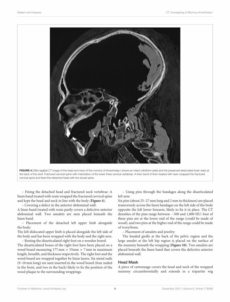
Mummification– Excerebration: The skull base is intact without evidence ofan attempt to remove the brain (Figure 3). The shrunken brainoccupies the posterior aspect of the skull cavity without evidenceof any embalming materials (Figure 4).
– Evisceration: There is no evidence of viscera inside the bodycavity. A vertical left flank opening measuring (90mm × 58mm× 51mm in length, transverse, and craniocaudal dimensions,respectively) was likely the incision used for evisceration. Thechest and abdominopelvic cavities are stuffed with loose linen(about−100HU) and packs with variable CT densities consistentwith linen treated with resin (70–120 HU). The heart is seen inthe left hemithorax.
– Packing: There is no evidence of orbital packing. Traces ofresin are noted on the desiccated eye globes. A small stopper oflinen treated with resin is inserted in each nostril, whichmeasures16mm× 5mm in the right nostril, and 7mm× 4mm in the left.The desiccated tongue is seen at the back of the mouth; no resin
or packs are seen within the mouth (Figure 4). No subcutaneouspacking is noted in the face or elsewhere in the body.
– Wrapping: There is CT evidence of the individual wrappingof the right upper limb and the left lower limb; the fingers andtoes are wrapped with the rest of the right hand and left foot, andnot being individually bandaged. The disarticulated left upperlimb has not been individually wrapped but bandaged along withthe body and with the right upper limb. The right thigh and legare individually wrapped. The disarticulated right foot has notbeen individually wrapped; it was bandaged to an underlyingwood board and with the left foot. The circumcised penis hasbeen independently wrapped. The body of themummy is coveredby transverse wrapping in a spiral fashion. The thickness of themummy wrapping is (78–112mm) in the front and (21–40mm)at the back of the body.
– Arms position: The right forearm of Amenhotep I crossesthe body with a right angle at the elbow. The disarticulated righthand is placed in front of the transversely oriented right forearm.The left upper limb is placed along the body side. The twomissingfingers from the left hand are seen inside the abdominal cavity.
Amulets and JewelryA total number of 30 amulets/jewelry pieces are found in thewrappedmummy of Amenhotep I. The details of the amulets andjewelry are listed in Table 1.
Reburial Embalming TreatmentThe mummy shows signs of repair:
Frontiers in Medicine | www.frontiersin.org 5 December 2021 | Volume 8 | Article 778498
Saleem and Hawass CT Unwrapping of Mummy Amenhotep I
FIGURE 4 | Mid-sagittal CT image of the head and neck of the mummy of Amenhotep I shows an intact cribriform plate and the preserved desiccated brain rests at
the back of the skull. Fractured cervical spine with malrotation of the lower three cervical vertebrae. A linen band of linen treated with resin wrapped the fractured
cervical spine and fixed the detached head with the dorsal spine.
– Fixing the detached head and fractured neck vertebrae: Alinen band treated with resin wrapped the fractured cervical spineand kept the head and neck in line with the body (Figure 4).
– Covering a defect in the anterior abdominal wall:A linen band treated with resin partly covers a defective anteriorabdominal wall. Two amulets are seen placed beneath thelinen band.
– Placement of the detached left upper limb alongsidethe body:The left dislocated upper limb is placed alongside the left side ofthe body and has been wrapped with the body and the right arm.
– Resting the disarticulated right foot on a wooden board:The disarticulated bones of the right foot have been placed on awood board measuring 177mm × 55mm × 7mm in maximumlength, breadth, and thickness respectively. The right foot and thewood board are wrapped together by linen layers. Six metal nails(9–10mm long) are seen inserted in the wood board (four nailedin the front, and two in the back) likely to fix the position of thewood plaque to the surrounding wrappings.
– Using pins through the bandages along the disarticulatedleft arm:Six pins (about 25–27mm long and 2mm in thickness) are placedtransversely across the linen bandages on the left side of the bodyopposite the left lower forearm, likely to fix it in place. The CTdensities of the pins range between −300 and 1,800 HU: four ofthese pins are at the lower end of the range (could be made ofwood), and two pins at the higher end of the range could be madeof ivory/bone.
– Placement of amulets and jewelry:The beaded girdle at the back of the pelvic region and the
large amulet at the left hip region is placed on the surface ofthe mummy beneath the wrapping (Figure 10). Two amulets areplaced beneath the linen band that covers the defective anteriorabdominal wall.
Head MaskA piece of cartonnage covers the head and neck of the wrappedmummy circumferentially and extends in a tripartite wig
Frontiers in Medicine | www.frontiersin.org 6 December 2021 | Volume 8 | Article 778498

Saleem and Hawass CT Unwrapping of Mummy Amenhotep I
FIGURE 5 | Three-dimensional frontal CT image of the lower torso and upper limbs of the mummy of Amenhotep I. The right forearm is flexed at the elbow and
crosses the lower abdomen transversely; the right hand is dislocated at the wrist and is displaced anterior to the forearm. The dislocated left arm and forearm are
placed extended along the left side of the body. The broken left (hand) has three flexed fingers; the missing two fingers are seen inside an anterior abdominal wall
defect (long arrow). The fractures were likely inflicted by tomb robbers. The initial position of the arms was probably crossed in front of the body. A short pin is placed
transversely across the bandages (short arrow) likely to fix the left disarticulated arm in place.
configuration till the mid-chest level in the front and back. Themaximum dimensions of the cartonnage: in length is 444mm infront and 399 at the back; in width is 320mm; 225mm in depth.The thickness of the cartonnage measures 5.5–6.5mm; it shows acentral low-density layer of linen/papyrus (−30 HU) covered bya thin layer of a higher density material (gesso plaster) from theoutside and another layer of gesso from inside (700–1,000 HU).
The face of the mask is formed of a separate piece of woodmounted on the molded front of the cartonnage. The woodpiece measures 1,200 HU in CT density and measures: 250mmin length, 184mm in width, and 12mm in thickness. The eyesof the mask are inlaid stones (Figure 11). The black pupil ofeach eye is made of a discoid structure with a homogeneousCT density (1,693–1,700 HU) corresponding to obsidian crystals.
Frontiers in Medicine | www.frontiersin.org 7 December 2021 | Volume 8 | Article 778498
Saleem and Hawass CT Unwrapping of Mummy Amenhotep I
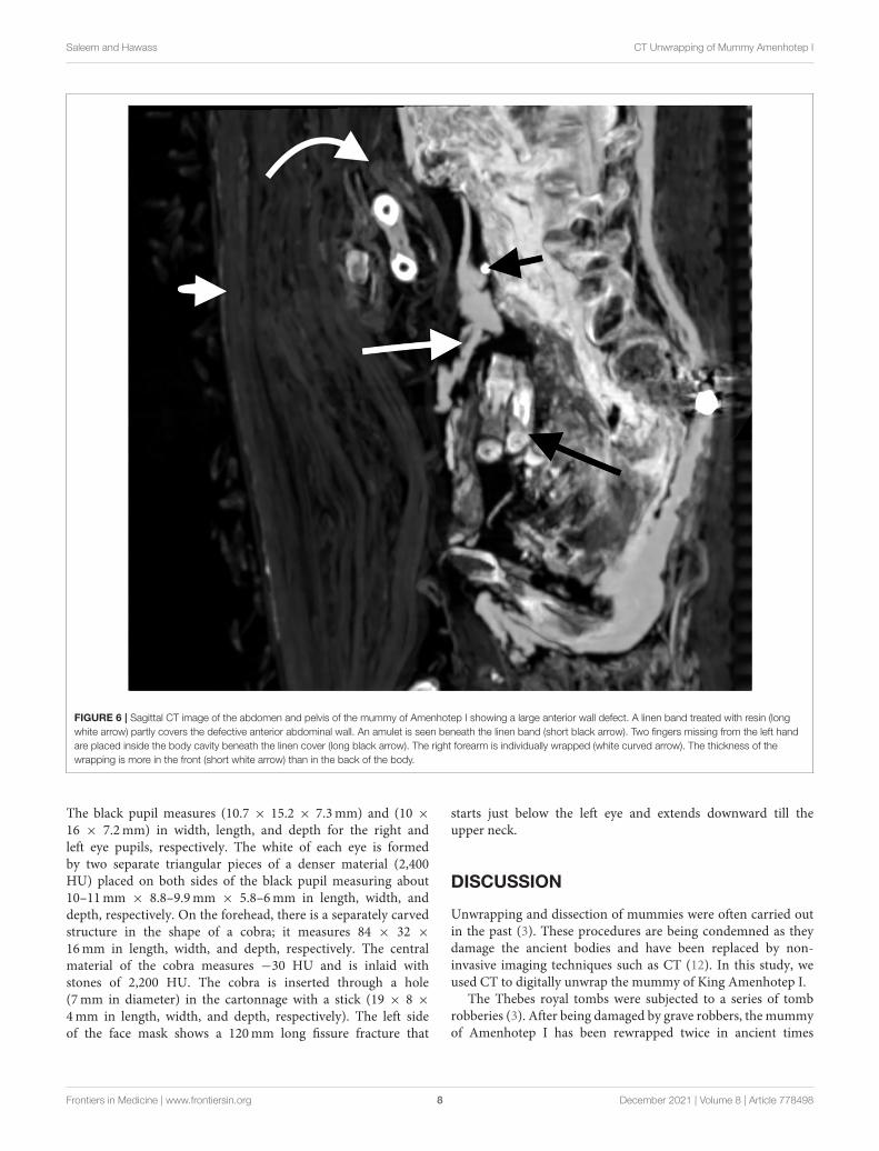
FIGURE 6 | Sagittal CT image of the abdomen and pelvis of the mummy of Amenhotep I showing a large anterior wall defect. A linen band treated with resin (long
white arrow) partly covers the defective anterior abdominal wall. An amulet is seen beneath the linen band (short black arrow). Two fingers missing from the left hand
are placed inside the body cavity beneath the linen cover (long black arrow). The right forearm is individually wrapped (white curved arrow). The thickness of the
wrapping is more in the front (short white arrow) than in the back of the body.
The black pupil measures (10.7 × 15.2 × 7.3mm) and (10 ×
16 × 7.2mm) in width, length, and depth for the right andleft eye pupils, respectively. The white of each eye is formedby two separate triangular pieces of a denser material (2,400HU) placed on both sides of the black pupil measuring about10–11mm × 8.8–9.9mm × 5.8–6mm in length, width, anddepth, respectively. On the forehead, there is a separately carvedstructure in the shape of a cobra; it measures 84 × 32 ×
16mm in length, width, and depth, respectively. The centralmaterial of the cobra measures −30 HU and is inlaid withstones of 2,200 HU. The cobra is inserted through a hole(7mm in diameter) in the cartonnage with a stick (19 × 8 ×
4mm in length, width, and depth, respectively). The left sideof the face mask shows a 120mm long fissure fracture that
starts just below the left eye and extends downward till theupper neck.
DISCUSSION
Unwrapping and dissection of mummies were often carried outin the past (3). These procedures are being condemned as theydamage the ancient bodies and have been replaced by non-invasive imaging techniques such as CT (12). In this study, weused CT to digitally unwrap the mummy of King Amenhotep I.
The Thebes royal tombs were subjected to a series of tombrobberies (3). After being damaged by grave robbers, the mummyof Amenhotep I has been rewrapped twice in ancient times
Frontiers in Medicine | www.frontiersin.org 8 December 2021 | Volume 8 | Article 778498
Saleem and Hawass CT Unwrapping of Mummy Amenhotep I
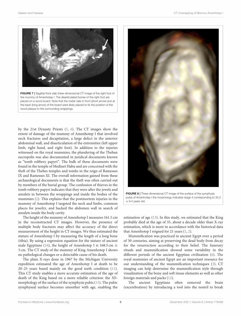
FIGURE 7 | Sagittal thick slab three-dimensional CT image of the right foot of
the mummy of Amenhotep I. The disarticulated bones of the right foot are
placed on a wood board. Note that the metal nails in front (short arrow) and at
the back (long arrow) of the board were likely placed to fix the position of the
wood plaque to the surrounding wrappings.
by the 21st Dynasty Priests (5, 6). The CT images show theextent of damage of the mummy of Amenhotep I that involvedneck fractures and decapitation, a large defect in the anteriorabdominal wall, and disarticulation of the extremities (left upperlimb, right hand, and right foot). In addition to the injurieswitnessed on the royal mummies, the plundering of the Thebannecropolis was also documented in juridical documents knownas “tomb robbery papyri”. The bulk of these documents werefound in the temple of Medinet Habu and are concerned with thetheft of the Thebes temples and tombs in the reign of RamessesIX and Ramesses XI. The overall information gained from thesearchaeological documents is that the theft was often carried outby members of the burial group. The confession of thieves in thetomb robbery papyri indicates that they were after the jewels andamulets in between the wrappings and inside the bodies of themummies (2). This explains that the postmortem injuries in themummy of Amenhotep I targeted the neck and limbs, commonplaces for jewelry, and hacked the abdomen wall in search ofamulets inside the body cavity.
The height of the mummy of Amenhotep I measures 161.5 cmin the reconstructed CT images. However, the presence ofmultiple body fractures may affect the accuracy of the directmeasurement of the height in CT images. We thus estimated thestature of Amenhotep I by measuring the length of a long bone(tibia). By using a regression equation for the stature of ancientmale Egyptians (16), the height of Amenhotep I is 168.5 cm ±
3 cm. The CT study of the mummy of King Amenhotep I showsno pathological changes or a detectable cause of his death.
The plain X-rays done in 1967 by the Michigan Universityexpedition estimated the age of Amenhotep I at death to be20–25 years based mainly on the good teeth condition (11).This CT study enables a more accurate estimation of the age ofdeath of the King based on a more reliable criterion: the 3D-morphology of the surface of the symphysis pubis (15). The pubicsymphyseal surface becomes smoother with age, enabling the
FIGURE 8 | Three-dimensional CT image of the surface of the symphysis
pubis of Amenhotep I the morphology indicates stage 4 corresponding to 35.2
± 9.4 years old.
estimation of age (13). In this study, we estimated that the Kingprobably died at the age of 35, about a decade older than X-rayestimation, which is more in accordance with the historical datathat Amenhotep I reigned for 21 years (1, 2).
Mummification was practiced in ancient Egypt over a periodof 30 centuries, aiming at preserving the dead body from decayfor the resurrection according to their belief. The funeraryrituals and mummification showed some variability in thedifferent periods of the ancient Egyptian civilization (6). Theroyal mummies of ancient Egypt are an important resource forour understanding of the mummification techniques (3). CTimaging can help determine the mummification style throughvisualization of the bony and soft tissue elements as well as otherforeign materials and packs (12).
The ancient Egyptians often removed the brain(excerebration) by introducing a tool into the nostril to break
Frontiers in Medicine | www.frontiersin.org 9 December 2021 | Volume 8 | Article 778498

Saleem and Hawass CT Unwrapping of Mummy Amenhotep I
FIGURE 9 | Three-dimensional CT image of the teeth of Amenhotep I in frontal view shows a full set of healthy teeth.
the weak part of the anterior skull base. The brain was removedand sometimes embalming materials were introduced insidethe skull. Attempts of excerebration were as early as the FourthDynasty (18). The CT images of the mummy of Amenhotep Ishow that there was no attempt to remove the brain which isseen shrank and occupying the back of the skull. Other royalmummies dated around the time of Amenhotep I (late 17thDynasty to early 18th Dynasty) have not been excerebrated(12). These include Seqenenre Taa II, Meritamun, Thutmose II,Thutmose III, and Hatshepsut (19–21). Excerebration becamepopular later in the 18th Dynasty and the peak of the procedureis suggested to be in the Ptolemaic Period (18).
To prevent body putrefaction, the ancient Egyptianembalmers removed the internal organs through an abdominalincision (6). The CT scan of the mummy of Amenhotep Irevealed a vertical left flank incision. Before the 18th Dynasty,incisions were vertical in the left flank and extended from theanterior superior iliac spine upwards. An example of a verticalleft flank incision has been reported in the CT of the mummyof Seqenenre Taa II (17th Dynasty) (12, 20). Later in the 18thDynasty, most of the royal mummies have oblique incisions
parallel to the left inguinal ligament as in Thutmose III (18thDynasty), Ramesses II (19th Dynasty), and Ramesses III (20thDynasty) (12). During the process of evisceration, the heart waspreserved (6). The CT images identified the presence of theheart within the chest of Amenhotep I. Similarly, previous CTstudies identified the preserved heart in other New Kingdomroyal mummies as in Thutmose II, Ramesses II, Ramesses III(12). The ancient Egyptian embalmers used different materialsto fill the emptied body cavity (6, 12). The CT images show thatthe body cavity of Amenhotep I, similar to other royal mummiesof the New Kingdom, is filled with materials with different CTdensities representing linen fibers, and linen packs treated withresin (12). To make the corpse look lifelike, the embalmers of theroyal mummies dated to the New Kingdom usually used packsin the eyes, nose, mouth, and under the skin (22). However, thisstudy shows that the mummy of Amenhotep I has not receivedsuch treatment.
Bandaging and wrapping the mummified body with linensheets was an important stage of the embalming process inancient Egypt (2). The bandaging techniques of the mummychanged during the different periods of ancient Egyptian
Frontiers in Medicine | www.frontiersin.org 10 December 2021 | Volume 8 | Article 778498

Saleem and Hawass CT Unwrapping of Mummy Amenhotep I
TABLE 1 | Computed tomography (CT) findings of Amulets and jewelry related to the mummy of Amenhotep I.
Region Number of
amulets
Description and identification
Right upper limb 7 – At the shoulder region (n = 3)
Anterior to gleno-humeral joint: quartz/faience Eye of Horus (19 × 18 × 6mm; 1,650 HU)
Posterior to gleno-humeral joint: quartz/faience Eye of Horus (13 × 11 × 2mm, 1,670 HU)
Anterior to scapula: Quartz/faience eye of Horus (19 × 13 × 4.3mm; 1,550 HU)
– Between the wrapping at the back of the right arm (n = 3):
At the upper arm: a gold Eye of Horus: (21 × 15 × 3mm; 3,059 HU)
At the lower arm: a gold Eye of Horus: (20 × 12mm × 3; 3,070 HU)
At the lower arm: quartz/faience scarab (11 × 9 mmx 3; 1,500 HU)
– Between the wrapping at the back of the forearm (n = 1):
An oblong shaped gold bead (7 × 3 × 4mm; 3,069 HU)
Inside torso cavity 3 Behind the left sterno-clavicular joint (n=1): a rectangular quartz/faience amulet (7.5 × 3.5mm; 1,790 HU)
– Anterior abdominal wall behind embalming pack at L1 level in midline and to the left side (n = 2): an oblong quartz/faience
bead (9 × 4 × 3mm; 1,800 HU); a fired clay/faience scarab amulet (8.3 × 7.5 × 4mm; HU 1,100 HU)
On torso surface and
between wrappings
10 – In front of the right sterno-clavicular joint (n = 1): a discoid gold amulet (7 × 5 × 3mm; 3,000 HU).
– Right posterior chest wall on outer wrapping (n = 2): a pointed gold amulet (7 × 4 × 3mm; 3,066 HU); a rectangular gold
amulet (6.5 × 4.5 × 3mm; 3,069 HU).
– At right lateral torso wall between wrappings from up to down (n = 5): a gold oblong bead (5 × 85mm; 3,000 HU); a double
plume quartz/faience amulet (19 × 12 × 5mm; 1,789 HU); an oblong-shaped (Wadji) gold amulet (10 × 4.5 × 4mm; 3,006
HU); a gold discoid amulet (5 × 4 × 2mm; 2,979 HU); a pyramid quartz/faience amulet (10 × 10.4 × 9mm); 1,800 HU).
– At the left lateral side of pelvis on the body surface (n = 1): an oblong shaped amulet with pointed ends (snail shell) made of
quartz/faience (45 × 20 × 17mm; HU 1,800 HU).
– At the back of the pelvic region on the body surface (n = 1): A girdle formed of 34 beads gold beads (7.2 to 10.1mm in
diameter; 3,036 HU). The beads are joined with double metal strings at the periphery
Left lower limb 10 In front of the wrapping on the upper end of left femur arranged transversely from medial to lateral (n = 5): A rod-shaped stone
amulet (18 × 7 × 6mm; 1,993 HU); a rectangular plate quartz/faience (6.5 × 6.3 × 4mm; 1,360 HU); a rectangular plate fired
clay amulet (8.6 × 8.3 × 6.4mm; 1,100 HU); an oblong (Wadji) quartz/faience (10 × 5 × 3mm; 1,370 HU); and a serpent
head fired clay amulet (13 × 9 × 5mm; 1,060 HU)
On the outer wrapping at the medial of upper thigh (n = 1): an oblong fired clay amulet (12 × 8 × 5mm; 1,300 HU)
Within the wrappings at the back of the upper thigh (the general wrapping) (n = 1): a gold spherical bead (5mm diameter;
3,028 HU)
On the surface of the upper lateral leg (n = 1): a fired clay scarab amulet (7 × 7 × 5mm; 1,400 HU)
In the wrapping at the lateral of the lower leg (n = 1): a stone bead with a central hole (4.7mm in diameter; 2,700 HU)
In the wrapping at the medial of the lower leg (n = 1): an oblong-shaped quartz/faience amulet (6 × 3 × 3mm; 1,900 HU).
Civilization. In the Middle Kingdom, a large sheet of linen wasused to wrap the mummy. In the New Kingdom, the wrappingbecame more elaborate as the embalmers used overlapping spiralbands to wrap each limb individually. Once all the body partswere wrapped, the embalmers started wrapping the body as awhole. As each part of the mummy was bandaged, the embalmersuttered spells and placed protective amulets on the body andthe different linen layers. In later periods, wrapping grew moresophisticated as the bodies were wrapped with narrow bandages,forming complex patterns (8). The bandages carried the nameor titles of the deceased (2). Little information is known aboutthe style of wrapping of the New Kingdom royal mummies sincemost of them were found in cached burials in Deir el Bahariand Amenhotep II tomb in the Valley of the Kings after beingrewrapped in the 21st Dynasty. The only non-cached burial royalmummy known is Tutankhamun; unfortunately, the wrappingshad become carbonized, and little is known about its pattern (8).The CT images show that the mummy of Amenhotep I has beenfully wrapped. We suggest that some bandages could be original,dated to the 18th Dynasty in addition to the reburial work in the21st Dynasty. The CT images show that the right upper limb andthe left lower limb are individually wrapped and have amulets on
the surface of the limb or in between the layers of the bandages.While the detached left upper limb and the disarticulated rightfoot have no individual bandages or related amulets and havebeen wrapped with the body and/or the contralateral limb. Weassume that the individual wrapping of these limbs was original,dated to the 18th Dynasty, and was a little disturbed by the tombrobbers. While the disarticulated left upper limb and right foothave lost their original individual bandages and amulets and werenot individually wrapped but only offered general bandaging withthe rest of the body by the 21st Dynasty priests. The circumcisedpenis has individual wrapping.
The right forearm of Amenhotep I crosses the body witha right angle at the elbow. The preservation of the individualwrapping and amulets of the right upper limb may support thehypothesis that the forearm was crossed at the time of the initialmummification in the 18th Dynasty. The disarticulated left armand forearm were likely placed alongside the body during thereburial in the 21st Dynasty. The crossing forearms on the bodyare commonly seen in the New Kingdom royal mummies. Themummy of King Ahmose, who preceded Amenhotep I, has botharms along the body (6). This makes the mummy of AmenhotepI likely to be the first to appear with crossed forearms in the
Frontiers in Medicine | www.frontiersin.org 11 December 2021 | Volume 8 | Article 778498
Saleem and Hawass CT Unwrapping of Mummy Amenhotep I
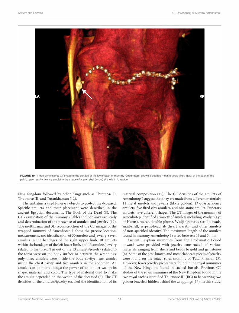
FIGURE 10 | Three-dimensional CT image of the surface of the lower back of mummy Amenhotep I shows a beaded metallic girdle (likely gold) at the back of the
pelvic region and a faience amulet in the shape of a snail shell (arrow) at the left hip region.
New Kingdom followed by other Kings such as Thutmose II,Thutmose III, and Tutankhamun (12).
The embalmers used funerary objects to protect the deceased.Specific amulets and their placement were described in theancient Egyptian documents, The Book of the Dead (8). TheCT examination of the mummy enables the non-invasive studyand determination of the presence of amulets and jewelry (12).The multiplanar and 3D reconstruction of the CT images of thewrapped mummy of Amenhotep I show the precise location,measurement, and identification of 30 amulets and jewelry: sevenamulets in the bandages of the right upper limb, 10 amuletswithin the bandages of the left lower limb, and 13 amulets/jewelryrelated to the torso. Ten out of the 13 amulets/jewelry related tothe torso were on the body surface or between the wrappings;only three amulets were inside the body cavity: heart amuletinside the chest cavity and two amulets in the abdomen. Anamulet can be many things; the power of an amulet was in itsshape, material, and color. The type of material used to makethe amulet depended on the wealth of the deceased (8). The CTdensities of the amulets/jewelry enabled the identification of its
material composition (17). The CT densities of the amulets ofAmenhotep I suggest that they are made from different materials:11 metal amulets and jewelry (likely golden), 13 quartz/faienceamulets, five fired clay amulets, and one stone amulet. Funeraryamulets have different shapes. The CT images of the mummy ofAmenhotep identified a variety of amulets includingWadjet (Eyeof Horus), scarab, double-plume, Wadji (papyrus scroll), beads,snail-shell, serpent-head, ib (heart scarab), and other amuletsof non-specified identity. The maximum length of the amuletsfound in mummy Amenhotep I varied between 45 and 5 mm.
Ancient Egyptian mummies from the Predynastic Periodonward were provided with jewelry constructed of variousmaterials ranging from shells and beads to gold and gemstones(8). Some of the best-known and most elaborate pieces of jewelrywere found on the intact royal mummy of Tutankhamun (3).However, fewer jewelry pieces were found in the royal mummiesof the New Kingdom found in cached burials. Previous CTstudies of the royal mummies of the New Kingdom found in thetwo royal caches identified Thutmose III (BC) to be wearing twogolden bracelets hidden behind the wrappings (17). In this study,
Frontiers in Medicine | www.frontiersin.org 12 December 2021 | Volume 8 | Article 778498

Saleem and Hawass CT Unwrapping of Mummy Amenhotep I
FIGURE 11 | Axial CT image of the face mask at the level of the eyes. The face mask is mounted on the cartonnage (long arrow); the cartonnage is formed of a
central low-density layer of linen/papyrus covered by denser layers of gesso (long arrow). Each eye of the mask is made of inlaid stones of a central discoid structure
that corresponds to the black obsidian pupil and denser pieces on each side for the white of the eye likely made from quartz.
the CT images show a belt formed of 34 gold beads placed directlyon the lower back of the mummy of Amenhotep I.
The wrapped mummy of Amenhotep I provides a uniqueopportunity to understand the intervention done during itsreburial by the 21st Dynasty priests. The hieratic dockets writtenin ink on the coffin gave information on the historical processof the reburial of the mummy Amenhotep I (2). The CTexamination suggests how the Theban priests of the 21st Dynastyrepaired the injuries of the mummy of Amenhotep I inflictedby the tomb robbers and restored the royal dead. The reburialtreatment included: fixing the detached head and fractured neckvertebrae with a resin-treated linen pad; covering the defectiveanterior abdominal wall with a linen band treated with resin;placement of the detached left upper limb extended alongsidethe body and placing the two detached fingers of the left handinside the abdomen; resting the disarticulated right foot on awooden board with metallic nails; wrapping the body; usingpins (likely made of ivory or bones) to fix the bandages atthe disarticulated left upper limb and right foot. Metal nailsand wooden pins were used in the manufacture of funeraryfurniture and coffins in ancient Egypt (23). Although nails havebeen known since the Old Kingdom, they were not used inwoodwork until the 18th Dynasty. During the Old Kingdom,the only metal available was copper. From the Middle Kingdom(c. 2160 BC to 1788 BC), bronze and copper were present.However, iron has not been used in funerary equipment until 700BC (24).
During the reburial of the mummy of Amenhotep I, wesuggest that the 21st Dynasty embalmers placed two amuletsbeneath the linen band they used to cover the anterior abdominalwall defect.We suggest that during reburial the embalmers placed
or maintained in place a golden beaded girdle at the back ofthe pelvis.
The mummy of Amenhotep I wears a mask. Mummy maskswere part of an elaborate burial ritual in ancient Egypt. Theyare usually made of cartonnage. Cartonnage is a cardboard-like material that was used to make coffins for mummies. Thecartonnage substance is made of glued linen or papyrus, coatedwith plaster and water, then molded to the desired shape. The drycartonnage could be then colored or gilded. Cartonnage couldbe made as one piece that covers the full mummy, or in smallerpieces to cover certain regions such as the head, pectoral, or legs.Cartonnage was used in the different periods of Egyptian ancientcivilization dating back to the end of the Old Kingdom (2686–2181 BC). Cartonnage became popular in the Third IntermediatePeriod (1069–664 BC), as well as later in the Ptolemaic-EarlyRoman Periods (330 BC−250 AD) (6). A CT scan can study amummy cartonnage (25). In this study, the CT images show theshape and full dimensions of the mask of Amenhotep I whichis partly hidden by the overlying garlands. The layers of thecartonnage have different CT densities based on their material;the linen/papyrus layer has a lower CT density than that ofthe plaster (gesso) layers on both the inner and outer surfaces.The CT images can help to understand how the parts of thehead mask were assembled. The CT scan shows the face of themask as a separate piece of wood mounted on the front of thecartonnage. The face of the mask has been individualized to thefacial features identical to that on the coffin of Amenhotep I (1).The facial features of the mask of Amenhotep I have given alife-like appearance by using eyes inlaid with stones. The blackeye pupil is discoid in shape made of Obsidian, a naturallyoccurring glass; while the eye white is formed with two separate
Frontiers in Medicine | www.frontiersin.org 13 December 2021 | Volume 8 | Article 778498
Saleem and Hawass CT Unwrapping of Mummy Amenhotep I
pieces made from a denser material (likely quartz) and are placedon both sides of each pupil. Different manufacturing methodsand materials have been identified in the inlaid eyes in ancientEgyptian collections including obsidian, rock crystal, quartz, andivory (6, 26). The manufacturer of the inlaid eye in the mask ofAmenhotep I used three separate pieces of stones which likelyrequired more work than other methods where the white eye wasmade of one piece with a tapering hole in the middle to receive adisc representing the pupil (26). The CT images identify the goodstatus of preservation of the cartonnage with no cracks or bentareas. However, the wood facial mask shows a long thin fissurefracture; a piece of information that may help in its restoration.
The embalmers covered the mummy with garlands of red,yellow, and blue flowers recognized as Delphinium orientale,Sesbania egyptiaca, Acacia nilotica, and Carmanthus tinctorius(8). It was the beautiful reburial job offered by the 21st Dynastypriests for the mummy of Amenhotep I that made Masperodecide not to unwrap the mummy or disturb its novelty (8). Thereburial project of the New Kingdom Royal mummies by the21st Dynasty priests was accused to be for the intention of theremoval and reuse of the royal burial equipment for the ThirdIntermediate Period Kings. Examples include the reemploymentof two coffins originally made for Thutmose I (18th Dynasty)for King Pinudjem I (21st Dynasty) (2). However, this CTexamination of the mummy of Amenhotep I reveals how theTheban priests of the 21st Dynasty had lovingly restored the royalmummified body of Amenhotep I and preserved or provided richamulets and jewelry. This study may make us have re-confidencein the goodwill of the reburial project of the Royal mummies bythe 21st Dynasty priests.
In this study we used CT to scan the mummy of KingAmenhotep I. CT is considered the diagnostic gold standardimaging modality for studying mummies. Magnetic resonanceimaging has a limited value in the examination of drymummifiedremains as this modality primarily provides information on thelocation of mobile hydrogen within the body. Future analysisof the mummy of Amenhotep I may include dual-energy CTscanning. Different materials have different linear attenuationcoefficients at different energy levels. Dual-energy CT scanninguses two different energy levels that may help to identify andcharacterize the embalming materials in the mummy. Dual-energy scanning can also help to reduce metal artifacts in CT
images induced by golden amulets and jewelry placed on themummy (27).
The specialized CT imaging technique in this study may haveapplications in paleo-anthropological and bio-archaeologicalstudies of mummies from Egypt as well as other cultures suchas Peru (28). CT is being used nowadays more frequently inforensic medicine. Postmortem CT imaging can provide valuableinformation in identifications in mass disasters, trauma, andhomicidal cases (27). The methodological tool in this studymay have potential applications in studying cadavers that havebeen preserved by natural mummification due to extremeenvironmental conditions such as dry hot deserts or freezingmountains (27, 28).
CONCLUSION
The digital unwrapping of the mummy of Amenhotep I using CTsets a unique opportunity to reveal non-invasively the physicalfeatures of the King, understand the mummification style early inthe 18th Dynasty, and recognize the reburial intervention done inthe 21st Dynasty.
DATA AVAILABILITY STATEMENT
The datasets presented in this article are not readily availablebecause the data collected during the current study areavailable from the authors on reasonable request and withpermission of the Egyptian Ministry of Antiquities andTourism. Requests to access the datasets should be directedto [email protected].
AUTHOR CONTRIBUTIONS
SS was responsible for the conception and design, acquisitionof data, analysis and interpretation of data, as well asdrafting of the manuscript, and generation of the figuresand accountable for the accuracy and integrity of thework. ZH made substantial contributions to the design,interpretation of the results, and revision of the intellectualcontent and agreed to be accountable for the integrityof any part of the work. All authors read and approvedthe manuscript.
REFERENCES
1. Partridge RB. Faces of Pharaohs Royal Mummies and Coffins from Ancient
Thebes. London: The Rubicon Press (1997).
2. Reeves N, Wilkinson RH. The Complete Valley of the Kings. London: Thames
& Hudson Ltd (1996).
3. Brier B. Egyptian Mummies. New York: William Morrow and
Company (1994).
4. Maspero G. History of Egypt, Chaldaea, Syria, Babylonia, and Assyria.
London: William Clowes And Sons, Limited (1903). Vol. 4. Project
Gutenberg EBook, Release Date: December 16, 2005. EBook #17324.
Available at: http://www.gutenberg.org/files/17324/17324-h/17324-h.htm#
link2HCH0001 (accessed May 12, 2020).
5. Maspero G. Les momies royale de Deir el-Bahari. Paris: Ernest Leroux (1889).
6. Ikram S, Dodson A. Royal Mummies in the Egyptian Museum. Cairo:
American University in Cairo Press (1997).
7. Kitchen K. The Third intermediate Period in Egypt: (1100-
650 BC). second revised edition. Warminster: Aris &
Philips (1986).
8. Smith G. The Royal Mummies: Cairo: Catalogue general des Antiquites
Epyptiennes du Musee du Caire. Nos 61051-61100. The Royal Mummies.
Cairo:L’Institut d’Archeologie Orientale (1912).
9. Derry DE. An X-ray examination of the mummy of King Amenophis I.
Annales du Service des Antiquite’s d’Egypte (ASAE). (1933/4) 34:47–48.
10. Harris JE, Weeks K. X-raying the Pharaohs. New York: Charles Scribner’s Sons
(1973). ISBN 684-13016-5
11. Harris JE, Wente EF. An X-ray Atlas of the Royal Mummies. Chicago:
University of Chicago Press (1980).
Frontiers in Medicine | www.frontiersin.org 14 December 2021 | Volume 8 | Article 778498

Saleem and Hawass CT Unwrapping of Mummy Amenhotep I
12. Hawass Z, Saleem SN. Scanning the Pharaohs: CT Imaging of the New Kingdom
Royal Mummies. AUC Press, New York (2016).
13. Telmon N, Gaston A, Chemla P, Blanc A, Joffre F, Rougé D. Application of the
Suchey-Brooks method to three-dimensional imaging of the pubic symphysis.
J Forensic Sci. (2005) 50:507–12. doi: 10.1520/JFS2004326
14. Lovejoy CO. Dental wear in the Libben population: its functional pattern and
role in the determination of adult skeletal age at death. Am J Phys Anthropol.
(1985) 68:47–56. doi: 10.1002/ajpa.1330680105
15. Pasquier E, De Saint Martin Pernot L, Burdin V, Mounayer C, Le Rest
C, Colin D. Determination of age at death: assessment of an algorithm
of age prediction using numerical three-dimensional CT data from pubic
bones. Am J Phys Anthropol. (1999) 108:261–8. doi: 10.1002/(SICI)1096-
8644(199903)108:3<261::AID-AJPA2>3.0.CO;2-B
16. Raxter MH, Ruff CB, Azab A, Erfan M, Soliman M, El-Sawaf A.
Stature estimation in Ancient Egyptians: a new technique based on
anatomical reconstruction of stature. Am J Phys Anthropol. (2008) 136:147–
55. doi: 10.1002/ajpa.20790
17. Saleem SN, Hawass Z. Multidetector computed tomographic study of
amulets, jewelry, and other foreign objects in royal Egyptian mummies dated
from the 18th to 20th dynasties. J Comput Assist Tomogr. (2014) 38:153–
8. doi: 10.1097/RCT.0b013e3182ab2221
18. Wade AD, Nelson AJ, Garvin GJ. A synthetic radiological study of
brain treatment in ancient Egyptian mummies. Homo. (2011) 62:248–
69. doi: 10.1016/j.jchb.2011.01.004
19. Saleem SN, Hawass Z. Variability in brain treatment during mummification of
royal Egyptians dated to the 18th−20th dynasties: MDCT findings correlated
with the archaeologic literature. AJR Am J Roentgenol. (2013) 200:W336–
44. doi: 10.2214/AJR.12.9405
20. Saleem SN, Hawass Z. Computed tomography study of the mummy of king
seqenenre Taa II: new insights into his violent death. Front Med. (2021)
8:637527. doi: 10.3389/fmed.2021.637527
21. Hawass Z, Saleem SN. Computed tomography examination of the
screaming mummy “Unknown-Woman-A”. Egypt J Radiol Nucl Med. (2020)
51:139 doi: 10.1186/s43055-020-00255-6
22. Saleem SN, Hawass Z. Subcutaneous packing in royal Egyptian mummies
dated from 18th to 20th dynasties. J Comput Assist Tomogr. (2015) 39:301–
6. doi: 10.1097/RCT.0000000000000205
23. Bracci S, Caruso O, Galeotti M, Iannaccone R, Magrini D, Picchi
D. Multidisciplinary approach for the study of an Egyptian coffin
(late 22nd/early 25th dynasty): combining imaging and spectroscopic
techniques. Spectrochim Acta A Mol Biomol Spectrosc. (2015) 145:511–
22. doi: 10.1016/j.saa.2015.02.052
24. Dixon DM. Timber in Ancient Egypt. Commonwealth Forestry Review. (1974)
53:205–9. URL: https://www.jstor.org/stable/42605377
25. Hughes S. Three-dimensional reconstruction of an ancient Egyptian mummy.
In: Imaging the Past. Electronic Imaging and Computer Graphics in museums
and archaeology. British Museum Occasional Paper. 114. London: The British
Museum (1996). p. 211–25.
26. Petrie WMF. Hawara, Biahmu, and Arsinoe. London: Field & Tuer, The
Leadenhall Press (1889).
27. Beckett RG, Conlogue GJ. Advances in Paleoimaging. Applications for
Paleoanthropology, Bioarchaeology, Forensics, and Cultural Artefacts. London:
CRC (2020). doi: 10.4324/9781315203089
28. Beckett RG, Conlogue GJ, Nelson A. Case Studies for Advances in
Paleoimaging and Other Non-Clinical Applications. Boca Raton: CRC
(2021). doi: 10.4324/9780429318597
Conflict of Interest: The authors declare that the research was conducted in the
absence of any commercial or financial relationships that could be construed as a
potential conflict of interest.
Publisher’s Note: All claims expressed in this article are solely those of the authors
and do not necessarily represent those of their affiliated organizations, or those of
the publisher, the editors and the reviewers. Any product that may be evaluated in
this article, or claim that may be made by its manufacturer, is not guaranteed or
endorsed by the publisher.
Copyright © 2021 Saleem and Hawass. This is an open-access article distributed
under the terms of the Creative Commons Attribution License (CC BY). The use,
distribution or reproduction in other forums is permitted, provided the original
author(s) and the copyright owner(s) are credited and that the original publication
in this journal is cited, in accordance with accepted academic practice. No use,
distribution or reproduction is permitted which does not comply with these terms.
Frontiers in Medicine | www.frontiersin.org 15 December 2021 | Volume 8 | Article 778498