COMMUNITY STRUCTURE OF BRACHYURAN CRAB IN SETIU LAGOON, TERENGGANU
Upload
khangminh22Category
view
0download
0
1 3
J Comp Physiol BDOI 10.1007/s00360-014-0815-2
OrIgInal PaPer
Digestive enzymes of two brachyuran and two anomuran land crabs from Christmas Island, Indian Ocean
Stuart M. Linton · Reinhard Saborowski · Alicia J. Shirley · Jake A. Penny
received: 16 December 2013 / revised: 28 January 2014 / accepted: 6 February 2014 © Springer-Verlag Berlin Heidelberg 2014
higher endo-β-1,4-glucanase, lichenase and laminarinase activities compared to that of the other species. Thus, C. perlatus may be efficient at digestion of cellulose and hemi-cellulose within plant material. Zymography indicated that the majority of protease, lipase, phosphatase, amylase, endo-β-1,4-glucanase, β-glucohydrolase and N-acetyl-β-d-glucosaminidase isozymes were common to all species, and hence were inherited from a common aquatic ancestor. Differences were observed for the phosphatase, lipase and endo-β-1,4-glucanase isozymes. These differences are dis-cussed in relation to phylogeny and possible evolution to cope with the adoption of a terrestrial diet.
Keywords land crabs · Digestion · Digestive enzymes · Zymography
Introduction
The gecarcinid land crabs, Gecarcoidea natalis (Pocock 1888) and Discoplax celeste (ng and Davie 2012) and the coenobitid land crabs, Birgus latro (linnaeus 1767) and Coenobita perlatus (Milne edwards 1853) are sympat-ric species on Christmas Island, Indian Ocean. all these species consume a variety of material ranging from plant material in the form of fruits, seeds and in some cases leaf litter to animal material in the form of carrion, dead con-specifics and in some cases prey. The detailed composi-tion of digestive enzymes in these species in terms of their activities, number of isoforms and molecular masses is unknown. However, given the ubiquitous nature of essen-tial nutrients they are likely to possess proteases for hydrol-ysis of protein, lipases for the hydrolysis of lipid and car-bohydrases such as α-amylase for the hydrolysis of storage carbohydrate.
Abstract The digestive ability of four sympatric land crabs species (the gecarcinids, Gecarcoidea natalis and Discoplax celeste and the anomurans, Birgus latro and Coe‑nobita perlatus) was examined by determining the activity of their digestive enzymes. The gecarcinids are detritivores that consume mainly leaf litter; the robber crab, B. latro, is an omnivore that preferentially consumes items high in lipid, carbohydrate and/or protein; C. perlatus is also an omnivore/detritivore. all species possess protease, lipase and amylase activity for hydrolysing ubiquitous protein, lipid and storage polysaccharides (glycogen and starch). Similarly all species possess enzymes such as N-acetyl-β-d-glucosaminidase, the cellulases, endo-β-1,4-glucanase and β-glucohydrolase and hemicellulases, lichenase and lamina-rinase for the respective hydrolysis of structural substrates chitin, cellulose and hemicelluloses, lichenan and lamina-rin. except for the enzyme activities of C. perlatus, enzyme activity could not be correlated to dietary preference. Per-haps others factors such as olfactory and locomotor ability and metabolic status may determine the observed dietary preferences. The digestive fluid of C. perlatus possessed
Communicated by I.D. Hume.
S. M. linton · a. J. Shirley · J. a. Penny School of life and environmental Sciences, Deakin University, Pigdons road, Waurn Ponds, VIC 3217, australia
S. M. linton (*) School of life and environmental Sciences, Deakin University, locked bag 20000, geelong, VIC 3220, australiae-mail: [email protected]
r. Saborowski Functional ecology, alfred Wegener Institute for Polar and Marine research, P.O. Box 120161, 27570 Bremerhaven, germany

J Comp Physiol B
1 3
although these four species are omnivorous/detritivorous, they have different foraging strategies and display a distinct preference for certain dietary items. The red crab, Gecar‑coidea natalis and the blue crab Discoplax celeste have small home ranges in the non-breeding season and their for-aging is restricted to these areas (Table 1). The diet of both species consists mainly of brown leaf litter (low in nitrogen and high in cellulose and hemicellulose content) with small amounts of fruits, seeds and animal material, when avail-able (Table 1). The anomuran land crabs, B. latro and the terrestrial hermit crabs, Coenobita sp belong to the family, Coenobitidae (Davie 2002). B. latro is an omnivore with a distinct preference for items such as fruits, seeds and ani-mal material which are high in carbohydrate, lipid and protein (linton and greenaway 2007; Wilde et al. 2004) (Table 1). They are capable of travelling large distances, up to 1 km per day, and possess an excellent olfactory ability which allow them to seek out their preferred food (Stens-myr et al. 2005; greenaway 2001; Fletcher et al. 1990). Ter-restrial hermit crabs such as C. perlatus are nocturnal and are restricted to areas near the shore such as beaches and terrace forest (Hartnoll 1988). Coenobita species are largely omnivores/detritivores, which consume a variety of plant, animal and shoreline detritus (Table 1) (grubb 1971; Barnes 1997; greenaway 2003). The diet for C. perlatus on Christ-mas Island species is largely unknown, but given the species is found near the shore and not in the terrace forest it may consume, like other Coenobita, strandline detritus, some plants and perhaps algae scrapped off the rocks at night.
given these differences in feeding preferences between the species, the question arises whether these prefer-ences are reflected in the complement (activity and pres-ence) of digestive enzymes? In particular are the activities of the digestive enzymes higher for a preferred substrate? Digestive enzymes are likely to be correlated to the dietary compounds present in the diet with the animal being eco-nomical, producing only enzymes it would require and not wasting energy-producing ones it would not use (Kara-sov et al. 2011). activities of digestive enzymes have been correlated to dietary preferences in other species of deca-pod crustaceans, amphipods and vertebrates such as fish and bats (family Phyllostomidae) (Johnston and Freeman 2005; Karasov et al. 2011; Johnston and Yellowlees 1998; Johnston et al. 2004; german et al. 2010; Schondube et al. 2001). generally, carnivorous species possess high protease activities, species which consume starchy items and algae possess high amylase and laminarinase activities, insectivo-rous species possess high chitinase and trehalase activities, while species which consume plant material have high cel-lulase activities (Johnston and Freeman 2005; Karasov et al. 2011; Johnston and Yellowlees 1998; Johnston et al. 2004; german et al. 2010; Schondube et al. 2001). a diverse range of digestive enzymes is likely in G. natalis, D. celeste and B.
latro given the diverse diet and previous demonstrations that these species are capable of digesting a range of substrates (greenaway and linton 1995; greenaway and raghaven 1998; Wilde et al. 2004). However given the differences in feeding ecology, the activities of particular classes of enzymes, may be higher to reflect the species preferred die-tary substrates. The gecarcinids may possess high cellulase and hemicellulase activities while B. latro may possess high protease, lipase and amylase activities.
activities of digestive enzymes were measured within the digestive fluid of the gecarcinids, G. natalis and D. celeste and the anomurans, B. latro and C. perlatus to determine one, the presence of the different types of diges-tive enzymes and two, if the dietary preferences of the spe-cies were reflected in the enzyme complement. Zymograms were also conducted to determine the number and estimated size of the digestive enzymes. Similarities in activity, num-ber and size of enzymes across all species may suggest that the enzymes were inherited from a common aquatic ances-tor. any difference between the gecarcinid and coenobitid groups may be indicative of phylogeny, while differences between closely related species, G. natalis and D. celeste or B. latro and C. perlatus may be indicative of adaptations to a terrestrial diet.
Methods
Sampling of digestive fluid
land crabs Birgus latro (linnaeus, 1767), Coenobita per‑latus (H. Milne edwards 1837), Gecarcoidea natalis (Poc-ock, 1888) and Discoplax celeste (ng and Davie 2012) were collected from the rainforest on Christmas Island, Indian Ocean and held at the Christmas Island research station for 24 h without food. Only male G. natalis and D. hirtipes were collected. The mass range of G. natalis and D. hirtipes was, respectively, 103–225 and 251–424 g. Mixed sex B. latro and C. perlatus were collected. The mass of B. latro ranged from 1 to 2 kg, while the mass of C. perlatus ranged from 16 to 60 g. Digestive fluid was then collected from the cardiac stomach of each crab. To do this, one experimenter held the crab, ventral side up while another experimenter inserted the narrow tip of a 10-ml eppendorf Combitip into the cardiac stomach and applied gentle suction to collect the fluid. Depending on the size of the crab, between 2 and 4 ml of digestive fluid was col-lected. Digestive fluid samples were immediately dispensed into 2-ml screw top tubes, and frozen in a liquid nitro-gen-cooled biological shipper (CX100 Taylor-Wharton). Samples were then air-freighted back to the laboratories at Deakin University, geelong, australia and the alfred Wegener Institute in Bremerhaven, germany.

J Comp Physiol B
1 3
Preparation of digestive fluid for enzyme assays
The digestive fluid samples were centrifuged at 10,000g for 10 min to pellet any particulate matter and then diluted with demineralised water. The samples were assayed for total protein and activities of the following enzymes, total protease, serine protease, cysteine protease, trypsin, chymotrypsin, alanine amino peptidase, lipase, ester-ase, alkaline phosphatase, N-acetyl-β-d-glucosaminidase (nagase), laminarinase, endo-β-1,4-glucanase, lichenase, amylase and β-1,4-glucosidase (Table 2). except for total protease activities, which were expressed as the change in absorbance per minute per mg protein, enzyme activities were expressed as units (U) (μmol of product produced or consumed) per minute per mg of protein. all enzyme assays were conducted at 30 °C. Zymograms were also conducted to estimate the size and number of the major digestive enzymes: total protease, esterase, N-acetyl-β-d-glucosaminidase (nagase), phosphatase, β-glucosidase and endo-β-1,4-glucanase (Table 3).
Total protein measurement
Protein in the digestive fluid was measured using a com-mercial Bradford reagent (Biorad, Hercules, Ca #500-0006) as per the manufacturers protocol (Bradford 1976). Solutions of bovine serum albumin at concentrations between 0 and 1 mg ml−1 were used as standards. Diges-tive fluid samples were diluted so that their absorbance in the protein assays was within the range of the standards.
enzymes assays
Protease enzymes
Using the methods of Teschke and Saborowski (2005) and Saborowski et al. (2004) total protease, trypsin, chymot-rypsin and alanine amino peptidase activities were meas-ured in the digestive fluid (Table 2). Inhibition assays were also conducted on the digestive fluid using the methods of Teschke and Saborowski (2005) to determine the percent-age of serine, cysteine and other proteases, possibly metal-loproteases within it (Table 2).
Lipase, esterase and N‑acetyl‑β‑d‑glucosaminidase
lipase, esterase and N-acetyl-β-d-glucosaminidase activi-ties were measured in digestive fluid using the respec-tive substrates 4-methylumbelliferyl oleate (75164, Fluka), 4-methylumbelliferyl butyrate (19362, Sigma) and 4-methylumbelliferyl N-acetyl-β-d-glucosaminide (M2133 Sigma) as per the method of Knotz et al. (2006) (Table 2). These assays measure the continuous release of Ta
ble
1 F
eedi
ng m
ode,
die
tary
item
s co
nsum
ed a
nd r
elat
ive
hom
e ra
nges
of
four
sym
patr
ic la
nd c
rab
spec
ies
on C
hris
tmas
Isl
and,
Ind
ian
Oce
an
Spec
ies
Feed
ing
mod
eD
ieta
ry it
ems
Hom
e ra
nge
ref
eren
ces
Com
mon
ly c
onsu
med
rar
ely
cons
umed
Gec
arco
idea
nat
alis
Det
ritiv
ore
Bro
wn
leaf
litte
rFr
uits
, see
ds, c
arri
on,
dead
con
spec
ifics
≈5
m2 in
non
-bre
edin
g se
ason
ada
mcz
ewsk
a an
d M
orri
s (2
001)
, l
into
n an
d g
reen
away
(20
07)
Dis
copl
ax c
eles
teD
etri
tivor
eg
reen
and
bro
wn
le
af li
tter
Frui
ts, s
eeds
, car
rion
, de
ad c
onsp
ecifi
csSm
all r
estr
icte
d to
wet
soa
ks a
nd
spri
ngs
Hic
ks e
t al.
(199
0), l
into
n an
d
gre
enaw
ay (
2007
)
Bir
gus
latr
oO
mni
vore
Frui
ts, s
eeds
, ani
mal
mat
eria
l [ca
rrio
n, d
ead
co
nspe
cific
s, p
rey
(≈1
red
crab
per
day
)]l
arge
, tra
vels
up
to 1
km
per
day
lin
ton
and
gre
enaw
ay (
2007
), W
ilde
et a
l. (2
004)
, gre
enaw
ay (
2001
)
Coe
nobi
ta p
erla
tus
Om
nivo
re/d
etri
tivor
eFa
llen
frui
ts, s
eeds
, man
grov
e pr
opag
ules
, alg
ae,
rotti
ng te
rres
tria
l veg
etat
ion,
str
andl
ine
detr
itus,
an
imal
fae
ces,
car
rion
Smal
l, re
stri
cted
to a
rea
adja
cent
to
the
shor
e du
e to
req
uire
men
t to
re
plen
ish
shel
l wat
er w
ith s
ea
wat
er f
or o
smor
egul
atio
n
gru
bb (
1971
), B
arne
s (1
997)
, g
reen
away
(20
03),
Har
tnol
l (19
88)

J Comp Physiol B
1 3
Tabl
e 2
Det
ails
of
the
enzy
me
assa
ys u
sed
in th
is s
tudy
enz
yme
Dilu
tion
fact
or o
f di
gest
ive
fluid
us
ed in
ass
ay
How
enz
yme
activ
ity w
as
mea
sure
dU
nits
of
enzy
me
activ
ityB
uffe
r us
ed in
en
zym
e as
say
Subs
trat
e or
inhi
bito
rFi
nal s
ubst
rate
co
ncen
trat
ion
or
effe
ctiv
e in
hibi
tor
co
ncen
trat
ion
ref
eren
ces
Tota
l pro
teas
e50
Hyd
roly
sis
of a
zoca
sein
to
rele
ase
azo
dye
Δ36
6 nm
min
−1 m
g−1
prot
ein
0.1
mol
l−
1 T
risH
Cl
pH =
7.5
azo
case
in (
Sigm
a a
2765
)1.
85 m
g m
l−
1Te
schk
e an
d Sa
b-or
owsk
i (20
05),
Sa
boro
wsk
i et a
l. (2
004)
Seri
ne p
rote
ase
50r
educ
tion
in to
tal p
rote
ase
activ
ity th
roug
h in
hibi
tion
of
seri
ne p
rote
ases
Δ36
6 nm
min
−1 m
g−1
prot
ein
0.1
mol
l−
1 T
risH
Cl
pH =
7.5
Seri
ne p
rote
ase
in
hibi
tor
4-(2
-am
ino-
ethy
l) b
enze
nesu
lfon
yl
fluor
ide
hydr
ochl
orid
e (a
eB
SF)
(Sig
ma
a84
56)
0.5
mm
ol l
−1
Tesc
hke
and
Sab-
orow
ski (
2005
)
Cys
tein
e pr
otea
se50
red
uctio
n in
tota
l pro
teas
e ac
tivity
thro
ugh
inhi
bitio
n of
cy
stei
ne p
rote
ases
Δ36
6 nm
min
−1 m
g−1
prot
ein
0.1
mol
l−
1 T
risH
Cl
pH =
7.5
Cys
tein
e pr
otea
se
inhi
bito
r tr
ans-
ep
oxy-
succ
inyl
-l-
leuc
ylam
ido-
(4-g
uani
dino
)
buta
ne (
e64
)
(Sig
ma
e31
32)
0.5
mm
ol l
−1
Tesc
hke
and
Sab-
orow
ski (
2005
)
Try
psin
20H
ydro
lysi
s of
sub
stra
te to
re
leas
e p-
nitr
ophe
nol
μm
ol p
-nitr
ophe
nol m
in−
1 (U
) m
g−1 p
rote
in0.
1 m
ol l
−1
Tri
sHC
l pH
= 8
N-α
-Ben
zoyl
-l-a
rg-p
-ni
troa
nilid
e hy
dro-
chlo
ride
(B
aPn
a)
(Sig
ma
B32
79)
1 m
mol
l−
1Sa
boro
wsk
i et a
l. (2
004)
Chy
mot
ryps
in1,
000
Hyd
roly
sis
of s
ubst
rate
to
rele
ase
p-ni
trop
heno
lμ
mol
p-n
itrop
heno
l min
−1
(U)
mg−
1 pro
tein
0.1
mol
l−
1 T
risH
Cl
pH =
8
N-S
ucci
nyl-
(ala
)2-P
ro-
Phe-
p-ni
troa
nilid
e (S
aPn
a)
(Sig
ma
S738
8)
1 m
mol
l−
1Sa
boro
wsk
i et a
l. (2
004)
ala
nine
am
inop
epti-
dase
5H
ydro
lysi
s of
sub
stra
te to
re
leas
e p-
nitr
ophe
nol
μm
ol p
-nitr
ophe
nol m
in−
1 (U
) m
g−1 p
rote
in0.
1 m
ol l
−1
Tri
sHC
l pH
= 8
l-a
lani
ne-4
-nitr
o an
ilide
hyd
roch
lori
de
(Sig
ma
a93
25)
1 m
mol
l−
1Sa
boro
wsk
i et a
l. (2
004)
lip
ase
2,50
0H
ydro
lysi
s of
sub
stra
te
to r
elea
se fl
uore
scen
t 4-
β-m
ethy
lum
belli
fero
ne
μm
ol
4-β
-met
hylu
mbe
llife
rone
m
in−
1 (U
) m
g−1 p
rote
in
0.1
mol
l−
1 T
risH
Cl
pH =
7
4-M
ethy
lum
belli
fery
l ol
eate
(Fl
uka
7516
4)0.
1 m
mol
l−
1K
notz
et a
l. (2
006)
est
eras
e2,
500
Hyd
roly
sis
of s
ubst
rate
to
rel
ease
fluo
resc
ent
4-β
-met
hylu
mbe
llife
rone
μm
ol
4-β
-met
hylu
mbe
llife
rone
m
in−
1 (U
) m
g−1 p
rote
in
0.1
mol
l−
1 T
risH
Cl
pH =
7
4-M
ethy
lum
belli
fery
l bu
tyra
te
(Flu
ka 1
9362
)
0.1
mm
ol l
−1
Kno
tz e
t al.
(200
6)
N-a
cety
l-β
-d-
gluc
osam
inid
ase
5H
ydro
lysi
s of
sub
stra
te
to r
elea
se fl
uore
scen
t 4-
β-m
ethy
lum
belli
fero
ne
μm
ol
4-β
-met
hylu
mbe
llife
rone
m
in−
1 (U
) m
g−1 p
rote
in
0.1
mol
l−
1 so
dium
citr
ate
pH =
5.5
4-M
ethy
lum
belli
fery
l N
-ace
tyl-
β
-d-g
luco
sam
inid
e (S
igm
a M
2133
)
0.1
mm
ol l
−1
Kno
tz e
t al.
(200
6)

J Comp Physiol B
1 3
Tabl
e 2
con
tinue
d
enz
yme
Dilu
tion
fact
or o
f di
gest
ive
fluid
us
ed in
ass
ay
How
enz
yme
activ
ity w
as
mea
sure
dU
nits
of
enzy
me
activ
ityB
uffe
r us
ed in
en
zym
e as
say
Subs
trat
e or
inhi
bito
rFi
nal s
ubst
rate
co
ncen
trat
ion
or
effe
ctiv
e in
hibi
tor
co
ncen
trat
ion
ref
eren
ces
alk
alin
e ph
osph
atas
e50
Hyd
roly
sis
of s
ubst
rate
to
rele
ase
p-ni
trop
heno
lμ
mol
p-n
itrop
heno
l min
−1
(U)
mg−
1 pro
tein
0.1
mol
l−
1 so
dium
citr
ate,
10
mM
CaC
l2
pH =
8, 9
, 10,
11
.4
p-n
itrop
heny
l pho
s-ph
ate
(Sig
ma
n46
45)
1 m
mol
l−
1
lam
inar
inas
e5
Hyd
roly
sis
of s
ubst
rate
to
rele
ase
redu
cing
sug
ars
μm
ol r
educ
ing
suga
rs
prod
uced
min
−1 (
U)
mg−
1 pr
otei
n
0.1
mol
l−
1 so
dium
ace
tate
pH
= 5
.5
lam
inar
in f
rom
Lam
i‑na
ria
digi
tata
(Si
gma
l96
34)
2.5
mg
ml
−1
lin
ton
and
gre
enaw
ay
(200
4, J
ue a
nd l
ipke
(1
985)
end
o-β
-1,4
-glu
cana
se5
Hyd
roly
sis
of s
ubst
rate
to
rele
ase
redu
cing
sug
ars
μm
ol r
educ
ing
suga
rs
prod
uced
min
−1 (
U)
mg−
1 pr
otei
n
0.1
mol
l−
1 so
dium
ace
tate
pH
= 5
.5
Car
boxy
met
hyl c
ellu
-lo
se (
Sigm
a C
5678
)5
mg
ml
−1
lin
ton
and
gre
ena-
way
(20
04),
Jue
and
l
ipke
(19
85)
lic
hena
se5
Hyd
roly
sis
of s
ubst
rate
to
rele
ase
redu
cing
sug
ars
μm
ol r
educ
ing
suga
rs
prod
uced
min
−1 (
U)
mg−
1 pr
otei
n
0.1
mol
l−
1 so
dium
ace
tate
pH
= 5
.5
lic
hena
n fr
om C
etra
ria
isla
ndic
a (M
P B
io-
chem
ical
s 15
5231
)
5 m
g m
l−
1l
into
n an
d g
reen
a-w
ay (
2004
), J
ue a
nd
lip
ke (
1985
)
am
ylas
e5
Hyd
roly
sis
of s
ubst
rate
to
rele
ase
redu
cing
sug
ars
μm
ol r
educ
ing
suga
rs
prod
uced
min
−1 (
U)
mg−
1 pr
otei
n
0.1
mol
l−
1 so
dium
ace
tate
pH
= 5
.5
Star
ch (
Sigm
a 33
615)
5 m
g m
l−
1l
into
n an
d g
reen
a-w
ay (
2004
), J
ue a
nd
lip
ke (
1985
)
β-g
luco
hydr
olas
e (m
easu
red
as β
-1,4
-gl
ucos
idas
e)
5g
luco
se p
rodu
ced
from
the
hydr
olys
is o
f ce
llobi
ose
μm
ol g
luco
se m
in−
1 (U
) m
g−1 p
rote
in0.
1 m
ol l
−1
sodi
um a
ceta
te
pH =
5.5
Cel
lobi
ose
(Sig
ma
C72
52)
29 m
mol
l−
1 (1
0 m
g m
l−
1 )l
into
n an
d g
reen
away
(2
004)

J Comp Physiol B
1 3
the fluorescent molecule 4-β-methylumbelliferone (MUF) from the hydrolysis of the substrates.
Alkaline phosphatase
alkaline phosphatase assays were conducted on digestive fluid, which was diluted 50 times at various alkaline pH values of 8, 9, 10 and 11.4. This assay used p-nitrophenyl phosphate as the substrate and measured the release of p-nitrophenol when it was hydrolysed. For these assays, 10 μl of 50 times diluted digestive fluid, 250 μl of 25 mmol l−1 Tris buffer adjusted to either pH 8, 9, 10 and 11.4 and 20 μl of 15 mmol l−1 p-nitrophenol phos-phate (final substrate concentration = 1 mmol l−1) was added to a 96-well plate and incubated at room tem-perature for 1 h. after this incubation, the reaction was stopped and the colour of p-nitrophenol was developed by the addition of 20 μl of 2 mol l−1 sodium hydroxide. absorbance of the samples within the 96-well plates was measured at 405 nm using a Dynatech Mr7000 micro-plate reader. absorbances for the p-nitrophenol were converted into μmol using the absorbance of p-nitro-phenol standards ranging in concentration from 2 μM to 1.25 mM.
Carbohydrases: laminarinase, endo‑β‑1,4‑glucanase, lichenase, β‑1,4‑glucosidase and amylase
activities of the carbohydrase enzymes, laminarinase, endo-β-1,4-glucanase, lichenase and amylase were measured by the rate of production of reducing sugars from the hydroly-sis of their respective substrates laminarin, carboxy methyl cellulose, lichenan and starch (Table 2). β-1,4-glucosidase (β-glucohydrolase) activities were measured by the pro-duction of glucose from the hydrolysis of cellobiose. The methods for all of these assays were as per linton and
greenaway (2004) and Figueiredo et al. (2001). reducing sugars were measured as per the method of Jue and lipke (1985).
Enzyme activity staining of SDS‑PAGE gels
Proteins and enzymes within the digestive fluid samples were separated with SDS polyacrylamide gel electropho-resis using an 8 × 10 cm, 12 % polyacrylamide gel run at 15 ma per gel, as per the method of laemmli (1970). Digestive fluid samples (15 μl) were mixed with the same amount of SDS-Page buffer (0.021 % bromophe-nol blue, 31 % glycerol, 4.2 % sodium dodecyl sulphate and 0.13 mol l−1 TrisHCl buffer pH = 6.8). To preserve the potential functionality of the proteins, the samples were neither heated nor treated with β-mercaptoethanol. after electrophoresis, the polyacrylamide gels were then stained for either N-acetyl-β-d-glucosaminidase, alkaline phosphatase, esterase and β-glucosidase activity using the methods modified from those of Prim et al. (2003) and allardyce et al. (2010) (Table 3). Different substrates and buffers were used for different enzymes (Table 3). In these gels, the enzymes hydrolyse the substrate to release the fluorescent molecule, 4-β-methylumbelliferone that produces a thin fluorescent band upon UV illumination. after activity staining, the gels were counterstained with Coomassie Brilliant blue r (0.05 % brilliant blue r dis-solved in 50 % water, 10 % acetic acid, 40 % methanol) to visualise the protein standards. Photographs of the same fluorescent and Coomassie blue stained gel were scaled to the same size within Corel PhotoPaint X3 and the size of the protein bands estimated by comparing their migration distance to that of the protein standards [either Invitrogen Mark 12 unstained standards (#lC5677), Carl roth roti-Mark standards (#T851) or Sigma low range molecular weight standards (#M3913)]. The method for
Table 3 Substrates, their concentration and the buffers used in the activity staining of the polyacrylamide gels for the enzyme activities listed
enzyme Substrate Substrate concentration Buffer reference for the methods
N-acetyl-β-d-glucosidase 4-Methylumbelliferyl N-acetyl-β-d-glucosaminide (Sigma M2133)
100 μmol l−1 0.1 mol l−1 sodium acetate pH 5.5
Prim et al. (2003)
alkaline phosphatase 4-Methylumbelliferyl phos-phate (Sigma M8883)
100 μmol l−1 0.1 mol l−1 TrisHCl, pH 8 for the gecarcinid species, pH 9 for the coenobitid species
Prim et al. (2003)
esterase 4-Methylumbelliferyl butyrate (Fluka 19362)
100 μmol l−1 0.1 mol l−1 TrisHCl pH 7 Prim et al. (2003)
β-1,4-glucosidase (β-glucohydrolase)
4-Methylumbelliferyl-β-d-glucopyranoside (Sigma M3633)
100 μmol l−1 0.1 mol l−1 sodium acetate pH 5.5
Prim et al. (2003)
Total proteinase Haemoglobin (Sigma H2625) 2.5 mg ml−1 0.1 mol l−1 TrisHCl, pH 7 del Toro et al. (2006)
J Comp Physiol B
1 3
staining of total protease activities were as per del Toro et al. (2006) and garcía-Carreño et al. (1993).
Statistical analysis
Mean enzyme activities among species were analysed sta-tistically using a one-way anOVa followed by Student–newman–Keuls post hoc tests. If the data were not nor-mally distributed or did not possess equal variance among the groups, then a Kruskal–Wallis non-parametric anOVa followed by either Student–newman–Keuls (equal group size) or Dunn’s (unequal group size) post hoc tests was conducted. Sigma Plot 11 was used to calculate statistical probabilities. Means were deemed to be significantly dif-ferent if the calculated probabilities were <0.05.
Results
Specific activities of the major digestive enzymes
Proteases
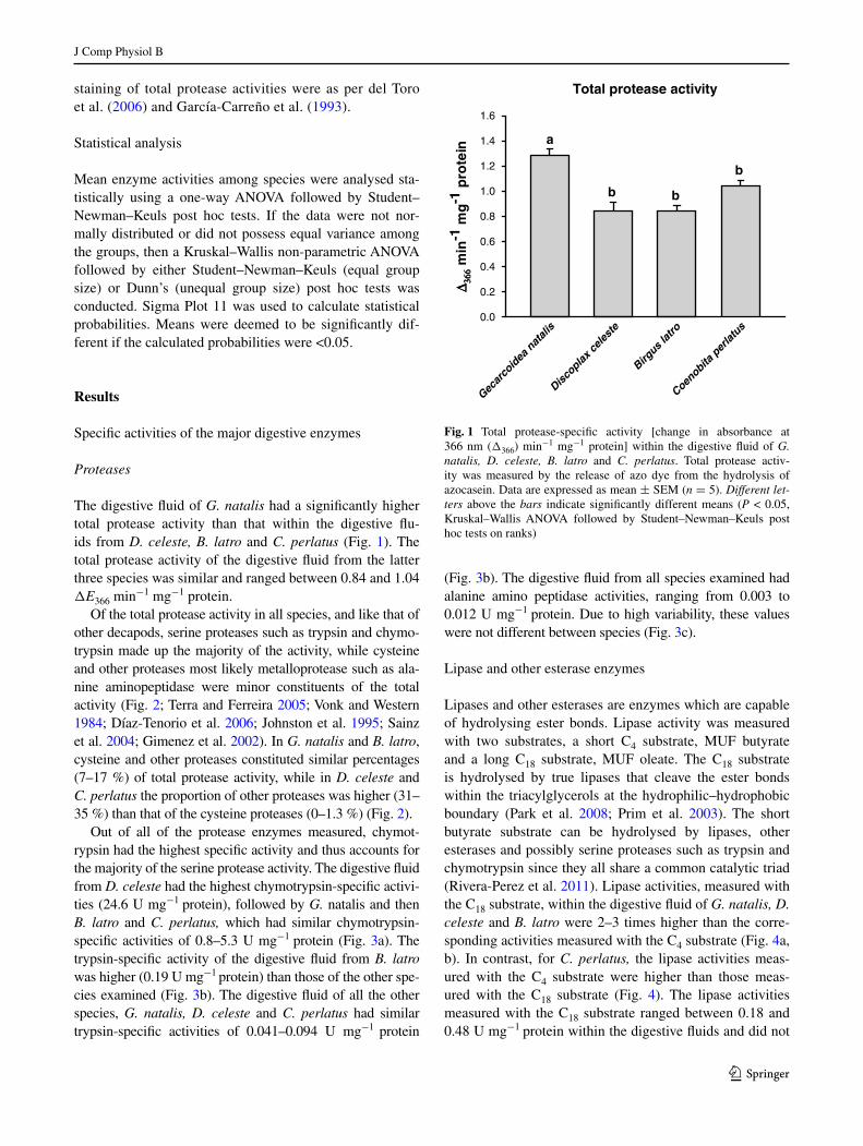
The digestive fluid of G. natalis had a significantly higher total protease activity than that within the digestive flu-ids from D. celeste, B. latro and C. perlatus (Fig. 1). The total protease activity of the digestive fluid from the latter three species was similar and ranged between 0.84 and 1.04 ΔE366 min−1 mg−1 protein.
Of the total protease activity in all species, and like that of other decapods, serine proteases such as trypsin and chymo-trypsin made up the majority of the activity, while cysteine and other proteases most likely metalloprotease such as ala-nine aminopeptidase were minor constituents of the total activity (Fig. 2; Terra and Ferreira 2005; Vonk and Western 1984; Díaz-Tenorio et al. 2006; Johnston et al. 1995; Sainz et al. 2004; gimenez et al. 2002). In G. natalis and B. latro, cysteine and other proteases constituted similar percentages (7–17 %) of total protease activity, while in D. celeste and C. perlatus the proportion of other proteases was higher (31–35 %) than that of the cysteine proteases (0–1.3 %) (Fig. 2).
Out of all of the protease enzymes measured, chymot-rypsin had the highest specific activity and thus accounts for the majority of the serine protease activity. The digestive fluid from D. celeste had the highest chymotrypsin-specific activi-ties (24.6 U mg−1 protein), followed by G. natalis and then B. latro and C. perlatus, which had similar chymotrypsin-specific activities of 0.8–5.3 U mg−1 protein (Fig. 3a). The trypsin-specific activity of the digestive fluid from B. latro was higher (0.19 U mg−1 protein) than those of the other spe-cies examined (Fig. 3b). The digestive fluid of all the other species, G. natalis, D. celeste and C. perlatus had similar trypsin-specific activities of 0.041–0.094 U mg−1 protein
(Fig. 3b). The digestive fluid from all species examined had alanine amino peptidase activities, ranging from 0.003 to 0.012 U mg−1 protein. Due to high variability, these values were not different between species (Fig. 3c).
lipase and other esterase enzymes
lipases and other esterases are enzymes which are capable of hydrolysing ester bonds. lipase activity was measured with two substrates, a short C4 substrate, MUF butyrate and a long C18 substrate, MUF oleate. The C18 substrate is hydrolysed by true lipases that cleave the ester bonds within the triacylglycerols at the hydrophilic–hydrophobic boundary (Park et al. 2008; Prim et al. 2003). The short butyrate substrate can be hydrolysed by lipases, other esterases and possibly serine proteases such as trypsin and chymotrypsin since they all share a common catalytic triad (rivera-Perez et al. 2011). lipase activities, measured with the C18 substrate, within the digestive fluid of G. natalis, D. celeste and B. latro were 2–3 times higher than the corre-sponding activities measured with the C4 substrate (Fig. 4a, b). In contrast, for C. perlatus, the lipase activities meas-ured with the C4 substrate were higher than those meas-ured with the C18 substrate (Fig. 4). The lipase activities measured with the C18 substrate ranged between 0.18 and 0.48 U mg−1 protein within the digestive fluids and did not
Total protease activity
Gecar
coid
ea n
atali
s
Discoplax
celes
te
Birgus l
atro
Coenobita
per
latus
∆∆ 336666
min
-1 m
g-1
pro
tein
0.0
0.2
0.4
0.6
0.8
1.0
1.2
1.4
1.6
a
b b
b
Fig. 1 Total protease-specific activity [change in absorbance at 366 nm (Δ366) min−1 mg−1 protein] within the digestive fluid of G. natalis, D. celeste, B. latro and C. perlatus. Total protease activ-ity was measured by the release of azo dye from the hydrolysis of azocasein. Data are expressed as mean ± SeM (n = 5). Different let‑ters above the bars indicate significantly different means (P < 0.05, Kruskal–Wallis anOVa followed by Student–newman–Keuls post hoc tests on ranks)

J Comp Physiol B
1 3
differ significantly among all species examined (Fig. 4a). The trends observed for the activities measured with the C4 substrate were different from that measured with the C18 substrate. The activities within the digestive fluid from D. celeste and C. perlatus were 0.28 and 0.30 U mg−1 protein, and thus significantly higher than those for G. natalis and B. latro, which amounted to 0.079 and 0.091 U mg−1 pro-tein, respectively (Fig. 4b). These differences between the activities measured with the two substrates may have been due to the lipase enzymes being less efficient at hydrolys-ing the shorter substrate in a more aqueous environment. alternatively the shorter substrate may possibly be hydro-lysed by other esterase enzymes besides true lipases.
amylase
amylase hydrolyses α-1,4-glycosidic bonds within storage polysaccharides such as starch and glycogen. The amyl-ase activities within the digestive fluid of G. natalis, D.
celeste, B. latro and C. perlatus ranged between 0.19 and 0.41 U mg−1 protein and did not differ statistically (Fig. 5).
Chitinase
Two enzymes are required to degrade chitin to its N-acetyl-glucosamine monomers. Chitinase cleaves internal β-1,4-glycosidic bonds to produce short oligomers of 2-3 monosaccharides in length. N-acetyl glucosamine is then hydrolysed from the non-reducing end by N-acetyl-d-glu-cosaminidase (eC 3.2.1.52) (espie and roff 1995; Terra and Ferreira 2005). Due to high variability, trends for N-acetyl-β-d-glucosaminidase activities were not as clear as for the other enzymes. The only clear difference was that the N-acetyl-β-d-glucosaminidase activity within the digestive fluid of C. perlatus was higher (8.0 × 10⁻⁵ U mg−1 pro-tein) than that from G. natalis (1.3 × 10⁻⁵ U mg−1 pro-tein) (Fig. 6). The N-acetyl-β-d-glucosaminidase activities within the digestive fluid from D. celeste, B. latro and C.
Fig. 2 Percentages of serine, cysteine and other proteases within the digestive fluid of G. natalis (a), D. celeste (b), B. latro (c) and C. perlatus (d). Data [mean ± SeM (n = 5)] are expressed as a percent-age of total protease activity. Serine and cysteine proteases were respectively inhibited by aeBSF and e64. Other proteases represent the propor-tion remaining after the serine and cysteine protease activities have been subtracted from the total protease activity. For each species, different letters above the bars indicate significantly different means (P < 0.05, Kruskal–Wallis anOVa fol-lowed by Student–newman–Keuls post hoc tests on ranks)
(a) Gecarcoidea natalis
Serin
e
Cyste
ine
OtherP
erce
nta
ge
of
tota
l pro
teas
e ac
tivi
ty
0
20
40
60
80
100
(b) Discoplax celeste
Serin
e
Cyste
ine
Other
Per
cen
tag
e o
f to
tal p
rote
ase
acti
vity
0
20
40
60
80
100
(c) Birgus latro
Serin
e
Cyste
ine
OtherP
erce
nta
ge
of
tota
l pro
teas
e ac
tivi
ty
0
20
40
60
80
100
(d) Coenobita perlatus
Serin
e
Cyste
ine
OtherP
erce
nta
ge
of
tota
l pro
teas
e ac
tivi
ty
0
20
40
60
80
100
a
b b
a
b
c
a
b b
a
b
c

J Comp Physiol B
1 3
perlatus were similar (Fig. 6). In addition, the N-acetyl-β-d-glucosaminidase activities within the digestive fluid from G. natalis were not significantly different from those of D. celeste and B. latro (Fig. 6).
Cellulase and hemicellulase
endo-β-1,4-glucanase is a cellulase which hydrolyses β-1,4-glycosidic bonds within cellulolytic substrates such as carboxy methyl cellulose. endo-β-1,4-glucanase activi-ties within the digestive fluids of D. celeste and C. perla‑tus were similar and higher than those of G. natalis and B. latro (Fig. 7). The endo-β-1,4-glucanase activities of these latter two species were similar (Fig. 7).
lichenase hydrolyses β-1,4-glycosidic bonds within mixed-linkage polysaccharides such as lichenin and those found within the cell walls of protozoans, fungi and algae.
lichenase activity within the digestive fluid from C. per‑latus was higher (0.85 U mg−1 protein) than that from D. celeste, G. natalis and B. latro (0.32–0.44 U mg−1 protein) (Fig. 8). The lichenase activities within the digestive fluid from these last three species were similar (Fig. 8).
β-glucohydrolase, measured as β-1,4-glucosidase activity, is an enzyme that complements endo-β-1,4-glucanase. It is thought to hydrolyse glucose off short polymers produced by endo-β-1,4-glucanase (allardyce et al. 2010). β-1,4-glucosidase activities within the diges-tive fluid of D. celeste were approximately 20 times higher (0.11 U mg−1 protein) than that for G. natalis, B. latro and C. perlatus, which were similar ranging between 0.002 and 0.007 U mg−1 protein (Fig. 9).
laminarinase hydrolyses β-1,3-glycosidic bonds within laminarin; a glucose polymer joined by mainly β-1,3-glycosidic bonds. The laminarinase activities ranged
Fig. 3 Chymotrypsin (a), trypsin (b), and alanine amino peptidase (c) specific activities within the digestive fluid of G. natalis, D. celeste, B. latro and C. perlatus. all specific activities were measured as the rate of release of p-nitrophenol from the hydrolysis of BaPna for trypsin, SaPna for chymotrypsin and l-alanine-4-nitroanilide for alanine amino peptidase. Data are presented as mean ± SeM (n = 5). For each of the proteases, different letters above the bars indicate signifi-cantly different means (Trypsin: P < 0.05, 1-way anOVa followed by Student–newman–Keuls post hoc tests. Chymot-rypsin and alanine amino pepti-dase: P < 0.05, Kruskal–Wallis one-way anOVa followed by Student–newman–Keuls post hoc tests on ranks.)
(a) Chymotrypsin
Gecar
coid
ea n
atali
s
Discoplax
celes
te
Birgus l
atro
Coenobita
per
latus
U. m
g-1
pro
tein
U. m
g-1
pro
tein
U. m
g-1
pro
tein
0
5
10
15
20
25
30
b
a
cc
(b) Trypsin
Gecar
coid
ea n
atali
s
Discoplax
celes
te
Birgus l
atro
Coenobita
per
latus
0.00
0.05
0.10
0.15
0.20
0.25
a a
b
a
(c) Alanine amino peptidase
Gecar
coid
ea n
atali
s
Discoplax
celes
te
Birgus l
atro
Coenobita
per
latus
0.000
0.002
0.004
0.006
0.008
0.010
0.012
0.014
0.016
0.018
0.020
a
aa
a

J Comp Physiol B
1 3
between 1.18 and 1.63 U mg−1 protein. The activity was significantly higher within the digestive fluid of C. perlatus than in the digestive fluids from G. natalis, D. celeste and B. latro (Fig. 10).
(a) 4-Methylumbelliferyl oleate
Gecar
coid
ea n
atali
s
Discoplax
celes
te
Birgus l
atro
Coenobita
per
latus
Lip
ase
spec
ific
act
ivit
y(U
. mg
-1 p
rote
in)
0.0
0.1
0.2
0.3
0.4
0.5
0.6
(b) 4-Methylumbelliferyl butyrate
Gecar
coid
ea n
atali
s
Discoplax
celes
te
Birgus l
atro
Coenobita
per
latus
Est
eras
e sp
ecif
ic a
ctiv
ity
(U. m
g-1
pro
tein
)
0.0
0.1
0.2
0.3
0.4
0.5
a
a
aa
a
bb
a
Fig. 4 lipase-specific activities (U mg−1 protein) within the diges-tive fluid of G. natalis, D. celeste, B. latro and C. perlatus. lipase activities were measured as the rate of release of the fluorescent molecule, 4-β-methylumbelliferone (MUF) from the hydrolysis of either MUF oleate (long C18 substrate) (a) or MUF butyrate (short C4 substrate) (b). Data are expressed as mean ± SeM (n = 3–4). For enzyme assays within either MUF oleate or MUF butyrate, similar letters above the bars indicate similar means (lipase: P > 0.05, one-way anOVa. esterase: P < 0.05, one-way anOVa followed by Stu-dent–newman–Keuls post hoc tests)
Amylase
Gecar
coid
ea n
atali
s
Discoplax
celes
te
Birgus l
atro
Coenobita
per
latus
U. m
g-1
pro
tein
0.0
0.1
0.2
0.3
0.4
0.5
0.6
a
a
a
a
Fig. 5 amylase-specific activities (U mg−1 protein) within the diges-tive fluid of G. natalis, D. celeste, B. latro and C. perlatus. amylase activities were measured as the rate of production of reducing sugars from the hydrolysis of starch. Common letters above the bars indicate similar means (P > 0.05, one-way anOVa). Data are expressed as mean ± SeM (n = 5)
N-acetyl-ββ-D-glucosaminidase
Gecar
coid
ea n
atali
s
Discoplax
celes
te
Birgus l
atro
Coenobita
per
latus
U. m
g-1
pro
tein
0
2.0x10⁻⁵
4.0x10⁻⁵
6.0x10⁻⁵
8.0x10⁻⁵
10-4
1.2x10⁻⁴
b
aab
ab
Fig. 6 N-acetyl-β-d-glucosaminidase-specific activities (U mg−1 protein) within the digestive fluid of G. natalis, D. celeste, B. latro and C. perlatus. N-acetyl-β-d-glucosaminidase activities were meas-ured as the rate of production of 4-methylumbelliferone from the hydrolysis of 4-methylumbelliferyl N-acetyl-β-d-glucosaminide. Dif‑ferent letters above the means indicate statistically different means (P < 0.05, Kruskal–Wallis anOVa followed by Dunn’s post hoc tests on ranks). Data are expressed as mean ± SeM (n = 4)

J Comp Physiol B
1 3
Endo-ββ-1,4-glucanase
Gecar
coid
ea n
atali
s
Discoplax
celes
te
Birgus l
atro
Coenobita
per
latus
U. m
g-1
pro
tein
0.0
0.1
0.2
0.3
0.4
0.5
0.6
aa
b b
Fig. 7 endo-β-1,4-glucanase-specific activities (U mg−1 protein) within the digestive fluid of G. natalis, D. celeste, B. latro and C. per‑latus. endo-β-1,4-glucanase activities were measured as the rate of production of reducing sugars from the hydrolysis of carboxy methyl cellulose. Different letters above the means indicate that they differed significantly (P < 0.05, Kruskal–Wallis one-way anOVa followed by Student–newman–Keuls post hoc tests on ranks). Values are mean ± SeM (n = 5)
Lichenase
Gecar
coid
ea n
atali
s
Discoplax
celes
te
Birgus l
atro
Coenobita
per
latus
U. m
g-1
pro
tein
0.0
0.2
0.4
0.6
0.8
1.0 a
b
b b
Fig. 8 lichenase-specific activities (U mg−1 protein) within the digestive fluid of G. natalis, D. celeste, B. latro and C. perlatus. lichenase activities were measured as the production of reducing sugars from the hydrolysis of lichenan. Different letters above the means indicate that they were statistically different (P < 0.05, one-way anOVa followed by Student–newman–Keuls post hoc tests). Values are mean ± SeM (n = 3–5)
ββ-glucohydrolase
Gecar
coid
ea n
atali
s
Discoplax
celes
te
Birgus l
atro
Coenobita
per
latus
U. m
g-1
pro
tein
0
5.0x10⁻³
10-2
1.5x10⁻²
10-1
1.2x10⁻¹ a
bb
b
Fig. 9 β-glucohydrolase-specific activities (U mg−1 protein) within the digestive fluid of G. natalis, D. celeste, B. latro and C. perlatus. β-glucohydrolase activities were measured as β-1,4-glucosidase activ-ity; that is, the production of glucose from the hydrolysis of cello-biose. Different letters above the means indicate that they differed significantly (P < 0.05, Kruskal–Wallis one-way anOVa followed by Student–newman–Keuls post hoc tests on ranks). Data are expressed as mean ± SeM (n = 5)
Laminarinase
Gecar
coid
ea n
atali
s
Discoplax
celes
te
Birgus l
atro
Coenobita
per
latus
U. m
g-1
pro
tein
0.0
0.2
0.4
0.6
0.8
1.0
1.2
1.4
1.6
1.8
2.0
aa
a
b
Fig. 10 laminarinase-specific activities (U mg−1 protein) within the digestive fluid of G. natalis, D. celeste, B. latro and C. perlatus. lam-inarinase activities were measured by the rate of production of reduc-ing sugars from the hydrolysis of laminarin. Different letters above the bars indicate significantly different means (P < 0.05, Kruskal–Wallis one-way anOVa followed by Student–newman–Keuls post hoc tests on ranks). Data are expressed as mean ± SeM (n = 5)

J Comp Physiol B
1 3
Size and number of digestive enzymes as revealed by zymography
Protease activity staining
Three protease bands of apparently 15, 19 and 21–22 kDa were observed for digestive fluid samples from G. natalis, B. latro and C. perlatus (Fig. 11; Table 4). The digestive
fluid from D. celeste produced two protease activity bands, apparently 15 and 18 kDa in size (Fig. 11; Table 4).
Lipase/esterase activity staining
Five lipase/esterase activity bands were observed from G. natalis. This included a strong fluorescent band at 129 kDa, and four smaller bands at 57, 51, 24.5 and 17 kDa (Fig. 12; Table 4). The bands of 24, 57 and 129 kDa were unique to this species. The smallest band at 17 kDa was next to the solvent front. For the other land crabs species exam-ined, three lipase bands at 17, 40–42 and 48–50 kDa were observed (Fig. 12; Table 4). all three bands were observed for B. latro and D. celeste (Table 4). Only one strong band at 33 kDa was observed for C. perlatus (Table 4).The smaller activity bands at 17 and 25 kDa may be due to serine proteases, such as trypsin and chymotrypsin, due to their similar sizes and they share the same catalytic triad as lipases and, thus, may possibly hydrolyse the butyrate sub-strate (rivera-Perez et al. 2011).
Alkaline phosphatase staining
alkaline phosphatases (eC 3.1.3.1) are unspecific phos-phatases that remove the phosphate, through hydrolysis of the phosphoester bond, from various compounds before assimilation (Terra and Ferreira 2005).There are clear differ-ences between the phosphatases present in the digestive flu-ids of the anomuran and brachyuran crabs. The pH optima
Fig. 11 Protease activity staining for the digestive fluid from G. natalis (gn), D. celeste (Dc), B. latro (Bl) and C. perlatus (Cp). all digestive fluid samples were diluted by a factor of 250 times. Pres-ence of protease activity was determined from the hydrolysis of 1 % haemoglobin, which had been allowed to infuse into the polyacryla-mide gel. gels were fixed and stained with 0.05 % Coomassie blue dissolved in a solution of 10 % acetic acid, 40 % methanol, 50 % water to visualise the halos of protease activity and the molecular mass markers (Invitrogen Mark 12 unstained marker) of the sizes indicated
Table 4 number of isoforms and apparent molecular mass (kDa) of the various digestive enzymes present in the digestive fluid of the species listed
Presence of the enzymes was determined by activity staining. number of replicates with each isoform is indicated in parentheses. For each enzyme, the highest number represents the total number of replicate digestive fluid samples examined
enzyme G. natalis D. celeste B. latro C. perlatus
Size (kDa) n Size (kDa) n Size (kDa) n Size (kDa) n
Protease 22 4 22 2 21 3
18 4 18 3 18 3 19 3
15 4 15 3 15 3 15 3
esterase 129 3
57 3
51 3 48 3 50 3
40 3 41 3 33 3
24.5 3
17 3 17 3 17 3
Phosphatase 75.5 4 73.5 2
50 1 48 4 55 5 52 3
amylase 58 2 59 2 58 2 60 2
β-glucosidase 126 2 125 2 119 4
32 3 31 2 30 4
28 2 28 2 28 4
25 3
N-acetyl-β-d-glucosaminidase 97 3 92 3 94.5 5 94.5 3
76.5 3 76.5 3 76.5 1 77 1

J Comp Physiol B
1 3
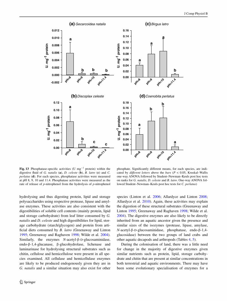
for the phosphatase activities of the anomuran crabs were between pH 9 and 10 while that of the brachyurans was 8, over the pH range tested (Fig. 13). Similarly, the zymo-grams revealed differences in the number and size of the phosphatases. The anomurans, B. latro and C. perlatus pos-sessed two phosphatases, one of apparently 73.5–75.5 kDa and another of 52–55 kDa (Fig. 14a; Table 4). In contrast, the brachyurans possessed only the smaller phosphatase which was apparently 48–50 kDa in size (Fig. 14a; Table 4).
Amylase activity staining
One amylase activity band of apparently 58–60 kDa was observed for the digestive fluids from G. natalis, D. celeste, B. latro and C. perlatus (Fig. 14b).
Activity staining for enzymes that hydrolyse β‑1,4‑glycosidic bonds (β‑glucohydrolase and endo‑β‑1,4‑glucanase)
The hydrolysis of the fluorescent substrate, 4-methylumbelliferyl-β-d-glucopyranoside by the diges-tive fluid samples produced activity bands at apparently
25, 28, 31–32 and 119–126 kDa (Fig. 14c; Table 4). This substrate is hydrolysed by enzymes that hydrolyse β-1,4-glycosidic bonds; this includes β-glucohydrolyses and endo-β-1,4-glucanases/lichenases (allardyce et al. 2010). The large fluorescent band at 119–126 kDa is thought to be due to β-glucohydrolase, while the three smaller activity bands (32, 28 and 25 kDa) are thought to be endo-β-1,4-glucanase/lichenase isozymes that have been characterised previously (linton and Shirley 2011; allardyce and linton 2008). These enzymes when run on SDS-Page gels with-out heating or reducing the sample with β-mercaptoethanol, the conditions used to create the zymograms, run further than samples which were heated and reduced. Thus under these conditions the 53-kDa isozyme runs at 32 kDa, the 47-kDa isozyme runs at 28 kDa and the 43-kDa isozyme runs at 25 kDa (linton and Shirley 2011). The digestive fluid from G. natalis produced three of the smaller endo-β-1,4-glucanase activity bands, that from D. celeste produced two and the digestive fluid from B. latro and C. perlatus produced only one small endo-β-1,4-glucanase activity band; 30 kDa for B. latro and 28 kDa for C. perlatus. There was also variation between individuals of G. natalis; the digestive fluid from two out of three replicates produced three endo-β-1,4-glucanase activity bands, while that for the remaining replicates produced only two activity bands. The large activity band at 119–126 kDa was feint for G. natalis and D. celeste and bright for C. perlatus. This band was not produced by the digestive fluid samples from B. latro.
N‑acetyl‑β‑d‑glucosamine activity staining
Two N-acetyl-β-d-glucosaminidase activity bands, one at apparently 77 kDa and another at 92–97 kDa, were pro-duced by the digestive fluid of the gecarcinids, G. natalis and D. celeste (Fig. 14d; Table 4). Both of these bands were present in all the replicates examined. Similarly, both bands were produced from the digestive fluid of the coenobitids B. latro and C. perlatus (Fig. 14d). For B. latro and C. per‑latus the larger band was present in all the replicates exam-ined, while the smaller band was only present in one out of five replicates for B. latro and one out of three replicates for C. perlatus (Table 4). Thus, individual polymorphism was observed for both coenobitid species.
Discussion
Major conclusions about the digestive enzyme activities
G. natalis, D. celeste, B. latro and C. perlatus possessed substantial activities of all the enzymes examined and this reflects their omnivorous nature. Thus they are capable of
Fig. 12 activity staining of polyacrylamide gels for esterase activity in the digestive fluid of the brachyuran land crabs, G. natalis (gn) and D. celeste (Dc) and the anomuran land crabs, B. latro (Bl) and C. perlatus(Cp). Presence of esterase enzyme activities was determined using the substrate 4-methylumbelliferyl butyrate. The substrate is hydrolysed by the enzyme to release the fluorescent molecule, 4-methylumbelliferone. The digestive fluid samples from G. natalis, D. celeste and B. latro were diluted 1 in 1,000 times while those from C. perlatus were diluted by a factor of 2,500 times. Molecular mass markers (Carl roth roti-Mark standards), with sizes (kDa) indicated, were visualised by counterstaining gels with 0.05 % Coomassie blue dissolved in a solution of 10 % acetic acid, 40 % methanol, 50 % water
J Comp Physiol B
1 3
hydrolysing and thus digesting protein, lipid and storage polysaccharides using respective protease, lipase and amyl-ase enzymes. These activities are also consistent with the digestibilities of soluble cell contents (mainly protein, lipid and storage carbohydrate) from leaf litter consumed by G. natalis and D. celeste and high digestibilities for lipid, stor-age carbohydrate (starch/glycogen) and protein from arti-ficial diets consumed by B. latro (greenaway and linton 1995; greenaway and raghaven 1998; Wilde et al. 2004). Similarly, the enzymes N-acetyl-β-d-glucosaminidase, endo-β-1,4-glucanase, β-glucohydrolase, lichenase and laminarinase for hydrolysing structural substrates such as chitin, cellulose and hemicellulose were present in all spe-cies examined. all cellulase and hemicellulase enzymes are likely to be produced endogenously given they are in G. natalis and a similar situation may also exist for other
species (linton et al. 2006; allardyce and linton 2008; allardyce et al. 2010). again, these activities may explain the digestion of these structural substrates (greenaway and linton 1995; greenaway and raghaven 1998; Wilde et al. 2004). The digestive enzymes are also likely to be directly inherited from an aquatic ancestor given the presence and similar sizes of the isozymes (protease, lipase, amylase, N-acetyl-β-d-glucosaminidase, phosphatase, endo-β-1,4-glucosidase) between the two groups of land crabs and other aquatic decapods and arthropods (Tables 4, 5).
During the colonisation of land, there was a little need for change in the majority of digestive enzymes given similar nutrients such as protein, lipid, storage carbohy-drate and chitin that are present at similar concentrations in both terrestrial and aquatic environments. There may have been some evolutionary specialisation of enzymes for a
(a) Gecarcoidea natalis
pH=8pH=9
pH=10
pH=11.4
U. m
g-1
pro
tein
-0.002
0.000
0.002
0.004
0.006
0.008
0.010
0.012
(b) Discoplax celeste
pH=8pH=9
pH=10
pH=11.4
U. m
g-1
pro
tein
0.00
0.02
0.04
0.06
0.08
0.10
0.12
(c) Birgus latro
pH=8pH=9
pH=10
pH=11.4
U. m
g-1
pro
tein
0.00
0.02
0.04
0.06
0.08
0.10
0.12
0.14
0.16
(d) Coenobita perlatus
pH=8pH=9
pH=10
pH=11.4
U. m
g-1
pro
tein
0.00
0.02
0.04
0.06
0.08
0.10
0.12
0.14
0.16
0.18
a
b b b
a
bc
c
a
a
a
b
a
bb
a
Fig. 13 Phosphatase-specific activities (U mg−1 protein) within the digestive fluid of G. natalis (a), D. celeste (b), B. latro (c) and C. perlatus (d). For each species, phosphatase activities were measured at pH 8, 9, 10 and 11.4. Phosphatase activities were measured as the rate of release of p-nitrophenol from the hydrolysis of p-nitrophenol
phosphate. Significantly different means, for each species, are indi-cated by different letters above the bars (P < 0.05, Kruskal–Wallis one-way anOVa followed by Student–newman–Keuls post hoc tests on ranks for G. natalis, D. celeste and B. latro. One-way anOVa fol-lowed Student–newman–Keuls post hoc tests for C. perlatus)

J Comp Physiol B
1 3
Fig. 14 activity staining of polyacrylamide gels for enzymes with phosphatase (a), amylase (b), β-glucosidase (c) and N-acetyl-β-d-glucosaminidase (d) activity, in the digestive fluid from the brachyuran land crabs, G. natalis (gn) and D. celeste (Dc) and the anomuran land crabs, B. latro (Bl) and C. perlatus (Cp). Pres-ence of amylase, β-glucosidase, N-acetyl-β-d-glucosaminidase and phosphatase enzyme activities was determined using the respective substrates 4-methylumbelliferyl-α-d-glucopyranoside, 4-methylumbelliferyl-β-d-glucopyranoside, 4-methylumbelliferyl N-acetyl-β-d-glucosaminide and 4-methylumbelliferyl phosphate. These substrates are hydrolysed by the relevant enzyme to release the fluorescent molecule, 4-methylumbelliferone. Digestive fluid from G. natalis, D. celeste and C. perlatus were diluted by a factor
of 10 times, while that from B. latro was diluted by a factor of 30 times for phosphatase activity staining (a). For amylase zymography (b), digestive fluid samples were diluted by a factor of 100 times for G. natalis, D. celeste and B. latro and 5 times for C. perlatus. For β-glucosidase activity (c), the digestive fluid samples were diluted by a factor of 5 times for large molecular mass isozymes and 100 times for low molecular mass isozymes. For N-acetyl-β-d-glucosaminidase activity staining (d), the digestive fluid samples were diluted by a fac-tor of ten times. Molecular mass markers [either Invitrogen Mark 12 unstained standard or SigmaMarker (M3913)], with sizes (kDa) indi-cated, were visualised by counterstaining gels with 0.05 % Coomas-sie blue dissolved in a solution of 10 % acetic acid, 40 % methanol, 50 % water

J Comp Physiol B
1 3
Table 5 apparent molecular mass (kDa) of digestive enzymes from other arthropods
native indicates that the samples were neither heated nor reduced prior to polyacrylamide electrophoresis. Denatured indicates that the samples were denatured and reduced prior to SDS polyacrylamide electrophoresis. Mass spec indicates that the mass of the enzyme was determined by its mass spectrum on a time of flight mass spectrometer. cDna indicates the size of the enzyme predicted from the translated amino acid sequence
enzyme Species and taxonomic group Size (kDa) references
Chymotrypsin Crangon crangon (shrimp) 35, 38, 43 (native) Teschke and Saborowski (2005)
Crangon allmani (shrimp) 20, 22, 23 (native) Teschke and Saborowski (2005)
Panulirus interruptus (lobster) 21, 60 (native) Celis-guerrero et al. (2004)
Trypsin Crangon crangon (shrimp) 20 (native) Teschke and Saborowski (2005)
Crangon allmani (shrimp) 20 (native) Teschke and Saborowski (2005)
Thenus orientalis (decapod) 35 (denatured) Johnston et al. (1995)
Penaeus vannamei (prawn) 21, 22, 23 (native) Sainz et al. (2004)
30.2, 32.9 (denatured)
31–32 (denatured)
Panulirus interruptus (lobster) 10, 22 (native) Celis-guerrero et al. (2004)
Penaeus japonicus (prawn) 25 (denatured) galgani et al. (1985)
Farfantepenaeus paulensis (shrimp)
14.6–21.7 lemos et al. (1999)
Cancer pagurus 26 (denatured) Hehemann et al. (2007), Saborowski et al. (2004)
lipase Penaeus vannamei (prawn) 44.8 (denatured) rivera-Perez et al. (2011)
Carcinus mediterraneus (crab) 65 (denatured) Cherif et al. (2007)
Cherax quadricarinatus (crayfish)
43, 46, 63, 118 (native) lópez-lópez et al. (2003)
Scorpio maurus (scorpion) 50 (denatured) Zouari et al. (2005)
amylase Eupagurus bernhardus (hermit crab)
55 (denature) Van Wormhoudt et al. (1995)
Procambarus clarkii (crayfish) 55 (denatured) Van Wormhoudt et al. (1995)
Carcinus maenas (crab) 30 (denatured) Van Wormhoudt et al. (1995)
Penaeus vannamei (prawn) 30 (denatured) Van Wormhoudt et al. (1995)
Palaemon elegans 30.5, 33, 53, 58, 75 (denatured) Van Wormhoudt and Favrel (1988)
Insects generally 48–60 Terra and Ferreira (2005)
alkaline phosphatase Homarus gammarus (lobster) 47 (native), 60 (denature) pH max 8–9 Saborowski (personal communica-tion)
Pandalus borealis (shrimp) 53–60 (cDna, denatured) subunits of dimeric protein. pH max 10–10.5
nilsen et al. (2001)
Penaeus monodon (Prawn) 35, 46 subunits of polymeric enzyme. pH max 9
lee and Chuang (1991)
N-acetyl-β-d-glucosaminidase Penaeus vannamei (prawn) 105 (2 × 45 subunits) (denatured) Xie et al. (2004)
Scylla serrata (crab) 132 (2 × 65.8 subunits) (denatured) Zhang et al. (2006)
Penaeus japonicus (prawn) 37 (denatured), 57 (mass spec) Kono et al. (1990)
endo-β-1,4-glucanase Cherax quadricarinatus 30, 40 (native) Xue et al. (1999)
48, 50 (mass spec) (Crawford et al. 2004)
Cherax destructor 53 (denatured) (allardyce and linton 2008)
Gecarcoidea natalis 43, 47, 53 (denatured) allardyce et al. (2010), linton and Shirley (2011), allardyce and linton (2008)
25, 28, 32 (native)
β-glucohydrolase (β-1,4-glucosidase)
Gecarcoidea natalis 130–169 (native) allardyce et al. (2010)

J Comp Physiol B
1 3
terrestrial, mainly leaf litter diet, particularly in the gecar-cinids, G. natalis and D. celeste since these species possess isozymes that were not observed in the coenobitids. In par-ticular, the gecarcinid crabs possessed additional endo-β-1,4-glucanase isozymes: G. natalis possessed two extra iso-forms and D. celeste one. Perhaps the increase in isozymes elevated cellulase activity that enhanced the efficiency of cellulose digestion. additional endo-β-1,4-glucanases may have arisen through gene duplication of the gHF9 gene. Two copies of a gHF9 gene are possessed by the isopod, Porcellio scaber (Kostanjšek et al. 2010) and individual termite species possess multiple copies of the gHF9 gene (lo et al. 2011). alternatively, the multiple endo-β-1,4-glucanases may be due to the expression of different glyco-syl hydrolase genes such as gHF1, gHF5, gHF9, gHF10 and gHF45 (Sakamoto et al. 2007, 2009; Sakamoto and Toyohara 2009a, b). The aquatic bivalve, Corbicula japon‑ica, possesses and expresses such genes which account for its endo-β-1,4-glucanse, xylanase and β-1,4-glucosidase activities (Sakamoto et al. 2007, 2009; Sakamoto and Toy-ohara 2009a, b). Similarly, G. natalis possessed an addi-tional lipase isozyme, a strong 129-kDa band that was not present in the other species examined. This enzyme may be a true lipase or a general esterase that can hydrolyse solu-ble substrates. gecarcinids consume substantial amounts of condensed and hydrolysable tannins with leaf litter and must be able to metabolise these compounds (Swain 1979; Hagerman and Butler 1991; linton and greenaway 2007). One possibility is the hydrolysis of hydrolysable tannins with an enzyme such as an esterase (linton and greenaway 2007). Differing in their pH optima and isozyme size, the phosphatases were also different between the gecarcinids and coenobitids. again, there may have been evolution of the enzymes or the differences may be due to inheritance from different brachyuran and anomuran ancestors.
activities of the digestive enzymes do not reflect the dietary preferences of the species examined
except for C. perlatus, dietary preferences of the other spe-cies, G. natalis, D. celeste and B. latro were not reflected in the activities of the digestive enzymes. Other factors may be determining the observed feeding preferences. In partic-ular, the restricted home ranges of G. natalis, D. celeste and C. perlatus may mean that the diet of these species is deter-mined by the abundance of items found within it (adam-czewska and Morris 2001) (Table 1). For the gecarcinids, given this is mostly leaf litter, this is what they mostly eat. In contrast, the excellent olfactory and locomotor ability of B. latro allows it to travel large distances (up to 1 km) to seek out its preferred dietary items (Table 1) (greenaway 2001; Fletcher et al. 1990; Drew et al. 2010). In contrast, in other marine and intertidal crabs, the activities of their
digestive enzymes commonly reflect their dietary prefer-ence. For example in other crab species, Plagusia chabrus and Thenus orientalis are carnivores with high activities of proteinase and amylase; Leptograpsus variegatus is an omnivore that consumes mainly green and brown algae and has high activities of amylase and laminarinase; Nectocar‑cinus integrifons is herbivorous, consuming sea grass, and possesses high cellulase activities; both Plagusia chabrus and Carcinus maenas are omnivores with similar levels of proteinase, amylase, laminarinase and cellulase (John-ston and Freeman 2005; Johnston et al. 1995). Similarly in talitrid amphipods, dietary preference is reflected in enzyme activity; Talorchestia marmorata consumes brown algae and possesses high laminarinase and lipase activi-ties to digest the laminarin and lipid esters; an intertidal Talorchestia sp. consumes mainly diatoms and possesses high activities of α- and β-glucosidases for the digestion of the cell wall and starch; like the herbivorous land crabs, the litter feeder, Keratroides vulgaris possess relative low activities of all digestive enzymes (Johnston et al. 2005). Perhaps, the land crabs are like the other omnivorous crustaceans that possess similar activities of the digestive enzymes to deal with the variety of compounds within their opportunistic diet.
The activities of cellulase (endo-β-1,4-glucanase) and hemicellulase (lichenase and laminarinase) enzymes within the digestive fluid of C. perlatus were higher than those of other species, particularly compared to B. latro. This suggests that C. perlatus is capable of substantial diges-tion of cellulose and hemicellulose, and may thus have a preference for lower grade plant items that contain these compounds. Conversely, the lower cellulase (endo-β-1,4-glucanase) and hemicellulase (laminarinase and lichenase) activities for B. latro compared to that for C. perlatus sug-gest that these compounds are not as important for energy as the highly digestible ones. However, the presence of the enzymes in B. latro is consistent with the digestion of small amounts of hemicellulose and cellulose by the crab when fed artificial plant-based diets (Wilde et al. 2004). B. latro would not derive substantial energy from cellulose diges-tion given its highly nutritious diet (Wilde et al. 2004). Thus, it is surprising that the digestive fluid possesses endo-β-1,4-glucanase activity. Indeed the apparent lack of β-glucohydrolyase in B. latro, as suggested by activity staining, suggests that this species is incapable of hydro-lysing cellulose to glucose. The carnivorous mud crab, Scylla serrata also possesses endo-β-1,4-glucanase activi-ties within its midgut gland (Pavasovic et al. 2004). like B. latro, this species would not derive a significant amount of energy from cellulose digestion. Perhaps, the endo-β-1,4-glucanase enzyme in these species contributes to the diges-tive efficiency. The activity of the endo-β-1,4-glucanase plus that of the gastric mill may help to degrade the fibrous

J Comp Physiol B
1 3
pulp of fruits and other plant materials to release the pre-ferred and highly digestible lipid, protein and carbohydrate (Wilde et al. 2004).
The activities for the enzymes that hydrolyse highly digestible substrates: total protease, amylase and lipase, within the digestive fluid of B. latro were not higher than those of other species. Thus, the preference of this species for highly digestible nutrients (protein, lipid and carbohy-drate) is not reflected in the enzyme activities (Wilde et al. 2004). However, the activities of these enzymes are pre-sumably high enough to achieve the high digestibility coef-ficients for nitrogen/protein, lipid and carbohydrate previ-ously measured for this species (Wilde et al. 2004).
The gecarcinids were surprising in that the activities of their digestive enzymes, in particular the cellulase and hemicellulase enzymes, were similar to each other and to B. latro but were lower than that for C. perlatus. The enzyme activities for G. natalis were particularly surprising in that endo-β-1,4-glucanase activity was not higher than that for other species; particularly, D. celeste and total pro-tease activities were higher than the total protease activities of other species. This is different to that described previ-ously where endo-β-1,4-glucanase activities for G. natalis were higher than that for D. celeste, laminarinase activi-ties similar, and lichenase activities higher for D. celeste (linton and greenaway 2004). as observed previously, β-glucohydrolase activity (β-glucosidase activity) was higher in D. celeste than G. natalis (linton and greenaway 2004). However, the substantial activities of the cellulase and hemicellulase enzymes can explain the substantial cel-lulose and hemicellulose digestibilities observed for these species when fed on leaf litter diets (greenaway and linton 1995; greenaway and raghaven 1998).
The digestive fluid was sampled from individuals of G. natalis at the end of the dry season, just before the start of the reproductive migration. These animals were mostly confined to their burrows and may have been in “dry sea-son mode” with a suppressed metabolism (different ener-getic metabolism to migrating wet season animals) (Mor-ris et al. 2010). There may have been phenotypic plasticity in the expression of their digestive enzymes, in particu-lar cellulase and hemicellulase enzymes may have been downregulated and the enzymes involved in the hydrolysis of highly nutritious substrates, lipase, protease and amyl-ase upregulated. This may be required to gain energy and nitrogen for reproductive purposes. alternatively, the high total protease activity may reflect the efficient hydroly-sis and thus digestion of the low amount of protein asso-ciated with a mainly leaf litter diet (linton and greena-way 2007). although phenotypic plasticity of digestive enzymes remains to be demonstrated in land crabs, it has been observed in other decapod crustaceans and insects (Locusta migratoria). activities of α-amylase in the
midgut gland of mud crabs, Scylla serrata increase when the crabs are fed diets increasing in starch (Pavasovic et al. 2004). Similarly, activities of the digestive enzymes, α-amylase, endo-β-1,4-glucanase and protease within the crayfish, C. quadricarinatus, are positively correlated to the protein and carbohydrate content of the diet (Pavasovic et al. 2007a, b). In contrast, the locust, Locusta migrato‑ria downregulates α-amylase and α-chymotrypsin levels in response to excessive intake of carbohydrate or protein (Clissold et al. 2010). This downregulation of enzyme activity is thought to decrease the absorption of excess nutrients as the animal tries to achieve its most optimal intake of both carbohydrate and protein from a very nutri-ent deficient diet. a similar mechanism may occur in G. natalis at the end of the dry season as this species would have a restricted diet of mainly leaf litter and would, con-sequently, have a high intake of structural carbohydrates such as cellulose and hemicellulose.
Acknowledgments The authors are grateful to the Hermon Slade Foundation for funding this work.
References
adamczewska aM, Morris S (2001) ecology and behavior of Gecar‑coidea natalis, the Christmas Island red Crab, during the annual breeding migration. Biol Bull 200:305–320
allardyce BJ, linton SM (2008) Purification and characterisation of endo-β-1,4-glucanase and laminarinase enzymes from the gecar-cinid land crab Gecarcoidea natalis and the aquatic crayfish Che‑rax destructor. J exp Biol 211(14):2275–2287
allardyce BJ, linton SM, Saborowski r (2010) The last piece in the cellulase puzzle: the characterisation of {beta}-glucosidase from the herbivorous gecarcinid land crab Gecarcoidea natalis. J exp Biol 213(17):2950–2957. doi:10.1242/jeb.041582
Barnes DKa (1997) ecology of tropical hermit crabs at Quirimba Island, Mozambique: a novel and locally important food source. Mar ecol Prog Ser 161:299–302
Bradford MM (1976) a rapid and sensitive method for the quantita-tion of microgram quantities of protein utilizing the principle of protein-dye binding. anal Biochem 72(1–2):248–254
Celis-guerrero le, garcía-Carreño F, del Toro Man (2004) Char-acterization of proteases in the digestive system of spiny lobster (Panulirus interruptus). Mar Biotechnol 6:262–269
Cherif S, Fendri a, Miled n, Trabelsi H, Mejdoub H, gargouri Y (2007) Crab digestive lipase acting at high temperature: purification and biochemical characterization. Biochimie 89(8):1012–1018
Clissold FJ, Tedder BJ, Conigrave aD, Simpson SJ (2010) The gastrointestinal tract as a nutrient-balancing organ. Proc royal Soc B: Biol Sci 277(1688):1751–1759. doi:10.1098/rspb.2009.20452009.2045
Crawford aC, Kricker Ja, anderson aJ, richardson na, Mather PB (2004) Structure and function of a cellulase gene in redclaw cray-fish Cherax quadricarinatus. gene 340:267–274
Davie PJF (2002) Crustacea: Malacostraca: eucarida (Part 2): Decap-oda–anomura, Brachyura. In: Wells a, Houston WWK (eds) Zoological catalogue of australia, vol 19.3B. CSIrO Publishing, Melbourne, p xiv 641

J Comp Physiol B
1 3
del Toro Man, garcía-Carreño Fl, lopez MD, Celis g, laura e, Saborowski r (2006) aspartic proteinases in the digestive tract of marine decapod crustaceans. J exp Zool 305a:645–654
Díaz-Tenorio lM, garcía-Carreño Fl, del Toro Man (2006) Characterization and comparison of digestive proteinases of the Cortez swimming crab, Callinectes bellicosus, and the arched swimming crab, Callinectes arcuatus. Invertebr Biol 125(2):125–135
Drew MM, Harzsch S, Stensmyr M, erland S, Hansson BS (2010) a review of the biology and ecology of the robber Crab, Birgus latro (linnaeus, 1767) (anomura: Coenobitidae). Zoologischer anzeiger—J Comp Zool 249(1):45–67
espie PJ, roff JC (1995) Characterization of Chitobiase from Daphnia magna and its relation to chitin flux. Physiol Zool 68(5):727–748
Figueiredo MSrB, Kricker Ja, anderson aJ (2001) Digestive enzyme activities in the alimentary tract of redclaw crayfish, Che‑rax quadricarinatus (Decapoda: Parastacidae). J Crustac Biol 21(2):334–344
Fletcher WJ, Brown IW, Fielder Dr (1990) Movement of coco-nut crabs, Birgus latro, in rainforest habitat in Vanuatu. Pac Sci 44(4):407–416
galgani Fg, Benyamin Y, Van Wormhoudt a (1985) Purification, properties and immunoassay of trypsin from the shrimp Penaeus japonicus. Comp Biochem Physiol Part B: Comp Biochem 81(2):447–452
garcía-Carreño Fl, Dimes le, Haard nF (1993) Substrate-gel electrophoresis for composition and molecular weight of pro-teinases or proteinaceous proteinase inhibitors. anal Biochem 214(1):65–69
german DP, nagle BC, Villeda JM, ruiz aM, Thomson aW, Con-treras Balderas S, evans DH (2010) evolution of herbivory in a carnivorous clade of minnows (Teleostei: Cyprinidae): effects on gut size and digestive physiology. Physiol Biochem Zool: PBZ 83(1):1–18. doi:10.1086/648510
gimenez aVF, garcía-Carreño Fl, del Toro Man, Fenucci Jl (2002) Digestive proteinases of Artemesia longinaris (Decapoda, Penaeidae) and relationship with molting. Comp Biochem Phys-iol B 132:593–598
greenaway P (2001) Sodium and water balance in free-ranging rob-ber crabs, Birgus latro (anomura: Coenobitidae). J Crustac Biol 21(2):317–327
greenaway P (2003) Terrestrial adaptations in the anomura (Crusta-cea: Decapoda). Mem Mus Vic 60(1):13–26
greenaway P, linton SM (1995) Dietary assimilation and food reten-tion time in the herbivorous terrestrial crab Gecarcoidea natalis. Physiol Zool 68(6):1006–1028
greenaway P, raghaven S (1998) Digestive strategies in two species of leaf-eating land crabs (Brachyura: gecarcinidae) in a rain for-est. Physiol Zool 71(1):36–44
grubb P (1971) ecology of terrestrial decapod crustaceans on aldabra. Philos Trans r Soc lond 260:411–416
Hagerman ae, Butler lg (1991) Tannins and lignins. In: rosenthal ga, Berenbaum Mr (eds) Herbivores their interactions with sec-ondary plant metabolites, vol 1, 2nd edn. academic Press, San Diego, pp 355–386
Hartnoll rg (1988) evolution, systematics, and geographical distri-bution. In: Burggren WW, McMahon Br (eds) Biology of the land crabs. Cambridge University Press, Cambridge, pp 7–53
Hehemann J-H, redecke l, Perbandt M, Saborowski r, Betzel C (2007) Crystallization and preliminary X-ray diffraction stud-ies of trypsin-like proteases from the gastric fluid of the marine crab Cancer pagurus. acta Crystallogr Sect F 63(3):242–245. doi:10.1107/s1744309107008524
Hicks J, rumpff H, Yorkston H (1990) Christmas crabs, 2nd edn. Christmas Island natural History association, Christmas Island
Johnston D, Freeman J (2005) Dietary preference and digestive enzymes activities as indicators of trophic resource utilization by six species of crab. Biol Bull 208:36–46
Johnston DJ, Yellowlees D (1998) relationship between dietary pref-erences and digestive enzyme complement of the slipper lob-ster Thenus orientalis (Decapoda: Scyllaridae). J Crustac Biol 18(4):656–665
Johnston D, Hermans JM, Yellowlees D (1995) Isolation and charac-terization of a trypsin from the slipper lobster, Thenus orientalis (lund). arch Biochem Biophys 324(1):35–40
Johnston D, ritar aJ, Thomas CW (2004) Digestive enzyme profiles reveal digestive capacity and potential energy sources in fed and starved spiny lobster (Jasus edwardsii) phyllosoma larvae. Comp Biochem Physiol B 138:137–144
Johnston M, Johnston D, richardson a (2005) Digestive capa-bilities reflect the major food sources in three species of talitrid amphipods. Comp Biochem Physiol—B Biochem Mol Biol 140(2):251–257
Jue DK, lipke Pn (1985) Determination of reducing sugars in the nanomole range with tetrazolium blue. J Biochem Biophys Meth-ods 11:109–115
Karasov WH, del rio CM, Caviedes-Vidal e (2011) ecologi-cal physiology of diet and digestive systems. In: Julius D, Clapham De (eds) annual review of physiology, vol 73. annual review of physiology. annual reviews, Palo alto, pp 69–93. doi:10.1146/annurev-physiol-012110-142152
Knotz S, Boersma M, Saborowski r (2006) Microassays for a set of enzymes in individual small marine copepods. Comp Biochem Physiol—Mol Integr Physiol 145(3):406–411
Kono M, Matusi T, Shimizu C, Koga D (1990) Purifications and some properties of chitinase from the liver of a prawn. Penaeus japoni-cus. agricultural Biological Chemistry 54(8):2145–2147
Kostanjšek r, Milatovic M, Štrus J (2010) endogenous origin of endo-β-1,4-glucanase in common woodlouse Porcellio scaber (Crustacea, Isopoda). J Comp Physiol (B) 180(8):1143–1153. doi:10.1007/s00360-010-0485-7
laemmli UK (1970) Cleavage of structural proteins during the assem-bly of the head of bacteriophage T4. nature 227(5259):680–685
lee a-C, Chuang n–n (1991) Characterization of different molecu-lar forms of alkaline phosphatase in the hepatopancreas from the shrimp Penaeus monodon (Crustacea: Decapoda). Comp Bio-chem Physiol Part B: Comp Biochem 99(4):845–850
lemos D, Hernández-Cortés MP, navarrete a, garcía-Carreño Fl, Phan Vn (1999) Ontogenetic variation in digestive proteinase activity of larvae and postlarvae of the pink shrimp Farfantepe‑naeus paulensis (Crustacea: Decapoda: Penaeidae). Mar Biol 135(4):653–662. doi:10.1007/s002270050666
linnaeus C (1767) Systema naturae per regna tria naturae, secundum classes, ordinies, genera, species cum character-bus, differentiis, synonymis, locis, 12 edn. Stockholm
linton SM, greenaway P (2004) Presence and properties of cellulase and hemicellulase enzymes of the gecarcinid land crabs Gecar‑coidea natalis and Discoplax hirtipes. J exp Biol 207:4095–4104
linton SM, greenaway P (2007) a review of feeding and nutrition of herbivorous land crabs: adaptations to low quality plant diets. J Comp Physiol B 177:269–286
linton SM, Shirley aJ (2011) Isozymes from the herbivorous gecar-cinid land crab, Gecarcoidea natalis that possess both lichenase and endo-β-1,4-glucanase activity. Comp Biochem Physiol—B Biochem Mol Biol 160(1):44–53
linton SM, greenaway P, Towle DW (2006) endogenous production of endo-β-1,4-glucanase by decapod crustaceans. J Comp Physiol B 176:339–348
lo n, Tokuda g, Watanabe H (2011) evolution and function of endogenous termite cellulases. Biology of termites: a modern synthesis. Springer, Heidelberg

J Comp Physiol B
1 3
lópez-lópez S, nolasco H, Vega-Villasante F (2003) Characteriza-tion of digestive gland esterase-lipase activity of juvenile redclaw crayfish Cherax quadricarinatus. Comp Biochem Physiol B: Bio-chem Mol Biol 135(2):337–347
Milne edwards H (1853) Histoire naturelle des Crustaces. librairie encyclopedique de roret (Paris) 2, p 532
Morris S, Postel U, Mrinalini P, Turner lM, Palmer J, Webster Sg (2010) The adaptive significance of crustacean hyperglycaemic hormone (CHH) in daily and seasonal migratory activities of the Christmas Island red crab Gecarcoidea natalis. J exp Biol 213(17):3062–3073. doi:10.1242/jeb.045153
ng PKl, Davie PJF (2012) The blue crab of Christmas Island, Dis‑coplax celeste, new species (Crustacea: Decapoda: Brachyura: gecarcinidae). raffles Bull Zool 60(1):89–100
nilsen IW, Øverbø K, Olsen rl (2001) Thermolabile alkaline phos-phatase from northern shrimp (Pandalus borealis): protein and cDna sequence analyses. Comp Biochem Physiol B: Biochem Mol Biol 129(4):853–861
Park J, Cho SY, Choi SJ (2008) Purification and characterization of hepatic lipase from Todarodes pacificus. J Biochem Mol Biol 41(3):254–258
Pavasovic M, richardson na, anderson aJ, Mann D, Mather PB (2004) effect of pH, temperature and diet on digestive enzyme profiles in the mud crab, Scylla serrata. aquaculture 242(1–4):641–654
Pavasovic a, anderson aJ, Mather PB, richardson na (2007a) effect of a variety of animal, plant and single cell-based feed ingredi-ents on diet digestibility and digestive enzyme activity in redclaw crayfish, Cherax quadricarinatus (Von Martens 1868). aquacul-ture 272(1–4):564–572. doi:10.1016/j.aquaculture.2007.08.027
Pavasovic a, anderson aJ, Mather PB, richardson na (2007b) Influence of dietary protein on digestive enzyme activity, growth and tail muscle composition in redclaw crayfish, Che‑rax quadricarinatus (von Martens). aquac res 38(6):644–652. doi:10.1111/j.1365-2109.2007.01708.x
Pocock rI (1888) On the arachnida, Myriopoda, and land-Crustacea of Christmas Island. Proc Zool Soc lond 56(1):556–564
Prim n, Sánchez M, ruiz C, Javier Pastor FI, Díaz P (2003) Use of methylumbeliferyl-derivative substrates for lipase activity charac-terization. J Mol Catal B enzym 22(5–6):339–346
rivera-Pérez C, del Toro Man, garcía-Carreño F (2011) Purification and characterization of an intracellular lipase from pleopods of whiteleg shrimp (Litopenaeus vannamei). Comp Biochem Phys-iol—B Biochem Mol Biol 158(1):99–105
Saborowski r, Sahlinga g, navarette del Toro Ma, Waltera I, garcía-Carreñob Fl (2004) Stability and effects of organic solvents on endopeptidases from the gastric fluid of the marine crab Cancer pagurus. J Mol Catal B: enzym 30(3–4):109–118
Sainz JC, garcía-Carreño Fl, Hernández-Cortés P (2004) Penaeus vannamei isotrypsins: purification and characterization. Comp Biochem Physiol B 138:155–162
Sakamoto K, Toyohara H (2009a) Molecular cloning of glycoside hydrolase family 45 cellulase genes from brackish water clam Corbicula japonica. Comp Biochem Physiol B: Biochem Mol Biol 152(4):390–396
Sakamoto K, Toyohara H (2009b) Putative endogenous xylanase from brackish-water clam Corbicula japonica. Comp Biochem Physiol B: Biochem Mol Biol 154(1):85–92
Sakamoto K, Touhata K, Yamashita M, Kasai a, Toyohara H (2007) Cellulose digestion by common Japanese freshwater clam Cor‑bicula japonica. Fish Sci 73:675–683
Sakamoto K, Uji S, Kurokawa T, Toyohara H (2009) Molecular clon-ing of endogenous [beta]-glucosidase from common Japanese brackish water clam Corbicula japonica. gene 435(1–2):72–79
Schondube Je, Herrera-M lg, Martínez del rio C (2001) Diet and the evolution of digestion and renal function in phyllostomid bats. Zoology 104(1):59–73. doi:10.1078/0944-2006-00007
Stensmyr MC, erland S, Hallberg e, Wallen r, greenaway P, Hans-son BS (2005) Insect-like olfactory adaptations in the terrestrial giant robber crab. Curr Biol 15:116–121
Swain T (1979) Tannins and lignins. In: rosenthal ga, Janzen DH (eds) Herbivores, their interaction with secondary plant metabo-lites. academic press, new York, pp 657–682
Terra Wr, Ferreira C (2005) Biochemistry of digestion. In: Sarjeet S (ed) Comprehensive molecular insect science. elsvier, amster-dam, new York, pp 171–224
Teschke M, Saborowski r (2005) Cysteine proteinases substitute for serine proteinases in the midgut glands of Crangon crangon and Crangon allmani (Decapod: Caridea). J exp Mar Biol ecol 316:213–229
Van Wormhoudt a, Favrel P (1988) electrophoretic characterization of Palaemon elegans (crustacea, decapoda) alpha-amylase sys-tem: study of amylase polymorphism during the intermolt cycle. Comp Biochem Physiol Part B: Comp Biochem 89(2):201–207
Van Wormhoudt a, Bourreau g, le Moullac g (1995) amylase poly-morphism in crustacea decapoda: electrophoretic and immuno-logical studies. Biochem Syst ecol 23(2):139–149
Vonk HJ, Western JrH (1984) Comparative biochemistry and physi-ology of enzymatic digestion. academic Press, new York
Wilde Je, linton SM, greenaway P (2004) Dietary assimilation and the digestive strategy of the omnivorous anomuran land crab Bir‑gus latro (Coenobitidae). J Comp Physiol B 174:299–308
Xie X-l, Chen Q-X, lin J-C (2004) Purification and some proper-ties of β-N-acetyl-d-glucosaminidase from prawn (Penaeus van‑namei). Mar Biol 146:143–148
Xue XM, aderson aJ, richardson na, anderson aJ, Xue gP, Mather PB (1999) Characterisation of cellulase activity in the digestive system of the redclaw crayfish (Cherax quadricarinatus). aqua-culture 180:373–386
Zhang J-P, Chen Q-X, Wang Q, Xie J–J (2006) Purification and some properties of β-N-acetyl-d-glucosaminidase from viscera of green crab (Scylla serrata). Biochemistry (Moscow) 71(Suppl. 1):S55–S59
Zouari n, Miled n, Cherif S, Mejdoub H, gargouri Y (2005) Purifi-cation and characterization of a novel lipase from the digestive glands of a primitive animal: the scorpion. Biochimica et Biophy acta—gen Subj 1726(1):67–74
Copyright © 2022 FDOKUMEN