Efficient Array-Based Identification of Novel Cardiac Genes through Differentiation of Mouse ESCs
Upload
khangminh22Category
view
0download
0
toxins
Article
Differentiation, Quantification and Identification of Abrin andAbrus precatorius Agglutinin
Sylvia Worbs 1, Bettina Kampa 1, Martin Skiba 1, Eva-Maria Hansbauer 1,2, Daniel Stern 1 , Hervé Volland 2,François Becher 2 , Stéphanie Simon 2 , Martin B. Dorner 1 and Brigitte G. Dorner 1,*
�����������������
Citation: Worbs, S.; Kampa, B.; Skiba,
M.; Hansbauer, E.-M.; Stern, D.;
Volland, H.; Becher, F.; Simon, S.;
Dorner, M.B.; Dorner, B.G.
Differentiation, Quantification and
Identification of Abrin and Abrus
precatorius Agglutinin. Toxins 2021, 13,
284. https://doi.org/10.3390/
toxins13040284
Received: 14 March 2021
Accepted: 13 April 2021
Published: 18 April 2021
Publisher’s Note: MDPI stays neutral
with regard to jurisdictional claims in
published maps and institutional affil-
iations.
Copyright: © 2021 by the authors.
Licensee MDPI, Basel, Switzerland.
This article is an open access article
distributed under the terms and
conditions of the Creative Commons
Attribution (CC BY) license (https://
creativecommons.org/licenses/by/
4.0/).
1 Biological Toxins, Centre for Biological Threats and Special Pathogens, Robert Koch Institute, Seestr. 10,13353 Berlin, Germany; [email protected] (S.W.); [email protected] (B.K.); [email protected] (M.S.);[email protected] (E.-M.H.); [email protected] (D.S.); [email protected] (M.B.D.)
2 Département Médicaments et Technologies pour la Santé, Université Paris Saclay, CEA, INRAE, SPI,91191 Gif-sur-Yvette, France; [email protected] (H.V.); [email protected] (F.B.);[email protected] (S.S.)
* Correspondence: [email protected]; Tel.: +49-30-18754-2500
Abstract: Abrin, the toxic lectin from the rosary pea plant Abrus precatorius, has gained considerableinterest in the recent past due to its potential malevolent use. However, reliable and easy-to-useassays for the detection and discrimination of abrin from related plant proteins such as Abrusprecatorius agglutinin or the homologous toxin ricin from Ricinus communis are sparse. To addressthis gap, a panel of highly specific monoclonal antibodies was generated against abrin and therelated Abrus precatorius agglutinin. These antibodies were used to establish two sandwich ELISAsto preferentially detect abrin or A. precatorius agglutinin (limit of detection 22 pg/mL for abrin;35 pg/mL for A. precatorius agglutinin). Furthermore, an abrin-specific lateral flow assay wasdeveloped for rapid on-site detection (limit of detection ~1 ng/mL abrin). Assays were validated forcomplex food, environmental and clinical matrices illustrating broad applicability in different threatscenarios. Additionally, the antibodies turned out to be suitable for immuno-enrichment strategiesin combination with mass spectrometry-based approaches for unambiguous identification. Finally,we were able to demonstrate for the first time how the developed assays can be applied to detect,identify and quantify abrin from a clinical sample derived from an attempted suicide case involvingA. precatorius.
Keywords: Abrin; Abrus precatorius; monoclonal antibodies; ELISA; mass spectrometry; lateral flowassay; clinical sample; food; suicide attempt
Key Contribution: The generation of highly specific monoclonal antibodies specific for the planttoxin abrin allowed us to establish immunological and mass spectrometric assays to detect, identifyand quantify abrin from complex clinical, food and environmental matrices.
1. Introduction
The shrub Abrus precatorius belongs to the Fabaceae family and is also known asjequirity bean, crab’s eye, rosary or paternoster pea plant. It can be found in many tropicaland subtropical areas across the globe. The seeds contain the highly toxic lectin abrin, amember of the type II ribosomal inactivating proteins (RIPs) family. These cytotoxic lectinsfacilitate cell death by halting protein synthesis by depurinating a specific adenine in thesarcin-loop of the ribosomal RNA. Type II RIPs consist of a catalytically active A-chain—the RNA N-glycosidase—and a sugar-binding B-chain—the lectin part—which mediatescellular binding and uptake [1]. A number of highly toxic plant proteins including ricin(castor oil plant, Ricinus communis) and viscumin (mistletoe, Viscum album) are members ofthe RIP family, which also includes shiga toxins produced by the bacteria Escherichia coliand Shigella dysenteriae [2–4]. Plant RIPs are thought to protect the producing organism
Toxins 2021, 13, 284. https://doi.org/10.3390/toxins13040284 https://www.mdpi.com/journal/toxins
Toxins 2021, 13, 284 2 of 27
against predators and fungi, whereas bacterial RIPs act as potent pathogenicity factors.Due to their high toxicity, RIPs are investigated in agriculture (pest control) or medicine(immunotoxins, anti-tumor and anti-viral activity) and some have also gained militaryor criminal interest [5–8]. Here, ricin is the best-known example, but abrin has alsobeen used with malevolent intent recently; consequently, both toxins are listed as selectagents [9,10]. Several reports cover the use of abrin in attempted murders or in a biothreatscenario [11–14], and accidental or suicidal poisonings with Abrus precatorius seeds havebeen reported [15]. Its lethality depends on the amount of seeds and the applicationroute. Fatality after oral ingestion is usually rare in humans, while injection seems tobe more severe [16–23]—a characteristic that has been noted for ricin previously [24].Toxicological data from animal studies indicate that the major toxin abrin—as with otherprotein toxins—is more potent when applied systemically (half maximal lethal dose, LD50,in rats: 0.3–0.5 µg/kg) compared to inhalation (rat LD50 3–4 µg/kg) and least toxic via theoral route (mice LD50 2–3 mg/kg) [15,25]; however, the exact concentration and purity ofthe abrin used in those studies remain elusive. Abrus precatorius roots and leaves have beenused in traditional (ayurvedic) medicine—e.g., to treat coughing—while the seeds can belaxative and abortive [7]. Moreover, boiled (detoxified) seeds can be part of the local diet inregions where Abrus is common [7]. Cooking or baking is used for other legumes (Fabaceae)as well, such as beans and lentils, to destroy their heat-labile toxic compounds, makingthem suitable for consumption.
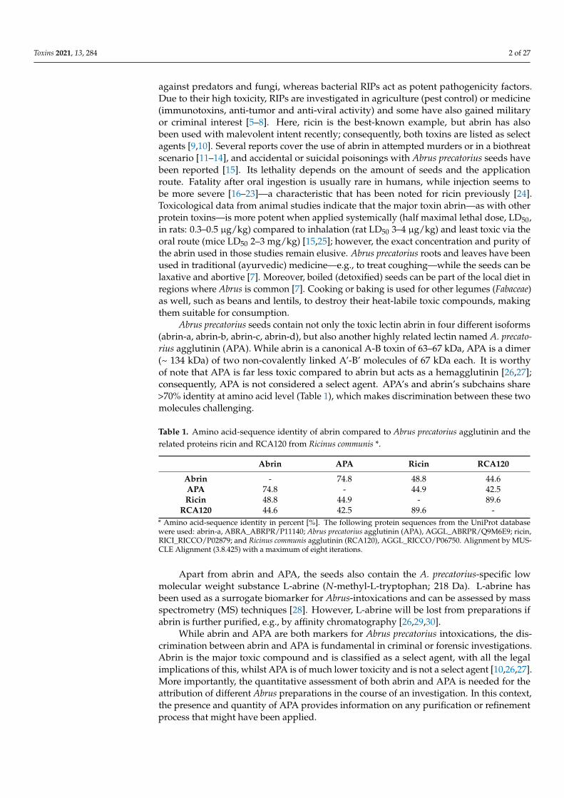
Abrus precatorius seeds contain not only the toxic lectin abrin in four different isoforms(abrin-a, abrin-b, abrin-c, abrin-d), but also another highly related lectin named A. precato-rius agglutinin (APA). While abrin is a canonical A-B toxin of 63–67 kDa, APA is a dimer(~ 134 kDa) of two non-covalently linked A’-B’ molecules of 67 kDa each. It is worthyof note that APA is far less toxic compared to abrin but acts as a hemagglutinin [26,27];consequently, APA is not considered a select agent. APA’s and abrin’s subchains share>70% identity at amino acid level (Table 1), which makes discrimination between these twomolecules challenging.
Table 1. Amino acid-sequence identity of abrin compared to Abrus precatorius agglutinin and therelated proteins ricin and RCA120 from Ricinus communis *.
Abrin APA Ricin RCA120
Abrin - 74.8 48.8 44.6APA 74.8 - 44.9 42.5Ricin 48.8 44.9 - 89.6
RCA120 44.6 42.5 89.6 -* Amino acid-sequence identity in percent [%]. The following protein sequences from the UniProt databasewere used: abrin-a, ABRA_ABRPR/P11140; Abrus precatorius agglutinin (APA), AGGL_ABRPR/Q9M6E9; ricin,RICI_RICCO/P02879; and Ricinus communis agglutinin (RCA120), AGGL_RICCO/P06750. Alignment by MUS-CLE Alignment (3.8.425) with a maximum of eight iterations.
Apart from abrin and APA, the seeds also contain the A. precatorius-specific lowmolecular weight substance L-abrine (N-methyl-L-tryptophan; 218 Da). L-abrine hasbeen used as a surrogate biomarker for Abrus-intoxications and can be assessed by massspectrometry (MS) techniques [28]. However, L-abrine will be lost from preparations ifabrin is further purified, e.g., by affinity chromatography [26,29,30].
While abrin and APA are both markers for Abrus precatorius intoxications, the dis-crimination between abrin and APA is fundamental in criminal or forensic investigations.Abrin is the major toxic compound and is classified as a select agent, with all the legalimplications of this, whilst APA is of much lower toxicity and is not a select agent [10,26,27].More importantly, the quantitative assessment of both abrin and APA is needed for theattribution of different Abrus preparations in the course of an investigation. In this context,the presence and quantity of APA provides information on any purification or refinementprocess that might have been applied.

Toxins 2021, 13, 284 3 of 27
Only a few immunological and MS-based assays have been described that directlydetect the presence of the toxic compound abrin. Immunological assays such as sandwichenzyme-linked immunosorbent assays (ELISAs) can detect between 100 to 4000 pg/mLabrin and often work quite well with complex matrices such as beverages or foods [31–34].Methods for on-site detection such as lateral flow assays (LFAs) can reach detection limitsbetween 100 and 50,000 pg/mL but deliver results in less than an hour compared to theapproximately four to six hours required for a conventional sandwich ELISA performed inmicrotiter plates [34–37]. LFAs are optimized for use by non-trained personnel in the field,and they are usually more prone to matrix effects due to the lack of washing steps. Althoughlab-based ELISAs are more time-consuming, they are cost-effective, can be automated andare applicable for high-throughput testing. Antibody and aptamer-based biosensors forabrin detection applying cantilevers, micro/nano optical fibres, Raman spectrometry orcolorimetry have also been reported but to date have not been challenged against detectionfrom complex matrices [38–41]. To assess the potential hazard in security and food safetyscenarios, the discrimination of abrin from APA and other related toxins such as ricin isan important issue. Due to the high sequence homology between abrin and APA, onlyvery few assays are able to distinguish between both molecules. The discrimination andunambiguous identification of abrin and APA has been achieved by MS-based methodsdelivering sequence information [42–44].
A basic prerequisite for any kind of method suitable for use in detection in complexmatrices—which can be seen in different fields from clinical diagnosis to food safety andto criminal/forensic investigations—is the availability of highly specific and sensitivedetection reagents. Antibodies—in particular, monoclonal antibodies (mAbs)—are stillunmatched by other binding reagents.
Here, we describe the generation and comprehensive characterization of a panel ofmAbs against abrin and APA. The antibodies provided the basis to develop and validateELISAs and LFAs suitable for the detection of abrin and APA from food, clinical andenvironmental samples. Additionally, selected mAbs turned out to be useful for immuno-enrichment strategies followed by MS-based identification and quantification. Finally, in areal case of attempted suicide by oral A. precatorius ingestion, the ELISA and MS methodswere successfully applied to confirm abrin poisoning from fecal samples.
2. Results2.1. Generation and Characterization of Monoclonal Antibodies against Abrin andA. precatorius Agglutinin
As a starting point for the generation and characterization of antibodies against abrinand APA, three different lectin preparations were purified from A. precatorius seeds: first, apurified mixture of A. precatorius lectins devoid of non-carbohydrate binding proteins andlow molecular weight metabolites containing abrin and APA in a ratio of approximately2:3 as determined by Matrix-Assisted Laser Desorption Ionization–Time of Flight MassSpectrometry (MALDI-TOF MS). Independently, a second preparation of highly pure abrincontaining all four isolectins abrin-a, abrin-b, abrin-c, and abrin-d and a third preparationof highly pure APA were purified by chromatographic separation. Since abrin and APAhave the same molecular weight under non-reducing conditions in an SDS-PAGE assay(Supplementary Figure S1), the quality control of the purified materials was performed byMALDI-TOF MS (Supplementary Figure S2). The purity of both the abrin and the APApreparation was estimated by mass spectrometry to be ≥97% [45]. While the mixture of A.precatorius lectins was used for the immunization of animals, the highly pure preparationsof abrin and APA were used to select hybridoma clones and to characterize the bindingprofiles of the corresponding antibodies.
In order to generate monoclonal and polyclonal antibodies against abrin and APA,mice and a rabbit were immunized with the purified mixture of A. precatorius lectins con-taining abrin and APA. Considering the toxicity of abrin, the mixture of A. precatorius lectinswas inactivated by formaldehyde treatment to generate a toxoid before administration asdescribed in [46]. Once mice had mounted a substantial titer, they were boosted with the

Toxins 2021, 13, 284 4 of 27
native A. precatorius lectin mixture to stimulate B-cells’ production of antibodies specificfor the active toxin. Hybridoma cells producing mAbs were obtained after the fusion ofsplenocytes with myeloma cells [46,47].
In the first screening round, hybridoma supernatants were tested for their specificityto the mixture of A. precatorius lectins (abrin and APA) and the related proteins ricin andRCA120. While abrin and APA are 74.8% identical at amino acid level, abrin shares 48.8%sequence identity with ricin and 44.6% with RCA120, respectively (Table 1). Antibodiesspecific to the mixture of A. precatorius lectins and showing cross-reactivity against ricinand RCA120 were excluded [48]. In a second screening round, hybridoma supernatantswere tested for their specific binding of abrin and/or APA using the above-mentionedhighly pure preparations of abrin or APA, respectively. In total, about 5300 hybridomasupernatants from three individual fusions were tested for the production of specificantibodies. Fifteen positive hybridoma clones were selected and subcloned at least twice,and the corresponding antibodies were purified for further characterization.
To determine antibody specificity, the 15 mAbs and the polyclonal rabbit antibody(KAP142) were tested for binding to abrin, APA, ricin and Ricinus communis agglutinin(RCA120) by ELISA (Figure 1) and/or Western blotting (Supplementary Figure S3). Asshown in Figure 1, three out of 15 monoclonal antibodies recognized abrin and APA equallywell (AP87, AP464, and AP708) indicating specific binding to an epitope shared betweenabrin and APA. While nine monoclonal antibodies preferentially detected abrin (AP12,AP54, AP69, AP188, AP406, AP430, AP3202, AP3659, and AP3808) and three preferentiallydetected APA (strong preference: AP2573; weak preference: AP267 and AP476), none ofthe mAbs showed cross-reactivity towards ricin or RCA120 (Figure 1). In contrast, thepolyclonal antibody KAP142 turned out to be reactive against both abrin and APA and wascross-reactive against ricin and RCA120.
Toxins 2021, 13, 284 4 of 27
In order to generate monoclonal and polyclonal antibodies against abrin and APA, mice and a rabbit were immunized with the purified mixture of A. precatorius lectins con-taining abrin and APA. Considering the toxicity of abrin, the mixture of A. precatorius lec-tins was inactivated by formaldehyde treatment to generate a toxoid before administra-tion as described in [46]. Once mice had mounted a substantial titer, they were boosted with the native A. precatorius lectin mixture to stimulate B-cells’ production of antibodies specific for the active toxin. Hybridoma cells producing mAbs were obtained after the fusion of splenocytes with myeloma cells [46,47].
In the first screening round, hybridoma supernatants were tested for their specificity to the mixture of A. precatorius lectins (abrin and APA) and the related proteins ricin and RCA120. While abrin and APA are 74.8% identical at amino acid level, abrin shares 48.8% sequence identity with ricin and 44.6% with RCA120, respectively (Table 1). Antibodies specific to the mixture of A. precatorius lectins and showing cross-reactivity against ricin and RCA120 were excluded [48]. In a second screening round, hybridoma supernatants were tested for their specific binding of abrin and/or APA using the above-mentioned highly pure preparations of abrin or APA, respectively. In total, about 5300 hybridoma supernatants from three individual fusions were tested for the production of specific an-tibodies. Fifteen positive hybridoma clones were selected and subcloned at least twice, and the corresponding antibodies were purified for further characterization.
To determine antibody specificity, the 15 mAbs and the polyclonal rabbit antibody (KAP142) were tested for binding to abrin, APA, ricin and Ricinus communis agglutinin (RCA120) by ELISA (Figure 1) and/or Western blotting (Supplementary Figure S3). As shown in Figure 1, three out of 15 monoclonal antibodies recognized abrin and APA equally well (AP87, AP464, and AP708) indicating specific binding to an epitope shared between abrin and APA. While nine monoclonal antibodies preferentially detected abrin (AP12, AP54, AP69, AP188, AP406, AP430, AP3202, AP3659, and AP3808) and three pref-erentially detected APA (strong preference: AP2573; weak preference: AP267 and AP476), none of the mAbs showed cross-reactivity towards ricin or RCA120 (Figure 1). In contrast, the polyclonal antibody KAP142 turned out to be reactive against both abrin and APA and was cross-reactive against ricin and RCA120.
A [45
0−620n
m]
Figure 1. Specificity of monoclonal antibodies against abrin or APA in an indirect ELISA. Abrin (red), APA (black), ricin (grey) and RCA120 (white) were coated as antigens at 500 ng/mL each in 50 µL PBS containing 1 µg/mL BSA. The binding of the indicated monoclonal antibodies selected in this work (AP12 to AP3808 at 10 µg/mL) to the coated antigens was tested. For comparison and as a positive control, the polyclonal antibody KAP142 was used in parallel. As a negative control for abrin and APA, the monoclonal antibody R109 [46] was applied which specifically binds to ricin and RCA120 but does not detect the Abrus lectins.
The binding of all mAbs to their cognate antigen was further characterized by surface plasmon resonance (SPR) spectroscopy using a dilution series of abrin or APA in
Figure 1. Specificity of monoclonal antibodies against abrin or APA in an indirect ELISA. Abrin (red), APA (black), ricin(grey) and RCA120 (white) were coated as antigens at 500 ng/mL each in 50 µL PBS containing 1 µg/mL BSA. The bindingof the indicated monoclonal antibodies selected in this work (AP12 to AP3808 at 10 µg/mL) to the coated antigens wastested. For comparison and as a positive control, the polyclonal antibody KAP142 was used in parallel. As a negativecontrol for abrin and APA, the monoclonal antibody R109 [46] was applied which specifically binds to ricin and RCA120but does not detect the Abrus lectins.
The binding of all mAbs to their cognate antigen was further characterized by surfaceplasmon resonance (SPR) spectroscopy using a dilution series of abrin or APA in equimolarconcentrations. SPR allowed us to determine the affinity KD as well as the binding kineticsand helped to assess the antibodies’ ability to capture native antigen from solution—a cru-cial prerequisite to work as capture antibodies in a sandwich ELISA or for immunoaffinityenrichment. Eight of the 15 mAbs exhibited binding to either abrin or APA or both in theSPR setting applied (Figure 2), whereas seven showed neither binding to abrin nor APA(Supplementary Figure S4). This corresponded to eight mAbs being able to capture their

Toxins 2021, 13, 284 5 of 27
cognate antigen from solution, and the SPR results confirmed the preferential recognitionof abrin or APA, respectively, as demonstrated previously by ELISA (Figure 1).
Toxins 2021, 13, 284 6 of 27
AP267 AP406 AP430 AP476
Ab
i
AA
(a) (b) (c) (d)
AP2573 AP3202 AP3659 AP3808
Ab
i
AA
(e) (f) (g) (h)
Figure 2. Binding kinetics of the monoclonal antibodies. Binding responses (in resonance units (RU)) of double referenced binding curves (red lines) are shown overlaid with fitting curves (black lines) from a 1:1 binding model for single cycle kinetic measurements of the indicated monoclonal anti-bodies (mAbs); (a) to (h) mAb binding either to abrin (upper panel) or binding to APA (lower panel). Five increasing concentrations of abrin or APA were injected consecutively for 120 s before a buffer was injected for 600 s after injection with the highest concentration (333.33 nM corresponding to 20 µg/mL abrin or 40 µg/mL APA, respectively).
Figure 2. Binding kinetics of the monoclonal antibodies. Binding responses (in resonance units (RU)) of double referencedbinding curves (red lines) are shown overlaid with fitting curves (black lines) from a 1:1 binding model for single cyclekinetic measurements of the indicated monoclonal antibodies (mAbs); (a–h) mAb binding either to abrin (upper panel) orbinding to APA (lower panel). Five increasing concentrations of abrin or APA were injected consecutively for 120 s before abuffer was injected for 600 s after injection with the highest concentration (333.33 nM corresponding to 20 µg/mL abrin or40 µg/mL APA, respectively).

Toxins 2021, 13, 284 6 of 27
The eight monoclonal antibodies showing binding to either abrin and/or APA in theSPR analysis demonstrated high affinities to their cognate antigens (KD between 10−8 and<10−10 M, Table 2) with the highest affinities for abrin measured for the antibodies AP430and AP3202 (KD 1.5 × 10⁻⁹ M and 3.3 × 10⁻⁹ M). Likewise, the antibodies AP476 andAP2573 demonstrated the highest affinities for APA (KD < 1 × 10⁻¹⁰ M).
Table 2. Characteristics of monoclonal antibodies against abrin or APA. Antibodies indicated in boldwere used for setting up ELISA, lateral flow assay (LFA) and/or mass spectroscopy (MS) analyses(this publication and [42,43]).
Antibody Isotype Affinity KD [M] Specificity *Abrin APA Abrin § APA §
AP12 IgG1 n. b. n. b. + –AP54 IgG1 n. b. n. b. +++ 0AP69 IgG1 n. b. n. b. +++ (B) 0AP87 IgG2a n. b. n. b. +++ +++AP188 IgG1 n. b. n. b. +++ (A) 0AP267 IgG2a 1.4 × 10⁻⁷ 6.8 × 10⁻¹⁰ ++ +++AP406 IgG1 9.1 × 10⁻⁹ 5.0 × 10⁻⁸ +++ 0AP430 IgG2a 1.5 × 10⁻⁹ 4.3 × 10⁻⁸ +++ (B) +AP464 IgG2a n. b. n. b. +++ (B) +++AP476 IgG2a 1.8 × 10⁻⁷ <10−10 # ++ +++AP708 IgG2a n. b. n. b. +++ (B) +++
AP2573 IgG1 5.9 × 10⁻⁸ <10−10 # 0 +++AP3202 IgG1 3.3 × 10⁻⁹ n. b. +++ (A) 0AP3659 IgG2a 1.1 × 10⁻⁸ n. b. +++ 0AP3808 IgG2a 1.1 × 10⁻⁸ n. b. +++ 0
* Specificity is shown as derived from indirect ELISA, Western blots and, where possible, by SPR experiments.# High affinity binding, dissociation out of measurement range of the instrument; § Based on A[450−620 nm] ELISAreadings (Figure 1) –: <0.2; 0: <0.5; + <1; ++: <2; +++: ≥2; n.b.: no binding in the surface plasmon resonance (SPR)setting applied (Supplementary Figure S4). (A) Epitope of the antibody localized on the abrin A-chain; (B) epitopeof the antibody localized on the abrin B-chain.
Generally, most of the seven mAbs which did not show binding in the SPR analysisto abrin and/or APA performed well in Western blotting (AP54, AP69, AP87, AP188,AP464, and AP708; Figures S3 and S4); of these, three mAbs recognized abrin and APAequally well (AP87, AP464, and AP708), indicating a preference for the denatured antigensand/or linear epitopes. Five mAbs provided suboptimal results in Western blotting (AP12,AP406, AP2573, AP3659, and AP3808) indicating the recognition of an epitope sensitive todenaturing conditions.
The affinity KD and further characteristics of the 15 monoclonal antibodies analyzedby ELISA, Western blot and SPR are summarized in Table 2.
For selected abrin-specific mAbs, the binding specificity could be further delineatedto the abrin A or B-chain by Western blotting under reducing conditions using a highlypurified preparation of isolated abrin-a (devoid of abrin-b, c, and d [49,50]). As shown inSupplementary Figure S5, AP188 and AP3202 recognized an epitope on the A-chain ofabrin-a, whereas AP69, AP430, AP464, and AP708 were specific for the B-chain (Table 2).
2.2. Establishment of Two Sandwich ELISAs for the Detection of Abrin andA. precatorius Agglutinin
Based on the characterization of the 15 mAbs, the next step was to develop twodifferent sandwich ELISAs to preferentially detect either abrin or APA with no cross-reactivity to the related lectins ricin and RCA120. To this end, different combinations ofeither abrin or APA-specific antibodies were tested. As expected, for both antigens, the bestresults were obtained when the mAbs with the highest affinity were combined (Table 2).For an abrin-specific ELISA with little cross-reactivity to APA, mAb AP430 was used as acapture antibody and combined with biotinylated AP3202 as a detection antibody. In order

Toxins 2021, 13, 284 7 of 27
to set up an APA-specific ELISA with little cross-reactivity to abrin, AP476 was selectedas a capture mAb and combined with biotinylated AP2573 as a detection antibody. Theperformance of the two different sandwich ELISAs against a dilution series of both abrinand APA (and the related ricin) is depicted in Figure 3. The two abrin and APA-specificsandwich ELISAs showed similar sensitivities with a half maximal effective concentration(EC50) of ~372 pg/mL for the abrin-specific ELISA and ~655 pg/mL for the APA-specificELISA. Considering the twofold higher molecular weight of APA compared to abrin, thisresulted in very similar molar EC50 concentrations.
Toxins 2021, 13, 284 7 of 27
2.2. Establishment of Two Sandwich ELISAs for the Detection of Abrin and A. precatorius Agglutinin
Based on the characterization of the 15 mAbs, the next step was to develop two dif-ferent sandwich ELISAs to preferentially detect either abrin or APA with no cross-reac-tivity to the related lectins ricin and RCA120. To this end, different combinations of either abrin or APA-specific antibodies were tested. As expected, for both antigens, the best re-sults were obtained when the mAbs with the highest affinity were combined (Table 2). For an abrin-specific ELISA with little cross-reactivity to APA, mAb AP430 was used as a capture antibody and combined with biotinylated AP3202 as a detection antibody. In or-der to set up an APA-specific ELISA with little cross-reactivity to abrin, AP476 was se-lected as a capture mAb and combined with biotinylated AP2573 as a detection antibody. The performance of the two different sandwich ELISAs against a dilution series of both abrin and APA (and the related ricin) is depicted in Figure 3. The two abrin and APA-specific sandwich ELISAs showed similar sensitivities with a half maximal effective con-centration (EC50) of ~372 pg/mL for the abrin-specific ELISA and ~655 pg/mL for the APA-specific ELISA. Considering the twofold higher molecular weight of APA compared to abrin, this resulted in very similar molar EC50 concentrations.
As shown in Figure 3, the abrin-specific sandwich ELISA showed a slight cross-reac-tivity of approximately 0.7% towards APA; the cross-reactivity of the APA-specific sand-wich ELISA with abrin was even lower (below 0.1%). No cross-reactivity towards ricin or RCA120 was observed for both ELISAs (Figure 3). Considering that abrin and APA were both purified to ≥97% from A. precatorius seeds, a low degree of cross-reactivity of the ELISAs was expected, and this was previously observed in ELISA for the related toxins ricin and RCA120 [48]. Indeed, a dedicated analysis by liquid chromatography coupled with electrospray ionization and tandem mass spectrometry (LC-ESI-MS/MS) of the puri-fied toxins used as ELISA antigens identified trace amounts of APA in the purified abrin preparation and, conversely, abrin in the purified APA preparation (≤3%, see 2.4.). Based on this, it is currently unclear if the data show a real cross-reactivity of the ELISAs or if the low amount of the respective non-target analyte is detected (e.g., for the abrin ELISA detection of the traces of abrin in the APA preparation).
A [45
0−620n
m]
A [45
0−620n
m]
(a) (b)
Figure 3. Sandwich ELISAs for the detection of abrin and APA. Serial dilutions of purified abrin (red), APA (black) as well as ricin (grey) were tested in (a) sandwich ELISA preferentially detecting abrin based on mAb AP430 as a capture antibody and biotinylated mAb AP3202, and (b) sandwich ELISA preferentially detecting APA based on mAb AP476 as a capture reagent and biotinylated mAb AP2573 as a detection reagent. Absorption was measured at 450 nm with a reference wave-length at 620 nm. Absorption was plotted against the log concentrations of the different toxins. Error bars indicate the standard deviation of two technical duplicates.
Figure 3. Sandwich ELISAs for the detection of abrin and APA. Serial dilutions of purified abrin (red), APA (black) aswell as ricin (grey) were tested in (a) sandwich ELISA preferentially detecting abrin based on mAb AP430 as a captureantibody and biotinylated mAb AP3202, and (b) sandwich ELISA preferentially detecting APA based on mAb AP476 as acapture reagent and biotinylated mAb AP2573 as a detection reagent. Absorption was measured at 450 nm with a referencewavelength at 620 nm. Absorption was plotted against the log concentrations of the different toxins. Error bars indicate thestandard deviation of two technical duplicates.
As shown in Figure 3, the abrin-specific sandwich ELISA showed a slight cross-reactivity of approximately 0.7% towards APA; the cross-reactivity of the APA-specificsandwich ELISA with abrin was even lower (below 0.1%). No cross-reactivity towardsricin or RCA120 was observed for both ELISAs (Figure 3). Considering that abrin andAPA were both purified to ≥97% from A. precatorius seeds, a low degree of cross-reactivityof the ELISAs was expected, and this was previously observed in ELISA for the relatedtoxins ricin and RCA120 [48]. Indeed, a dedicated analysis by liquid chromatographycoupled with electrospray ionization and tandem mass spectrometry (LC-ESI-MS/MS)of the purified toxins used as ELISA antigens identified trace amounts of APA in thepurified abrin preparation and, conversely, abrin in the purified APA preparation (≤3%,see Section 2.4). Based on this, it is currently unclear if the data show a real cross-reactivityof the ELISAs or if the low amount of the respective non-target analyte is detected (e.g., forthe abrin ELISA detection of the traces of abrin in the APA preparation).
Validation of the Abrin-Specific Sandwich ELISA
In case of a criminal or forensic investigation or to support medical diagnosis, theaccreditation of the method used according to international standards ISO 15189 andISO/IEC 17025 is desirable if not mandatory. To fulfill ISO standard criteria, the abrin-specific sandwich ELISA was comprehensively validated as described in the Materials

Toxins 2021, 13, 284 8 of 27
and Methods section and finally accredited according to ISO 15189 (clinical matrices) andISO/IEC 17025 (food, feed and environmental matrices) by the German accreditationbody DAkkS.
The validation study started with the determination of the half maximal effectiveconcentration (EC50) of the abrin-specific ELISA as the point of highest precision withrespect to quantification and resulted in an EC50 of 372 ± 60 pg/mL (Table 3). As describedin the Materials and Methods section, the limit of detection (LOD) was determined tobe 22 ± 6 pg/mL. The working range for the quantification of the abrin-specific ELISA,as the range in which the obtained results had a coefficient of variation of ≤20% and atrueness of 80%–120%, was experimentally determined, and the lower and upper limitsof quantification were determined to be 109 ± 20 pg/mL and 1270 ± 210 pg/mL, respec-tively. Intra-assay and inter-assay coefficients of variation of the EC50 concentration weredetermined at 8% and 12%, respectively, with n = 10 as the number of intra or inter-assayreplicates analyzed in technical duplicates (Table 3).
Table 3. Key features of the abrin-specific ELISA as determined in a validation study.
Parameter * Abrin-Specific ELISA
EC50 (pg/mL) 372 ± 60 pg/mLLOD (pg/mL) 22 ± 6 pg/mL
LLOQ 109 ± 20 pg/mLULOQ 1270 ± 210 pg/mL
CVintra (EC50) 8%CVinter (EC50) 12%
* EC50, half maximal effective concentration; LOD, the limit of detection; LLOQ and ULOQ, lower and upperlimits of quantification; CVintra and CVinter, intra-assay and inter-assay coefficients of vari-ation measured at EC50.
While the APA-specific ELISA did not undergo a full validation, two key featureswere determined. The LOD was calculated from the standard curve as the mean blankreading plus 10 times the standard deviation of the blank as about 35 pg/mL and the EC50value was determined at ~655 pg/mL.
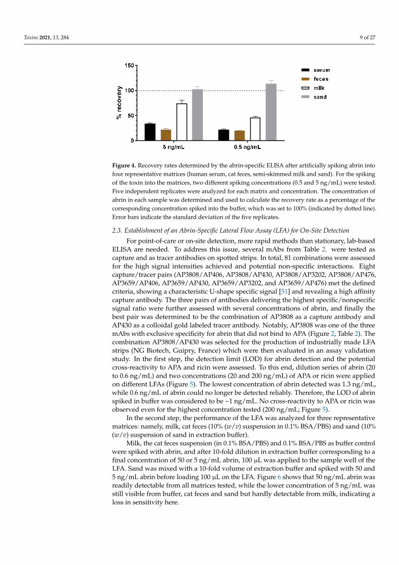
To assess potential matrix effects which could be encountered when analyzing clinical,food or environmental samples, four representative matrices were selected. As clinicalmatrices, pooled human donor serum and cat feces were analyzed. As the food matrix, semi-skimmed milk was evaluated, and as an environmental matrix, a commercially availablestandardized soil (100% sand) was evaluated. In the first step, the effect of the blankmatrices in different amounts or concentrations without the addition of toxin on the abrin-specific sandwich ELISA was analyzed. No interfering matrix effects such as increasedbackground signals were observed. In the second step, the influence of the matrices onthe detection and quantitation of abrin was investigated. For this purpose, either thebuffer or the four matrices were spiked with 5 and 0.5 ng/mL of abrin. Five independentreplicates were analyzed for each matrix and concentration. The concentration of abrin ineach sample was determined and used to calculate the recovery rate as a percentage ofthe corresponding concentration spiked into the buffer, which was set to 100%. Figure 4shows the results for the four matrices (human serum, cat feces, semi-skimmed milk andsand) spiked with abrin. The highest recovery rate was determined with the inert matrixsand (around 100%). For milk, a recovery rate between 50% and 75% was found. Serumand feces were more challenging matrices with recovery rates between 20–30%. Differentcomponents of these two matrices—e.g., glyco-structures—seemed to mask the toxin orinterfere with the toxin−antibody interaction. The matrix feces contained insoluble organicparticles, so a loss of abrin binding to the particles might be an additional reason forreduced recovery.
Toxins 2021, 13, 284 9 of 27
Toxins 2021, 13, 284 9 of 27
interfere with the toxin−antibody interaction. The matrix feces contained insoluble organic particles, so a loss of abrin binding to the particles might be an additional reason for re-duced recovery.
Figure 4. Recovery rates determined by the abrin-specific ELISA after artificially spiking abrin into four representative matrices (human serum, cat feces, semi-skimmed milk and sand). For the spik-ing of the toxin into the matrices, two different spiking concentrations (0.5 and 5 ng/mL) were tested. Five independent replicates were analyzed for each matrix and concentration. The concentration of abrin in each sample was determined and used to calculate the recovery rate as a percentage of the corresponding concentration spiked into the buffer, which was set to 100% (indicated by dotted line). Error bars indicate the standard deviation of the five replicates.
2.3. Establishment of an Abrin-Specific Lateral Flow Assay (LFA) for On-Site Detection For point-of-care or on-site detection, more rapid methods than stationary, lab-based
ELISA are needed. To address this issue, several mAbs from Table 2. were tested as cap-ture and as tracer antibodies on spotted strips. In total, 81 combinations were assessed for the high signal intensities achieved and potential non-specific interactions. Eight cap-ture/tracer pairs (AP3808/AP406, AP3808/AP430, AP3808/AP3202, AP3808/AP476, AP3659/AP406, AP3659/AP430, AP3659/AP3202, and AP3659/AP476) met the defined cri-teria, showing a characteristic U-shape specific signal [51] and revealing a high affinity capture antibody. The three pairs of antibodies delivering the highest specific/nonspecific signal ratio were further assessed with several concentrations of abrin, and finally the best pair was determined to be the combination of AP3808 as a capture antibody and AP430 as a colloidal gold labeled tracer antibody. Notably, AP3808 was one of the three mAbs with exclusive specificity for abrin that did not bind to APA (Figure 2, Table 2). The com-bination AP3808/AP430 was selected for the production of industrially made LFA strips (NG Biotech, Guipry, France) which were then evaluated in an assay validation study. In the first step, the detection limit (LOD) for abrin detection and the potential cross-reactiv-ity to APA and ricin were assessed. To this end, dilution series of abrin (20 to 0.6 ng/mL) and two concentrations (20 and 200 ng/mL) of APA or ricin were applied on different LFAs (Figure 5). The lowest concentration of abrin detected was 1.3 ng/mL, while 0.6 ng/mL of abrin could no longer be detected reliably. Therefore, the LOD of abrin spiked in buffer was considered to be ~1 ng/mL. No cross-reactivity to APA or ricin was observed even for the highest concentration tested (200 ng/mL; Figure 5).
In the second step, the performance of the LFA was analyzed for three representative matrices: namely, milk, cat feces (10% (w/v) suspension in 0.1% BSA/PBS) and sand (10% (w/v) suspension of sand in extraction buffer).
Milk, the cat feces suspension (in 0.1% BSA/PBS) and 0.1% BSA/PBS as buffer control were spiked with abrin, and after 10-fold dilution in extraction buffer corresponding to a final concentration of 50 or 5 ng/mL abrin, 100 µL was applied to the sample well of the LFA. Sand was mixed with a 10-fold volume of extraction buffer and spiked with 50 and 5 ng/mL abrin before loading 100 µL on the LFA. Figure 6 shows that 50 ng/mL abrin was readily detectable from all matrices tested, while the lower concentration of 5 ng/mL was
Figure 4. Recovery rates determined by the abrin-specific ELISA after artificially spiking abrin intofour representative matrices (human serum, cat feces, semi-skimmed milk and sand). For the spikingof the toxin into the matrices, two different spiking concentrations (0.5 and 5 ng/mL) were tested.Five independent replicates were analyzed for each matrix and concentration. The concentration ofabrin in each sample was determined and used to calculate the recovery rate as a percentage of thecorresponding concentration spiked into the buffer, which was set to 100% (indicated by dotted line).Error bars indicate the standard deviation of the five replicates.
2.3. Establishment of an Abrin-Specific Lateral Flow Assay (LFA) for On-Site Detection
For point-of-care or on-site detection, more rapid methods than stationary, lab-basedELISA are needed. To address this issue, several mAbs from Table 2. were tested ascapture and as tracer antibodies on spotted strips. In total, 81 combinations were assessedfor the high signal intensities achieved and potential non-specific interactions. Eightcapture/tracer pairs (AP3808/AP406, AP3808/AP430, AP3808/AP3202, AP3808/AP476,AP3659/AP406, AP3659/AP430, AP3659/AP3202, and AP3659/AP476) met the definedcriteria, showing a characteristic U-shape specific signal [51] and revealing a high affinitycapture antibody. The three pairs of antibodies delivering the highest specific/nonspecificsignal ratio were further assessed with several concentrations of abrin, and finally thebest pair was determined to be the combination of AP3808 as a capture antibody andAP430 as a colloidal gold labeled tracer antibody. Notably, AP3808 was one of the threemAbs with exclusive specificity for abrin that did not bind to APA (Figure 2, Table 2). Thecombination AP3808/AP430 was selected for the production of industrially made LFAstrips (NG Biotech, Guipry, France) which were then evaluated in an assay validationstudy. In the first step, the detection limit (LOD) for abrin detection and the potentialcross-reactivity to APA and ricin were assessed. To this end, dilution series of abrin (20to 0.6 ng/mL) and two concentrations (20 and 200 ng/mL) of APA or ricin were appliedon different LFAs (Figure 5). The lowest concentration of abrin detected was 1.3 ng/mL,while 0.6 ng/mL of abrin could no longer be detected reliably. Therefore, the LOD of abrinspiked in buffer was considered to be ~1 ng/mL. No cross-reactivity to APA or ricin wasobserved even for the highest concentration tested (200 ng/mL; Figure 5).
In the second step, the performance of the LFA was analyzed for three representativematrices: namely, milk, cat feces (10% (w/v) suspension in 0.1% BSA/PBS) and sand (10%(w/v) suspension of sand in extraction buffer).
Milk, the cat feces suspension (in 0.1% BSA/PBS) and 0.1% BSA/PBS as buffer controlwere spiked with abrin, and after 10-fold dilution in extraction buffer corresponding to afinal concentration of 50 or 5 ng/mL abrin, 100 µL was applied to the sample well of theLFA. Sand was mixed with a 10-fold volume of extraction buffer and spiked with 50 and5 ng/mL abrin before loading 100 µL on the LFA. Figure 6 shows that 50 ng/mL abrin wasreadily detectable from all matrices tested, while the lower concentration of 5 ng/mL wasstill visible from buffer, cat feces and sand but hardly detectable from milk, indicating aloss in sensitivity here.

Toxins 2021, 13, 284 10 of 27
Toxins 2021, 13, 284 10 of 27
still visible from buffer, cat feces and sand but hardly detectable from milk, indicating a loss in sensitivity here.
(a) 20 ng/mL 10 ng/mL 5 ng/mL 2.5 ng/mL 1.3 ng/mL 0.6 ng/mL 0 ng/mL
(b) 200 ng/mL 20 ng/mL (c) 200 ng/mL 20 ng/mL
Figure 5. Performance of the abrin LFA for the detection of abrin spiked into buffer. The LFA was based on AP3808 as a capture antibody and colloidal gold labeled AP430 as a tracer antibody. For (a) abrin, 20, 10, 5, 2.5, 1.3, 0.6, and 0 ng/mL as final concentrations were used, whereas for (b) APA and (c) ricin, 200 and 20 ng/mL were tested. Abrin, APA and ricin were first diluted in 0.1% BSA/PBS followed by a 1:10 dilution in extraction buffer in accordance with the manufacturer’s instructions, and 100 µL was applied to the LFA. Results were read out after 30 min by the naked eye. C denotes the control line (anti-mouse capture antibody) and T denotes the test line (anti-abrin antibody).
(a) 50 ng/mL 5 ng/mL 0 ng/mL (b) 50 ng/mL 5 ng/mL 0 ng/mL
(c) 50 ng/mL 5 ng/mL 0 ng/mL (d) 50 ng/mL 5 ng/mL 0 ng/mL
Figure 6. Detection of abrin in matrices using the abrin LFA. Two concentrations of abrin were spiked into (a) 0.1% BSA/PBS, (b) semi-skimmed milk and (c) a 10% (w/v) suspension of cat feces in 0.1% BSA/PBS, which were all further diluted with a 10-fold volume of extraction buffer resulting in the indicated final concentrations of abrin. (d) A 10% (w/v) suspension of sand in extraction buffer was spiked with abrin at 50 and 5 ng/mL and the sand was removed by centrifugation. Then, 100 µL of each solution was applied to the sample well. Results were read out after 30 min by the naked
Figure 5. Performance of the abrin LFA for the detection of abrin spiked into buffer. The LFA wasbased on AP3808 as a capture antibody and colloidal gold labeled AP430 as a tracer antibody. For (a)abrin, 20, 10, 5, 2.5, 1.3, 0.6, and 0 ng/mL as final concentrations were used, whereas for (b) APA and(c) ricin, 200 and 20 ng/mL were tested. Abrin, APA and ricin were first diluted in 0.1% BSA/PBSfollowed by a 1:10 dilution in extraction buffer in accordance with the manufacturer’s instructions,and 100 µL was applied to the LFA. Results were read out after 30 min by the naked eye. C denotesthe control line (anti-mouse capture antibody) and T denotes the test line (anti-abrin antibody).
Toxins 2021, 13, 284 10 of 27
still visible from buffer, cat feces and sand but hardly detectable from milk, indicating a loss in sensitivity here.
(a) 20 ng/mL 10 ng/mL 5 ng/mL 2.5 ng/mL 1.3 ng/mL 0.6 ng/mL 0 ng/mL
(b) 200 ng/mL 20 ng/mL (c) 200 ng/mL 20 ng/mL
Figure 5. Performance of the abrin LFA for the detection of abrin spiked into buffer. The LFA was based on AP3808 as a capture antibody and colloidal gold labeled AP430 as a tracer antibody. For (a) abrin, 20, 10, 5, 2.5, 1.3, 0.6, and 0 ng/mL as final concentrations were used, whereas for (b) APA and (c) ricin, 200 and 20 ng/mL were tested. Abrin, APA and ricin were first diluted in 0.1% BSA/PBS followed by a 1:10 dilution in extraction buffer in accordance with the manufacturer’s instructions, and 100 µL was applied to the LFA. Results were read out after 30 min by the naked eye. C denotes the control line (anti-mouse capture antibody) and T denotes the test line (anti-abrin antibody).
(a) 50 ng/mL 5 ng/mL 0 ng/mL (b) 50 ng/mL 5 ng/mL 0 ng/mL
(c) 50 ng/mL 5 ng/mL 0 ng/mL (d) 50 ng/mL 5 ng/mL 0 ng/mL
Figure 6. Detection of abrin in matrices using the abrin LFA. Two concentrations of abrin were spiked into (a) 0.1% BSA/PBS, (b) semi-skimmed milk and (c) a 10% (w/v) suspension of cat feces in 0.1% BSA/PBS, which were all further diluted with a 10-fold volume of extraction buffer resulting in the indicated final concentrations of abrin. (d) A 10% (w/v) suspension of sand in extraction buffer was spiked with abrin at 50 and 5 ng/mL and the sand was removed by centrifugation. Then, 100 µL of each solution was applied to the sample well. Results were read out after 30 min by the naked
Figure 6. Detection of abrin in matrices using the abrin LFA. Two concentrations of abrin werespiked into (a) 0.1% BSA/PBS, (b) semi-skimmed milk and (c) a 10% (w/v) suspension of cat feces in0.1% BSA/PBS, which were all further diluted with a 10-fold volume of extraction buffer resultingin the indicated final concentrations of abrin. (d) A 10% (w/v) suspension of sand in extractionbuffer was spiked with abrin at 50 and 5 ng/mL and the sand was removed by centrifugation. Then,100 µL of each solution was applied to the sample well. Results were read out after 30 min by thenaked eye. C denotes the control line (anti-mouse capture antibody) and T denotes the test line(anti-abrin antibody).

Toxins 2021, 13, 284 11 of 27
2.4. Application of the Monoclonal Antibodies for LC-ESI-MS/MS with Immuno-Enrichment
Generally, immunological methods based on highly specific and affine antibodies offerseveral advantages such as exquisite sensitivity and compatibility with routine applications,but they usually do not deliver unambiguous results. Here, MS-based techniques areclearly advantageous as they enable the unambiguous identification of a protein basedon its peptide fingerprint—an issue that is highly relevant in the course of a forensicinvestigation [52]. Additionally, MS-based methods can deliver detailed information onknown and even unknown sample contents, especially when using scanning-mode MSapproaches, thereby adding an open view to the diagnostic workflow [53]. Moreover,absolute quantification can be achieved by spiking stable-isotope-labeled peptides to thesamples. However, MS methods can be severely hampered by the presence of otherpeptides or proteins in excessive amounts. Thus, affinity purification steps prior to MSanalysis have been used to extract the target analyte(s) from complex sample matrices. Inthis context, we have recently published two manuscripts applying the antibodies describedin this work for immuno-affinity enrichment followed by the LC-ESI-MS/MS-based orMALDI-TOF-based identification and quantification of abrin in complex matrices [42,43].Based on our previous work, we identified a combination of four mAbs—namely AP430,AP3808, AP3659—directed against abrin plus AP476 specific for APA, as an optimal mixturefor the immuno-enrichment of both abrin and APA from samples, followed by a trypticdigest and MS analysis.
In the context of the current work, we conducted a trace analysis study, using thepurified abrin and APA preparations reported herein to characterize the antibodies’ bindingprofiles (Figures 1–3). The question was if low amounts of APA in excessive amounts ofabrin and, conversely, abrin in APA could be clearly identified considering their highidentity on the amino acid level (74.8%; Table 1). To this end, approximately 80 µg of theabrin or the APA preparation was subjected to immuno-enrichment using the previouslyestablished protocol followed by tryptic digestion and LC-ESI-MS/MS analysis. As shownin Supplementary Figure S6, all four isoforms of abrin (abrin-a to d [26]) were identifiedand sequenced with a sequence coverage of 53% to 60% in the purified abrin preparation.Despite the high purity of the abrin preparation (>97%), peptides specific for APA couldbe identified and sequenced with a sequence coverage of 39% indicating a low cross-contamination of abrin with APA.
Likewise, the same procedure applied to the purified APA preparation delivered 52%sequence coverage for APA-specific peptides (Supplementary Figure S7). Additionally,a low amount of cross-contaminating abrin (isoforms abrin-a, b, and d) with a sequencecoverage of 43% to 54% was identified as well, accounting for some 2%–3% of the material(Supplement Figure S7).
2.5. Application of the Monoclonal Antibodies in Diagnostics of an Attempted Suicide Case
In order to demonstrate the diagnostic value of the different methodologies establishedin this work, we applied both the abrin-specific ELISA and the LC-ESI-MS/MS approach inan attempted suicide case with oral A. precatorius uptake in Germany. Here, a single humanfecal sample was obtained approximately 24 h after the ingestion of at least one chewed A.precatorius seed (according to the patient). The stool was extracted for 30 min with either0.1% BSA/PBS or 0.1% BSA/PBS containing 1% (v/v) Triton X-100 and 250 mM galactose(gal-TX buffer) to increase toxin recovery by facilitating desorption from glyco-structures.The extracts were clarified by centrifugation and analyzed using the abrin-specific ELISA(Figure 3a). As shown in Figure 7, ELISA quantification provided about 660 ng abrin pergram of stool in the 0.1% BSA/PBS buffer extract, but 3840 ng abrin per gram of stool in thegal-TX buffer, indicating a 5.8-fold increase in toxin recovery from the challenging matrixfeces when using a galactose/detergent-containing extraction buffer.

Toxins 2021, 13, 284 12 of 27
Toxins 2021, 13, 284 12 of 27
the challenging matrix feces when using a galactose/detergent-containing extraction buffer.
Figure 7. Analysis of a fecal sample from a suicide attempt with Abrus precatorius using abrin-spe-cific ELISA. Stool was extracted for 30 min 1:5 (w/v) with either 0.1% BSA/PBS or with the same buffer supplemented with 1% Triton X-100 and 250 mM galactose. Suspension was clarified by cen-trifugation and supernatants were measured applying the abrin-specific ELISA. Quantitation was based on a standard curve using the purified abrin preparation described in this work.
In order to confirm the presence of abrin in the fecal sample, an immuno-affinity en-richment protocol was applied to the stool extract, followed by a tryptic digest and LC-ESI-MS/MS detection (Figure 8). In this challenging clinical matrix, abrin could be identi-fied with a sequence coverage of 24% for the isoform abrin-b and 9% for abrin-a, confirm-ing the previous ELISA results.
(a) (b)
Figure 8. Protein sequence coverage of proteins identified in a human fecal sample from a suicide attempt after immuno-affinity enrichment, tryptic digest and non-targeting liquid chromatography coupled with electrospray ionization and tandem mass spectrometry (LC-ESI-MS/MS) analysis. Se-quences of identified abrin isoforms (a) abrin-a (UniProt P11140) and (b) abrin-b (Q06077) are shown after a MASCOT server search against a self-assembled UniProt/NCBI database containing all abrin isoforms and Abrus precatorius agglutinin as well as an NCBI database containing all Abrus precato-rius proteins. Amino acids highlighted in red were experimentally identified with a sequence cov-erage of 9% for abrin-a and 24% for abrin-b. Underlined peptides represent proteotypic peptides for (a) abrin-a or (b) abrin-b, respectively. Asparagine (N), highlighted in turquoise, represents poten-tial N-linked glycosylation sites. The linker peptide sequence between the two chains of both abrin isoforms is underlined in black.
P11140, Abrin-a; Protein sequence coverage: 9% Q06077, Abrin-b; Protein sequence coverage: 24%
1 QDRPIKFSTE GATSQSYKQF IEALRERLRG GLIHDIPVLP DPTTLQERNR 1 QDQVIKFTTE GATSQSYKQF IEALRQRLTG GLIHGIPVLP DPTTLQERNR
51 YITVELSNSD TESIEVGIDV TNAYVVAYRA GTQSYFLRDA PSSASDYLFT 51 YISVELSNSD TESIEAGIDV SNAYVVAYRA GNRSYFLRDA PTSASRYLFT
101 GTDQHSLPFY GTYGDLERWA HQSRQQIPLG LQALTHGISF FRSGGNDNEE 101 GTQQYSLRFN GSYIDLERLA RQTRQQIPLG LQALRHAISF LQSGTDDQEI
151 KARTLIVIIQ MVAEAARFRY ISNRVRVSIQ TGTAFQPDAA MISLENNWDN 151 ARTLIVIIQM ASEAARYRFI SYRVGVSIRT NTAFQPDAAM ISLENNWDNL
201 LSRGVQESVQ DTFPNQVTLT NIRNEPVIVD SLSHPTVAVL ALMLFVCNPP 201 SGGVQQSVQD TFPNAVTLRS VNNQPVIVDS LTHQSVAVLA LMLFVCNPPN
251 NANQSPLLIR SIVEKSKICS SRYEPTVRIG GRDGMCVDVY DNGYHNGNRI 251 ANQSPLLIRS IVEKSKICSS RYEPTVRIGG RNGMCVDVYD DGYHNGNRII
301 IMWKCKDRLE ENQLWTLKSD KTIRSNGKCL TTYGYAPGSY VMIYDCTSAV 301 AWKCKDRLEE NQLWTLKSDK TIRSNGKCLT TEGYAPGNYV MIYDCTSAVA
351 AEATYWEIWD NGTIINPKSA LVLSAESSSM GGTLTVQTNE YLMRQGWRTG 351 EATYWEIWDN GTIINPKSAL VLSAESSSMG GTLTVQTNEY LMRQGWRTGN
401 NNTSPFVTSI SGYSDLCMQA QGSNVWMADC DSNKKEQQWA LYTDGSIRSV 401 NTSPFVTSIS GYSDLCMQAQ GSNVWLAYCD NNKKEQQWAL YTDGSIRSVQ
451 QNTNNCLTSK DHKQGSTILL MGCSNGWASQ RWVFKNDGSI YSLYDDMVMD 451 NTNNCLTSKD HKQGSPIVLM ACSNGWASQR WLFRNDGSIY NLHDDMVMDV
501 VKGSDPSLKQ IILWPYTGKP NQIWLTLF
501 KRSDPSLKEI ILHPYHGKPN QIWLTLF
Figure 7. Analysis of a fecal sample from a suicide attempt with Abrus precatorius using abrin-specific ELISA. Stool was extracted for 30 min 1:5 (w/v) with either 0.1% BSA/PBS or with the samebuffer supplemented with 1% Triton X-100 and 250 mM galactose. Suspension was clarified bycentrifugation and supernatants were measured applying the abrin-specific ELISA. Quantitation wasbased on a standard curve using the purified abrin preparation described in this work.
In order to confirm the presence of abrin in the fecal sample, an immuno-affinityenrichment protocol was applied to the stool extract, followed by a tryptic digest andLC-ESI-MS/MS detection (Figure 8). In this challenging clinical matrix, abrin could beidentified with a sequence coverage of 24% for the isoform abrin-b and 9% for abrin-a,confirming the previous ELISA results.
Toxins 2021, 13, 284 12 of 27
the challenging matrix feces when using a galactose/detergent-containing extraction buffer.
Figure 7. Analysis of a fecal sample from a suicide attempt with Abrus precatorius using abrin-spe-cific ELISA. Stool was extracted for 30 min 1:5 (w/v) with either 0.1% BSA/PBS or with the same buffer supplemented with 1% Triton X-100 and 250 mM galactose. Suspension was clarified by cen-trifugation and supernatants were measured applying the abrin-specific ELISA. Quantitation was based on a standard curve using the purified abrin preparation described in this work.
In order to confirm the presence of abrin in the fecal sample, an immuno-affinity en-richment protocol was applied to the stool extract, followed by a tryptic digest and LC-ESI-MS/MS detection (Figure 8). In this challenging clinical matrix, abrin could be identi-fied with a sequence coverage of 24% for the isoform abrin-b and 9% for abrin-a, confirm-ing the previous ELISA results.
(a) (b)
Figure 8. Protein sequence coverage of proteins identified in a human fecal sample from a suicide attempt after immuno-affinity enrichment, tryptic digest and non-targeting liquid chromatography coupled with electrospray ionization and tandem mass spectrometry (LC-ESI-MS/MS) analysis. Se-quences of identified abrin isoforms (a) abrin-a (UniProt P11140) and (b) abrin-b (Q06077) are shown after a MASCOT server search against a self-assembled UniProt/NCBI database containing all abrin isoforms and Abrus precatorius agglutinin as well as an NCBI database containing all Abrus precato-rius proteins. Amino acids highlighted in red were experimentally identified with a sequence cov-erage of 9% for abrin-a and 24% for abrin-b. Underlined peptides represent proteotypic peptides for (a) abrin-a or (b) abrin-b, respectively. Asparagine (N), highlighted in turquoise, represents poten-tial N-linked glycosylation sites. The linker peptide sequence between the two chains of both abrin isoforms is underlined in black.
P11140, Abrin-a; Protein sequence coverage: 9% Q06077, Abrin-b; Protein sequence coverage: 24%
1 QDRPIKFSTE GATSQSYKQF IEALRERLRG GLIHDIPVLP DPTTLQERNR 1 QDQVIKFTTE GATSQSYKQF IEALRQRLTG GLIHGIPVLP DPTTLQERNR
51 YITVELSNSD TESIEVGIDV TNAYVVAYRA GTQSYFLRDA PSSASDYLFT 51 YISVELSNSD TESIEAGIDV SNAYVVAYRA GNRSYFLRDA PTSASRYLFT
101 GTDQHSLPFY GTYGDLERWA HQSRQQIPLG LQALTHGISF FRSGGNDNEE 101 GTQQYSLRFN GSYIDLERLA RQTRQQIPLG LQALRHAISF LQSGTDDQEI
151 KARTLIVIIQ MVAEAARFRY ISNRVRVSIQ TGTAFQPDAA MISLENNWDN 151 ARTLIVIIQM ASEAARYRFI SYRVGVSIRT NTAFQPDAAM ISLENNWDNL
201 LSRGVQESVQ DTFPNQVTLT NIRNEPVIVD SLSHPTVAVL ALMLFVCNPP 201 SGGVQQSVQD TFPNAVTLRS VNNQPVIVDS LTHQSVAVLA LMLFVCNPPN
251 NANQSPLLIR SIVEKSKICS SRYEPTVRIG GRDGMCVDVY DNGYHNGNRI 251 ANQSPLLIRS IVEKSKICSS RYEPTVRIGG RNGMCVDVYD DGYHNGNRII
301 IMWKCKDRLE ENQLWTLKSD KTIRSNGKCL TTYGYAPGSY VMIYDCTSAV 301 AWKCKDRLEE NQLWTLKSDK TIRSNGKCLT TEGYAPGNYV MIYDCTSAVA
351 AEATYWEIWD NGTIINPKSA LVLSAESSSM GGTLTVQTNE YLMRQGWRTG 351 EATYWEIWDN GTIINPKSAL VLSAESSSMG GTLTVQTNEY LMRQGWRTGN
401 NNTSPFVTSI SGYSDLCMQA QGSNVWMADC DSNKKEQQWA LYTDGSIRSV 401 NTSPFVTSIS GYSDLCMQAQ GSNVWLAYCD NNKKEQQWAL YTDGSIRSVQ
451 QNTNNCLTSK DHKQGSTILL MGCSNGWASQ RWVFKNDGSI YSLYDDMVMD 451 NTNNCLTSKD HKQGSPIVLM ACSNGWASQR WLFRNDGSIY NLHDDMVMDV
501 VKGSDPSLKQ IILWPYTGKP NQIWLTLF
501 KRSDPSLKEI ILHPYHGKPN QIWLTLF
Figure 8. Protein sequence coverage of proteins identified in a human fecal sample from a suicide attempt after immuno-affinity enrichment, tryptic digest and non-targeting liquid chromatography coupled with electrospray ionization andtandem mass spectrometry (LC-ESI-MS/MS) analysis. Sequences of identified abrin isoforms (a) abrin-a (UniProt P11140)and (b) abrin-b (Q06077) are shown after a MASCOT server search against a self-assembled UniProt/NCBI databasecontaining all abrin isoforms and Abrus precatorius agglutinin as well as an NCBI database containing all Abrus precatoriusproteins. Amino acids highlighted in red were experimentally identified with a sequence coverage of 9% for abrin-a and24% for abrin-b. Underlined peptides represent proteotypic peptides for (a) abrin-a or (b) abrin-b, respectively. Asparagine(N), highlighted in turquoise, represents potential N-linked glycosylation sites. The linker peptide sequence between thetwo chains of both abrin isoforms is underlined in black.

Toxins 2021, 13, 284 13 of 27
3. Discussion
In the current work, a panel of 15 mAbs specific for either abrin, APA or both wasgenerated and comprehensively characterized by ELISA, SPR and Western blotting, andsuitable mAbs were implemented into sandwich ELISA, LFA and MS-based approaches.Key features of the methodologies were highlighted and the approaches were applied toanalyze representative complex clinical, food and environmental matrices, including aclinical sample from a human case of A. precatorius intoxication. An overview of the use ofthe different mAbs in the different applications is given in Table 4, with antibodies depictedin bold showing the superior performance when applied in the indicated methods.
Table 4. Performance of the mAbs generated in this work in various applications.
Antibody ShortName
Application of Antibodies #
WesternBlot SPR Sandwich
ELISA * LFA *
Immuno-Enrichment
followed by MSAnalysis * §
AP12 – –AP54 X –AP69 X –AP87 X –AP188 X –AP267 X XAP406 – XAP430 X X X X XAP464 X –AP476 X X X XAP708 X –
AP2573 – X XAP3202 X X XAP3659 – X XAP3808 – X X X
* selected antibodies with superior performance for the indicated method (bold); # X: good performance; –: no orpoor binding; § immuno-enrichment for MS as described in this publication and in Hansbauer et al. [42] andLivet et al. [43].
Antibodies are still the most versatile tools to specifically detect their target moleculein a broad range of matrices. Here, mAbs in comparison to polyclonal antibodies (pAbs)have been shown to offer the advantage of defined specificity and often higher sensitivity,provided that high-affinity mAbs are used. In terms of quality management, mAbs derivedfrom stable hybridoma clones can be produced with a constant quality over time, thusincreasing the reproducibility of experimental data and preventing the lot-to-lot variabilityobserved with pAbs. In line with recommendations on mAb validation [54,55], the mAbsdescribed in this work were characterized to demonstrate their fitness for purpose, assess-ing their target antigen, binding selectivity (cross-reactivity), binding strength (affinity) andthe influence of non-target substances (matrix effects). Not surprisingly, different mAbsturned out to be optimal for different applications, with some of them targeting epitopeson the native antigen, making them suitable tools for sandwich ELISA or immuno-affinityenrichment, and others targeting denatured epitopes relevant in Western blotting (Table 4).Interestingly, the panel of mAbs comprised antibodies showing a strong preference foreither abrin (AP3202, AP3659, AP3808), for APA (AP2573) or both (AP87, AP464, AP708),with the latter three applicable only in Western blotting and indirect ELISA. By carefullyselecting highly affine mAbs that preferentially detect abrin over APA or vice versa, twoELISA systems were developed in this work which allowed us to discriminate betweenpurified abrin and purified APA, with less than 0.7% (abrin-specific ELISA) or 0.1% (APA-specific ELISA) cross-reactivity between the two related lectins. It is worthy of note that bycombining a capture mAb directed against the B-chain of abrin (AP430) with a detection

Toxins 2021, 13, 284 14 of 27
mAb recognizing the A-chain (AP3202), the abrin-specific ELISA detected only the intactA-B toxin. In previous works, the issue of mAb / ELISA selectivity for abrin versus APAhas rarely been addressed; only a few groups have reported either cross-reactive or specificmAbs for abrin and APA derived from mouse or llama [32,56,57]. In a broader context,sandwich ELISAs able to differentiate between related toxin subtypes or isolectins havebeen successfully established based on highly specific mAbs directed against unique do-mains found in the related molecules (e.g., for ricin/RCA120 or for the related botulinumneurotoxins BoNT/C, CD, DC, and D [47,48]). Additionally, based on specific mAbs, asurface plasmon resonance sensor has been developed to simultaneously differentiate andquantify ricin from RCA120 in real time in less than 30 min [58]—an application that isnow open to be explored for abrin and APA as well.
In comparison, the pAb described here, KAP142, was unable to distinguish betweenthe select agent abrin and the related APA, which are 74.8% identical at amino acid level,thus preventing its use in criminal or forensic investigations where discrimination betweenthese two molecules is mandatory. Even worse, KAP142 also reacted with the relatedplant lectins ricin and RCA120, which share 48.8% or 44.6% sequence identity with abrin,respectively. This type of cross-reactivity between abrin/APA and ricin/RCA120 haspreviously been observed for other pAbs [30,59,60]; it has even been reported that mAbs—similarly to pAbs—showed cross-reactivity between abrin and ricin [61,62]. Interestingly,these mAbs were derived from naïve human or llama phage-display libraries and not froman immunized host.
With respect to matrix interference, pAbs are more likely to react with non-targetsubstances in complex samples, which often results in elevated background signals [31].This is due to their polyclonal nature and the presence of antigen-unrelated antibodies. Theapplication of the mAbs presented in this work in a sandwich ELISA to detect abrin fromrepresentative clinical (serum, feces), food (milk) and environmental (sand) samples didnot result in an elevated background. When abrin was spiked into the four representativematrices, ELISA results delivered recovery rates between 20–110%. It is well known thatmatrix effects play a major role in assay performance [46,63–65] since components of thematrix can interfere with the non-covalent interactions between antibodies and antigens sta-bilized by electrostatic forces, hydrogen bonds, van der Waals forces and/or hydrophobicforces. Additionally, matrix components might mask or expose antibody-binding epitopes,leading to decreased or increased antibody–antigen binding. The difficulty is that theextent to which these effects occur with different matrices cannot be anticipated but has tobe assessed empirically. Therefore, although challenging matrices have been tested, thevalidation study initiated in this work has to be extended in the future to include a broaderspectrum of clinical, food and environmental matrices.
Regarding assay sensitivity, the two stationary ELISAs developed in this work deliv-ered excellent detection limits: for the abrin-specific ELISA, an LOD of 22 pg/mL, andfor the APA-specific ELISA, an LOD of ~35 pg/mL were determined. In comparison,previously reported ELISAs for abrin delivered detection limits between 100 pg/mL and7800 pg/mL [31–34,57]; therefore, the current work significantly advances the field, offeringtools with higher sensitivity as well as increased specificity and selectivity.
In a potential biothreat scenario, the rapid detection of threat agents is beneficial tosupport an immediate risk assessment and to protect first responders entering the scene. Inthis situation, a stationary ELISA as described above, although highly sensitive and specific,turns out to be of limited use, due to its long assay time (4–6 h) and the requirement of alaboratory surrounding with trained personnel. To address this, LFAs and biosensors havebeen developed and optimized for use by non-trained personnel in the field, deliveringresults in less than one hour [34–37]. The mAbs described in this work were tested andincorporated into LFA cartridges at CEA and were selected for industrial production witha commercial partner. Unlike most other LFAs, the LFAs established here preferentiallydetected abrin over APA (LOD ~1 ng/mL) and as such would provide first responderswith a more robust risk assessment compared to cross-reactive LFAs. To date, there is only

Toxins 2021, 13, 284 15 of 27
one other product described which is able to discriminate between abrin and APA [34].Due to the lack of washing steps, LFAs might encounter problems with complex matrices.To address this, the LFA was tested with the representative matrices of milk, feces andsand. Concentrations of 5–50 ng/mL could still be detected, which was well in the rangeof 0.3 to 50 ng/mL reported for other LFAs [34,36,37]. It should be noted that, in the caseof fecal samples, feeding, drinking and other living conditions will presumably alter theextraction efficacy to lower or higher rates, meaning that the sensitivity cannot generallybe anticipated.
Apart from methods targeting the toxin itself, techniques addressing the biologicalactivity of abrin are important to assess the threat potential in an incident and are requiredto complement a comprehensive analysis of evidences in a criminal or forensic investigation.As described above, our sandwich ELISA detects both the A and the B chain present inintact abrin. However, this is not yet sufficient evidence of biological activity. In order toassess the toxin’s functional activity, in vivo experiments, cell-based cytotoxicity assaysand assays measuring the depurination of the rRNA can be performed—approachesthat have been previously described for ricin as well [66–68]. As both toxins result insimilar biological responses based on the same functional mechanism within the cell, adiscrimination between abrin and ricin by the use of specific neutralizing antibodies isimportant [68,69].
In this work, a major factor to delineate the specificity and selectivity of the mAbsand the corresponding assays was access to highly purified abrin (containing all fourisoforms abrin-a, b, c, and d) and APA preparations. Actually, the separation of the fourabrin isoforms from APA is challenging, since they share a high sequence identity (seeabove) and run at similar molecular weights in an SDS-PAGE assay under non-reducingconditions [50]. Based on previous publications [50], an optimized purification protocolwas developed that delivered abrin and APA preparations of an estimated purity ≥97%as determined by MALDI-TOF MS and SDS-PAGE [45]. This protocol will serve as astarting point to further develop a candidate reference material for abrin in the currentEuropean project EuroBioTox [70], which aims at establishing validated procedures forthe detection and identification of biological toxins, including the plant toxins abrin andricin. The challenges ahead in producing and characterizing certified reference materialshave recently been summarized by the consortium [71]. In the context of reference materialproduction, one critical issue is that the main component(s) as well as any impuritieshave to be identified and quantified by a combination of biochemical, immunological andspectrometric methods [48,71]. In preparation for this endeavor, we performed a traceanalysis applying an immuno-affinity enrichment protocol followed by a tryptic digest andLC-MS/MS analysis developed on the basis of our mAbs [42,43]. Starting with the purifiedabrin and purified APA preparations, it was our goal to determine if low amounts of APAin excessive amounts of abrin and, conversely, of abrin in APA could be clearly identifiedconsidering their high identity on the amino acid level. Indeed, the analysis showed thatboth the main component(s), and even the impurities which accounted for below ≤3%could be identified by LC-ESI-MS/MS with a high sequence coverage (52%–60% sequencecoverage for the main component(s) and 39–54% sequence coverage for the impurity). Thiswill serve as starting point for a more comprehensive characterization of the future abrinreference material; e.g., by applying MS-based quantification of the components based onlabeled AQUA peptides [42,43].
In order to demonstrate the diagnostic value of the sandwich ELISA and the immuno-affinity enrichment LC-ESI-MS/MS approach based on the mAbs described in this work, weapplied the methods in an attempted suicide case with oral A. precatorius uptake in Germany.Here, human feces were obtained approximately 24 h after ingestion of at least one chewedA. precatorius seed according to the patient’s statement. In order to optimize samplepreparation, we tested two buffers: one containing 0.1% BSA/PBS, the other one 0.1%BSA/PBS plus 1% (v/v) Triton X-100 and 0.25 M galactose. For the latter buffer, we tookadvantage of previous works in animal models of ricin intoxication, where either 0.25 M of

Toxins 2021, 13, 284 16 of 27
lactose or galactose was used for sample preparation [72,73]. Ricin and abrin as lectins bothbind to tissue or matrix components containing carbohydrates, so a high concentrationof galactose in the homogenization buffer was thought to aid detachment [72]. Indeed,we obtained a 5.8-fold increase in abrin recovery from the challenging matrix feces whenthe galactose/detergent-containing extraction buffer was used for ELISA quantification.Notably, the ELISA results could be confirmed by the immuno-affinity enrichment LC-ESI-MS/MS approach, delivering five proteotypic peptides for abrin-a (one peptide) and b(four peptides). The sequence coverage obtained was, as expected, low but still enabledunambiguous identification with 9% sequence coverage for abrin-a and 24% for abrin-b; theisolectins abrin-c and d could not be detected. In order to put these results into perspectivewith other cases of A. precatorius intoxication worldwide, we performed a literature searchfor case descriptions of A. precatorius intoxication. Since 1961, we found 23 case descriptionsof human A. precatorius intoxications in the literature, either linked to accidental, voluntaryor suicidal uptake (22 cases of oral uptake, one case of injectional uptake). For the majorityof cases, the link to A. precatorius was demonstrated by circumstantial evidence based ondetails of the case report; e.g., known or observed uptake of plant seeds or the finding ofplant material, but explicitly not by the detection of abrin. In four out of 23 cases, diagnosticassays successfully detected and identified the low molecular weight molecule L-abrine(N-methyl-L-tryptophan) in urine as a surrogate marker for abrin intoxication [18–20,74].In none of the cases was abrin itself detected. Therefore, to the best of our knowledge, thisis the first case where ELISA-based detection and quantification as well as LC-ESI-MS/MS-based identification were successfully implemented for abrin detection in a real-life case ofhuman A. precatorius intoxication.
4. Materials and Methods4.1. Toxins
All toxins as well as the mixture of A. precatorius lectins were handled by trainedpersonnel under a class II vertical laminar flow cabinet (Heraeus Herasafe, Thermo Scien-tific, Dreieich, Germany) in a dedicated toxin laboratory. Toxin-containing solutions wereinactivated with sodium hydroxide in a final concentration of 5% overnight, and solidwaste containing traces of toxin was inactivated by autoclaving (134 ◦C, 1 h).
Purified abrin, purified APA, a mixture of A. precatorius lectins, ricin and R. communisagglutinin were all produced in-house. Ricin and RCA120 were purified from the seeds ofR. communis variety carmencita pink similar to protocols described earlier [30,69,75]. Thematerial was extensively characterized and purity was determined as≥97% [48]. Abrin andAPA were purified and separated from each other following a similar strategy. The purityof both preparations was determined by mass spectrometry as ≥97% (SupplementaryFigures S1 and S2) [48,73].
For the purified mixture of A. precatorius lectins, proteins with lectin properties werepurified from the extract of A. precatorius seeds by affinity-chromatography using anXK16/20 column (Cytiva, Freiburg, Germany) packed in-house with lactosyl-sepharose.Analysis by MALDI-TOF MS showed the presence of abrin and APA in an estimated ratioof 2:3.
4.2. Matrices
A human serum pool was kindly provided by MH Hannover, Germany, and semi-skimmed milk was bought from a local retail store. Artificial soil #2 (100% sand, stan-dardized reference soil) was obtained from Ros Consulting and Development AB, Sweden.For reconstitution 20 g of dry sand was mixed with 2 mL of distilled water by rotationovernight at 4 ◦C until sand was completely wetted. Cat feces was collected, autoclaved at120 ◦C, homogenized in 0.1% BSA/PBS pH 7.4 (1:10) and filtered through a 212 µm sieveto remove residual fur. The 10% cat feces suspension was used for the spiking experimentsof matrices.
Toxins 2021, 13, 284 17 of 27
4.3. Clinical Sample Material of an A. precatorius Intoxication Case
A fecal sample of an Abrus precatorius intoxication was homogenized to a ratio of 1:5(w/v) with assay buffer (PBS, 0.1% BSA) or gal-TX buffer (PBS, 0.1% (w/v) BSA, 0.25 Mgalactose and 1% (v/v) Triton X-100) and extracted under 30 min of shaking at 4 ◦C followedby a centrifugation step (5 min, 12,000× g, 4 ◦C). The supernatant was used for analysisin the abrin-specific ELISA and immuno-enrichment for LC-ESI-MS/MS analysis. Forthe abrin-specific ELISA, the supernatants were used undiluted and diluted further withassay buffer.
For immuno-enrichment, 250 µL of sample (extract containing assay buffer) was takenfor immuno-affinity-enrichment using mAb AP3202 and AP3808 coupled to magneticDynabeads® (Invitrogen, Karlsruhe Germany) and on-bead tryptic digest (0.5 µg trypsin,120 min at 37 ◦C) followed by non-targeting LC-ESI-MS/MS analysis (for details, see thesection on Mass Spectrometry).
4.4. Sequence Analysis
For sequence comparison, the following protein sequences from the UniProt databasewere used: abrin-a, ABRA_ABRPR/P11140; Abrus precatorius agglutinin, AGGL_ABRPR/Q9M6E9; ricin, RICI_RICCO/P02879; and Ricinus communis agglutinin, AGGL_RICCO/P06750. Sequences were uploaded into the Geneious Prime 2020.2 software package(Biomatters Ltd., Auckland, New Zealand). Protein sequences were aligned and distancescalculated using MUSCLE Alignment (3.8.425) with a maximum of eight iterations.
4.5. Generation of Monoclonal and Polyclonal Antibodies
Handling of laboratory animals was performed in compliance with the regulationsof the German Animal Welfare Act and European legislation for the protection of animalsused for scientific purposes (Directive 2010/63/EU). Immunizations of mice to generatemAbs were approved by the State Office for Health and Social Affairs in Berlin (LAGeSoBerlin, Germany) under the registration numbers H129/19 and H109/03. Sacrifice of micefor the removal of thymocytes was registered by the LAGeSo under the number T0060/08.
Monoclonal antibodies (mAb) were generated as described previously [46,47,76]. Inbrief, BALB/c or NMRI mice (Charles River, Sulzfeld, Germany) were used at the age of8 weeks. Three female mice were immunized with ~50 µg of a mixture of A. precatoriuslectins inactivated by formaldehyde. The inactivated mixture of A. precatorius lectins wasprepared by adding 37% formaldehyde to the mixture, resulting in a final concentrationof 0.5% formaldehyde, followed by incubation for 21 days at 37 ◦C. Mice were boostedseveral times with similar doses of the same antigen in adjuvant at four-week intervals.Once animals had mounted a substantial titer, they were boosted with 5 µg of the nativeA. precatorius lectin mixture in adjuvant. On day −3, −2 and −1 before fusion, 5 µg ofthe native A. precatorius lectin mixture in phosphate-buffered saline (PBS) was appliedintraperitoneally. Hybridomas were produced by fusing spleen cells from immunizedmice with myeloma cells (P3-X63-Ag8.653, American Type Culture Collection) at a ratioof 2:1 in polyethylene glycol 1500 (PEG, Roche Diagnostics, Mannheim, Germany). Cellsfused together with thymocytes as feeder cells were grown in selective RPMI1640 mediacontaining 20% (v/v) fetal bovine serum, 5.78 µM azaserine, 100 µM hypoxanthine, 50 µM2-mercaptoethanol and 500 U/mL murine interleukin-6 (IL-6). Hybridoma supernatantswere screened by an indirect ELISA against the A. precatorius lectin mixture and additionallyagainst ricin/RCA120 to exclude cross-reactive antibodies at days 10 to 14 post-fusion.Positive hybridoma clones were subcloned at least twice. Immunoglobulins (IgG) werepurified from hybridoma supernatants grown in RPMI media supplemented with IgG-freefetal bovine serum by affinity chromatography over a HiTrap MabSelect SuRe columnusing a Fast Protein Liquid Chromatography System (ÄKTA, GE Healthcare Bio-SciencesAB, Uppsala, Sweden). The isotype of all purified monoclonal antibodies (mAbs) wasdetermined using an antibody isotyping sandwich ELISA (antibodies and controls fromSouthernBiotech/Biozol Diagnostica Vertrieb, Eching, Germany).

Toxins 2021, 13, 284 18 of 27
Purified antibodies were coupled to biotin according to the manufacturer’s instructions(EZ-Link Sulfo-NHS-LC-biotin; Pierce, Rockford, IL, USA). Biotinylated antibodies werestored in PBS with 0.2% (w/v) bovine serum albumin (BSA; Serva, Heidelberg, Germany)and 0.05% (w/v) NaN3 (Carl Roth, Karlsruhe, Germany).
Generation of pAb: polyclonal antibodies were generated in white New Zealandrabbits immunized subcutaneously with ~50 µg of a mixture of A. precatorius lectinsinactivated by formaldehyde. Blood was collected every four weeks after the secondbooster immunization for serum preparation. Serum was affinity purified over a proteinG column on an ÄKTA LC-instrument to obtain the IgG fraction (ÄKTA, GE HealthcareBio-Sciences AB, Uppsala, Sweden).
4.6. Indirect ELISA and Sandwich ELISA
For indirect ELISA, MaxiSorp microtiter plates (Nunc MaxiSorp flat bottom, ThermoScientific, Dreieich, Germany) were coated with abrin, APA, ricin or RCA120 at 500 ng/mLin 50 µL PBS/1 µg/mL BSA overnight at 4 ◦C and blocked with 2% skimmed milk in PBST(phosphate buffered saline with 0.05% (v/v) Tween 20, Merck, Darmstadt, Germany) for1 h at room temperature. After washing, 50 µL of antibody (10 µg/mL anti-abrin/APAantibodies or anti-ricin/RCA120 antibody R109), or in the initial hybridoma screening,50 µL of undiluted hybridoma supernatant, was added and incubated for 1 h at roomtemperature. After washing, the ELISA was developed by incubation with horseradish per-oxidase (HRP)-labeled goat anti-mouse IgG (Fc-γ specific; Dianova, Hamburg, Germany;for detection of mouse mAbs) or HRP-labeled goat anti-rabbit IgG (Dianova, Hamburg,Germany; for detection of polyclonal antibody KAP142) diluted in 2% skimmed milk inPBST (30 min, room temperature), followed by washing and incubation with substrate3,3′,5,5′-tetramethylbenzidine (TMB, SeramunBlau slow2 50, Seramun Diagnostika, Hei-desee, Germany). The color reaction was stopped with 0.25 M sulfuric acid and theabsorption was determined at 450 nm (referenced to 620 nm) using a microtiter plate reader(Infinite M200, Tecan, Männedorf, Switzerland).
For sandwich ELISA, MaxiSorp microtiter plates were coated with 5 µg/mL of pri-mary mAb AP430 (abrin-specific ELISA) or AP476 (APA-specific ELISA) in 50 µL of PBSovernight at 4 ◦C and blocked with casein buffer (Senova, Jena, Germany) for 1 h at roomtemperature. After washing, 50 µL of toxin was added in serial dilutions from 100 ng/mLto 0.3 pg/mL in assay buffer (PBS, 0.1% BSA (Sigma-Aldrich, Munich, Germany)) andincubated for 2 h at room temperature. After washing, the sandwich ELISA was devel-oped by incubation with biotin-labeled secondary antibody (AP3202 for abrin-specificELISA; AP2573 for APA-specific ELISA) diluted in casein buffer (1 h, room temperature),followed by washing and detection with Streptavidin-PolyHRP40 (0.5 ng/mL, Senova,Jena, Germany) and substrate TMB. The color reaction was stopped with 0.25 M sulfuricacid and the absorption was determined at 450 nm (referenced to 620 nm) using a microtiterplate reader.
4.7. Validation of Sandwich ELISA
For statistical analysis, the standard curve of the abrin-specific sandwich ELISAwas measured in 10 independent runs over 10 days, with two technical replicates perconcentration. Differences in absorbance at 450 nm and reference wavelength at 620 nmwere plotted against the logarithmic concentration of the abrin standard and fitted againsta sigmoidal dose–response curve (four-parametric non-linear regression analysis) in Prism8.4 (GraphPad, La Jolla, CA, USA).
The limit of detection (LOD) was calculated from the regression curve by using theabsorption value (LOD(A450–620 nm)) calculated according to Equation (1) [77,78]. The limitof blank (LOB) determined was the mean absorbance (A450–620 nm) of 178 determinationsof blanks. For the calculation of the standard deviation of low concentrations, in total80 determinations of the absorbance of low concentrated samples were performed.
LOD(A450–620 nm) = LOB + 1.645 × SD(LowC) (1)

Toxins 2021, 13, 284 19 of 27
where LOD is limit of detection, LOB is limit of blank, SD is standard deviation and LowCis low concentration.
The lower and upper limits of quantification (LLOQ and ULOQ) were calculatedbased on the sigmoidal standard curve. The LLOQ and ULOQ flank the linear range ofthe sigmoidal curve between the inflection points of the first derivative of the sigmoidalregression curve and were computed as the maxima and minima of the second derivative.
The intra-assay coefficient of variation (CV%intra) at the half maximal effective concen-tration (EC50) was determined as the standard deviation divided by the mean concentrationof 10 double determinations of the EC50 within plates multiplied by 100.
The inter-assay coefficient of variation (CV%inter) at the EC50 was calculated in relationto the concentrations determined between 10 separate and independent runs, measured intechnical duplicates.
The recovery of abrin in different matrices with the abrin-specific abrin ELISA wasevaluated by analyses of selected matrices spiked with two concentrations of abrin (5 and0.5 ng/mL) or without toxin (blank matrices). As a spiking control, buffer (0.1% BSA/PBS)was spiked with the same concentrations and treated as the spiked matrices. Buffer,human serum pool, semi-skimmed milk and 10% (w/v) cat feces suspensions were spikeddirectly with abrin (1:100 ratio toxin/matrix). The wettened sand was first resuspendedin buffer and spiked with abrin. Afterwards, spiked and blank matrices were incubatedunder rotation for 30 min at 4 ◦C followed by centrifugation for 3 min with 12,000× g.Supernatants were analyzed by abrin-specific ELISA. Five independent replicates wereprepared and analyzed for each matrix and concentration. The concentration of abrin ineach sample was determined and used to calculate the recovery rate as a percentage of thecorresponding concentration spiked into buffer, which was set to 100%.
4.8. SDS-PAGE
In total, 2 µg abrin and APA were separated on a 12% SDS-PAGE under non-reducingconditions, or 15 µg abrin-a was separated on a 12% SDS-PAGE under reducing conditions,respectively, followed by staining with colloidal Coomasssie Brilliant Blue overnight.Images were captured by a CCD camera (ChemiDoc, BioRad, Feldkirchen, Germany).
4.9. Western Blot
In total, 100 ng abrin, APA, ricin or BSA were separated on a 12% SDS-PAGE underreducing conditions and transferred onto an Immuno-Blot 0.45 µm PVDF membrane(Invitrogen, Karlsruhe, Germany). After blocking the membrane in blocking buffer (2%skimmed milk in PBST) at 4 ◦C overnight, diluted primary anti abrin/APA antibodies (finalconcentration 5 µg/mL) in blocking buffer were added to the membrane for 1 h. Afterthree washing steps, the membrane was incubated with biotin-labeled goat anti-mouse IgG(1:5000; Dianova, Hamburg, Germany; for detection of mouse mAbs) or biotin-labeled goatanti-rabbit IgG (1:5000; Dianova, Hamburg, Germany; for detection of polyclonal antibodyKAP142) in blocking buffer at room temperature for 30 min and was developed withavidin–alkaline phosphatase (incubation for 20 min, final concentration 0.5 µg/mL in PBST;Avidx™-AP, Fisher Scientific, Bremen, Germany) and the chemiluminescent substrateCDP-Star (Perkin Elmer, Waltham, MA, USA). Images were captured by a CCD camera(ChemiDoc, BioRad, Feldkirchen, Germany).
4.10. Surface Plasmon Resonance (SPR) Measurements
The affinity and kinetics of all mAbs for binding to abrin (molecular weight approxi-mately 60 kDa) and APA (molecular weight approximately 120 kDa) were determined asdescribed previously with minor modifications [58]. Briefly, a series S sensor chip CM5was modified with rabbit anti-mouse antibodies using the mouse antibody capture kit andthe amine coupling kit (all Cytiva, Freiburg, Germany) according to the manufacturer’sinstructions. The rabbit anti-mouse antibody bound all IgG subclasses used in this workequally well and showed a highly stable binding of the captured antibodies. Thus, the

Toxins 2021, 13, 284 20 of 27
determined binding kinetics were not affected by the antibody isotype and/or driftingbaselines by instable capturing. Before usage, the modified sensor chip was conditioned byinjections of purified polyclonal mouse IgG (Dianova, Hamburg, Germany) at 100 µg/mLfor 300 s at a flow rate of 5 µL/min over all four flow cells (Fc) and regeneration usinginjections of 10 mM glycine/HCl buffer at pH 1.7 (Cytiva) at 10 µL/min for 180 s, repeatedthree times. All measurements were performed on a Biacore T200 (Cytiva) at 25 ◦C usingHBS-EP+ buffer (10 mM HEPES, pH 7.4, 150 mM NaCl, 3 mM EDTA, 0.05% (v/v) Tween 20)supplemented with 10 mg/mL D-galactose (Carl Roth, Karlsruhe, Germany) to suppressthe unspecific binding of abrin or APA with immobilized antibodies due to their lectinactivity. MAbs were diluted to 2 µg/mL and captured on Fc 2 and 4 for 60 s at a flowrate of 5 µL/min leading to ligand immobilization levels between approximately 190 and350 resonance units (RUs). Binding kinetics were determined using single-cycle kineticsby injecting increasing concentrations of abrin or APA in a 1:3 dilution series rangingfrom 4.17 nM to 333.33 nM (20 µg/mL abrin or 40 µg/mL agglutinin). Association wasmonitored by injecting abrin or APA for 120 s, while dissociation was monitored for 600 sby injecting buffer at flow rates of 30 µL/min. Regeneration between runs was performedas described above using 10 mM glycine/HCl buffer at pH 1.7. Before and after eachmeasurement, buffer was injected over immobilized mAbs as blank measurements, whichwere used for double referencing binding curves [79]. To determine kinetic binding param-eters, a 1:1 Langmuir binding model (A + B = AB) with Rmax fitted globally and an RI setto 0 was fit to the measured binding curves using the Biacore T200 Evaluation SoftwareVersion 3.2 (Cytiva, Freiburg, Germany). For APA, instead of blank measurement withbuffer injections, binding curves after injection of APA over non-binding mAbs (AP12,AP54, AP69, AP87) were used as blank measurements. This was done due to the highlectin-mediated binding of APA to both control (FC 1 and 3) and measurement flow cells(FC 2 and 4), leading to artefacts in the binding curves after subtracting the signals from Fc1 from 2 and Fc 3 from 4. Although this efficiently solved the issue of artificially distortedbinding curves in the lower concentration range, distortion was still visible in the higherrange (111.11 and 333.33 nM), which is why those concentrations were excluded fromthe analysis for curve fitting. Mean binding kinetics and affinities were determined andcalculated from two technical replicate measurements for all mAbs tested.
4.11. Lateral Flow Assay4.11.1. Evaluation of Antibodies for Lateral Flow Assay
To select the best mAb pairs, and in order to develop a two-site lateral flow test, acombinatorial analysis was carried out using each mAb either as a capture or gold-labeledantibody. In this study, nine antibodies selected on the basis of their previous performancewere evaluated (AP406, AP430, AP3202, AP267, AP476, AP2573, AP3659, AP3808, andKAP142).
4.11.2. Preparation of Colloidal Gold Labeled Abrin Antibodies
Colloidal gold was prepared as previously described [80]. In total, 2 mL of the colloidalgold solution was centrifuged for 15 min at 15,000× g, and the pellet was suspended in1.6 mL of water. Then, 200 µL of a 100 µg/mL solution of each mAb in 0.02 M phosphatebuffer at pH 7.4 was added to the colloidal/gold and incubated for 1 h at 20 ◦C, leadingto the ionic adsorption of the mAbs on the surface of the gold particles. Then, 200 µL ofphosphate buffer 20 mM at pH 7.4 containing BSA 1% (w/v) was added, and the mixturewas centrifuged for 15 min at 15,000× g. The supernatant was discarded and the pelletsuspended in 1 mL phosphate buffer 2 mM at pH 7.4 with BSA 0.1% (w/v), sonicated a fewseconds and centrifuged for 15 min at 15,000× g. The supernatant was discarded and thepellet suspended in 500 µL of phosphate buffer 2 mM at pH 7.4 with BSA 0.1% (w/v) andstored at 4 ◦C in the dark.

Toxins 2021, 13, 284 21 of 27
4.11.3. Selection and Assessment of the Best Pairs of Antibodies
The spotting method was performed as previously described [81]. In total, 1 µL ofeach mAb (100 µg/mL in 50 mM phosphate buffer pH 7.4) was applied on the stripsand allowed to dry. Then, 100 µL of an abrin solution (30 ng/mL in 100 mM potassiumphosphate at pH 7.4 with 0.1% (w/v) BSA, 150 mM NaCl, 0.01% (w/v) NaN3, 0.5% (v/v)Tween 20) and 10 µL of colloidal gold labeled mAb were delivered in microtiter platewells (Greiner, Les Ulis, France). After a 5 min reaction, the strips were immersed into thesolution, and results were read out after 30 min of migration.
For the assessment of superior mAb pairs, a conventional strip format was used. Thestrips (0.5 cm in width and 4.5 cm in length) were composed of three parts: (i) a samplepad (Standard 14; Whatman) (0.5 cm in length), (ii) a nitrocellulose membrane (Prima 40,Cytiva, Velizy-Villacoublay, France) (2.5 cm in length) and (iii) an absorption pad (Cellulosegrade 470; Whatman) (1.5 cm in length), all attached to a backing card. The detection zonecontained immobilized goat anti-mouse antibodies as a control line and an anti-abrin mAbas a test line (1 mg/mL in 50 mM sodium phosphate buffer; pH 7.4) dispensed at 1 µL/cmusing an automatic dispenser (Biojet XYZ 3050; BioDot, Norton, UK). After drying for30 min at 37 ◦C in an air oven, the membrane was incubated with a blocking solution(10 mM sodium phosphate pH 7.4, 150 mM NaCl containing 0.5% (w/v) BSA) for 30 min atRT. The membrane was washed three times with deionized water, incubated for 30 min atRT in a preserving solution (10 mM sodium phosphate pH 7.4, 150 mM NaCl containing0.1% (v/v) Tween 20 and 7.5% (w/v) glucose) and then dried for 30 min at 37 ◦C in an airoven. After the absorption pad and the sample pad were fixed to the top and the bottom ofthe membrane, respectively, the card was cut into strips 5 mm in width using an automaticprogrammable cutter (CM4000 Guillotine cutting system; BioDot, Norton, UK).
Dilutions of abrin (40, 20, 10, 5, 2.5, 0.1, and 0 ng/mL) were performed in an extractionbuffer (Tris 100 mM, pH 8, NaCl 0.15 M, BSA 0.1% (w/v), 0.5% (v/v) Tween 20, 1% (w/v)CHAPS). In total, 100 µL of this solution was incubated for 5 min with 10 µL of conjugatebefore dripping the strip. Results were read out after 30 min of migration time.
4.11.4. Evaluation of the LFA
For the evaluation of the limit of detection (LOD), industrially made strips (NGBiotech, Guipry, France) were used. In total, 50 µL of diluted abrin (200, 100, 50, 25, 12.5, 6,and 0 ng/mL) in 0.1% BSA/PBS was further diluted to a ratio of 1:10 in extraction buffer(100 mM Tris pH 8, 150 mM NaCl, 0.1% BSA (w/v), 0.5% (v/v) Tween 20, and 1% (w/v)CHAPS) according to the manufacturer’s instructions. Then, 100 µL of this solution wasapplied to the sample well of the abrin LFA. Analysis was done with the naked eye after30 min.
To assess matrix compatibility, the buffer (0.1% BSA/PBS), semi-skimmed milk and10% cat feces suspension were spiked with 500 and 50 ng/mL abrin and incubated for30 min under rotation at 4 ◦C. After centrifugation, 50 µL was mixed with 450 µL extractionbuffer (1:10 dilution, according to manufacturer’s instructions) and 100 µL of this solutionwas applied to the sample well of the abrin LFA. Analysis was done with the naked eyeafter 30 min.
One part of reconstituted soil sample was mixed with nine parts of extraction bufferand spiked with 50 or 5 ng/mL abrin. After 30 min of incubation under rotation at 4 ◦C andcentrifugation, 100 µL was directly applied to the sample well of the abrin LFA. Analysiswas done with the naked eye after 30 min.
4.12. Mass Spectrometry4.12.1. Peptide Mass Fingerprinting by MALDI-TOF-MS
For purity control, the purified abrin and APA preparations were diluted to approx-imately 600 ng in 25 µL of trypsin digest buffer (40 mM NH4HCO3 containing 9% (v/v)acetonitrile). Reduction was performed with 1.5 µL of 400 mM dithiothreitol for 10 min at95 ◦C under shaking. Alkylation was carried out using 3.0 µL of 500 mM iodoacetamide
Toxins 2021, 13, 284 22 of 27
at 37 ◦C for 30 min in the dark. Reduced and alkylated samples were digested with15 µL of trypsin (0.02 ng/µL, proteomics grade; Sigma-Aldrich, Taufkirchen, Germany) at37 ◦C overnight. Reaction was stopped with 4 µL of 0.1% trifluoroacetic acid. Digestedpeptides were further desalted and concentrated with ZipTip C18 resin (Merck Millipore,Darmstadt, Germany), which was carried out according to the manufacturer’s instructions.Sample analysis was done utilizing an autoflex speed MALDI-TOF/TOF mass spectrom-eter (Bruker Daltonics, Bremen, Germany) with a polished steel MTP 384 target plate(Bruker Daltonics, Bremen, Germany). One microliter of sample was mixed with 1 µLof A-cyano-4-hydroxycinnamic acid (12 mg/mL; Bruker Daltonics, Bremen, Germany),and 1 µL of the mixture was deposited on the target to let it dry. For matrix suppression,deflection was set to 600, and mass spectra were acquired over the mass range of 600–4500.External calibration was performed with peptide calibration standard II (Bruker Daltonics,Bremen, Germany). Spectra were processed by flexAnalysis 2.4 (Bruker Daltonics, Bremen,Germany) and MASCOT server 2.4 software (Matrix Science Ltd., London, UK).
4.12.2. LC-ESI-MS/MS with Immuno-Affinity-Enrichment
Monoclonal antibodies directed against abrin (namely AP430, AP3659, AP3808) orAPA (AP476) were immobilized on M-280 tosylactivated magnetic Dynabeads® (Life Tech-nologies, Oslo Norway) as described by Kull et al. [82]. Briefly, resuspended Dynabeads®
(250 µL) were washed twice with 800 µL of buffer A (0.1 M sodium phosphate buffer, pH7.4), mixed separately with 150 µg of each mAb and incubated at 37 ◦C overnight, underrotation. The reaction was stopped by washing the beads twice with 800 µL of buffer B(0.1% BSA/PBS) for 5 min each at 4 ◦C, resuspending in 800 µL of buffer C (0.2 M Triscontaining 0.1% BSA, pH 8.5) and incubating at 37 ◦C for 4 h. Beads were washed with800 µL of buffer B and stored in 500 µL of buffer B at 4 ◦C.
For the immuno-enrichment of abrin and APA, an antibody–bead mix containing8 µL of AP430–Dynabeads, 4 µL of AP3808–Dynabeads, 4 µL of AP3659–Dynabeads and8 µL of AP476–Dynabeads was added to 50 µL abrin (~75 µg abrin) or APA (~85 µg APA)in a KingFisherTM deep well plate (Thermo Fisher Scientific, Bremen, Germany). Thesample was diluted with 400 µL 1× phosphate buffered saline with 0.05% (v/v) Tween 20(PBST) as well as with 50 µL of 10× PBST. The deep well plate was placed in a KingFisherflex purification system (Thermo Fisher Scientific, Bremen, Germany) for automated beadshaking (2 h) and washing, which included two washes with 1 mL each of PBST followedby one wash with 1 mL of PBS. Beads were eluted into 1 mL of water, removed from theKingFisher flex system and separated manually on a DynaMag-2 magnet (Life Technolo-gies, Oslo, Norway). Supernatants were discarded and the toxin was eluted with 25 µL of0.1% (v/v) Trifluoroacetic acid (TFA, Merck, Darmstadt, Germany) in Ultra LC-MS-gradewater (Carl Roth, Karlsruhe, Germany) for 10 min. Supernatants were transferred to afresh LoBind Eppendorf tube (Hamburg, Germany) and neutralized with 7 µL of 400 mMNH4HCO3. Dithiosulfide bond reduction and alkylation was performed by adding 1.5 µLof 400 mM dithiothreitol (DTT, Sigma-Aldrich, Munich, Germany) and submitting themixture to 10 min of shaking at 95 ◦C. After cooling to RT, 3 µL of 500 mM 2-iodoacetamide(Sigma-Aldrich, Munich, Germany) was added and incubated for 30 min at 37 ◦C. Proteindigestion was achieved by the addition of 5 µL of 20 µg/mL proteomics grade trypsin so-lution (Sigma-Aldrich, Munich, Germany) followed by o/n incubation at 37 ◦C. Digestionreaction was stopped by adding 4 µL of 10% trifluoroacetic acid (TFA, Sigma-Aldrich, Mu-nich, Germany). The sample was desalted with ZipTip C18 (Merck, Darmstadt, Germany)according to the manufacturer’s protocol. ZipTip eluate was dried in a speedvac concentra-tor (Thermo Fisher Scientific, Bremen, Germany) and resuspended in 15 µL of 0.1% formicacid (Thermo Scientific, Bremen, Germany). The concentration of digested peptides wasdetermined by absorbance measurement at 280 nm in a NanoPhotometer (Thermo FisherScientific, Bremen, Germany). Peptides were analyzed on a nanoLC (EASY-nanoLC 1200,Thermo Fisher Scientific, Bremen, Germany) coupled online to an Orbitrap mass spectrom-eter (Q ExactiveTM Plus or Q ExactiveTM HF, Thermo Fisher Scientific, Bremen). Peptide
Toxins 2021, 13, 284 23 of 27
solution (5 µL) was loaded on an AcclaimTM PepMapTM trap column (20 mm × 75 µm i.d.,100 Å C18, 3 µm; Thermo Fisher Scientific, Bremen, Germany) at a flow rate of 3 µL/min,followed by peptide separation on a 200 cm µPAC column (PharmaFluidics, Ghent, Bel-gium) using a linear 60 min gradient of 4% to 43% acetonitrile in 0.1% of formic acid at a300 nL/min flow rate. The temperature of the LC column was set to 50 ◦C. The mass spec-trometer was operated in a data-dependent acquisition mode and the following settingswere applied: full scan spectra (MS1) were recorded with a scan resolution of 70,000 in ascan range of 300 to 1650 m/z. The MS1 automatic gain control (AGC) target value was setto 5 × 10⁵ with a maximum injection time of 20 ms. Fragment spectra (MS2) were obtainedby higher-energy c-trap dissociation (HCD) with a normalized collision energy (NCE) of25% for up to the 12 most intense 2+ to 5+ charged ions. MS2 scan resolution was 17,500at 200 m/z. MS2 AGC target value was set to 1 × 10⁵ with a maximum injection time of50 ms and an isolation window of 1.5 m/z. The minimum AGC target value was set to1 × 104 and a dynamic exclusion of 30 s within a 10 ppm window. Peptides were ionizedusing electrospray with a stainless-steel emitter, I.D. 30 µm, (Proxeon, Odense, Denmark)at a spray voltage of 2.0 kV and a heated capillary temperature of 275 ◦C. Mass data wereprocessed by Proteome Discover software (Thermo Fisher Scientific, Bremen, Germany) aswell as MASCOT server 2.4 software (Matrix Science Ltd., London, UK).
Supplementary Materials: The following are available online at https://www.mdpi.com/article/10.3390/toxins13040284/s1, Figure S1: Purified abrin and A. precatorius agglutinin (APA) analyzed bySDS-PAGE and Coomassie staining. Figure S2: Purified abrin and A. precatorius agglutinin (APA)analyzed by MALDI-TOF MS. Figure S3: Detection of purified abrin, APA or ricin by Westernblot using the monoclonal antibodies generated in this work. Figure S4: Binding kinetics of thenewly generated monoclonal antibodies to abrin and APA. Figure S5: Binding specificity of selectedmonoclonal antibodies targeting the A or B-chain of purified abrin-a. Figure S6: Protein sequencecoverage of proteins identified in the purified abrin preparation. Figure S7: Protein sequence coverageof proteins identified in the purified APA preparation.
Author Contributions: Conceptualization, S.W., F.B., S.S., M.B.D., and B.G.D.; methodology, S.W.,B.K., M.S., E.-M.H., D.S., H.V., F.B., and M.B.D.; validation, S.W., B.K., M.S., E.-M.H., H.V., andF.B.; formal analysis, S.W., B.K., M.S., E.-M.H., H.V., and F.B.; investigation, S.W., M.S., M.B.D.;resources, S.W., E.-M.H., F.B.; writing—original draft preparation, S.W., M.S., M.B.D., and B.G.D.;writing—review and editing, all authors; visualization, S.W.; supervision, F.B., S.S., and B.G.D.;project administration, F.B., S.S., and B.G.D.; funding acquisition, S.S. and B.G.D. All authors haveread and agreed to the published version of the manuscript.
Funding: This research was supported by the German Federal Ministry of Education and Research,grant number 13N132223 (GEFREASE project) to B.G.D. The authors also acknowledge the cofundingof the EuroBioTox project under the European Union’s Horizon 2020 research and innovation programunder Grant Agreement No 740189 (B.G.D., S.S.).
Institutional Review Board Statement: Within the study, the handling of laboratory animals com-plied with the regulations of the German Animal Welfare Act and European legislation for theprotection of animals used for scientific purposes (Directive 2010/63/EU).
Informed Consent Statement: Informed consent was obtained from all human blood donors. In adiagnostic setting, a clinical sample was analyzed to confirm abrin intoxication. Patient consent waswaived because the information given on the case and the patient is not traceable to an individual.
Data Availability Statement: Data is contained within the manuscript or the supplementary infor-mation. The data presented in this study is available in Worbs, S.; Kampa, B.; Skiba, M.; Hansbauer,E.-M.; Stern, D.; Volland, H.; Becher, F.; Simon, S.; Dorner, M.B.; Dorner, B.G. Differentiation,Quantification and Identification of Abrin and Abrus precatorius Agglutinin. Toxins 2021, 13, 284.https://doi.org/10.3390/toxins13040284.
Acknowledgments: The authors would like to thank Anna Kowalczyk, Jacek Millert, and HeidrunRanisch (all RKI) for excellent technical assistance. Thanks to Daniel Jansson, FOI, SE, and to AndreasRummel, MHH, DE, for providing us with standardized reference sand and a human serum pool inthe framework of EuroBioTox.

Toxins 2021, 13, 284 24 of 27
Conflicts of Interest: The authors declare no conflict of interest. The funders had no role in the designof the study; in the collection, analyses, or interpretation of data; in the writing of the manuscript, orin the decision to publish the results.
References1. Endo, Y.; Mitsui, K.; Motizuki, M.; Tsurugi, K. The mechanism of action of ricin and related toxic lectins on eukaryotic ribosomes.
The site and the characteristics of the modification in 28 S ribosomal RNA caused by the toxins. J. Biol. Chem. 1987, 262, 5908–5912.[CrossRef]
2. Schrot, J.; Weng, A.; Melzig, M.F. Ribosome-inactivating and related proteins. Toxins 2015, 7, 1556–1615. [CrossRef] [PubMed]3. Lapadula, W.J.; Sánchez Puerta, M.V.; Juri Ayub, M. Revising the taxonomic distribution, origin and evolution of ribosome
inactivating protein genes. PLoS ONE 2013, 8, e72825. [CrossRef] [PubMed]4. Fredriksson, S.-Å.; Artursson, E.; Bergström, T.; Östin, A.; Nilsson, C.; Åstot, C. Identification of RIP-II toxins by affinity
enrichment, enzymatic digestion and LC-MS. Anal. Chem. 2015, 87, 967–974. [CrossRef] [PubMed]5. Stirpe, F. Ribosome-inactivating proteins: From toxins to useful proteins. Toxicon 2013, 67, 12–16. [CrossRef]6. Lin, J.Y.; Tserng, K.Y.; Chen, C.C.; Lin, L.T.; Tung, T.C. Abrin and ricin: New anti-tumour substances. Nature 1970, 227, 292–293.
[CrossRef]7. Bhakta, S.; Das, S.K. The medicinal values of Abrus precatorius: A review study. J. Adv. Biotechnol. Exp. 2020, 3, 84–91. [CrossRef]8. Clark, G.C.; Casewell, N.R.; Elliott, C.T.; Harvey, A.L.; Jamieson, A.G.; Strong, P.N.; Turner, A.D. Friends or foes? Emerging
impacts of biological toxins. Trends Biochem. Sci. 2019, 44, 365–379. [CrossRef] [PubMed]9. Roxas-Duncan, V.I.; Smith, L.A. Of beans and beads: Ricin and abrin in bioterrorism and biocrime. J. Bioterror. Biodef. 2012, S7,
1–8. [CrossRef]10. U.S. Government. HHS and USDA Select Agents and Toxins: 7CFR Part 331, 9 CFR Part 121, and 42 CFR Part 73. Available
online: https://www.selectagents.gov/sat/list.html (accessed on 4 January 2021).11. Florida Man Sentenced to 110 Months in Prison for Conspiring to Murder a Foreign National; Sale and Smuggling of Deadly
Toxins. Available online: https://www.justice.gov/opa/pr/florida-man-sentenced-110-months-prison-conspiring-murder-foreign-national-sale-and-smuggling (accessed on 16 February 2021).
12. Woman Tried to Poison Mother in Plot Inspired by Breaking Bad, Court Told. Available online: https://www.theguardian.com/uk-news/2014/sep/22/woman-poison-plot-mother-breaking-bad-court (accessed on 16 February 2021).
13. Arianti, V. Biological Terrorism in Indonesia: Terrorists in West Java Planned to Use a Biological Toxic Agent in a Bomb Attack.What Does This Signify for the Bioterrorism Threat in Indonesia? Available online: https://thediplomat.com/2019/11/biological-terrorism-in-indonesia/ (accessed on 16 February 2021).
14. Norton, R.A. Abrin–Another Poison Being Experimented with by Terrorists. Available online: https://www.food-safety.com/articles/6437-abrin-another-poison-being-experimented-with-by-terrorists (accessed on 16 February 2021).
15. Dickers, K.J.; Bradberry, S.M.; Rice, P.; Griffiths, G.D.; Vale, J.A. Abrin poisoning. Toxicol. Rev. 2003, 22, 137–142. [CrossRef]16. Alhamdani, M.; Brown, B.; Narula, P. Abrin poisoning in an 18-month-old child. Am. J. Case Rep. 2015, 16, 146–148. [CrossRef]
[PubMed]17. Jang, D.H.; Hoffman, R.S.; Nelson, L.S. Attempted suicide, by mail order: Abrus precatorius. J. Med. Toxicol. 2010, 6, 427–430.
[CrossRef]18. Horowitz, B.Z.; Castelli, R.; Hughes, A.; Hendrickson, R.G.; Johnson, R.C.; Thomas, J.D. Massive fatal overdose of abrin with
progressive encephalopathy. Clin. Toxicol. (Phila.) 2020, 58, 417–420. [CrossRef]19. Rinner, G.R.; Watkins, S.A.; Shirazi, F.M.; Fernández, M.C.; Hess, G.; Mihalic, J.; Runcorn, S.; Waddell, V.; Ritter, J.; Reagan-Steiner,
S.; et al. Fatal abrin poisoning by injection. Clin. Toxicol. 2020, 1–3. [CrossRef] [PubMed]20. Wooten, J.V.; Pittman, C.T.; Blake, T.A.; Thomas, J.D.; Devlin, J.J.; Higgerson, R.A.; Johnson, R.C. A case of abrin toxin poisoning,
confirmed via quantitation of L-abrine (N-methyl-L-tryptophan) biomarker. J. Med. Toxicol. 2014, 10, 392–394. [CrossRef]21. Reedman, L.; Shih, R.D.; Hung, O. Survival after an intentional ingestion of crushed Abrus seeds. West. J. Emerg. Med. 2008, 9,
157–159.22. Pillay, V.V.; Bhagyanathan, P.V.; Krishnaprasad, R.; Rajesh, R.R.; Vishnupriya, N. Poisoning due to white seed variety of Abrus
precatorius. J. Assoc. Physicians India 2005, 53, 317–319. [PubMed]23. Karthikeyan, A.; Amalnath, S.D. Abrus precatorius poisoning: A retrospective study of 112 patients. Indian J. Crit. Care Med. 2017,
21, 224–225. [CrossRef]24. Worbs, S.; Köhler, K.; Pauly, D.; Avondet, M.A.; Schaer, M.; Dorner, M.B.; Dorner, B.G. Ricinus communis intoxications in human
and veterinary medicine—A summary of real cases. Toxins 2011, 3, 1332–1372. [CrossRef]25. Garber, E.A.E. Toxicity and detection of ricin and abrin in beverages. J. Food Prot. 2008, 71, 1875–1883. [CrossRef]26. Lin, J.Y.; Lee, T.C.; Hu, S.T.; Tung, T.C. Isolation of four isotoxic proteins and one agglutinin from jequiriti bean (Abrus precatorius).
Toxicon 1981, 19, 41–51. [CrossRef]27. Olsnes, S.; Saltvedt, E.; Pihl, A. Isolation and comparison of galactose-binding lectins from Abrus precatorius and Ricinus communis.
J. Biol. Chem. 1974, 249, 803–810. [CrossRef]28. Johnson, R.C.; Zhou, Y.; Jain, R.; Lemire, S.W.; Fox, S.; Sabourin, P.; Barr, J.R. Quantification of L-abrine in human and rat urine: A
biomarker for the toxin abrin. J. Anal. Toxicol. 2009, 33, 77–84. [CrossRef] [PubMed]
Toxins 2021, 13, 284 25 of 27
29. Olsnes, S.; Pihl, A. Isolation and properties of abrin: A toxic protein inhibiting protein synthesis. Eur. J. Biochem. 1973, 35, 179–185.[CrossRef] [PubMed]
30. Hegde, R.; Podder, S.K. Studies on the variants of the protein toxins ricin and abrin. Eur. J. Biochem. 1992, 204, 155–164. [CrossRef][PubMed]
31. Garber, E.A.; Walker, J.L.; O’Brien, T.W. Detection of abrin in food using enzyme-linked immunosorbent assay and electrochemi-luminescence technologies. J. Food Prot. 2008, 71, 1868–1874. [CrossRef]
32. He, X.; Patfield, S.; Cheng, L.W.; Stanker, L.H.; Rasooly, R.; McKeon, T.A.; Zhang, Y.; Brandon, D.L. Detection of abrin holotoxinusing novel monoclonal antibodies. Toxins 2017, 9, 386. [CrossRef]
33. Zhou, Y.; Tian, X.L.; Li, Y.S.; Pan, F.G.; Zhang, Y.Y.; Zhang, J.H.; Wang, X.R.; Ren, H.L.; Lu, S.Y.; Li, Z.H.; et al. Development of amonoclonal antibody-based sandwich-type enzyme-linked immunosorbent assay (ELISA) for detection of abrin in food samples.Food Chem. 2012, 135, 2661–2665. [CrossRef]
34. Xu, C.; Li, X.; Liu, G.; Xu, C.; Xia, C.; Wu, L.; Zhang, H.; Yang, W. Development of ELISA and colloidal gold-pAb conjugate-basedimmunochromatographic assay for detection of abrin-a. Monoclon. Antib. Immunodiagn. Immunother. 2015, 34, 341–345. [CrossRef]
35. Yang, W.; Li, X.B.; Liu, G.W.; Zhang, B.B.; Zhang, Y.; Kong, T.; Tang, J.J.; Li, D.N.; Wang, Z. A colloidal gold probe-basedsilver enhancement immunochromatographic assay for the rapid detection of abrin-a. Biosens. Bioelectron. 2011, 26, 3710–3713.[CrossRef]
36. Liu, X.; Zhao, Y.; Sun, C.; Wang, X.; Wang, X.; Zhang, P.; Qiu, J.; Yang, R.; Zhou, L. Rapid detection of abrin in foods with anup-converting phosphor technology-based lateral flow assay. Sci. Rep. 2016, 6, 34926. [CrossRef] [PubMed]
37. Gao, S.; Nie, C.; Wang, J.; Wang, J.; Kang, L.; Zhou, Y.; Wang, J.L. Colloidal gold-based immunochromatographic test strip forrapid detection of abrin in food samples. J. Food Prot. 2012, 75, 112–117. [CrossRef]
38. Liu, G.; Li, K. Micro/nano optical fibers for label-free detection of abrin with high sensitivity. Sens. Actuators B Chem. 2015, 215,146–151. [CrossRef]
39. Zhao, R.; Ma, W.; Wen, Y.; Yang, J.; Yu, X. Trace level detections of abrin with high SNR piezoresistive cantilever biosensor. Sens.Actuators B: Chem. 2015, 212, 112–119. [CrossRef]
40. Tang, J.J.; Sun, J.F.; Lui, R.; Zhang, Z.M.; Liu, J.F.; Xie, J.W. New surface-enhanced Raman sensing chip designed for on-sitedetection of active ricin in complex matrices based on specific depurination. ACS Appl. Mater. Interfaces 2016, 8, 2449–2455.[CrossRef] [PubMed]
41. Hu, J.; Ni, P.; Dai, H.; Sun, Y.; Wang, Y.; Jiang, S.; Li, Z. Aptamer-based colorimetric biosensing of abrin using catalytic goldnanoparticles. Analyst 2015, 140, 3581–3586. [CrossRef] [PubMed]
42. Hansbauer, E.M.; Worbs, S.; Volland, H.; Simon, S.; Junot, C.; Fenaille, F.; Dorner, B.G.; Becher, F. Rapid detection of abrin toxinand its isoforms in complex matrices by immuno-extraction and quantitative high resolution targeted mass spectrometry. Anal.Chem. 2017, 89, 11719–11727. [CrossRef]
43. Livet, S.; Worbs, S.; Volland, H.; Simon, S.; Dorner, M.B.; Fenaille, F.; Dorner, B.G.; Becher, F. Development and evaluation of animmuno-MALDI-TOF mass spectrometry approach for quantification of the abrin toxin in complex food matrices. Toxins 2021, 13,52. [CrossRef]
44. Kalb, S.R.; Becher, F. Unambiguous identification of ricin and abrin with advanced mass spectrometric assays. In Applications inForensic Proteomics: Protein Identification and Profiling; American Chemical Society: Washington, DC, USA, 2019; Volume 1339,pp. 175–184.
45. Worbs, S.; Kampa, B.; Skiba, M.; Mierzala, A.-S.; Becher, F.; Puustinen, A.; Jansson, D.; Zeleny, R.; Dorner, B.G. Purification andin-depth characterization of an abrin reference material derived from Abrus precatorius. Manucript in preparation.
46. Pauly, D.; Kirchner, S.; Stoermann, B.; Schreiber, T.; Kaulfuss, S.; Schade, R.; Zbinden, R.; Avondet, M.A.; Dorner, M.B.; Dorner, B.G.Simultaneous quantification of five bacterial and plant toxins from complex matrices using a multiplexed fluorescent magneticsuspension assay. Analyst 2009, 134, 2028–2039. [CrossRef] [PubMed]
47. Hansbauer, E.M.; Skiba, M.; Endermann, T.; Weisemann, J.; Stern, D.; Dorner, M.B.; Finkenwirth, F.; Wolf, J.; Luginbühl, W.;Messelhäußer, U.; et al. Detection, differentiation, and identification of botulinum neurotoxin serotypes C, CD, D, and DC byhighly specific immunoassays and mass spectrometry. Analyst 2016, 141, 5281–5297. [CrossRef] [PubMed]
48. Worbs, S.; Skiba, M.; Söderström, M.; Rapinoja, M.-L.; Zeleny, R.; Russmann, H.; Schimmel, H.; Vanninen, P.; Fredriksson, S.-Å.;Dorner, B.G. Characterization of ricin and R. communis agglutinin reference materials. Toxins 2015, 7, 4906–4934. [CrossRef][PubMed]
49. Herrmann, M.S.; Behnke, W.D. A characterization of abrin A from the seeds of the Abrus precatorius plant. Biochim. Biophys. Acta1981, 667, 397–410. [CrossRef]
50. Hegde, R.; Maiti, T.K.; Podder, S.K. Purification and characterization of three toxins and two agglutinins from Abrus precatoriusseed by using lactamyl-Sepharose affinity chromatography. Anal. Biochem. 1991, 194, 101–109. [CrossRef]
51. Boutal, H.; Naas, T.; Devilliers, K.; Oueslati, S.; Dortet, L.; Bernabeu, S.; Simon, S.; Volland, H. Development and validation ofa lateral flow immunoassay for rapid detection of NDM-producing Enterobacteriaceae. J. Clin. Microbiol. 2017, 55, 2018–2029.[CrossRef]
52. O’Bryon, I.; Tucker, A.E.; Kaiser, B.L.D.; Wahl, K.L.; Merkley, E.D. Constructing a tandem mass spectral library for forensic ricinidentification. J. Proteome Res. 2019, 18, 3926–3935. [CrossRef]

Toxins 2021, 13, 284 26 of 27
53. Dorner, B.G.; Zeleny, R.; Harju, K.; Hennekinne, J.-A.; Vanninen, P.; Schimmel, H.; Rummel, A. Biological toxins of potentialbioterrorism risk: Current status of detection and identification technology. Trends Anal. Chem. 2016, 85 Pt B, 89–102. [CrossRef]
54. Weller, M.G. Ten basic rules of antibody validation. Anal. Chem. Insights 2018, 13, 1177390118757462. [CrossRef]55. Taussig, M.J.; Fonseca, C.; Trimmer, J.S. Antibody validation: A view from the mountains. N. Biotechnol. 2018, 45, 1–8. [CrossRef]56. Goldman, E.R.; Anderson, G.P.; Zabetakis, D.; Walper, S.; Liu, J.L.; Bernstein, R.; Calm, A.; Carney, J.P.; O’Brien, T.W.; Walker, J.L.;
et al. Llama-derived single domain antibodies specific for Abrus agglutinin. Toxins 2011, 3, 1405–1419. [CrossRef] [PubMed]57. Li, X.B.; Yang, W.; Zhang, Y.; Zhang, Z.G.; Kong, T.; Li, D.N.; Tang, J.J.; Liu, L.; Liu, G.W.; Wang, Z. Preparation and identification
of monoclonal antibody against abrin-a. J. Agric. Food Chem. 2011, 59, 9796–9799. [CrossRef] [PubMed]58. Stern, D.; Pauly, D.; Zydek, M.; Müller, C.; Avondet, M.A.; Worbs, S.; Lisdat, F.; Dorner, M.B.; Dorner, B.G. Simultaneous
differentiation and quantification of ricin and agglutinin by an antibody-sandwich surface plasmon resonance sensor. Biosens.Bioelectron. 2016, 78, 111–117. [CrossRef]
59. Pappenheimer, A.M.; Olsnes, S.; Harper, A.A. Lectins from Abrus precatorius and Ricinus communis: I. Immunochemical relation-ships between toxins and agglutinins. J. Immunol. 1974, 113, 835–841.
60. Mechaly, A.; Alcalay, R.; Noy-Porat, T.; Epstein, E.; Gal, Y.; Mazor, O. Novel phage display-derived anti-abrin antibodies conferpost-exposure protection against abrin intoxication. Toxins 2018, 10, 80. [CrossRef]
61. Glaven, R.H.; Anderson, G.P.; Zabetakis, D.; Liu, J.L.; Long, N.C.; Goldman, E.R. Linking single domain antibodies that recognizedifferent epitopes on the same target. Biosensors 2012, 2, 43–56. [CrossRef]
62. Zhou, H.; Zhou, B.; Ma, H.; Carney, C.; Janda, K.D. Selection and characterization of human monoclonal antibodies against abrinby phage display. Bioorg. Med. Chem. Lett. 2007, 17, 5690–5692. [CrossRef] [PubMed]
63. Lindström, M.; Korkeala, H. Laboratory diagnostics of botulism. Clin. Microbiol. Rev. 2006, 19, 298–314. [CrossRef] [PubMed]64. Garber, E.A.; Eppley, R.M.; Stack, M.E.; McLaughlin, M.A.; Park, D.L. Feasibility of immunodiagnostic devices for the detection
of ricin, amanitin, and T-2 toxin in food. J. Food Prot. 2005, 68, 1294–1301. [CrossRef] [PubMed]65. Sharma, S.K.; Ferreira, J.L.; Eblen, B.S.; Whiting, R.C. Detection of type A, B, E, and F Clostridium botulinum neurotoxins in foods
by using an amplified enzyme-linked immunosorbent assay with digoxigenin-labeled antibodies. Appl. Environ. Microbiol. 2006,72, 1231–1238. [CrossRef]
66. Tam, C.C.; Henderson, T.D.; Stanker, L.H.; He, X.; Cheng, L.W. Abrin toxicity and bioavailability after temperature and pHtreatment. Toxins 2017, 9, 66. [CrossRef]
67. Rasooly, R.; Do, P.; Hernlem, B. CCD based detector for detection of abrin toxin activity. Toxins 2020, 12, 120. [CrossRef]68. Makdasi, E.; Laskar, O.; Milrot, E.; Schuster, O.; Shmaya, S.; Yitzhaki, S. Whole-cell multiparameter assay for ricin and abrin
activity-based digital holographic microscopy. Toxins 2019, 11, 174. [CrossRef] [PubMed]69. Pauly, D.; Worbs, S.; Kirchner, S.; Shatohina, O.; Dorner, M.B.; Dorner, B.G. Real-time cytotoxicity assay for rapid and sensitive
detection of ricin from complex matrices. PLoS ONE 2012, 7, e35360. [CrossRef]70. The EuroBioTox Consortium. European Programme for the Establishment of Validated Procedures for the Detection and
Identification of Biological Toxins. Available online: www.eurobiotox.eu (accessed on 29 May 2019).71. Zeleny, R.; Rummel, A.; Jansson, D.; Dorner, B.G. Challenges in the Development of Reference Materials for Protein Toxins. In
Applications in Forensic Proteomics: Protein Identification and Profiling; Merkley, E., Ed.; American Chemical Society: Washington,DC, USA, 2019. [CrossRef]
72. Leith, A.G.; Griffiths, G.D.; Green, M.A. Quantification of ricin toxin using a highly sensitive avidin/biotin enzyme-linkedimmunosorbent assay. J. Forensic Sci. Soc. 1988, 28, 227–236. [CrossRef]
73. Griffiths, G.D.; Knight, S.J.; Holley, J.L.; Thullier, P. Evaluation by ELISA of ricin concentration in fluids and tissues after exposureto aerosolised ricin, and evaluation of an immunochromatographic test for field diagnosis. J. Clin. Toxicol. 2013, 3, 1000162.[CrossRef]
74. Ninan, E.C.; James, E. Acute disseminated encephalomyelitis due to Abrus precatorius poisoning-A case report. Saudi Pharm. J.2019, 27, 521–524. [CrossRef]
75. Lin, T.T.S.; Li, S.S.L. Purification and physicochemical properties of ricins and agglutinins from Ricinus communis. Eur. J. Biochem.1980, 105, 453–459. [CrossRef]
76. Dorner, B.G.; Steinbach, S.; Hüser, M.B.; Kroczek, R.A.; Scheffold, A. Single-cell analysis of the murine chemokines MIP-1α,MIP-1β, RANTES and ATAC/lymphotactin by flow cytometry. J. Immunol. Methods 2003, 274, 83–91. [CrossRef]
77. Tholen, D.W.; Linnet, K.; Kondratovich, M.; Armbruster, D.A.; Garrett, P.E.; Jones, R.L.; Kroll, M.H.; Lequin, R.M.; Pankratz, T.J.;Scassellati, G.A.; et al. Protocols for determination of limits of detection and limits of quantitation; approved guideline. Nccls Doc.Ep17-A 2004, 24, 52.
78. Armbruster, D.A.; Pry, T. Limit of blank, limit of detection and limit of quantitation. Clin. Biochem. Rev. 2008, 29 (Suppl. 1),S49–S52.
79. Myszka, D.G. Improving biosensor analysis. J. Mol. Recognit. 1999, 12, 279–284. [CrossRef]80. Khreich, N.; Lamourette, P.; Boutal, H.; Devilliers, K.; Creminon, C.; Volland, H. Detection of Staphylococcus enterotoxin B
using fluorescent immunoliposomes as label for immunochromatographic testing. Anal. Biochem. 2008, 377, 182–188. [CrossRef][PubMed]

Toxins 2021, 13, 284 27 of 27
81. O’Farrell, B. Lateral Flow Immunoassay Systems: Evolution from the Current State of the Art to the Next Generation of HighlySensitive, Quantitative Rapid Assays. In The Immunoassay Handbook: Theory and Applications of Ligand Binding, ELISA, and RelatedTechniques, 4th ed.; Elsevier: Oxford, MA, USA, 2013; pp. 89–107.
82. Kull, S.; Pauly, D.; Störmann, B.; Kirchner, S.; Stämmler, M.; Dorner, M.B.; Lasch, P.; Naumann, D.; Dorner, B.G. Multiplexdetection of microbial and plant toxins by immunoaffinity enrichment and matrix-assisted laser desorption/ionization massspectrometry. Anal. Chem. 2010, 82, 2916–2924. [CrossRef] [PubMed]
Copyright © 2022 FDOKUMEN