Changed Genome Heterochromatinization Upon Prolonged Activation of the Raf/ERK Signaling Pathway

Søndergaard et al. Journal of Translational Medicine 2010, 8:39http://www.translational-medicine.com/content/8/1/39
Open AccessR E S E A R C H
ResearchDifferential sensitivity of melanoma cell lines with BRAFV600E mutation to the specific Raf inhibitor PLX4032Jonas N Søndergaard1,2,8, Ramin Nazarian†3, Qi Wang†3, Deliang Guo4, Teli Hsueh1, Stephen Mok1, Hooman Sazegar1, Laura E MacConaill5,6, Jordi G Barretina5,6, Sarah M Kehoe5,6, Narsis Attar1, Erika von Euw2, Jonathan E Zuckerman1, Bartosz Chmielowski1, Begoña Comin-Anduix2, Richard C Koya2, Paul S Mischel4,7, Roger S Lo3,7 and Antoni Ribas*1,2,7
AbstractBlocking oncogenic signaling induced by the BRAFV600E mutation is a promising approach for melanoma treatment. We tested the anti-tumor effects of a specific inhibitor of Raf protein kinases, PLX4032/RG7204, in melanoma cell lines. PLX4032 decreased signaling through the MAPK pathway only in cell lines with the BRAFV600E mutation. Seven out of 10 BRAFV600E mutant cell lines displayed sensitivity based on cell viability assays and three were resistant at concentrations up to 10 μM. Among the sensitive cell lines, four were highly sensitive with IC50 values below 1 μM, and three were moderately sensitive with IC50 values between 1 and 10 μM. There was evidence of MAPK pathway inhibition and cell cycle arrest in both sensitive and resistant cell lines. Genomic analysis by sequencing, genotyping of close to 400 oncogeninc mutations by mass spectrometry, and SNP arrays demonstrated no major differences in BRAF locus amplification or in other oncogenic events between sensitive and resistant cell lines. However, metabolic tracer uptake studies demonstrated that sensitive cell lines had a more profound inhibition of FDG uptake upon exposure to PLX4032 than resistant cell lines. In conclusion, BRAFV600E mutant melanoma cell lines displayed a range of sensitivities to PLX4032 and metabolic imaging using PET probes can be used to assess sensitivity.
BackgroundImproved knowledge of the oncogenic events in mela-noma indicates that a majority of mutations activate themitogen-activated protein kinase (MAPK) pathway [1,2].The most frequent mutation in the MAPK pathway is inthe BRAF gene, present in 60-70% of malignant melano-mas [3]. NRAS mutations occur in approximately 15% ofmelanomas [1,4,5] and are mutually exclusive with BRAFmutations [6,7]. The majority of mutations in BRAF areaccounted for by a single nucleotide transversion fromthymidine to adenosine leading to a substitution of valineby glutamic acid at position 600 (termed BRAFV600E)[3,4,8], which leads to a 500-fold increase in activity com-pared to the wild type protein kinase [8].
PLX4032 (also known as RG7204) was developed as aspecific inhibitor of Raf. It is an analogue of the pre-clini-cally tested PLX4720 [9]. PLX4720 inhibits the mutatedB-Raf kinase at 13 nM, while the wild type kinase requirestenfold higher concentration (160 nM) [9], thus predict-ing high specificity for BRAFV600E mutant cell lines. Thebasis of this specificity for the mutated kinase is thoughtto be the preferential inhibition of the active conforma-tion of B-Raf. In addition, its access to a Raf-selectivepocket accounts for the selectivity against most othernon-Raf kinases, which require concentrations 100 to1000 times higher for kinase inhibition. The only excep-tion is the breast tumor kinase (BRK), which is inhibitedat 130 nM, a one-log difference compared to the V600Emutated B-Raf kinase [9].
In the current studies we analyzed a panel of humanmelanoma cell lines with defined oncogenic alterationsfor sensitivity to PLX4032. In addition, with a view todevelopment of a biomarker to indicate response to tar-
* Correspondence: [email protected] Department of Medicine, Division of Hematology/Oncology, University of California Los Angeles (UCLA), Los Angeles, CA, USA† Contributed equallyFull list of author information is available at the end of the article
BioMed Central© 2010 Søndergaard et al; licensee BioMed Central Ltd. This is an Open Access article distributed under the terms of the Creative Com-mons Attribution License (http://creativecommons.org/licenses/by/2.0), which permits unrestricted use, distribution, and reproduc-tion in any medium, provided the original work is properly cited.

Søndergaard et al. Journal of Translational Medicine 2010, 8:39http://www.translational-medicine.com/content/8/1/39
Page 2 of 11
geted therapy, we investigated a non-invasive method ofimaging resistance versus sensitivity in vivo. We describethat PLX4032 works differentially in melanoma cell lineswith BRAFV600E mutations and that the positron emissiontomography (PET) tracer 2-fluoro-2-deoxy-D-glucose(FDG) can be used in non-invasive PET imaging to dis-tinguish between sensitive and resistant cell lines.
Materials and methodsReagents and cell linesPLX4032 (also known as RG7204 or RO5185426) wasobtained under a materials transfer agreement (MTA)with Plexxikon (Berkeley, CA) and dissolved in DMSO(Fisher Scientific, Morristown, NJ) to a stock concentra-tion of 10 mM. SKMEL28 was obtained from AmericanType Culture Collection (ATCC, Rockville, MD), and theremaining human melanoma cell lines (M series) wereestablished from patient's biopsies under UCLA IRBapproval #02-08-067. Cells were cultured in RPMI 1640with L-glutamine (Mediatech Inc., Manassas, VA) con-taining 10% (unless noted, all percentages represent vol-ume to volume) fetal bovine serum (FBS, OmegaScientific, Tarzana, CA) and 1% penicillin, streptomycin,and amphotericin (Omega Scientific). All cell lines weremycoplasma free when periodically tested using a Myco-alert assay (Lonza, Rockland, ME).
BRAFV600E mutation analysisGenomic DNA was extracted using FlexiGene DNA Kit(Qiagen, Valencia, CA) and the 200 bp region flanking themutation site was amplified by PCR using Invitrogenonline primer design (Invitrogen, Calsbad, CA) asdescribed [10]. The PCR products were purified usingQIAquick PCR Purification Kit (Qiagen), sequenced(Laragen Inc., Los Angeles, CA) and aligned with theBRAF gene (http://www.ncbi.nlm.nih.gov, accession no.NT_007914).
Oncomap 3 core mass-spectrometric genotypingSamples were run through OncoMap 3 which interro-gates 396 somatic mutations across 33 genes. Wholegenome amplified DNA at 5 ng/μl was used as input formultiplex PCR as described previously [11]. Single-base-pair primer extension (iPLEX) was performed in a 2 μlreaction volume using iPLEX Gold single base extensionenzyme (Sequenom, San Diego, CA). Products were res-ined and transferred to SpectroCHIPs for analysis byMALDI-TOF mass spectrometry [11]. All mutationswere confirmed by direct sequencing of the relevant genefragment.
SNP array analysisDNA extracted from the full panel of 13 human mela-noma cell lines was hybridized onto Illumina Beadchip
Human Exon 510S-Duo (Illumina Inc., San Diego, CA).DNA copy number was calculated using PennCNV (*) asdescribed [12]. Eight of the cell lines (M202, M207, M229,M249, M255, M257, M263, M308) were additionally ana-lyzed using Affymetrix GeneChip® Human Mapping 250KNsp Array (Affymetrix, Santa Clara, CA).
Cell proliferation and viability assaysMelanoma cell lines were treated in triplicates withPLX4032 and parallel vehicle control in the given concen-trations for 120 hours. Viable cells was measured using atetrazolium compound [3-(4,5-dimethylthiazol-2-yl)-5-(3-carboxymethoxyphenyl)-2-(4-sulfophenyl)-2H-tetra-zolium (MTS)-based colorimetric cell proliferation assay(Promega, Madison, WI). Cell line doubling time weredetermined from cell numbers measured in duplicatesevery 24 hours for a period of 9 to 12 days using a Vi-CELL series cell viability analyzer (Beckman Coulter).The doubling time in 24 hours was calculated by the for-mula 1/{[((logC2)-(logC1))×3.32]/T}, where C1 = the ini-tial cell number, C2 = the final cell number, and T = 24hours. The average of day 3, 4, 5 was used as the optimaldoubling time for the given experimental condition.
Phosphoflow stainingCells were plated and treated with 1 μM PLX4032 orvehicle control for 1 or 20 hours, fixed in 1.6% paraform-aldehyde (Electron Microscopy Sciences, Hatfield, PA),permeabilized in 4°C 100% methanol (Fisher Scientific)and stained with Alexafluor 647-conjugated human anti-phospho-Erk1/2 (T202/Y204, BD Biosciences, San Jose,CA) in sterile PBS (Mediatech Inc.) containing 0.5% albu-min bovine serum and 0.01% sodium azide (both fromSigma-Aldrich, St. Louis, MO). Flow cytometry was per-formed on FACSCalibur or FACScan (BD Biosciences)and data was analyzed using FlowJo (Tree Star Inc,Asland, OR).
Cell cycle analysisCells were treated with 1 μM PLX4032 and parallel vehi-cle control for 20 to 120 hours, fixed in 70% ethanol(Pharmco-Aaper, Shelbyville, KY), and then resuspendedin sterile PBS containing 0.5% albumin bovine serum, 180μL/ml propidium iodide staining solution (BD Biosci-ences) and 50 μg/mL ribonuclease A from bovine pan-creas (Sigma-Aldrich). Flow cytometry was performed onFACSCalibur or FACScan and data was analyzed usingFlowJo.
Apoptosis analysisMelanoma cell lines were treated with increasing concen-trations of PLX4032, DMSO vehicle control, or 1 μM ofstaurosporine as a positive control, for 120 hours. Cellswere trypsinized and transferred to FACS tubes andstained with Annexin V-FITC and propidium iodide fol-

Søndergaard et al. Journal of Translational Medicine 2010, 8:39http://www.translational-medicine.com/content/8/1/39
Page 3 of 11
lowing the manufacturer's instructions (BD Biosciences)and analyzed by flow cytometry using FACSCalibur asdescribed [13].
Western BlottingWestern blotting was performed as previously described[14]. Primary antibodies included p-Akt Ser473 andThr308, Akt, p-S6K, S6K, p-S6 Ser235/236, S6, PTEN, p-ERK Thr204/205, ERK, p-AMPK, AMPK (all from CellSignaling Technology, Danvers, MA), and α-actin (Sigma-Aldrich). The immunoreactivity was revealed by use of anECL kit (Amersham Biosciences Co, Piscataway, NJ).
In vitro metabolic tracer uptake assay104 cells/well were plated on 0.001% poly-L-lysine(Sigma-Aldrich) pre-incubated filter bottom 96-wellplates (multiscreen HTS GV 0.22 μm opaque, Millipore,Billerica, MA) and rested for 24 hours. 1 μM PLX4032and parallel vehicle control were added in triplicates for20 hours. Cells were incubated for 1 hour with 0.5 μCiwith one of the three metabolic tracers with analoguesused as PET tracers: 2-FDG [5,6-3H] (American Radiola-beled Chemicals Inc., St. Louis, MO) in glucose-freeDMEM (Invitrogen), or 2'-Deoxy-2'-fluoroarabinofura-nosylcytosine-[3H], and thymidine [methyl-3H] (FAC andthymidine, Moravek Biochemicals Inc., Brea, CA) inRPMI 1640. Extracellular metabolic tracer was washedoff using a multiscreen HTS vacuum manifold system(Millipore). 100 μL scintillation fluid (Perkin Elmer,Waltham, MA) was added to each well and tritium countwas measured on a 1450 microbeta trilux microplate(Perkin Elmer).
In vivo microCT and microPET studiesMice with established subcutaneous human melanomaxenografts were treated for 3 days with 100 mg/kgPLX4032 in corn oil or vehicle control twice daily by oralgavage. The last treatment was given one hour prior tointraperitoneal injection of 200 μCi [18F]-FDG, which wasallowed to distribute in the tissues for 1 hour beforemicroPET scanning as previously described [15].
Statistical analysisContinuous variables were compared using a paired Stu-dent's t-test with two-tailed P values.
ResultsPLX4032 specifically blocks the MAPK pathway in melanoma cell lines with the BRAFV600E mutationWe tested the ability of PLX4032 to differentially blockMAPK pathway signaling in a panel of human melanomacell lines (Table 1) by quantitating the inhibition of phos-phorylated Erk (pErk), a downstream target of B-Rafactivity, using intracellular phosphospecific flow cytome-try (Figure 1A). As expected, cell lines with BRAFV600E
mutation had a fast (detectable at 1 hour) and sustained(persistent at 20 hours, Figure 1B) inhibition of pErk,although one of the cell lines (M263) had lower inhibitionof pErk than the rest. There was no pErk inhibition in twocell lines with NRAS Q61L mutation (M202 and M207)and a cell line wild type for both oncogenes (M257). Infact, there was a markedly increased pErk signal in oneNRAS Q61L mutated cell line (M207), an observationconsistent with data from others that has been attributedto loss of negative regulatory pathways [16,17] andenhanced signaling through C-Raf [18,19]. Therefore,PLX4032 inhibits MAPK pathway signaling specifically incell lines that harbor the BRAFV600E mutation.
Differential sensitivity to PLX4032 in BRAFV600E mutated melanoma cell linesMelanoma cell lines with different NRAS/BRAF muta-tional status were treated in vitro with a range of concen-trations of PLX4032 for 5 days. The three cell lineswithout BRAFV600E mutation were resistant to PLX4032.Seven BRAFV600E mutant cell lines were sensitive toPLX4032, including four highly sensitive cell lines withhalf maximal inhibitory concentration (IC50) valuesbelow 1 μM. Surprisingly, in three cell lines withBRAFV600E mutation we could not determine an IC50 withincreasing concentrations of PLX4032 up to 10 μM, sug-gesting that these cell lines are resistant to this agent in a5-day exposure in vitro (Figure 1C). Similar results havebeen obtained in 3-day viability assays and whenPLX4032 is added daily to the cultures or just at thebeginning of the experiment (data not shown).
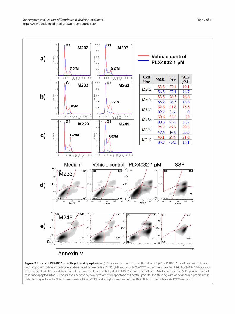
PLX4032 has similar inhibitory effects on cell cycle in sensitive and resistant BRAFV600E mutant cell linesTo study effects of PLX4032 on cell cycle progressiondownstream of B-Raf signaling we used propidium iodideflow cytometric staining. As expected, PLX4032 had noeffect on cell cycle progression in melanoma cell lineswithout a BRAFV600E mutation (Figure 2A). In contrast,PLX4032 exposure for one (data not shown) or 20 hours(Figure 2B and 2C) led to a similar and profound G1arrest in all BRAFV600E mutant cell lines regardless of theirin vitro sensitivity to PLX4032.
PLX4032 leads to apoptotic death in sensitive BRAFV600E
but not in resistant BRAFV600E mutated melanoma cell linesWe then analyzed the ability of PLX4032 to differentiallyinduce apoptotic effects against melanoma cell lines withthe BRAFV600E mutation. Using a BRAFV600E mutant mela-noma cell line with a good response to PLX4032 (M249)and another one that was poorly responsive to PLX4032(M233) based on cell viability assays, we analyzed apop-totic induction using flow cytometry based on the incor-poration of propidium iodide and Annexin V. After

Søndergaard et al. Journal of Translational Medicine 2010, 8:39http://www.translational-medicine.com/content/8/1/39
Page 4 of 11
Table 1: Genomic characterization, growth kinetics and sensitivity towards PLX4032 for a panel of human melanoma cell lines.
Cell Line NRAS/BRAF Number of BRAF
Gene Copies
Other Oncogenic Events Cell line doubling time
(hours)
PLX4032 IC50 (μM)
M257 Wild type 3 CDKN2A R80 31.4 Not reached
M202 NRAS Q61L 2 EGFR amplificationCDKN2A homozygous deletion
26.1 Not reached
M207 NRAS Q61L 2 MITF amplificationPTEN heterozygous deletion
25.2 Not reached
M233 BRAFV600E
Heterozygous3 AKT1 amplification
CCND1 amplificationEGFR amplificationCDKN2A homozygous deletionPTEN homozygous deletion
29.6 Not reached
M255 BRAFV600E
Heterozygous2 AKT2 amplification
CCND1 amplificationEGFR amplificationCDKN2A homozygous deletion
48.6 Not reached
M308 BRAFV600E
Heterozygous3 MITF amplification
AKT2 amplificationEGFR amplificationCDKN2A heterozygous deletion
35.0 Not reached
M263 BRAFV600E
Heterozygous2 CDKN2A heterozygous deletion 23.3 10
M321 BRAFV600E
Homozygous2 34.1 7.5
SKMEL28 BRAFV600E
Homozygous2 EGFR P753S
MITF amplificationCCND1 amplificationCDKN2A heterozygous deletionPTEN heterozygous deletion
26.9 4.6
M229 BRAFV600E
Homozygous4 MITF amplification
AKT1 amplificationPTEN heterozygous deletion
27.9 0.2
M238 BRAFV600E
Heterozygous2 CDKN2A homozygous deletion
PTEN heterozygous deletion28.1 0.7
M249 BRAFV600E
Heterozygous3 MITF amplification
AKT2 amplificationPTEN homozygous deletion
21.2 0.8
M262 BRAFV600E
Homozygous2 AKT1 E17K
AKT1 amplificationEGFR amplificationCDKN2A homozygous deletion
47.4 0.1

Søndergaard et al. Journal of Translational Medicine 2010, 8:39http://www.translational-medicine.com/content/8/1/39
Page 5 of 11
PLX4032-treatment, the increase in Annexin V positivecells, with or without being double positive for propidiumiodide, was greater in the PLX4032-responsive M249cells compared to the poorly responding M233 cells (Fig-ure 2D and 2E). Similar results were obtained with M238and M263 (data not shown). Taken together with the dataon cell cycle inhibition, these data suggest that PLX4032has cytostatic effects in BRAFV600E mutant cell lines witha poor response, while it has cytostatic and cytotoxiceffects in cell lines with a good response to PLX4032 incell viability assays.
Functional and genomic characterization of BRAFV600E
mutated cell lines with different sensitivity to PLX4032We tested if the differences in sensitivity to PLX4032were due to markedly different doubling times. ResistantBRAFV600E mutated cell lines tended to have a slower dou-bling time compared to the sensitive BRAFV600E mutatedcell lines (P = 0.24, Table 1). The lack of significance wasdue to outliers in a small group, most notably the highlysensitive cell line M262 having a doubling time close to 50hours. Interestingly, all cell lines homozygous for theBRAFV600E mutation were moderately to highly sensitiveto PLX4032, and cell lines resistant to PLX4032 were allheterozygous for BRAFV600E (P = 0.16). However, therewere also two highly sensitive heterozygous cell lines withIC50 values below 1 μM of PLX4032, and the sensitivity ofhomozygous cell lines spreads through one-log differ-ences in PLX4032 concentrations (Table 1). We then usedhigh throughput analysis of over 500 gene mutationsusing mass-spectrometry based genotyping [11] andhigh-density SNP arrays to explore other genomic altera-tions. Two different platforms (Illumina and Affymetrix)gave highly concordant results (data not shown) demon-strating that out of the 10 cell lines with BRAFV600E muta-tion, four have amplification of the BRAF locus (Table 1).There was no clear relationship between these amplifica-tion events and the BRAFV600E zygosity or the sensitivityto PLX4032. There were very few secondary mutations inthis group of cell lines, with one cell line having a muta-tion in EGFR, and one cell line with a mutation in AKT(Table 1). In addition, the M257 cell line, which is wildtype for both NRAS and BRAF and is highly resistant toPLX4032, was found to have 3 copies of wild type BRAFand a point mutation in CDKN2A. The distribution ofamplification events in MITF and EGFR were also spreadamong the cell lines. Of note, there was no clear trendregarding the activation of the PI3K/Akt pathway basedon activating mutations, or amplifications of AKT1/2 seg-regating the resistant and sensitive cell lines. Supervisedhierarchical clustering comparing SNP array data fromPLX4032-resistant and -sensitive BRAFV600E mutant celllines did not point to specific genomic areas with concor-
dant alterations differentiating the two groups of celllines.
Modulation of MAPK and PI3k/Akt signaling pathways in sensitive and resistant cell linesTo further explore how cell lines with BRAFV600E muta-tion respond differently to PLX4032 we chose twoextreme examples of cell lines with similar growth kinet-ics to perform an extended analysis of signaling pathways(Figure 3). M229 is one of the two most sensitive celllines, while M233 proved to be very resistant despite hav-ing a short in vitro doubling time (Table 1). Exposure toPLX4032 resulted in a marked decrease in pErk in bothcell lines, but this was more prominent and durable in thesensitive M229 compared to the resistant M233. M229has a heterozygous PTEN deletion by SNP array analysis,and had a detectable band for PTEN protein by Westernblot that did not change with PLX4032 exposure. Theresistant M233 cell line has a homozygous PTEN deletionand has no PTEN protein by Western blot. This corre-lates with baseline pAkt detectable in M233 but notM229, as well as increase in pAkt upon PLX4032-expo-sure in the resistant M233 but not in the sensitive M229cell line. Interestingly, pS6 decreased in both cell linesupon PLX4032 exposure. Finally, we explored if there wasmodulation of AMPK, which has been recently describedas a downstream modulator of glucose metabolism inBRAFV600E mutants [20]. There was a low level of induc-tion of pAMPK. These studies demonstrate that PLX4032has complex effects on MAPK and PI3k/Akt signalingpathways that may be dependent on secondary oncogenicevents beyond B-Raf.
Non-invasive imaging of PLX4032 anti-tumor activityWe analyzed the uptake profile of three different meta-bolic tracers that can be used in PET scans: two nucleo-side analogs (thymidine and FAC [21]) and FDG, aglucose analog widely used as a PET tracer. As expected,BRAF wild type cell lines had no significant change inuptake of thymidine or FAC upon PLX4032-exposure.Conversely, all BRAFV600E mutated cell lines, irrespectiveof their sensitivity to PLX4032, had markedly decreaseduptake of these two nucleoside analogues (Figure 4a and4b). The greatest difference between PLX4032-sensitiveand -resistant BRAFV600E mutants was in FDG uptake.The percentage decrease in FDG uptake was roughlydouble in PLX4032-sensitive BRAFV600E mutants com-pared to PLX4032-resistant cell lines (P = 0.009, Figure4c). Finally, we tested if [18F]-FDG uptake could be usedas a pharmacodynamic marker of B-RafV600E inhibition byPLX4032 in vivo. Mice with established subcutaneousM249 melanoma xenografts, a cell line highly sensitive toPLX4032 in vitro, were treated for 3 days with PLX4032twice daily by oral gavage, and then analyzed by com-

Søndergaard et al. Journal of Translational Medicine 2010, 8:39http://www.translational-medicine.com/content/8/1/39
Page 6 of 11
Figure 1 PLX4032 modulation of the MAPK pathway and melanoma cell line viability. Melanoma cell lines treated with 1 μM PLX4032 for 20 hours were stained with pErk antibody and analyzed by flow cytometry. a) Representative flow cytometry histogram showing the fluorescence inten-sity of pErk in cells treated with vehicle control or PLX4032. b) Comparison of percentage change in pErk for a panel of 10 melanoma cell lines with different NRAS/BRAF mutational status. c) In vitro cell viability upon culture with increasing concentrations of PLX4032 (from 0.001-10 μM) for 120 hours. Cell viability was determined using an MTS-based assay.
c)
a)
Unstained Vehicle/medium
PLX4032 (80.1% decrease)
M238
WT
NRAS Q61L
BRAF V600E heterozygous
BRAF V600E homozygous
b)
Søndergaard et al. Journal of Translational Medicine 2010, 8:39http://www.translational-medicine.com/content/8/1/39
Page 7 of 11
Figure 2 Effects of PLX4032 on cell cycle and apoptosis. a-c) Melanoma cell lines were cultured with 1 μM of PLX4032 for 20 hours and stained with propidium iodide for cell cycle analysis gated on live cells. a) NRAS Q61L mutants, b) BRAFV600E mutants resistant to PLX4032, c) BRAFV600E mutants sensitive to PLX4032. d-e) Melanoma cell lines were cultured with 1 μM of PLX4032, vehicle control, or 1 μM of staurosporine (SSP - positive control to induce apoptosis) for 120 hours and analyzed by flow cytometry for apoptotic cell death upon double-staining with Annexin V and propidium io-dide. Testing included a PLX4032-resistant cell line (M233) and a highly sensitive cell line (M249), both of which are BRAFV600E mutants.
0 2 0 0 4 0 0 6 0 0 8 0 0 1 0 0F L 2 - A
0
2 0
4 0
6 0
8 0
1 0 0
% o
f Ma
x
M 2 0
M202
0 2 0 0 4 0 0 6 0 0 8 0 0 1 0 0F L 2 - A
0
2 0
4 0
6 0
8 0
1 0 0
% o
f Ma
x
0
M 2 0
M207
0 2 0 0 4 0 0 6 0 0 8 0 01 0F L 2 - A
0
2 0
4 0
6 0
8 0
1 0 0
% o
f Ma
x
M 2
M229
0 2 0 0 4 0 06 0 0 8 0 01 0F L 2 - A
0
2 0
4 0
6 0
8 0
1 0 0
% o
f Ma
x
M 2
M249
Vehicle controlPLX4032 1 µM
a)
b)
c)
0 2 0 0 4 0 0 6 0 0 8 0 0 1 0 0F L 2 - A
0
2 0
4 0
6 0
8 0
1 0 0
% o
f Ma
x
0
M 2 3
M233
0 2 0 0 4 0 0 6 0 0 8 0 0 1 0 0F L 2 - A
0
2 0
4 0
6 0
8 0
1 0 0
% o
f Ma
x
M 2 6
M263
Medium Vehicle control PLX4032 1 µM SSP
M233
P.I.
Annexin V
d)
e)
P.I.
M249
G1
G2/M
G1
G2/M
G2/MG2/M
G2/M G2/M
G1G1
G1 G1

Søndergaard et al. Journal of Translational Medicine 2010, 8:39http://www.translational-medicine.com/content/8/1/39
Page 8 of 11
bined microPET and microCT using [18F]-FDG as PETtracer. There was a 32% decrease in [18F]-FDG uptakecompared to the vehicle control mice, even though tumorsizes were not different at this early time point (Figure4d). In conclusion, inhibition of [18F]-FDG uptake can beused as an early marker of effective B-RafV600E inhibitionby PLX4032.
DiscussionThe BRAFV600E mutation is one of the most commonkinase domain mutations in human cancer with a partic-ularly high incidence in malignant melanoma [3,7]. TheRaf-inhibitors PLX4720 and PLX4032 have the preclini-cal characteristics of functioning as specific inhibitors ofthe BRAFV600E mutated kinase with a favorable profilecompared to wild type kinases [9,22]. Understanding thepatterns of sensitivity and resistance in melanomas withdifferent oncogenic events is of high importance for clini-cal translation. Our studies confirmed the high specificity
of PLX4032 for a subset of BRAFV600E mutant cell lines[22]. Surprisingly, we noted differences in the sensitivityto PLX4032, with some BRAFV600E mutants demonstrat-ing resistance to the cytotoxic effects of PLX4032. Inmost cases, these cells had a tendency towards slowergrowth kinetics and being heterozygous for BRAFV600E.
This differential response to PLX4032 in BRAFV600E
mutant melanoma cell lines may be explained by severalmechanisms. It may be that there is preferential MAPKpathway-addiction in sensitive cell lines, and cells withlower sensitivity are less dependent on the BRAFV600E
oncogenic signaling, relying on the co-activation of othersignaling pathways including the PI3K/Akt pathway. Weexplored this possibility with SNP arrays and highthroughput oncogene sequencing with a particular inter-est in looking at this pathway. The genomic analysisrevealed that alterations in PI3K/Akt, including deletionsof PTEN, amplifications of AKT and activating mutationsin AKT were distributed throughout the cell line list with
Figure 3 Western blot analysis of phosphorylated and total amount of key proteins in the MAPK and PI3k/Akt pathways. a) The PLX4032-sensitive M229 cell line and the PLX4032-resistant M233 cell line were cultured in different concentrations of PLX4032 for 24 hours and lysates were analyzed by Western blot. b) M229 and M233 cells were treated by PLX4032 in a time course over 24 hours, and cell lysates were analyzed by Western blot.
p-ERK Thr202/204
ERK
S6
p-S6 Ser235/236
p-S6K Thr389
p-AMPK Thr172
AMPK
�-actin
p-Akt Thr308
p-Akt Ser473
Akt
PTEN
p-ERK Thr202/204
ERK
S6
p-S6 Ser235/236
p-S6K Thr389
p-AMPK Thr172
AMPK
�-actin
p-Akt Thr308
p-Akt Ser473
Akt
PTEN
0 1 2 5 0 1 2 5 uM PLX4032
M229 M233
0’ 30’ 2h 4h 8h 24h 0’ 30’ 2h 4h 8h 24h PLX4032
M229 M233
A B

Søndergaard et al. Journal of Translational Medicine 2010, 8:39http://www.translational-medicine.com/content/8/1/39
Page 9 of 11
Figure 4 Metabolic tracer uptake profile upon exposure to PLX4032. a-c) in vitro PET tracer uptake profiles for 11 different melanoma cell lines. Tritium counts was measured on a micro-beta reader and PLX4032 treated cells were compared to vehicle control and relative PET tracer uptake cal-culated. a) [3H]-thymidine uptake profile, b) [3H]-FAC uptake profile, c) [3H]-FDG uptake profile. The black lines and the number next to them represent the average change in PET tracer uptake of the cell lines with the same mutational status and sensitivity towards PLX4032. d) [18F]FDG PET tracer up-take in vivo. SCID/beige mice with 5-7 mm M249 melanoma xenografts on the left lower flank were treated twice daily with 100 mg/kg of PLX4032 or vehicle control by oral gavage. Three days later mice were imaged by microPET scanning upon administration of [18F]-FDG.
Søndergaard et al. Journal of Translational Medicine 2010, 8:39http://www.translational-medicine.com/content/8/1/39
Page 10 of 11
no clear pattern of correlation with sensitivity or resis-tance to PLX4032. However, in two cell lines phospho-specific Western blot staining suggested that the resistantcell line had increased Akt signaling upon PLX4032 expo-sure. Another possibility is that PLX4032-resistantBRAFV600E mutants have alternative signaling at the levelof Raf, as has been described for cell lines with acquiredresistance to a different Raf-inhibitor, AZ628, which showincreased signaling through C-Raf [23]. The increase inpErk in an NRAS Q61L mutant cell line could beexplained by abrogation of negative feedback loops medi-ated mainly by dual specificity phosphatases (MKPs/DUSPs), as reported with Mek inhibitors [17,24], and therecent description of increased C-Raf signaling when het-erodimerizing with inhibited B-Raf in BRAF wild typecells [18,19]. Therefore, the modulation of feed-backloops and alteration of Raf dimerization upon treatmentwith Raf inhibitors may also have a role in the differentialsensitivity to PLX4032 in BRAFV600E mutant cell lines.Finally, differences in expression of pro- and anti-apop-totic molecules like Bim and Bad [25] may allow someBRAFV600E mutant cell lines to undergo growth arrest butnot die from apoptosis upon exposure to PLX4032. Stud-ies are ongoing to further explore these possibilities.
We explored the use of PET imaging as a mean to non-invasively detect PLX4032-sensitivity. In vitro we foundthat any of the three PET tracers FDG, FLT and FACcould be used to distinguish between melanomas with aNRAS or a BRAFV600E mutation based on the differentialeffects of PLX4032 on cell cycle and metabolism. FDGcould furthermore be used to distinguish betweenBRAFV600E mutant melanomas with high or low sensitiv-ity to PLX4032. The PI3K/Akt pathway has been widelyregarded as having a role in the regulation of glucosemetabolism through mTOR, but recently the LKB1-AMPK pathway has been found to be regulated by onco-genic BRAFV600E signaling [20], which together mayexplain the marked and rapid effects of PLX4032 oninhibiting FDG uptake. We explored this possibility intwo cell lines. Our data suggests a minor increase inpAMPK upon PLX4032 exposure, which may be in linewith the proposed hypothesis [20].
ConclusionsThese studies in melanoma cell lines may allow to betterinterpret the results of a recently reported phase I clinicaltrial with PLX4032 [26], with an objective response inexcess of 70% of patients with BRAFV600E positive meta-static melanoma. The characterization of PLX4032-sensi-tive and -resistant BRAFV600E mutant melanoma cell linesmay provide information about the molecular mecha-nisms that dictate sensitivity and resistance to PLX4032.In addition, molecular imaging with [18F]FDG PET scans
may help in providing an early readout of complete orincomplete pharmacodynamic effects of PLX4032 andtherefore predict lesions that may or may not respond totherapy.
Abbreviations(BRK): Breast tumor kinase; (MKPs/DUSPs): Dual specificity phosphatases; (FDG):2-fluoro-2-deoxy-D-glucose; (FAC): 2'-Deoxy-2'-fluoroarabinofuranosylcyto-sine-[3H]; (MTA): Materials transfer agreement; (MTS): 3-(4,5-dimethylthiazol-2-yl)-5-(3-carboxymethoxyphenyl)-2-(4-sulfophenyl)-2H-tetrazolium; (IC50): Halfmaximal inhibitory concentration; (MAPK): Mitogen-activated protein kinase;(pErk): Phosphorylated Erk; (PET): Positron emission tomography; (thymidine):Thymidine [methyl-3H]; (UCLA): University of California Los Angeles.
Competing interestsThe authors declare that they have no competing interests.
Authors' contributionsJNS, RN, QW, DG, TH, SM, HS, LEM, JGB, SK, NA, EVE, JZ, BC, BAC, RCK: Performedexperiments.JNS, PMRSL, AR: Planned the studies and wrote the manuscript.All authors have read and approved the final manuscript.
AcknowledgementsWe would like to thank Dr. Gideon Bollag from Plexxikon for providing PLX4032 and for helpful discussions regarding these studies. We would also like to thank Drs. William Tap and Dennis Slamon at UCLA, and Peter Hirth at Plexxikon for helpful discussions. This work was funded in part by the Jonsson Cancer Center Foundation (JCCF), the NIH award P50 CA086306 and by the Caltech-UCLA Joint Center for Translational Medicine (to AR); and the Dermatology Founda-tion, the STOP CANCER Foundation and the Burroughs Welcome Fund (to RSL).
Author Details1Department of Medicine, Division of Hematology/Oncology, University of California Los Angeles (UCLA), Los Angeles, CA, USA, 2Department of Surgery, Division of Surgical Oncology, UCLA, Los Angeles, CA, USA, 3Department of Medicine, Division of Dermatology, UCLA, Los Angeles, CA, USA, 4Department of Pathology and Laboratory Medicine, UCLA, Los Angeles, CA, USA, 5The Broad Institute of MIT and Harvard, Cambridge, MA USA, 6Departments of Medical and Pediatric Oncology and Center for Cancer Genome Discovery, Dana-Farber Cancer Institute, Harvard Medical School, Boston, MA, USA, 7Jonsson Comprehensive Cancer Center at UCLA, Los Angeles, CA, USA and 8Current address: Department of Systems Biology, Molecular Immune Regulation at the Center for Biological Sequence Analysis, Technical University of Denmark, Lyngby, Denmark
References1. Gray-Schopfer V, Wellbrock C, Marais R: Melanoma biology and new
targeted therapy. Nature 2007, 445:851-857.2. Smalley KS, Nathanson KL, Flaherty KT: Genetic subgrouping of
melanoma reveals new opportunities for targeted therapy. Cancer Res 2009, 69:3241-3244.
3. Davies H, Bignell GR, Cox C, Stephens P, Edkins S, Clegg S, Teague J, Woffendin H, Garnett MJ, Bottomley W, et al.: Mutations of the BRAF gene in human cancer. Nature 2002, 417:949-954.
4. Fecher LA, Amaravadi RK, Flaherty KT: The MAPK pathway in melanoma. Curr Opin Oncol 2008, 20:183-189.
5. Fecher LA, Cummings SD, Keefe MJ, Alani RM: Toward a molecular classification of melanoma. J Clin Oncol 2007, 25:1606-1620.
6. Haluska FG, Tsao H, Wu H, Haluska FS, Lazar A, Goel V: Genetic alterations in signaling pathways in melanoma. Clin Cancer Res 2006, 12:2301s-2307s.
7. Curtin JA, Fridlyand J, Kageshita T, Patel HN, Busam KJ, Kutzner H, Cho KH, Aiba S, Brocker EB, LeBoit PE, et al.: Distinct sets of genetic alterations in melanoma. N Engl J Med 2005, 353:2135-2147.
8. Wan PT, Garnett MJ, Roe SM, Lee S, Niculescu-Duvaz D, Good VM, Jones CM, Marshall CJ, Springer CJ, Barford D, Marais R: Mechanism of
Received: 24 February 2010 Accepted: 20 April 2010 Published: 20 April 2010This article is available from: http://www.translational-medicine.com/content/8/1/39© 2010 Søndergaard et al; licensee BioMed Central Ltd. This is an Open Access article distributed under the terms of the Creative Commons Attribution License (http://creativecommons.org/licenses/by/2.0), which permits unrestricted use, distribution, and reproduction in any medium, provided the original work is properly cited.Journal of Translational Medicine 2010, 8:39

Søndergaard et al. Journal of Translational Medicine 2010, 8:39http://www.translational-medicine.com/content/8/1/39
Page 11 of 11
activation of the RAF-ERK signaling pathway by oncogenic mutations of B-RAF. Cell 2004, 116:855-867.
9. Tsai J, Lee JT, Wang W, Zhang J, Cho H, Mamo S, Bremer R, Gillette S, Kong J, Haass NK, et al.: Discovery of a selective inhibitor of oncogenic B-Raf kinase with potent antimelanoma activity. Proc Natl Acad Sci USA 2008, 105:3041-3046.
10. Kumar R, Angelini S, Hemminki K: Activating BRAF and N-Ras mutations in sporadic primary melanomas: an inverse association with allelic loss on chromosome 9. Oncogene 2003, 22:9217-9224.
11. Thomas RK, Baker AC, Debiasi RM, Winckler W, Laframboise T, Lin WM, Wang M, Feng W, Zander T, MacConaill L, et al.: High-throughput oncogene mutation profiling in human cancer. Nat Genet 2007, 39:347-351.
12. Wang K, Li M, Hadley D, Liu R, Glessner J, Grant SF, Hakonarson H, Bucan M: PennCNV: an integrated hidden Markov model designed for high-resolution copy number variation detection in whole-genome SNP genotyping data. Genome Res 2007, 17:1665-1674.
13. Comin-Anduix B, Boros LG, Marin S, Boren J, Callol-Massot C, Centelles JJ, Torres JL, Agell N, Bassilian S, Cascante M: Fermented wheat germ extract inhibits glycolysis/pentose cycle enzymes and induces apoptosis through poly(ADP-ribose) polymerase activation in Jurkat T-cell leukemia tumor cells. J Biol Chem 2002, 277:46408-46414.
14. Guo D, Hildebrandt IJ, Prins RM, Soto H, Mazzotta MM, Dang J, Czernin J, Shyy JY, Watson AD, Phelps M, et al.: The AMPK agonist AICAR inhibits the growth of EGFRvIII-expressing glioblastomas by inhibiting lipogenesis. Proc Natl Acad Sci USA 2009, 106:12932-12937.
15. Shu CJ, Radu CG, Shelly SM, Vo DD, Prins R, Ribas A, Phelps ME, Witte ON: Quantitative PET reporter gene imaging of CD8+ T cells specific for a melanoma-expressed self-antigen. Int Immunol 2009, 21:155-165.
16. Dougherty MK, Muller J, Ritt DA, Zhou M, Zhou XZ, Copeland TD, Conrads TP, Veenstra TD, Lu KP, Morrison DK: Regulation of Raf-1 by direct feedback phosphorylation. Mol Cell 2005, 17:215-224.
17. Pratilas CA, Taylor BS, Ye Q, Viale A, Sander C, Solit DB, Rosen N: (V600E)BRAF is associated with disabled feedback inhibition of RAF-MEK signaling and elevated transcriptional output of the pathway. Proc Natl Acad Sci USA 2009, 106:4519-4524.
18. Heidorn SJ, Milagre C, Whittaker S, Nourry A, Niculescu-Duvas I, Dhomen N, Hussain J, Reis-Filho JS, Springer CJ, Pritchard C, Marais R: Kinase-dead BRAF and oncogenic RAS cooperate to drive tumor progression through CRAF. Cell 2010, 140:209-221.
19. Poulikakos PI, Zhang C, Bollag G, Shokat KM, Rosen N: RAF inhibitors transactivate RAF dimers and ERK signalling in cells with wild-type BRAF. Nature 2010, 464(7287):427-30.
20. Zheng B, Jeong JH, Asara JM, Yuan YY, Granter SR, Chin L, Cantley LC: Oncogenic B-RAF negatively regulates the tumor suppressor LKB1 to promote melanoma cell proliferation. Mol Cell 2009, 33:237-247.
21. Radu CG, Shu CJ, Nair-Gill E, Shelly SM, Barrio JR, Satyamurthy N, Phelps ME, Witte ON: Molecular imaging of lymphoid organs and immune activation by positron emission tomography with a new [18F]-labeled 2'-deoxycytidine analog. Nat Med 2008, 14:783-788.
22. Sala E, Mologni L, Truffa S, Gaetano C, Bollag GE, Gambacorti-Passerini C: BRAF silencing by short hairpin RNA or chemical blockade by PLX4032 leads to different responses in melanoma and thyroid carcinoma cells. Mol Cancer Res 2008, 6:751-759.
23. Montagut C, Sharma SV, Shioda T, McDermott U, Ulman M, Ulkus LE, Dias-Santagata D, Stubbs H, Lee DY, Singh A, et al.: Elevated CRAF as a potential mechanism of acquired resistance to BRAF inhibition in melanoma. Cancer Res 2008, 68:4853-4861.
24. Friday BB, Yu C, Dy GK, Smith PD, Wang L, Thibodeau SN, Adjei AA: BRAF V600E disrupts AZD6244-induced abrogation of negative feedback pathways between extracellular signal-regulated kinase and Raf proteins. Cancer Res 2008, 68:6145-6153.
25. Boisvert-Adamo K, Aplin AE: Mutant B-RAF mediates resistance to anoikis via Bad and Bim. Oncogene 2008, 27:3301-3312.
26. Flaherty K, Puzanov I, Sosman J, Kim K, Ribas A, McArthur G, Lee RJ, Grippo JF, Nolop K, Chapman P: Phase I study of PLX4032: proof of concept for V600E BRAF mutation as a therapeutic target in human cancer. J Clin Oncol 2009, 27(15s):. suppl; abstr 9000
doi: 10.1186/1479-5876-8-39Cite this article as: Søndergaard et al., Differential sensitivity of melanoma cell lines with BRAFV600E mutation to the specific Raf inhibitor PLX4032 Journal of Translational Medicine 2010, 8:39
Copyright © 2022 FDOKUMEN