(mit: Ralf Adelmann) ›Man versucht sich zu öffnen.‹ Video und RAF – eine abgeschlossene Geschichte

Genes & Cancer1(5) 409 –420© The Author(s) 2010Reprints and permission: sagepub.com/journalsPermissions.navDOI: 10.1177/1947601910373795http://ganc.sagepub.com
Introduction
Cancer is a disease caused by uncontrolled cell growth and proliferation. When localized, the formation of the tumor by uncontrolled cell proliferation is easily treated by sur-gery and various therapeutic drugs; however, complica-tions arise when the tumor metastasizes. Once metastasized, the cancer becomes deadly and is extremely difficult to treat due to accumulated resistance to chemotherapy, radio-therapy, and immunotherapy. When metastasizing, the tumor cells invade the basement membrane and circulatory system, extravasate through the endothelial lining of the blood vessels, and form colonies in the new tissue sites. The migration from the epithelial layer into the mesenchy-mal layer before reaching the basement membrane is known as “epithelial to mesenchymal transition” (EMT). During EMT, the polarized epithelial cells acquire certain attributes of mesenchymal cells and, thus, are able to pen-etrate the mesenchymal layer through the basement mem-brane and invade neighboring tissues.1 Breaching of the mesenchymal layer leads to infiltration into the blood ves-sels and migration to other parts of the body. This review will focus on the role of B-RAF mutations in melanoma and the induction of EMT.
Metastasis in Melanoma
Melanoma is one of the most highly metastatic cancers. Melanoma is a type of cancer that arises from melanocytes, cells dedicated to melanin production and located in the basal layer of the epidermis. It represents 2% to 3% of malignant tumors in both the United States and Northern Europe. The incidence of melanoma has doubled in the last 20 years; a 4% increase every year is documented in the United States.2 Whereas the incidence is continuously increasing, the mortality seems to be decreasing, most likely due to the increase of screening programs and early
1Department of Microbiology, Immunology & Molecular Genetics, David Geffen School of Medicine, Jonsson Comprehensive Cancer Center, University of California at Los Angeles, Los Angeles, CA, USA2Department of Biomedical Sciences, University of Catania, Catania, Italy3Plastic Surgery Section, Department of Medicine and Surgery Specialities, University of Catania, Catania, Italy
Corresponding Author:Benjamin Bonavida, Department of Microbiology, Immunology & Molecular Genetics, David Geffen School of Medicine, Jonsson Comprehensive Cancer Center, University of California at Los Angeles, Los Angeles, CA 90095-1747 Email: [email protected]
The Role of B-RAF Mutations in Melanoma and the Induction of EMT via Dysregulation of the NF-κB/Snail/RKIP/PTEN Circuit
Kimberly Lin1, Stavroula Baritaki1, Loredana Militello1, Graziella Malaponte2, Ylenia Bevelacqua3, and Benjamin Bonavida1
Submitted 13-Mar-2010; Revised 01-May-2010; Accepted 03-May-2010
AbstractMelanoma is a highly metastatic cancer, and there are no current therapeutic modalities to treat this deadly malignant disease once it has metastasized. Melanoma cancers exhibit B-RAF mutations in up to 70% of cases. B-RAF mutations are responsible, in large part, for the constitutive hyperactivation of survival/antiapoptotic pathways such as the MAPK, NF-κB, and PI3K/AKT. These hyperactivated pathways regulate the expression of genes targeting the initiation of the metastatic cascade, namely, the epithelial to mesenchymal transition (EMT). EMT is the result of the expression of mesenchymal gene products such as fibronectin, vimentin, and metalloproteinases and the invasion and inhibition of E-cadherin. The above pathways cross-talk and regulate each other’s activities and functions. For instance, the NF-κB pathway directly regulates EMT through the transcription of gene products involved in EMT and indirectly through the transcriptional up-regulation of the metastasis inducer Snail. Snail, in turn, suppresses the expression of the metastasis suppressor gene product Raf kinase inhibitor protein RKIP (inhibits the MAPK and the NF-κB pathways) as well as PTEN (inhibits the PI3K/AKT pathway). The role of B-RAF mutations in melanoma and their direct role in the induction of EMT are not clear. This review discusses the hypothesis that B-RAF mutations are involved in the dysregulation of the NF-κB/Snail/RKIP/PTEN circuit and in both the induction of EMT and metastasis. The therapeutic implications of the dysregulation of the above circuit by B-RAF mutations are such that they offer novel targets for therapeutic interventions in the treatment of EMT and metastasis.
KeywordsB-RAF mutations, Snail, RKIP, PTEN, EMT
Review

410 Genes & Cancer / vol 1 no 5 (2010)
detection. Cutaneous melanoma is the most frequently occurring melanoma; however, melanoma can occur in other sites, such as the uvea. Occurrence of melanoma in the uvea accounts for the greatest percentage of malignant melanomas other than melanoma of the skin.3 Other possi-ble sites of melanoma include mucosal melanomas (female genitalia, head and neck, anorectal region); however, these occurrences are less common.4 The main risk factors for cutaneous melanoma are related to sun exposure (UV), phe-notype (Fitzpatrick Classification Scale), distribution of existing melanocytic nevous, type and location (lower limbs in females, posterior trunk in males), and family his-tory.5 Melanoma has the greatest potential for metastasis, even compared to other skin cancers such as basal cell car-cinoma and squamous cell carcinoma.6 However, if discov-ered in its early stages, the primary tumor can be removed surgically, and there is a high probability of complete recov-ery.6 Approximately 80% of melanoma cases are detected at an early stage in which the melanoma is localized in the primary site and can be excised.7 Melanoma tumors can be classified into 2 groups, those in the radial growth phase (RGP) and those in the vertical growth phase (VGP). Tumors classified in the RGP are associated with early stage melanoma and are confined, for the most part, to the epidermis with a low probability of dissemination. Once progressed to the VGP classification, the tumor cells are now tumorigenic, characterized by important dermal inva-sion, and become metastastic.8,9
Metastatic melanoma is refractory to current therapies and has a very poor prognosis, with a median survival rate of 6 months.10 Chemotherapy treatment of melanoma is not very effective due to the high refractory nature of this malignancy to most standard cytotoxic agents. In a prospec-tive randomized controlled trial, adjuvant high-dose inter-feron was shown to increase relapse-free survival and overall survival (OS) when compared to patients with high-risk resected cutaneous melanoma.11 Melanoma that has already spread to distant sites is rarely curable with stan-dard therapy, although high-dose interleukin-2 (IL-2) has been reported to produce durable responses in a small num-ber of patients.12 However, the most promising treatments of melanoma seem to be targeted to mutations in transduc-tion/survival pathways that are common in melanomas. Recent progress in the molecular understanding of the sig-naling pathways involved in melanomagenesis has led researchers to develop targeted therapies for this disease. These include selective inhibitors of the RAF and MEK kinases and inhibitors of the PI3K pathway.7
Inhibitors of the RAF kinases serve as a targeted therapy for melanoma because a direct correlation has been found between the presence of mutated signaling protein B-RAF and the progressive stages of melanoma growth; there is a higher incidence of B-RAF mutations in VPG tumors
compared to tumors of the RGP.13 Therefore, studies of the role of mutations in B-RAF signaling and in cell survival are important to decipher the underlying mechanisms of melanoma tumorigenesis and its progression. This review will focus on B-RAF signaling and the gene products that play major roles in the regulation of EMT in melanoma and how these gene products work in concert, namely, the NF-κB/ Snail/RKIP/PTEN circuit. Evidence supports B-RAF muta-tions in the regulation of this above circuit and the induction of EMT in melanoma (see schematic diagram in Fig. 1).
B-RAF Mutations in MelanomaB-RAF and B-RAF Mutations
B-RAF is part of a family of serine/threonine kinase pro-teins designated as the RAF proteins. RAF proteins are acti-vated by RAS, the small membrane-bound G protein that is regulated upstream by extracellular signals. The RAF fam-ily has 3 functional proteins, namely, A-RAF, B-RAF, and C-RAF (also known as RAF-1); all 3 have a high degree of homology in 3 conserved regions, and their activation is regulated by various phosphorylation sites14-16 (Fig. 2A). Two of the conserved regions of the RAF family of pro-teins, CR1 and CR2, are found in the N-terminus, while the third, CR3, is located in the C-terminus and codes for the proteins’ kinase activity. Several phosphorylation sites are conserved, while some are unique to each protein. The pres-ence of both conserved and unique phosphorylation sites indicates that, although there are common mechanisms for regulating the RAF proteins, each can be regulated
B-RAF
NF-κB
Snail
RKIP PTEN
EMT
Figure 1. The role of the B-RAF/NF-κB/Snail/RKIP/PTEN circuitry in the regulation of EMT in melanoma. B-RAF induces EMT in melanoma through the constitutive activation of NF-κB and resulting in the up-regulation of the metastasis inducer, Snail. Inhibition of B-RAF by either up-regulation of RKIP or PTEN inhibits Snail expression. Expression of Snail down-regulates both RKIP and PTEN, thus providing a positive feedback loop for self-amplification and EMT.

B-RAF mutations and EMT in melanoma / Lin et al. 411
independently of the others. In tumor cells, as well as in normal cells, B-RAF is the protein providing the most sig-naling for the transduction pathway, whereas the role of A-RAF and C-RAF is to refine the signal and make small adjustments at the level of signaling. B-RAF is the protein used most often to transduce signals in cellular signaling pathways due to its elevated basal kinase activity in relation to its isoforms A-RAF and C-RAF. This elevated basal kinase activity is due to the presence of an aspartic acid just upstream of the conserved region CR3 that is not present in either A-RAF or C-RAF. Because of the negative charge provided by aspartic acid, B-RAF has a greater propensity towards activation (Fig. 2A). Both A-RAF and C-RAF proteins require not only phosphorylation but also additional activa-tion factors such as activation by the tyrosine kinase Src; therefore, point mutations are not sufficient to induce activ-ity.17 Mutations in B-RAF easily result in hyperactivation due to its propensity towards activation by the aspartic acid and because only phosphorylation is required for activation.
As a result, it is shown that the majority of activating muta-tions occur in B-RAF.
The most common B-RAF mutations are observed in the kinase domain of the protein, particularly within the activa-tion segment. The most common mutation in the B-RAF protein consists of the substitution of glutamic acid for valine at codon 600 in exon 15 (Val600Glu; B-RAFV600E) (Fig. 2B).18 This substitution leads to constitutive activation of the protein, which results in an increase in its basal kinase activity. In vitro, the kinase activity of mutant B-RAF is approximately 500-fold greater than that of its wild-type counterpart, and the transformation rates in vivo are 70 to 138 times more efficient than transformation by the wild-type B-RAF.19,20 The effectiveness of B-RAF mutations is implicated in its common presence in various cancers. B-RAF has been found to be mutated in several human can-cers including but not limited to colorectal (5%-22%), ovar-ian (30%), thyroid (36%-53%), and most commonly in melanoma (27%-70%).20
B-RAF mutations have been reported to be important in the development and progression of melanoma; however, B-RAF mutations alone are not sufficient to cause mela-noma tumorigenesis. Observations show that 82% of benign nevi have high frequencies of B-RAF mutations relative to frequencies seen in RGP tumors.21 This disparity in the fre-quency of activating B-RAF mutations between benign nevi and RGP tumors implicates that B-RAF mutations alone cannot induce melanoma but must cooperate with other signaling pathways such as the PI3K/AKT pathway and/or additional genetic alterations such as loss of p53 or PTEN.22,23 In addition, the microenvironment has long been acknowledged as a facilitator of melanoma initiation and progression; recent studies have illuminated tumor-associated factors of the microenvironment, including hypoxia and the extracellular matrix, as important mediators of melanocyte transformation and transdifferentiation to melanoma.24
The dynamic role of genetic and epigenetic changes, including B-RAF mutations in cooperation with other genetic alterations and the microenvironment, in the tran-sition between melanocytic and melanoma cell pheno-types has been extensively reported. As a representative example, in the zebrafish-expressing constitutively active B-RAFV600E, but not wild-type B-RAF, dramatic patches of ectotopic melanocytes were formed, but no evidence of transformation was seen. When p53-deficient transgenic zebrafish were generated to express B-RAFV600E, not only were melanocyte lesions formed, but these lesions also rapidly developed into invasive melanomas.25 In accordance, recent studies have determined that hypoxia may be essential for melanocyte transformation, and aggressive melanoma-conditioned extracellular matrices can epigenetically transdiffentiate normal melanocytes toward an invasive melanoma-like phenotype.26,27
Figure 2. Conformations and mutations in RAF proteins. (A)Conformations of A-RAF, B-RAF, and C-RAF. The presence of an aspartic acid in B-RAF just upstream of the conserved region CR3 is not present in either A-RAF or C-RAF; this difference accounts for B-RAF’s natural propensity towards activation. Oncogenic mutations often occur in the activation segment. (B) Common mutations in the activation segment of B-RAF. The activation segment is the region in which oncogenic mutations commonly occur. Common mutations occur in the first few codons of the activation segment (boxed region). The most common mutation occurs at codon 600 in exon 15; valine (V) is substituted for glutamic acid (E).

412 Genes & Cancer / vol 1 no 5 (2010)
To summarize, activating B-RAF mutations constitute one epigenetic change, which plays a profound role throughout melanoma progression. However, only coopera-tion of B-RAF mutations with other signaling pathways and the microenvironment can actively promote the earliest steps of melanocyte transformation and later enhance aggressive phenotypes including invasion, vasculogenic mimicry, and angiogenesis. In the following sections, we further discuss how B-RAF signaling synergizes with key molecular pathways and the tumor microenvironment in the induction of EMT in melanoma (Fig. 3).
Cell Signaling with B-RAF Mutations and EMTIn cancer, the dysregulation of several regulatory pathways has been implicated to promote metastasis of tumor cells. The MAPK, NF-κB, and PI3K/ATK pathways are some of the most commonly dysregulated pathways. The dysregula-tion of MAPK, NF-κB, and PI3K/AKT pathways is com-mon in melanoma; these 3 pathways are constitutively hyperactivated.20,28,29 A mechanism of hyperactivation of the MAPK and NF-κB pathways, resulting in increased metastatic potential, is through mutation and constitutive activation of the upstream signaling protein, B-RAF. Deletion of PTEN accounts for the hyperactivation of the PI3K/AKT pathway, which collaborates with B-RAF to hyperactivate the MAPK and NF-κB pathways (Fig. 1). Mutated B-RAF has been shown to induce constitutive ERK signaling in NIH3T3 (mouse embryonic fibroblast cells) and induce tumor formation in vivo.20,30 When B-RAF mutations were observed in LOX-IMVI and A375 melanoma cell lines, the same results as the NIH3T3 cells were observed. LOX-IMVI and A375 cells trans-fected with mutant B-RAF when injected into mice resulted in tumorigenesis. Inhibition of the mutant B-RAF in vivo led to inhibition of tumorigenesis and regression of tumor growth.31 Oncogenic B-RAF has also been shown to increase IKK activation and simulta-neous IκB degradation. Conversely, knockdown of B-RAF signaling resulted in a decrease of IKK activa-tion and IκB degradation.32 Loss of PTEN has been found in several kinds of solid tumors and plays an important role in tumorigenesis and angiogenesis.33 From these above findings, B-RAF is a likely major factor in the regulation of EMT via the deregulation of the MAPK, NF-κB, and PI3K/AKT survival pathways as will be described below.
Activation of the MAPK Pathway by B-RAF MutationsThe mitogen-activated protein kinase (MAPK) pathway, also known as the Ras/Raf/MEK/ERK pathway, is a com-monly studied pathway associated with mutant B-RAF. The
MAPK pathway is a signal transduction cascade that relays extracellular signals from the plasma membrane to the nucleus through a series of kinase-induced phosphoryla-tions. Studies have shown that the MAPK pathway is hyperactivated in the majority of human melanomas; the hyperactivation of this pathway leads to cell proliferation, survival, and the induction of EMT.34 The signaling path-way of a normal cell through the MAPK pathway is first initiated by the activation of RAS and recruitment of cyto-solic RAF to the cellular membrane. At the cell membrane, RAF becomes activated by tyrosine kinases, such as the Src-family kinases, and in turn activates the MAP kinases MEK 1 and 2. ERK 1 and 2 are then phosphorylated and translocated into the nucleus to bind promoters of genes involved in the induction of cell growth and proliferation (Fig. 3A).35,36 Most commonly, when B-RAF is mutated, it is constitutively activated without the need for signaling from RAS. The downstream effect of constitutively acti-vated B-RAF is continual translocation of ERK to the nucleus promoting cell proliferation and invasion. Other B-RAF mutations, such as D593V, are found to have a lower kinase activity, 0.3-fold activity of wild-type B-RAF, resulting in impaired signaling activity directed to the MAPK pathway. However, these mutations with lower kinase activation continue to regulate translocation of ERK through activation of endogenous C-RAF and, conse-quently, activation of MAPK.16 Regardless of the type of mutation, the MAPK pathway is constitutively activated, and ERK is continuously translocated to the nucleus to pro-mote cell growth, proliferation, and EMT. ERK has been found to be constitutively activated in several cancer types including 90% of melanomas37; this finding has been cor-related with high frequencies of activating B-RAF muta-tions in melanoma. However, B-RAF is only one factor in ERK1/2 activation; additional factors and mechanisms, mostly related to the tumor microenvironment, also contrib-ute to ERK hyperactivation during melanomagenesis.
The MAPK pathway is negatively regulated by Raf-1 kinase inhibitor protein (RKIP). RKIP is a metastasis sup-pressor gene product and is a member of the phosphatidyl-ethanolamine-binding protein (PEBP) family.38 RKIP has not only been associated with cellular signaling but has also been shown to be involved in neurological functions and reproduction.39 RKIP has been shown to regulate MAPK activity via inhibition of B-RAF activity.38,40 This inhibition of B-RAF activity in normal cells by RKIP pre-vents uncontrolled proliferation and invasion by regulat-ing MAPK signaling through B-RAF signaling. RKIP not only regulates MAPK but is also responsible for the regu-lation of the NF-κB pathway, another pathway hyperacti-vated in melanoma by B-RAF mutations. The MAPK regulates gene products involved in the induction of EMT (see below).
B-RAF mutations and EMT in melanoma / Lin et al. 413
Activation of the NF-κB Pathway by B-RAF Mutations
The NF-κB pathway is an important cell survival signaling pathway in tumor cells that regulates the expression of anti-apoptotic, proproliferative, and prometastatic genes.32 The NF-κB pathway is activated through IKK (IκB kinase). IKK triggers the phosphorylation and degradation of IκBα by the proteasome leading to translocation of NF-κB to the nucleus.41 NF-κB then promotes metastasis through the up-regulation of several metastatic genes such as COX-2, metalloproteinases, VEGF, and most importantly the metas-tasis inducer Snail.42,43 Constitutive activation of NF-κB has become a hallmark of melanoma. One of the potential mechanisms by which the NF-κB pathway is constitutively active is through mutant B-RAF signaling. Mutant B-RAF activates the canonical NF-κB pathway through IKKβ, which promotes degradation of IκB and translocation of NF-κB to the nucleus.28,44,45 Regulation of NF-κB is exe-cuted by RKIP. RKIP, as previously stated, is able to regu-late B-RAF activity and therefore also regulates the downstream activity of NF-κB. RKIP regulates NF-κB activity using 2 different pathways: through direct RKIP inhibition of B-RAF activity and through direct inhibition of the NF-κB pathway via physical association and inhibi-tion of the NF-κB–inducing kinase (NIK), TGF-beta–acti-vated kinase (TAK), and IκB kinase (IKK) (Fig. 3B).46 Thus, both the MAPK and the NF-κB pathways are regu-lated negatively by RKIP and positively by B-RAF. Up-regulation of B-RAF activity is necessary in order to overcome the negative regulation of RKIP, and this up-regulation of B-RAF is carried out by deregulation and constitutive activation of the PI3K/AKT pathway.
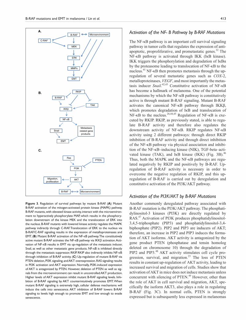
Activation of the PI3K/AKT by B-RAF MutationsAnother commonly deregulated pathway associated with B-RAF mutation is the PI3K/AKT pathway. The phosphati-dylinositol-3 kinases (PI3K) are directly regulated by RAS.47 Activation of PI3K produces phosphatidylinositol-3,4,5-triphosphate (PIP3) and phosphatidylinositol-3,4- biphosphate (PIP2). PIP2 and PIP3 are inducers of AKT; therefore, an increase in PIP2 and PIP3 induces the forma-tion of AKT isoforms. AKT activity is antagonized by the gene product PTEN (phosphatase and tensin homolog deleted on chromosome 10) through the degradation of PIP2 and PIP3.48 AKT activity stimulates cell cycle pro-gression, survival, and migration.23 The loss of PTEN results in constant up-regulation of AKT activity, leading to increased survival and migration of cells. Studies show that activation of AKT in mice does not induce metastasis unless concurrent with silencing of PTEN.49 However, other than the role of AKT in cell survival and migration, AKT, spe-cifically the isoform AKT3, also plays a role in regulating B-RAF (Fig. 3C). In normal cells, PTEN is strongly expressed but is subsequently less expressed in melanomas
A.B-RAF
MEK
ERK
C-RAF
MMPs
EMT
Microenvironment
B.B-RAF
RKIP
IKKβ
Snail
NF-κB
EMT
TAK
Metastasisrelated gene
products
C.
Ras
PI3K
AKT
PTEN
Migration,proliferation
B-RAF Microenvironment
Figure 3. Regulation of survival pathways by mutant B-RAF. (A) Mutant B-RAF activation of the mitogen-activated protein kinase (MAPK) pathway. B-RAF mutants, with elevated kinase activity, interact with the microenviron-ment to hyperactively phosphorylate MAP, which results in the phosphory-lation downstream of the kinase MEK and the translocation of ERK into the nucleus. B-RAF mutants with lowered kinase activity regulate the MAPK pathway indirectly through C-RAF. Translocation of ERK to the nucleus via B-RAF/C-RAF signaling results in the expression of metalloproteinases and EMT. (B) Mutant B-RAF activation of the NF-κB pathway. The constitutively active mutant B-RAF activates the NF-κB pathway via IKKβ activation. Acti-vation of NF-κB results in EMT via up-regulation of the metastasis inducer, Snail, as well as other metastatic gene products. NF-κB is inhibited directly through the metastasis suppressor, RKIP. RKIP also indirectly inhibits NF-κB through inhibition of B-RAF activity. (C) Up-regulation of mutant B-RAF via PTEN deletion, PI3K signaling, and AKT overexpression. RAS signaling results in PI3K activation and AKT expression. Normally, PI3K-induced expression of AKT is antagonized by PTEN. However, deletion of PTEN as well as sig-nals from the microenvironment can result in uncontrolled AKT production. Higher levels of AKT expression inhibit mutant B-RAF signaling levels. Inhi-bition of B-RAF signaling by AKT counterintuitively promotes EMT. When mutant B-RAF signaling is extremely high, cellular defense mechanisms will induce the cells into senescence. AKT inhibition of B-RAF lowers B-RAF signaling to levels high enough to promote EMT and low enough to evade senescence.

414 Genes & Cancer / vol 1 no 5 (2010)
and completely lost in the metastatic lesions.50 Decreased PTEN expression levels up-regulate AKT activity. The up-regulation of AKT activity is important for the regulation of B-RAF signaling. AKT3 inhibits B-RAF activity, which, counterintuitively, promotes cell proliferation. Mutant B-RAF, which expresses high levels of signaling, induces cellular defense mechanisms targeted against activated oncogenes, and the induction of these cellular defense mechanisms leads to cell senescence. Inhibition of B-RAF by AKT3 regulates B-RAF to levels high enough to pro-mote proliferation, survival, and metastasis but low enough to prevent activation of cellular defense mechanisms and cell senescence.51,52 Without dysregulation of the PI3K/AKT pathway, AKT3 expression would not be high enough to optimize mutant B-RAF activity, and neither the MAPK nor the NF-κB pathway would be hyperactivated.
Within each of the above mentioned pathways, several gene products are regulated by NF-κB, particularly impor-tant in the regulation of EMT, and include Snail, RKIP, and PTEN. The implication of these gene products and their association with B-RAF are important for understanding their role in transformation and the induction of EMT in melanoma tumors.
Cross-Talk between the Microenvironment and Genetic Alterations in Melanoma during the Induction of EMT
Tumor cells communicate bidirectionally with the sur-rounding microenvironment, which consists of elements as diverse as extracellular matrix components and growth factors (including VEGF, nutrients, and varying concen-trations of oxygen). Collectively, these interfere with the genetic cell programming and supply signals that control cell survival, growth, differentiation, and invasion. The microenvironment has emerged as a major player in mela-nocyte transformation and transdifferentiation and in pro-moting melanoma cell invasion and metastasis. For EMT induction in melanoma, all genetic abnormalities includ-ing common mutations and/or deregulated expressions of B-RAF, N-RAS, and PTEN seem to synergize with sev-eral microenvironmental factors. Among these factors are the loss of cell-cell adhesions, resulting from 1) altered expression of cadherins, catenins, and integrins; 2) the interaction between melanoma cells and surrounding keratinocytes, fibroblasts, and immune cells; and 3) the tumor angiogenetic efficiency.53 In addition, it has been shown that hypoxia promotes melanocyte transdifferentia-tion and melanoma migratory and invasive abilities through up-regulation of genes normally associated with the extracellular matrix remodeling and invasion; these
genes include laminin 5 γ2 (Ln-5 γ2), Urokinase, and genes encoding matrix metalloproteinases.54,55 Other stud-ies have shown that constitutively active AKT, which is observed in a high percentage of melanomas, can trans-form melanocytes when oxygen levels are low.26 Further-more, different microenvironments may be selective for higher levels of both B-RAF–dependent and B-RAF–independent ERK1/2 activation; these microenvironments are critical for tumor cell proliferation and spread. Immu-nohistochemical studies in melanoma samples have shown that expression levels of phosphorylated ERK1/2 are not always correlated with the mutational status of B-RAF or N-RAS,56 suggesting that other B-RAF–independent or B-RAF–complementary factors and mechanisms promote ERK1/2 activation. Among these factors are growth factor autocrine loops, such as the CSF-dependent activation of c-Kit, and extracellular signals, such as derived from epi-dermal and nerve growth factors.57-61 On the other hand, Conner et al. have shown that adhesion-regulated ERK1/2 activation in melanocytes is by-passed by B-RAF muta-tions in malignant melanoma cells, although melanoma cells may still be dependent on integrin-activated PI3K and AKT signaling for survival and migration.62 The above findings exemplify how the microenvironment can complement aberrant genetic changes such as mutations in B-RAF to promote melanomagenesis and to support an invasive cell phenotype. The contribution of the microen-vironment in cell fate might further explain why RGP tumors are characterized by a relatively low frequency of B-RAF mutations; EMT is important for promoting inva-sion and transition to VGP cells.13 In contrast to general conceptions that invasion and dissemination occur during the later vertical phase, recent findings show that early dissemination of tumor cells that have not yet fully pro-gressed is a contributor to subsequent development of metastasis. This finding is in accordance with similar observations in breast cancer and suggests that genetic alterations are not autonomous in determining cell behav-ior and cell fate; however, with the support of microenvi-ronmental factors, even the less “progressed” tumor cells with few genetic alterations may accumulate additional alterations that will favor ectopic growth.63
The Regulation of NF-κB Activity and Downstream Snail, RKIP, and PTEN Gene Products by B-RAF Mutations and Induction of EMTEvidence suggests a strong association of B-RAF muta-tions with NF-κB, Snail, RKIP, and PTEN in the regula-tion of EMT. Each of these has been individually shown to be directly associated in the regulation of EMT in melanoma.

B-RAF mutations and EMT in melanoma / Lin et al. 415
Role of NF-κB Activation by B-RAF Mutations and Induction of EMT in Melanoma
Metastatic prostate cancer cells have elevated NF-ĸB activ-ity and constitutively active IKK, which is responsible for the activation of NF-ĸB.64 These findings are observed in several cancers including melanoma.65 Knockdown of NF-ĸB by RNAi reversed the mesenchymal-like phenotype and suppressed the motility and invasion capacity of meta-static cancer cells. In vitro motility and invasion assays showed that both the motility and invasive potentials were reduced in cells in which NF-ĸB was inhibited.66 In vivo, inhibition of NF-ĸB in Ras-transformed epithelial cells led to an actively suppressed EMT gene program. One mecha-nism of regulation of NF-κB activation is through the B-RAF signaling cascade via IKK activation and IκB deg-radation. Hyperactivation of NF-κB via mutant B-RAF greatly increases the metastatic potential. Several studies have provided evidence for potential underlying mecha-nisms by which activated NF-ĸB is able to induce metasta-sis.67 Matrix metalloproteinases (MMPs) have been shown to be important for invasion and angiogenesis and are highly implicated in metastasis.68,69 Regulatory elements within the 5′ flanking sequence of the human MMP-9 gene have consensus sequences for NF-ĸB. NF-ĸB activation has been shown to be necessary for inducing MMP-9 mRNA and protein expression.69 In addition to these findings, NF-ĸB has also been shown to activate transcription of the metastasis-inducer Snail transcription factor.
Role of Snail Activation by B-RAF Mutations and Induction of EMT in MelanomaSnail is a transcription factor that is a member of the zinc-finger protein family and has been linked to metastasis and resistance against apoptosis. Snail has been shown to induce metastasis by repression of E-cadherin.70 E-cadherin is a cell adhesion molecule; loss of cell-cell adhesion by reduc-tion of E-cadherin leads to EMT. Epithelial cells that over-express Snail acquire metastatic and invasive potential and are correlated with down-regulation of E-cadherin expres-sion.71 Snail is also involved in E-cadherin–independent EMT through the down-regulation of claudins and occlu-dins. Claudins and occludins are associated with establish-ing and maintaining cell polarity.72,73 Loss of these molecules in tumor cells results in a deregulation of cell polarity and increased potential for EMT. Snail is also dem-onstrated to induce metalloproteinases; induction of metal-loproteinases is associated with portal invasion, metastasis, and recurrence in human cancers.74,75 B-RAF has been shown to up-regulate Snail and thus provides a potential mechanism for B-RAF–induced EMT.76 Hyperactivation in mutant B-RAF results in overexpression of Snail and, therefore, increased metastatic potential of the melanoma.
An important molecule antagonizing the metastatic effects of B-RAF and Snail is the metastasis suppressor, RKIP.
Role of the Inhibition of RKIP Expression by B-RAF Mutations and Induction of EMT in MelanomaRKIP is a metastasis suppressor gene product. Studies have shown that overexpression of RKIP inhibited metastasis and down-regulation of RKIP increases the metastatic potential. In mice, RKIP that was exogenously expressed led to a decreased level of metastasis and invasion in trans-formed metastatic cells.77 RKIP also plays a role as a pro-apoptotic factor in drug-resistant tumors; induced overexpression of RKIP in drug-resistant cells showed an increase in apoptosis-matching levels of apoptosis induced by the constitutively active proapoptotic factor t-Bid.78 RKIP has been shown to be able to directly down-regulate the metastasis-inducing pathway, the NF-ĸB pathway. As discussed above, NF-ĸB activation requires the phosphory-lation and degradation of IĸB. RKIP has been shown to negatively modulate the activating phosphorylations of IKKα and IKKβ by upstream kinases.46 RKIP is also able to down-regulate NF-κB activity via B-RAF signaling. Inhibi-tion of B-RAF signaling by RKIP is achieved by down-regulation of B-RAF kinase activity; this inhibition of B-RAF kinase activity results in the down-regulation of all metastatic pathways associated with B-RAF signaling.40 RKIP is not the only regulator of B-RAF; PTEN is also an important regulatory molecule in B-RAF–associated path-ways and metastasis.
Role of Inhibition of PTEN Expression by B-RAF Mutations and Induction of EMT in MelanomaPhosphatase and tensin homolog deleted on chromosome 10, also known as PTEN, is a tumor suppressor gene located at 10q23-24 on human chromosome 10.47 PTEN is a mem-ber of the protein tyrosine phosphatase (PTP) family and has a relatively high constitutive phosphatase activity in vitro and in vivo.79 The amino acid sequence of PTEN resembles that of 2 proteins, protein tyrosine phosphatases as well as lipid phosphatases.80 Sequence similarities have also been found between PTEN and the cytoskeleton pro-tein tensin, whose role involves interaction with integrins in integrin-signaling complexes.81 Integrins are involved in forming complexes of signal transduction molecules such as MAPK,82,83 Ras,84 NF-κB,85 and PI3K.86 Because of the sequence similarity between PTEN and tensin, it also inter-acts with integrin-signaling complexes.87,88 PTEN plays a significant role in cell cycle arrest, regulation of cell adhesion, migration, and proliferation. PTEN is demonstrated to be critical for embryonic development, and the expression of PTEN is ubiquitous in all human tissues.89,90 Loss of PTEN and/or its function is found in many tumors as well as various

416 Genes & Cancer / vol 1 no 5 (2010)
other diseases. PTEN loss occurs through homozygous gene deletion or point mutation, resulting in loss of its expression.91-93 Several mechanisms have been described for the mutation of PTEN; most mutations occur within the coding region of the gene and inactivate phosphatase activ-ity.79 The deletion of PTEN in melanoma results in hyperac-tivation of the PI3K/AKT pathway and overexpression of AKT3. Overexpression of AKT3 is necessary as a collabo-rating molecule in B-RAF signaling. Levels of AKT3 must be up-regulated in order to optimize B-RAF signaling levels; optimization of B-RAF signaling levels induces cell prolif-eration, survival, and metastasis via the B-RAF–associated pathways MAPK and NF-κB.51,52
As discussed above, B-RAF mutations regulate NF-κB, Snail, RKIP, and PTEN activities in melanoma and metas-tasis. Noteworthy, when examined collectively, it appears that these activities are part of an NF-κB/Snail/RRKIP/PTEN circuit such that this circuit regulates EMT.
Dysregulation by B-RAF Mutations of the NF-κB/Snail/RKIP/PTEN Circuit and Induction of EMT
All 3 pathways associated with B-RAF mutations, namely, the MAPK, NF-κB, and PI3K/AKT pathways, result in the up-regulation of the metastasis inducer, Snail. The PI3K/AKT pathway has been shown to activate the NF-κB path-way via IKKβ.94 The PI3K/AKT pathway is normally antagonized by PTEN; however, PTEN somatic mutations are found in 40% to 60% of melanoma cell lines and 10% to 20% of primary melanomas.95 Therefore, NF-κB is up-regulated not only by B-RAF but also by the uninhibited PI3K/AKT pathway as well. Not only is PTEN found to be commonly mutated, but it is also down-regulated by the transcription factor Snail (Fig. 4A).96 Thus, the NF-κB pathway is shown to be constitutively activated by the PI3K/AKT pathway due to the mutation/suppression of PTEN as well as Snail-induced inhibition of PTEN.
Regulation of the NF-κB pathway is also achieved through the MAPK pathway through MEK signaling. MEK has been shown to induce NF-κB activation through the IKK complex.97 The MAPK pathway also mediates the NF-κB pathway via the translocation factor, ERK. ERK regulates downstream the translocation of NF-κB into the nucleus through up-regulation of Snail expression (Fig. 4B).76 When B-RAF is mutated, the MAPK pathway is con-stitutively activated and, therefore, constitutively up-regu-lates Snail indirectly via NF-κB activation and directly by ERK translocation.
Snail has been shown as a negative regulator of RKIP98; up-regulation of Snail through the PI3K/AKT and MAPK pathways inhibits both PTEN and RKIP, which then, in turn, amplifies the expression of Snail through constitutively
active NF-κB (Fig. 4C). From the cross-talks among the different B-RAF–associated pathways, it is clear that the regulation of Snail via PTEN and RKIP is an important mechanism in the regulation of EMT.
B-RAF Mutations in Melanoma and Therapeutic Targets Directed at the Dysregulation of the NF-κB/Snail/RKIP/PTEN Circuit to Prevent EMT
One RAF kinase inhibitor, sorafenib (BAY 43-9006), is an oral multikinase inhibitor that targets RAF kinase as well as the VEGF receptor.99 Sorafenib has been observed to inhibit growth of melanoma xenografts in mice; however, no anti-tumor effects were seen in advanced melanoma patients. Sorafenib is well tolerated but has little effect in advanced melanoma patients as a single agent.100 RAF kinase inhibi-tors could potentially be the most potent drugs; inhibition of the RAF kinase could inhibit B-RAF activity and all down-stream signaling pathways. Other inhibitors currently in clinical trials are the MEK kinase inhibitors PD0325901 and AZD6244.7 B-RAF–mutated tumors seem to be partic-ularly sensitive to clinically available MEK inhibitors.101 It appears that MEK inhibitors are more often agents that induce growth arrest rather than cell death in most B-RAF–mutated models.18 Inhibition of MEK would result in inhi-bition of ERK translocation as well as inhibition of NF-κB translocation and, therefore, a decrease in the metastatic potential. The PI3K pathway is another potential target for chemotherapy; however, current trials using CCI-779 and RAD001 to target the PI3K pathway do not have a high specificity in targeting melanoma cells and could poten-tially, counterproductively, activate AKT.102,103 If new PI3K inhibitors are developed, overexpression of AKT could be controlled, and overexpression of mutant B-RAF would induce tumor cell senescence. Current treatments with indi-vidual drugs have not yielded satisfactory results. These are, in part, due to the complicated interrelations among the pathways involved in metastasis, namely, the cross-talks among the PI3K/AKT, NF-κB, and MAPK pathways. Com-binational treatments with the above drugs may lead to syn-ergistic effects. Hopeful results have already been seen in the combination of sorafenib with carboplatin and pacli-taxel.104 PLX4032 is a novel, oral, small molecule that spe-cifically targets B-RAFV600E. It has recently been tested in clinical trials and has yielded positive responses in patients with metastatic melanoma. In phase I trials, a response was seen in 78% of the patients. However, despite the positive results obtained in phase I trials, several chal-lenges have been uncovered. Toxic side effects such as the development of squamous cell carcinoma occurred in 20% to 30% of treated patients and could, therefore, deter long-term usage of the drug.105,106

B-RAF mutations and EMT in melanoma / Lin et al. 417
Conclusions and Future Directions
Additional potential therapies that might prove useful in the treatment of malignant melanoma include the targeting of RKIP and Snail. Such potential drugs could include protea-some inhibitors, such as bortezomib and NPI-0052, which are shown to induce RKIP expression and inhibit the activ-ity of NF-κB and the transcription of Snail.107 Induction of RKIP would also inhibit metastasis through the MAPK pathway. Targeting for RKIP induction would be more effi-cient than targeting downstream effectors such as NF-κB and Snail. Drugs inducing the expression of PTEN could also prove effective. Overexpression of PTEN would sup-press AKT expression and induce cell senescence caused by extreme hyperactivation of mutant B-RAF. Combinational therapies should continue to be pursued in order to concur-rently inhibit all potential mechanisms of EMT and metas-tasis. Further research is required to identify other possible gene products that are needed to cooperate with mutant B-RAF for initiation, maintenance, and progression of mel-anoma. Discovery of additional genetic alterations that cooperate with B-RAF will provide future therapeutic tools for the treatment and diagnosis of malignant melanoma.
Declaration of Conflicting Interests
The author(s) declared no potential conflicts of interest with respect to the authorship and/or publication of this article.
Funding
This study was supported by various donors and by the Jonsson Comprehensive Cancer Center at UCLA.
References
1. Geiger TR, Peeper DS. Metastasis mechanisms. Biochim Biophys
Acta 2009;1796(2):293-308.
2. Jemal A, Siegel R, Ward E, Hao Y, Xu J, Thun MJ. Cancer statistics,
2009. CA Cancer J Clin 2009;59(4):225-49.
3. Grin JM, Grant-Kels JM, Grin CM, MD, Berke A, Kels BD. Ocular
melanomas and melanocytic lesions of the eye. J Am Acad Dermatol
1998;38(5 Pt 1):716-30.
4. Hussein MR. Extracutaneous malignant melanomas. Cancer Invest
2008;26(5):516-34.
5. Moan J, Porojnicu AC, Dahlback A. Ultraviolet radiation and malig-
nant melanoma. Adv Exp Med Biol 2008;624:104-16.
6. American Cancer Society. Cancer facts & figures, 2009. Atlanta:
American Cancer Society; 2009.
7. Russo AE, Torrisi E, Bevelacqua Y, Perrotta R, Libra M, McCubrey
JA, et al. Melanoma: molecular pathogenesis and emerging target
therapies. Int J Oncol 2009;34(6):1481-9.
8. Balch CM, Buzaid AC, Soong SJ, Atkins MB, Cascinelli N, Coit DG,
et al. Final version of the American Joint Committee on Cancer staging
system for cutaneous melanoma. J Clin Oncol 2001;19(16):3635-48.
9. Veronesi U, Cascinelli N. Narrow excision (1-cm margin): a safe pro-
cedure for thin cutaneous melanoma. Arch Surg 1991;126(4):438-41.
A.
EMT
Snail
IKK
NF-κB
PI3K PTEN
AKT
RAS
EMT
Snail
ERK
Ras
B-RAF
MEK
C-RAF
IKK
NF-κB
B.
C.
B-RAF
Snail PI3K/AKTMAPK
PTENRKIP
Figure 4. Activation of NF-κB by the PI3K/AKT and MAPK pathways. (A) Activation of NF-κB via the PI3K/AKT pathway. Activation of the PI3K pathway by RAS results in the production of AKT. AKT activates NF-κB through the activation of IKK. Activation of NF-κB results in Snail-induced EMT. PI3K signaling is antagonized by PTEN; however, up-regulation in Snail inhibits PTEN, resulting in a self-amplification cycle. (B) Activation of NF-κB via the MAPK pathway NF-κB is regulated through MEK-induced activation of IKK and ERK-induced up-regulation of Snail. (C) Regulation of Snail via RKIP and PTEN up-regulation of Snail through the PI3K/AKT and MAPK pathways inhibits both PTEN and RKIP, which then, in turn, amplifies the expression of Snail.

418 Genes & Cancer / vol 1 no 5 (2010)
10. Miller AJ, Mihm MC Jr. Melanoma. N Engl J Med 2006;355:51-65.
11. Kirkwood JM, Strawderman MH, Ernstoff MS, Smith TJ, Borden EC,
Blum RH. Interferon alfa-2b adjuvant therapy of high-risk resected
cutaneous melanoma: the Eastern Cooperative Oncology Group Trial
EST 1684. J Clin Oncol 1996;14(1):7-17.
12. Atkins MB, Kunkel L, Sznol M, Rosenberg SA. High-dose recombi-
nant interleukin-2 therapy in patients with metastatic melanoma: long-
term survival update. Cancer J Sci Am 2000;6(suppl 1):S11-S14.
13. Dong J, Phelps RG, Qiao R, Yao S, Benard O, Ronai Z, et al. BRAF
oncogenic mutations correlate with progression rather than initiation
of human melanoma. Cancer Res 2003;63(14):3883-5.
14. Zhang BH, Guan KL. Activation of B-Raf kinase requires phos-
phorylation of the conserved residues Thr598 and Ser601. EMBO J
2000;19(20):5429-39.
15. Chong H, Lee J, Guan KL. Positive and negative regulation of
Raf kinase activity and function by phosphorylation. EMBO J
2001;20(14):3716-27.
16. Wan PT, Garnett MJ, Roe SM, Lee S, Niculescu-Duvaz D, Good VM,
et al. Mechanism of activation of the RAF-ERK signaling pathway by
oncogenic mutations of B-RAF. Cell 2004;116(6):855-67.
17. Wellbrock C, Karasarides M, Marais R. The RAF proteins take centre
stage. Nat Rev Mol Cell Biol 2004;5(11):875-85.
18. Halilovic E, Solit DB. Therapeutic strategies for inhibiting oncogenic
BRAF signaling. Curr Opin Pharmacol 2008;8(4):419-26.
19. Davies H, Bignell GR, Cox C, Stephens P, Edkins S, Clegg S,
et al. Mutations of the BRAF gene in human cancer. Nature
2002;417(6892):949-54.
20. Garnett MJ, Marais R. Guilty as charged: B-RAF is a human onco-
gene. Cancer Cell 2004;6(4):313-9.
21. Pollock PM, Harper UL, Hansen KS, Yudt LM, Stark M, Robbins
CM, et al. High frequency of BRAF mutations in nevi. Nat Genet
2003;33(1):19-20.
22. Davies H, Bignell GR, Cox C, Stephens P, Edkins S, Clegg S,
et al. Mutations of the BRAF gene in human cancer. Nature
2002;417(6892):949-54.
23. Palmieri G, Capone M, Ascierto ML, Gentilcore G, Stroncek DF,
Casula M, et al. Main roads to melanoma. J Transl Med 2009;7:86.
24. Houghton AN, Polsky D. Focus on melanoma. Cancer Cell
2002;2(4):275-8.
25. Patton EE, Widlund HR, Kutok JL, Kopani KR, Amatruda JF,
Murphey RD, et al. BRAF mutations are sufficient to promote nevi
formation and cooperate with p53 in the genesis of melanoma. Curr
Biol 2005;15(3):249-54.
26. Bedogni B, Welford SM, Cassarino DS, Nickoloff BJ, Giaccia AJ,
Powell MB. The hypoxic microenvironment of the skin contributes to
Akt-mediated melanocyte transformation. Cancer Cell 2005;8(6):443-54.
27. Seftor EA, Brown KM, Chin L, Kirschmann DA, Wheaton WW,
Protopopov A, et al. Epigenetic transdifferentiation of normal mela-
nocytes by a metastatic melanoma microenvironment. Cancer Res
2005;65(22):10164-9.
28. Dhawan P, Richmond A. A novel NF-kappa B-inducing kinase-MAPK
signaling pathway up-regulates NF-kappa B activity in melanoma
cells. J Biol Chem 2002;277(10):7920-8.
29. Robertson GP. Functional and therapeutic significance of Akt deregu-
lation in malignant melanoma. Cancer Metastasis Rev 2005;24(2):273-85.
30. Brose MS, Volpe P, Feldman M, Kumar M, Rishi I, Gerrero R, et al.
BRAF and RAS mutations in human lung cancer and melanoma.
Cancer Res 2002;62(23):6997-7000.
31. Hoeflich KP, Jaiswal B, Davis DP, Seshagiri S. Inducible BRAF sup-
pression models for melanoma tumorigenesis. Methods Enzymol
2008;439:25-38.
32. Liu J, Suresh Kumar KG, Yu D, Molton SA, McMahon M, Herlyn M,
et al. Oncogenic BRAF regulates beta-Trcp expression and NF-kap-
paB activity in human melanoma cells. Oncogene 2007;26(13):1954-8.
33. Jiang BH, Liu LZ. PI3K/PTEN signaling in angiogenesis and tumori-
genesis. Adv Cancer Res 2009;102:19-65.
34. Dhomen N, Marais R. BRAF signaling and targeted therapies in mela-
noma. Hematol Oncol Clin North Am 2009;23(3):529-45, ix.
35. Pearson G, Robinson F, Beers Gibson T, Xu BE, Karandikar M,
Berman K, et al. Mitogen-activated protein (MAP) kinase pathways:
regulation and physiological functions. Endocr Rev 2001;22(2):153-83.
36. Chambard JC, Lefloch R, Pouysségur J, Lenormand P. ERK implication
in cell cycle regulation. Biochim Biophys Acta 2007;1773(8):1299-310.
37. Cohen C, Zavala-Pompa A, Sequeira JH, Shoji M, Sexton DG, Cotsonis
G, et al. Mitogen-actived protein kinase activation is an early event in
melanoma progression. Clin Cancer Res 2002;8(12):3728-33.
38. Yeung K, Seitz T, Li S, Janosch P, McFerran B, Kaiser C, et al. Sup-
pression of Raf-1 kinase activity and MAP kinase signalling by RKIP.
Nature 1999;401(6749):173-7.
39. Klysik J, Theroux SJ, Sedivy JM, Moffit JS, Boekelheide K. Signaling
crossroads: the function of Raf kinase inhibitory protein in cancer, the
central nervous system and reproduction. Cell Signal 2008;20(1):1-9.
40. Park S, Yeung ML, Beach S, Shields JM, Yeung KC. RKIP down-
regulates B-Raf kinase activity in melanoma cancer cells. Oncogene
2005;24(21):3535-40.
41. Lenardo MJ, Baltimore D. NF-kappa B: a pleiotropic mediator of
inducible and tissue-specific gene control. Cell 1989;58(2):227-9.
42. Amiri KI, Richmond A. Role of nuclear factor-kappa B in melanoma.
Cancer Metastasis Rev 2005;24(2):301-13.
43. Barberà MJ, Puig I, Domínguez D, Julien-Grille S, Guaita-Esteruelas
S, Peiró S, et al. Regulation of Snail transcription during epithelial to
mesenchymal transition of tumor cells. Oncogene 2004;23(44):7345-54.
44. Ikenoue T, Hikiba Y, Kanai F, Tanaka Y, Imamura J, Imamura T,
et al. Functional analysis of mutations within the kinase activation
segment of B-Raf human colorectal tumors. Cancer Res 2003;63(23):
8132-7.
45. Ikenoue T, Hikiba Y, Kanai F, Aragaki J, Tanaka Y, Imamura J, et al.
Different effects of point mutations within the B-Raf glycine-rich loop
in colorectal tumors on mitogen-activated protein/extracellular sig-
nal-regulated kinase kinase/extracellular signal-regulated kinase and
nuclear factor kappaB pathway and cellular transformation. Cancer Res
2004;64(10):3428-35.
46. Yeung KC, Rose DW, Dhillon AS, Yaros D, Gustafsson M, Chatterjee
D, et al. Raf kinase inhibitor protein interacts with NF-kappaB-inducing
kinase and TAK1 and inhibits NF-kappaB activation. Mol Cell Biol
2001;21(21):7207-17.

B-RAF mutations and EMT in melanoma / Lin et al. 419
47. Downward J. Ras signalling and apoptosis. Curr Opin Genet Dev
1998;8(1):49-54.
48. Datta SR, Brunet A, Greenberg ME. Cellular survival: a play in three
Akts. Genes Dev 1999;13(22):2905-27.
49. Dankort D, Curley DP, Cartlidge RA, Nelson B, Karnezis AN,
Damsky WE, Jr., , et al. Braf(V600E) cooperates with Pten loss to
induce metastatic melanoma. Nat Genet 2009;41(5):544-52.
50. Singh RS, Diwan AH, Zhang PS, Prieto VG. Phosphoinositide
3-kinase is not overexpressed in melanocytic lesions. J Cutan Pathol
2007;34(3):220-5.
51. Madhunapantula SV, Robertson GP. The PTEN-AKT3 signaling cas-
cade as a therapeutic target in melanoma. Pigment Cell Melanoma Res
2009;22(4):400-19.
52. Cheung M, Sharma A, Madhunapantula SV, Robertson GP. Akt3 and
mutant V600E B-Raf cooperate to promote early melanoma develop-
ment. Cancer Res 2008;68(9):3429-39.
53. Ch’ng S, Tan ST. Genetics, cellular biology and tumor microenviron-
ment of melanoma. Front Biosci 2009;14:918-28.
54. Postovit LM, Seftor EA, Seftor RE, Hendrix MJ. Influence of the
microenvironment on melanoma cell fate determination and pheno-
type. Cancer Res 2006;66(16):7833-6.
55. Seftor EA, Brown KM, Chin L, Kirschmann DA, Wheaton WW,
Protopopov A, et al. Epigenetic transdifferentiation of normal mela-
nocytes by a metastatic melanoma microenvironment. Cancer Res
2005;65(22):10164-9.
56. Houben R, Vetter-Kauczok CS, Ortmann S, Rapp UR, Broecker EB,
Becker JC. Phospho-ERK staining is a poor indicator of the muta-
tional status of BRAF and NRAS in human melanoma. J Invest Der-
matol 2008;128(8):2003-12.
57. Chambard JC, Lefloch R, Pouysségur J, Lenormand P. ERK implication
in cell cycle regulation. Biochim Biophys Acta 2007;1773(8):1299-310.
58. Lange-Carter CA, Johnson GL. Ras-dependent growth factor reg-
ulation of MEK kinase in PC12 cells. Science 1994;265(5177):
1458-61.
59. Babchia N, Calipel A, Mouriaux F, Faussat AM, Mascarelli F. The
PI3K/Akt and mTOR/P70S6K signaling pathways in human uveal
melanoma cells: interaction with B-Raf/ERK. Invest Ophthalmol Vis
Sci 2010;51(1):421-9.
60. Lefevre G, Glotin AL, Calipel A, Mouriaux F, Tran T, Kherrouche
Z, et al. Roles of stem cell factor/c-Kit and effects of Glivec/
STI571 in human uveal melanoma cell tumorigenesis. J Biol Chem
2004;279(30):31769-79.
61. Lefèvre G, Babchia N, Calipel A, Mouriaux F, Faussat AM, Mrzyk S,
et al. Activation of the FGF2/FGFR1 autocrine loop for cell prolifera-
tion and survival in uveal melanoma cells. Invest Ophthalmol Vis Sci
2009;50(3):1047-57.
62. Conner SR, Scott G, Aplin AE. Adhesion-dependent activation of
the ERK1/2 cascade is by-passed in melanoma cells. J Biol Chem
2003;278(36):34548-54.
63. Ossowski L, Aguirre-Ghiso JA. Dormancy of metastatic melanoma.
Pigment Cell Melanoma Res 2010;23(1):41-56.
64. Gasparian AV, Yao YJ, Kowalczyk D, Lyakh LA, Karseladze A,
Slaga TJ, et al. The role of IKK in constitutive activation of NF-
kappaB transcription factor in prostate carcinoma cells. J Cell Sci
2002;115(Pt 1):141-51.
65. Yang J, Richmond A. Constitutive IkappaB kinase activity correlates
with nuclear factor-kappaB activation in human melanoma cells. Can-
cer Res 2001;61(12):4901-9.
66. Zhang L, Chen W, Li X. A novel anticancer effect of butein: inhibition
of invasion through the ERK1/2 and NF-kappa B signaling pathways
in bladder cancer cells. FEBS Lett 2008;582(13):1821-8.
67. Huber MA, Azoitei N, Baumann B, Grünert S, Sommer A, Peham-
berger H, et al. NF-kappaB is essential for epithelial-mesenchymal
transition and metastasis in a model of breast cancer progression. J
Clin Invest 2004;114(4):569-81.
68. Cox G, O’Byrne KJ. Matrix metalloproteinases and cancer. Antican-
cer Res 2001;21(6B):4207-19.
69. Liu J, Zhan M, Hannay JA, Das P, Bolshakov SV, Kotilingam D, et al.
Wild-type p53 inhibits nuclear factor-kappaB-induced matrix metallo-
proteinase-9 promoter activation: implications for soft tissue sarcoma
growth and metastasis. Mol Cancer Res 2006;4(11):803-10.
70. Poser I, Domínguez D, de Herreros AG, Varnai A, Buettner R, Bosser-
hoff AK. Loss of E-cadherin expression in melanoma cells involves
up-regulation of the transcriptional repressor Snail. J Biol Chem
2001;276(27):24661-6.
71. Cano A, Pérez-Moreno MA, Rodrigo I, Locascio A, Blanco MJ,
del Barrio MG. The transcription factor snail controls epithelial-
mesenchymal transitions by repressing E-cadherin expression. Nat
Cell Biol 2000;2(2):76-83.
72. Ohkubo T, Ozawa M. The transcription factor Snail downregulates the
tight junction components independently of E-cadherin downregula-
tion. J Cell Sci 2004;117(Pt 9):1675-85.
73. Ikenouchi J, Matsuda M, Furuse M, Tsukita S. Regulation of tight
junctions during the epithelium-mesenchyme transition: direct repres-
sion of the gene expression of claudins/occludin by Snail. J Cell Sci
2003;116(Pt 10):1959-67.
74. Miyoshi A, Kitajima Y, Sumi K, Sato K, Hagiwara A, Koga Y, et al.
Snail and SIP1 increase cancer invasion by upregulating MMP family
in hepatocellular carcinoma cells. Br J Cancer 2004;90(6):1265-73.
75. Yokoyama K, Kamata N, Fujimoto R, Tsutsumi S, Tomonari M, Taki
M, et al. Increased invasion and matrix metalloproteinase-2 expres-
sion by Snail-induced mesenchymal transition in squamous cell carci-
nomas. Int J Oncol 2003;22(4):891-8.
76. Massoumi R, Kuphal S, Hellerbrand C, Haas B, Wild P, Spruss T,
et al. Down-regulation of CYLD expression by Snail promotes tumor
progression in malignant melanoma. J Exp Med 2009;206(1):221-32.
77. Fu Z, Smith PC, Zhang L, Rubin MA, Dunn RL, Yao Z, et al. Effects
of raf kinase inhibitor protein expression on suppression of prostate
cancer metastasis. J Natl Cancer Inst 2003;95(12):878-89.
78. Chatterjee D, Bai Y, Wang Z, Beach S, Mott S, Roy R, et al. RKIP
sensitizes prostate and breast cancer cells to drug-induced apoptosis. J
Biol Chem 2004;279(17):17515-23.
79. Leslie NR, Downes CP. PTEN function: how normal cells control it
and tumour cells lose it. Biochem J 2004;382(Pt 1):1-11.
80. Maehama T, Dixon JE. The tumor suppressor, PTEN/MMAC1,
dephosphorylates the lipid second messenger, phosphatidylinositol
3,4,5-trisphosphate. J Biol Chem 1998;273(22):13375-8.
81. Miyamoto S, Teramoto H, Coso OA, Gutkind JS, Burbelo PD,
Akiyama SK, et al. Integrin function: molecular hierarchies of cyto-
skeletal and signaling molecules. J Cell Biol 1995;131(3):791-805.

420 Genes & Cancer / vol 1 no 5 (2010)
82. Morino N, Mimura T, Hamasaki K, Tobe K, Ueki K, Kikuchi K, et al.
Matrix/integrin interaction activates the mitogen-activated protein
kinase, p44erk-1 and p42erk-2. J Biol Chem 1995;270(1):269-73.
83. Chen Q, Kinch MS, Lin TH, Burridge K, Juliano RL. Integrin-medi-
ated cell adhesion activates mitogen-activated protein kinases. J Biol
Chem 1994;269(43):26602-5.
84. Kapron-Bras C, Fitz-Gibbon L, Jeevaratnam P, Wilkins J, Dedhar S.
Stimulation of tyrosine phosphorylation and accumulation of GTP-
bound p21ras upon antibody-mediated alpha 2 beta 1 integrin activa-
tion in T-lymphoblastic cells. J Biol Chem 1993;268(28):20701-4.
85. Yebra M, Filardo EJ, Bayna EM, Kawahara E, Becker JC, Cheresh
DA. Induction of carcinoma cell migration on vitronectin by NF-
kappa B-dependent gene expression. Mol Biol Cell 1995;6(7):841-50.
86. Chen HC, Guan JL. Association of focal adhesion kinase with its
potential substrate phosphatidylinositol 3-kinase. Proc Natl Acad Sci
U S A 1994;91(21):10148-52.
87. Li J, Yen C, Liaw D, Podsypanina K, Bose S, Wang SI, et al. PTEN,
a putative protein tyrosine phosphatase gene mutated in human brain,
breast, and prostate cancer. Science 1997;275(5308):1943-7.
88. Steck PA, Pershouse MA, Jasser SA, Yung WK, Lin H, Ligon AH,
et al. Identification of a candidate tumour suppressor gene, MMAC1,
at chromosome 10q23.3 that is mutated in multiple advanced cancers.
Nat Genet 1997;15(4):356-62.
89. Tamura M, Gu J, Tran H, Yamada KM. PTEN gene and integrin sig-
naling in cancer. J Natl Cancer Inst 1999;91(21):1820-8.
90. Di Cristofano A, Pesce B, Cordon-Cardo C, Pandolfi PP. Pten is
essential for embryonic development and tumour suppression. Nat
Genet 1998;19(4):348-55.
91. Li J, Yen C, Liaw D, Podsypanina K, Bose S, Wang SI, et al. PTEN,
a putative protein tyrosine phosphatase gene mutated in human brain,
breast, and prostate cancer. Science 1997;275(5308):1943-7.
92. Steck PA, Pershouse MA, Jasser SA, Yung WK, Lin H, Ligon AH, et al.
Identification of a candidate tumour suppressor gene, MMAC1, at
chromosome 10q23.3 that is mutated in multiple advanced cancers.
Nat Genet 1997;15(4):356-62.
93. Whang YE, Wu X, Suzuki H, Reiter RE, Tran C, Vessella RL, et al.
Inactivation of the tumor suppressor PTEN/MMAC1 in advanced
human prostate cancer through loss of expression. Proc Natl Acad Sci
U S A 1998;95(9):5246-50.
94. Madrid LV, Mayo MW, Reuther JY, Baldwin AS Jr. Akt stimu-
lates the transactivation potential of the RelA/p65 Subunit
of NF-kappa B through utilization of the Ikappa B kinase and
activation of the mitogen-activated protein kinase p38. J Biol Chem
2001;276(22):18934-40.
95. Tsao H, Zhang X, Benoit E, Haluska FG. Identification of PTEN/
MMAC1 alterations in uncultured melanomas and melanoma cell
lines. Oncogene 1998;16(26):3397-402.
96. Escrivà M, Peiró S, Herranz N, Villagrasa P, Dave N, Montserrat-Sentís
B, et al. Repression of PTEN phosphatase by Snail1 transcriptional
factor during gamma radiation-induced apoptosis. Mol Cell Biol
2008;28(5):1528-40.
97. Nakano H, Shindo M, Sakon S, Nishinaka S, Mihara M, Yagita H,
et al. Differential regulation of IkappaB kinase alpha and beta by two
upstream kinases, NF-kappaB-inducing kinase and mitogen-activated
protein kinase/ERK kinase kinase-1. Proc Natl Acad Sci U S A 1998;
95(7):3537-42.
98. Beach S, Tang H, Park S, Dhillon AS, Keller ET, Kolch W, et al. Snail
is a repressor of RKIP transcription in metastatic prostate cancer cells.
Oncogene 2008;27(15):2243-8.
99. Egberts F, Kahler KC, Livingstone E, Hauschild A. Metastatic mela-
noma: scientific rationale for sorafenib treatment and clinical results.
Onkologie 2008;31(7):398-403.
100. Eisen T, Ahmad T, Flaherty KT, Gore M, Kaye S, Marais R, et al.
Sorafenib in advanced melanoma: a phase II randomised discontinua-
tion trial analysis. Br J Cancer 2006;95(5):581-6.
101. Solit DB, Garraway LA, Pratilas CA, Sawai A, Getz G, Basso A, et al.
BRAF mutation predicts sensitivity to MEK inhibition. Nature
2006;439(7074):358-62.
102. Hocker TL, Singh MK, Tsao H. Melanoma genetics and therapeutic
approaches in the 21st century: moving from the benchside to the bed-
side. J Invest Dermatol 2008;128(11):2575-95.
103. Dancey JE. Therapeutic targets: MTOR and related pathways. Cancer
Biol Ther 2006;5(9):1065-73.
104. Flaherty KT. Chemotherapy and targeted therapy combinations in
advanced melanoma. Clin Cancer Res 2006;12(7 Pt 2):2366s-70s.
105. Shepherd C, Puzanov I, Sosman JA. B-RAF inhibitors: an evolv-
ing role in the therapy of malignant melanoma. Curr Oncol Rep
2010;12(3):146-52.
106. Brower V. BRAF inhibitors: research accelerates in wake of positive
findings. J Natl Cancer Inst 2010;102(4):214-5.
107. Baritaki S, Yeung K, Palladino M, Berenson J, Bonavida B. Pivotal
roles of snail inhibition and RKIP induction by the proteasome inhibi-
tor NPI-0052 in tumor cell chemoimmunosensitization. Cancer Res
2009;69(21):8376-85.
Copyright © 2022 FDOKUMEN