Vitamin A Is a Negative Regulator of Osteoblast Mineralization
Upload
grandceremonyCategory
view
1download
0
Available online at www.sciencedirect.com
Journal of Nutritional Biochemistry 22 (2011) 318–327
Differential effects of formononetin and cladrin on osteoblast function,peak bone mass achievement and bioavailability in rats☆,☆☆
Abnish K. Gautam, MSca,1, Biju Bhargavan, PhDa,1, Abdul M. Tyagi, MSca, Kamini Srivastava, MSca,Dinesh K. Yadav, MScb, Manmeet Kumar, MScb, Akanksha Singh, MScb, Jay S. Mishra, MScc,
Amar Bahadur Singh, MScc, Sabyasachi Sanyal, PhDc, Rakesh Maurya, PhDb, Lakshmi Manickavasagam, MScd,Sheelendra P. Singh, MScd, Wahajuddin Wahajuddin, PhDd, Girish K. Jain, PhDd,
Naibedya Chattopadhyay, PhDa, Divya Singh, PhDa,⁎
aDivision of Endocrinology, Central Drug Research Institute (Council of Scientific and Industrial Research), Chattar Manzil, P.O. Box 173, Lucknow, IndiabDivision of Medicinal & Process Chemistry, Central Drug Research Institute (Council of Scientific and Industrial Research), Chattar Manzil, P.O. Box 173, Lucknow, IndiacDrug Target Discovery and Development, Central Drug Research Institute (Council of Scientific and Industrial Research), Chattar Manzil, P.O. Box 173, Lucknow, India
dDivision of Pharmacokinetics & Metabolism, Central Drug Research Institute (Council of Scientific and Industrial Research), Chattar Manzil, P.O. Box 173, Lucknow, India
Received 15 October 2009; received in revised form 12 February 2010; accepted 18 February 2010
Abstract
Dietary soy isoflavones including genistein and daidzein have been shown to have favorable effects during estrogen deficiency in experimental animals andhumans. We have evaluated osteogenic effect of cladrin and formononetin, two structurally related methoxydaidzeins found in soy food and other naturalsources. Cladrin, at as low as 10 nM, maximally stimulated both osteoblast proliferation and differentiation by activating MEK-Erk pathway. On the other hand,formononetin maximally stimulated osteoblast differentiation at 100 nM that involved p38 MAPK pathway but had no effect on osteoblast proliferation. Unlikedaidzein, these two compounds neither activated estrogen receptor in osteoblast nor had any effect on osteoclast differentiation. Daily oral administration ofeach of these compounds at 10.0 mg kg−1 day−1 dose to recently weaned female Sprague–Dawley rats for 30 consecutive days, increased bone mineral density atvarious anatomic positions studied. By dynamic histomorphometry of bone, we observed that rats treated with cladrin exhibited increased mineral appositionand bone formation rates compared with control, while formononetin had no effect. Cladrin had much better plasma bioavailability compared withformononetin. None of these compounds exhibited estrogen agonistic effect in uteri. Our data suggest that cladrin is more potent among the two in promotingparameters of peak bone mass achievement, which could be attributed to its stimulatory effect on osteoblast proliferation and better bioavailability. To the bestof our knowledge, this is the first attempt to elucidate structure–activity relationship between the methoxylated forms of daidzein and their osteogenic effects.© 2011 Elsevier Inc. All rights reserved.
Keywords: Osteogenic; Proliferation; Differentiation; MAPK signaling; Peak bone mass
1. Introduction
The adult bone mass of an individual is critically dependent on theachievement of peak bone mass (PBM) during skeletal growth [1].PBM is described as bonemass and strength achieved at the end of thegrowth period and is negatively correlated with the risk ofosteoporosis fractures occurring after menopause. Phytoestrogens,found in many edible plants are diverse groups of biologically active
☆ Supporting grants: Ministry of Heath and Family Welfare, Council ofScientific and Industrial Research, University Grants Commission, Govern-ment of India, Department of Biotechnology.
☆☆ Disclosure: Authors have no conflict of interest.⁎ Corresponding author. Tel.: +91 522 2612411 418x4246; fax: +91 522
2623938.E-mail address: [email protected] (D. Singh).1Authors contributed equally to this work.
0955-2863/$ – see front matter © 2011 Elsevier Inc. All rights reserved.doi:10.1016/j.jnutbio.2010.02.010
compounds with structural similarity to estradiol [2]. The majorestrogenic isoflavones, including daidzein, genistein and biochainin A,have been shown to have important role in reducing symptomsassociated with estrogen deficiency disorders. These compounds maybe protective against osteoporosis due to their ability to exertosteogenic and anti-resorptive actions on bone, particularly on boneturnover and growth [3]. High dietary intake of these isoflavones havebeen reported to increase bonemineral density (BMD) in lumbar spineof Japanese [4], Chinese [5] andAmerican [6] postmenopausalwomen.Perinatal exposure to phytoestrogens has been reported to lead to ahigher BMD later in life [1]. However, the effect of phytoestrogens onPBM achievement has not been investigated in detail.
Daidzein is an extensively studied phytoestrogen with respect toits skeletal effects. It promotes osteoblast functions and inhibitsosteoclast functions in vitro [7]. These effects of daidzein aremediatedvia the estrogen receptors (ERs) [8]. Daidzein may also exhibitestrogenicity at the uterine level [9,10]. In addition to its direct effect,
319A.K. Gautam et al. / Journal of Nutritional Biochemistry 22 (2011) 318–327
uterine estrogenicity of daidzein is partly contributed by its highlyestrogenic metabolite, equol [10]. Ten weeks of daily injection ofdaidzein at 16.6 mg kg−1 dose to growing ovariectomized (Ovx) ratsexhibited significant bone forming effects [11]. In adult Ovx mice onhigh calcium diet, daidzein at 100 mg kg−1 day−1 oral dose for 12weeks favorably influenced both trabecular and cortical bone [12]. Anintriguing report shows that daidzein fed immature mice exhibitedsexually dimorphic skeletal effect with increased BMD and boneformation in males and decreased in females when compared withcontrols [13]. From these reports, it appears that daidzein couldfavorably affect PBM achievement, likely via its metabolite, equol [14].
Formononetin [7-hydroxy-3(4-methoxypheny)chromone or 4′-methoxy daidzein] is a soy isoflavonoid that is found abundantly intraditional Chinese medicine Astragalus mongholicus (Bunge) [15]and Trifolium pretense L. (red clover) [15], and in an Indian medicinalplant, Butea monosperma [16]. Crude extract of Butea monosperma isused for rapid healing of fracture in Indian traditional medicine [16].In addition to formononetin, crude extracts of Butea monospermaabundantly contains another structurally related methoxylateddaidzein, namely cladrin (3′,4′-dimethoxy daidzein). We hypothe-sized that formononetin and cladrin have osteogenic action andtherefore investigated the effects of formononetin and cladrin inosteoblast functions in vitro and bone formation in vivo. Fundamentaldifferences were apparent between these two methoxylated daid-zeins and daidzein. Our study also reveals possible structuralattributes and pharmacokinetic properties that may contribute todifferences in their bone forming activity.
2. Materials and methods
2.1. Reagents and chemicals
Cell culture media and supplements were purchased from Invitrogen (Carlsbad,CA, USA). All fine chemicals were purchased from Sigma Aldrich (St. Louis, MO, USA).p38 MAP kinase ELISA kit was purchased from Cell Signaling Technologies, Danvers,MA, USA. ECL kit was purchased from Amersham Pharmacia, USA. All antibodies forWestern blot analysis were obtained from Cell Signaling Technologies. bromodeox-yuridine (BrdU) ELISA kit was procured from Roche (USA).
Reference standards of daidzein and equol were purchased from Indofine Chemical(Hillsborough, NJ, USA). 4-Hydroxymephenytoin (internal standard) was purchasedfrom Sigma Aldrich. High-performance liquid chromatography (HPLC) grade acetoni-trile and methanol were purchased from Sisco Research Laboratories (Mumbai, India).Diethyl ether was purchased from TKM Pharma (Hyderabad, India). Glacial acetic acidAR was purchased from E Merck (Mumbai, India). Milli-Q pure water was obtainedfrom a Millipore Elix water purification system purchased from Millipore India (NewDelhi, India). Heparin sodium injection i.p. (1000 IU/ml) was purchased from GlandPharma (Hyderabad, India). Blank, drug free plasma samples were collected from adult,healthy female Sprague–Dawley rats at Division of Laboratory Animals of Central DrugResearch Institute (Lucknow, India). Plasma was obtained by centrifuging theheparinised blood (25 IU/ml) at 13,000 rpm for 10 min. Prior approval from theInstitutional Animal Ethics Committee was sought for maintenance, experimentalstudies, euthanasia and disposal of animal carcass.
HPLC system consisting of Series 200 pumps and auto sampler with temperaturecontrolled Peltier-tray (Perkin-Elmer instruments, Norwalk, CA, USA) was used to inject10-μL aliquots of the processed samples on a Supelco Discovery C18 column. Massspectrometric detection was performed on an API 4000 mass spectrometer (AppliedBiosystems,MDS Sciex Toronto, Canada) equippedwith an electrospray ionization source.
2.2. Synthesis of compounds
Cladrin and formononetin, initially isolated from Butea monosperma [16], weresubsequently synthesized in gram scale for all in vitro and in vivo studies. Cladrin and
Fig. 1. Structures of daidzein, cl
formononetin were synthesized by previously published protocol [17]. Synthesizedcompounds were matched with the data of the authentic samples and the purities ofthe compounds were confirmed by HPLC and nuclear magnetic resonance analyticalmethods [16]. Structures of the compounds are shown in Fig. 1.
2.3. In vitro studies with osteoblasts
2.3.1. Culture of calvarial osteoblastsRat calvarial osteoblasts were obtained following our previously published
protocol of sequential digestion [18]. Briefly, calvaria from 1- to 2-day-old Sprague–Dawley rats (both sexes) were pooled. After surgical isolation from the skull and theremoval of sutures and adherent mesenchymal tissues, calvaria were subjected to fivesequential (10–15 min) digestions at 37°C in a solution containing 0.1% dispase and0.1% collagenase P. Cells released from the second to fifth digestions were pooled,centrifuged, resuspended, and plated in T-25cm2 flasks in α-minimum essentialmedium Eagle (MEM) containing 10% fetal calf serum (FCS) and 1% penicillin/streptomycin (complete growth medium).
2.3.2. Osteoblast proliferationFor the measurement of osteoblast proliferation, osteoblasts at ∼80% confluence
were trypsinized and 103 cells/well were seeded in 96-well plates. Cells were treatedwith different concentrations of the compounds for 24 h inα-MEM supplemented with2% charcoal-treated FCS (osteoblast growth medium). After culturing for 22 h, the cellswere pulsed with BrdU for 2 h, and the cell population entering S phase wasdetermined by quantifying BrdU incorporation colorimetrically using a kit (Roche).
2.3.3. Osteoblast differentiationFor determination of alkaline phosphatase (ALP) activity, 2×10³ cells/well were
seeded in 96-well plates. Cells were treated with different concentrations of thecompounds for 48 h in α-MEM supplemented with 5% charcoal treated FCS, 10 mM β-glycerophosphate, 50 μg/ml ascorbic acid and 1% penicillin/streptomycin (osteoblastdifferentiation medium). At the end of incubation period, total ALP activity wasmeasured using p-nitrophenylphosphate as substrate and quantitated colorimetricallyat 405 nm [19].
2.3.4. Mineralization of bone marrow cellsFor mineralization studies, bone marrow cells (BMCs) from female Sprague–
Dawley rats weighing ∼40 g were isolated and cultured according to a previouslypublished protocol from our laboratory [18]. Briefly, the femurs were excisedaseptically, cleaned of soft tissues, and washed 3×, 15 min each, in a culture mediumcontaining 10 times the usual concentration of antibiotics as mentioned above. Theepiphyses of femur were cut off and the marrow flushed out in 20 ml of culturemedium consisting of α-MEM, supplemented with 15% charcoal treated 10% charcoaltreated FCS, 10−7 M dexamethasone, 50 μg/ml ascorbic acid and 10 mM β-glycerophosphate. Released BMCs were collected and plated (2×10⁶ cells/well of six-well plate) in the culture medium, consisting of α-MEM, supplemented with 15%charcoal treated fetal calf serum, 10−7 M dexamethazone, 50 μg/ml ascorbic acid and10 mM β-glycerophosphate. Cells were cultured with and without the compounds for21 days at 37°C in a humidified atmosphere of 5% CO2 and 95% air, and the mediumwaschanged every 48 h. After 21 days, the attached cells were fixed in 4% formaldehyde for20 min at room temperature and rinsed once in phosphate-buffered saline. Afterfixation, the specimens were processed for staining with 40 mM Alizarin Red-S, whichstains areas rich in nascent calcium.
For quantification of alizarin red-S staining, 800 μl of 10% (v/v) acetic acid wasadded to each well, and plates were incubated at room temperature for 30 min withshaking. Themonolayer, now loosely attached to the plate, was then scraped with a cellscraper and transferred with 10% (v/v) acetic acid to a 1.5-ml tube. After vortexing for30 s, the slurry was overlaid with 500 μl mineral oil (Sigma–Aldrich), heated to exactly85°C for 10 min and transferred to ice for 5 min. The slurry was then centrifuged at20,000×g for 15 min and 500 μl of the supernatant was removed to a new tube. Then200 μl of 10% (v/v) ammonium hydroxide was added to neutralize the acid. In somecases, the pH was measured at this point to ensure that it was between 4.1 and 4.5. OD(405 nm) of 150 μl aliquots of the supernatant were measured in 96-well format usingopaque-walled, transparent-bottomed plates [16,20].
For studying cell signaling events, treatment of inhibitors (ICI182780, U0126,SB203580, SP600125 and LY294002) were given 30 min prior to thecompound treatments.
adrin and formononetin.
320 A.K. Gautam et al. / Journal of Nutritional Biochemistry 22 (2011) 318–327
2.3.5. p-38 MAPK ELISAFor measuring total and phospho-p38 mitogen activated protein kinese (MAPK),
osteoblasts (20×103 cells/well) were seeded in six-well plates. Cells were exposed toformononetin for different time intervals (0, 15, 30, 60, 240 and 1440 min). Cells werelysed and protein quantification was made by Bradford method (Sigma, Aldrich). Totaland phospho-p38 MAPK levels were determined by enzyme-linked immunosorbentassay (ELISA) kit (Cell Signaling Technologies) following manusfacturer's instruction.Inhibitor treatments were made as described in the figure legends [21].
2.3.6. Transfection assayIn order to validate whether or not the two compounds were able to activate ER-
mediated transcription, a mammalian two-hybrid assay was performed. Huh7 (kindgift from Dr. Iannis Talianidis, Alexander Fleming Biomedical Sciences Research Center,Greece) cells were maintained in Dulbecco's modified Eagle's medium (DMEM; highglucose) plus 10% charcoal treated FBS. Twenty-four hours before transfection, cellswere seeded into 24-well plates and transfections with indicated DNAs were carriedout with lipofectamine 2000 (Invitrogen) according to manufacturer's instructions.Sixteen hours after transfection, cells were treated with indicated amounts ofcompounds and ligands for 24 h, following which cells were lysed and luciferase andGreen fluorescence protein (GFP;internal control) were measured. In all wells, totalDNAwas kept at 700 ng (including empty vectors). The data represent mean±S.E.M. ofthree independent experiments performed in duplicates.
2.3.7. Western blottingCells were grown to 60–70% confluence following which they were exposed to
compounds for different time periods. The cells were then homogenized with tritonlysis buffer (50 mM Tris–HCl, pH 8 containing 150 mM NaCl, 1% Triton X-100, 0.02%
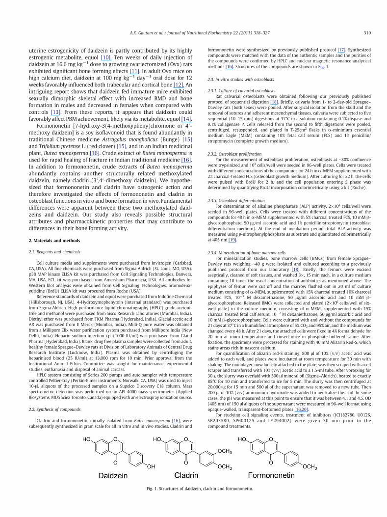
Fig. 2. Formononetin (Formo) stimulates osteoblast differentiation by p38MAPK pathway. (A)osteoblasts. Data shown as mean±S.E.M.; n=3; ⁎⁎⁎Pb.001, compared with vehicle treated cellsdifferentiation medium and treated with formononetin (10−7 M) for 21 d (as described in thered-S. Stain was extracted, and optical density (OD)wasmeasured colorimetrically. Data showninduces osteoblast differentiation via p38 MAPK pathway. Osteoblasts were treated with varioare shown as mean±S.E.M.; n=3; ⁎⁎Pb.01; ⁎⁎⁎Pb.001 compared with vehicle treated ce0126+formononetin. (D) Osteoblasts were treated with 10−7M formononetin for different timmean±S.E.M.; n=3; ⁎⁎Pb.01, ⁎⁎⁎Pb.001.
sodium azide, 10 mM EDTA, 10 μg/ml aprotinin and 1 μg/ml aminoethylbenzenesulfo-nyl fluoride). Protein samples were loaded onto 10% sodium dodecyl sulfate-polyacrylamide gel electrophoresis (SDS-PAGE) gel. After electrophoresis, proteinswere transferred to PVDF membranes. The membranes were incubated with phosphoand non-phospho Erk1/2 antibodies. The bands were developed using ECL kit.
2.4. In vivo experiments
The study was conducted in accordance with current legislation on animalexperiments (Institutional Animal Ethical Committee) at C.D.R.I. 21d immature femaleSprague–Dawley rats were used for the study [13]. All rats were housed at 21°C, in 12-h light:12-h dark cycles. Normal chow diet and water were provided ad libitum.
2.4.1. Assessment of various bone parametersRats were treated with 10.0 mg kg−1 body weight doses of individual compound or
vehicle (gum acacia in distilled water) once daily for 30 consecutive days by oralgavage. Each animal received intraperitoneal injection of fluorochromes tetracycline(20 mg kg−1 body weight dose) and calcein (20 mg kg−1 body weight dose) on days 15and 28 of treatment, respectively. At autopsy lumbar vertebrae, femur and tibia weredissected and separated from adjacent tissue, cleaned, fixed in 70% ethanol and storedat 4°C until bone strength and BMD measurements. Initial and final body weight anduterine weight were recorded. Uteri were carefully excised, gently blotted, weighedand fixed for histology and histomorphometry as we reported earlier [18].
BMD measurements of regions of interest were performed using a bonedensitometer (Model 4500 Elite, Hologic) fitted with commercially available software(QDR 4500 ACCLAIM series). After BMDmeasurement, the bones were embedded in anacrylic material for the determination of bone formation rate (BFR), mineral
Effect of formononetin on osteoblast differentiation as assessed by ALP production by. (B) Bone marrow cells (25,000 cells/well) from rats were seeded into 12-well plates inMaterials and methods). At the end of the incubation, cells were stained with alizarinasmean±S.E.M.; n=3; ⁎⁎Pb.01 compared with vehicle treated cells. (C) Formononetin
us inhibitors alone or in combination with formononetin as described in the panel. Datalls; aPb.05, SP-600125 vs. SP-600125 + formononetin and bPb.01, U-0126 vs. U-e points, and p38 MAPK was measured by ELISA kit as described before. Data shown as
321A.K. Gautam et al. / Journal of Nutritional Biochemistry 22 (2011) 318–327
appositional rate (MAR) and mineralization surface (MS). Sections 50 μm were madeusing Isomet Bone cutter, and photographs were taken under fluorescent microscopeaided with appropriate filters. The calculations were done according to previous report[22]. Estrogen agonistic and antagonistic activities were evaluated as reported earlierfrom our laboratory [23].
2.4.2. Plasma pharmacokineticsIn-vivo oral pharmacokinetic study was performed in female Sprague–Dawley rats
(n=3, weight range 200–220 g). Formononetin and cladrin were administered singleoral dose of 50 and 10 mg/kg respectively to two separate sets of rats. Blood sampleswere collected from the retro-orbital plexus of rats under light ether anesthesia intomicrofuge tubes containing heparin as an anti-coagulant at 0.08, 0.25, 0.50, 1, 2, 4, 6,8 and 10 h post-dosing. Plasma was harvested by centrifuging the blood at 13000 rpmfor 10min and stored frozen at−70±10°C until analysis. Plasma (100 μl) samples werespiked with IS (internal standard) and processed as described above. Along with theplasma samples, QC (quality control) samples were distributed among calibrators andunknown samples and analyzed by LC MS/MS.
2.5. Statistics
Data are expressed as mean±S.E.M. The data obtained in experiments withmultiple treatments were subjected to one-way analysis of variance followed by posthoc Tukey test of significance using MINITAB 13.1 software. Student's t test was used tostudy statistical significance in experiments with only two treatments.
3. Results
3.1. Structure of the compounds
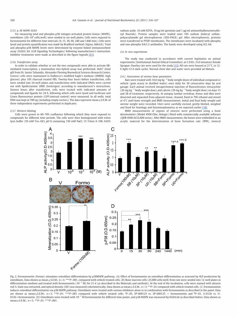
Fig. 1 shows structures of daidzein (4′,7-dihydroxyisoflavone),cladrin (7-hydroxy, 3′, 4′-dimethoxy isoflavone or 3′, 4′-dimethoxy
Fig. 3. Effects of cladrin on osteoblast proliferation, differentiation and mineralization of bonconcentrations of cladrin (Clad) for 24 h and proliferation was determined by BrdU ELISA. Dacladrin for 48 h, and ALP activity was determined as described in the Materials and methods.differentiation medium and treated with cladrin (10−8M) for 21 days (as described in the MatStain was extracted, and OD was measured colorimetrically. Data shown as mean±S.E.M.; n=
daidzein) and formononetin (4′-methoxy-7-hydroxy isoflavone or 4′-methoxy daidzein).
3.2. Formononetin stimulates osteoblast differentiation by p38MAPK pathway
Unlike cladrin, formononetin had no effect on the proliferation ofosteoblasts (data not shown). However, formononetin (10−8−10−6
M) stimulated osteoblast differentiation, assessed by osteoblast ALPproduction (Fig. 2A).The effective concentration was 10−7 M incomparison to 10−8 M required for cladrin to stimulate ALPproduction from osteoblasts. Furthermore, rat BMCs cultured in thepresence of 100 nM formononetin for 21 d under osteoblastdifferentiation condition resulted in increased formation of mineral-ized nodules compared with vehicle (Fig. 2B), suggesting thatformononetin stimulated formation of osteoporgenitor cells intomature osteoblasts in the bone marrow.
Treatment with p38 MAPK inhibitor, SB203580 (10.0 μM), but notJNK and Erk inhibitors, abolished formononetin-stimulated ALP produc-tion from osteoblasts (Fig. 2C). Consistent with this result, it wasobserved that formononetin rapidly activated phosphorylation of p38MAPK inosteoblast, peaking at 30min anddeclining at 240min (Fig. 2D).
3.3. Cladrin stimulates osteoblast proliferation and differentiation byMEK-Erk pathway
From 10−9−10−7 M, cladrin stimulated proliferation of ratneonatal calvarial osteoblasts, assessed by BrdU incorporation
e marrow osteoprogenitor cells. (A) Rat calvarial osteoblasts were exposed to variousta are mean±S.E.M.; n=3; Pb.001. (B) Cells were exposed to various concentrations of(C) Bone marrow cells (25,000 cells/well) from rats were seeded into 12-well plates inerials and methods). At the end of the incubation, cells were stained with alizarin red-S.3; ⁎⁎Pb.01; ⁎⁎⁎Pb.001 compared with vehicle-treated cells.
322 A.K. Gautam et al. / Journal of Nutritional Biochemistry 22 (2011) 318–327
(Fig. 3A). In addition, cladrin (10−9−10−7 M) stimulated osteoblastdifferentiation, assessed by osteoblast ALP production (Fig. 3B).Furthermore, rat BMCs cultured in presence of 10−8 M cladrin for 21d under osteoblast differentiation condition resulted in increasedformation of mineralized nodules compared with vehicle (Fig. 3C),suggesting that cladrin stimulates formation of osteoporgenitor cellsinto mature osteoblasts in the bone marrow.
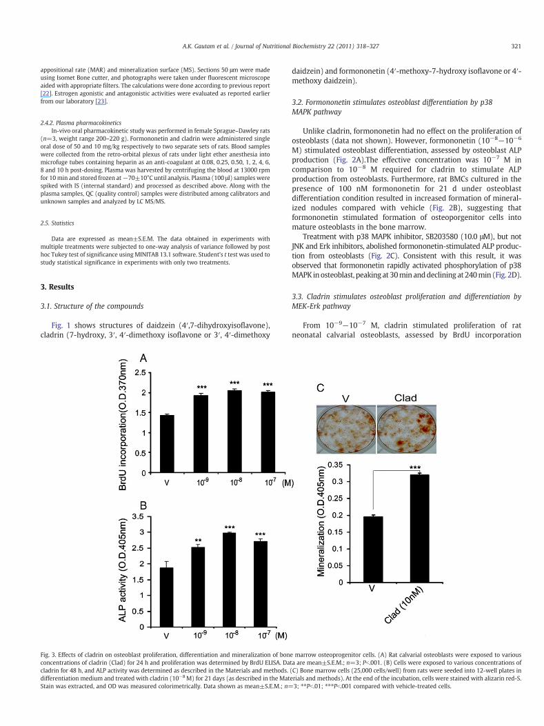
The impact of cladrin-stimulated osteoblast proliferation (asassessed by BrdU incorporation) was abolished by U0126, a mitogenactivated extracellular kinase (MEK)1/2 inhibitor whereas LY294002,an inhibitor of PI3 kinase had no effect, suggesting that cladrinstimulated osteoblast proliferation by activating MEK/Erk but not Aktpathway (Fig. 4A). U0126 also inhibited cladrin-stimulated produc-
Fig. 4. Cladrin promotes osteoblast proliferation and differentiation by MEK-Erk1/2pathway. (A) Effect of U0126 on osteoblast proliferation induced by cladrin. Cellswere pre-treated with U0126 and Ly294002 (10 μM each) for 0.5 h followed bytreatment with or without cladrin for 24 h. Results showed that U0126 abolishedosteoblast proliferation induced by cladrin. Data shown as mean±S.E.M.; n=3;⁎⁎Pb.01 compared with vehicle treated cells; aPb.01, Ly-294002 vs. Ly-294002+cladrin. (B) Effect of p38, JNK and Erk inhibitors on osteoblast cells induced bycladrin. Cells were pretreated with various inhibitors and then stimulated with 10−8
M cladrin for 48 h. Data shown as mean±S.E.M.; n=3; ⁎Pb.05; ⁎⁎Pb.01; ⁎⁎⁎Pb.001compared with vehicle treated cells, aPb.01 SB-203580 vs. SB-203580+cladrin,bPb.001, SP-600125 vs. SP-600125+cladrin. (C) Cladrin stimulates Erk phosphoryla-tion in osteoblasts. Cells were treated with 10−8 M cladrin and lysate were resolvedon SDS-PAGE. Levels of Erk and their phosphorylated forms were determined bywestern blot analysis using specific antibody. Representative gel of three indepen-dent experiments with similar results.
tion of ALP in osteoblasts, suggesting the involvement of MEK-Erkpathway in mediating cladrin action on osteoblast differentiation(Fig. 4B). We confirmed the inhibitor data by demonstrating thatcladrin had a rapid (30 min) but sustained activation (up to 1440min) on phosphorylation of Erk1/2 (Fig. 4C).
3.4. Cladrin and formononetin act independent of ER
Reports show that isoflavones such as genistein and daidzein actvia the ER in osteoblasts [24]. We tested whether cladrin orformononetin mediated their actions in osteoblasts through ER.Our data show that the presence of an ER antagonist, ICI-182780,failed to abolish cladrin- or formononetin-induced osteoblastfunctions such as proliferation and differentiation (Fig. 5A and B).Furthermore, in Huh 7 cells transfected with human ERα and ERβ,neither cladrin nor formononetin transactivated these reporter geneconstructs (Fig. 5C and D), suggesting lack of ER-mediated signalingby these compounds.
3.5. Cladrin and formononetin differentially promote BMD and boneformation rate
We next assessed in vivo effect of cladrin and formononetin ingrowing rats where bone formation is the dominant event. FemaleSprague–Dawley rats at weaning were given either cladrin orformononetin at 10.0 mg kg−1 day−1 dose by oral gavage for 30consecutive days. Gum acacia was used as vehicle (control group).
Fig. 6 shows the effects of these compounds on BMD levels. Forexample, at 10.0 mg kg−1 day−1 dose of cladrin, BMD levels wereincreased in femur (midshaft), all regions of tibia and lumbarvertebra (global, L-1, L-2 and L-4). In case of formononetin, at 10.0mg kg−1 day−1 dose, BMD levels were higher at femur (midshaft),all regions of tibia and all segments of lumbar vertebra. From thesedata, it appears that cladrin and formononetin are comparable inpromoting BMD levels of growing rats compared with controls.
Dynamic histomorphometric studies by double fluorochrome(tetracycline-calcein) labeling experiment allowed determination ofnew bone formation during the period of administration of eithercladrin or formononetin or vehicle (control). Fig. 7A and B showthat while cladrin treatment to rats, increased MAR and BFR byN2.0-fold compared with control rats, formononetin treatment hadcomparables values with that of control. When compared withcontrols, cladrin or formononetin treatment modestly but signifi-cantly increased mineralizing surface (MS) (Fig. 7B).
3.6. Assessment of uterine estrogen agonistic/antagonistic activity ofcladrin and formononetin
Daidzein is known to possess varied degrees of estrogenic or anti-estrogenic effects in vivo [25–29]. We tested whether cladrin orformononetin had such effect at the uterine level. Weaning (21-day-old) Sprague–Dawley rats were Ovx, followed by oral administrationof cladrin or formononetin at 10 mg kg−1 day−1 dose for 3consecutive days with or without 17β-E2. Table 1 shows that bothcladrin and formononetin had no estrogenic or anti-estrogenic effectsas assessed by uterine wet weight. Furthermore, histologicalevaluation of uteri under various treatments revealed that whereas17 β-E2 treatment resulted in hypertrophy of luminal and glandularepithelium, neither of the two compounds had such effect (compa-rable to control) (Fig. 8).
3.7. Plasma pharmacokinetic studies
Pilot studies revealed that formononetin had substantiallylesser oral bioavailability than cladrin (data not shown). Because
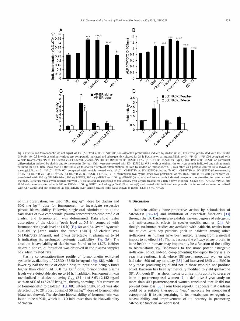
Fig. 5. Cladrin and formononetin do not signal via ER. (A) Effect of ICI-182780 (ICI) on osteoblast proliferation induced by cladrin (Clad). Cells were pre-treated with ICI-182780(1.0 nM) for 0.5 h with or without various test compounds indicated and subsequently cultured for 24 h. Data shown as mean±S.E.M.; n=3; ⁎⁎Pb.01; ⁎⁎⁎Pb.001 compared withvehicle treated cells; aPb.01, ICI-182780 vs. ICI-182780+cladrin; bPb.001, ICI-182780 vs. ICI-182780+17β-E2; cPb.01, ICI-182780 vs. 17β-E2. (B) Effect of ICI-182780 on osteoblastdifferentiation induced by cladrin and formononetin (Formo). Cells were pre-treated with ICI-182780 for 0.5 h with or without the test compounds indicated and subsequentlycultured for 48 h. Data show that ICI-182780 failed to abolish osteoblast differentiation induced by cladrin or formononetin. E2 was taken as a positive control. Data shown asmean±S.E.M.; n=3; ⁎⁎Pb.01; ⁎⁎⁎Pb.001 compared with vehicle treated cells; aPb.01, ICI-182780 vs. ICI-182780+cladrin; bPb.001, ICI-182780 vs. ICI-182780+formononetin;cPb.05, ICI-182780 vs. 17β-E2; dPb.05, ICI-182780 vs. ICI-182780+17β-E2. (C) A mammalian two-hybrid assay was performed where, Huh7 cells in 24-well plates were co-transfected with 200 ng GAL4-UAS-Luc, 100 ng EGFPC1, 100 ng pMTIF-2 and 100 ng VP16-ER (α or −β) and treated with indicated compounds as described in materials andmethods. Luciferase values were normalized with GFP values and are expressed as fold activity over vehicle treated cells. Data shown as mean±S.E.M.; n=3; ⁎Pb.05; ⁎⁎Pb.01. (D)Huh7 cells were transfected with 200 ng ERE-Luc, 100 ng EGFPC1 and 40 ng pcDNA3 ER (α or −β) and treated with indicated compounds. Luciferase values were normalizedwith GFP values and are expressed as fold activity over vehicle treated cells. Data shown as mean±S.E.M.; n=3. ⁎Pb.05.
323A.K. Gautam et al. / Journal of Nutritional Biochemistry 22 (2011) 318–327
of this observation, we used 10.0 mg kg−1 dose for cladrin and50.0 mg kg−1 dose for formononetin to investigate respectiveplasma bioavailability. Following single oral administration at thesaid doses of two compounds, plasma concentration-time profile ofcladrin and formononetin was determined. Data show fasterabsorption of the cladrin (peak level at 0.5 h) compared withformononetin (peak level at 1.0 h) (Fig. 8A and B). Overall systemicavailability [area under the curve (AUC)] of cladrin was571.0±73.25 h⁎ng/ml, and it was detectable in plasma up to 24h indicating its prolonged systemic availability (Fig. 8A). Theabsolute bioavailability of cladrin was found to be 13.7%. Neitherdaidzein nor equol formation was observed in the plasma samplesof cladrin treated rats.
Plasma concentration-time profile of formononetin exhibitedsystemic availability of 278.30±38.50 hr⁎ng/ml (Fig. 8B), which islower by half the value of cladrin despite input dose being 5 timeshigher than cladrin. At 50.0 mg kg−1 dose, formononetin plasmalevels were detectable also up to 24 h. In addition, formononetin wasmetabolized to daidzein, having Cmax (24 h) of 8.63±2.152 ng/mlwith an AUC of 147.2488 h⁎ng/ml, thereby showing ∼50% conversionof formononetin to daidzein (Fig. 8B). Interestingly, equol was alsodetected up to 28 h post dosing of 50 mg kg−1 dose of formononetin(data not shown). The absolute bioavailability of formononetin wasfound to be 4.294%, which is ∼3.0-fold lesser than the bioavailabilityof cladrin.
4. Discussion
Daidzein affords bone-protective action by stimulation ofosteoblast [30–32] and inhibition of osteoclast functions [33]through the ER. Daidzein also exhibits varying degrees of estrogenicand anti-estrogenic effects in species-specific manner [24]. Al-though, no human studies are available with daidzein, results fromthe studies with soy proteins (rich in daidzein among otherisoflavones) in humans have been mixed, ranging from a modestimpact to no effect [34]. That is because the efficacy of soy protein inbone health in humans may importantly be a function of the abilityto biotransform soy isoflavones to the more potent estrogenicisoflavone, equol. Indeed, complementing the equol theory is a 2-year interventional trial, where 108 postmenopausal women whohad taken 500 ml soy milk/day [35], had increased BMD and BMC inonly those producing equol and not in those who did not produceequol. Daidzein has been synthetically modified to yield ipriflavone(IP). Although IP, has shown some promise in its ability to preservebone in postmenopausal women [7], a definitive 3-year study onmore than 400 postmenopausal women concluded that IP did notprevent bone loss [36]. From these reports, it appears that daidzeincould be a suitable therapeutic “lead” molecule for menopausalosteoporosis if issues pertaining to its metabolism, estrogenicity,bioavailability and improvement of its potency in promotingosteoblast function are addressed.
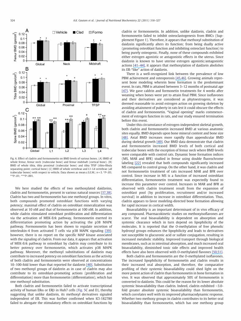
Fig. 6. Effect of cladrin and formononetin on BMD levels of various bones. (A) BMD ofwhole femur, femur neck (trabecular bone) and femur midshaft (cortical bone). (B)BMD of whole tibia, tibia proximal (trabecular bone) and tibia TFSP (tibio-fibulaseparating point; cortical bone) (C) BMD of whole vertebrae and L1–L4 vertebrae (alltrabecular bones) with respect to vehicle. Data shown as mean±S.E.M.; n=3. ⁎Pb.05;⁎⁎Pb.01; ⁎⁎⁎Pb.001.
324 A.K. Gautam et al. / Journal of Nutritional Biochemistry 22 (2011) 318–327
We here studied the effects of two methoxylated daidzeins,cladrin and formononetin, present in various natural sources [37,38].Cladrin has two and formononetin has one methoxyl groups. In vitro,both compounds promoted osteoblast functions with varyingpotency; maximal effect of cladrin on osteoblast mineralization wasobserved at 10 nM and that of formononetin at 100 nM. In addition,while cladrin stimulated osteoblast proliferation and differentiationvia the activation of MEK-Erk pathway, formononetin exerted itsdifferentiation promoting action by activating the p38 MAPKpathway. Formononetin has been shown to regulate secretion ofinterleukin-4 from activated T cells via p38 MAPK signaling [39];however, there is no report on the specific MAP kinase associatedwith the signaling of cladrin. From our data, it appears that activationof MEK-Erk pathway in osteoblast by cladrin may contribute to itsbetter potency over formononetin, which activates p38 MAPKpathway. Moreover, the methoxyl substitutions of daidzein maycontribute to increased potency on osteoblast functions as the activityof both cladrin and formononetin were observed at concentrations10–100-fold lower than that reported for daidzein [40]. Substitutionof two methoxyl groups of daidzein as in case of cladrin may alsocontribute to its osteoblast-promoting actions (proliferation anddifferentiation) more than formononetin (differentiation), which hasone methoxyl substitution.
Both cladrin and formononetin failed to activate transcriptionalactivity of human ERα or ERβ in Huh7 cells (Fig. 5C and D), therebysuggesting that unlike daidzein, its methoxyl derivatives signaledindependent of ER. This was further confirmed when ICI-182780failed to abrogate the stimulatory effects on osteoblast functions by
cladrin or formononetin. In addition, unlike daidzein, cladrin andformononetin failed to inhibit osteoclastogenesis from BMCs (Sup-plement Figure 1). Therefore, it appears that methoxyl substitution ofdaidzein significantly alters its function; from being dually active(promoting osteoblast function and inhibiting osteoclast function) tobeing purely osteogenic. Finally, none of these compounds exhibitedeither estrogen agonistic or antagonistic effects in the uterus. Sincedaidzein is known to have uterine estrogen agonistic/antagonisticactions [41–44], it appears that methoxylation of daidzein abolishesits ER-“like” action of daidzein.
There is a well-recognized link between the prevalence of lowPBM achievement and osteoporosis [45,46]. Growing animals repre-sent bone modeling wherein bone formation is the predominantevent. In rats, PBM is attained between 3–12 months of postnatal age[47]. We gave caldrin and formonetin treatments for 4 weeks afterweaning when bones were yet to attain final PBM. Since isoflavonesand their derivatives are considered as phytoestrogens, it wasdeemed reasonable to avoid estrogen action on growing skeleton byavoiding attainment of puberty in rats lest it could obscure the effectsof cladrin and formononetin. “Vaginal opening” marks commence-ment of estrogen function in rats, and our study ensured terminationbefore this event.
Under this circumstance of estrogen-independent skeletal growth,both cladrin and formononetin increased BMD at various anatomicsites equally. BMD depends upon bone mineral content and bone size[48]. Axial BMD increases more rapidly than appendicular BMDduring skeletal growth [49]. Our BMD data demonstrate that cladrinand formononetin increased BMD levels of both cortical andtrabecular bones with the exception of femur neck where BMD levelswere comparable with control rats. Dynamic bone formation indices(MS, MAR and BFR) studied in femur using double fluorochromelabeling [22] revealed that both compounds significantly increasedMS compared to control group. On the other hand, it was cladrin andnot formononetin treatment of rats increased MAR and BFR overcontrol. Since increase in MS is a function of increased osteoblastdifferentiation, formononetin treatment was expectedly found toincrease this parameter over control. Increases in MAR and BFR asobserved with cladrin treatment result from the expansion ofosteoblast pool (by proliferation, recruitment and/or increasedsurvival) in addition to increase in osteoblast differentiation. Thus,cladrin appears to favor modeling-directed bone formation allowingfor rapid increase in cortical width.
Bioavailability is an important determinant of in vivo efficacy ofany compound. Pharmacokinetic studies on methoxyisoflavones arescarce. The oral bioavailability is dependent on absorption andsystemic clearance which in turn depend on lipophilicity of themolecules. It is reported that the O-methylation of free phenolichydroxyl groups enhances the lipophilicity and leads to derivativesnot susceptible to glucuronic acid or sulfate conjugation, resulting inincreased metabolic stability. Improved transport through biologicalmembranes, such as in intestinal absorption, and much increased oralbioavailability, diminished toxic side effects and improved healtheffects have also been observed with O-methylated flavones [50,51].
Both cladrin and formononetin are the O-methylated isoflavones.The increased lipophilicity of formononetin and cladrin results intheir increased oral absorption, and therefore, the comparativeprofiling of their systemic bioavailability could shed light on themore potent action of cladrin than formononetin in bone formation invivo. It was observed that approximately 50% of formononetin isconverted to daidzein. This could be the reason for its lower absolutesystemic bioavailability than cladrin. Indeed, cladrin exhibited ∼3.0-fold greater absolute systemic bioavailability than formononetin,which correlates well with its improved pharmacodynamic response.Whether two methoxy groups in cladrin contributes to its better oralbioavailability than formononetin, which has one methoxy group
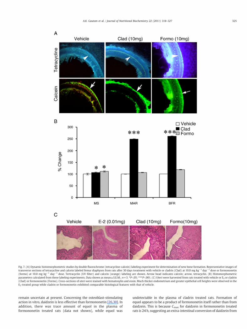
Fig. 7. (A) Dynamic histomorphometric studies by double fluorochrome (tetracycline-calcein) labeling experiment for determination of new bone formation. Representative images oftransverse sections of tetracycline and calcein labeled femur diaphyses from rats after 30 days treatment with vehicle or cladrin (Clad) at 10.0 mg kg−1 day−1 dose or formononetin(formo) at 10.0 mg kg−1 day−1 dose. Tertracyclin (UV filter) and calcein (orange) labeling are shown. Arrow head indicates calcein; arrow, tetracyclin. (B) Histomorphometricparameters calculated from these labeling experiments. Data shown as mean±S.E.M.; n=3. ⁎Pb.05; ⁎⁎⁎Pb.001. (C) Uteri were harvested from rats treated with vehicle or E2 or cladrin(Clad) or formononetin (Formo). Cross-sections of uteri were stained with hematoxylin and eosin. Much thicker endometrium and greater epithelial cell heights were observed in theE2-treated group while cladrin or formononetin exhibited comparable histological features with that of vehicle.
325A.K. Gautam et al. / Journal of Nutritional Biochemistry 22 (2011) 318–327
remain uncertain at present. Concerning the osteoblast-stimulatingaction in vitro, daidzein is less effective than formononetin [26,30]. Inaddition, there was trace amount of equol in the plasma offormononetin treated rats (data not shown), while equol was
undetectable in the plasma of cladrin treated rats. Formation ofequol appears to be a product of formononetin itself rather than fromdaidzein. This is because Cmax for daidzein in formononetin treatedrats is 24 h, suggesting an extra-intestinal conversion of daidzein from
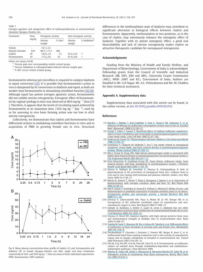
Table 1Estrogen agonistic and antagonistic effect of methoxyisoflavones in ovariectomizedimmature Sprague–Dawley rats
Treatment Dosemg/kg
Estrogenic activity Anti-estrogenic activity
Uterineweight
% Gaina Uterineweight
% Inhibitionb
Vehicle - 14.7±2.2Ethynyl estradiol 0.01 98.3c±11.5 568Cladrin 10 14.9±1.0 1.3 91.2±5.0 7Formononetin 10 17.5±2.0 19 97.4±4.8 1
Values are mean±S.E.M.a Percent gain over corresponding vehicle control group.b Percent inhibition in ethynylestradiol-induced uterine weight gain.c Pb.001 versus vehicle treated group.
326 A.K. Gautam et al. / Journal of Nutritional Biochemistry 22 (2011) 318–327
formononetin whereas gut microflora is required to catalyze daidzeinto equol conversion [52]. It is possible that formononetin's action invivo is dampened by its conversions to daidzein and equol, as both areweaker than formononetin in stimulating osteoblast function [26,30].Although equol has potent estrogen agonistic action, formononetindid not exhibit uterine estrogenicity. Estrogenic effect of formonone-tin by vaginal cytology inmice was observed at 40.0 mg kg−1 dose [53]. Therefore, it appears that the levels of circulating equol achieved byformononetin at its maximum dose (10.0 mg kg−1 day−1) used byus for assessing in vivo bone forming action was too low to elicituterine estrogenicity.
Collectively, we demonstrate that cladrin and formononetin havedifferential actions in modulating osteoblast functions in vitro and inacquisition of PBM in growing female rats in vivo. Structural
Fig. 8. Mean plasma concentration-time profile of cladrin (A) and, formononetin anddaidzein (B) in female Sprague–Dawley rats after single oral dose treatmentsrespectively of 10.0- and 50.0 mg kg−1. Data are mean of three individual experiments.FMN, formononetin; DZN, daidzein.
differences in the methoxylation state of daidzein may contribute tosignificant alteration in biological effects between cladrin andformononetin. Apparently, methoxylation at two positions, as in thecase of cladrin, may enormously enhance the osteogenic effect ofdaidzein. Together with its potent osteogenic effect, a good oralbioavailability and lack of uterine estrogenicity makes cladrin anattractive therapeutic candidate for menopausal osteoporosis.
Acknowledgments
Funding from the Ministry of Health and Family Welfare, andDepartment of Biotechnology, Government of India is acknowledged.Fellowship grants from the Council of Scientific and IndustrialResearch (BB, DKY, JSM and ABS), University Grants Commission(AKG), MOH (AMT and KS), Government of India. Authors arethankful to Mr. G.K Nagar, Mr. A.L. Vishwakarma and Ms. M. Chabbrafor their technical assistances.
Appendix A. Supplementary data
Supplementary data associated with this article can be found, inthe online version, at doi:10.1016/j.jnutbio.2010.02.010.
References
[1] Mardon J, Mathey J, Kati-Coulibaly S, Puel C, Davicco MJ, Lebecque P, et al.Influence of lifelong soy isoflavones consumption on bonemass in the rat. Exp BiolMed (Maywood) 2008;233:229–37.
[2] Uesugi T, Fukui Y, Yamori Y. Beneficial effects of soybean isoflavone supplemen-tation on bone metabolism and serum lipids in postmenopausal Japanese women:a four-week study. J Am Coll Nutr 2002;21:97–102.
[3] Anderson JJ, Garner SC. Phytoestrogens and bone. Baillieres Clin Endocrinol Metab1998;12:543–57.
[4] Somekawa Y, Chiguchi M, Ishibashi T, Aso T. Soy intake related to menopausalsymptoms, serum lipids, and bone mineral density in postmenopausal Japanesewomen. Obstet Gynecol 2001;97:109–15.
[5] Mei J, Yeung SS, Kung AW. High dietary phytoestrogen intake is associated withhigher bone mineral density in postmenopausal but not premenopausal women. JClin Endocrinol Metab 2001;86:5217–21.
[6] Kritz-Silverstein D, Goodman-Gruen DL. Usual dietary isoflavone intake, bonemineral density, and bone metabolism in postmenopausal women. J WomensHealth Gend Based Med 2002;11:69–78.
[7] Sharan K, Siddiqui JA, Swarnkar G, Maurya R, Chattopadhyay N. Role ofphytochemicals in the prevention of menopausal bone loss: evidence from invitro and in vivo, human interventional and pharma-cokinetic studies. Curr MedChem 2009;16:1138–57.
[8] Morito K, Aomori T, Hirose T, Kinjo J, Hasegawa J, Ogawa S, et al. Interaction ofphytoestrogens with estrogen receptors alpha and beta (II). Biol Pharm Bull2002;25:48–52.
[9] Diel P, Schulz T, Smolnikar K, Strunck E, Vollmer G, Michna H. Ability of xeno- andphytoestrogens to modulate expression of estrogen-sensitive genes in rat uterus:estrogenicity profiles and uterotropic activity. J Steroid Biochem Mol Biol2000;73:1–10.
[10] Selvaraj V, Zakroczymski MA, Naaz A, Mukai M, Ju YH, Doerge DR, et al.Estrogenicity of the isoflavone metabolite equol on reproductive and non-reproductive organs in mice. Biol Reprod 2004;71:966–72.
[11] Somjen D, Katzburg S, Kohen F, Gayer B, Livne E. Daidzein but not otherphytoestrogens preserves bone architecture in ovariectomized female rats in vivo.J Cell Biochem 2008;103:1826–32.
[12] Fonseca D, Ward WE. Daidzein together with high calcium preserve bone massand biomechanical strength at multiple sites in ovariectomized mice. Bone2004;35:489–97.
[13] FujiokaM, Sudo Y, OkumuraM,Wu J, Uehara M, Takeda K, et al. Differential effectsof isoflavones on bone formation in growing male and female mice. Metabolism2007;56:1142–8.
[14] Phrakonkham P, Chevalier J, Desmetz C, Pinnert MF, Berges R, Jover E, et al.Isoflavonoid-based bone-sparing treatments exert a low activity on reproductiveorgans and on hepatic metabolism of estradiol in ovariectomized rats. ToxicolAppl Pharmacol 2007;224:105–15.
[15] Wu JH, Li Q, Wu MY, Guo DJ, Chen HL, Chen SL, et al. Formononetin, an isoflavone,relaxes rat isolated aorta through endothelium-dependent and endothelium-independent pathways. J Nutr Biochem 2009.
[16] Maurya R, Yadav DK, Singh G, Bhargavan B, Narayana Murthy PS, Sahai M, et al.Osteogenic activity of constituents from Butea monosperma. Bioorg Med ChemLett 2009;19:610–3.
327A.K. Gautam et al. / Journal of Nutritional Biochemistry 22 (2011) 318–327
[17] Bass RJ. Synthesis of chromones by cyclization of 2-hydroxyphenyl ketones withboron trifluoride — diethyl ether and methanesulphonyl chloride. J Chem SocChem Commun 1976:78–9.
[18] Trivedi R, Kumar S, Kumar A, Siddiqui JA, Swarnkar G, Gupta V, et al. Kaempferolhas osteogenic effect in ovariectomized adult Sprague–Dawley rats. Mol CellEndocrinol 2008;289:85–93.
[19] Ishizuya T, Yokose S, Hori M, Noda T, Suda T, Yoshiki S, et al. Parathyroid hormoneexerts disparate effects on osteoblast differentiation depending on exposure timein rat osteoblastic cells. J Clin Invest 1997;99:2961–70.
[20] Gregory CA, Gunn WG, Peister A, Prockop DJ. An Alizarin red-based assay ofmineralization by adherent cells in culture: comparison with cetylpyridiniumchloride extraction. Anal Biochem 2004;329:77–84.
[21] Yamaguchi T, Chattopadhyay N, Kifor O, Sanders JL, Brown EM. Activation ofp42/44 and p38 mitogen-activated protein kinases by extracellular calcium-sensing receptor agonists induces mitogenic responses in the mouse osteoblasticMC3T3-E1 cell line. Biochem Biophys Res Commun 2000;279:363–8.
[22] Hara K, Kobayashi M, Akiyama Y. Vitamin K2 (menatetrenone) inhibits bone lossinduced by prednisolone partly through enhancement of bone formation in rats.Bone 2002;31:575–81.
[23] Srivastava SR, Keshri G, Bhargavan B, Singh C, Singh MM. Pregnancy interceptiveactivity of the roots of Calotropis gigantea Linn. in rats. Contraception 2007;75:318–22.
[24] Choi SY, Ha TY, Ahn JY, Kim SR, Kang KS, Hwang IK, et al. Estrogenic activities ofisoflavones and flavones and their structure-activity relationships. Planta Med2008;74:25–32.
[25] Ju YH, Fultz J, Allred KF, Doerge DR, Helferich WG. Effects of dietary daidzein andits metabolite, equol, at physiological concentrations on the growth of estrogen-dependent human breast cancer (MCF-7) tumors implanted in ovariectomizedathymic mice. Carcinogenesis 2006;27:856–63.
[26] Ohtomo T, Uehara M, Penalvo JL, Adlercreutz H, Katsumata S, Suzuki K, et al.Comparative activities of daidzein metabolites, equol and O-desmethylangolen-sin, on bone mineral density and lipid metabolism in ovariectomized mice and inosteoclast cell cultures. Eur J Nutr 2008;47:273–9.
[27] Zhao L, Mao Z, Brinton RD. A select combination of clinically relevantphytoestrogens enhances estrogen receptor beta-binding selectivity and neuro-protective activities in vitro and in vivo. Endocrinology 2009;150:770–83.
[28] Han DH, Denison MS, Tachibana H, Yamada K. Relationship between estrogenreceptor-binding and estrogenic activities of environmental estrogens andsuppression by flavonoids. Biosci Biotechnol Biochem 2002;66:1479–87.
[29] Rachon D, Vortherms T, Seidlova-Wuttke D, Wuttke W. Effects of dietary equol onthe pituitary of the ovariectomized rats. Horm Metab Res 2007;39:256–61.
[30] Jia TL,Wang HZ, Xie LP,Wang XY, Zhang RQ. Daidzein enhances osteoblast growththat may be mediated by increased bone morphogenetic protein (BMP)production. Biochem Pharmacol 2003;65:709–15.
[31] Setchell KD, Lydeking-Olsen E. Dietary phytoestrogens and their effect on bone:evidence from in vitro and in vivo, human observational, and dietary interventionstudies. Am J Clin Nutr 2003;78:593S–609S.
[32] Sugimoto E, Yamaguchi M. Stimulatory effect of Daidzein in osteoblastic MC3T3-E1 cells. Biochem Pharmacol 2000;59:471–5.
[33] Rassi CM, Lieberherr M, Chaumaz G, Pointillart A, Cournot G. Down-regulation ofosteoclast differentiation by daidzein via caspase 3. J Bone Miner Res 2002;17:630–8.
[34] Branca F. Dietary phyto-oestrogens and bone health. Proc Nutr Soc 2003;62:877–87.
[35] Lydeking-Olsen E, Beck-Jensen JE, Setchell KD, Holm-Jensen T. Soymilk orprogesterone for prevention of bone loss — a 2 year randomized, placebo-controlled trial. Eur J Nutr 2004;43:246–57.
[36] Alexandersen P, Toussaint A, Christiansen C, Devogelaer JP, Roux C, FechtenbaumJ, et al. Ipriflavone in the treatment of postmenopausal osteoporosis: arandomized controlled trial. Jama 2001;285:1482–8.
[37] Zheng Z, Song C, Liu D, Hu Z. Determination of 6 isoflavonoids in the hairy rootcultures of Astragalus membranaceus by HPLC. Yao Xue Xue Bao 1998;33:148–51.
[38] Kawakita S, Marotta F, Naito Y, Gumaste U, Jain S, Tsuchiya J, et al. Effect of anisoflavones-containing red clover preparation and alkaline supplementation onbone metabolism in ovariectomized rats. Clin Interv Aging 2009;4:91–100.
[39] Park J, Kim SH, Cho D, Kim TS. Formononetin, a phyto-oestrogen, and itsmetabolites up-regulate interleukin-4 production in activated T cells viaincreased AP-1 DNA binding activity. Immunology 2005;116:71–81.
[40] Yamaguchi M, Sugimoto E. Stimulatory effect of genistein and daidzein on proteinsynthesis in osteoblastic MC3T3-E1 cells: activation of aminoacyl-tRNA synthe-tase. Mol Cell Biochem 2000;214:97–102.
[41] De Wilde A, Lieberherr M, Colin C, Pointillart A. A low dose of daidzein acts as anERbeta-selective agonist in trabecular osteoblasts of young female piglets. J CellPhysiol 2004;200:253–62.
[42] Farmakalidis E, Hathcock JN, Murphy PA. Oestrogenic potency of genistin anddaidzin in mice. Food Chem Toxicol 1985;23:741–5.
[43] Matsumura A, Ghosh A, Pope GS, Darbre PD. Comparative study of oestrogenicproperties of eight phytoestrogens in MCF7 human breast cancer cells. J SteroidBiochem Mol Biol 2005;94:431–43.
[44] Shao ZM, Alpaugh ML, Fontana JA, Barsky SH. Genistein inhibits proliferationsimilarly in estrogen receptor-positive and negative human breast carcinoma celllines characterized by P21WAF1/CIP1 induction, G2/M arrest, and apoptosis. J CellBiochem 1998;69:44–54.
[45] Cooper C, Harvey N, Cole Z, Hanson M, Dennison E. Developmental origins ofosteoporosis: the role of maternal nutrition. Adv Exp Med Biol 2009;646:31–9.
[46] Cooper C, Harvey N, Javaid K, Hanson M, Dennison E. Growth and bonedevelopment. Nestle Nutr Workshop Ser Pediatr Program 2008;61:53–68.
[47] Ke HZ, Crawford DT, Qi H, Chidsey-Frink KL, Simmons HA, Li M, et al. Long-termeffects of aging and orchidectomy on bone and body composition in rapidlygrowing male rats. J Musculoskelet Neuronal Interact 2001;1:215–24.
[48] Seeman E. Growth in bone mass and size — are racial and gender differences inbone mineral density more apparent than real? J Clin Endocrinol Metab 1998;83:1414–9.
[49] Finkelstein JS, Neer RM, Biller BM, Crawford JD, Klibanski A. Osteopenia in menwith a history of delayed puberty. N Engl J Med 1992;326:600–4.
[50] Walle T. Methylation of dietary flavones greatly improves their hepatic metabolicstability and intestinal absorption. Mol Pharm 2007;4:826–32.
[51] Wen X, Walle T. Methylated flavonoids have greatly improved intestinalabsorption and metabolic stability. Drug Metab Dispos 2006;34:1786–92.
[52] Jackman KA, Woodman OL, Sobey CG. Isoflavones, equol and cardiovasculardisease: pharmacological and therapeutic insights. Curr Med Chem 2007;14:2824–30.
[53] Wang W, Tanaka Y, Han Z, Higuchi CM. Proliferative response of mammaryglandular tissue to formononetin. Nutr Cancer 1995;23:131–40.
Copyright © 2022 FDOKUMEN