P2X4 receptors control the fate and survival of activated microglia
Upload
independentCategory
view
0download
0
Different patterns of Ca2+ signals are induced by lowcompared to high concentrations of P2Yagonists in microglia
S. Visentin & C. De Nuccio & G. C. Bellenchi
Received: 9 December 2005 /Revised: 19 June 2006 /Accepted: 19 June 2006 / Published online: 13 September 2006# Springer Science + Business Media B.V. 2006
Abstract Brain-resident macrophages (microglia) are keycellular elements in the preservation of tissue integrity. Onthe other hand, they can also contribute to the developmentof pathological events by causing an extensive andinappropriate inflammatory response. A growing numberof reports indicate the involvement of nucleotides in thecontrol of microglial functions. With this study on P2Yreceptors in rat microglia, we want to contribute to thedefinition of their expression profile and to the character-isation of their signalling mechanisms leading to Ca2+
movements. Endogenous nucleotides, when applied at aconcentration of 100 μM, elicited robust Ca2+ transients,thanks to a panel of metabotropic receptors comprisingmainly P2Y2, P2Y6 and P2Y12 subtypes. The involvementof P2Y12 receptors in Ca2+ responses induced by adeninenucleotides was confirmed by the pharmacological andpertussis toxin sensitivity of the response induced byadenosine diphosphate (ADP). Beside the G proteininvolved, Gi and Gq respectively, adenine and uracilnucleotides differed also for induction by the latter of acapacitative Ca2+ plateau. Moreover, when applied at low(sub-micromolar) concentrations with a long-lasting chal-lenge, uracil nucleotides elicited oscillatory Ca2+ changeswith low frequency of occurrence (≤1 min−1), sometimessuperimposed to an extracellular Ca2+-dependent sustainedCa2+ rise. We conclude that different patterns of Ca2+
transients are induced by low (i.e., oscillatory Ca2+ activity)compared to high (i.e., fast release followed by sustained
raise) concentrations of nucleotides, which can suggestdifferent roles played by receptor stimulation depending notonly on the type but also on the concentration ofnucleotides.
Key words calcium . capacitative . mitochondria .
oscillation
Introduction
Distributed in the central nervous system (CNS) parenchy-ma in a ramified morphology, microglial cells play acontinuous “resting activity” of surveillance. Signals notwell identified as yet seem to be required to keep microgliain such a quiescent but alert state. Similarly, signalsresulting from an ongoing acute insult or from a chronicdegenerative condition drive toward or maintain microgliain a state of cell activation. Synthesis of pro- and anti-inflammatory cytokines, reactive oxygen species, nitricoxide, prostaglandins, expression of major histocompatibil-ity complex (MHC) molecules and re-arrangement of theion channel profile are only some of the changes acquiredby activated microglial cells [1–3]. In principle, these newlyacquired functional capabilities would tend to favourprotection and recovery of homeostasis, but in someinstances, control on the overall inflammatory response islost and an exacerbation of the pathology takes place.Participation in the pathogenesis of virtually all acute andchronic diseases has been suggested for microglia. Thedichotomy of microglia between guardian and potentiallypromoter of recovery on one hand and pathogenic cellularentity on the other hand has drawn interest to the searchfor signals driving microglia to beneficial capabilities andthe control of pro-inflammatory and potentially harmfulactivities [4, 5].
Purinergic Signalling (2006) 2: 605–617DOI 10.1007/s11302-006-9023-1
S. Visentin (*) : C. De Nuccio :G. C. BellenchiDepartment of Cell Biology and Neuroscience,Section of Degenerative and Inflammatory Neurological Diseases,Istituto Superiore di Sanità,Viale Regina Elena 299,00161 Rome, Italye-mail: [email protected]

Adenosine triphosphate (ATP) and other nucleotides havebeen extensively described as multi-functional agents actingon neuronal and glial cells. ATP acts as a fast synapticneurotransmitter by binding to post-synaptic P2X receptorsin a variety of neuronal cells, in keeping with their widedistribution predominantly on brain neurons [6]. ATP alsoacts on postsynaptic P2Y receptors, modulating voltage-dependent K+ and Ca2+ currents [7, 8]. P2Y receptors werealso described to interfere with N-methyl-D-aspartate(NMDA) but not with alpha-amino-3-hydroxy-5-methyl-4-isoxazole propionic acid (AMPA) glutamate receptors,probably by inducing phosphorylation of the receptorprotein [9]. ATP and analogues acting on P2Y receptorsregulate the release of neurotransmitters such as acetyl-choline, noradrenaline, dopamine, serotonin and gluta-mate (6 for references) in concert with adenosine.
Also, all glial cell types have been described to react toATP and other nucleotides. ATP and analogues were shownto induce astrocyte proliferation, stellation and increase ofglial fibrillary acidic protein (GFAP) expression, all symp-toms of astrogliosis [10–12], by stimulating P2Y receptors.Ca2+ wave propagation in mechanically or pharmacologi-cally stimulated cultured astrocytes are known to depend onthe release of ATP and on its effect on adjacent cells [13].ATP released from stimulated dorsal root ganglia (DRG)cells was also described to modulate the differentiation andproliferation of Schwann cells in a in vitro co-culturesystem [14]. Recently, we identified ATP and adenosinediphosphate (ADP) as agents able to control differentiationand promote migration of oligodendrocyte progenitors byactivation of P2Y1 receptors [15]. A number of differenteffects of nucleotides on microglial cells have also beenreported. In microglia, great attention has been dedicated toP2X ionotropic receptors, and thanks to this, their profile ofexpression and a set of functions clearly linked to eachsubtype has been delineated. P2X7 receptors, first describedfor their pro-inflammatory capabilities due to the inductionof interleukin (IL)-1β and tumour necrosis factor (TNF)-αrelease [16, 17], as well as for their cytotoxicity due tothe opening of a large conductance pore [18], are nowconsidered also as involved in the fine control of microglialproliferation (Bianco et. al., this issue). A second ionotropicATP receptor that we tentatively proposed in microglia asthe P2X4 subtype on the basis of kinetic and pharmaco-logical properties [19] was then clearly identified in this celltype, and its role in the induction of tactile peripheralallodynia following nerve injury was proposed [20]. Anumber of studies have addressed the capabilities ofnucleotides to mobilise Ca2 and to induce the opening ofstore-operated Ca2+ channels by acting on microglial P2Yreceptors [21, 22]. Chemotactic activity induced by ATPwas described to involve P2Y12 receptors [23] while therelease of pro- and anti-inflammatory cytokines was shown
to be under the control of as yet unidentified P2Y receptors[24–26].
Glial cells are not only targets of nucleotide signalling,they also are more generally responsible of shaping thenucleotide signalling system, being also sources of nucleo-tides and in charge of their degradation. Regardless of themechanisms utilised—vesicular, mediated by ATP-bindingcassette (ABC) transportes or through connexin emi-channels—during Ca2+ waves propagation, astrocytes re-lease ATP, which acts as an extracellular messenger inhomo-typical (i.e., astrocyte to astrocyte) and hetero-typical(i.e., astrocyte to microglia) intercellular communication[13, 27]. When challenged with the pro-inflammatory factorlipopolysaccharide (LPS), microglia was described torelease ATP, which, by acting as an autocrine factor,contributed to processing and release of cytokines IL-β1and IL-10 [16, 25]. Finally, astrocytes and microglial cellsare deputed to the degradation of extracellular nucleotidesin virtue of the expression of ecto-enzymes ecto-ATPase,ecto-apyrase and ecto-5′-nucleotidase [28]. Interestingly,increase of expression of these enzymes after transient fore-brain ischaemia in the rat was shown to involve mainlymicroglia [29], which makes these cells capable of reactingto the increase of nucleotide release in the attempt to avoidexcessive nucleotide stimulation.
It is known that high doses of nucleotides released bytissue undergoing stress, cell damage or abnormal deathmight act as “danger signals” and cause release of pro-inflammatory cytokines (IL-1β, TNF-α, IL-6), prostaglan-dins and reactive oxygen species [30] from immune cells.On the other hand, it was recently suggested that at lowerdoses, nucleotides might play a regulatory role on inflam-matory reactions aimed at the prevention of tissue damage[31].
In this study, we focused on P2 metabotropic receptorsin rat microglial cells, and by molecular [reverse transcrip-tion polymerase chain reaction (RT-PCR)] and functional(Ca2+ video imaging) approaches, we characterised expres-sion profile, functional capability and some aspects of thepathways involved in receptor activation. By comparing theeffects induced by low versus high concentrations ofmetabotropic agonists, we then aimed to depict dependenceon the availability of nucleotides of cytoplasmic Ca2+
signalling.
Materials and methods
Cell culture
Microglial cells were obtained from the cerebral cortex of1- to 2-day-old rats [32]. All experiments were carried outin accordance with directives of the Council of the
606 Purinergic Signalling (2006) 2: 605–617

European Communities N. 86/609/CEE. Briefly, after pupswere rendered hypothermic on an ice-cold surface, theywere decapitated. The cerebral cortex was dissected out,and meninges were removed. The tissue was dissociated bya two-step procedure comprising enzymatic digestionfollowed by mechanical dissociation. Mixed primarycultures were grown on poly-L-lysine-coated culture flasksfor 7–10 days in Basal Medium Eagle (BME) supple-mented with 10% endotoxin-free foetal calf serum (FCS),2 mM glutamine and 100 μg ml−1 gentamicin (37°C, 5%CO2). After mild shaking, microglial cells were harvestedand plated on un-coated glass coverslips sealed on home-made silicon wells at a density of 15 × 104 cells/cm2. Tofurther improve the purity of microglial cultures, non-adhering cells were removed after 20 min by changing themedium.
RT-PCR
Microglia cells obtained from rat brain at postnatal day 2were used to extract total ribonucleic acid (RNA) usingTrizol reagent according with the data sheet. Twomicrograms of total RNA were digested for 30 min withDnase (Invitrogen) and retro-transcribed using the Super-Script synthesis system (Life Technologies). Specificoligonucleotides with similar annealing temperatures weredesigned to amplify the following purinergic receptors:P2Y1 (NM012800.1), P2Y2 (NM017255.1), P2Y4
(NM031680 . 1 ) , P2Y6 (NM057124 . 1 ) , P2Y1 2
(NM022800.1), P2Y13 (NM001002853.1) and P2Y14
(NM133577.1). β-actin (NM031144) was always ampli-fied as control. Samples were analysed on agarose gelcontaining ethidium bromide.
The following primers were used:
P2Y1: forward: 5′-AACAGTACAATCGCCTCGAC-3′; reverse: 5′-TTCGCAGGTACTCATCTGAC-3′.Band size of 559 bp.P2Y2: forward: 5′AACGGACGTGAGCATCCAA3′;reverse: 5′-AGGTGTCGTGGCAGGTGATT-3′. Bandsize of 639 bp.P2Y4: forward: 5′-CCATCCTGTGCCATGACACT-3′;reverse: 5′-CATAGCTGCTGGAGCTGGTT-3′. Bandsize of 418 bp.P2Y6: forward: 5′-CGGCAGCATCCTGTTCCTCA-3′;reverse 5′- TGGAGCGCACAGCCAAGTAG-3′. Bandsize of 470 bp.P2Y12: forward: 5′-GGTGTGCCAAGTCACTTCAG;reverse 5′-ATCAAGGCAGGCGTTCAAGG-3′. Bandsize of 598 bp.P2Y13: forward: 5′-GATGACACAGCTGCTGTTCC-3′; reverse: 5′- TGACTGCTGTGGTGCTCATC-3′.Band size of 915 bp.
P2Y14: forward: 5 ′-GCACAAGGCGTCTAAC-TATGTC-3′; reverse: 5′-CCACATGCCACTTCTC-CAT TCG-3′. Band size of 692 bp.β-Actin: forward: 5′-ACCTTCAACACCCCAGCCATGTAC G-3′; reverse: 5′-CTGATCCACATCTGCTGGAAGGTGG-3′. Band size of 698 bp.
Intracellular Ca2+ recording
To record intracellular Ca2+ we employed the video-imaging technique with the Ca2+-sensitive probe Fura-2.Acetoxymethyl Fura-2 (Fura-2-AM; Molecular Probes,Leiden, The Netherlands) was dissolved in a solutioncontaining pluronic acid and dimethylsulfoxide (DMSO)(1:4 ratio in weight) and then sonicated for 5 min. Micro-glial cultures were exposed to a solution containing 2.5 μMFura-2-AM for 50 min at room temperature to allow cellloading. To achieve a better hydrolysis of Fura-2-AM, thecultures were recorded 30 min after washing of the dye.Silicon wells were removed, and the glass coverslips wereplaced in a recording chamber on the stage of an Axiovert35 inverted microscope (Zeiss, Milan, Italy). Fura-2-loadedcells were exposed every 1–5 s to the excitation wave-lengths 340 and 380 nm by means of a monochromator(Polychrome II, T.I.L.L. Photonics, Planegg, Germany).The emission light at 510 nm was collected by a digitalcamera (Sensicam, PCO, Kelheim, Germany) and recordedon the hard disk of a PC computer. Recording and analysisof data were made possible by the Imaging Workbench 5.2software package (Indec BioSystems, Santa Clara, CA,USA ). Ratio values were converted to free Ca2+ concen-trations by utilising an established equation [33], withRmin = 0.28, Rmax = 6.45, β = 4.5.
Unless otherwise stated, the bulk solution had thefollowing composition (mM): 140 NaCl, 5 KCl, 2.5 CaCl2,1 MgCl2, 10 D-glucose, 10 HEPES/NaOH (room temper-ature, pH 7.4, 290 mosmol l−1). Ca2+-free solutions weremade by replacing Ca2+ with an equal amount of Mg2+. Inparallel with a perfusion system allowing the change of thebulk solution of the bath, a local perfusion system was used(Rapid Solution Changer: RSC 100; Biologic, Grenoble,France). It consisted of a set of reservoirs containingdifferent solutions connected to polyethylene tubings withan internal diameter of 300 μm glued together in a parallelarray from which the flow of solution was made possible byoperating on solenoid valves. Amplitudes of Ca2+ signalswere calculated as Ca2+ increase from the pre-applicationCa2+ level. Peak amplitudes were calculated on single cells,and their averages were calculated pooling similarexperiments (from a minimum of two experiments foreach condition). Not to underestimate an eventual effecton sub-populations of cells, we thought it useful to
Purinergic Signalling (2006) 2: 605–617 607

represent the data as frequency distributions of theexperimental values measured in the single cells. Theresults obtained with uridine 5'-diphosphate (UDP) werenot statistically different from those obtained with uridine5'-triphosphate (UTP), which we showed to represent uracilnucleotides in all figures following Figure 2.
Results
mRNA expression of P2Y receptors
In order to understand which P2Y receptors are expressedin primary microglial cells, we performed RT-PCR analysison purified messenger ribonucleic acid (mRNA) obtainedfrom primary microglial cells. Specific amplificationprofiles are shown in Figure 1. High mRNA expressionwas detected for P2Y2, P2Y6, P2Y12 and P2Y14. The weakband detected for P2Y1 and P2Y4 could not exclude theirpresence. Negative control (PCR without Retro transcrip-tion) was always performed (data not shown).
Effects of endogenous nucleotides on Ca2+
To contribute to the determination of P2Y receptorsfunctionally active in our rat microglial cell cultures, weutilised a number of endogenous nucleotides (ATP, ADP,
UTP, UDP, UDP-glucose). When briefly applied at a highconcentration (100 μM for 20 s), all of them, with theexception of UDP-glucose, were able to induce a fast Ca2+
rise followed by a slower phase of recovery in virtuallyall microglial cells (Figure 2). The lack of P2Y14 protein(to be tested when an appropriately validated antibody isavailable) or the expression of a protein not functionallycoupled to Ca2+ release [15] can explain the apparentincongruence between PCR and Ca2+ data.
The speed of rise and average Ca2+ amplitude weregenerally unaffected by removal of Ca2+, with the excep-tion of a slight decrease of the Ca2+ concentration reachedwhen ATP was used (data not shown). Molecular andfunctional data are compatible with a P2Y receptor profilein which the P2Y12 is responsible for adenine nucleotide-induced Ca2+ signals and P2Y2 and P2Y6 for UTP- andUDP-induced Ca2+ signals. To confirm this description,especially the role of P2Y12 in Ca2+ signalling, furtherstudies testing the pharmacological and pertussis toxinsensitivity of the ADP response were carried out.
Pharmacological blockade of P2Y receptors
For a pharmacological characterisation of P2Y receptors,we utilised PPADS and MRS2179. PPADS is a non-selective but non-universal P2 receptor antagonist. Itefficiently inhibits P2X receptors, but it discriminatesbetween ADP and UTP responses on P2Y receptors.
Figure 1 Expression pattern of P2Y receptors analysed by reversetranscription polymerase chain reaction (RT-PCR). (a) In microglialcells, we found the expression of P2Y2, P2Y6, P2Y12 and P2Y14
receptors. Moreover, a weak amplification of the P2Y1 and P2Y4
receptors was detected. Ethidium-bromide-stained agarose gel shows
RT-PCR products generated from total ribonucleic acid (RNA) ofmicroglia. (b) In control samples, complementary deoxyribonucleicacid (cDNA) was reverse transcribed and amplified for β-actin. Allsamples were preincubated with DNAse before retro-transcription
608 Purinergic Signalling (2006) 2: 605–617

Hence, we used it to confirm the specificity of the effect ofADP and uracil nucleotides. At a concentration of 100 μM,PPADS completely inhibited the response to ADP and UTPin 78% and 18% of the cells (Figure 3a, c), respectively,
and only partially affected the responses induced in theremaining cells.
MRS2179 can be reliably utilised to detect involvementof P2Y1 receptors, as we recently confirmed in oligoden-
Figure 2 Adenosine triphosphate (ATP), adenosine diphosphate((ADP), uridine 5'-triphosphate (UTP) and uridine 5'-diphosphate(UDP) but not UDP-glucose (P2Y14 agonist) induce intracellular Ca2+
release in microglia. Microglial cells were challenged with a panel ofnaturally occurring nucleotides for 20 s at a concentration of 100 μM.
With the exception of UDP-glucose, nucleotide application caused invirtually all cells challenged a fast Ca2+ rise followed by a variablephase of recovery. The exemplifying traces depict the fact that uracilnucleotides (UTP and UDP) more consistently induced a Ca2+ plateaufollowing the initial peak
Figure 3 Ca2+ release inducedby nucleotides is differentlyaffected by pharmacologicalinhibitors. Ca2+ transients in-duced by adenosine diphosphate(ADP) and uridine 5'-triphos-phate (UTP) in the absence ofCa2+ were recorded in the ab-sence or during the applicationof P2 antagonists (100 μM, with2 min of pre-treatment). Eachpanel shows frequency distribu-tion of amplitudes of the Ca2+
transients recorded in the ab-sence and presence of the an-tagonist. (a), (c) PPADS moreefficiently inhibits the ADP-compared with UTP-inducedCa2+ responses (n = 64−103).(b), (d) As expected, the P2Y1
antagonist MRS2179 does notaffect the response to UTP but itdoes not inhibit even that toADP, outlining the presence ofADP-sensitive receptors otherthan P2Y1 (n = 59−164)
Purinergic Signalling (2006) 2: 605–617 609

drocyte progenitors [15]. MRS2179, up to a concentrationof 100 μM, did not inhibit Ca2+ responses induced by UTP,as expected considering that UTP acts on receptors otherthan P2Y1 (Figure 3d). However, it was unable to inhibiteven ADP-induced responses (Figure 3b), so confirmingthat P2Y1 receptor activation is not appreciably involved inthe induction of Ca2+movements by ADP in rat microglia.On the other hand, this observation is in keeping with a roleplayed by the P2Y12 subtype in ADP-induced Ca2+
responses.
G protein involvement
As mentioned above, P2Y receptors act by preferentiallybinding and activating specific hetero-trimeric G proteins.Only 10% of microglial cells treated overnight with PTxwere able to show a Ca2+ response when challenged withADP (Figure 4a) while 70% of cells maintained a Ca2+
response to UTP (Figure 4c) though lacking the capacita-tive plateau (Figure 4d). PTx-treated cells still respondingto ADP showed a reduced Ca2+ signal (236 ± 6 nM and134 ± 13 nM in control and treated cultures, respectively)while cells still responding to UTP after PTx-treatmentmaintained a similar Ca2+ signal (190 ± 8 nM versus
172 ± 6 nM). Involvement of PTx-sensitive Gi in themechanism leading to Ca2+ rise by ADP further supportsthe idea that activation of P2Y12 receptors is responsible forADP-induced Ca2+ movements in microglia. When ATPwas tested in PTx-treated cultures, it elicited a Ca2+
transient in only a fraction of cells in the absence of Ca2+,and it caused small Ca2+ rises in most cells when Ca2+ waspresent, depicting Ca2+ influx through ionotropic receptors(Figure 4b).
Capacitative plateau
According to the above data, a striking difference amongthe signals triggered by adenine and uracil nucleotides isthe main G protein involved. A second difference is themuch greater capability of uracil nucleotides to induce,after the rapid Ca2+ rise, a sustained Ca2+ plateausometimes appearing as a second and clearly distinguish-able phenomenon (Figure 2). According to extracellularCa2+ dependence (data not shown) and blockade bySKF96395 (Figure 5a), it was identified as the capacitativeplateau described in a number of cell types. According tothe canonical description, its induction is due to the openingof store-operated Ca2+ (SOC) channels on the plasma
Figure 4 Pertussis toxin com-pletely inhibits Ca2+ release in-duced by adenine nucleotidesbut not by uridine 5'-triphos-phate (UTP). (a), (c) Ca2+
transients induced by nucleoti-des (100 μM for 20 s in theabsence of Ca2+ ) were recordedfrom control microglial culturesand cultures treated overnightwith pertussis toxin (PTx)50 ng/ml. More than 90% ofPTx-treated cells were unable torespond to adenosine diphophate(ADP) (n=107−271) while only30% did not respond to UTP(n = 98−289). (b) ExemplifyingCa2+ traces recorded from PTx-treated cultures show the lack ofresponse to ADP, the rare re-sponse to ATP in the absence ofCa2+ and the lower but consis-tent Ca2+ transients triggered byATP in the presence of Ca2+,depicting the lack of sensitivityto PTx of the ATP-inducedionotropic response. (d) Exem-plifying Ca2+ traces recordedfrom PTx-treated culturesshowing UTP-induced Ca2+
signals with amplitudes compa-rable with those obtained incontrol conditions but without asustained plateau
610 Purinergic Signalling (2006) 2: 605–617

membrane, following (through a not as yet clearlydetermined mechanism) Ca2+ depletion in the endoplasmicreticulum. However, this latter mechanism by itself canunlikely account for the induction of SOC opening inmicroglia since ADP and UTP share the same endoplasmicreticulum (ER) Ca2+ stores (Figure 5c, d), as depicted by“cross-desensitisation” experiments, and at the dose usedboth agents are able to efficiently induce TER Ca2+
depletion. A second mechanism triggered by uracil nucleo-
tides but not by ADP and acting in parallel to storedepletion must be involved.
Ca2+ oscillations induced by low doses of nucleotides
In the attempt to describe P2Y signalling in microglia, wehave so far depicted some peculiar features of adeninecompared with uracil nucleotides, which can cause differentfunctional roles of P2Y subtypes and of their endogenousagonists. Then we wanted to investigate the possibility thatdifferences in the availability of nucleotides for receptoroccupancy and activation could cause qualitatively differentpatterns of signalling and, in turn, functional outcomes. Along-lasting challenge with nucleotides at a concentration of100 μM (which is a high dose for metabotropic receptors)caused an initial Ca2+ peak followed by a maintained Ca2+
rise only slowly declining in the continuous presence of theagonist (Figure 6a,b). On the contrary, when using sub-micromolar concentrations (100–300 nM), a fraction ofcells reacted to the challenge with a first Ca2+ peak. Moreimportantly, a maintained Ca2+ activity was rarely detectedwhen using ADP (Figure 6c) while a low-frequency(≤1 min−1) oscillatory Ca2+ activity was detected whencells were challenged with UTP (Figure 6d). In a fraction ofcells showing oscillatory Ca2+ movements, a slow Ca2+ riseoverlapped Ca2+ oscillations (Figure 7a). When the extra-cellular Ca2+ dependence was tested, the slow rise of Ca2+
disappeared in the absence of extracellular Ca2+ while thefraction of cells showing oscillatory activity did not change(Figure 7b). These observations are compatible with Ca2+
movements from intracellular compartments and Ca2+
entrance as the mechanisms causing oscillatory activityand slow Ca2+ rise, respectively. Moreover, in the time spanevaluated (5–10 min), the frequency of oscillations seemedto be unaffected by Ca2+ removal, so depicting a mecha-nism probably independent from extracellular Ca2+. Themost probable source for Ca2+ oscillations is the ER. To testthis hypothesis, ER were depleted by repeated challengeswith high doses of nucleotides or by pre-treatment with theER Ca2+-ATPase inhibitor thapsigargin (Figure 8a, b). Inneither of the cases could a subsequent application of a lowdose of UTP trigger Ca2+ oscillations, so confirming thatthe ER was the source of Ca2+.
Potential regulatory mechanisms of Ca2+ oscillations
Once we ascertained the dual nature of the effect of lowdoses of uracil nucleotides and the ER as the source of Ca2+
oscillations, we wanted to investigate their possiblemechanisms of regulation. According to the previousliterature on Ca2+ oscillations in other cell types, twomechanisms might also apply to microglial cells: aninhibitory mechanisms triggered by capacitative Ca2+ influx
Figure 5 Nucleotide-induced capacitative Ca2+ plateau. (a), (b)Uridine 5'-triphosphate (UTP)-induced Ca2+ transients were recordedfrom control cells (left panel) and cells treated with the store-operatedCa2+ (SOC) channel blocker SKF96395 (30 μM). The Ca2+ plateauinduced by UTP was abrogated by SKF96395, depicting the nature ofthe plateau phase, while the Ca2+ peak (due to Ca2+ release) wasunaffected (n= 81). (c), (d) Cross-desensitisation of he adenosinediphosphate (ADP)- and UTP-induced responses. In the absence ofCa2+, a series of challenges with the same nucleotide (100 μM for20 s) was followed by a single challenge with a different nucleotide.Both nucleotides (ADP and UTP) very promptly caused Ca2+
depletion since in this experiment, after two challenges Ca2+, releasewas abolished. Cross-desensitisation of responses elicited by the twonucleotides depicts the fact that the receptors activated by the differentnucleotides share the same Ca2+ stores (n = 116−186)
Purinergic Signalling (2006) 2: 605–617 611

[34] and a facilitating role played by mitochondria [35].When a challenge with a low dose of UTP followedprevious application of a high dose of the same nucleotide,to induce Ca2+ influx, Ca2+ oscillations were not affected(Figure 9a). Neither the percentage of cells showing Ca2+
oscillations (59 ± 6% and 51 ± 12% of the cells in controland after UTP 100 μM, respectively) nor the frequency ofCa2+ oscillations (0.37 ± 0.04 and 0.49 ± 0.06 min−1, incontrol and after UTP 100 μM, respectively) significantlychanged after a pre-application of 100 μM UTP. In afurther attempt to detect such regulatory mechanism, weused the calcineurin inhibitor cyclosporin A since capaci-tative Ca2+ influx was shown to inhibit Ca2+ oscillation bya mechanism involving calcineurin [34]. Also in this case,neither the percentage of cells showing Ca2+ oscillations(56 ± 6%) nor the frequency of Ca2+ oscillations(0.45 ± 0.06 min−1) were affected. We then focused onmitochondria as possible regulators of oscillations. To this aim,we evaluated the effect on oscillatory activity of the mitochon-drial uncoupler carbonyl cyanide p-(trifluoromethoxy) phenyl-hydrazone (FCCP). After a 2- to 4-min-long pre-treatment withthis agent, to cause mitochondrial depolarisation, the percent-
age of cells showing Ca2+ oscillations dropped to 14%,suggesting a possible role played by mitochondria in theestablishment of Ca2+ oscillations.
Discussion
With this study, we wanted to contribute to the understand-ing of metabotropic P2 receptor signalling in microglia byoffering our description of P2Y receptor expression pattern,by depicting some of the features of their signalling and byfocusing on the role of agonist availability on the pattern ofthe Ca2+ signals.
Molecular and functional expression profileof P2Y receptors
Since the first electrophysiological reports on microglialcells, nucleotides are known to stimulate both ionotropic andmetabotropic receptors. According to these first studies,activation of the former caused inward currents and depola-risation of membrane potential and activation of the latter
Figure 6 Different patterns ofCa2+ signals are induced by highcompared to low concentrationsof nucleotides. (a), (b) Long-lasting challenges with a highdose of nucleotides [adenosinediphosphate (ADP) and uridine5'-triphosphate (UTP): 100 μMfor (2–5 min) were applied tomicroglial cells. The exemplify-ing Ca2+ traces depict the fastCa2+ increase followed by along-lasting and slowly decay-ing Ca2+ plateau caused by bothnucleotides (left panel: ADP;right panel: UTP). C, D Long-lasting challenges with lowdoses of nucleotides (ADP andUTP: 300 nM for 5–10 min)were applied to microglial cells.As depicted by the exemplifyingCa2+ traces, ADP triggered asingle Ca2+ peak (c) while UTPcaused a long-lasting oscillatoryCa2+ activity (d)
612 Purinergic Signalling (2006) 2: 605–617

induced a K+ outward current and Ca2+ release [36–38].Since then, several insights have been added to the picture ofP2 receptor signalling in microglial cells, contributing todelineate a complex array of possible functions. Neverthe-less, focused studies on receptor expression, especially ofP2Y metabotropic receptors, were missing. Only recently anexhaustive picture of P2 receptor expression in microglia waspublished, thanks to the work by Verderio and collaborators[39]. Our description of the P2Y receptor profile divergesfrom the one proposed in the above article for the presenceof functional P2Y1 and P2Y14 receptors, which we did notdetect in our study. P2Y1 and P2Y12 receptors were found toco-exist in platelets and both to concur to the control ofplatelet aggregation [40]. Based on the insensitivity toMRS2179 and on the PTx-induced inhibition of the Ca2+
signal induced by ADP, we depicted P2Y12 rather than P2Y1
receptors as the ones responsible for the Ca2+ transientselicited by ADP stimulation. However, we cannot exclude aslight expression of the mRNA coding the P2Y1 receptor.
Moreover, as already suggested [39], we depicted the capa-bility of P2Y12 receptors to move Ca2+ by a mechanism thatwe describe as PTx sensitive. These aspects of P2Y12
signalling are particularly interesting if we consider the rolethese receptors play in microglial chemotaxis induced byadenine nucleotides [23] and the observation that microgliaare the only cells belonging to the monocyte/macrophagelineage that express P2Y12 receptors [41]. If this holds true,P2Y12 receptors could be the only specific marker distinguish-ing microglia from peripheral macrophages so far identified.
Regarding P2Y14 receptors, we found mRNA expressionby PCR but did not detect any Ca2+ response when usingthe endogenous agonist UDP-glucose. Nevertheless, sinceP2Y14 receptors are known to activate Gi/o proteins [42],we cannot exclude that the receptor proteins, if expressed,are able to accomplish other functions, such as the controlof cyclic adenosine monophosphate (cAMP) accumulation.Interestingly, Bianco et al. [39] described the functionalcapability of P2Y14 receptors as highly dependent on cell
Figure 7 Ca2+ signal induced by low doses of uridine 5'-triphosphate(UTP) only partially depend on the presence of extracellular Ca2+.(a) When challenged with a low dose of UTP, microglial cells reactedwith Ca2+ signals with the following patterns: a single Ca2+ peak, anoscillatory Ca2+ activity and an oscillatory Ca2+ activity superimposedto a slowly raising Ca2+ plateau. (b) Different populations of microglial
cells were challenged with UTP 300 nM in the presence or absence ofCa2+, and the percentage of cells reacting with the different patterns ofCa2+ signals was calculated (n = 198−247). The absence of Ca2+ didnot affect the percentage of occurrence of the single peak nor of theoscillatory activity while completely abrogated the slow rise of Ca2+
Purinergic Signalling (2006) 2: 605–617 613

activation. We can hypothesise that microglial activationpromotes P2Y14 protein expression or coupling of thereceptor to Ca2+ release. We should also consider thatculture conditions slightly differing from lab to lab make itillusory to have a completely overlapping picture of acomplex signalling system such as the P2Y, even more soin plastic cells such as microglia. Moreover, we found itinteresting to consider some other aspects as causes ofdifference. Among these, the areas of the brain from whichcell cultures come (cortex versus hippocampus), thedevelopmental phase (embryonic versus post-natal) andspecies differences (rat versus murine N9 cell line). All ofthese aspects should be considered as interesting topics forfurther, more focused, investigations.
Ca2+ responses induced by adenineversus uracil nucleotides
According to our data, the Ca2+ signals triggered byadenine and uracil nucleotides differ from two main
aspects: the main G protein involved and the more efficientinduction of capacitative influx by uracil nucleotides. Weutilised ADP to activate P2Y receptors sensitive to adenine
Figure 8 Oscillatory Ca2+ activity is abrogated by depleting Ca2+
from the endothelial reticulum (ER) stores. (a), (b) Ca2+ depletionfrom the ER stores was achieved by a repetitive receptor activation (a)or by applying the sarcoplasmic/endoplasmic reticulum calciumATPase (SERCA) inhibitor thapsigargin (b) in the absence of Ca2+.Following a series of applications of uridine 5'-triphosphate (UTP)100 μM or a 2-min-long treatment with thapsigargin (100 nM),300 nM UTP was applied to microglial cells. The exemplifying Ca2+
traces show the absence of oscillatory activity following endothelialreticulum (ER) Ca2+ depletion (n= 60−94)
Figure 9 Oscillatory Ca2+ activity is regulated by mitochondrial“energy charge” but not by a previous activation of the capacitativeinflux. (a) To point out the eventual regulatory role on Ca2+
oscillations of a high dose of uridine 5'-triphosphate (UTP), anapplication of 100 μM of the nucleotide was applied prior to anapplication of 300 nM of the same nucleotide. As depicted by theexemplifying Ca2+ signals, Ca2+ oscillatory activity did not differ fromthat recorded from non-pre-treated cells (n = 64). (b) To evaluate theregulatory role of calcineurin activated by a previous Ca2+ influxthrough capacitative channels, an application of 100 μM UTP wasfollowed by an application of 300 nM of the same nucleotide in thepresence of the calcineurin inhibitor cyclosporin-A (1 μM). Also inthis case, the oscillatory Ca2+ activity was not affected (n = 90). (c) Toevaluate the regulatory role of mitochondrial activity on oscillatoryCa2+ activity, microglial cells were treated with the mitochondrialuncoupler carbonyl cyanide p-(trifluoromethoxy) phenylhydrazone(FCCP) (2 μM for 3 min) prior to 300 nM UTP. As shown by theexemplifying Ca2+ traces, FCCP pre-treatment completely abrogatedUTP-induced oscillatory Ca2+ activity, suggesting the involvement ofmitochondria in the regulation of the oscillatory Ca2+ activity(n = 273)
614 Purinergic Signalling (2006) 2: 605–617
nucleotides. According to inhibition by PTx, ADP-inducedCa2+ release occurred by a PTx-dependent mechanismwhile UTP-induced Ca2+ signals were almost unaffected byPTx pre-treatment. Involvement of different G proteinsdetermined an early divergence of signal transductionpathways following receptor and G protein activation. Inthe case of uracil nucleotides, known to act mainly onGq/11 and here found to be only partially PTx sensitive,Gα subunit would trigger PLC+ activation and Ca2+
release through IP3 receptor/channels [43]. In the case ofP2Y12 receptors activated by ADP, the Gβγ subunit wouldtrigger PLCβ activation and Ca2+ release while the Gαisubunit would probably cause a concomitant inhibition ofadenylyl cyclase and cAMP reduction. However, even if thesignalling pathways are so different, they converge to thesame ER stores that the two nucleotides, when used at aconcentration of 100 μM, deplete with similar efficacy, asdepicted by cross-desensitisation experiments. This bring usto the other difference between the Ca2+ signalling inducedby the two receptor types. Depletion of Ca2+ from the ERstores is known to be the starting signal of induction of thecapacitative Ca2+ influx mainly described in non-excitablecells, among them microglia [21]. The nature and identityof the agent (or event) following Ca2+ depletion is still amatter of debate, and it might also be that differentmechanisms put in place by ER Ca2+ depletion act indifferent cells [44]. In any case, in microglial cells, Ca2+
depletion might be necessary but certainly is not sufficientto cause capacitative Ca2+ influx. As already shown [21],uracil nucleotides (UTP and UDP) were clearly moreefficient than ADP in inducing the Ca2+ plateau, whichwe identified as being due to capacitative influx on thebasis of the sensitivity to SKF96395 and extracellular Ca2+.
Regardless the mechanisms utilised, the capability totrigger capacitative Ca2+ influx adds further functionalcapability to the receptors sensitive to uracil nucleotides, asdepicted by the specificity of some functional outcomeslinked to capacitative influx besides ER Ca2+ storereplenishment: examples of this specificity are the modu-lation of some Ca2+-dependent adenylyl cyclase isoforms[45] and Ca2+-calmudulin-induced activation of nuclearfactor of activated T cells (NFAT), which specifically needcapacitative Ca2+ [44].
Oscillatory Ca2+ activity induced by low dosesof nucleotides
The release of nucleotides can occur not only following celllysis but also by highly regulated mechanisms in the courseof cell activity, such as action potential propagation,synaptic activity and astrocytic Ca2+ waves [13, 27]. Canthe different availability of nucleotides for receptor activa-tion be utilised by microglia as an indicator of the condition
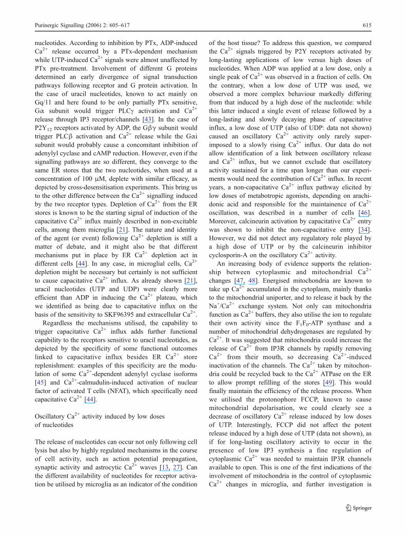
of the host tissue? To address this question, we comparedthe Ca2+ signals triggered by P2Y receptors activated bylong-lasting applications of low versus high doses ofnucleotides. When ADP was applied at a low dose, only asingle peak of Ca2+ was observed in a fraction of cells. Onthe contrary, when a low dose of UTP was used, weobserved a more complex behaviour markedly differingfrom that induced by a high dose of the nucleotide: whilethis latter induced a single event of release followed by along-lasting and slowly decaying phase of capacitativeinflux, a low dose of UTP (also of UDP: data not shown)caused an oscillatory Ca2+ activity only rarely super-imposed to a slowly rising Ca2+ influx. Our data do notallow identification of a link between oscillatory releaseand Ca2+ influx, but we cannot exclude that oscillatoryactivity sustained for a time span longer than our experi-ments would need the contribution of Ca2+ influx. In recentyears, a non-capacitative Ca2+ influx pathway elicited bylow doses of metabotropic agonists, depending on arachi-donic acid and responsible for the maintainence of Ca2+
oscillation, was described in a number of cells [46].Moreover, calcineurin activation by capacitative Ca2+ entrywas shown to inhibit the non-capacitative entry [34].However, we did not detect any regulatory role played bya high dose of UTP or by the calcineurin inhibitorcyclosporin-A on the oscillatory Ca2+ activity.
An increasing body of evidence supports the relation-ship between cytoplasmic and mitochondrial Ca2+
changes [47, 48]. Energised mitochondria are known totake up Ca2+ accumulated in the cytoplasm, mainly thanksto the mitochondrial uniporter, and to release it back by theNa+/Ca2+ exchange system. Not only can mitochondriafunction as Ca2+ buffers, they also utilise the ion to regulatetheir own activity since the F1F0-ATP synthase and anumber of mitochondrial dehydrogenases are regulated byCa2+. It was suggested that mitochondria could increase therelease of Ca2+ from IP3R channels by rapidly removingCa2+ from their mouth, so decreasing Ca2+-inducedinactivation of the channels. The Ca2+ taken by mitochon-dria could be recycled back to the Ca2+ ATPase on the ERto allow prompt refilling of the stores [49]. This wouldfinally maintain the efficiency of the release process. Whenwe utilised the protonophore FCCP, known to causemitochondrial depolarisation, we could clearly see adecrease of oscillatory Ca2+ release induced by low dosesof UTP. Interestingly, FCCP did not affect the potentrelease induced by a high dose of UTP (data not shown), asif for long-lasting oscillatory activity to occur in thepresence of low IP3 synthesis a fine regulation ofcytoplasmic Ca2+ was needed to maintain IP3R channelsavailable to open. This is one of the first indications of theinvolvement of mitochondria in the control of cytoplasmicCa2+ changes in microglia, and further investigation is
Purinergic Signalling (2006) 2: 605–617 615

needed to come to an unambiguous interpretation of thisresult since FCCP can have a number of outcomes togetherwith the decrease of Ca2+ uptake by mitochondria, such asATP depletion and pH changes. In any case, it isnoteworthy that during microglia activation, mitochondriachange their cellular distribution, morphology and internalorganisation, probably to better sustain energy-consumingfunctions of activated microglia [50].
In this study, we pointed out the capability of P2Yreceptors to cause different patterns of cytoplasmic Ca2+
changes involving capacitative Ca2+ influx and intracellularCa2+ mobilisation, depending not only on the type but alsoon the concentration of the agonist. Intracellular Ca2+
changes can independently affect a multitude of functionsand events depending on the vicinity of the Ca2+ sources tospecific target proteins, on the concentration reached andalso on the time evolution of the Ca2+ variations. It isknown that sustained and oscillatory Ca2+ changes candifferently affect target proteins (for a review, see 51).Protein kinases, phospholipases and transcription factorsare examples of the multitude of proteins that can bedifferently regulated by Ca2+.
In conclusion we speculate that the different patternsof Ca2+ signals elicited by different concentrations ofnucleotides would render P2Y receptors capable of signal-ling to microglia the state of the host tissue and, hopefully,regulate microglial responses in concert with the needs ofthe tissue.
Acknowledgements The authors thank Mrs. Estella Sansonetti forexcellent technical and editorial assistance. This work was supportedby the Italian Ministry of Education, University and Research (FIRB-MIUR, Grant no. H91/1: “Synaptic plasticity and brain repair.”
References
1. Kreutzberg GW (1996) Microglia: a sensor for pathological eventsin the CNS. Trends Neurosci 19:312–318
2. Raivich G, Bohatschek M, Kloss CU, Werner A, Jones LL,Kreutzberg GW (1999) Neuroglial activation repertoire in theinjured brain: graded response, molecular mechanisms and cues tophysiological function. Brain Res Brain Res Rev 30:77–105
3. Visentin S, Agresti C, Patrizio M, Levi G (1995) Ion channels inrat microglia and their different sensitivity to lipopolysaccharideand interferon-gamma. J Neurosci Res 42:439–451
4. De Simone R, Ajmone-Cat MA, Minghetti L (2004) Atypicalantiinflammatory activation of microglia induced by apoptoticneurons: possible role of phosphatidylserine phosphatidylserinereceptor interaction. Mol Neurobiol 29(2):197–212
5. Minghetti L (2005) Role of inflammation in neurodegenerativediseases. Curr Opin Neurol 18(3):315–321
6. Franke H, Illes P (2006) Involvement of P2 receptors in thegrowth and survival of neurons in the CNS. Pharmacol Ther 109(3):297–324
7. Dave S, Mogul DJ (1996) ATP receptor activation potentiates avoltage-dependent Ca channel in hippocampal neurons. Brain Res715(1–2):208–216
8. Ikeuchi Y, Nishizaki T (1995) Dual effects of ATP on thepotassium channel and intracellular Ca2+ release in smooth musclecells of the bovine brain arteries. Biochem Biophys Res Commun215(3):1071–1077
9. Wirkner K, Koles L, Thummler S, Luthardt J, Poelchen W, FrankeH, Furst S, Illes P (2002) Interaction between P2Y and NMDAreceptors in layer V pyramidal neurons of the rat prefrontal cortex.Neuropharmacology 42(4):476–488
10. Abbracchio MP, Saffrey MJ, Hopker V, Burnstock G (1994)Modulation of astroglial cell proliferation by analogues ofadenosine and ATP in primary cultures of rat striatum. Neurosci-ence 59(1):67–76
11. Ciccarelli R, Di Iorio P, Ballerini P, Ambrosini G, Giuliani P,Tiboni GM, Caciagli F (1994) Effects of exogenous ATP andrelated analogues on the proliferation rate of dissociated primarycultures of rat astrocytes. J Neurosci Res 39(5):556–566
12. Franke H, Krugel U, Illes P (1999) P2 receptor-mediatedproliferative effects on astrocytes in vivo.Glia 28(3):190–200
13. Venance L, Stella N, Glowinski J, Giaume C (1997) Mechanisminvolved in initiation and propagation of receptor-inducedintercellular calcium signaling in cultured rat astrocytes. J Neurosci17(6):1981–1992
14. Fields RD, Stevens B (2000) ATP: an extracellular signallingmolecule between neurons and glia. Trends Neurosci 23:625–633
15. Agresti C, Meomartini ME, Amadio S, Ambrosini E, Volonte C,Aloisi F, Visentin S (2005) ATP regulates oligodendrocyteprogenitor migration, proliferation, and differentiation: involve-ment of metabotropic P2 receptors. Brain Res Brain Res Rev 48(2):157–165
16. Ferrari D, Chiozzi P, Falzoni S, Hanau S, Di Virgilio F (1997)Purinergic modulation of interleukin-1 beta release from micro-glial cells stimulated with bacterial endotoxin. J Exp Med 185(3):579–582
17. Hide I, Tanaka M, Inoue A, Nakajima K, Kohsaka S, Inoue K,Nakata Y (2000) Extracellular ATP triggers tumor necrosis factor-alpha release from rat microglia. J Neurochem 75(3):965–972
18. Ferrari D, Chiozzi P, Falzoni S, Dal Susino M, Collo G, Buell G,Di Virgilio F (1997) ATP-mediated cytotoxicity in microglialcells. Neuropharmacology 36(9):1295–1301
19. Visentin S, Renzi M, Frank C, Greco A, Levi G (1999) Twodifferent ionotropic receptors are activated by ATP in rat micro-glia. J Physiol 519(Pt 3):723–736
20. Tsuda M, Shigemoto-Mogami Y, Koizumi S, Mizokoshi A,Kohsaka S, Salter MW, Inoue K (2003) P2X4 receptors inducedin spinal microglia gate tactile allodynia after nerve injury. Nature424(6950):778–783
21. Toescu EC, Moller T, Kettenmann H, Verkhratsky A (1998) Long-term activation of capacitative Ca2+ entry in mouse microglialcells. Neuroscience 86(3):925–935
22. Moller T, Kann O, Verkhratsky A, Kettenmann H (2000)Activation of mouse microglial cells affects P2 receptor signaling.Brain Res 853(1):49–59
23. Honda S, Sasaki Y, Ohsawa K, Imai Y, Nakamura Y, Inoue K,Kohsaka S (2001) Extracellular ATP or ADP induce chemotaxisof cultured microglia through Gi/o-coupled P2Y receptors. JNeurosci 21(6):1975–1982
24. Ogata T, Chuai M, Morino T, Yamamoto H, Nakamura Y,Schubert P (2003) Adenosine triphosphate inhibits cytokinerelease from lipopolysaccharide-activated microglia via P2yreceptors. Brain Res 981(1–2):174–183
616 Purinergic Signalling (2006) 2: 605–617

25. Seo DR, Kim KY, Lee YB (2004) Interleukin-10 expression inlipopolysaccharide-activated microglia is mediated by extracellu-lar ATP in an autocrine fashion. Neuroreport 15(7):1157–1161
26. Shigemoto-Mogami Y, Koizumi S, Tsuda M, Ohsawa K, KohsakaS, Inoue K (2001) Mechanisms underlying extracellular ATP-evoked interleukin-6 release in mouse microglial cell line, MG-5.J Neurochem 78(6):1339–1349
27. Verderio C, Matteoli M (2001) ATP mediates calcium signalingbetween astrocytes and microglial cells: modulation by IFN-gamma. J Immunol 166(10):6383–6391
28. Zimmermann H (2000) Extracellular metabolism of ATP andother nucleotides. Naunyn Schmiedebergs Arch Pharmacol 362(4–5):299–309
29. Braun N, Zhu Y, Krieglstein J, Culmsee C, Zimmermann H (1998)Upregulation of the enzyme chain hydrolyzing extracellular ATP aftertransient forebrain ischemia in the rat. J Neurosci 18(13):4891–4900
30. Gallucci S, Matzinger P (2001) Danger signals: SOS to theimmune system. Curr Opin Immunol 13(1):114–119
31. la Sala A, Ferrari D, Di Virgilio F, Idzko M, Norgauer J,Girolomoni G (2003) Alerting and tuning the immune response byextracellular nucleotides. J Leukoc Biol 73(3):339–343
32. Levi G, Patrizio M, Bernardo A, Petrucci TC, Agresti C (1993)Human immunodeficiency virus coat protein gp120 inhibits thebeta-adrenergic regulation of astroglial and microglial functions.Proc Natl Acad Sci USA 90(4):1541–1545
33. Grynkiewicz G, Poenie M, Tsien RY (1985) A new generation ofCa2+ indicators with greatly improved fluorescence properties. JBiol Chem 260(6):3440–3450
34. Mignen O, Thompson JL, Shuttleworth TJ (2003) Calcineurindirects the reciprocal regulation of calcium entry pathways innonexcitable cells. J Biol Chem 278(41):40088–40096
35. Hoth M, Fanger CM, Lewis RS (1997) Mitochondrial regulationof store-operated calcium signaling in T lymphocytes. J Cell Biol137(3):633–648
36. Kettenmann H, Banati R, Walz W (1993) Electrophysiologicalbehavior of microglia. Glia 7(1):93–101
37. Norenberg W, Langosch JM, Gebicke-Haerter PJ, Illes P (1994)Characterization and possible function of adenosine 5′-triphos-phate receptors in activated rat microglia. Br J Pharmacol 111(3):942–950
38. Langosch JM, Gebicke-Haerter PJ, Norenberg W, Illes P (1994)Characterization and transduction mechanisms of purinoceptors inactivated rat microglia. Br J Pharmacol 113(1):29–34
39. Bianco F, Fumagalli M, Pravettoni E, D_Ambrosi N, Volonte C,
Matteoli M, Abbracchio MP, Verderio C (2005) Pathophysiolog-ical roles of extracellular nucleotides in glial cells: differentialexpression of purinergic receptors in resting and activated micro-glia. Brain Res Brain Res Rev 48(2):144–156
40. Hollopeter G, JantzenHM,Vincent D, Li G, England L, RamakrishnanV, Yang RB, Nurden P, Nurden A, Julius D, Conley PB (2001)Identification of the platelet ADP receptor targeted by antithromboticdrugs. Nature 409(6817):202–207
41. Sasaki Y, Hoshi M, Akazawa C, Nakamura Y, Tsuzuki H, InoueK, Kohsaka S (2003) Selective expression of Gi/o-coupled ATPreceptor P2Y12 in microglia in rat brain. Glia 44(3):242–250
42. Abbracchio MP, Boeynaems JM, Barnard EA, Boyer JL, KennedyC, Miras-Portugal MT, King BF, Gachet C, Jacobson KA, WeismanGA, Burnstock G (2003) Characterization of the UDP-glucosereceptor (re-named here the P2Y14 receptor) adds diversity to theP2Y receptor family. Trends Pharmacol Sci 24(2):52–55
43. Communi D, Janssens R, Suarez-Huerta N, Robaye B, BoeynaemsJM (2000) Advances in signalling by extracellular nucleotides: therole and transduction mechanisms of P2Y receptors. Cell Signal12(6):351–360
44. Venkatachalam K, van Rossum DB, Patterson RL, Ma HT, GillDL (2002) The cellular and molecular basis of store-operatedcalcium entry. Nat Cell Biol 4(11):E263–E272
45. Fagan KA, Mons N, Cooper DM (1998) Dependence of the Ca2+-inhibitable adenylyl cyclase of C6-2B glioma cells on capacitativeCa2+ entry. J Biol Chem 273(15):9297–9305
46. Shuttleworth TJ, Mignen O (2003) Calcium entry and the controlof calcium oscillations. Biochem Soc Trans 31(Pt 5):916–919
47. Brookes PS, Yoon Y, Robotham JL, Anders MW, Sheu SS (2004)Calcium, ATP, and ROS: a mitochondrial love-hate triangle. Am JPhysiol Cell Physiol 287(4):C817–C833
48. Duchen MR (2004) Mitochondria in health and disease: perspectiveson a new mitochondrial biology. Mol Aspects Med 25(4):365–451
49. Poburko D, Kuo KH, Dai J, Lee CH, van Breemen C (2004)Organellar junctions promote targeted Ca2+ signaling in smoothmuscle: why two membranes are better than one. TrendsPharmacol Sci 25(1):8–15
50. Banati RB, Egensperger R, Maassen A, Hager G, Kreutzberg GW,Graeber MB (2004) Mitochondria in activated microglia in vitro. JNeurocytol 33(5):535–541
51. Johnson JD, Chang JP (2000) Function- and agonist-specific Ca2+
signalling: the requirement for and mechanism of spatial andtemporal complexity in Ca2+ signals. Biochem Cell Biol 78(3):217–240
Purinergic Signalling (2006) 2: 605–617 617
Copyright © 2022 FDOKUMEN