Different Cellular and Genetic Basis of Noise-Related Endocochlear Potential Reduction in CBA/J and...
14
Different Cellular and Genetic Basis of Noise-Related Endocochlear Potential Reduction in CBA/J and BALB/cJ Mice KEVIN K. OHLEMILLER 1,3 ,ALLYSON D. ROSEN 3 ,ERIN A. RELLINGER 3 ,SCOTT C. MONTGOMERY 2 , AND P ATRICIA M. GAGNON 1 , 1 Department of Otolaryngology, Washington University School of Medicine, 660 S. Euclid, St. Louis, MO 63110, USA 2 Department of Otolaryngology, Saint Louis University School of Medicine, St. Louis, MO, USA 3 Program in Audiology and Communication Sciences, Washington University School of Medicine, St. Louis, MO, USA Received: 28 July 2010; Accepted: 20 September 2010; Online publication: 5 October 2010 ABSTRACT The acute and permanent effects of noise exposure on the endocochlear potential (EP) and cochlear lateral wall were evaluated in BALB/cJ (BALB) inbred mice, and compared with CBA/J (CBA) and C57BL/6 (B6) mice. Two-hour exposure to broadband noise (4–45 kHz) at 110 dB SPL leads to a ~50 mV reduction in the EP in BALB and CBA, but not B6. EP reduction in BALB and CBA is reliably associated with characteristic acute cellular pathology in stria vascularis and spiral ligament. By 8 weeks after exposure, the EP in CBA mice has returned to normal. In BALBs, however, the EP remains depressed by an average ~10 mV, so that permanent EP reduction contributes to permanent threshold shifts in these mice. We recently showed that the CBA noise phenotype in part reflects the influence of a large effect quantitative trait locus on Chr. 18, termed Nirep (Ohlemiller et al., Hear Res 260:47–53, 2010b). While CBA “EP susceptibility” alleles are dominant to those in B6, examination of (B6×BALB) F1 hybrid mice and (F1×BALB) N2 backcross mice revealed that noise-related EP reduction and associated cell pathology in BALBs are inherited in an autosomal recessive manner, and are dependent on multiple genes. Moreover, while N2 mice formed from B6 and CBA retain strong correspondence between acute EP reduction, ligament pathology, and strial pathology, N2s formed from B6 and BALB include subsets that dissociate pathology of ligament and stria. We conclude that the genes and cascades that govern the very similar EP susceptibility phenotypes in BALB and CBA mice need not be the same. BALBs appear to carry alleles that promote more pronounced long term effects of noise on the lateral wall. Separate loci in BALBs may preferentially impact stria versus ligament. Keywords: stria vascularis, spiral ligament, spiral limbus, Reissner’s membrane, fibrocytes, C57BL/6, noise-induced hearing loss, presbycusis INTRODUCTION From decades of study of noise-induced hearing loss (NIHL), some consensus has emerged concerning both the reversible cochlear changes underlying the transient component of hearing loss, and irreversible changes associated with permanent hearing loss. Reversible changes may include altered spatial relations within the organ of Corti (Beagley 1965; Harding et al. 1992; Nordmann et al. 2000), injury to hair cell stereocilia (Liberman and Kiang 1978; Liberman and Mulroy 1982; Liberman and Dodds 1984), and transient depression of the endocochlear potential (EP) (Syka et al. 1981). Critical irreversible changes (those accounting for permanent hearing loss) have appeared concentrated in the organ of Corti. These include hair cell loss and non-lethal hair cell injury (e.g., Covell 1953; Johnson and Hawkins 1976; Hamernik et al. 1989; Ou et al. 2000b; Wang et al. 2002) as well as newly recognized delayed and progressive Correspondence to : Kevin K. Ohlemiller & Department of Otolaryngol- ogy & Washington University School of Medicine & 660 S. Euclid, St. Louis, MO 63110, USA. Telephone: +1-314-7477179; fax: +1-314- 7477230; email: [email protected] JARO 12: 45–58 (2011) DOI: 10.1007/s10162-010-0238-z D 2010 Association for Research in Otolaryngology 45 JARO Journal of the Association for Research in Otolaryngology
Transcript of Different Cellular and Genetic Basis of Noise-Related Endocochlear Potential Reduction in CBA/J and...

Different Cellular and Genetic Basis of Noise-RelatedEndocochlear Potential Reduction in CBA/J and BALB/cJ Mice
KEVIN K. OHLEMILLER1,3, ALLYSON D. ROSEN
3, ERIN A. RELLINGER3, SCOTT C. MONTGOMERY
2, ANDPATRICIA M. GAGNON
1,1Department of Otolaryngology, Washington University School of Medicine, 660 S. Euclid, St. Louis, MO 63110, USA2Department of Otolaryngology, Saint Louis University School of Medicine, St. Louis, MO, USA3Program in Audiology and Communication Sciences, Washington University School of Medicine, St. Louis, MO, USA
Received: 28 July 2010; Accepted: 20 September 2010; Online publication: 5 October 2010
ABSTRACT
The acute and permanent effects of noise exposureon the endocochlear potential (EP) and cochlearlateral wall were evaluated in BALB/cJ (BALB) inbredmice, and compared with CBA/J (CBA) and C57BL/6(B6) mice. Two-hour exposure to broadband noise(4–45 kHz) at 110 dB SPL leads to a ~50 mVreduction in the EP in BALB and CBA, but not B6.EP reduction in BALB and CBA is reliably associatedwith characteristic acute cellular pathology in striavascularis and spiral ligament. By 8 weeks afterexposure, the EP in CBA mice has returned tonormal. In BALBs, however, the EP remainsdepressed by an average ~10 mV, so that permanentEP reduction contributes to permanent thresholdshifts in these mice. We recently showed that theCBA noise phenotype in part reflects the influence ofa large effect quantitative trait locus on Chr. 18,termed Nirep (Ohlemiller et al., Hear Res 260:47–53,2010b). While CBA “EP susceptibility” alleles aredominant to those in B6, examination of (B6×BALB)F1 hybrid mice and (F1×BALB) N2 backcross micerevealed that noise-related EP reduction and associatedcell pathology in BALBs are inherited in an autosomalrecessive manner, and are dependent on multiplegenes. Moreover, while N2 mice formed from B6 andCBA retain strong correspondence between acute EPreduction, ligament pathology, and strial pathology, N2sformed from B6 and BALB include subsets that
dissociate pathology of ligament and stria. We concludethat the genes and cascades that govern the very similarEP susceptibility phenotypes in BALB and CBA miceneed not be the same. BALBs appear to carry alleles thatpromote more pronounced long term effects of noiseon the lateral wall. Separate loci in BALBs maypreferentially impact stria versus ligament.
Keywords: stria vascularis, spiral ligament, spirallimbus, Reissner’s membrane, fibrocytes, C57BL/6,noise-induced hearing loss, presbycusis
INTRODUCTION
From decades of study of noise-induced hearing loss(NIHL), some consensus has emerged concerningboth the reversible cochlear changes underlying thetransient component of hearing loss, and irreversiblechanges associated with permanent hearing loss.Reversible changes may include altered spatialrelations within the organ of Corti (Beagley 1965;Harding et al. 1992; Nordmann et al. 2000), injuryto hair cell stereocilia (Liberman and Kiang 1978;Liberman and Mulroy 1982; Liberman and Dodds1984), and transient depression of the endocochlearpotential (EP) (Syka et al. 1981). Critical irreversiblechanges (those accounting for permanent hearing loss)have appeared concentrated in the organ of Corti.These include hair cell loss and non-lethal hair cellinjury (e.g., Covell 1953; Johnson and Hawkins 1976;Hamernik et al. 1989; Ou et al. 2000b; Wang et al. 2002)as well as newly recognized delayed and progressive
Correspondence to: Kevin K. Ohlemiller & Department of Otolaryngol-ogy & Washington University School of Medicine & 660 S. Euclid, St.Louis, MO 63110, USA. Telephone: +1-314-7477179; fax: +1-314-7477230; email: [email protected]
JARO 12: 45–58 (2011)DOI: 10.1007/s10162-010-0238-zD 2010 Association for Research in Otolaryngology
45
JAROJournal of the Association for Research in Otolaryngology

injury to afferent neurons (Kujawa and Liberman 2009).The latter may not contribute to hearing loss perse, but rather to supra-threshold deficits. PermanentEP reduction has not generally been held to be acommon contributor to permanent NIHL. EPreduction by noise may reflect two very differentmechanisms, depending on the severity of the noiseand the model. Traumatic noise that tears Reissner’smembrane or breaches the reticular lamina can reducethe EP by eliminating the required ion gradients.Reversible EP reduction by noise that does not violatethe boundaries of scalamedia has been demonstrated insome mouse strains (CBA/J and CBA/CaJ) and notothers (C57BL/6J, or B6), and thus to reflect geneticallymodifiable factors intrinsic to the lateral wall (Hiroseand Liberman 2003; Ohlemiller and Gagnon 2007).Following a single moderate noise exposure (4–45 kHz,110 dB sound pressure level (SPL), 2 h), CBA/J mice(henceforth CBA) show a 40–50 mV reversible EPreduction that is reliably accompanied by certain formsof strial and ligament pathology. For the same exposure,B6 mice show neither EP reduction nor significantlateral wall pathology. We recently showed thatdifferences in the vulnerability of the EP betweenCBA and B6 mice in part reflect the influence of amajor effect quantitative trait locus (QTL) on Chr.18, termed Nirep (Ohlemiller et al. 2010b). Nirep wasfound to account for about 12% of EP variance,with an effect size of ~19 mV. Here we show that,like CBAs, BALB/cJ (BALB) mice also show EPreduction following the same noise exposure.Although the acute cellular pathology associatedwith EP reduction in BALBs is quite similar to thatin CBAs, BALBs show several major differences.Among these are a different mode of inheritance,clear involvement of multiple loci, and dissociabilityof injury to stria vascularis and spiral ligament. Mostsignificantly, unlike CBA the EP remains somewhatdepressed in BALBs 8 weeks after noise exposure.Thus, BALBs appear to carry alleles that promotelong term effects of noise on the cochlear lateralwall. Similar alleles and influences may operate inthe human cochlea, and could impact the longterm stability of NIHL and the manifestation ofpresbycusis.
MATERIALS AND METHODS
Animals
All procedures were approved by the WashingtonUniversity Institutional Animal Care andUseCommittee.The basic procedure involved exposing inbred, hybrid,and backcross mice one time to broadband noisefollowed by hearing assessment, recording of the EP,and sacrifice for histology either 1–3 h, 24 h, or 8 weeks
after exposure. Mice were 3–4 months of age at the timeof exposure. All groups were roughly evenly balanced bygender and no gender effects were evident in any ofthe data we present. Mice were purchased directlyfrom The Jackson Laboratory (JAX) or were derivedfrom breeders purchased from JAX. Subjects included66 BALB/cJ, 29 CBA/J (CBA), and 26 C57BL/6J (B6),and 27 B6.CAST-Cdh23CAST (B6.CAST) mice. Becausewe previously showed that the effect of noise on the EPwas the same in B6 and B6.CAST mice (that is, theCdh23ahl allele does not play a role) (Ohlemiller andGagnon 2007), data from B6 and B6.CAST werecombined for assessment of acute threshold and EPchanges. Also included were data from 36 (B6×CBA) F1hybrids, 55 (B6×BALB) F1 hybrids and 56 [(B6×BALB)×BALB] N2 backcross mice.
Noise exposure
Noise exposures were performed in a foam-lined,single-walled soundproof room. The noise exposureapparatus consisted of a 21×21×11 cm wire cagemounted on a pedestal inserted into a B&K 3921turntable. To ensure a uniform sound field, the cagewas rotated at 1 revolution/80 s within a 42×42 cmmetal frame. A Motorola KSN1020A piezo ceramicspeaker (four total) was attached to each side of theframe. Opposing speakers were displaced so as notto lie on the same axis and driven by separatechannels of a Crown D150A power amplifier.Broadband noise was generated by General Radio1310 generators and band-passed at 4–45 kHz byKrohn–Hite 3550 filters. Noise levels at various points inthe exposure cage, measured using a B&K 4135 1/4inch microphone in combination with a B&K 2231sound level meter, ranged from 110–113 dB SPL. Micewere exposed in pairs for 2 h.
CAP recording
Compound action potential (CAP) recordings wereconducted as terminal procedures, so that each animalwas assessed once immediately prior to sacrifice.Animals were anesthetized (60 mg/kg sodiumpentobarbital, IP) and positioned ventrally in a customheadholder. Core temperature was maintained at 37.5±1.0°C using a thermostatically controlled heating padin conjunction with a rectal probe (Yellow SpringsInstruments Model 73A). An incision was madealong the midline of the neck and soft tissues wereblunt dissected and displaced laterally to expose thetrachea and animal’s left bulla. A tracheostomy wasthen made and the musculature over the bulla wascut posteriorly to expose the bone overlying theround window. Using a hand drill, a small hole wasmade over the round window. The recording
46 OHLEMILLER ET AL.: Noise-Related Endocochlear Potential Reduction in Mice

electrode was a modified platinum needle electrode(Grass) insulated with epoxy except for the tip,which was inserted into round window antrum usinga micromanipulator. Additional platinum electrodesinserted into the neck musculature and hind leg servedas reference and ground, respectively. Electrodes wereled to a Grass P15 differential amplifier (100–3,000 Hz, ×100), then to a custom amplifierproviding another ×1,000 gain, then digitized at30 kHz using a Cambridge Electronic DesignMicro1401 in conjunction with SIGNAL™ andcustom signal averaging software operating on a120 MHz Pentium PC. Sinewave stimuli generatedby a Hewlett Packard 3325A oscillator were shapedby a custom electronic switch to 5 ms total duration,including 1 ms rise/fall times. The stimulus wasamplified by a Crown D150A power amplifier andoutput to a KSN1020A piezo ceramic speakerlocated 7 cm directly lateral to the left ear. Stimuliwere presented freefield and calibrated using aB&K 4135 1/4 inch microphone placed where themouse’s ear would normally be for recording.Toneburst stimuli at each frequency and level werepresented 100 times at 3/s. The minimum soundpressure level required for visual detection of aresponse (N1) was determined at 5, 10, 20, 28.3,and 40 kHz, using a 5 dB minimum step size.
Endocochlear potential recording
The EP was measured using a ventral approachimmediately after CAP recording. Using a fine drill,a hole was made in the left cochlear capsule directlyover scala media of the lower basal turn. Undernormal conditions there exists in mouse cochleae a9–10 mV EP gradient favoring the base (Ohlemilleret al. 2006). When the EP is acutely reduced bynoise (Ohlemiller and Gagnon 2007), or chroni-cally reduced with age (Ohlemiller et al. 2010a),this gradient may be reversed, resulting in slightlyhigher EPs (≤5 mV) in the apex. Thus the use ofthe lower basal turn EP in the present workprovides a reasonable indication of the extent ofEP reduction by noise throughout the cochlea.Glass capillary pipettes (40–80 MΩ) filled with0.15 M KCl were mounted on a hydraulic micro-drive (Frederick Haer) and advanced until a stablepositive potential was observed that did not changewith increased electrode depth. The signal from therecording electrode was led to an AM SystemsModel 1600 intracellular amplifier. EP changes bycondition (Fig. 3) were compared within each strainby one-way analysis of variance (ANOVA) usingyoung unexposed mice as controls. Sample sizes bystrain and condition varied from six to 20.
Tissue processing for histology
At the end of recording, animals were overdosedand perfused transcardially with cold 2.0% parafor-maldehyde/2.0% glutaraldehyde in 0.1 M phos-phate buffer (pH 7.4). Each cochlea was rapidlyisolated, immersed in the same fixative, and thestapes was immediately removed. Complete infiltrationof the cochlea by fixative was ensured by making asmall hole at the apex of the cochlear capsule, andgently circulating the fixative over the cochlea usinga transfer pipet. After decalcification in sodiumEDTA for 72 h, cochleas were post-fixed in buffered1% osmium tetroxide, dehydrated in an ascendingacetone series, and embedded in Epon. Cochleaswere sectioned in the mid-modiolar plane at4.0 μm, then stained with toluidine blue for brightfield viewing with a Nikon Optiphot™ light microscopeusing a calibrated grid ocular.
Morphometric analysis
Analyses were based on left cochleas, from which allrecordings were obtained. Tissue sections were scoredblindly. Since the mouse cochlea features just over twocomplete turns (Bohne et al. 2001), a typical “core”mid-modiolar section shows four profiles of the organof Corti and lateral wall: lower base, upper base, lowerapex, and upper apex (which sometimes appears onlyin partial profile). Quantitative assessment of cochleasfocused on lateral wall, Reissner’s membrane, andspiral limbus of the upper basal turn in a regionexpected to correspond to 10 kHz (±0.5 octave) (Ouet al. 2000a). This was done to avoid possible artifactsassociated with electrode entry in the lower basal turn.Changes we characterize in the upper basal turn werequalitatively similar elsewhere, although generallymore extensive in the lower base than in the apex.Metrics obtained depended upon whether a cochleawas taken for examination 1–3 h after noise (in whichcase they were designed to detect changes in theappearance of cells and structures, not cell loss), or8 weeks after noise (wherein metrics were aimed atcell loss). Examination of cochleas taken 24 h afterexposure was purely qualitative. DIC images used forillustration were obtained on a Zeiss LSM 700 laserscanning confocal microscope using ZEN™ software,then further processed using CANVAS™.
Assessment of acute noise injury (1–3 h after noise)
Quantitative assessment of acute noise injury wasbased on 8 noise-exposed BALB mice, seven non-exposed controls, and 21 exposed N2 backcrossmice. Every fifth section was analyzed through adistance of 184 μm for a total of ten sections ineach animal. Acute pathology was characterized
OHLEMILLER ET AL.: Noise-Related Endocochlear Potential Reduction in Mice 47

non-parametrically using subjective judgments of“normal” or “abnormal” applied to stria vascularis,Reissner’s membrane, spiral ligament, and spirallimbus. Determination depended upon the clearpresence of cell shrinkage, cell vacuolization, voidspaces between cells, nuclear condensation, ornuclear enlargement. The cells upon which wefocused were based on our previous observationsin CBA/J and CBA/CaJ mice (Ohlemiller and Gagnon2007), and included all strial cell types, types I, II, IV, andV fibrocytes in ligament, mesothelial cells of Reissner’s,and fibrocytes in the central zone of spiral limbus. Theoverall proportion of sections scored “normal” wascompared in exposed and non-exposed BALBs by z test(Fig. 6), or calculated for each animal and tested forcorrelation with EP or other metrics across animals(Figs. 12 and 13).
Assessment of permanent noise injury (8 week after noise)
Quantitative assessment of permanent noise injury wasbased on 8 BALB mice 6–7 months old at time ofexamination and seven age-matched non-exposed con-trols. Every fifth section was analyzed through a distance
FIG. 1. A Mean±SD CAP thresholds for noise-exposed andunexposed control BALB, B6, and (B6×BALB) F1 mice. B Mean±SD CAP thresholds only for control BALB mice and BALB at varioustimes after noise. Unexposed control and acute exposed mice (1–3 hafter noise) are replotted from A to facilitate comparison. All noiseexposures were 2 h, 4–45 kHz, 110 dB SPL. NR no response at highestsound level available.
FIG. 2. A Typical cochlear duct of the upper basal turn in aBALB mouse 1–3 h after noise. B Expanded view of box centeredon spiral limbus in (A). Stellate fibrocytes of limbus show subtlevacuolization (arrowhead). C Expanded view of organ of Corti.Pillars are bent and Nuel’s space is collapsed (arrowhead), but nobreach of reticular lamina is apparent. SpL spiral ligament, StVstria vascularis, SV scala vestibuli, ST scala tympani, SM scalamedia, spLim spiral limbus, RM Reissner’s membrane, SpG spiralganglion, TM tectorial membrane.
FIG. 3. Mean±SD basal turn EP in unexposed controls and atvarious times after noise exposure in inbred BALB, B6, and CBA/Jmice, and in F1 hybrids formed from crossing BALB and CBA withB6. EP reduction by noise is dominant versus B6 in CBA; recessive inBALB. EP does not completely recover in BALBs. Asterisk Signifi-cantly different from unexposed controls of same strain by one-wayANOVA.
48 OHLEMILLER ET AL.: Noise-Related Endocochlear Potential Reduction in Mice
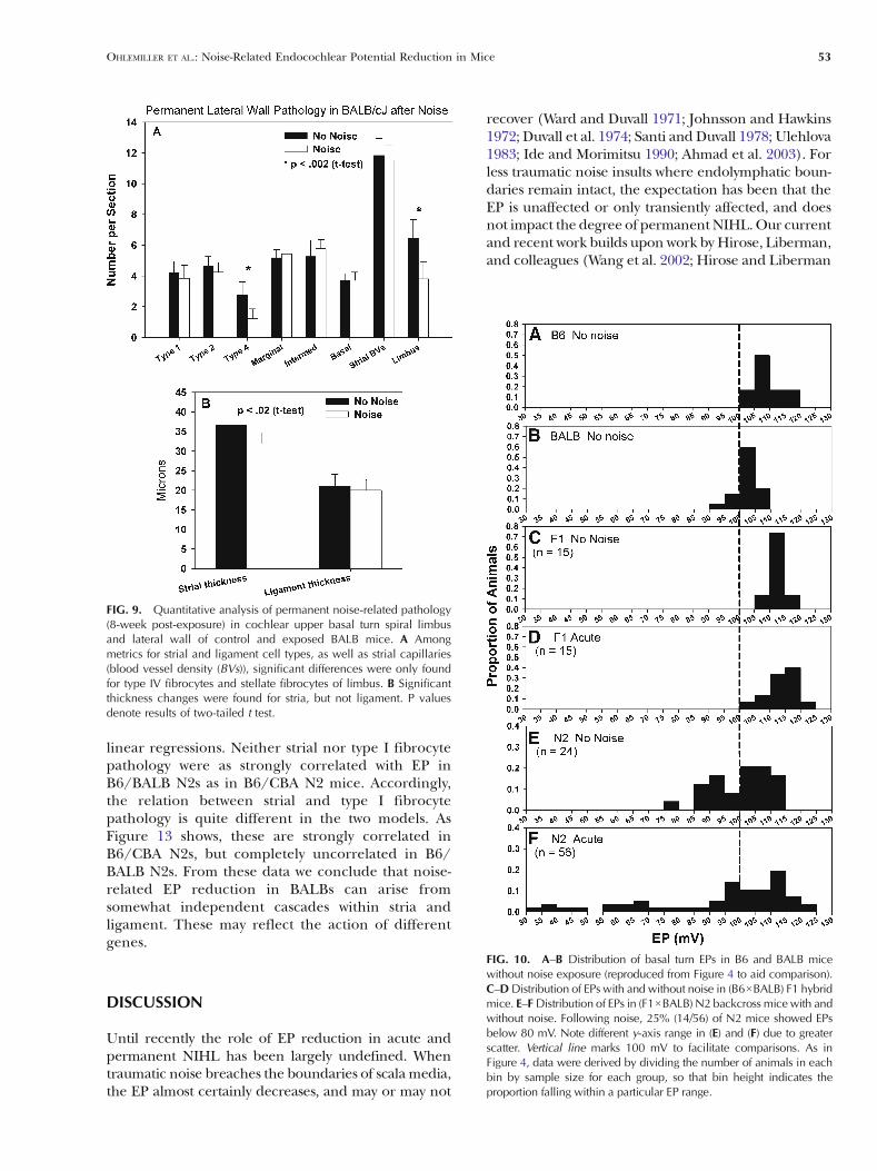
of 184 μm for a total of ten sections per animal.Quantitative measures included strial thickness, strialmarginal cell density, intermediate cell density, basal celldensity, ligament thickness, density of types I, II, and IVfibrocytes in the ligament, and density of fibrocytes inadjacent spiral limbus. Only nucleated profiles wereincluded in cell counts. Strial thickness was measuredorthogonal to the midpoint. Marginal cells, intermedi-ate cells, and basal cells were counted in an 80 μm linearsegment of stria, centered at the midpoint. No attemptwas made to distinguish between lower and upper levelintermediate cells, a distinction recently made bySchulte and Spicer (Spicer and Schulte 2005b). Liga-
ment thickness was measured on an axis co-linear withthe strial midline. Types I, II, and IV fibrocytes werecounted in a 1,600 μm2 area. These were identifiedbased on location, an approach taken in previousstudies (Hequembourg and Liberman 2001; Lang etal. 2002; Hirose and Liberman 2003). Fibrocytes inspiral limbus were also counted in a roughly centered1,600 μm2 area, expected to be populated principally bystellate cells (Kimura et al. 1990). For each metric, theten estimates were averaged to yield an overall averagefor each animal. Parametric data from noise-exposedanimals were compared with non-exposed controls ofthe same strain by t test (Fig. 9).
RESULTS
CAP thresholds in inbred and F1 mice
Noise exposure led to nearly complete loss of CAPresponses above 5 kHz in both BALB and B6mice in theacute period after noise (Fig. 1A). F1 hybrids formedfromB6 and BALB seemed to gain some resistance, withmost F1s exhibiting recordable responses at all frequen-cies. While threshold shifts are not the focus of thispaper, we considered to what extent acute thresholdshifts in Figure 1Amay reflect the EP changes describedbelow. As we will show, acute threshold differencesbetween B6 and F1 mice cannot reflect EP differences,as neither of these exhibit EP reduction after noise.Note, however, that residual hearing at 5 kHz was~30 dB better in B6 and F1 hybrid mice than in BALBmice, in which we demonstrate EP reduction. Wepreviously showed that the dependence of thresholdson the EP after noise exposure in our mice is about−0.4 dB/mV at most test frequencies (Ohlemiller andGagnon 2007; Ohlemiller 2009), compared with` slopescloser to −1.0 dB/mVobtained using furosemide in catsand gerbils (Sewell 1984; Schmiedt et al. 2002). We haveinterpreted this to indicate that acute threshold shifts aredominated by injury to the organ of Corti. Assuming aslope much less than −1.0 dB/mV, and that the organ ofCorti in the 5 kHz region sustained less injury than morebasal regions, the threshold difference at 5 kHz betweenBALBs and the other strains is in line with the ~50 mVaverage EP reduction in these mice. By 8 weeks post-exposure, thresholds in BALBs had resolved somewhat(Fig. 1B), but remained elevated by an average ~50 dB atmost frequencies. As we will see, inmany cases part of thisis due to permanent EP reduction.
Consistent with the assumption that acute thresholdshifts are dominated by the organ of Corti, lightmicroscopy revealed striking abnormalities in the organof Corti of BALBs shortly after exposure. The examplein Figure 2 shows that the organ of Corti in the upperbasal turn of BALBs was distorted, featuring bendingof pillars and collapse of Nuel’s space (Fig. 2C). In
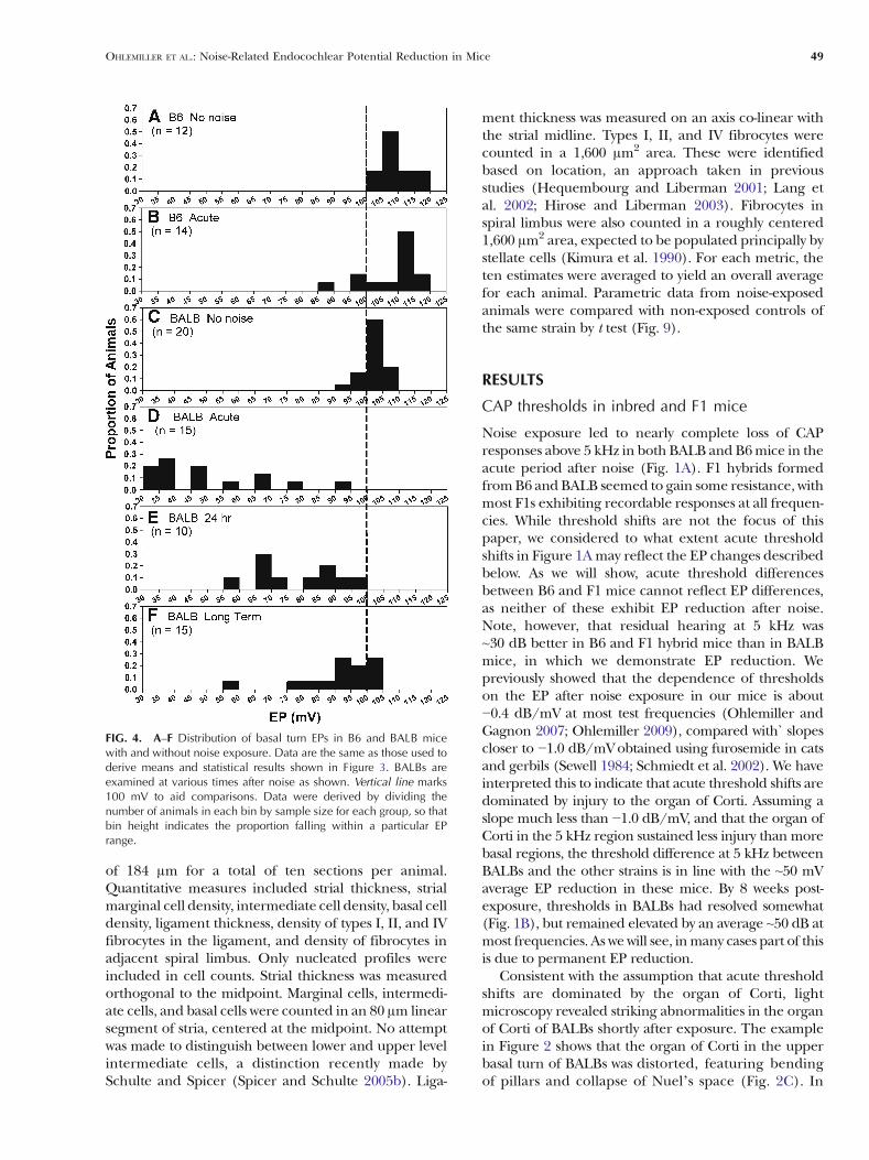
FIG. 4. A–F Distribution of basal turn EPs in B6 and BALB micewith and without noise exposure. Data are the same as those used toderive means and statistical results shown in Figure 3. BALBs areexamined at various times after noise as shown. Vertical line marks100 mV to aid comparisons. Data were derived by dividing thenumber of animals in each bin by sample size for each group, so thatbin height indicates the proportion falling within a particular EPrange.
OHLEMILLER ET AL.: Noise-Related Endocochlear Potential Reduction in Mice 49

no case, however, did the reticular lamina appearruptured, nor was there any other sign of breachingof the boundaries of scala media. In addition, spirallimbus was significantly more likely to show vacuo-lization of stellate fibrocytes than in unexposedcontrols (Figs. 2B and 6).
Effects of noise on EP in inbreds and F1s
Figure 3 compares the average EP in BALBs andcomparison groups in controls, and at various timesafter noise. The EP in BALBs was reduced by 50–60 mVin the hours after exposure. The EP remained signifi-cantly depressed 24 h later, and still remained signifi-
cantly depressed by an average ~10 mV at 8 weeks post-exposure. B6 mice showed no EP reduction at any time.Like CBAs, F1s formed from B6 and CBA showed EPreduction that recovered completely by 8 weeks. Bycontrast, F1s formed from B6 and BALB showed no EPreduction by noise at any time. The fact that these F1mice do not display an intermediate phenotype suggeststhat all EP susceptibility alleles in BALBs act in a simplerecessive manner, and may be few in number.
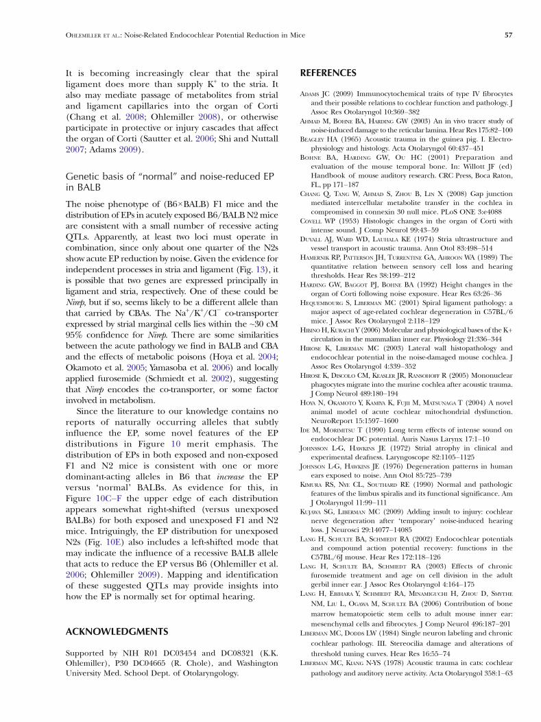
Figure 4 re-analyzes data from Figure 3, showing thedistribution of EPs in BALBs at various times after noise,and comparing these with B6 mice. In each graph, eachbin represents the proportion of animals having an EPwithin a particular 5 mV range. Slightly different modalvalues in unexposed B6 and BALB mice (compareFig. 4A, C) are in keeping with our previous observation(Ohlemiller et al. 2006) that young ‘normal’ BALBshave an EP that is on average ~10 mV lower than in B6.In the hours after noise, nearly all BALBs showed an EPbelow 80 mV, while B6 mice showed no average change.By 24 h later, the EP in some BALBs had returned tonear-normal (Fig. 4E). However, even 8 weeks afternoise the EP remained significantly depressed onaverage as a result of marked leftward skewing of theEP distribution (Figs. 3 and 4F).
Morphological correlates of acute EP changes
We previously showed that several lateral wall celltypes in the cochlear upper basal turn of CBA miceshow acute pathology that correlates with EPreduction within hours after noise (Ohlemiller andGagnon 2007; Ohlemiller et al. 2010b). This pathologywas manifested not as cell loss at this early time point,but rather in the form of cell vacuolization, shrinkage,
FIG. 6. Quantitative analysis of acute (1–3 h post) noise-relatedpathology of lateral wall and spiral limbus in noise-exposed andcontrol BALB mice. Sections were blindly scored “normal” or“abnormal” for each cell type or structure, and section totals werecombined for each experimental group. P values indicate z testresults.
FIG. 5. A Cochlear upper basal turn lateral wallin example BALB mouse 1–3 h after noise.Animal is the same as shown in Figure 2. BExpanded view of box B in (A) showing enlargedpale nucleus of mesothelial cell of Reissner’smembrane (arrowhead). C Expanded view of boxC in (A) showing vacuolated type V fibrocytes(arrowheads). D–E Expanded view of boxes Dand E in (A) showing vacuolated basal cells andpossibly intermediate cells in stria with adjacentshrunken type I fibrocytes in spiral ligament(arrowheads). Shrinkage of fibrocytes is attendedby increased clear space in ligament. F Expandedview of box F in (A) showing vacuolated type IIfibrocytes (arrowheads). G Expanded view of boxG in (A) showing vacuolated type IV fibrocytes(arrowheads). Roman numerals in (C–G) denotemajor constituent fibrocyte types.
50 OHLEMILLER ET AL.: Noise-Related Endocochlear Potential Reduction in Mice
or nuclear pyknosis. BALBs showed similar effects invirtually all the same cells as CBAs, as shown by examplein Figure 5, and quantitatively in Figure 6 (Note that wedid not quantify as many cell types in the earlier CBAstudy.). The acute effects of noise were apparent inseveral structures, including Reissner’s membrane,stria vascularis, spiral ligament, and spiral limbus.Noise-related changes within Reissner’s membrane,stria, and ligament were highly distinctive and noteasily confused with processing artifacts, andincluded enlargement and paling of the nucleus ofmesothelial cells on Reissner’s upper surface(Fig. 5B), vacuolization of types II, IV, and V fibrocytes(Fig. 5C, F, G), and shrinkage of type I fibrocytes(Fig. 5E). The stria itself showed vacuoles that wereusually confined to the basal cell layer, but occasionallyextended into the middle strial layers (Fig. 5D). Strialcapillaries rarely appeared distended, collapsed, orotherwise abnormal. At the light microscope level,
acute strial changes appeared mostly intracellular,and not as edema.
Judged quantitatively, differences versus non-exposedcontrols in the percent of sections scored abnormal afternoise were highly significant (Fig. 6). The cells leastreliably distinguishable in noise versus control animalswere type IV fibrocytes, and cells of spiral limbus. TypeIV’s were scored abnormal in controls in ~10% ofsections, and scored abnormal in exposed animalsin ~50% of sections, which was nevertheless a significantdifference by z test (pG .001). The relatively highpercentage of type IV’s scored abnormal may in partreflect an attempt to apply inclusive criteria. Upon re-inspection, type IV fibrocytes labeled abnormal incontrols tended to be labeled based on their pyknoticnuclei, while those in noise-exposed animals tended toshow vacuoles like those shown in Figure 5G. Cells ofspiral limbus were scored abnormal in ~30% of controlssections and ~55% of exposed animals, a smaller differ-ence that was nevertheless significant (p=.04). Thefrequent abnormal appearance of the limbus includedvacuolization of stellate cells, and pyknotic nuclei of bothstellate cells and interdental cells. It is not clear whetherabnormal features of ligament and limbus in nominallynormal animals reflect sub-optimal preservation, orperhaps ongoing loss and turnover of these cells.
Consistent with previous reports (Duvall et al.1974; Ide and Morimitsu 1990; Wang et al. 2002;Hirose and Liberman 2003), by 24 h after noiseexposure the manner of abnormal appearance ofthe stria changed from vacuolization of some cellsto swelling of the intrastrial space, imparting aswollen profile to the stria (arrowheads inFig. 7A). By this time, signs of fibrocyte deathbegan to appear, particularly among type II and IVfibrocytes of the ligament and stellate fibrocytes ofspiral limbus (Fig. 8A–C). More so than weobserved in CBAs, BALBs also tended towarddarkening of the cells of the spiral prominenceepithelium at this time (see arrow in Fig. 7A).
Permanent effects of noise in inbreds
We previously showed that most of the same cells andstructures exhibiting acute effects of noise in CBA micealso showed changes 8 weeks later (Ohlemiller andGagnon 2007). As determined by morphometry of theupper basal turn, all the lateral wall and limbus cell typesnoted to be abnormal acutely after noise in CBAs,including type I and II fibrocytes and strial basal cells,were significantly reduced in number at 8 weeks. Inaddition, strial capillaries were reduced in number inCBAs, and both stria and ligament became significantlythinner. Given that the EP completely recovers in CBAs,but shows incomplete recovery in BALBs, we anticipatedgreater cell loss and permanent injury in BALBs. This
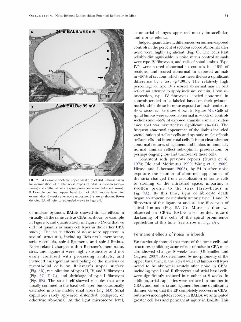
FIG. 7. A Example cochlear upper basal turn of BALB mouse takenfor examination 24 h after noise exposure. Stria is swollen (arrow-heads) and epithelial cells of spiral prominence are darkened (arrow).B Example cochlear upper basal turn of BALB mouse taken forexamination 8 weeks after noise exposure. EPs are as shown. Boxesdenoted 8A–8F refer to expanded views in Figure 8.
OHLEMILLER ET AL.: Noise-Related Endocochlear Potential Reduction in Mice 51

was not the case, however. Amore extensive inventory ofcell loss and other structural changes in the upper base8 weeks after exposure in BALBs revealed significantloss of only type IV fibrocytes and stellate fibrocytes oflimbus (Figs. 7B, 8D–F, and 9A). While the stria showedsignificant thinning (Fig. 9B), ligament showed nodifference. Acute pathology of the epithelium ofspiral prominence frequently observed in BALBsdid not lead to clear degeneration of epithelial cellsat 8 weeks. A thin layer of cells covering theprominence was seen in all cases.
Inheritance pattern of EP and lateral wall injury inN2 backcross mice
As an initial approach to the genetics of the different EPphenotypes in BALB and B6 mice, we bred and noise-exposed (B6×BALB) F1 hybrids and (F1×BALB) N2backcross mice. Figure 10 compares the distribution ofEPs measured acutely after noise in both models. Tofacilitate comparison, data from the unexposed inbredlines have been recapitulated (Fig. 10A, B). The F1 datasuggest a hybrid advantage in both unexposed andexposed mice (Fig. 10C, D). Both show EPs that appearslightly higher on average than in unexposed, althoughthis trend was not significant by one-way ANOVA. EPsfrom unexposed N2 mice were surprisingly broadlydistributed, and included a mode at distinctly lower EPsthan seen in either of the parent strains (compareFig. 10A, B, E). These N2s may be homozygous forBALB alleles responsible for the slightly low “normal”EP in BALB inbreds. EPs in noise-exposed N2s werehighly skewed toward lower values (Fig. 10F), with onequarter (14/56) of the mice showing EPs below 80 mV.This is consistent with a requirement of homozygosityfor BALB alleles at two or more loci for the EP to bereduced by noise.
Histologic correlates of acute EP reduction in N2mice
In N2 mice formed from B6 and CBA, we previouslydemonstrated strong correlations between metrics foracute pathology of stria, spiral ligament, and Reissner’smembrane versus the EP (Ohlemiller and Gagnon2007; Ohlemiller et al. 2010b) (Table 1). We thereforeconducted similar analyses in noise-exposed N2sformed from B6 and BALB, selecting mice with EPsamong the lowest recorded (n=11), and the highestrecorded (n=10). Unlike B6/CBA N2 mice, twodistinct patterns were noted among B6/BALB N2mice with a low EP, based on the appearance of striavascularis and type I fibrocytes. Some mice with a lowEP featured a normal appearing stria with abnormaltype I’s (Fig. 11B, D). Other mice with a low EPcombined a clearly abnormal stria with normalappearing type I’s (Fig. 11C, F, G). Mice showing alow EP reliably exhibited abnormal appearance ofReissner’s membrane (example not shown) and typeII fibrocytes (Fig. 11B, C, E, H).
Trends described above could be demonstratedquantitatively, as shown in Figure 12. Among allmetrics, the appearance of Reissner’s membraneand type II fibrocytes were most strongly correlatedwith EP (R2 ≥.70) (Fig. 12A, B). Abnormal appearanceof stria, type V fibrocytes, and type I fibrocyteswere progressively less reliable predictors of a lowEP (Fig. 11C–E), although correlations were stillsignificant. The appearance of type IV fibrocytes(Fig. 11F) and spiral limbus (not shown) werecompletely uncorrelated with EP. The very differentrelations between the predictive value for the EP ofthe appearance of stria vascularis and type Ifibrocytes in CBA- versus BALB-derived N2 miceare highlighted in Table 1, which compares thecoefficients of determination of their respective
FIG. 8. Expanded views of boxed regions inFigure 7. A–C At 24 h post-exposure, pyknoticnuclei and condensed cells of spiral limbus andin regions of ligament dominated by type IVfibrocytes and type II fibrocytes, respectively,indicate dying cells (arrowheads). D–F By 8weeks post-exposure, stellate fibrocytes of lim-bus are entirely missing in upper base and lowerapex (arrowhead in (D)). Type IV fibrocytes arereduced in number. Arrowhead in (E) showsdying type IV. Type II fibrocytes (F, arrowheads)appear generally normal.
52 OHLEMILLER ET AL.: Noise-Related Endocochlear Potential Reduction in Mice
linear regressions. Neither strial nor type I fibrocytepathology were as strongly correlated with EP inB6/BALB N2s as in B6/CBA N2 mice. Accordingly,the relation between strial and type I fibrocytepathology is quite different in the two models. AsFigure 13 shows, these are strongly correlated inB6/CBA N2s, but completely uncorrelated in B6/BALB N2s. From these data we conclude that noise-related EP reduction in BALBs can arise fromsomewhat independent cascades within stria andligament. These may reflect the action of differentgenes.
DISCUSSION
Until recently the role of EP reduction in acute andpermanent NIHL has been largely undefined. Whentraumatic noise breaches the boundaries of scala media,the EP almost certainly decreases, and may or may not
recover (Ward and Duvall 1971; Johnsson and Hawkins1972; Duvall et al. 1974; Santi and Duvall 1978; Ulehlova1983; Ide and Morimitsu 1990; Ahmad et al. 2003). Forless traumatic noise insults where endolymphatic boun-daries remain intact, the expectation has been that theEP is unaffected or only transiently affected, and doesnot impact the degree of permanentNIHL.Our currentand recent work builds upon work by Hirose, Liberman,and colleagues (Wang et al. 2002; Hirose and Liberman
FIG. 9. Quantitative analysis of permanent noise-related pathology(8-week post-exposure) in cochlear upper basal turn spiral limbusand lateral wall of control and exposed BALB mice. A Amongmetrics for strial and ligament cell types, as well as strial capillaries(blood vessel density (BVs)), significant differences were only foundfor type IV fibrocytes and stellate fibrocytes of limbus. B Significantthickness changes were found for stria, but not ligament. P valuesdenote results of two-tailed t test.
FIG. 10. A–B Distribution of basal turn EPs in B6 and BALB micewithout noise exposure (reproduced from Figure 4 to aid comparison).C–DDistribution of EPs with and without noise in (B6×BALB) F1 hybridmice. E–FDistribution of EPs in (F1×BALB) N2 backcross mice with andwithout noise. Following noise, 25% (14/56) of N2 mice showed EPsbelow 80 mV. Note different y-axis range in (E) and (F) due to greaterscatter. Vertical line marks 100 mV to facilitate comparisons. As inFigure 4, data were derived by dividing the number of animals in eachbin by sample size for each group, so that bin height indicates theproportion falling within a particular EP range.
OHLEMILLER ET AL.: Noise-Related Endocochlear Potential Reduction in Mice 53

2003; Hirose et al. 2005) by demonstrating that (1) EPreduction by noise can occur in instances where theboundaries of scala media are maintained; (2) WhetherEP reduction occurs depends on genetically modifiable
factors that are probably intrinsic to the lateral wall;(3) EP reduction may reflect independent cascadeswithin stria vascularis and the spiral ligament; and(4) Depending on the genes and alleles involved,EP reduction may not be completely reversible. Forour particular noise exposure the degree of perma-nent reduction averaged a modest ~10 mV in BALBmice. Rather than supporting any particular amountof EP reduction or any required exposure condi-tion, we regard the noise phenotype of BALBssimply as an indication that permanent EP reduc-tion by noise is neither special nor atypical. Onlyone noise condition and relatively little of theextant genetic diversity of mice had to be “mined”to demonstrate permanent EP reduction. Thus wesuppose that alleles favoring a wide range of lateral wallresponses to noise will be found across animal andhuman populations, and that there exists no single
TABLE 1
Coefficients of determination (R2) for EP versus lateral wallinjury metrics in [(B6×CBA)×B6] N2 and [(B6×BALB)×
BALB] N2 mice
B6/CBA N2 B6/BALB N2
EP versus R2 P R2 P
Reissner’s pathology 0.9a G.001 0.72 G.001Strial pathology 0.78b G.001 0.55 G.001Type II pathology 0.69b G.001 0.70 G.001Type I pathology 0.57b G.001 0.27 0.013
aFrom Ohlemiller et al. 2010bbFrom Ohlemiller and Gagnon 2007
FIG. 11. Examples of the three major patterns ofcellular appearance observed in the cochlear upperbasal turn 1–3 h after noise in B6/BALB N2 back-cross mice. EPs are shown for each animal. ANormal EP attended by minimal pathology, asdetected by light microscope. B Mouse withreduced EP combining minimal strial pathologywith clear pathology of type I fibrocytes (expandedin (D), arrowheads) and type II fibrocytes (expandedin (E), arrowheads). C Mouse combining clear strialpathology (expanded in (F), arrowheads) withpathology of type II fibrocytes (expanded in (H),arrowheads), but showing minimal pathology oftype I fibrocytes (expanded in (G)).
54 OHLEMILLER ET AL.: Noise-Related Endocochlear Potential Reduction in Mice

cellular pattern of noise-related cochlear pathology inmammals.
Anatomic correlates of acute EP reduction
Our acute observations in BALB inbred mice point toa broad range of affected non-sensory cell types andstructures, including strial basal cells, and mostconstituent cells of spiral ligament and limbus. Weinterpreted a similar pattern in CBA mice as indicat-ing that all the affected cells participate in the samepathologic cascade, and considered which cell(s) arelikely to express the product encoded by the NirepQTL (Ohlemiller et al. 2010b). The accepted broadconceptual framework for operation of the lateral wallis that of return path for K+ that is “recycled” afterpassing through the organ of Corti (Spicer et al. 1996;Wangemann 2002). The concentration gradients driv-ing K+ movement are largely established by ATPases intype II fibrocytes and strial marginal cells. If the most“downstream” point—that is, the stria—were tobecome functionally impaired, then K+ could build up
in ligament. Assuming K+ accumulates inside of type IIfibrocytes but outside of type I fibrocytes, the verydifferent appearance of these cells (vacuolation of typeII, shrinkage of type I) might be explained. Further,accumulation of K+ and other ions outside type Ifibrocytes could occur if the normally ion-tight boun-dary of the lateral stria were rendered porous by noise.That boundary is composed of tight junctions betweenbasal cells, the most obviously affected strial cell type inour mice. All these events could thus be linked in CBAmice, with the probable site of Nirep expression lyingwithin the stria. Our finding of somewhat independentpathology of stria and type I’s in B6/BALB N2 miceseems less compatible with a ‘leaky stria’ scenario. Inboth BALB and CBA, however, the condition of bothtype II fibrocytes and Reissner’s membrane was highlycorrelated with the EP, suggesting these spatially sepa-rated cells participate in a common affected process.
To extract strain differences from broad patternsof change, we deliberately focused on changesdiscernable at the light microscope level. Thus ourobservation that basal cells appeared to be the mostaffected strial cell type in BALBs is not meant to imply
FIG. 12. A–F Correlation betweenbasal turn EP and pathology of specificcells and structures in the upper basalturn of B6/BALB N2 backcross mice,observed 1–3 h after noise. The con-dition of Reissner’s membrane and typeII fibrocytes of ligament showed thestrongest correlation with EP (A, B),followed by progressively weaker cor-relation between EP and the appear-ance of the stria (C), type V fibrocytes(D), and type I fibrocytes (E). Theappearance of type IV fibrocytes (F)and spiral limbus (not shown) wasuncorrelated with EP.
OHLEMILLER ET AL.: Noise-Related Endocochlear Potential Reduction in Mice 55

that intermediate and marginal cells were unaffected bynoise. The extent of intermediate and marginal cellinvolvement clearly depends on exposure conditions(Hirose and Liberman 2003). Marginal cells expressseveral components key to the generation of the EP(Hibino and Kurachi 2006; Wangemann 2006), andappear to be metabolically labile (e.g., Spicer andSchulte 2005a; Ohlemiller et al. 2009). They are alsothe principal strial site of expression of the Na+/K+/Cl-
exchanger (Slc12a2), a strong candidate for Nirep(Ohlemiller et al. 2010b).
The B6/BALB N2 data suggest that acute pathologyof type IV fibrocytes and spiral limbus cells are indirectlyrelated or unrelated to EP reduction, since changes inthese were poorly correlated with EP (Fig. 12). Strictlynoise-related pathology was also hardest to show inthese cells (Fig. 6), potentially obscuring the truerelationship. It may be the case that BALBs harboralleles that promote noise injury in type IV andlimbus fibrocytes independent of EP change and its
genetics. While we did not quantify type IV pathol-ogy in CBAs, we and others have identified type IVsas a target of noise in both CBA and B6 mice(Hirose et al. 2005; Ohlemiller and Gagnon 2007).Permanent limbus pathology, however, appears tosegregate with acute EP vulnerability, being readilyobserved in both CBA and BALB, but less so in B6(Ohlemiller and Gagnon 2007). We could not usethe N2 mice to try to separate permanent limbuspathology from EP vulnerability, since there wouldbe no way of knowing whether mice exhibiting clearloss of limbus fibrocytes 8 weeks after noise wouldhave shown acute EP reduction. It presentlyremains unclear whether spiral limbus pathologycan be viewed as part of the cascade that reducesthe EP in BALB and CBA mice, and what this mayreveal about mechanisms.
Anatomic correlates of permanent EP reduction
Given the overall match between acute and permanentpathology we observed in CBA mice (Ohlemiller andGagnon 2007; Ohlemiller et al. 2010b), which do notshow permanent EP reduction fromour noise exposure,we expected even more prominent permanent changesin the cochlear lateral wall of BALB mice. Instead, wewere surprised to observe only modest changesrestricted to type IV fibrocytes, spiral limbus, andstrial thickness (Fig. 9). Light microscopy alone isnot adequate to explain this discrepancy, and ultra-structural analysis or examination of major lateralwall protein expression (e.g., Na+/K+-ATPase, Na+/K+/Cl− co-transporter, KCNJ10) (Hibino and Kurachi2006; Wangemann 2006) maybe required. Because itwas easy to find dying type II fibrocytes in spiral ligament24 h after noise (Fig. 8), it was particularly surprisingthat no permanent loss of these cells was detected.Renewal of cells in spiral ligament has been demon-strated in mice and other models (Lang et al. 2003,2006; Yamasoba et al. 2003), and it maybe that renewalprocesses are more robust in BALBs than in CBAs.
We previously showed that by 19 months of ageBALB/c mice exhibit age-associated EP reductionwhose major anatomic correlates are marginal cellloss and thinning of spiral ligament (Ohlemiller et al.2006; Ohlemiller 2009). Although it is not clear whichstrial cell changes underlie strial thinning after noise,neither marginal cell loss nor thinning of theligament was clearly exacerbated (Fig. 9). Thus thereappears no obvious overlap between the targets ofnoise and aging in the BALB lateral wall, at least at6 months of age. It remains to be seen whether thetypical aging trajectory of the BALB lateral wall canbe altered by earlier noise exposure. Permanentnoise injury to the lateral wall may exert effects onhearing apart from any impact on the EP, of course.
FIG. 13. Direct comparison the correlation between acute noise-related pathology of stria vascularis and type I fibrocytes of spiralligament in B6/BALB N2 mice (A) and B6/CBA N2 mice (B). Thesewere highly correlated in B6/CBA N2s, but completely uncorrelatedin B6/BALB N2s.
56 OHLEMILLER ET AL.: Noise-Related Endocochlear Potential Reduction in Mice
It is becoming increasingly clear that the spiralligament does more than supply K+ to the stria. Italso may mediate passage of metabolites from strialand ligament capillaries into the organ of Corti(Chang et al. 2008; Ohlemiller 2008), or otherwiseparticipate in protective or injury cascades that affectthe organ of Corti (Sautter et al. 2006; Shi and Nuttall2007; Adams 2009).
Genetic basis of “normal” and noise-reduced EPin BALB
The noise phenotype of (B6×BALB) F1 mice and thedistribution of EPs in acutely exposed B6/BALBN2miceare consistent with a small number of recessive actingQTLs. Apparently, at least two loci must operate incombination, since only about one quarter of the N2sshow acute EP reduction by noise. Given the evidence forindependent processes in stria and ligament (Fig. 13), itis possible that two genes are expressed principally inligament and stria, respectively. One of these could beNirep, but if so, seems likely to be a different allele thanthat carried by CBAs. The Na+/K+/Cl− co-transporterexpressed by strial marginal cells lies within the ~30 cM95% confidence for Nirep. There are some similaritiesbetween the acute pathology we find in BALB and CBAand the effects of metabolic poisons (Hoya et al. 2004;Okamoto et al. 2005; Yamasoba et al. 2006) and locallyapplied furosemide (Schmiedt et al. 2002), suggestingthat Nirep encodes the co-transporter, or some factorinvolved in metabolism.
Since the literature to our knowledge contains noreports of naturally occurring alleles that subtlyinfluence the EP, some novel features of the EPdistributions in Figure 10 merit emphasis. Thedistribution of EPs in both exposed and non-exposedF1 and N2 mice is consistent with one or moredominant-acting alleles in B6 that increase the EPversus ‘normal’ BALBs. As evidence for this, inFigure 10C–F the upper edge of each distributionappears somewhat right-shifted (versus unexposedBALBs) for both exposed and unexposed F1 and N2mice. Intriguingly, the EP distribution for unexposedN2s (Fig. 10E) also includes a left-shifted mode thatmay indicate the influence of a recessive BALB allelethat acts to reduce the EP versus B6 (Ohlemiller et al.2006; Ohlemiller 2009). Mapping and identificationof these suggested QTLs may provide insights intohow the EP is normally set for optimal hearing.
ACKNOWLEDGMENTS
Supported by NIH R01 DC03454 and DC08321 (K.K.Ohlemiller), P30 DC04665 (R. Chole), and WashingtonUniversity Med. School Dept. of Otolaryngology.
REFERENCES
ADAMS JC (2009) Immunocytochemical traits of type IV fibrocytesand their possible relations to cochlear function and pathology. JAssoc Res Otolaryngol 10:369–382
AHMAD M, BOHNE BA, HARDING GW (2003) An in vivo tracer study ofnoise-induced damage to the reticular lamina.Hear Res 175:82–100
BEAGLEY HA (1965) Acoustic trauma in the guinea pig. I. Electro-physiology and histology. Acta Otolaryngol 60:437–451
BOHNE BA, HARDING GW, OU HC (2001) Preparation andevaluation of the mouse temporal bone. In: Willott JF (ed)Handbook of mouse auditory research. CRC Press, Boca Raton,FL, pp 171–187
CHANG Q, TANG W, AHMAD S, ZHOU B, LIN X (2008) Gap junctionmediated intercellular metabolite transfer in the cochlea incompromised in connexin 30 null mice. PLoS ONE 3:e4088
COVELL WP (1953) Histologic changes in the organ of Corti withintense sound. J Comp Neurol 99:43–59
DUVALL AJ, WARD WD, LAUHALA KE (1974) Stria ultrastructure andvessel transport in acoustic trauma. Ann Otol 83:498–514
HAMERNIK RP, PATTERSON JH, TURRENTINE GA, AHROON WA (1989) Thequantitative relation between sensory cell loss and hearingthresholds. Hear Res 38:199–212
HARDING GW, BAGGOT PJ, BOHNE BA (1992) Height changes in theorgan of Corti following noise exposure. Hear Res 63:26–36
HEQUEMBOURG S, LIBERMAN MC (2001) Spiral ligament pathology: amajor aspect of age-related cochlear degeneration in C57BL/6mice. J Assoc Res Otolaryngol 2:118–129
HIBINOH, KURACHI Y (2006)Molecular and physiological bases of the K+circulation in the mammalian inner ear. Physiology 21:336–344
HIROSE K, LIBERMAN MC (2003) Lateral wall histopathology andendocochlear potential in the noise-damaged mouse cochlea. JAssoc Res Otolaryngol 4:339–352
HIROSE K, DISCOLO CM, KEASLER JR, RANSOHOFF R (2005) Mononuclearphagocytes migrate into the murine cochlea after acoustic trauma.J Comp Neurol 489:180–194
HOYA N, OKAMOTO Y, KAMIYA K, FUJII M, MATSUNAGA T (2004) A novelanimal model of acute cochlear mitochondrial dysfunction.NeuroReport 15:1597–1600
IDE M, MORIMITSU T (1990) Long term effects of intense sound onendocochlear DC potential. Auris Nasus Larynx 17:1–10
JOHNSSON L-G, HAWKINS JE (1972) Strial atrophy in clinical andexperimental deafness. Laryngoscope 82:1105–1125
JOHNSON L-G, HAWKINS JE (1976) Degeneration patterns in humanears exposed to noise. Ann Otol 85:725–739
KIMURA RS, NYE CL, SOUTHARD RE (1990) Normal and pathologicfeatures of the limbus spiralis and its functional significance. AmJ Otolaryngol 11:99–111
KUJAWA SG, LIBERMAN MC (2009) Adding insult to injury: cochlearnerve degeneration after ‘temporary’ noise-induced hearingloss. J Neurosci 29:14077–14085
LANG H, SCHULTE BA, SCHMIEDT RA (2002) Endocochlear potentialsand compound action potential recovery: functions in theC57BL/6J mouse. Hear Res 172:118–126
LANG H, SCHULTE BA, SCHMIEDT RA (2003) Effects of chronicfurosemide treatment and age on cell division in the adultgerbil inner ear. J Assoc Res Otolaryngol 4:164–175
LANG H, EBIHARA Y, SCHMIEDT RA, MINAMIGUCHI H, ZHOU D, SMYTHE
NM, LIU L, OGAWA M, SCHULTE BA (2006) Contribution of bonemarrow hematopoietic stem cells to adult mouse inner ear:mesenchymal cells and fibrocytes. J Comp Neurol 496:187–201
LIBERMAN MC, DODDS LW (1984) Single neuron labeling and chroniccochlear pathology. III. Stereocilia damage and alterations ofthreshold tuning curves. Hear Res 16:55–74
LIBERMAN MC, KIANG N-YS (1978) Acoustic trauma in cats: cochlearpathology and auditory nerve activity. Acta Otolaryngol 358:1–63
OHLEMILLER ET AL.: Noise-Related Endocochlear Potential Reduction in Mice 57

LIBERMAN MC, MULROY MJ (1982) Acute and chronic effects of acoustictrauma: cochlear pathology and auditory nerve pathophysiology.In: Hamernik RP, Henderson D, Salvi R (eds) New perspectives onnoise-induced hearing loss. Raven Press, New York, pp 105–135
NORDMANN AS, BOHNE BA, HARDING GW (2000) Histopathologicaldifferences between temporary and permanent threshold shift.Hear Res 139:13–30
OHLEMILLER KK (2008) Recent findings and emerging questions incochlear noise injury. Hear Res 245:5–17
OHLEMILLER KK (2009) Mechanisms and genes in human strialpresbycusis from animal models. Brain Res 1277:70–83
OHLEMILLER KK, GAGNON PM (2007) Genetic dependence of cochlearcells and structures injured by noise. Hear Res 224:34–50
OHLEMILLER KK, LETT JM, GAGNON PM (2006) Cellular correlates ofage-related endocochlear potential reduction in a mouse model.Hear Res 220:10–26
OHLEMILLER KK, RICE MR, LETT JM, GAGNON PM (2009) Absence ofstrial melanin coincides with age associated marginal cell lossand endocochlear potential decline. Hear Res 249:1–14
OHLEMILLER KK, DAHL AR, GAGNON PM (2010a) Divergent agingcharacteristics in CBA/J and CBA/CaJ mouse cochleae. J AssocRes Otolaryngol doi:10.1007/s10162-010-0228-1
OHLEMILLER KK, ROSEN AD, GAGNON PM (2010b) A major effect QTLon chromosome 18 for noise injury to the mouse cochlearlateral wall. Hear Res 260:47–53
OKAMOTO Y, HOYA N, KAMIYA K, FUJII M, OGAWA K, MATSUNAGA T (2005)Permanent threshold shifts caused by acute cochlear mitochon-drial dysfunction is primarily mediated by degeneration of thelateral wall of the cochlea. Audiol Neuro-Otol 10:220–233
OU HC, HARDING GW, BOHNE BA (2000a) An anatomically basedfrequency-placemap for themouse cochlea. Hear Res 145:123–129
OU HC, BOHNE BA, HARDING GW (2000b) Noise damage in theC57BL/CBA mouse cochlea. Hear Res 145:111–122
SANTI PA, DUVALL AJ (1978) Stria vascularis pathology and recoveryfollowing noise exposure. Otolaryngology 86:354–361
SAUTTER NB, SHICK EH, RANSOHOFF RM, CHARO IF, HIROSE K (2006)CC chemokine receptor 2 is protective against noise-inducedhair cell death: studies in CX3CR1+/GFP mice. J Assoc Res
Otolaryngol 7:361–372SCHMIEDT RA, LANG H, OKAMURA H, SCHULTE BA (2002) Effects of
furosemide applied chronically to the round window: a model ofmetabolic presbycusis. J Neurosci 22:9643–9650
SEWELLW (1984) The effects of furosemide on the endocochlear potentialand auditory nerve fiber tuning curves in cats. Hear Res 14:305–314
SHI X, NUTTALL AL (2007) Expression of adhesion molecularproteins in the cochlear lateral wall of normal and PARP-1mutant mice. Hear Res 224:1–14
SPICER SS, SCHULTE BA (2005a) Pathologic changes of presbycusisbegin in secondary processes and spread to primary processes ofstrial marginal cells. Hear Res 205:225–240
SPICER SS, SCHULTE BA (2005b) Novel structures in marginal andintermediate cells presumably relate to functions of basal versusapical strata. Hear Res 200:87–101
SPICER SS, SAMUEL S, SCHULTE BA (1996) The fine structure of spiralligament cells relates to ion return to the stria and varies withplace-frequency. Hear Res 100:80–100
SYKA J, MELICHAR I, ULEHLOVA L (1981) Longitudinal distribution ofcochlear potentials and the K+concentration in the endolymphafter acoustic trauma. Hear Res 4:287–298
ULEHLOVA L (1983) Stria vascularis in acoustic trauma. ArchOtorhinolaryngol 237:133–138
WANG Y, HIROSE K, LIBERMAN MC (2002) Dynamics of noise-inducedcellular injury and repair in the mouse cochlea. J Assoc ResOtolaryngol 3:248–268
WANGEMANN P (2002) K+recycling and the endocochlear potential.Hear Res 165:1–9
WANGEMANN P (2006) Supporting sensory transduction: cochlear fluidhomeostasis and the endocochlear potential. J Physiol 576(1):11–21
WARD WD, DUVALL AJ (1971) Behavioral and ultrastructural corre-lates of acoustic trauma. Ann Otol 80:881–896
YAMASOBA T, KONDO K, MIYAJIMA C, SUZUKI M (2003) Changes in cellproliferation in rat and guinea pig cochlea after aminoglycoside-induced damage. Neurosci Lett 347:171–174
YAMASOBA T, GOTO Y, KOMAKI H, MIMAKI M, SUDO K, SUZUKI M (2006)Cochlear damage due to germanium-induced mitochondrialdysfunction in guinea pigs. Neurosci Lett 395:18–22
58 OHLEMILLER ET AL.: Noise-Related Endocochlear Potential Reduction in Mice


![CBA - MLCF - Wpg Final_TL_TR[5] - UFCW832](https://static.fdokumen.com/doc/165x107/633ce8afb30159f8b4087b8d/cba-mlcf-wpg-finaltltr5-ufcw832.jpg)