Differences in Al tolerance between Plantago algarbiensis and P. almogravensis reflect their ability...
11
Differences in Al tolerance between Plantago algarbiensis and P. almogravensis reflect their ability to respond to oxidative stress Neusa Martins • Maria Leonor Oso ´rio • Sandra Gonc ¸alves • Ju ´lio Oso ´rio • Anabela Romano Received: 4 October 2012 / Accepted: 28 March 2013 / Published online: 8 April 2013 Ó Springer Science+Business Media New York 2013 Abstract We evaluated the impact of low pH and aluminum (Al) on the leaves and roots of Plantago almogravensis Franco and Plantago algarbiensis Samp., focusing on energy partitioning in photosystem II, H 2 O 2 levels, lipid peroxidation, electrolyte leakage (EL), protein oxidation, total soluble protein content and antioxidant enzyme activities. In both species, Al triggered more changes in oxidative metabolism than low pH alone, particularly in the roots. We found that Al increased the levels of H 2 O 2 in P. algarbiensis roots, but reduced the levels of H 2 O 2 in P. almogravensis leaves and roots. Neither low pH nor Al affected the spatial heterogeneity of chlorophyll fluorescence, the maximum photochemical efficiency of PSII (F v /F m ), the actual quantum efficiency of PSII (/ PSII ) or the quantum yields of regulated (/ NPQ ) and nonregulated (/ NO ) energy dissipation, and there was no significant change in total soluble protein content and EL. In P. algarbiensis, Al increased the carbonyl content and the activities of superoxide dismutase (SOD) and catalase (CAT) in the roots, and also CAT, ascorbate peroxidase and guaiacol peroxidase activities in the leaves. In P. almogravensis, Al reduced the level of malondialdehyde in the roots as well as SOD activity in the leaves and roots. We found that P. almogravensis plantlets could manage the oxidative stress caused by low pH and Al, whereas the P. algarbiensis antioxidant system was unable to suppress Al toxicity completely, leading to the accumulation of H 2 O 2 and consequential protein oxidation in the roots. Keywords Antioxidant enzymes Á Energy partitioning Á Lipid peroxidation Á Oxidative stress Á Protein oxidation Á Reactive oxygen species Abbreviations APX Ascorbate peroxidase CAT Catalase EL Electrolyte leakage F v /F m Maximum photochemical efficiency of PSII FW Fresh weight GPX Guaiacol peroxidase MDA Malondialdehyde MS Murashige and Skoog NBT Nitroblue tetrazolium PSII Photosystem II PVPP Polyvinylpolypyrrolidone ROS Reactive oxygen species SOD Superoxide dismutase TBA Thiobarbituric acid TCA Trichloroacetic acid N. Martins Á M. L. Oso ´rio Á S. Gonc ¸alves Á A. Romano (&) IBB/CGB, Faculty of Sciences and Technology, University of Algarve, Campus de Gambelas, Ed. 8, 8005-139 Faro, Portugal e-mail: [email protected] J. Oso ´rio ICAAM (Institute of Mediterranean Agricultural and Environment Sciences), Faculty of Sciences and Technology, University of Algarve, Campus de Gambelas, Ed. 8, 8005-139 Faro, Portugal 123 Biometals (2013) 26:427–437 DOI 10.1007/s10534-013-9625-3
-
Upload
independent -
Category
Documents
-
view
1 -
download
0
Transcript of Differences in Al tolerance between Plantago algarbiensis and P. almogravensis reflect their ability...

Differences in Al tolerance between Plantago algarbiensisand P. almogravensis reflect their ability to respondto oxidative stress
Neusa Martins • Maria Leonor Osorio •
Sandra Goncalves • Julio Osorio •
Anabela Romano
Received: 4 October 2012 / Accepted: 28 March 2013 / Published online: 8 April 2013
� Springer Science+Business Media New York 2013
Abstract We evaluated the impact of low pH and
aluminum (Al) on the leaves and roots of Plantago
almogravensis Franco and Plantago algarbiensis
Samp., focusing on energy partitioning in photosystem
II, H2O2 levels, lipid peroxidation, electrolyte leakage
(EL), protein oxidation, total soluble protein content
and antioxidant enzyme activities. In both species, Al
triggered more changes in oxidative metabolism than
low pH alone, particularly in the roots. We found that
Al increased the levels of H2O2 in P. algarbiensis roots,
but reduced the levels of H2O2 in P. almogravensis
leaves and roots. Neither low pH nor Al affected the
spatial heterogeneity of chlorophyll fluorescence, the
maximum photochemical efficiency of PSII (Fv/Fm),
the actual quantum efficiency of PSII (/PSII) or the
quantum yields of regulated (/NPQ) and nonregulated
(/NO) energy dissipation, and there was no significant
change in total soluble protein content and EL. In
P. algarbiensis, Al increased the carbonyl content and
the activities of superoxide dismutase (SOD) and
catalase (CAT) in the roots, and also CAT, ascorbate
peroxidase and guaiacol peroxidase activities in the
leaves. In P. almogravensis, Al reduced the level of
malondialdehyde in the roots as well as SOD activity in
the leaves and roots. We found that P. almogravensis
plantlets could manage the oxidative stress caused by
low pH and Al, whereas the P. algarbiensis antioxidant
system was unable to suppress Al toxicity completely,
leading to the accumulation of H2O2 and consequential
protein oxidation in the roots.
Keywords Antioxidant enzymes � Energy
partitioning � Lipid peroxidation � Oxidative stress �Protein oxidation � Reactive oxygen species
Abbreviations
APX Ascorbate peroxidase
CAT Catalase
EL Electrolyte leakage
Fv/Fm Maximum photochemical efficiency of PSII
FW Fresh weight
GPX Guaiacol peroxidase
MDA Malondialdehyde
MS Murashige and Skoog
NBT Nitroblue tetrazolium
PSII Photosystem II
PVPP Polyvinylpolypyrrolidone
ROS Reactive oxygen species
SOD Superoxide dismutase
TBA Thiobarbituric acid
TCA Trichloroacetic acid
N. Martins � M. L. Osorio � S. Goncalves �A. Romano (&)
IBB/CGB, Faculty of Sciences and Technology,
University of Algarve, Campus de Gambelas, Ed. 8,
8005-139 Faro, Portugal
e-mail: [email protected]
J. Osorio
ICAAM (Institute of Mediterranean Agricultural and
Environment Sciences), Faculty of Sciences and
Technology, University of Algarve, Campus de
Gambelas, Ed. 8, 8005-139 Faro, Portugal
123
Biometals (2013) 26:427–437
DOI 10.1007/s10534-013-9625-3
/NPQ Quantum yield of regulated energy
dissipation
/NO Quantum yield of nonregulated energy
dissipation
/PSII Actual quantum efficiency of PSII
Introduction
Acidic soils account for 30–40 % of the word’s arable
land and are spreading due to natural, agricultural and
industrial processes (von Uexkull and Mutert 1995).
Aluminum (Al) is the main cause of toxicity to plants in
acidic soils because it is solubilized to yield the toxic
cation Al3? when the pH falls below 5 (von Uexkull
and Mutert 1995). Al affects many physiological
processes by disrupting redox homeostasis and induc-
ing the accumulation of reactive oxygen species (ROS)
(Mittler 2002; Achary et al. 2012). Al stress in plants
inhibits the photosynthetic apparatus by reducing the
photochemical efficiency of photosystem II (PSII) and
restricting electron flow, often due to structural dam-
age to the thylakoids (Pereira et al. 2000; Inostroza-
Blancheteau et al. 2011). This photo-oxidative damage
can be prevented by the dissipation of excess excitation
energy as heat in the antenna pigment complexes
through the xanthophyll cycle (Demmig-Adams and
Adams 1996) or alternative routes such as photores-
piration, the water–water cycle and cyclic electron
transport (Murchie and Niyogi 2011).
Kramer et al. (2004) have proposed a model in
which photosynthetic energy is partitioned into three
fractions: (1) energy utilized photochemically by PSII
(/PSII); (2) energy dissipated thermally by regulated
quenching mechanisms (/NPQ); and (3) energy dissi-
pated by nonregulated quenching mechanisms (/NO).
These fractions satisfy the condition /PSII ? /NPQ ?
/NO = 1. The /PSII and /NPQ pathways work as
protective mechanisms, whereas /NO can be regarded
as excess energy. Therefore, the use of this model
provides a sensitive and reliable method for under-
standing and quantifying damage to PSII caused by Al.
If photochemical and nonphotochemical capacities
are exceeded, the surplus energy is transferred to O2,
and ROS are produced (Wilson et al. 2006). Although
ROS are important signaling molecules, they can also
affect a variety of physiological functions (Gill and
Tuteja 2010). To mitigate oxidative injury, all plant
cells have developed mechanisms to scavenge ROS
using both enzymatic and non-enzymatic antioxidant
systems (Ma et al. 2012). Al stress has been shown to
induce the antioxidant enzymes superoxide dismutase
(SOD; E.C.1.15.1.1), which converts the superoxide
anion to hydrogen peroxide (H2O2), and catalase
(CAT; E.C. 1.11.1.6), ascorbate peroxidase (APX;
E.C. 1.11.1.11) and guaiacol peroxidase (GPX; E.C.
1.11.1.7), which are required to eliminate H2O2
(Giannakoula et al. 2010; Inostroza-Blancheteau
et al. 2011). When the amounts of ROS exceed the
capacity of the scavenging systems, the excess may
cause lipid peroxidation, protein oxidation and DNA
damage leading to mutation (Ma et al. 2012).
Plants have evolved morphological, physiological
and biochemical mechanisms that allow them to
survive in acidic, Al-rich soils (Kochian 1995). Al-
tolerant plants are better equipped to deal with ROS
because they have a higher baseline redox capacity
than Al-sensitive plants and can respond more quickly
to Al in the environment (Ramırez-Benıtez and
Hernandez-Sotomayor 2008). It is therefore important
to study the functional components of Al-tolerant wild
plant species to gain insight into the mechanisms
underlying Al resistance allowing this knowledge to
be applied in breeding strategies for crops growing in
acidic soils.
Plantago almogravensis Franco and Plantago alg-
arbiensis Samp. are members of the Plantaginaceae
family endemic to Portugal (southwest costal and
west/central Algarve region, respectively). Both spe-
cies colonize acidic soils and are morphologically
similar, so it is interesting to compare their responses
to Al. Since both species are protected under the
European Habitats Directive and by Portuguese law,
we established micropropagation protocols (Goncal-
ves et al. 2009) in order to provide enough material for
experiments, allowing us to confirm their ability to
grow normally in low pH medium (Martins et al.
2011). Our recent results indicate that micropropagat-
ed shoots and plantlets accumulate considerable
amounts of Al (Martins et al. 2013a, b, c) and suggest
that organic acids play an important role in Al
detoxification, mainly in P. almogravensis (Martins
et al. 2013b). We also investigated the impact of low
pH and Al stress on different aspects of both species,
including growth, nutrient concentrations, chlorophyll
a fluorescence, photosynthetic pigment content, and
proline and carbohydrate accumulation in shoots and
428 Biometals (2013) 26:427–437
123
plantlets (Martins et al. 2013a). Although we found
that both species are tolerant to Al and low pH,
P. almogravensis appeared to be better adapted to
maintain normal physiology and growth under those
conditions (Martins et al. 2013a).
Differences in tolerance towards Al and low pH
may reflect different response mechanisms in each
species, e.g. differences in the efficiency of ROS
scavenging, which is linked to Al stress tolerance
(Giannakoula et al. 2010; Inostroza-Blancheteau et al.
2011). We therefore investigated the impact of low pH
and Al stress on factors such as energy partitioning in
photosystem II, H2O2 levels, oxidative biomarkers
(lipid peroxidation, electrolyte leakage and protein
oxidation) and the activities of antioxidant enzymes
in the leaves and roots of P. algarbiensis and
P. almogravensis. We sought to determine whether
ow pH alone or combined with Al induces oxida-
tive stress in P. algarbiensis and P. almogravensis,
whether those effects are related to H2O2 accumula-
tion, whether the enzymatic antioxidant system is
enough to suppress oxidative stress, and whether
previously observed differences in Al tolerance
between the species reflect different responses to
oxidative stress.
Materials and methods
Plant material and growth conditions
Plantago algarbiensis shoots (*6 cm in length) and
P. almogravensis shoots (*3 cm in length) were
separated from in vitro cultures proliferating for
6 weeks in MS medium (Murashige and Shoog 1962)
containing 0.2 mg l-1 6-benzyladenine as described
by Goncalves et al. (2009). Plantlets were obtained by
cultivating the shoots for 3 weeks in rooting medium
(�MS containing 0.5 mg l-1 indole-3-acetic acid;
Goncalves et al. 2009). The plantlets were maintained
at 25 ± 2 �C with a 16-h photoperiod (cool white
fluorescent lamps, 69 lmol m-2 s-1).
Stress treatments
Low pH stress was applied by transferring plantlets to
�MS liquid medium with the pH adjusted to 4.00
(stress) or 5.75 (control). Al stress was applied by
transferring plantlets to �MS liquid medium with the
pH adjusted to 4.00 containing 400 lM AlCl3, corre-
sponding to 140 lM Al3?, as estimated by Geochem-
EZ (Shaff et al. 2010). The Al concentration was
selected based on preliminary assays in which a range
of Al concentrations were tested. Plantlets were
inoculated individually in 32 9 200 mm test tubes
containing 20 ml liquid medium on filter paper
bridges, and incubated for 7 days under the conditions
described above.
Chlorophyll fluorescence
Chlorophyll (Chl) fluorescence was determined using
a mini blue version of the Imaging-PAM Chl fluo-
rometer (IMAG-MIN/B, Walz, Effeltrich, Germany).
Spatial heterogeneity was evaluated by testing three
areas of interest on the leaves. Pixel value images of
the fluorescence parameters were displayed using false
color ranging from black (0.000) through red, yellow,
green and blue to pink (ending at 1.000). Leaves were
darkened for 20 min prior to measurement. Images of
the minimum fluorescence (F0) were obtained by
applying measuring light pulses modulated at 1 Hz,
whereas images of the maximum fluorescence yield
(Fm) were obtained using a saturating blue pulse
(6,000 lmol quanta m-2 s-1 PPFD) at 10 Hz. The
images of F0 and Fm were subtracted and divided by
Fm to generate the image of the maximum quantum
efficiency of PSII photochemistry Fv/Fm = (Fm -
F0)/Fm. Afterwards, saturating pulses of actinic illu-
mination were applied at 20-s intervals for 5 min to
determine the maximum fluorescence yield (Fm’) and
the Chl fluorescence yield (Fs) during illumination.
For each interval and saturation pulse, we captured
images and fluorescence parameters such as the actual
quantum efficiency of PSII (/PSII), quantum yield of
regulated energy dissipation (/NPQ) and quantum
yield of non-regulated energy dissipation (/NO) at
PSII (Kramer et al. 2004).
Determination of hydrogen peroxide content
The H2O2 content was determined as described by
Loreto and Velikova (2001). Fresh plant material
(100 mg) was homogenized in 1 ml 0.1 % (w/v)
trichloroacetic acid (TCA) at 4 �C. The homogenate
was centrifuged at 12,0009g for 15 min and 0.2 ml of
supernatant was added to 0.2 ml 10 mM potassium
phosphate buffer (pH 7.0) and 0.4 ml 1 M KI. The
Biometals (2013) 26:427–437 429
123

reaction was developed for 30 min in darkness and the
H2O2 content was quantified at 390 nm against a set of
H2O2 standards, after subtracting a blank sample
lacking the plant extract. The results were expressed as
lmol g-1 fresh weight (FW).
Determination of lipid peroxidation and electrolyte
leakage
Oxidative damage to lipids was evaluated as lipid
peroxidation, which was determined by the amount of
malondialdehyde (MDA) available to react with
2-thiobarbituric acid (TBA) (Hodges et al. 1999).
Fresh plant material (100 mg) was crushed in 0.1 %
(w/v) TCA and centrifuged at 10,0009g for 5 min.
The supernatant was added to either 20 % (w/v) TCA
(-TBA solution) or 0.5 % (w/v) TBA in 20 % (w/v)
TCA (?TBA solution). The mixture was heated at
95 �C for 30 min and then cooled in an ice bath. The
samples were centrifuged at 3,0009g for 10 min. The
absorbance of the supernatant was measured at 532,
600 and 440 nm and MDA equivalents were calcu-
lated as described by Hodges et al. (1999).
Electrolyte leakage (EL) was measured using an
electrical conductivity meter as described by Lutts
et al. (1996). Fresh plant material (100 mg) was
excised and washed three times with distilled water to
remove surface contamination. Leaves and roots were
cut into 1-cm segments and incubated in distilled
water overnight at 25 �C on a rotary shaker. The
electrical conductivity (EC1) of the bathing solution
was recorded. These samples were then boiled for
30 min to release all electrolytes, cooled to 25 �C and
the final electrical conductivity (EC2) was measured.
The EL was expressed as EL = (EC1/EC2) 9 100.
Protein oxidation
Protein oxidation was measured by the reaction of
carbonyls with 2,4-dinitrophenyldrazine (DNPH) as
described by Levine et al. (1994). Fresh plant material
(100 mg) was homogenized in 20 mM phosphate
buffer (pH 6.8) containing 0.1 mM ethylenediamine-
tetraacetic acid (EDTA) and 1 % (w/v) polyvinyl-
polypyrrolidone (PVPP). The homogenate was
centrifuged at 20,0009g for 1 min at 4 �C. Two equal
aliquots of the supernatant were precipitated with
equal volume of 20 % (w/v) TCA. The pellets were
suspended with 2 N HCl with or without (blank)
10 mM DNPH and left for 1 h at room temperature.
The samples were then precipitated with 20 % TCA
for 10 min and the supernatant discarded. After
washing with 1:1 (v/v) ethanol:ethyl acetate, the
pellets were dissolved in 20 mM sodium phosphate
buffer (pH 6.8) containing 6 M guanidine hydrochlo-
ride and centrifuged at 6,0009g at 4 �C. The carbonyl
content was calculated from the difference in absor-
bance at 380 nm (e = 22 mM-1 cm-1) for DNPH-
treated and HCl-treated (blank) samples and expressed
as nmol carbonyl mg-1 protein.
Enzyme assays and total soluble protein
SOD, CAT, GPX and APX activities were evaluated
after plantlets were cultured for 7 days. Fresh tissue
(100 mg) was ground in a pre-chilled mortar using a
homogenization medium consisting of 50 mM sodium
phosphate buffer (pH 7.0), 0.1 mM EDTA, 1 % (w/v)
PVPP and 2.5 mM dithiothreitol (and 5 mM ascorbate
in the case of APX). The homogenate was centrifuged
at 20,0009g for 10 min at 4 �C and the supernatant
was used for subsequent enzymes assays.
SOD activity was determined by the reduction of
nitroblue tetrazolium (NBT) as described by Beauchamp
and Fridovich (1971). The photo-reduction of NBT was
measured at 560 nm for 6 min. One unit of SOD was
defined as the amount of enzyme required to inhibit
NBT reduction by 50 %. CAT activity was determined
based on the ability to degrade H2O2 (Aebi 1983).
H2O2 decomposition was monitored at 240 nm
(e = 39.4 mM-1 cm-1). One unit of CAT was defined
as the amount of enzyme required to degrade 1 lmol
H2O2 per min. GPX activity was measured by the
modification of guaiacol as described by Egley et al.
(1983). Tetraguaiacol formation was monitored at
470 nm (e = 26.6 mM-1 cm-1). One unit of GPX
was defined as the amount ofenzyme required to produce
1 lmol tetraguaiacol per min. APX activity was deter-
mined by the oxidation of ascorbate as described by
Nakano and Asada (1981). The H2O2-dependent
oxidation of ascorbate was monitored at 290 nm
(e = 2.8 mM-1 cm-1). One unit of APX was defined
as the amount of enzyme required to degrade 1 lmol of
ascorbate per min. The specific enzyme activity for all
enzymes was expressed as enzyme units per milligram of
protein, based on the measurement of total soluble
protein levels according to the method of Bradford
(1976) using bovine serum albumin as a standard.
430 Biometals (2013) 26:427–437
123
Statistical analysis
The values obtained were expressed as means ± stan-
dard errors of five replicates. We carried out a
one-way analysis of variance (ANOVA) to assess
treatment differences using the SPSS statistical pack-
age for Windows (release 19.0; SPSS Inc., Chicago,
IL, USA). Significant differences between means
were determined using Duncan’s New Multiple Range
Test.
0
1
P. almogravensisP. algarbiensisF v
/Fm
φφ NP
Qφ N
Oφ P
SII
pH 4.00, 400 µM AlpH 4.00, 0 µM AlpH 5.75, 0 µM Al pH 5.75, 0 µM Al pH 4.00, 0 µM Al pH 4.00, 400 µM Al
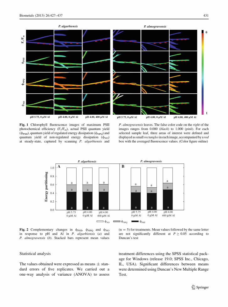
Fig. 1 Chlorophyll fluorescence images of maximum PSII
photochemical efficiency (Fv/Fm), actual PSII quantum yield
(/PSII), quantum yield of regulated energy dissipation (/NPQ) and
quantum yield of non-regulated energy dissipation (/NO)
at steady-state, captured by scanning P. algarbiensis and
P. almogravensis leaves. The false color code on the right of the
images ranges from 0.000 (black) to 1.000 (pink). For each
selected sample leaf, three areas of interest were defined and
displayed as small rectangles in each image, accompanied by a red
box with the averaged fluorescence values. (Color figure online)
Ene
rgy
part
itio
ning
0.0
0.2
0.4
0.6
0.8
1.0
P. algarbiensis P. almogravensis
A B
pH 5.750 M Al
pH 4.000 M Al
pH 4.00400 M Al
pH 5.750 M Al
pH 4.000 M Al
pH 4.00400 M Al
PSIINPQNO
a
a aa
aa
a a
a
a
aa
a
a
aa
a a
μ μ μ μ μ μ
φ φ φ
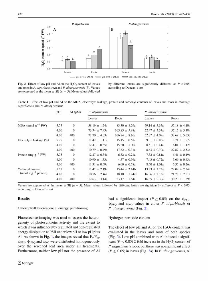
Fig. 2 Complementary changes in /PSII, /NPQ and /NO
in response to pH and Al in P. algarbiensis (a) and
P. almogravensis (b). Stacked bars represent mean values
(n = 5) for treatments. Mean values followed by the same letter
are not significantly different at P C 0.05 according to
Duncan’s test
Biometals (2013) 26:427–437 431
123
Results
Chlorophyll fluorescence: energy partitioning
Fluorescence imaging was used to assess the hetero-
geneity of photosynthetic activity and the extent to
which it was influenced by regulated and non-regulated
energy dissipation at PSII under low pH or low pH plus
Al. As shown in Fig. 1, the images reveal that Fv/Fm,
/PSII, /NPQ and /NO were distributed homogeneously
over the screened leaf area under all treatments.
Furthermore, neither low pH nor the presence of Al
had a significant impact (P C 0.05) on the /PSII,
/NPQ and /NO values in either P. algarbiensis or
P. almogravensis (Fig. 2).
Hydrogen peroxide content
The effect of low pH and Al on the H2O2 content was
evaluated in the leaves and roots of both species
(Fig. 3). Low pH combined with Al induced a signif-
icant (P \ 0.05) 2-fold increase in the H2O2 content of
P. algarbiensis roots, but there was no significant effect
(P C 0.05) in leaves (Fig. 3a). In P. almogravensis, Al
P. almogravensis
Leaves Roots
P. algarbiensis
Leaves Roots
H2O
2 co
nten
t ( μ
mol
g-1
FW
)
0.0
0.5
1.0
1.5
2.0
2.5
3.0BA
pH 5.75, 0 μM Al pH 4.00, 0 μM Al pH 4.00, 400 μM Al
a
b
ab
aa
a
b
aba
aa
Fig. 3 Effect of low pH and Al on the H2O2 content of leaves
and roots in P. algarbiensis (a) and P. almogravensis (b). Values
are expressed as the mean ± SE (n = 5). Mean values followed
by different letters are significantly different at P \ 0.05,
according to Duncan’s test
Table 1 Effect of low pH and Al on the MDA, electrolyte leakage, protein and carbonyl contents of leaves and roots in Plantago
algarbiensis and P. almogravensis
pH Al (lM) P. algarbiensis P. almogravensis
Leaves Roots Leaves Roots
MDA (nmol g-1 FW) 5.75 0 58.19 ± 1.74a 83.30 ± 8.29a 59.14 ± 5.35a 55.18 ± 4.10a
4.00 0 73.34 ± 7.93a 105.85 ± 5.98a 52.47 ± 3.37a 57.12 ± 5.18a
4.00 400 71.70 ± 4.03a 106.84 ± 8.16a 52.87 ± 4.09a 38.69 ± 5.03b
Electrolyte leakage (%) 5.75 0 11.42 ± 1.11a 15.15 ± 0.67a 9.01 ± 0.83a 18.71 ± 1.57a
4.00 0 12.41 ± 0.65a 15.20 ± 1.00a 8.51 ± 0.41a 16.01 ± 1.12a
4.00 400 10.79 ± 0.49a 17.62 ± 0.31a 8.63 ± 0.70a 22.87 ± 2.53a
Protein (mg g-1 FW) 5.75 0 12.27 ± 0.34a 6.32 ± 0.21a 7.32 ± 0.81a 6.41 ± 0.19a
4.00 0 10.90 ± 1.33a 4.57 ± 0.56a 7.43 ± 0.72a 5.66 ± 0.43a
4.00 400 11.31 ± 0.69a 6.00 ± 0.58a 8.60 ± 1.01a 6.35 ± 0.26a
Carbonyl content
(nmol mg-1 protein)
5.75 0 11.42 ± 2.19a 15.44 ± 2.14b 13.33 ± 2.23a 28.09 ± 2.54a
4.00 0 10.56 ± 2.46a 18.10 ± 1.24ab 16.06 ± 2.13a 21.77 ± 2.61a
4.00 400 12.63 ± 3.14a 23.17 ± 1.64a 16.65 ± 2.30a 30.23 ± 1.29a
Values are expressed as the mean ± SE (n = 5). Mean values followed by different letters are significantly different at P \ 0.05,
according to Duncan’s test
432 Biometals (2013) 26:427–437
123
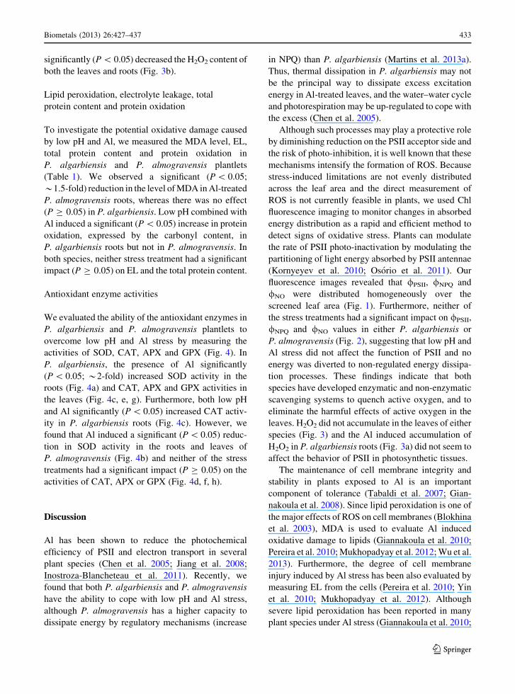
significantly (P \ 0.05) decreased the H2O2 content of
both the leaves and roots (Fig. 3b).
Lipid peroxidation, electrolyte leakage, total
protein content and protein oxidation
To investigate the potential oxidative damage caused
by low pH and Al, we measured the MDA level, EL,
total protein content and protein oxidation in
P. algarbiensis and P. almogravensis plantlets
(Table 1). We observed a significant (P \ 0.05;
*1.5-fold) reduction in the level of MDA in Al-treated
P. almogravensis roots, whereas there was no effect
(P C 0.05) in P. algarbiensis. Low pH combined with
Al induced a significant (P \ 0.05) increase in protein
oxidation, expressed by the carbonyl content, in
P. algarbiensis roots but not in P. almogravensis. In
both species, neither stress treatment had a significant
impact (P C 0.05) on EL and the total protein content.
Antioxidant enzyme activities
We evaluated the ability of the antioxidant enzymes in
P. algarbiensis and P. almogravensis plantlets to
overcome low pH and Al stress by measuring the
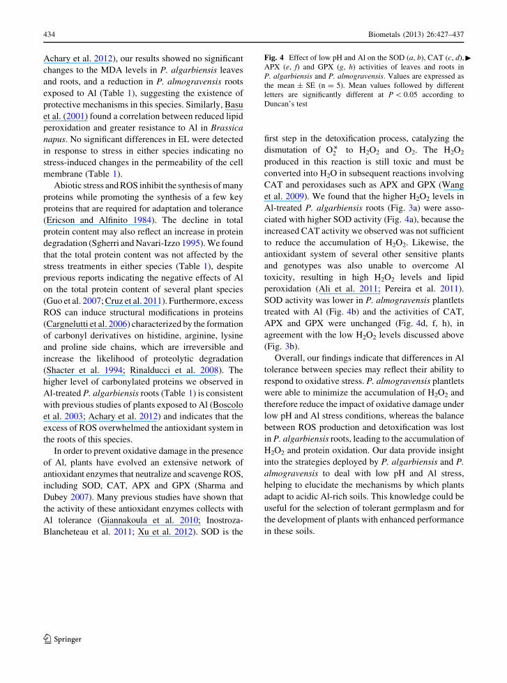
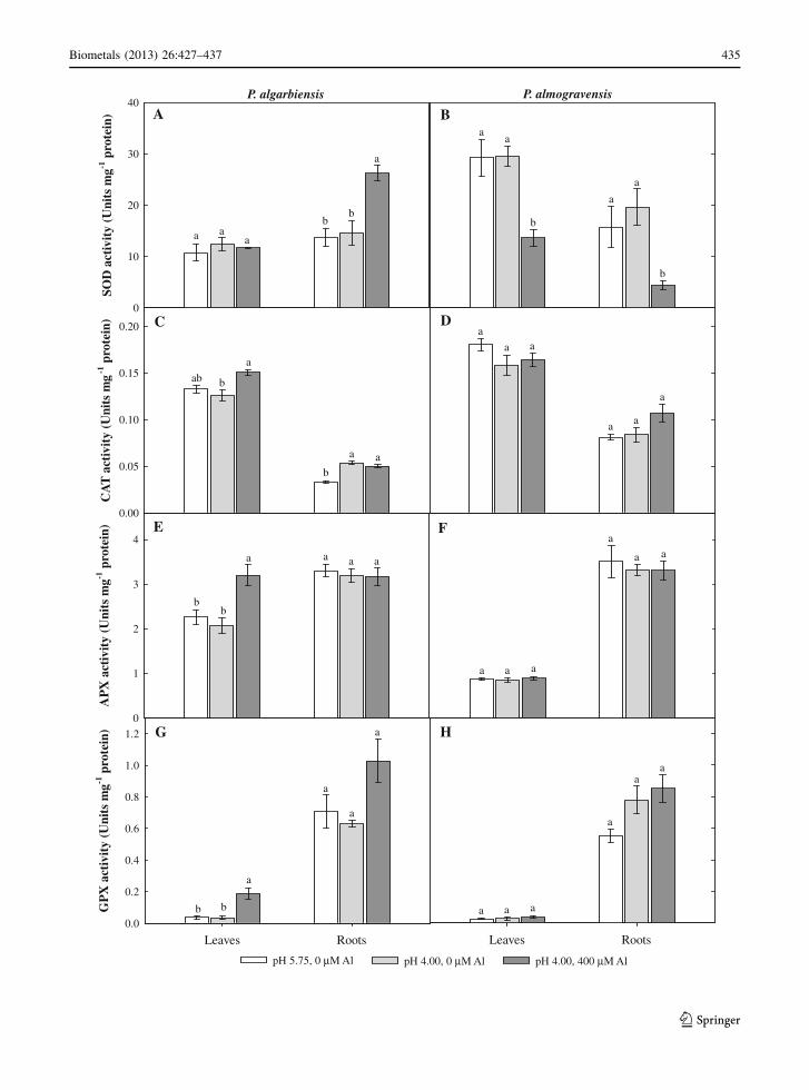
activities of SOD, CAT, APX and GPX (Fig. 4). In
P. algarbiensis, the presence of Al significantly
(P \ 0.05; *2-fold) increased SOD activity in the
roots (Fig. 4a) and CAT, APX and GPX activities in
the leaves (Fig. 4c, e, g). Furthermore, both low pH
and Al significantly (P \ 0.05) increased CAT activ-
ity in P. algarbiensis roots (Fig. 4c). However, we
found that Al induced a significant (P \ 0.05) reduc-
tion in SOD activity in the roots and leaves of
P. almogravensis (Fig. 4b) and neither of the stress
treatments had a significant impact (P C 0.05) on the
activities of CAT, APX or GPX (Fig. 4d, f, h).
Discussion
Al has been shown to reduce the photochemical
efficiency of PSII and electron transport in several
plant species (Chen et al. 2005; Jiang et al. 2008;
Inostroza-Blancheteau et al. 2011). Recently, we
found that both P. algarbiensis and P. almogravensis
have the ability to cope with low pH and Al stress,
although P. almogravensis has a higher capacity to
dissipate energy by regulatory mechanisms (increase
in NPQ) than P. algarbiensis (Martins et al. 2013a).
Thus, thermal dissipation in P. algarbiensis may not
be the principal way to dissipate excess excitation
energy in Al-treated leaves, and the water–water cycle
and photorespiration may be up-regulated to cope with
the excess (Chen et al. 2005).
Although such processes may play a protective role
by diminishing reduction on the PSII acceptor side and
the risk of photo-inhibition, it is well known that these
mechanisms intensify the formation of ROS. Because
stress-induced limitations are not evenly distributed
across the leaf area and the direct measurement of
ROS is not currently feasible in plants, we used Chl
fluorescence imaging to monitor changes in absorbed
energy distribution as a rapid and efficient method to
detect signs of oxidative stress. Plants can modulate
the rate of PSII photo-inactivation by modulating the
partitioning of light energy absorbed by PSII antennae
(Kornyeyev et al. 2010; Osorio et al. 2011). Our
fluorescence images revealed that /PSII, /NPQ and
/NO were distributed homogeneously over the
screened leaf area (Fig. 1). Furthermore, neither of
the stress treatments had a significant impact on /PSII,
/NPQ and /NO values in either P. algarbiensis or
P. almogravensis (Fig. 2), suggesting that low pH and
Al stress did not affect the function of PSII and no
energy was diverted to non-regulated energy dissipa-
tion processes. These findings indicate that both
species have developed enzymatic and non-enzymatic
scavenging systems to quench active oxygen, and to
eliminate the harmful effects of active oxygen in the
leaves. H2O2 did not accumulate in the leaves of either
species (Fig. 3) and the Al induced accumulation of
H2O2 in P. algarbiensis roots (Fig. 3a) did not seem to
affect the behavior of PSII in photosynthetic tissues.
The maintenance of cell membrane integrity and
stability in plants exposed to Al is an important
component of tolerance (Tabaldi et al. 2007; Gian-
nakoula et al. 2008). Since lipid peroxidation is one of
the major effects of ROS on cell membranes (Blokhina
et al. 2003), MDA is used to evaluate Al induced
oxidative damage to lipids (Giannakoula et al. 2010;
Pereira et al. 2010; Mukhopadyay et al. 2012; Wu et al.
2013). Furthermore, the degree of cell membrane
injury induced by Al stress has been also evaluated by
measuring EL from the cells (Pereira et al. 2010; Yin
et al. 2010; Mukhopadyay et al. 2012). Although
severe lipid peroxidation has been reported in many
plant species under Al stress (Giannakoula et al. 2010;
Biometals (2013) 26:427–437 433
123
Achary et al. 2012), our results showed no significant
changes to the MDA levels in P. algarbiensis leaves
and roots, and a reduction in P. almogravensis roots
exposed to Al (Table 1), suggesting the existence of
protective mechanisms in this species. Similarly, Basu
et al. (2001) found a correlation between reduced lipid
peroxidation and greater resistance to Al in Brassica
napus. No significant differences in EL were detected
in response to stress in either species indicating no
stress-induced changes in the permeability of the cell
membrane (Table 1).
Abiotic stress and ROS inhibit the synthesis of many
proteins while promoting the synthesis of a few key
proteins that are required for adaptation and tolerance
(Ericson and Alfinito 1984). The decline in total
protein content may also reflect an increase in protein
degradation (Sgherri and Navari-Izzo 1995). We found
that the total protein content was not affected by the
stress treatments in either species (Table 1), despite
previous reports indicating the negative effects of Al
on the total protein content of several plant species
(Guo et al. 2007; Cruz et al. 2011). Furthermore, excess
ROS can induce structural modifications in proteins
(Cargnelutti et al. 2006) characterized by the formation
of carbonyl derivatives on histidine, arginine, lysine
and proline side chains, which are irreversible and
increase the likelihood of proteolytic degradation
(Shacter et al. 1994; Rinalducci et al. 2008). The
higher level of carbonylated proteins we observed in
Al-treated P. algarbiensis roots (Table 1) is consistent
with previous studies of plants exposed to Al (Boscolo
et al. 2003; Achary et al. 2012) and indicates that the
excess of ROS overwhelmed the antioxidant system in
the roots of this species.
In order to prevent oxidative damage in the presence
of Al, plants have evolved an extensive network of
antioxidant enzymes that neutralize and scavenge ROS,
including SOD, CAT, APX and GPX (Sharma and
Dubey 2007). Many previous studies have shown that
the activity of these antioxidant enzymes collects with
Al tolerance (Giannakoula et al. 2010; Inostroza-
Blancheteau et al. 2011; Xu et al. 2012). SOD is the
first step in the detoxification process, catalyzing the
dismutation of O��2 to H2O2 and O2. The H2O2
produced in this reaction is still toxic and must be
converted into H2O in subsequent reactions involving
CAT and peroxidases such as APX and GPX (Wang
et al. 2009). We found that the higher H2O2 levels in
Al-treated P. algarbiensis roots (Fig. 3a) were asso-
ciated with higher SOD activity (Fig. 4a), because the
increased CAT activity we observed was not sufficient
to reduce the accumulation of H2O2. Likewise, the
antioxidant system of several other sensitive plants
and genotypes was also unable to overcome Al
toxicity, resulting in high H2O2 levels and lipid
peroxidation (Ali et al. 2011; Pereira et al. 2011).
SOD activity was lower in P. almogravensis plantlets
treated with Al (Fig. 4b) and the activities of CAT,
APX and GPX were unchanged (Fig. 4d, f, h), in
agreement with the low H2O2 levels discussed above
(Fig. 3b).
Overall, our findings indicate that differences in Al
tolerance between species may reflect their ability to
respond to oxidative stress. P. almogravensis plantlets
were able to minimize the accumulation of H2O2 and
therefore reduce the impact of oxidative damage under
low pH and Al stress conditions, whereas the balance
between ROS production and detoxification was lost
in P. algarbiensis roots, leading to the accumulation of
H2O2 and protein oxidation. Our data provide insight
into the strategies deployed by P. algarbiensis and P.
almogravensis to deal with low pH and Al stress,
helping to elucidate the mechanisms by which plants
adapt to acidic Al-rich soils. This knowledge could be
useful for the selection of tolerant germplasm and for
the development of plants with enhanced performance
in these soils.
Fig. 4 Effect of low pH and Al on the SOD (a, b), CAT (c, d),
APX (e, f) and GPX (g, h) activities of leaves and roots in
P. algarbiensis and P. almogravensis. Values are expressed as
the mean ± SE (n = 5). Mean values followed by different
letters are significantly different at P \ 0.05 according to
Duncan’s test
c
434 Biometals (2013) 26:427–437
123
Leaves RootsLeaves Roots
GP
X a
ctiv
ity
(Uni
ts m
g-1 p
rote
in)
0.0
0.2
0.4
0.6
0.8
1.0
1.2
AP
X a
ctiv
ity
(Uni
ts m
g-1 p
rote
in)
0
1
2
3
4
CA
T a
ctiv
ity
(Uni
ts m
g-1
pro
tein
)
0.00
0.05
0.10
0.15
0.20
P. algarbiensisSO
D a
ctiv
ity
(Uni
ts m
g-1 p
rote
in)
0
10
20
30
40P. almogravensis
A
DC
B
E
HG
F
a
bb
a
bb
b
a
a
a
a
b
a
aa
ab
b
b
a
b b
pH 5.75, 0 μM Al pH 4.00, 0 μM Al pH 4.00, 400 μM Al
a aa
a
aa
aa
a
aaa
aaa
aa
a
a
a
a
a a a
a aa
Biometals (2013) 26:427–437 435
123
Acknowledgments N. Martins, M.L. Osorio and S. Goncalves
acknowledge grants from the Portuguese Science and
Technology Foundation (FCT, SFRH/BD/48379/2008, SFRH/
BPD/35410/2007 and SFRH/BPD/31534/2006, respectively).
This work was supported by the FCT project PTDC/AGR-
AAM/102664/2008.
References
Achary VMM, Patnaik AR, Panda BB (2012) Oxidative bio-
markers in leaf tissue of barley seedlings in response to
aluminum stress. Ecotoxicol Environ Saf 75:16–26
Aebi HE (1983) Catalase. In: Bergmeyer HU (ed) Methods of
enzymatic analysis. Chemie, Berlin, pp 273–286
Ali S, Bai P, Zeng F, Cai S, Shamsi IH, Qiu B, Wu F, Zhang G
(2011) The ecotoxicological and interactive effects of
chromium and aluminum on growth, oxidative damage and
antioxidant enzymes on two barley genotypes differing in
Al tolerance. Environ Exp Bot 70:185–191
Basu U, Good AG, Taylor GJ (2001) Transgenic Brassica napus
plants overexpressing aluminium-induced mitochondrial
manganese superoxide dismutase cDNA are resistant to
aluminium. Plant Cell Environ 24:1269–1278
Beauchamp CO, Fridovich I (1971) Superoxide dismutase:
improved assays and assays applicable to acrylamide gels.
Anal Biochem 44:276–287
Blokhina O, Virolainen E, Fagerstedt KV (2003) Antioxidants,
oxidative damage and oxygen deprivation stress: a review.
Ann Bot 91:179–194
Boscolo PRS, Menossi M, Jorge RA (2003) Aluminum-induced
oxidative stress in maize. Phytochemistry 62:181–189
Bradford M (1976) A rapid and sensitive method for the quan-
tification of microgram quantities of protein utilizing the
principle of protein-dye binding. Anal Biochem 72:
248–254
Cargnelutti D, Tabaldi LA, Spanevello RM, Jucoski GO, Bat-
tisti V, Redin M, Linares CEB, Dressler VL, Flores EMM,
Nicoloso FT, Morsch VM, Schetinger MRC (2006) Mer-
cury toxicity induces oxidative stress in growing cucumber
seedlings. Chemosphere 65:999–1006
Chen LS, Qi YP, Liu XH (2005) Effects of aluminum on light
energy utilization and photoprotective systems in citrus
leaves. Ann Bot 96:35–41
Cruz FJR, Lobato AKS, Costa RCL, Lopes MJS, Neves HKB,
Neto CFO, Silva MHL, Filho BGS, Junior JAL, Okumura
RS (2011) Aluminum negative impact on nitrate reductase,
nitrogen compounds and morphological parameters in
sorghum plants. Aust J Crop Sci 5:641–645
Demmig-Adams B, Adams WW (1996) Xanthophyll cycle and
light stress in nature: uniform response to excess direct
sunlight among higher plant species. Planta 198:460–470
Egley GH, Paul RN, Vaughn KC, Duke SO (1983) Role of
peroxidase in the development of water impermeable seed
coats in Sida spinosa L. Planta 157:224–232
Ericson MC, Alfinito AE (1984) Proteins produced during salt
stress in tobacco cell cultures. Plant Physiol 74:506–509
Giannakoula A, Moustakas M, Mylona P, Papadakis I, Yupsanis
T (2008) Aluminum tolerance in maize is correlated with
increased levels of mineral nutrients, carbohydrates and
proline, and decreased levels of lipid peroxidation and Al
accumulation. J Plant Physiol 165:385–396
Giannakoula A, Moustakas M, Syros T, Yupsanis T (2010)
Aluminum stress induces up-regulation of an efficient
antioxidant system in the Al-tolerant maize line but not in
the Al-sensitive line. Environ Exp Bot 67:487–494
Gill SS, Tuteja N (2010) Reactive oxygen species and antioxi-
dant machinery in abiotic stress tolerance in crop plants.
Plant Physiol Biochem 48:909–930
Goncalves S, Martins N, Romano A (2009) Micropropagation
and conservation of endangered species Plantago algar-
biensis and P. almogravensis. Biol Plant 53:774–778
Guo TR, Zhang GP, Zhang YH (2007) Physiological changes in
barley plants under combined toxicity of aluminum, copper
and cadmium. Colloids Surf B 57:182–188
Hodges DM, Delong JM, Forney CF, Prange RK (1999)
Improving the thiobarbituric acid-reactive-substances
assay for estimating lipid peroxidation in plant tissues
containing anthocyanin and other interfering compounds.
Planta 207:604–611
Inostroza-Blancheteau C, Reyes-Dıaz M, Aquea F, Nunes-Nesi
A, Alberdi M, Arce-Johnson P (2011) Biochemical and
molecular changes in response to aluminium-stress in
highbush blueberry (Vaccinium corymbosum L.). Plant
Physiol Biochem 49:1005–1012
Jiang HX, Chen LS, Zheng JG, Han S, Tang N, Smith BR (2008)
Aluminum-induced effects on photosystem II photo-
chemistry in citrus leaves assessed by the chlorophyll a
fluorescence transient. Tree Physiol 28:1863–1871
Kochian LV (1995) Cellular mechanisms of aluminum toxicity
and resistance in plants. Annu Rev Plant Physiol Plant Mol
Biol 46:237–260
Kornyeyev D, Logan BA, Holaday AS (2010) Excitation pres-
sure as a measure of the sensitivity of photosystem II to
photoinactivation. Funct Plant Biol 37:943–951
Kramer DM, Johnson G, Kiirats O, Edwards GE (2004) New
fluorescence parameters for the determination of QA redox
state and excitation energy fluxes. Photosynth Res 79:
209–218
Levine RL, Willams JA, Stadtman ER, Shacter E (1994) Car-
bonyl assay for determination of oxidatively modified
proteins. Methods Enzymol 233:346–363
Loreto F, Velikova V (2001) Isoprene produced by leaves pro-
tects the photosynthetic apparatus against ozone damage,
quences ozone products, and reduces lipid peroxidation of
cellular membranes. Plant Physiol 127:781–787
Lutts S, Kinet JM, Bouharmont J (1996) NaCl-induced senes-
cence in leaves of rice (Oriza sativa L.) cultivar differing in
salinity resistance. Ann Bot 78:389–398
Ma B, Gao L, Zhang H, Cui J, Shen Z (2012) Aluminum-
induced oxidative stress and changes in antioxidant
defenses in the roots of rice varieties differing in Al tol-
erance. Plant Cell Rep 31:687–696
Martins N, Goncalves S, Palma T, Romano A (2011) The
influence of low pH on in vitro growth and biochemical
parameters of Plantago almogravensis and P. algarbiensis.
Plant Cell Tissue Organ Cult 107:113–121
Martins N, Osorio ML, Goncalves S, Osorio J, Palma T, Romano
A (2013a) Physiological responses of Plantago algarbien-
sis and P. almogravensis shoots and plantlets to low pH and
aluminum stress. Acta Physiol Plant 35:615–625
436 Biometals (2013) 26:427–437
123
Martins N, Goncalves S, Andrade P, Valentao P, Romano A
(2013b) Changes on organic acid secretion and accumu-
lation in Plantago almogravensis Franco and P. algarbi-
ensis Samp under aluminum stress. Plant Sci 198:1–6
Martins N, Goncalves S, Romano A (2013c) Metabolism and
aluminum accumulation in Plantago almogravensis and P.
algarbiensis in response to low pH and aluminum stress.
Biol Plant 57:325–331
Mittler R (2002) Oxidative stress, antioxidants and stress tol-
erance. Trends Plant Sci 7:405–410
Mukhopadyay M, Bantawa P, Das A, Sarkar B, Bera B, Ghosh
P, Mondal TK (2012) Changes of growth, photosynthesis
and alteration of leaf antioxidative defence system of tea
[Camellia sinensis (L.) O. Kuntze] seedlings under alu-
minum stress. Biometals 25:1141–1154
Murashige T, Shoog F (1962) A revised medium for rapid
growth and bio-assays with tobacco tissue cultures. Physiol
Plant 15:473–497
Murchie EH, Niyogi KK (2011) Manipulation of photoprotec-
tion to improve plant photosynthesis. Plant Physiol 155:
86–92
Nakano Y, Asada K (1981) Hydrogen peroxide is scavenged by
ascorbate-specific peroxidase in spinach chloroplasts.
Plant Cell Physiol 22:867–880
Osorio ML, Osorio J, Vieira AC, Goncalves S, Romano A
(2011) Influence of enhanced temperature on photosyn-
thesis, photooxidative damage, and antioxidant strategies
in Ceratonia siliqua L. seedlings subjected to water deficit
and rewatering. Photosynthetica 49:3–12
Pereira WE, Siqueira DL, Martınez CA, Puiatti M (2000) Gas
exchange and chlorophyll fluorescence in four citrus
rootstocks under Al stress. J Plant Physiol 157:513–520
Pereira LB, Mazzanti CM, Goncalves JF, Cargnelutti D, Tabaldi
LA, Becker AG, Calgaroto NS, Farias JG, Battisti V,
Bohrer D, Nicoloso FT, Morsch VM, Schetinger MR
(2010) Aluminum-induced oxidative stress in cucumber.
Plant Physiol Biochem 48:683–689
Pereira LB, Mazzanti CMA, Cargnelutti D, Rossato LV, Gon-
calves JF, Calgaroto N, Dressler V, Nicoloso FT, Federizzi
LC, Morsch VM, Shetinger MRC (2011) Differential
responses of oat genotypes: oxidative stress provoked by
aluminum. Biometals 24:73–83
Ramırez-Benıtez JE, Hernandez-Sotomayor SMT (2008) Role
of reactive oxygen species (ROS) in aluminium-induced
signaling and aluminium resistance in plants. Curr Top
Biochem Res 19:79–89
Rinalducci S, Murgiano L, Zolla L (2008) Redox proteomics:
basic principles and future perspectives for the detection of
protein oxidation in plants. J Exp Bot 59:3781–3801
Sgherri CLM, Navari-Izzo F (1995) Sunflower seedling sub-
jected to increasing water deficit stress: oxidative stress and
defense mechanisms. Physiol Plant 93:25–30
Shacter E, Williams JA, Lim M, Levine RL (1994) Differential
susceptibility of plasma proteins to oxidative modification.
Examination by Western blot immunoassay. Free Rad Biol
Med 17:429–437
Shaff JE, Schultz BA, Craft EJ, Clark RT, Kochian LV (2010)
GEOCHEM-EZ: a chemical speciation program with
greater power and flexibility. Plant Soil 330:207–214
Sharma P, Dubey RS (2007) Involvement of oxidative stress and
role of antioxidative defense system in growing rice
seedlings exposed to toxic concentrations of aluminum.
Plant Cell Rep 26:2027–2038
Tabaldi LA, Nicoloso FT, Castro GY, Cargnelutti D, Goncalves
JF, Rauber R, Skresky EC, Schetinger MRC, Morsch VM,
Bisognin DA (2007) Physiological and oxidative stress
responses of four potato clones to aluminum in nutrient
solution. Braz J Plant Physiol 19:211–222
von Uexkull HR, Mutert E (1995) Global extent, development
and economic impact of acid soils. Plant Soil 171:1–15
Wang W-B, Kim Y-H, Lee H-S, Kim K-Y, Deng X-P, Kwak S–
S (2009) Analysis of antioxidant enzyme activity during
germination of alfalfa under salt and drought stresses. Plant
Physiol Biochem 47:570–577
Wilson KE, Ivanov AG, Oquist G, Grodzinski B, Sarhan F,
Huner NPA (2006) Energy balance, organellar redox status
and acclimation to environmental stress. Can J Bot 84:
1355–1370
Wu K, Xiao S, Chen Q, Wang Q, Zhang Y, Li K, Yu Y, Chen L
(2013) Changes in the activity and transcription of anti-
oxidant enzymes in response to Al stress in black soybeans.
Plant Mol Biol Rep 31:141–150
Xu FJ, Li G, Jin CW, Liu WJ, Zhang YS, Lin XY (2012) Alu-
minum-induced changes in reactive oxygen species accu-
mulation, lipid peroxidation and antioxidant capacity in
wheat root tips. Biol Plant 56:89–96
Yin L, Mano J, Wang S, Tsuji W, Tanaka K (2010) The
involvement of lipid peroxide-derived aldehydes in alu-
minum toxicity of tobacco roots. Plant Physiol 152:1406–
1417
Biometals (2013) 26:427–437 437
123